Embed Size (px)
Citation preview
2014 - Northwest Region Emergency Medical Services & Trauma Care Council
1
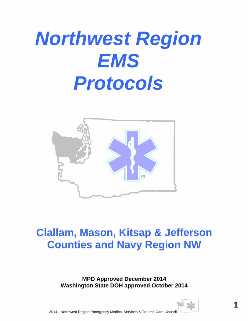
Clallam, Mason, Kitsap & Jefferson
Counties and Navy Region NW
MPD Approved December 2014 Washington State DOH approved October 2014
Northwest Region EMS
Protocols
2014 - Northwest Region Emergency Medical Services & Trauma Care Council
2
2014 - Northwest Region Emergency Medical Services & Trauma Care Council
3
Sections are color coded as follows:
Introduction
Regional Guidelines ...................................................................................................................... 8 NW Region Patient Care Procedures ............................................................................................ 8 CDC National Trauma Triage Procedure ....................................................................................... 9 Clallam .................................................................................................................................... 10-A Jefferson ................................................................................................................................. 10-B Mason ..................................................................................................................................... 10-C Kitsap ...................................................................................................................................... 10-D West Olympic Penninsula ....................................................................................................... 10-E Prehospital Provider Conduct ...................................................................................................... 11 Infection Control Standards ......................................................................................................... 11 Patient Refusal of Medical Evaluation ......................................................................................... 11
Protocols
Adult/Pediatric START/JumpSTART Triage ................................................................................ 12 Universal Patient Care ................................................................................................................ 13 Suspected Abuse ........................................................................................................................ 14
Cardiac
Cardiac Arrest ............................................................................................................................. 15 Non-Traumatic Shock.................................................................................................................. 16 Bradycardia ................................................................................................................................. 17 Narrow Complex Tachycardia ..................................................................................................... 18 Wide Complex Tachycardia ........................................................................................................ 19 V-Fib/Pulseless V-Tac ................................................................................................................. 20 Asystole / PEA (no shock advised) .............................................................................................. 21 Hyperkalemia .............................................................................................................................. 22 Chest Pain / Acute Coronary Syndrome ...................................................................................... 23
Respiratory
Airway (Adult) .............................................................................................................................. 24 Failed Airway (Adult) ................................................................................................................... 25 Reactive Airway Disease ............................................................................................................. 26 Pulmonary Edema ....................................................................................................................... 27 Post Resuscitation Management ................................................................................................. 28
Medical
Abdominal Pain ........................................................................................................................... 29 Allergic Reaction ......................................................................................................................... 30 Altered Mental Status / Diabetic Emergency ............................................................................... 31 General Illness ............................................................................................................................ 32 Overdose / Poisoning .................................................................................................................. 33 Pain Management ....................................................................................................................... 34 Psychological / Emotional / Excited Delirium ............................................................................... 35 Seizure ........................................................................................................................................ 36 Stroke .......................................................................................................................................... 37
OB / GYN
Pregnancy Induced Hypertension ............................................................................................... 38 Postpartum .................................................................................................................................. 39
2014 - Northwest Region Emergency Medical Services & Trauma Care Council
4
Environmental
Environmental ................................................................................................................................ 40 Burns .............................................................................................................................................. 41 SCUBA Emergencies ..................................................................................................................... 42 Drowning / Near Drowning ............................................................................................................. 43 Head Injury ..................................................................................................................................... 44 Multi-system Trauma ...................................................................................................................... 45
Pediatric Protocols
Pediatric Airway ............................................................................................................................ 46 Pediatric Rapid Sequence Intubation ............................................................................................. 47 Pediatric Cardiac Arrest ................................................................................................................. 48 Pediatric Bradycardia ..................................................................................................................... 49 Pediatric Narrow Complex Tachycardia ......................................................................................... 50 Pediatric Wide Complex Tachycardia ............................................................................................. 51 Pediatric V-Fib/Pulseless V-Tach (shock advised) ......................................................................... 52 Pediatric PEA / Asystole ................................................................................................................. 53 Pediatric Post Resuscitation Management ..................................................................................... 54 Pediatric Anaphylaxis ..................................................................................................................... 55 Pediatric Apparent Life Threatening Event (ALTE) ......................................................................... 56 Pediatric Breathing Difficulty .......................................................................................................... 57 Pediatric Diabetic Ketoacidosis / Hyperglycemia ............................................................................ 58 Pediatric Hypoglycemia .................................................................................................................. 59 Newborn Resuscitation/Post Delivery Care .................................................................................... 60 Pediatric Known Toxic Exposure .................................................................................................... 61 Pediatric Pain Management ........................................................................................................... 62 Pediatric Fever ............................................................................................................................... 63 Pediatric Shock Non-Traumatic ...................................................................................................... 64 Pediatric Seizure ............................................................................................................................ 65 Pediatric Multi-System Trauma ...................................................................................................... 66 Pediatric Near Drowning ................................................................................................................ 67 Pediatric Burns ............................................................................................................................... 68 Pediatric Environmental Emergencies ............................................................................................ 69 Pediatric Concussion/Traumatic Brain Injury .................................................................................. 70
Pediatric Procedures
Pediatric Assessment ..................................................................................................................... 71 Pediatric References ...................................................................................................................... 72 Airway Needle Cricothyrotomy (Pediatric) ...................................................................................... 73 APGAR Scale ................................................................................................................................. 74 AVPU Infant / Child ........................................................................................................................ 74 CUPS Pediatric .............................................................................................................................. 74 Neonatal Resuscitation .................................................................................................................. 75 Pain Assessment and Documentation – Pediatric .......................................................................... 76 Venous Access - Intraosseous Pediatric ........................................................................................ 77

2014 - Northwest Region Emergency Medical Services & Trauma Care Council
5
Procedures
Airway I-gel Supraglottic Airway .................................................................................................. 78 Airway Capnography ................................................................................................................... 79 Airway Combitube ....................................................................................................................... 80 Airway King LTS-D™ .................................................................................................................. 81 Airway Laryngeal Mask Airway (LMA) ......................................................................................... 82 Airway Cricothyrotomy Needle (Adult) ......................................................................................... 83 Airway Cricothyrotomy Surgical (Adult) ....................................................................................... 84 Airway Difficult Airway Assesment (LEMON) .............................................................................. 85 Airway Passive pre-oxygenation ................................................................................................. 86 Airway Intubation Confirmation End-Tidal Co2 Detector ............................................................. 87 Airway Intubation Confirmation Esophageal Bulb ........................................................................ 88 Airway Orotracheal Intubation ..................................................................................................... 89 Airway (AAA) Intubation w/Eschmann Catheter, Tracheal Tube introducer,Gum Elastic Bougie 90 Airway (AAA) Nasotracheal Intubation ........................................................................................ 91 Airway Nebulizer Inhalation Therapy ........................................................................................... 92 Airway Non-invasive Positive Pressure Ventilation (NIPPV)........................................................ 93 Airway Rapid Sequence Intubation (RSI) .................................................................................... 94 Airway Video Assisted Laryngoscopy .......................................................................................... 95 Airway Suctioning – Basic ........................................................................................................... 96 Airway Suctioning – Advanced .................................................................................................... 97 Airway Tracheostomy Tube Change ........................................................................................... 98 Airway Ventilator Operation ......................................................................................................... 99 Blood Product Administration .................................................................................................... 100 Cardiac Automatic Implantable cardio defibrillator (AIDC) Deactivation .................................... 101 Cardiac 12 Lead ECG ............................................................................................................... 102 Synchronized Cardioversion ..................................................................................................... 103 Cardiac Defibrillation Automated ............................................................................................... 104 Cardiac Defibrillation Manual .................................................................................................... 105 Cardiac Transcutaneous Pacing ............................................................................................... 106 Central Venous Device ....................................................................................................... 107-108 Chest Decompression ............................................................................................................... 109 Childbirth/Fundal Massage ........................................................................................................ 110 Stroke FAST Assesment ........................................................................................................... 111 CPR (High Density) ................................................................................................................... 112 Discontinuation of CPR /Do Not Attempt Resuscitation/Determination of Field Death ..................... 113 Glucometry ................................................................................................................................ 114 Glasgow Coma Score ............................................................................................................... 115 Injections – Subcutaneous, Intramuscular ................................................................................. 116 Intranasal Medication Delivery .................................................................................................. 117 Nasogastric Tube Insertion ....................................................................................................... 118 Orthostatic Blood Pressure Measurement ................................................................................. 119 Pain Assessment and Documentation - Adult ........................................................................... 120 Pelvic Fracture Stabilization ...................................................................................................... 121 Pulse Oximetry .......................................................................................................................... 122 Restraints ........................................................................................................................... 123-124 Spinal Motion Restriction ........................................................................................................... 125 Spinal Immobilization ................................................................................................................ 126 Splinting .................................................................................................................................... 127 Taser Dart Removal .................................................................................................................. 128 Temperature Measurement ....................................................................................................... 129 Thrombolytic Screen ................................................................................................................. 130

2014 - Northwest Region Emergency Medical Services & Trauma Care Council
6
Venous Access - Blood Draw .................................................................................................... 131 Venous Access - External Jugular Access ................................................................................ 132 Venous Access - Extremity ....................................................................................................... 133 Venous Access - Intraosseous Adult ......................................................................................... 134 Wound Care / Hemorrhage Control ........................................................................................... 135
Drug Formulary
Acetaminophen (Tylenol) .......................................................................................................... 136 Acetylsalicylic Acid / Aspirin (Bayer/Ecotrin) .............................................................................. 136 Activated Charcoal (Actidose-Aqua/Insta-Char) ........................................................................ 137 Adenosine (Adenocard) ............................................................................................................. 137 Albuterol Sulfate (Proventil, Ventolin) ........................................................................................ 138 Amiodarone (Cordarone) .................................................................................................... 139-140 Atropine (Atreza) ....................................................................................................................... 140 Bumetanide (Bumex)................................................................................................................. 141 Calcium Chloride (CaCl2) .......................................................................................................... 141 Clopidogrel Bisulfate (Plavix) .................................................................................................... 142 Dexamethasone (Decadron) ..................................................................................................... 142 Dextrose / D50W / D25W (DGlucose) ....................................................................................... 143 Diazepam (Valium) .................................................................................................................... 144 Diltiazem (Cardizem) ................................................................................................................. 145 Diphenhydramine (Benadryl) ..................................................................................................... 145 Dopamine (Intropin) .................................................................................................................. 146 Droperidol (Inapsine) ................................................................................................................. 147 Epinephrine (Adrenaline) ........................................................................................................... 148 Etomidate (Amidate) ................................................................................................................. 149 Fentanyl (Sublimaze) ................................................................................................................ 150 Furosemide (Lasix) ................................................................................................................... 150 Glucagon ................................................................................................................................... 151 Glucose Oral (Glucose Paste) ................................................................................................... 151 Heparin Sodium ........................................................................................................................ 152 Hydromorphone (Dilaudid) ........................................................................................................ 152 Insulin ........................................................................................................................................ 153 Ipratropium (Atrovent / Ipramide) ............................................................................................... 153 Ketamine ............................................................................................................................ 154-155 Ketorolac (Toradol) ................................................................................................................... 155 Labetalol (Trandate, Normodyne) .............................................................................................. 156 Lidocaine (Xylocaine) ................................................................................................................ 157 Lorazepam (Ativan) ................................................................................................................... 158 Magnesium Sulfate (MgSo4) ..................................................................................................... 158 Methylprednisolone (Solu-Medrol / Amethapred) ...................................................................... 159 Metoprolol (Lopressor) .............................................................................................................. 160 Midazolam (Versed) .................................................................................................................. 161 Morphine ................................................................................................................................... 162 Naloxone (Narcan) .................................................................................................................... 162 Nitroglycerin (NitroStat / NitroQuick) ......................................................................................... 163 Nitrous Oxide (Nitronox) ............................................................................................................ 163 Norepinephrine Bitartrate (Levophed) ....................................................................................... 164 Ondansetron (Zofran) ................................................................................................................ 165 Oxymetazoline (Afrin) ................................................................................................................ 165 Oxytocin (Pitocin) ...................................................................................................................... 166 Prednisone ................................................................................................................................ 166 Procainamide (Pronestyl) .......................................................................................................... 167
2014 - Northwest Region Emergency Medical Services & Trauma Care Council
7
Promethazine (Phenergan) ....................................................................................................... 168 Propofol ..................................................................................................................................... 169 Rocuronium (Zemuron) ............................................................................................................. 170 Sodium Bicarbonate .................................................................................................................. 170 Succinylcholine (Anectine) ........................................................................................................ 171 Tenecteplase (TNKase) ............................................................................................................ 171 Thiamine (Betalin, Biamine, Vitamin B1) ................................................................................... 172 Vasopressin (Pitressin) ............................................................................................................. 172 Vecuronium (Norcuron) ............................................................................................................. 173 Drug Reference ......................................................................................................................... 174
Miscellaneous
Air Ambulance Transports ......................................................................................................... 175 Physician on Scene ................................................................................................................... 175 Emergency at Physician's Office ............................................................................................... 176 Patient Care Reports ................................................................................................................. 176 10 Critical Steps for Handling Possible Bioterrorism Events............................................... 171-178 Medical Spanish ................................................................................................................. 179-180 Mnemonic's ........................................................................................................................ 181-182 Phone Numbers ........................................................................................................................ 183 Future additions ................................................................................................................. 184-186 Personal Information ................................................................................................................. 187
Any reproduction of this document must be approved by the Northwest Region Emergency Medical Services and Trauma Care Council.
2014 - Northwest Region Emergency Medical Services & Trauma Care Council
8
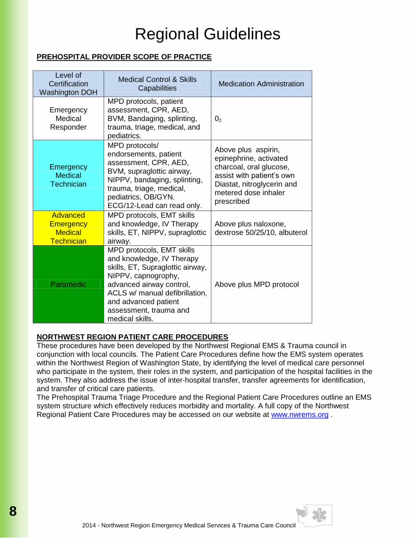
Regional Guidelines PREHOSPITAL PROVIDER SCOPE OF PRACTICE
Level of
Certification Washington DOH
Medical Control & Skills Capabilities
Medication Administration
Emergency Medical
Responder
MPD protocols, patient assessment, CPR, AED, BVM, Bandaging, splinting, trauma, triage, medical, and pediatrics.
02
Emergency Medical
Technician
MPD protocols/ endorsements, patient assessment, CPR, AED, BVM, supraglottic airway, NIPPV, bandaging, splinting, trauma, triage, medical, pediatrics, OB/GYN. ECG/12-Lead can read only.
Above plus aspirin, epinephrine, activated charcoal, oral glucose, assist with patient’s own Diastat, nitroglycerin and metered dose inhaler prescribed
Advanced Emergency
Medical Technician
MPD protocols, EMT skills and knowledge, IV Therapy skills, ET, NIPPV, supraglottic airway.
Above plus naloxone, dextrose 50/25/10, albuterol
Paramedic
MPD protocols, EMT skills and knowledge, IV Therapy skills, ET, Supraglottic airway, NIPPV, capnogrophy, advanced airway control, ACLS w/ manual defibrillation, and advanced patient assessment, trauma and medical skills.
Above plus MPD protocol
NORTHWEST REGION PATIENT CARE PROCEDURES These procedures have been developed by the Northwest Regional EMS & Trauma council in conjunction with local councils. The Patient Care Procedures define how the EMS system operates within the Northwest Region of Washington State, by identifying the level of medical care personnel who participate in the system, their roles in the system, and participation of the hospital facilities in the system. They also address the issue of inter-hospital transfer, transfer agreements for identification, and transfer of critical care patients. The Prehospital Trauma Triage Procedure and the Regional Patient Care Procedures outline an EMS system structure which effectively reduces morbidity and mortality. A full copy of the Northwest Regional Patient Care Procedures may be accessed on our website at www.nwrems.org .
2014 - Northwest Region Emergency medical Services & Trauma Care Council
9
9
Measure vital signs and level of consciousness
CDC National Trauma Triage
Glasgow Coma Scale <14 or
Systolic blood pressure <90 mmHg or
Respiratory rate <10 or > 29 breaths/minute (<20 in infant < one year)1
2
3
Assess anatomy of Injury
• All penetrating injuries to head, neck, torso, and extremities proximal
to elbow and knee
• Flail chest
• Two or more proximal long-bone fractures
• Crushed, degloved, or mangled extremity
• Amputation proximal to wrist and ankle
• Pelvic fractures
• Open or depressed skull fracture
• Paralysis
Assess mechanism of Injury and evidence of high-energy impact
Falls
• Adults: > 20ft. (one story is equal to 10 ft.)
• Children: > 10ft. or 2-3 times the height of the child
High-Risk Auto Crash
• Intrusion: > 12in. Occupant site; > 18in. Any site
• Ejection (partial or complete) from automobile
• Death in same passenger compartment
• Vehicle telementry data consistent with high risk of injury
Auto v. pedestrian/Bicyclist Thrown, Run Over, or with Significant
(> 20 mph) Impact
Motorcycle Crash > 20 mph
Assess special patient or system considerations
Age
• Older Adults: Risk of injury death increases after age 55 years
• Children: Should be triaged preferentially to pediatric capable trauma
centers
Anticoagulation and Bleeding Disorders
Burns
• Without other trauma mechanism: Triage to burn facility
• With trauma mechanism: Triage to trauma center
Time Sensitive Extremity Injury
End-Stage Renal Disease Requiring Dialysis
Pregnancy > 20 Weeks
EMS Provider Judgment
No
Take to a trauma center. Steps 1
and 2 attempt to identify the most
seriously injured patients. These
patients should be transported
preferentially to the highest level of
care within the trauma system
YES
YES
No
No
YES
Transport to closest appropriate
trauma center, which depending
on the trauma system, need not be
the the highest level trauma center
Contact medical control and
consider transport to a trauma
center or a specific resource
hospital.
YES
Transport according to protocol
No
4
2014 - Northwest Region Emergency Medical Services & Trauma Care Council
10
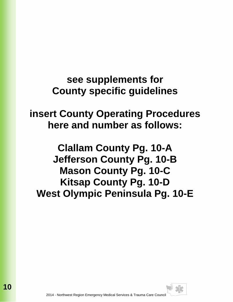
see supplements for County specific guidelines
insert County Operating Procedures
here and number as follows:
Clallam County Pg. 10-A Jefferson County Pg. 10-B
Mason County Pg. 10-C Kitsap County Pg. 10-D
West Olympic Peninsula Pg. 10-E
2014 - Northwest Region Emergency medical Services & Trauma Care Council
11
11
PREHOSPITAL PROVIDER CONDUCT
1. Northwest Region EMS Providers must maintain the highest standard of professional conduct. 2. Competent medical care must be provided with compassion and dignity for all persons regardless of
nationality, race, creed, religion, sex or status. 3. Providers must refuse to participate in unethical activities and/or activities which may impair
professional judgment and the ability to act competently. 4. Matters of disagreement between prehospital providers regarding patient care must be handled
professionally without alarming anyone on the scene. Medical Control contact will be made for immediate direction. Providers should not threaten, degrade, insult or verbally abuse each other.
5. Patient Confidentiality will be maintained at all times in compliance with Health Insurance Portability and Accountability Act (HIPAA) of 1996.
INFECTION CONTROL STANDARDS
1. Infection Control Standards assume that all contact with blood, other bodily fluids and potentially infectious materials is infectious.
2. The standards of use of Universal Precautions / Body Substance Isolation, which includes safe work practices, correct use of engineering controls and personal protective equipment is mandated by WISHA, and must be adhered to.
3. EMS Providers must protect themselves at all times from “reasonably anticipated potential for exposure”. The following is a list of mandated items: Gloves, Masks, Face Shields, Safety Glasses, High Efficiency Particulate Air (HEPA) Filters, Resuscitation Equipment, and Protective Clothing.
PATIENT REFUSAL OF MEDICAL EVALUATION
1. Consent a. The patient has responsibility to consent to or refuse treatment. If the patient is unable to
do so, a responsible relative or guardian has this right. b. If waiting to obtain lawful consent from the authorized person would present a serious risk
of death, serious impairment of health, or would prolong severe pain or suffering to the patient, treatment may be undertaken to avoid these risks without consent. In no event should legal consent procedures be allowed to delay immediately required treatment.
c. The patient must be eighteen years of age or emancipated to legally refuse treatment. d. If the patient is under age, consent should be from a natural parent, adopted parent, or
legal guardian only.
2. Mental competence
a. A person is mentally competent if: 1. Capable of understanding the nature and consequence of the proposed treatment. 2. Sufficient emotional control, judgment, and discretion to manage their own affairs are
present. b. A person is not mentally competent if he/she has impaired cerebral perfusion, presents in
shock, is postictal, or under the influence of drugs or alcohol. c. Medical Control contact with the Base physician is necessary for all patients refusing
transport in those counties requiring it. d. Nurses may speak for the Medical Control physician if the physician is unable to come to
the telephone. The nurse must give the prehospital care provider the name of the Base physician who is directing the nurse
2014 - Northwest Region Emergency Medical Services & Trauma Care Council
12
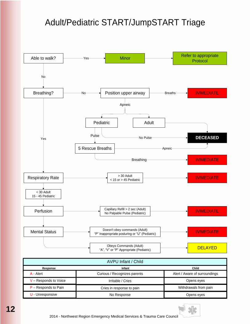
Adult/Pediatric START/JumpSTART Triage
Yes
Pulse
MinorRefer to appropriate
Protocol
No Pulse
Position upper airway
Able to walk?
Yes
Pediatric
Breathing?
No
No Breaths IMMEDIATE
Adult
Respiratory Rate
Perfusion
Mental Status
5 Rescue Breaths
DECEASED
IMMEDIATE
Apneic
Apneic
Breathing
EMT- A
EMT- P
MC Order
V – Responds to Voice
P – Responds to Pain
Opens eyes
Withdraws from Pain
U - Unresponsive No resonse
Response Infant Child
AVPU Infant / Child
A - Alert Alert / Aware of surroundings
> 30 Adult
< 15 or > 45 PediatricIMMEDIATE
< 30 Adult
15 - 45 Pediatric
Capillary Refill > 2 sec (Adult)
No Palpable Pulse (Pediatric)IMMEDIATE
Doesn't obey commands (Adult)
“P” Inappropriate posturing or “U” (Pediatric)IMMEDIATE
Obeys Commands (Adult)
“A”, “V” or “P” Appropriate (Pediatric)DELAYED
Curious / Recognizes parents
Irritable / Cries
Cries in response to pain
No Response
Withdrawals from pain
Opens eyes
2014 - Northwest Region Emergency Medical Services & Trauma Care Council
13
13
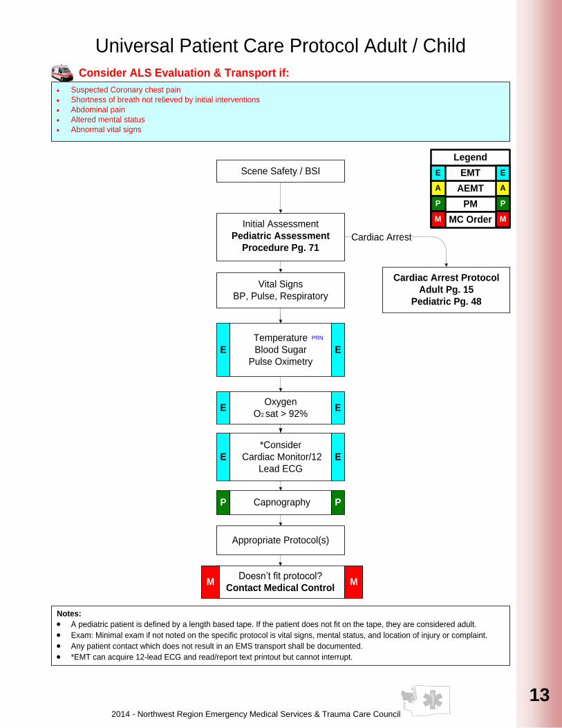
Notes:
· A pediatric patient is defined by a length based tape. If the patient does not fit on the tape, they are considered adult.
· Exam: Minimal exam if not noted on the specific protocol is vital signs, mental status, and location of injury or complaint.
· Any patient contact which does not result in an EMS transport shall be documented.
· *EMT can acquire 12-lead ECG and read/report text printout but cannot interrupt.
Scene Safety / BSI
Universal Patient Care Protocol Adult / Child
Initial Assessment
Pediatric Assessment
Procedure Pg. 71
Cardiac Arrest Protocol
Adult Pg. 15
Pediatric Pg. 48
Cardiac Arrest
Vital Signs
BP, Pulse, Respiratory
Appropriate Protocol(s)
Doesn’t fit protocol?
Contact Medical ControlM M
Oxygen
O2 sat > 92% E E
PRN
Temperature
Blood Sugar
Pulse Oximetry
E E
PRN
Consider ALS Evaluation & Transport if:
· Suspected Coronary chest pain
· Shortness of breath not relieved by initial interventions
· Abdominal pain
· Altered mental status
· Abnormal vital signs
*Consider
Cardiac Monitor/12
Lead ECG
E E
CapnographyP P
AEMT
PM
MC Order
A
P
A
P
M M
EMT
Legend
E E
2014 - Northwest Region Emergency Medical Services & Trauma Care Council
14
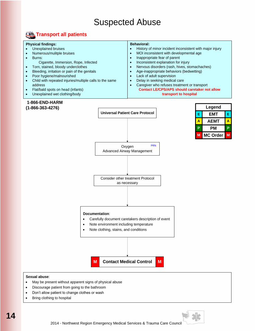
Suspected Abuse
Behavioral:
· History of minor incident inconsistent with major injury
· MOI inconsistent with developmental age
· Inappropriate fear of parent
· Inconsistent explanation for injury
· Nervous disorders (rash, hives, stomachaches)
· Age-inappropriate behaviors (bedwetting)
· Lack of adult supervision
· Delay in seeking medical care
· Caregiver who refuses treatment or transport
Contact LE/CPS/APS should caretaker not allow
transport to hospital
Physical findings:
· Unexplained bruises
· Numerous/mulitple bruises
· Burns:
Cigarette, Immersion, Rope, Infected
· Torn, stained, bloody underclothes
· Bleeding, irritation or pain of the genitals
· Poor hygiene/malnourished
· Child with repeated injuries/multiple calls to the same
address
· Flat/bald spots on head (infants)
· Unexplained wet clothing/body
Oxygen
Advanced Airway Management
PRN
Consider other treatment Protocol
as necessary
Documentation:
· Carefully document caretakers description of event
· Note environment including temperature
· Note clothing, stains, and conditions
Sexual abuse:
· May be present without apparent signs of physical abuse
· Discourage patient from going to the bathroom
· Don’t allow patient to change clothes or wash
· Bring clothing to hospital
Universal Patient Care Protocol
Contact Medical ControlM M
Transport all patients
1-866-END-HARM
(1-866-363-4276)
AEMT
PM
MC Order
A
P
A
P
M M
EMT
Legend
E E
2014 - Northwest Region Emergency Medical Services & Trauma Care Council
15
15
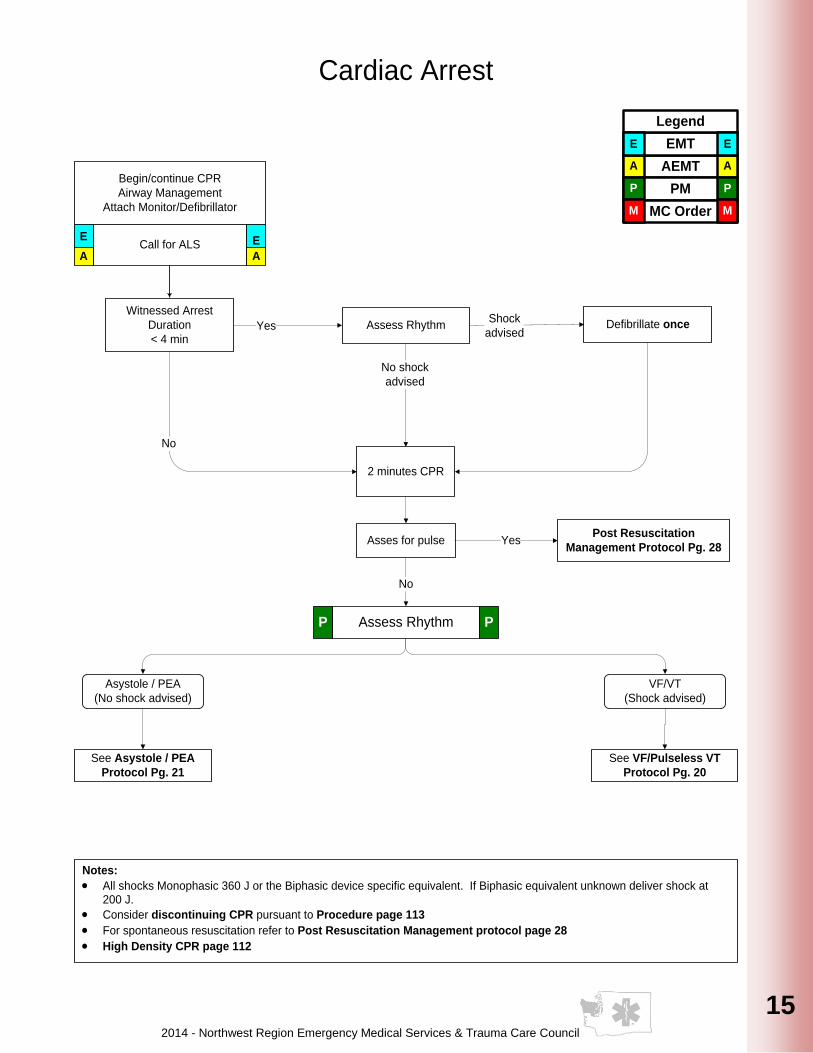
Cardiac Arrest
2 minutes CPR
Begin/continue CPR
Airway Management
Attach Monitor/Defibrillator
Call for ALSE E
A A
Asystole / PEA
(No shock advised)
VF/VT
(Shock advised)
See Asystole / PEA
Protocol Pg. 21
See VF/Pulseless VT
Protocol Pg. 20
Shock
advised
No shock
advised
Yes
No
Notes:
· All shocks Monophasic 360 J or the Biphasic device specific equivalent. If Biphasic equivalent unknown deliver shock at 200 J.
· Consider discontinuing CPR pursuant to Procedure page 113
· For spontaneous resuscitation refer to Post Resuscitation Management protocol page 28
· High Density CPR page 112
Witnessed Arrest
Duration
< 4 min
Assess RhythmP P
Asses for pulse
No
YesPost Resuscitation
Management Protocol Pg. 28
Assess Rhythm Defibrillate once
AEMT
PM
MC Order
A
P
A
P
M M
EMT
Legend
E E

2014 - Northwest Region Emergency Medical Services & Trauma Care Council
16
History:
· Cardiac ischemia (MI, CHF)
· Medications
Signs/Symptoms:
· Hypotension
· Rales & pulmonary edema on exam
· Altered mental status
· Weakness, dizziness
· Weak, rapid pulse
· Pale, cool, clammy skin
Differential:
· Dysrhythmias
· Vasovagal
· Allergic reaction
· Anaphylaxis
· Sepsis
· Neurogenic
Universal Patient Care Protocol
Non-Traumatic Shock
Obtain IV/IO accessA A
Fluid Bolus NS/LR
250cc-1,000cc may
repeat x 1 if not in
Pulmonary Edema
Goal SBP > 100
A A
Norepinephrine
Titrate drip 7-35 mcg/min
IV/IO SBP > 100
Or
Epinephrine
Titrate drip 2-10 mcg/min
IV/IO SBP > 100
Or
Dopamine
Titrate 5–20 mcg/kg/min
IV/IO SBP >100
P P
Contact Medical ControlM M
ECG / 12 leadP P
PRN
AEMT
PM
MC Order
A
P
A
P
M M
EMT
Legend
E E
2014 - Northwest Region Emergency Medical Services & Trauma Care Council
17
17
17
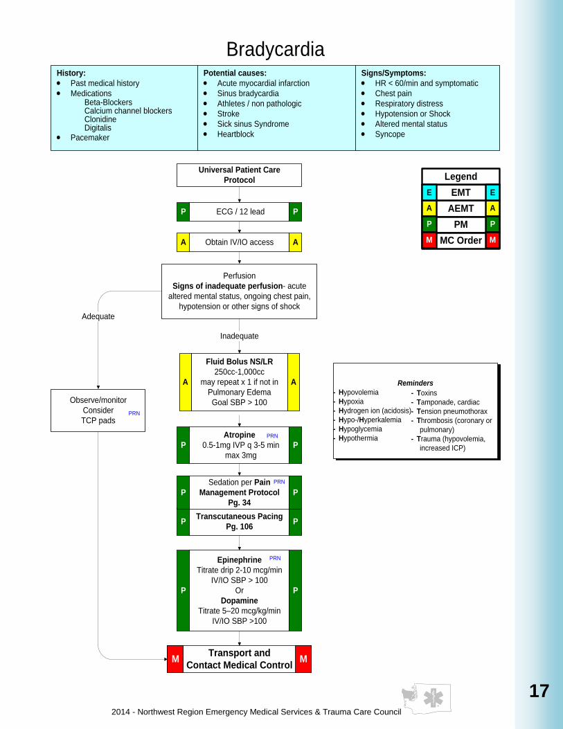
History:
· Past medical history
· MedicationsBeta-BlockersCalcium channel blockersClonidineDigitalis
· Pacemaker
Potential causes:
· Acute myocardial infarction
· Sinus bradycardia
· Athletes / non pathologic
· Stroke
· Sick sinus Syndrome
· Heartblock
Universal Patient Care
Protocol
Bradycardia
Perfusion
Signs of inadequate perfusion- acute
altered mental status, ongoing chest pain,
hypotension or other signs of shock
Adequate
Observe/monitor
Consider
TCP pads
Inadequate
Transcutaneous Pacing
Pg. 106P P
Atropine
0.5-1mg IVP q 3-5 min
max 3mg
P P
Transport and
Contact Medical ControlM M
Reminders
- Hypovolemia
- Hypoxia
- Hydrogen ion (acidosis)
- Hypo-/Hyperkalemia
- Hypoglycemia
- Hypothermia
- Toxins
- Tamponade, cardiac
- Tension pneumothorax
- Thrombosis (coronary or
pulmonary)
- Trauma (hypovolemia,
increased ICP)
PRN
Sedation per Pain
Management Protocol
Pg. 34
P P
PRN
Signs/Symptoms:
· HR < 60/min and symptomatic
· Chest pain
· Respiratory distress
· Hypotension or Shock
· Altered mental status
· Syncope
ECG / 12 leadP P
Obtain IV/IO accessA A
PRN
Epinephrine
Titrate drip 2-10 mcg/min
IV/IO SBP > 100
Or
Dopamine
Titrate 5–20 mcg/kg/min
IV/IO SBP >100
P P
PRN
AEMT
PM
MC Order
A
P
A
P
M M
EMT
Legend
E E
Fluid Bolus NS/LR
250cc-1,000cc
may repeat x 1 if not in
Pulmonary Edema
Goal SBP > 100
A A
2014 - Northwest Region Emergency Medical Services & Trauma Care Council
18
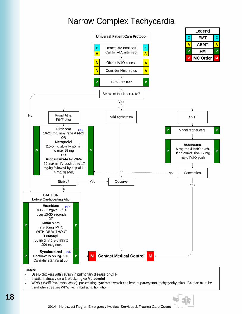
Universal Patient Care Protocol
Notes:
· Use β-blockers with caution in pulmonary disease or CHF
· If patient already on a β-blocker, give Metoprolol
· WPW ( Wolff Parkinson White): pre-existing syndrome which can lead to paroxysmal tachydysrhytmias. Caution must be
used when treating WPW with rabid atrial fibrilation.
Obtain IV/IO accessA A
Rapid Atrial
Fib/Flutter
Narrow Complex Tachycardia
Vagal maneuversP P
Adenosine
6 mg rapid IV/IO push.
If no conversion 12 mg
rapid IV/IO push
P P
Conversion
Contact Medical ControlM M
Diltiazem
10-25 mg, may repeat PRN
OR
Metoprolol
2.5-5 mg slow IV q5min
to max 15 mg
OR
Procainamide for WPW
20 mg/min IV push up to 17
mg/kg followed by drip of 1-
4 mg/kg IV/IO
P P
PRN
No
Synchronized
Cardioversion Pg. 103
Consider starting at 50j
P P
YesStable?
No
ObserveYes
SVT
Stable at this Heart rate?
Yes
Mild SymptomsNo
Consider Fluid BolusA A
ECG / 12 leadP P
Etomidate
0.1-0.3 mg/kg IV/IO
over 15-30 seconds
OR
Midazolam
2.5-10mg IV/ IO
WITH OR WITHOUT
Fentanyl
50 mcg IV q 3-5 min to
200 mcg max
P P
Immediate transport
Call for ALS intercept
E E
A A
PRN
PRN
CAUTION
before Cardioverting Afib
AEMT
PM
MC Order
A
P
A
P
M M
EMT
Legend
E E
2014 - Northwest Region Emergency Medical Services & Trauma Care Council
19
19
19
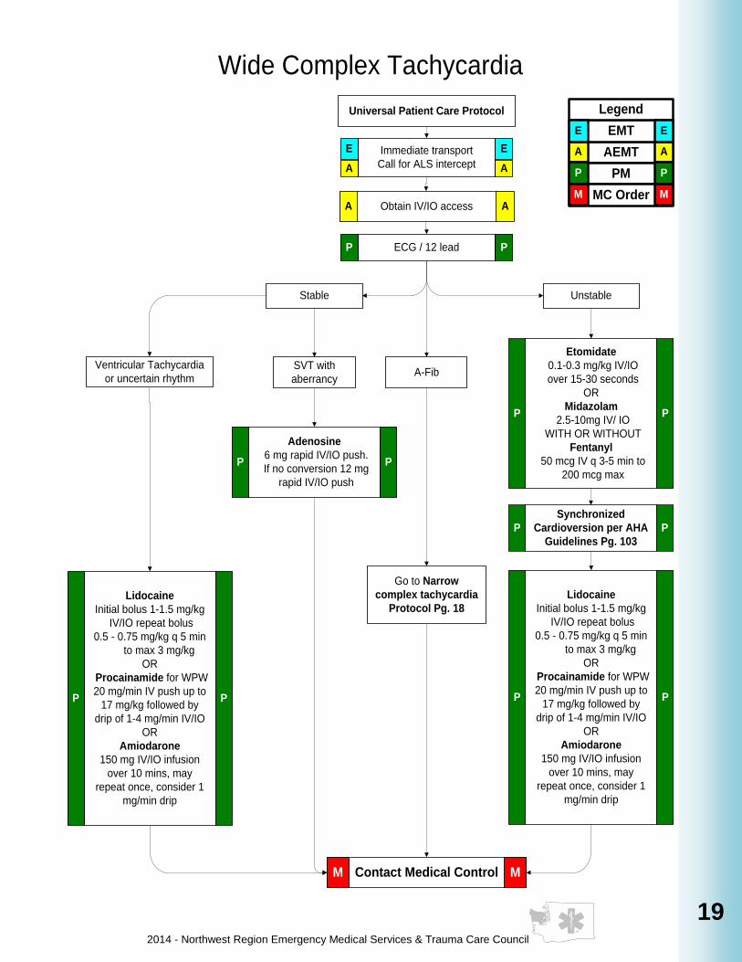
Wide Complex Tachycardia
Universal Patient Care Protocol
SVT with
aberrancy
Go to Narrow
complex tachycardia
Protocol Pg. 18
Contact Medical ControlM M
Stable Unstable
A-Fib
Adenosine
6 mg rapid IV/IO push.
If no conversion 12 mg
rapid IV/IO push
P P
Etomidate
0.1-0.3 mg/kg IV/IO
over 15-30 seconds
OR
Midazolam
2.5-10mg IV/ IO
WITH OR WITHOUT
Fentanyl
50 mcg IV q 3-5 min to
200 mcg max
P P
Ventricular Tachycardia
or uncertain rhythm
ECG / 12 lead P P
Synchronized
Cardioversion per AHA
Guidelines Pg. 103
P P
A AObtain IV/IO access
Lidocaine
Initial bolus 1-1.5 mg/kg
IV/IO repeat bolus
0.5 - 0.75 mg/kg q 5 min
to max 3 mg/kg
OR
Procainamide for WPW
20 mg/min IV push up to
17 mg/kg followed by
drip of 1-4 mg/min IV/IO
OR
Amiodarone
150 mg IV/IO infusion
over 10 mins, may
repeat once, consider 1
mg/min drip
P P
Immediate transport
Call for ALS intercept
E E
A A
AEMT
PM
MC Order
A
P
A
P
M M
EMT
Legend
E E
Lidocaine
Initial bolus 1-1.5 mg/kg
IV/IO repeat bolus
0.5 - 0.75 mg/kg q 5 min
to max 3 mg/kg
OR
Procainamide for WPW
20 mg/min IV push up to
17 mg/kg followed by
drip of 1-4 mg/min IV/IO
OR
Amiodarone
150 mg IV/IO infusion
over 10 mins, may
repeat once, consider 1
mg/min drip
P P
2014 - Northwest Region Emergency Medical Services & Trauma Care Council
20
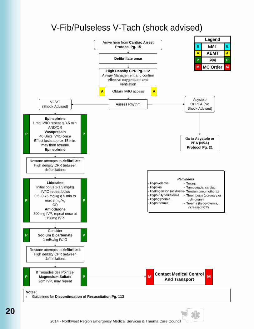
Arrive here from Cardiac Arrest
Protocol Pg. 15
V-Fib/Pulseless V-Tach (shock advised)
Epinephrine
1 mg IV/IO repeat q 3-5 min.
AND/OR
Vasopressin
40 Units IV/IO once
Effect lasts approx 15 min.
may then resume
Epinephrine
P P
Asystole
Or PEA (No
Shock Advised)
VF/VT
(Shock Advised)
Notes:
· Guidelines for Discontinuation of Resuscitation Pg. 113
Contact Medical Control
And TransportM M
High Density CPR Pg. 112
Airway Management and confirm
effective oxygenation and
ventilation
Obtain IV/IO accessA A
Defibrillate once
Assess Rhythm
Resume attempts to defibrillate
High density CPR between
defibrillations
If Torsades des Pointes-
Magnesium Sulfate
2gm IVP, may repeat
P P
Go to Asystole or
PEA (NSA)
Protocol Pg. 21
Resume attempts to defibrillate
High density CPR between
defibrillations
Consider
Sodium Bicarbonate
1 mEq/kg IV/IO
P P
Lidocaine
Initial bolus 1-1.5 mg/kg
IV/IO repeat bolus
0.5 -0.75 mg/kg q 5 min to
max 3 mg/kg
OR
Amiodarone
300 mg IVP, repeat once at
150mg IVP
P P
Reminders
- Hypovolemia
- Hypoxia
- Hydrogen ion (acidosis)
- Hypo-/Hyperkalemia
- Hypoglycemia
- Hypothermia
- Toxins
- Tamponade, cardiac
- Tension pneumothorax
- Thrombosis (coronary or
pulmonary)
- Trauma (hypovolemia,
increased ICP)
AEMT
PM
MC Order
A
P
A
P
M M
EMT
Legend
E E

2014 - Northwest Region Emergency Medical Services & Trauma Care Council
21
21
21
Arrive here from Cardiac Arrest
protocol Pg. 15
Asystole / PEA (no shock advised)
Assess Rhythm
Asystole or PEA
(No Shock Advised)
Notes:
· Guidelines for Discontinuation of Resuscitation Pg. 113
Obtain IV/IO accessA A
See VF/Pulseless
VT (SA) Protocol
Pg. 20
Contact Medical ControlM M
Consider
Sodium Bicarbonate
1 mEq/kg IV/IO
if preexisting hyperkalemia
(renal failure), TCA or
ASA OD Consider TCP for
Bradycardic rhythms
P P
Atropine
for slow (<60 bpm) PEA
1 mg IV/IO
repeat PRN to 3 mg max
P P
VF/VT (Shock Advised)
High Density CPR Pg. 112
Airway Management and confirm
effective oxygenation and
ventilation
Search for at treat possible causes:
- Hypovolemia
- Hypoxia
- Hydrogen ion (acidosis)
- Hypo-/Hyperkalemia
- Hypoglycemia
- Hypothermia
- Toxins
- Tamponade, cardiac
- Tension pneumothorax
- Thrombosis (coronary or
pulmonary)
- Trauma (hypovolemia,
increased ICP)
Epinephrine
1 mg IV/IO repeat q 3-5 min.
AND/OR
Vasopressin
40 Units IV/IO once
Effect lasts approx 15 min.
may then resume
Epinephrine
P P
PRN
AEMT
PM
MC Order
A
P
A
P
M M
EMT
Legend
E E
Consider Hyperkalemia Protocol
Pg. 22
2014 - Northwest Region Emergency Medical Services & Trauma Care Council
22
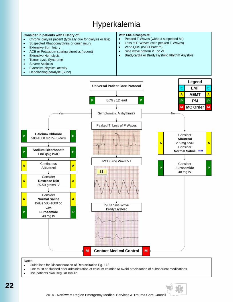
Hyperkalemia
Symptomatic Arrhythmia?
Notes:
· Guidelines for Discontinuation of Resuscitation Pg. 113
· Line must be flushed after administration of calcium chloride to avoid precipitation of subsequent medications.
· Use patients own Regular Insulin
Yes No
Contact Medical ControlM M
AEMT
PM
MC Order
A
P
A
P
M M
EMT
Legend
E EUniversal Patient Care Protocol
Consider in patients with History of:
· Chronic dialysis patient (typically due for dialysis or late)
· Suspected Rhabdomyolysis or crush injury
· Extensive Burn Injury
· ACE or Potassium sparing diuretics (recent)
· Extensive Hemolysis
· Tumor Lysis Syndrome
· Severe Acdiosis
· Extensive physical activity
· Depolarizing paralytic (Succ)
With EKG Changes of:
· Peaked T-Waves (without suspected MI)
· Loss of P-Waves (with peaked T-Waves)
· Wide QRS (IVCD Pattern)
· Sine wave pattern VT or VF
· Bradycardia or Bradyasystolic Rhythm Asystole
ECG / 12 lead P P
Consider
Albuterol
2.5 mg SVN
Consider
Normal Saline
A A
PRN
Consider
Furosemide
40 mg IV
PP
Calcium Chloride
500-1000 mg IV- SlowlyP P
Sodium Bicarbonate
1 mEq/kg IV/IOP P
Consider
Dextrose D50
25-50 grams IV
A A
Consider
Normal Saline
Bolus 500-1000 cc
A A
with
Furosemide
40 mg IV
PP
Continuous
AlbuterolA A
Peaked T, Loss of P Waves
IVCD Sine Wave VT
IVCD Sine Wave
Bradyasystolic

2014 - Northwest Region Emergency Medical Services & Trauma Care Council
23
23
23
History:
· Viagra, Cialis, Levitra
(Male or Female)
· Onset
· Palliation / Provocation
· Quality (crampy, constant,
sharp, dull, etc.)
· Region / Radiation /
Referred
· Severity (1-10)
· Time (duration / repetition)
Signs/Symptoms:
· CP (pain, pressure,
aching, tightness)
· Radiation of pain
· Pale, diaphoresis
· Shortness of breath
· Nausea, vomiting,
dizziness
· Abnormal vital signs
Differential:
· Aortic dissection or
aneurysm
· Trauma vs. Medical
· Pericarditis
· Pulmonary embolism
· Asthma / COPD
· Pneumothorax
· GE reflux or Hiatal hernia
· Esophageal spasm
· Chest wall injury or pain
· Pleural pain
Chest Pain / Acute Coronary Syndrome
Aspirin
325 mg chewable POE E
Nitroglycerin
If BP > 100 systolic
per patient Rx, 0.4 mg SL q 3-5 min
E E
Obtain IV/IO accessA A
Contact Medical ControlM M
Notes:
· Avoid Nitroglycerin in any patient (man or woman) who has used sexual performance enhancement drugs
(ie Viagra, Levitra, etc.) in the past 24 hours due to possible severe hypotension.
· If positive ECG changes, establish a second IV while en route to the hospital.
· Monitor for hypotension after administration of nitroglycerin and morphine.
· *Metroprolol - contraindicated with CNS stimulant.
- consider ½ doses for elderly, asthma & COPD
Consider repeat ECG / 12 leadP P
consider
Morphine
2 mg IV q 3-5 min to 20mg max
OR
Fentanyl
25 mcg IV q 3-5 min to 200 mcg max
OR
Hydromorphone
0.5mg IV q 3-5 min to 4mg max
P P
Universal Patient Care Protocol
Suspect Acute Coronary Syndrome
Interpret ECG / 12 LeadP P
Cardiac Risk Factors:
· Previous MI / Known Cardiac
disease
· Hypotention
· Diabetes
· Smoking tobacco use
· Family History
· High Cholesterol
· Hyperlipidemia
ALS Transport if:
*Metroprolol
2.5 - 5 mg IV, over 2 mins q 3-5 mins
to max 15 mg
Hold if SBP < 120 or Heart rate < 75
P P
See STEMI Follow
County Operating
Procedure Appendix
Pg. 10
P P
AEMT
PM
MC Order
A
P
A
P
M M
EMT
Legend
E E
consider
Lorazepam
0.5-4 mg IV/IN/IM
OR
Midazolam
2.5-5 mg IV/IN/IM
OR
Diazepam
2-5 mg IV/IN/IM
P P
If suspected CNS stimulant

2014 - Northwest Region Emergency Medical Services & Trauma Care Council
24
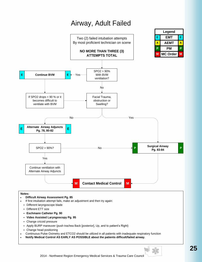
Notes:
· Capnometry or capnography is mandatory with all methods of intubation. Document results.
· For this protocol, adult is defined any person who does not fit the length based tape
· EMT’s must have multi-lumen airway training to use Supraglottic Airway Adjuncts.
· Maintain C-spine immobilization for patients with suspected spinal injury
· Paramedics should consider Supraglotic Airway Adjuncts.
· Reconfirm ETT placement each time patient is moved
· Continuous pulse oximetry should be utilized in all patients with compromised respiratory function
Airway, Adult
Assess ABC’s, respiratory rate,
effort, adequacy
Supplemental Oxygen
Preoxygenation Procedure
PG. 86
Inadequate
Basic Maneuvers
first-- open airway;
nasal/oral airway;
bag-valve mask
Obstruction
Remove
obstruction per
AHA guidelines
Successful
removal
Unsuccessful
Alternate Airway
Adjuncts Pg. 78, 80-82E E
Establish Advanced
Airway Pg. 89-91, 95P P
Inadequate
Inadequate
Failed Airway
Protocol Pg. 25
Adequate
Contact Medical ControlM M
Pulse OximetryE E
Adequate
Universal Patient Care Protocol
AEMT
PM
MC Order
A
P
A
P
M M
EMT
Legend
E E
2014 - Northwest Region Emergency Medical Services & Trauma Care Council
25
25
25
Notes:
· Difficult Airway Assessment Pg. 85
· If first intubation attempt fails, make an adjustment and then try again:
§ Different laryngoscope blade
§ Different ETT size
§ Eschmann Catheter Pg. 90
§ Video Assisted Laryngoscopy Pg. 95
§ Change cricoid pressure
§ Apply BURP maneuver (push trachea Back [posterior], Up, and to patient’s Right)
§ Change head positioning
· Continuous Pulse Oximetry and ETCO2 should be utilized in all patients with inadequate respiratory function
· Notify Medical Control AS EARLY AS POSSIBLE about the patients difficult/failed airway.
Airway, Adult Failed
Two (2) failed intubation attempts
By most proficient technician on scene
NO MORE THAN THREE (3)
ATTEMPTS TOTAL
SPO2 > 90%
With BVM
ventilation?
Continue BVME E Yes
If SPO2 drops < 90 % or it
becomes difficult to
ventilate with BVM
Facial Trauma,
obstruction or
Swelling?
No
Surgical Airway
Pg. 83-84P PSPO2 > 90%? No
No Yes
Continue ventilation with
Alternate Airway Adjuncts
Yes
Alternate Airway Adjuncts
Pg. 78, 80-82E E
Contact Medical ControlM M
AEMT
PM
MC Order
A
P
A
P
M M
EMT
Legend
E E
2014 - Northwest Region Emergency Medical Services & Trauma Care Council
26
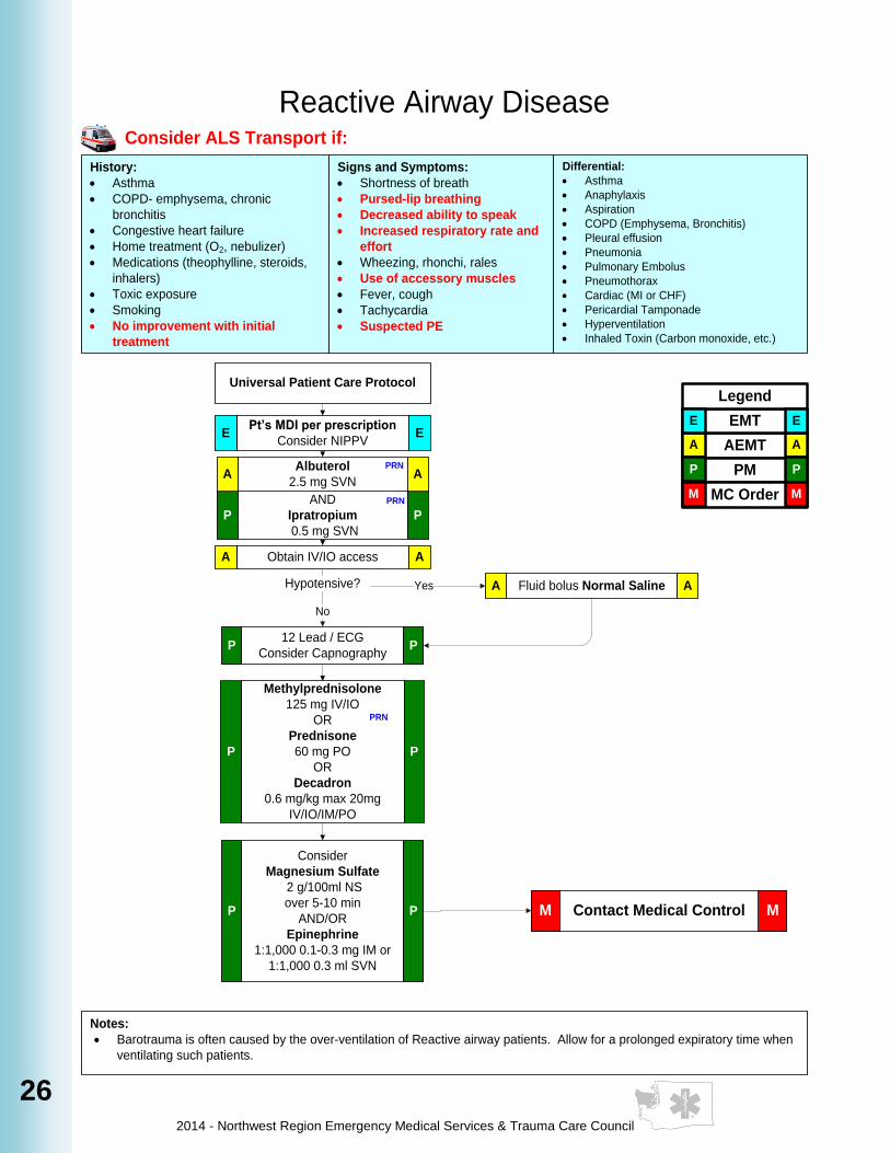
Reactive Airway Disease
History:
· Asthma
· COPD- emphysema, chronic
bronchitis
· Congestive heart failure
· Home treatment (O2, nebulizer)
· Medications (theophylline, steroids,
inhalers)
· Toxic exposure
· Smoking
· No improvement with initial
treatment
Signs and Symptoms:
· Shortness of breath
· Pursed-lip breathing
· Decreased ability to speak
· Increased respiratory rate and
effort
· Wheezing, rhonchi, rales
· Use of accessory muscles
· Fever, cough
· Tachycardia
· Suspected PE
Differential:
· Asthma
· Anaphylaxis
· Aspiration
· COPD (Emphysema, Bronchitis)
· Pleural effusion
· Pneumonia
· Pulmonary Embolus
· Pneumothorax
· Cardiac (MI or CHF)
· Pericardial Tamponade
· Hyperventilation
· Inhaled Toxin (Carbon monoxide, etc.)
Notes:
· Barotrauma is often caused by the over-ventilation of Reactive airway patients. Allow for a prolonged expiratory time when
ventilating such patients.
Universal Patient Care Protocol
Pt’s MDI per prescription
Consider NIPPVE E
Fluid bolus Normal SalineA A
Obtain IV/IO accessA A
Hypotensive?
No
Methylprednisolone
125 mg IV/IO
OR
Prednisone
60 mg PO
OR
Decadron
0.6 mg/kg max 20mg
IV/IO/IM/PO
P P
Contact Medical ControlM M
Consider
Magnesium Sulfate
2 g/100ml NS
over 5-10 min
AND/OR
Epinephrine
1:1,000 0.1-0.3 mg IM or
1:1,000 0.3 ml SVN
P P
PRN
12 Lead / ECG
Consider CapnographyP P
Albuterol
2.5 mg SVNA A
AND
Ipratropium
0.5 mg SVN
P P
PRN
PRN
Consider ALS Transport if:
Yes
AEMT
PM
MC Order
A
P
A
P
M M
EMT
Legend
E E

2014 - Northwest Region Emergency Medical Services & Trauma Care Council
27
27
27
History:
· Congestive heart failure
· Past medical history
· Medications (digoxin, lasix, HCTZ)
· Viagra, Levitra, Cialis
· Cardiac history- past myocardial
infarction
Signs/Symptoms:
· Respiratory distress, rales
· Apprehension, orthopnea
· Jugular vein distention
· Pink, frothy sputum
· Peripheral edema, diaphoresis
· Hypotension, shock
· Chest pain
Differential:
· Myocardial infarction
· Congestive heart failure
· Asthma
· Anaphylaxis
· Aspiration
· COPD
· Pleural effusion
· Pneumonia
· Pulmonary Embolus
· Pericardial tamponade
Universal Patient Care Protocol
Notes:
· Avoid Nitroglycerin in any patient (man or woman) who has used sexual performance enhancement drugs
(ie Viagra, Levitra, etc.) in the past 24 hours due to possible severe hypotension.
· If patient has taken nitroglycerin without relief, consider potency of the medication.
· Consider myocardial infarction in all these patients.
· Allow the patient to be in their position of comfort to maximize their breathing effort.
Pulmonary Edema
Mild-Moderate- able to speak sentences, crackles base only, O2 sat ≥ 92%
Severe- respiratory distress, crackles throughout, O2 sat < 92%
Near Death- Decreased LOC, cyanosis, dropping sats, ineffective respiratory drive
Contact Medical ControlM M
Nitroglycerin if SBP > 100
0.4 mg SL
may repeat q 3-5 min
A A
consider
Furosemide
40-80 mg IV
PP
ECG/ 12 Lead PP
Consider CapnographyP P
Support Adult
Airway Protocol
Pg. 24
Yes
Obtain IV/IO accessA A
consider
Midazolam
1 - 2 mg IV/ IO
Or
Diazepam
1 - 2 mg IV / IM / IO
Or
Lorazepam
0.5 - 1 mg IV/IN/IM. May
repeat PRN
P P
ALS transport this patient if available
AEMT
PM
MC Order
A
P
A
P
M M
EMT
Legend
E E
consider
Morphine
2 mg IV q 3-5 min to 20mg max
OR
Fentanyl
25 mcg IV q 3-5 min to 200 mcg max
OR
Hydromorphone
0.5mg IV q 3-5 min to 4mg max
P P
Consider
Nitroglycerin drip
Hold if SBP <100
Nitro Paste 1-2' to chest wall
P P
Non-Invasive positive
pressure ventilation
NIPPV if available
E E
May repeat q 1 minP P
Fever or Purulent Sputum?
Consider mild sedationP P
No
2014 - Northwest Region Emergency Medical Services & Trauma Care Council
28
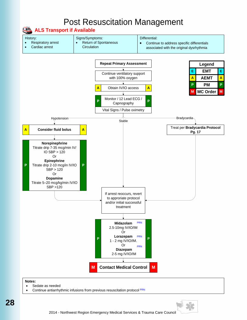
History:
· Respiratory arrest
· Cardiac arrest
Signs/Symptoms:
· Return of Spontaneous
Circulation
Differential:
· Continue to address specific differentials
associated with the original dysrhythmia
Repeat Primary Assessment
Notes:
· Sedate as needed
· Continue antiarrhythmic infusions from previous resuscitation protocol
Post Resuscitation Management
Continue ventilatory support
with 100% oxygen
Obtain IV/IO accessA A
Monitor / 12 Lead ECG /
CapnographyP P
Vital Signs / Pulse oximetry
Treat per Bradycardia Protocol
Pg. 17Consider fluid bolusA A
BradycardiaHypotension
If arrest reoccurs, revert
to approriate protocol
and/or initial successful
treatment
Contact Medical ControlM M
Midazolam
2.5-10mg IV/IO/IM
Or
Lorazepam
1 - 2 mg IV/IO/IM.
Or
Diazepam
2-5 mg IV/IO/IM
P P
PRN
PRN
Stable
PRN
PRN
ALS Transport if Available
AEMT
PM
MC Order
A
P
A
P
M M
EMT
Legend
E E
Norepinephrine
Titrate drip 7-35 mcg/min IV/
IO SBP > 120
Or
Epinephrine
Titrate drip 2-10 mcg/in IV/IO
SBP > 120
Or
Dopamine
Titrate 5–20 mcg/kg/min IV/IO
SBP >120
P P
2014 - Northwest Region Emergency Medical Services & Trauma Care Council
29
29
29
29
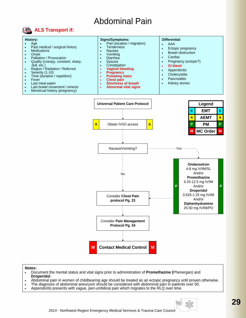
History:· Age· Past medical / surgical history· Medications· Onset· Palliation / Provocation· Quality (crampy, constant, sharp, dull, etc.)· Region / Radiation / Referred· Severity (1-10)· Time (duration / repetition)· Fever· Last meal eaten· Last bowel movement / emesis· Menstrual history (pregnancy)
Signs/Symptoms:· Pain (location / migration)· Tenderness· Nausea· Vomiting· Diarrhea· Dysuria· Constipation· Vaginal bleeding· Pregnancy· Pulsating mass· Chest pain· Shortness of breath· Abnormal vital signs
Differential:
· AAA
· Ectopic pregnancy
· Bowel obstruction
· Cardiac
· Pregnancy (ectopic?)
· GI bleed
· Appendicitis
· Cholecystitis
· Pancreatitis
· Kidney stones
Universal Patient Care Protocol
Notes:· Document the mental status and vital signs prior to administration of Promethazine (Phenergan) and
Droperidol.· Abdominal pain in women of childbearing age should be treated as an ectopic pregnancy until proven otherwise.· The diagnosis of abdominal aneurysm should be considered with abdominal pain in patients over 50.· Appendicitis presents with vague, peri-umbilical pain which migrates to the RLQ over time.
Nausea/Vomiting?
Obtain IV/IO accessA A
Ondansetron
4-8 mg IV/IM/SL
And/or
Promethazine
6.25-12.5 mg IV/IM
And/or
Droperidol
0.625-1.25 mg IV/IM
And/or
Diphenhydramine
25-50 mg IV/IM/PO
P P
Yes
Consider Chest Pain
protocol Pg. 23
Consider Pain Management
Protocol Pg. 34
Contact Medical ControlM M
Abdominal Pain
No
ALS Transport if:
AEMT
PM
MC Order
A
P
A
P
M M
EMT
Legend
E E
2014 - Northwest Region Emergency Medical Services & Trauma Care Council
30
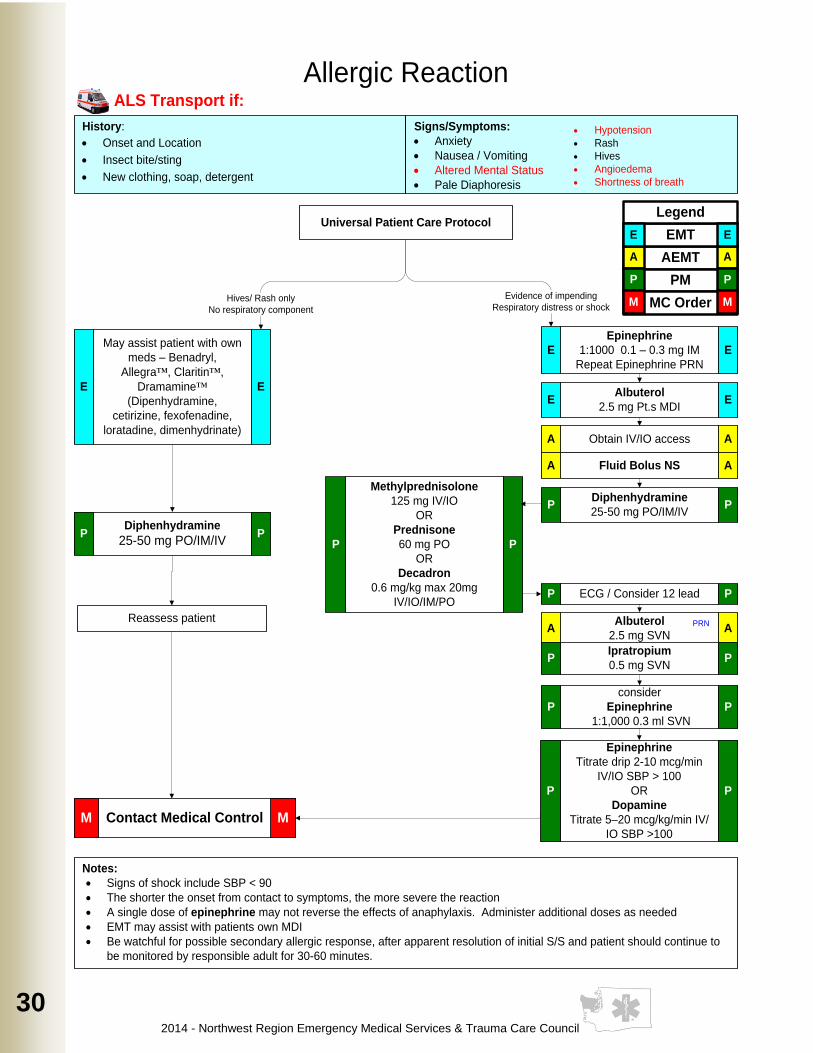
History:
· Onset and Location
· Insect bite/sting
· New clothing, soap, detergent
Signs/Symptoms:
· Anxiety
· Nausea / Vomiting
· Altered Mental Status
· Pale Diaphoresis
Universal Patient Care Protocol
Notes:
· Signs of shock include SBP < 90
· The shorter the onset from contact to symptoms, the more severe the reaction
· A single dose of epinephrine may not reverse the effects of anaphylaxis. Administer additional doses as needed
· EMT may assist with patients own MDI
· Be watchful for possible secondary allergic response, after apparent resolution of initial S/S and patient should continue to
be monitored by responsible adult for 30-60 minutes.
Allergic Reaction
Epinephrine
1:1000 0.1 – 0.3 mg IM
Repeat Epinephrine PRN
E E
Reassess patient
Contact Medical ControlM M
Obtain IV/IO access A A
Methylprednisolone
125 mg IV/IO
OR
Prednisone
60 mg PO
OR
Decadron
0.6 mg/kg max 20mg
IV/IO/IM/PO
P P
ECG / Consider 12 leadP P
Evidence of impending
Respiratory distress or shock Hives/ Rash only
No respiratory component
Fluid Bolus NS A A
Diphenhydramine
25-50 mg PO/IM/IV P P
May assist patient with own
meds – Benadryl,
Allegra™, Claritin™,
Dramamine™
(Dipenhydramine,
cetirizine, fexofenadine,
loratadine, dimenhydrinate)
E E
Diphenhydramine
25-50 mg PO/IM/IV P P
Albuterol
2.5 mg Pt.s MDI E E
Albuterol
2.5 mg SVN A A
PRN
ALS Transport if:
· Hypotension
· Rash
· Hives
· Angioedema
· Shortness of breath
AEMT
PM
MC Order
A
P
A
P
M M
EMT
Legend
E E
Epinephrine
Titrate drip 2-10 mcg/min
IV/IO SBP > 100
OR
Dopamine
Titrate 5–20 mcg/kg/min IV/
IO SBP >100
P P
Ipratropium
0.5 mg SVNP P
consider
Epinephrine
1:1,000 0.3 ml SVN
P P
2014 - Northwest Region Emergency Medical Services & Trauma Care Council
31
31
31
31
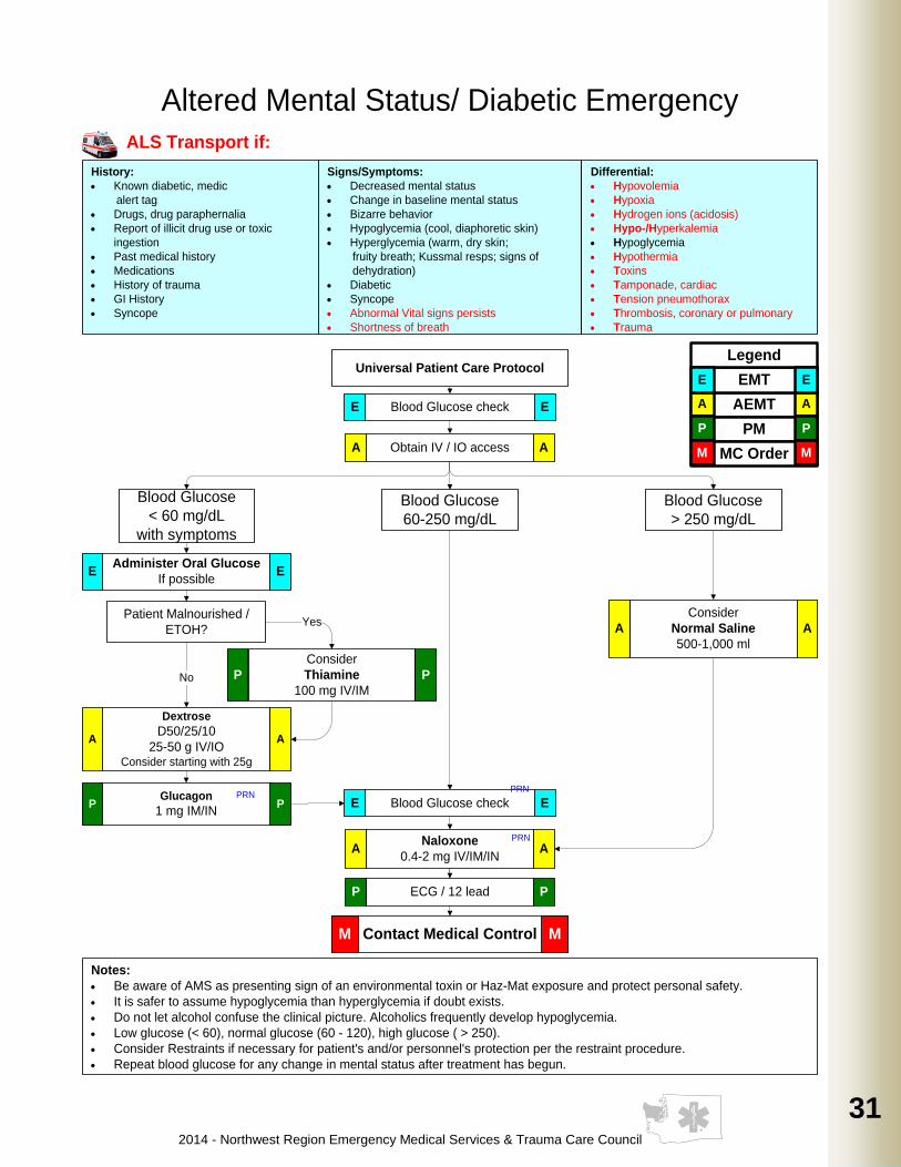
History:
· Known diabetic, medic
alert tag
· Drugs, drug paraphernalia
· Report of illicit drug use or toxic
ingestion
· Past medical history
· Medications
· History of trauma
· GI History
· Syncope
Signs/Symptoms:
· Decreased mental status
· Change in baseline mental status
· Bizarre behavior
· Hypoglycemia (cool, diaphoretic skin)
· Hyperglycemia (warm, dry skin;
fruity breath; Kussmal resps; signs of
dehydration)
· Diabetic
· Syncope
· Abnormal Vital signs persists
· Shortness of breath
Differential:
· Hypovolemia
· Hypoxia
· Hydrogen ions (acidosis)
· Hypo-/Hyperkalemia
· Hypoglycemia
· Hypothermia
· Toxins
· Tamponade, cardiac
· Tension pneumothorax
· Thrombosis, coronary or pulmonary
· Trauma
Universal Patient Care Protocol
Notes:
· Be aware of AMS as presenting sign of an environmental toxin or Haz-Mat exposure and protect personal safety.
· It is safer to assume hypoglycemia than hyperglycemia if doubt exists.
· Do not let alcohol confuse the clinical picture. Alcoholics frequently develop hypoglycemia.
· Low glucose (< 60), normal glucose (60 - 120), high glucose ( > 250).
· Consider Restraints if necessary for patient's and/or personnel's protection per the restraint procedure.
· Repeat blood glucose for any change in mental status after treatment has begun.
Administer Oral Glucose
If possibleE E
Blood Glucose
< 60 mg/dL
with symptoms
Blood Glucose
60-250 mg/dL
Blood Glucose
> 250 mg/dL
Altered Mental Status/ Diabetic Emergency
Contact Medical ControlM M
Dextrose
D50/25/10
25-50 g IV/IOConsider starting with 25g
A A
Consider
Thiamine
100 mg IV/IM
P P
Patient Malnourished /
ETOH?Yes
Glucagon
1 mg IM/INP P
Consider
Normal Saline
500-1,000 ml
A A
Obtain IV / IO accessA A
Naloxone
0.4-2 mg IV/IM/INA A
PRN
No
PRN
ECG / 12 leadP P
Blood Glucose checkE E
ALS Transport if:
AEMT
PM
MC Order
A
P
A
P
M M
EMT
Legend
E E
Blood Glucose checkE E
PRN
2014 - Northwest Region Emergency Medical Services & Trauma Care Council
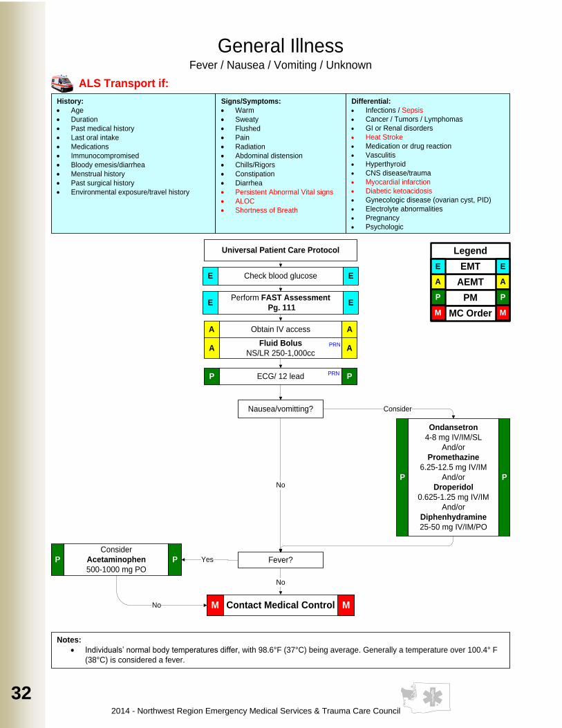
32
History:
· Age
· Duration
· Past medical history
· Last oral intake
· Medications
· Immunocompromised
· Bloody emesis/diarrhea
· Menstrual history
· Past surgical history
· Environmental exposure/travel history
Signs/Symptoms:
· Warm
· Sweaty
· Flushed
· Pain
· Radiation
· Abdominal distension
· Chills/Rigors
· Constipation
· Diarrhea
· Persistent Abnormal Vital signs
· ALOC
· Shortness of Breath
Differential:
· Infections / Sepsis
· Cancer / Tumors / Lymphomas
· GI or Renal disorders
· Heat Stroke
· Medication or drug reaction
· Vasculitis
· Hyperthyroid
· CNS disease/trauma
· Myocardial infarction
· Diabetic ketoacidosis
· Gynecologic disease (ovarian cyst, PID)
· Electrolyte abnormalities
· Pregnancy
· Psychologic
Universal Patient Care Protocol
Notes:
· Individuals’ normal body temperatures differ, with 98.6°F (37°C) being average. Generally a temperature over 100.4° F
(38°C) is considered a fever.
General IllnessFever / Nausea / Vomiting / Unknown
Check blood glucose E E
Perform FAST Assessment
Pg. 111E E
Obtain IV accessA A
Fluid Bolus
NS/LR 250-1,000cc A A
PRN
ECG/ 12 leadP PPRN
Nausea/vomitting?
Fever?
Consider
Acetaminophen
500-1000 mg PO
P P
Contact Medical ControlM M
Yes
Consider
No
No
No
ALS Transport if:
AEMT
PM
MC Order
A
P
A
P
M M
EMT
Legend
E E
Ondansetron
4-8 mg IV/IM/SL
And/or
Promethazine
6.25-12.5 mg IV/IM
And/or
Droperidol
0.625-1.25 mg IV/IM
And/or
Diphenhydramine
25-50 mg IV/IM/PO
P P
2014 - Northwest Region Emergency Medical Services & Trauma Care Council
33
33
33
33
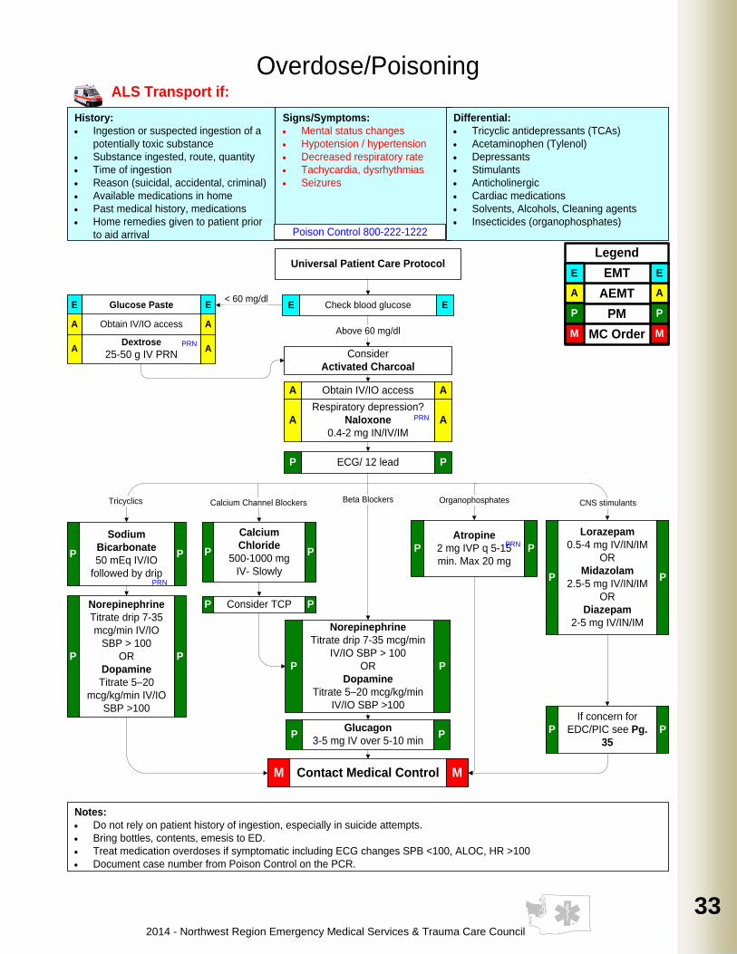
History:
· Ingestion or suspected ingestion of a
potentially toxic substance
· Substance ingested, route, quantity
· Time of ingestion
· Reason (suicidal, accidental, criminal)
· Available medications in home
· Past medical history, medications
· Home remedies given to patient prior
to aid arrival
Signs/Symptoms:
· Mental status changes
· Hypotension / hypertension
· Decreased respiratory rate
· Tachycardia, dysrhythmias
· Seizures
Differential:
· Tricyclic antidepressants (TCAs)
· Acetaminophen (Tylenol)
· Depressants
· Stimulants
· Anticholinergic
· Cardiac medications
· Solvents, Alcohols, Cleaning agents
· Insecticides (organophosphates)
Notes:
· Do not rely on patient history of ingestion, especially in suicide attempts.
· Bring bottles, contents, emesis to ED.
· Treat medication overdoses if symptomatic including ECG changes SPB <100, ALOC, HR >100
· Document case number from Poison Control on the PCR.
Overdose/Poisoning
Universal Patient Care Protocol
Obtain IV/IO accessA A
Respiratory depression?
Naloxone
0.4-2 mg IN/IV/IM
A A
Above 60 mg/dl
Consider
Activated Charcoal
Dextrose
25-50 g IV PRNA A
PRN
Glucose PasteE E
Obtain IV/IO accessA A
< 60 mg/dl
PRN
Sodium
Bicarbonate
50 mEq IV/IO
followed by drip
P P
Calcium
Chloride
500-1000 mg
IV- Slowly
P P
Norepinephrine
Titrate drip 7-35 mcg/min
IV/IO SBP > 100
OR
Dopamine
Titrate 5–20 mcg/kg/min
IV/IO SBP >100
P P
Atropine
2 mg IVP q 5-15
min. Max 20 mg
P P
Lorazepam
0.5-4 mg IV/IN/IM
OR
Midazolam
2.5-5 mg IV/IN/IM
OR
Diazepam
2-5 mg IV/IN/IM
P P
Glucagon
3-5 mg IV over 5-10 minP P
CNS stimulantsOrganophosphatesCalcium Channel BlockersTricyclics
Consider TCPP P
Beta Blockers
Norepinephrine
Titrate drip 7-35
mcg/min IV/IO
SBP > 100
OR
Dopamine
Titrate 5–20
mcg/kg/min IV/IO
SBP >100
P P
Contact Medical ControlM M
PRN
ALS Transport if:
Check blood glucose E EAEMT
PM
MC Order
A
P
A
P
M M
EMT
Legend
E E
PRN
ECG/ 12 leadP P
If concern for
EDC/PIC see Pg.
35
P P
Poison Control 800-222-1222
2014 - Northwest Region Emergency Medical Services & Trauma Care Council
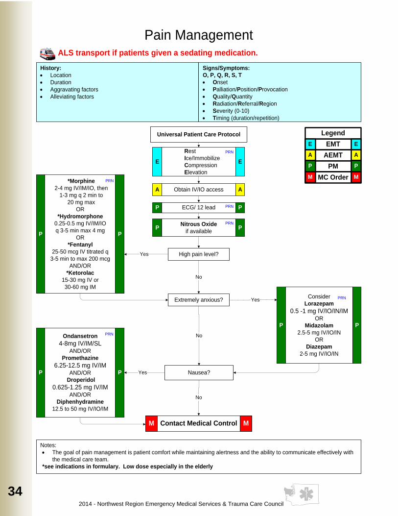
34
History:
· Location
· Duration
· Aggravating factors
· Alleviating factors
Signs/Symptoms:
O, P, Q, R, S, T
· Onset
· Palliation/Position/Provocation
· Quality/Quantity
· Radiation/Referral/Region
· Severity (0-10)
· Timing (duration/repetition)
Universal Patient Care Protocol
Notes:
· The goal of pain management is patient comfort while maintaining alertness and the ability to communicate effectively with
the medical care team.
*see indications in formulary. Low dose especially in the elderly
Obtain IV/IO accessA A
Contact Medical ControlM M
Ondansetron
4-8mg IV/IM/SLAND/OR
Promethazine
6.25-12.5 mg IV/IM AND/OR
Droperidol
0.625-1.25 mg IV/IMAND/OR
Diphenhydramine
12.5 to 50 mg IV/IO/IM
P P
PRN
Pain Management
Consider
Lorazepam
0.5 -1 mg IV/IO/IN/IMOR
Midazolam
2.5-5 mg IV/IO/IN
OR
Diazepam
2-5 mg IV/IO/IN
P P
*Morphine
2-4 mg IV/IM/IO, then
1-3 mg q 2 min to
20 mg max
OR
*Hydromorphone
0.25-0.5 mg IV/IM/IO
q 3-5 min max 4 mg
OR
*Fentanyl
25-50 mcg IV titrated q
3-5 min to max 200 mcg
AND/OR
*Ketorolac
15-30 mg IV or
30-60 mg IM
P P
PRN
Nitrous Oxide
if availableP P
PRN
ECG/ 12 leadP PPRN
Rest
Ice/Immobilize
Compression
Elevation
E E
PRN
High pain level?Yes
Extremely anxious?
No
Yes
Nausea?
PRN
Yes
No
No
ALS transport if patients given a sedating medication.
AEMT
PM
MC Order
A
P
A
P
M M
EMT
Legend
E E

2014 - Northwest Region Emergency Medical Services & Trauma Care Council
35
35
35
35
History:
· Situational crisis
· Psychiatric illness
· Medications
· Injury to self or threats to
others
· Medic alert tag
· Substance abuse / overdose
· Diabetes
Signs/Symptoms:
· Anxiety, agitation, confusion
· Affect change, hallucinations
· Delusional thoughts, bizarre
behavior
· Combative violent
· Expression of suicidal /
homicidal thoughts
Differential:
· Alcohol Intoxication
· Toxin / Substance abuse
· Medication effect / overdose
· Withdrawal syndromes
· Sepsis / Meningitis
· See Altered Mental Status differential
Universal Patient Care Protocol
Notes:
· Be sure to consider all possible medical/trauma causes for behavior.
· Do not overlook the possibility of associated domestic violence or child abuse.
· Do not stop Propofol until the patient is at the ED.
· *Doses may be repeated for the immediate safety of the provider or the patient.
Psychological/Emotional/Excited Delirium
ECG / 12 LeadP P
Midazolam*
2.5-5 mg IV/IO/IN/IM
AND/OR
Lorazepam*
0.5-2 mg IV/IO/IN/IM
AND/OR
Diazepam*
2-5 mg IV/IO/IN
P P
Contact Medical ControlM M
Leave scene immediately
Obtain IV/IO accessA APRN
Restraint Procedure Pg. 123-124 PRN
Or monitor situation and await Police
Await Police
No
Assess and treat
patient per
appropriate protocol
YesAEMT
PM
MC Order
A
P
A
P
M M
EMT
Legend
E E
Droperidol*
2.5-5 mg IV/IMP P
ALS transport if patients given a sedating medication.
Immediate risk of harm
to self or others?
Ketamine*
5-10 mg/kg IM/INP P
Droperidol*
2.5-10 mg IMP P
Midazolam*
2.5-10 mg IN/IM
AND/OR
Lorazepam*
0.5-4 mg IN/IM
AND/OR
Diazepam*
2-10 mg IN
P P
Patient remains aggressive or
refractory to TASER?
No
Consider for long transport
RSI Pg. 94
Propofol*
1-2 mg/kg IV followed by
drip 2.5 mcg/kg/min
keep BP > 90
P P
Yes
2014 - Northwest Region Emergency Medical Services & Trauma Care Council
36
History:
· Prior history of seizures
· Seizure medications
· Reported seizure activity
· History of recent head trauma
· Congenital abnormality
· Pregnancy (see Pregnancy Induced
Hypertension - Eclampsia)
· Medical alert tag
· If given sedation medication by EMS
Signs/Symptoms:
· Observed seizure activity
· Altered mental status
· Tonic / clonic activity
· Status epilepticus
· Incontinence
· Mouth trauma
· First time Seizure
Universal Patient Care Protocol
Notes:
· Be prepared to assist ventilations especially if a benzodiazepine is used.
· If evidence or suspicion of trauma, spine should be immobilized.
· Consider nasopharyngeal airway and elevate head of bed to 30 degrees.
· * Clallam County Versed IM or IN for seizures.
Seizure
Lorazepam
1-2 mg IV/IO/IN/IM
OR
Diazepam
2-5 mg IV/IO/IN
P P
Position patient on side to
prevent aspiration
Dextrose
D10/25/50 10-25g IV/IOA A/
Active Seizure?
Assess for cause of seizure
Treat per appropriate protocol
Repeat seizures? Yes
No
Yes
Contact Medical ControlM M
Differential:
· Hypovolemia
· Hypoxia
· Hydrogen ions (acidosis)
· Hypo-/Hyperkalemia
· Hypoglycemia
· Hypothermia
· Toxins
· Tamponade, cardiac
· Tension pneumothorax
· Thrombosis, coronary or pulmonary
· Trauma
Blood Glucose <60mg/dL?
Glucagon
1 mg IM/IN may repeat x 1P P
Obtain IV/IO accessA A
Pregnancy Induced
Hypertension Pg. 38
Yes, Pregnant Patient
ALS transport if patients given a sedating medication and / or:
PRN
PRN
Diastat *
* Patients own RXE E
AEMT
PM
MC Order
A
P
A
P
M M
EMT
Legend
E E
PRN
PRN
No
Yes
Midazolam
5-10 mg IN/IMP P
PRN
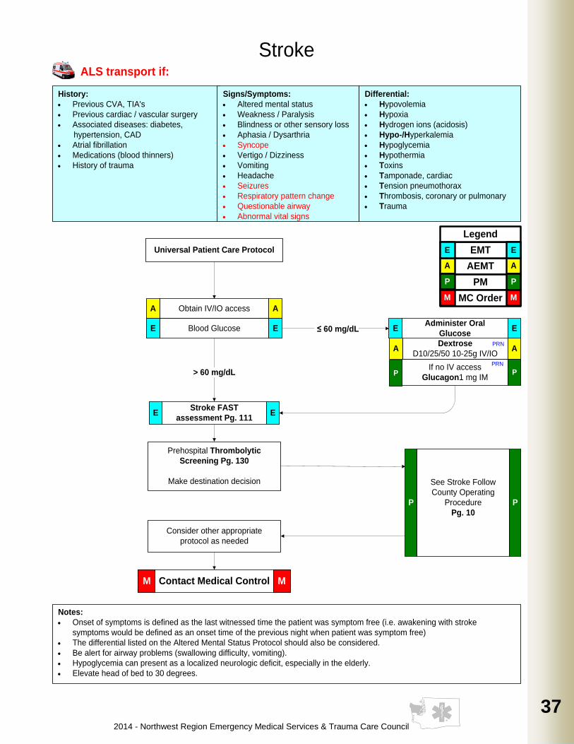
2014 - Northwest Region Emergency Medical Services & Trauma Care Council
37
37
37
37
History:
· Previous CVA, TIA's
· Previous cardiac / vascular surgery
· Associated diseases: diabetes,
hypertension, CAD
· Atrial fibrillation
· Medications (blood thinners)
· History of trauma
Signs/Symptoms:
· Altered mental status
· Weakness / Paralysis
· Blindness or other sensory loss
· Aphasia / Dysarthria
· Syncope
· Vertigo / Dizziness
· Vomiting
· Headache
· Seizures
· Respiratory pattern change
· Questionable airway
· Abnormal vital signs
Differential:
· Hypovolemia
· Hypoxia
· Hydrogen ions (acidosis)
· Hypo-/Hyperkalemia
· Hypoglycemia
· Hypothermia
· Toxins
· Tamponade, cardiac
· Tension pneumothorax
· Thrombosis, coronary or pulmonary
· Trauma
Universal Patient Care Protocol
Notes:
· Onset of symptoms is defined as the last witnessed time the patient was symptom free (i.e. awakening with stroke
symptoms would be defined as an onset time of the previous night when patient was symptom free)
· The differential listed on the Altered Mental Status Protocol should also be considered.
· Be alert for airway problems (swallowing difficulty, vomiting).
· Hypoglycemia can present as a localized neurologic deficit, especially in the elderly.
· Elevate head of bed to 30 degrees.
Stroke
Stroke FAST
assessment Pg. 111E E
Contact Medical ControlM M
Dextrose
D10/25/50 10-25g IV/IOA A
Obtain IV/IO accessA A
E EBlood Glucose ≤ 60 mg/dL
If no IV access
Glucagon1 mg IM> 60 mg/dL
PRN
P P
Prehospital Thrombolytic
Screening Pg. 130
Make destination decision
Consider other appropriate
protocol as needed
PRN
Administer Oral
GlucoseE E
ALS transport if:
AEMT
PM
MC Order
A
P
A
P
M M
EMT
Legend
E E
See Stroke Follow
County Operating
Procedure
Pg. 10
P P
2014 - Northwest Region Emergency Medical Services & Trauma Care Council
38
History:
· Past medical history
· Prenatal care
· Medications/drugs
· Familial incidence
· Primigravida
· Renal disease
Signs/Symptoms:
· Seizure
· Hypertension
· Tachycardia
· Edema
· Headache
· Visual disturbance
· Abdominal pain
· Amnesia and/or other change in mental status
Differential:
· Hypertension
· Multiple fetuses
· Gestational diabetes
· Microthrombi
· Improper placental
implantation
Universal Patient Care Protocol
Notes:
· Eclampsia can present up to two months postpartum
· Severe headache, vision changes, or RUQ pain may indicate preeclampsia.
· In the setting of pregnancy, hypertension is defined as a BP greater than 140 systolic or greater than 90 diastolic, or a
relative increase of 30 systolic and 20 diastolic from the patient's normal (pre-pregnancy) blood pressure.
· Maintain patient in a left lateral position to minimize risk of supine hypotensive syndrome.
Place in left lateral recumbent
position
Obtain IV/IO accessA A
ECGP P
Doppler Fetal Heart Tones
if availableP P
Seizing? (Eclamptic)Magnesium Sulfate
4 g IVPP P
Contact Medical ControlM M
No
Yes
Pregnancy Induced Hypertension ALS transport gently and quietly
Lorazepam
1-2 mg IV/IO/IN/IM
Or
Midazolam
2.5-5 mg IN/IM
Or
Diazepam
2-5 mg IV/IN/IO q5min
P P
PRN
AEMT
PM
MC Order
A
P
A
P
M M
EMT
Legend
E E
2014 - Northwest Region Emergency Medical Services & Trauma Care Council
39
39
39
39
39
39
39
39
History:
· Prenatal care?
· Due date and gestational age
· Multiple gestation (twins etc.)
· Meconium
· Delivery difficulties
· Medications (maternal)
· Maternal risk factors
· Substance abuse
Signs/Symptoms:
· Excess vaginal bleeding
· Signs of shock
Differential:
· Secretions
· Infection
· Hypovolemia
· Hypoglycemia
Universal Patient Care Protocol
Postpartum
Encourage breast feeding
Obtain IV/IO accessA A
Oxytocin
10 units IM
then
P P
Placenta delivered?
P P
Oxytocin
20 units in 1000 mL
NS titrated to severity
of bleeding
Contact Medical ControlM M
Yes
No
Fundal Massage Pg. 110
Newborn Resuscitation/Post-
Delivery Care Pg. 60
ALS transport if available:
AEMT
PM
MC Order
A
P
A
P
M M
EMT
Legend
E E
PRN
PRN
2014 - Northwest Region Emergency Medical Services & Trauma Care Council
40
History:
· Age
· Exposure to increased/decreased
temperatures and/or humidity
· Past medical history/medications
· Extreme exertion
· Time and length of exposure
· Poor PO intake
Signs/Symptoms:
· Altered mental status or
unconsciousness
· Hot, dry or sweaty skin
· Hypotension or shock
· Seizure
· Nausea
· Fatigue and / or muscle
cramping
Differential:
· Fever (Infection) / Sepsis
· Dehydration
· Medications
· Hyperthyroidism (Storm)
· Delirium tremens (DT's)
· Heat exhaustion
· Heat stroke
· Hypoglycemia
· Poisoning/overdose
Universal Patient Care Protocol
Notes:
· ATTEMPT REWARMING BEFORE CEASING RESUSCITATION EFFORTS.
· Extremes of age are more prone to temperature emergencies (i.e. young and old).
· Core temperature is the most reliable measure- for pts with ALOC and pts < 2 years old this should be the method of
measurement.
· Heat emergencies can be precipitated by use of: tricyclic antidepressants, phenothiazines, anticholinergic medications, and
alcohol.
· Cocaine, Amphetamines, and Salicylates may elevate body temperatures.
· Shivering stops below 32° C (90° F).
· Sweating generally disappears as body temperature rises above 104° F (40° C).
· With temperature less than 31° C (88° F) ventricular fibrillation is common cause of death. Handling patients gently may
prevent this (rarely responds to defibrillation).
· Hypothermia may produce severe bradycardia.
Environmental Emergencies
Document patient temperature
Cooling Measures Warming Measures
Hyperthermic? Hypothermic?
Blood Glucose check
Contact Medical ControlM M
Treat per appropriate protocol
No
PRN
YesShivering?
Obtain IV/IO accessA A
Fluid bolus NSA A
ECG/ 12 leadP PPRN
PRN
Consider
Lorazepam
0.5 -1 mg IV/IO/IN/IMOR
Midazolam
2.5-5 mg IM/INOR
Diazepam
2-5 mg IV/IO
P P
ALS transport if available:
AEMT
PM
MC Order
A
P
A
P
M M
EMT
Legend
E E
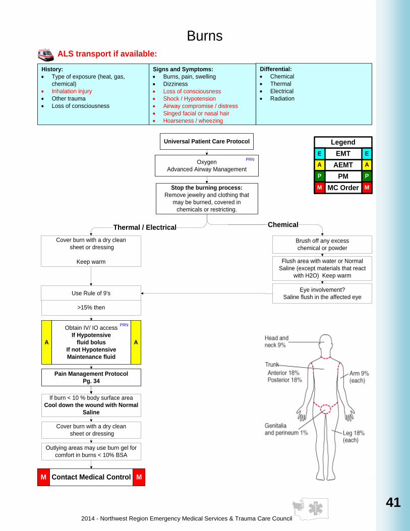
2014 - Northwest Region Emergency Medical Services & Trauma Care Council
41
41
41
41
41
41
41
41
Burns
Stop the burning process:
Remove jewelry and clothing that
may be burned, covered in
chemicals or restricting.
Brush off any excess
chemical or powder
Cover burn with a dry clean
sheet or dressing
Keep warm
Eye involvement?
Saline flush in the affected eye
Flush area with water or Normal
Saline (except materials that react
with H2O) Keep warm
Thermal / Electrical Chemical
Pain Management Protocol
Pg. 34
Obtain IV/ IO access
If Hypotensive
fluid bolus
If not Hypotensive
Maintenance fluid
A A
Oxygen
Advanced Airway Management
PRN
PRN
Use Rule of 9's
Contact Medical ControlM M
>15% then
Universal Patient Care Protocol
Differential:
· Chemical
· Thermal
· Electrical
· Radiation
History:
· Type of exposure (heat, gas,
chemical)
· Inhalation injury
· Other trauma
· Loss of consciousness
Signs and Symptoms:
· Burns, pain, swelling
· Dizziness
· Loss of consciousness
· Shock / Hypotension
· Airway compromise / distress
· Singed facial or nasal hair
· Hoarseness / wheezing
If burn < 10 % body surface area
Cool down the wound with Normal
Saline
Cover burn with a dry clean
sheet or dressing
Outlying areas may use burn gel for
comfort in burns < 10% BSA
ALS transport if available:
AEMT
PM
MC Order
A
P
A
P
M M
EMT
Legend
E E
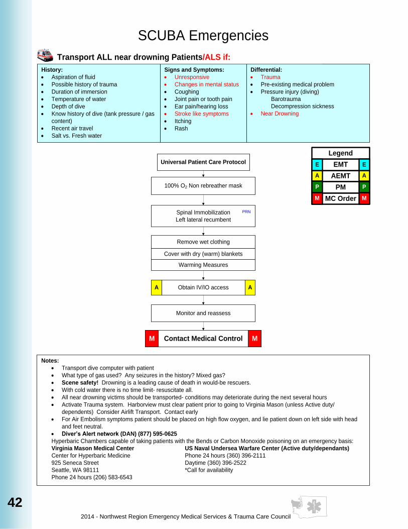
2014 - Northwest Region Emergency Medical Services & Trauma Care Council
42
History:
· Aspiration of fluid
· Possible history of trauma
· Duration of immersion
· Temperature of water
· Depth of dive
· Know history of dive (tank pressure / gas
content)
· Recent air travel
· Salt vs. Fresh water
Signs and Symptoms:
· Unresponsive
· Changes in mental status
· Coughing
· Joint pain or tooth pain
· Ear pain/hearing loss
· Stroke like symptoms
· Itching
· Rash
Differential:
· Trauma
· Pre-existing medical problem
· Pressure injury (diving)
Barotrauma
Decompression sickness
· Near Drowning
Notes:
· Transport dive computer with patient
· What type of gas used? Any seizures in the history? Mixed gas?
· Scene safety! Drowning is a leading cause of death in would-be rescuers.
· With cold water there is no time limit- resuscitate all.
· All near drowning victims should be transported- conditions may deteriorate during the next several hours
· Activate Trauma system. Harborview must clear patient prior to going to Virginia Mason (unless Active duty/
dependents) Consider Airlift Transport. Contact early
· For Air Embolism symptoms patient should be placed on high flow oxygen, and lie patient down on left side with head
and feet neutral.
· Diver’s Alert network (DAN) (877) 595-0625
Hyperbaric Chambers capable of taking patients with the Bends or Carbon Monoxide poisoning on an emergency basis:
Virginia Mason Medical Center US Naval Undersea Warfare Center (Active duty/dependants)
Center for Hyperbaric Medicine Phone 24 hours (360) 396-2111
925 Seneca Street Daytime (360) 396-2522
Seattle, WA 98111 *Call for availability
Phone 24 hours (206) 583-6543
SCUBA Emergencies
Spinal Immobilization
Left lateral recumbent
Monitor and reassess
Obtain IV/IO accessA A
Remove wet clothing
Cover with dry (warm) blankets
Warming Measures
Universal Patient Care Protocol
Contact Medical ControlM M
PRN
100% O2 Non rebreather mask
AEMT
PM
MC Order
A
P
A
P
M M
EMT
Legend
E E
Transport ALL near drowning Patients/ALS if:
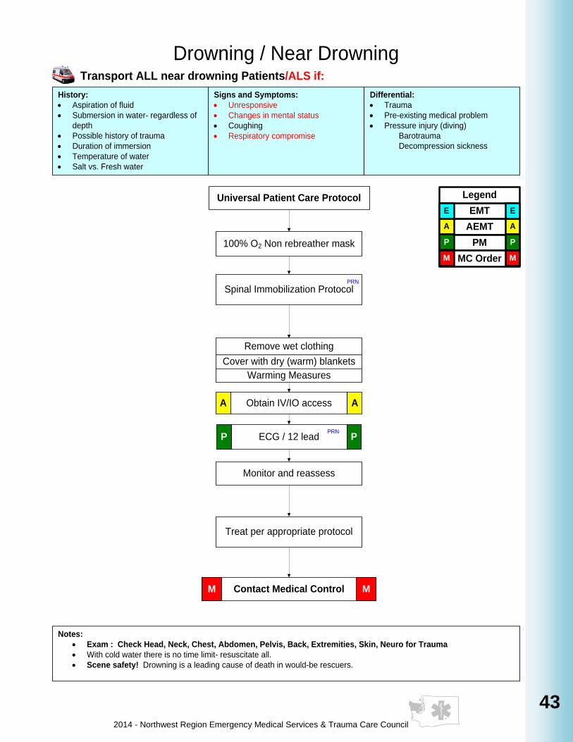
2014 - Northwest Region Emergency Medical Services & Trauma Care Council
43
43
43
43
43
43
43
43
History:
· Aspiration of fluid
· Submersion in water- regardless of
depth
· Possible history of trauma
· Duration of immersion
· Temperature of water
· Salt vs. Fresh water
Signs and Symptoms:
· Unresponsive
· Changes in mental status
· Coughing
· Respiratory compromise
Differential:
· Trauma
· Pre-existing medical problem
· Pressure injury (diving)
Barotrauma
Decompression sickness
Universal Patient Care Protocol
Notes:
· Exam : Check Head, Neck, Chest, Abdomen, Pelvis, Back, Extremities, Skin, Neuro for Trauma
· With cold water there is no time limit- resuscitate all.
· Scene safety! Drowning is a leading cause of death in would-be rescuers.
Drowning / Near Drowning
Spinal Immobilization Protocol
Monitor and reassess
Contact Medical ControlM M
Obtain IV/IO accessA A
Remove wet clothing
Cover with dry (warm) blankets
Warming Measures
Treat per appropriate protocol
Transport ALL near drowning Patients/ALS if:
100% O2 Non rebreather mask
AEMT
PM
MC Order
A
P
A
P
M M
EMT
Legend
E E
PRN
ECG / 12 leadP PPRN
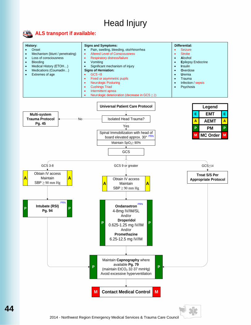
2014 - Northwest Region Emergency Medical Services & Trauma Care Council
44
History:
· Onset
· Mechanism (blunt / penetrating)
· Loss of consciousness
· Bleeding
· Medical History (ETOH…)
· Medications (Coumadin…)
· Extremes of age
Signs and Symptoms:
· Pain, swelling, bleeding, oto/rhinorrhea
· Altered Level of Consciousness
· Respiratory distress/failure
· Vomiting
· Significant mechanism of injury
Signs of Herniation:
· GCS <8
· Fixed or asymmetric pupils
· Neurologic Posturing
· Cushings Triad
· Intermittent apnea
· Neurologic deterioration (decrease in GCS ≥ 2)
Differential:
· Seizure
· Stroke
· Alcohol
· Epilepsy Endocrine
· Insulin
· Overdose
· Uremia
· Trauma
· Infection / sepsis
· Psychosis
Head Injury
Universal Patient Care Protocol
Spinal Immobilization with head of
board elevated approx. 30°
GCS
Isolated Head Trauma?
Multi-system
Trauma Protocol
Pg. 45
No
Yes
GCS 9 or greater GCS≥14GCS 3-8
Maintain SpO2≥ 90%
Treat S/S Per
Appropriate Protocol
M MContact Medical Control
Obtain IV access
Maintain
SBP ≥ 90 mm Hg
A A
Obtain IV access
Maintain
SBP ≥ 90 mm Hg
A A
ALS transport if available:
AEMT
PM
MC Order
A
P
A
P
M M
EMT
Legend
E E
PRN
Ondansetron
4-8mg IV/IM/SLAnd/or
Droperidol
0.625-1.25 mg IV/IMAnd/or
Promethazine
6.25-12.5 mg IV/IM
P P
PRNIntubate (RSI)
Pg. 94P P
PRN
Maintain Capnography where
available Pg. 79
(maintain EtCO2 32-37 mmHg)
Avoid excessive hyperventilation
P P
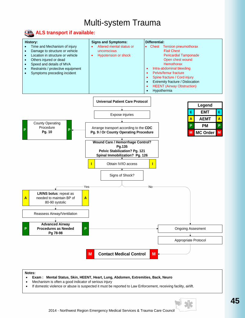
2014 - Northwest Region Emergency Medical Services & Trauma Care Council
45
45
45
45
45
45
45
45
History:
· Time and Mechanism of injury
· Damage to structure or vehicle
· Location in structure or vehicle
· Others injured or dead
· Speed and details of MVA
· Restraints / protective equipment
· Symptoms preceding incident
Signs and Symptoms:
· Altered mental status or
unconscious
· Hypotension or shock
Differential:
· Chest Tension pneumothorax
Flail Chest
Pericardial Tamponade
Open chest wound
Hemothorax
· Intra-abdominal bleeding
· Pelvis/femur fracture
· Spine fracture / Cord injury
· Extremity fracture / Dislocation
· HEENT (Airway Obstruction)
· Hypothermia
Multi-system Trauma
Universal Patient Care Protocol
Notes:
· Exam : Mental Status, Skin, HEENT, Heart, Lung, Abdomen, Extremities, Back, Neuro
· Mechanism is often a good indicator of serious injury
· If domestic violence or abuse is suspected it must be reported to Law Enforcement, receiving facility, airlift.
Arrange transport according to the CDC
Pg. 9 / Or County Operating Procedure
Signs of Shock?
Reassess Airway/Ventilation
Appropriate Protocol
Obtain IV/IO accessI I
No
Contact Medical ControlM M
Yes
Expose injuries
ALS transport if available:
LR/NS bolus: repeat as
needed to maintain BP of
80-90 systolic
A A
Wound Care / Hemorrhage Control?
Pg.135
Pelvic Stabilization? Pg. 121
Spinal Immobilization? Pg. 126
Ongoing Assesment
AEMT
PM
MC Order
A
P
A
P
M M
EMT
Legend
E E
County Operating
Procedure
Pg. 10P P
Advanced Airway
Procedures as Needed
Pg 78-98
P P

2014 - Northwest Region Emergency Medical Services & Trauma Care Council
46
46
Notes:
· For this Guideline, child is defined as less than 8 years old.
· Limit intubation attempts to 3 per patient
· Maintain C-spine immobilization for patients with suspected spinal injury
· Reconfirm ETT placement each time patient is moved
· All choking victims need to be transported to the hospital. Children who have possibly aspirated anything
may not be transported POV, but can be transported BLS if stable.
Pediatric Airway
Assess ABC’s,
respiratory rate,
effort, adequacy
Supplemental Oxygen
Inadequate
Adequate
Basic Maneuvers
first-- open airway;
nasal/oral airway;
bag-valve mask
ObstructionAttempt to remove obstruction
per AHA guidelines
Pediatric Needle
Cricothyrotomy
procedure Pg. 73
P P
Successful removal
Unsuccessful
Oral-tracheal Intubation
Pg. 89P P
Inadequate
Continue BVME E
Adequate
Oxygenate, Ventilate,
Position, Reassess
Rapid Transport
Stabilized
Deteriorating
PRN
Successful
Pulse OximetryE E
Pulse OximetryE E
Contact Medical ControlM M
ALS transport if available
AEMT
PM
MC Order
A
P
A
P
M M
EMT
Legend
E E
Unsuccessful
Unsuccessful
Alternate Airway
Management
Pg. 78-98
P P
Unsuccessful
History:
· Know difficult Airway
· Neck or head trauma
· Trisomy 21
· Congenital malformations
Differential:
· Physical Examination
• Small jaw or limited jaw opening
• Limited cervical spine movement swollen tongue,
oropharynx, or neck, midface hypoplasia
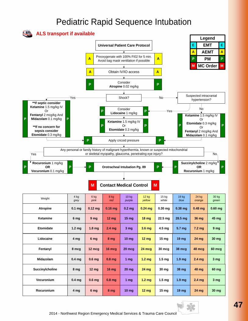
2014 - Northwest Region Emergency Medical Services & Trauma Care Council
47
47
47
47
47
47
47
47
47
47
Pediatric Rapid Sequence Intubation
Weight4 kg
grey
6 kg
pink
8 kg
red
10 kg
purple
12 kg
yellow
15 kg
white
19 kg
blue
30 kg
green
24 kg
orange
Atropine 0.1 mg 0.12 mg 0.16 mg 0.2 mg 0.24 mg 0.30 mg 0.38 mg 0.60 mg0.48 mg
Ketamine 6 mg 9 mg 12 mg 15 mg 18 mg 22.5 mg 28.5 mg 45 mg36 mg
Preoxygenate with 100% FI02 for 5 min.
Avoid bag mask ventilation if possibleA A
Consider
Atropine 0.02 mg/kgP P
Shock?YesSuspected intracranial
hypertension?
Consider
Lidocaine 1 mg/kgP P
Ketamine 1.5 mg/kg IV
Or
Etomidate 0.3 mg/kg
Or
Fentanyl 2 mcg/kg And
Midazolam 0.1 mg/kg
P PKetamine 1.5 mg/kg IV
Or
Etomidate 0.3 mg/kgP P
No
YesNo
Apply cricoid pressureP P
Any personal or family history of malignant hyperthermia, known or suspected mitochondrial
or skeletal myopathy, glaucoma, penetrating eye injury?
Rocuronium 1 mg/kg
OR
Vecuronium 0.1 mg/kgP P
Succinylcholine 2 mg/kg
OR
Rocuronium 1 mg/kg
P P
Yes No
Lidocaine 4 mg 6 mg 8 mg 10 mg 12 mg 15 mg 19 mg 30 mg24 mg
Fentanyl 8 mcg 12 mcg 16 mcg 20 mcg 24 mcg 30 mcg 38 mcg 60 mcg48 mcg
Midazolam 0.4 mg 0.6 mg 0.8 mg 1 mg 1.2 mg 1.5 mg 1.9 mg 3 mg2.4 mg
Succinylcholine 8 mg 12 mg 16 mg 20 mg 24 mg 30 mg 38 mg 60 mg48 mg
Vecuronium 0.4 mg 0.6 mg 0.8 mg 1 mg 1.2 mg 1.5 mg 1.9 mg 3 mg2.4 mg
Rocuronium 4 mg 6 mg 8 mg 10 mg 12 mg 15 mg 19 mg 30 mg24 mg
Obtain IV/IO accessA A
**If septic consider
Ketamine 1.5 mg/kg IV
Or
Fentanyl 2 mcg/kg And
Midazolam 0.1 mg/kg
**If no concern for
sepsis consider
Etomidate 0.3 mg/kg
P P
Universal Patient Care Protocol
Contact Medical ControlM M
ALS transport if available
AEMT
PM
MC Order
A
P
A
P
M M
EMT
Legend
E E
Orotracheal Intubation Pg. 89P P
Etomidate 1.2 mg 1.8 mg 2.4 mg 3 mg 3.6 mg 4.5 mg 5.7 mg 9 mg7.2 mg
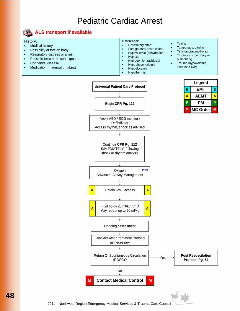
2014 - Northwest Region Emergency Medical Services & Trauma Care Council
48
48
Pediatric Cardiac Arrest
Differential:
· Respiratory effort
· Foreign body obstructions
· Hypovolemia (dehydration)
· Hypoxia
· Hydrogen ion (acidosis)
· Hypo-/hyperkalemia
· Hypoglycemia
· Hypothermia
History:
· Medical history
· Possibility of foreign body
· Respiratory distress or arrest
· Possible toxic or poison exposure
· Congenital disease
· Medication (maternal or infant)
· Toxins
· Tamponade, cardiac
· Tension pneumothorax
· Thrombosis (coronary or
pulmonary)
· Trauma (hypovolemia,
increased ICP)
Begin CPR Pg. 112
Apply AED / ECG monitor /
Defibrillator
Access rhythm, shock as advised
Continue CPR Pg. 112
IMMEDIATELY following
shock or rhythm analysis
Oxygen
Advanced Airway Management
PRN
Fluid bolus 20 ml/kg IV/IO
May repeat up to 60 ml/kgA A
Ongoing assessment
Consider other treatment Protocol
as necessary
No
Return Of Spontaneous Circulation
(ROSC)?Yes
Post Resuscitation
Protocol Pg. 54
Obtain IV/IO accessA A
Universal Patient Care Protocol
Contact Medical ControlM M
ALS transport if available
AEMT
PM
MC Order
A
P
A
P
M M
EMT
Legend
E E
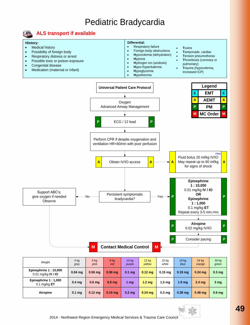
2014 - Northwest Region Emergency Medical Services & Trauma Care Council
49
49
49
49
49
49
49
49
49
49
Pediatric Bradycardia
Epinephrine
1 : 10,000
0.01 mg/kg IV / IO
OR
Epinephrine
1 : 1,000
0.1 mg/kg ET
Repeat every 3-5 min.PRN
P P
Differential:
· Respiratory failure
· Foreign body obstructions
· Hypovolemia (dehydration)
· Hypoxia
· Hydrogen ion (acidosis)
· Hypo-/hyperkalemia
· Hypoglycemia
· Hypothermia
History:
· Medical history
· Possibility of foreign body
· Respiratory distress or arrest
· Possible toxic or poison exposure
· Congenital disease
· Medication (maternal or infant)
Oxygen
Advanced Airway Management
ECG / 12 leadP P
Fluid bolus 20 ml/kg IV/IO
May repeat up to 60 ml/kg
for signs of shockA A
PRN
Atropine
0.02 mg/kg IV/IOP P
Consider pacingP P
Support ABC’s;
give oxygen if needed
Observe
YesNo
· Toxins
· Tamponade, cardiac
· Tension pneumothorax
· Thrombosis (coronary or
pulmonary)
· Trauma (hypovolemia,
increased ICP)
Perform CPR if despite oxygenation and
ventilation HR<60/mn with poor perfusion
Persistent symptomatic
bradycardia?
Weight4 kg
grey
6 kg
pink
8 kg
red
10 kg
purple
12 kg
yellow
15 kg
white
19 kg
blue
30 kg
green
24 kg
orange
Epinephrine 1 : 10,000
0.01 mg/kg IV / IO0.04 mg 0.06 mg 0.08 mg 0.1 mg 0.12 mg 0.15 mg 0.19 mg 0.3 mg0.24 mg
Epinephrine 1 : 1,000
0.1 mg/kg ET0.4 mg 0.6 mg 0.8 mg 1 mg 1.2 mg 1.5 mg 1.9 mg 3 mg2.4 mg
Atropine 0.1 mg 0.12 mg 0.16 mg 0.2 mg 0.24 mg 0.3 mg 0.38 mg 0.6 mg0.48 mg
Obtain IV/IO accessA A
Universal Patient Care Protocol
Contact Medical ControlM M
ALS transport if available
AEMT
PM
MC Order
A
P
A
P
M M
EMT
Legend
E E

2014 - Northwest Region Emergency Medical Services & Trauma Care Council
50
50
Attempt Vagal maneuver
Pediatric Narrow Complex Tachycardia
Borderline
Adenosine
0.1 mg/kg
PRN Repeat dose
0.2 mg/kg
P P
Weight4 kg
grey
6 kg
pink
8 kg
red
10 kg
purple
12 kg
yellow
15 kg
white
19 kg
blue
30 kg
green
24 kg
orange
Adenosine
0.1 mg/kg – 1st dose0.6 mg 0.8 mg 1 mg 1.2 mg 1.5 mg 1.9 mg 3 mg2.4 mg
Adenosine
0.2 mg/kg – 2nd dose
0.4 mg
1.2 mg 1.6 mg 2 mg 2.4 mg 3 mg 3.8 mg 6 mg4.8 mg
Oxygen
Advanced Airway Management
PRN
Differential:
Sinus Tachycardia vs. SVT
· Heart disease (congenital)
· Electrolyte imbalance
· Hypotension
· Fever / infection / sepsis
· Medication / toxin / drugs
· Pulmonary Embolism
· Tension pneumothorax
History:
· Medications or toxins
· Congenital heart disease
· Respiratory distress
· Syncope
· Volume loss (diarrhea / vomiting)
Adequately perfused Poorly perfused
Probable
Sinus
Tacycardia
Infant rate
< 220 bpm
Children
<180 bpm
0.8 mg
Probable
SVT
Infant rate
≥ 220 bpm
Children
≥ 180 bpm
Search and
treat causes
Consider
Vagal
Maneuvers
(no delays)
Synchronized
Cardioversion
Pg. 103
P P
Obtain IV/IO accessA A
12 lead / ECGP PPRN
Midazolam
0.1 mg/kg IV / IO
Or
Diazepam
0.1 mg/kg IV / IO
Or
Lorazepam
0.1mg/kg IV / IO
P P
Universal Patient Care Protocol
Contact Medical ControlM M
Midazolam 0.4 mg 0.6 mg 0.8 mg 1 mg 1.2 mg 1.5 mg 1.9 mg 2 mg2 mg
Diazepam 0.4 mg 0.6 mg 0.8 mg 1 mg 1.2 mg 1.5 mg 1.9 mg 2 mg2 mg
Lorazepam 0.4 mg 0.6 mg 0.8 mg 1 mg 1.2 mg 1.5 mg 1.9 mg 2 mg2 mg
ALS transport if available
AEMT
PM
MC Order
A
P
A
P
M M
EMT
Legend
E E
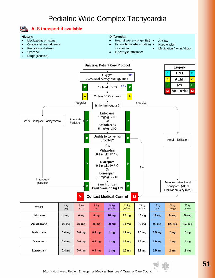
2014 - Northwest Region Emergency Medical Services & Trauma Care Council
51
51
51
51
51
51
51
51
51
51
Pediatric Wide Complex Tachycardia
Wide Complex Tachycardia
Is rhythm regular?
Atrial Fibrillation
Monitor patient and
transport. (Atrial
Fibrillation very rare)
Synchronized
Cardioversion Pg.103P P
Adequate
Perfusion
IrregularRegular
Differential:
· Heart disease (congenital)
· Hypovolemia (dehydration)
or anemia
· Electrolyte imbalance
History:
· Medications or toxins
· Congenital heart disease
· Respiratory distress
· Syncope
· Drugs (cocaine)
Lidocaine
1 mg/kg IV/IO
Or
Amiodarone
5 mg/kg IV/IO
P P
Unable to convert or
unstable?P P
Yes
No
Oxygen
Advanced Airway Management
PRN
12 lead / ECGP PPRN
Obtain IV/IO accessA A
Inadequate
perfusion
Weight4 kg
grey
6 kg
pink
8 kg
red
10 kg
purple
12 kg
yellow
15 kg
white
19 kg
blue
30 kg
green
24 kg
orange
Lidocaine 4 mg 6 mg 8 mg 10 mg 12 mg 15 mg 19 mg 30 mg24 mg
Amiodarone 20 mg 30 mg 40 mg 50 mg 60 mg 75 mg 95 mg 150 mg120 mg
Universal Patient Care Protocol
Contact Medical ControlM M
Midazolam
0.1 mg/kg IV / IO
Or
Diazepam
0.1 mg/kg IV / IO
Or
Lorazepam
0.1mg/kg IV / IO
P P
· Anxiety
· Hypotension
· Medication / toxin / drugs
Midazolam 0.4 mg 0.6 mg 0.8 mg 1 mg 1.2 mg 1.5 mg 1.9 mg 2 mg2 mg
Diazepam 0.4 mg 0.6 mg 0.8 mg 1 mg 1.2 mg 1.5 mg 1.9 mg 2 mg2 mg
Lorazepam 0.4 mg 0.6 mg 0.8 mg 1 mg 1.2 mg 1.5 mg 1.9 mg 2 mg2 mg
ALS transport if available
AEMT
PM
MC Order
A
P
A
P
M M
EMT
Legend
E E
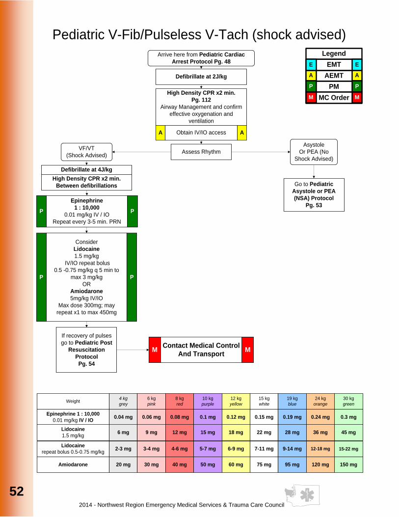
2014 - Northwest Region Emergency Medical Services & Trauma Care Council
52
52
Arrive here from Pediatric Cardiac
Arrest Protocol Pg. 48
Pediatric V-Fib/Pulseless V-Tach (shock advised)
Asystole
Or PEA (No
Shock Advised)
VF/VT
(Shock Advised)
Contact Medical Control
And TransportM M
High Density CPR x2 min.
Pg. 112
Airway Management and confirm
effective oxygenation and
ventilation
Obtain IV/IO accessA A
Defibrillate at 2J/kg
Assess Rhythm
Go to Pediatric
Asystole or PEA
(NSA) Protocol
Pg. 53
Consider
Lidocaine
1.5 mg/kg
IV/IO repeat bolus
0.5 -0.75 mg/kg q 5 min to
max 3 mg/kg
OR
Amiodarone
5mg/kg IV/IO
Max dose 300mg; may
repeat x1 to max 450mg
P P
AEMT
PM
MC Order
A
P
A
P
M M
EMT
Legend
E E
Defibrillate at 4J/kg
High Density CPR x2 min.
Between defibrillations
Epinephrine
1 : 10,000
0.01 mg/kg IV / IO
Repeat every 3-5 min. PRN
P P
Weight4 kg
grey
6 kg
pink
8 kg
red
10 kg
purple
12 kg
yellow
15 kg
white
19 kg
blue
30 kg
green
24 kg
orange
Epinephrine 1 : 10,000
0.01 mg/kg IV / IO0.04 mg 0.06 mg 0.08 mg 0.1 mg 0.12 mg 0.15 mg 0.19 mg 0.3 mg0.24 mg
Lidocaine
repeat bolus 0.5-0.75 mg/kg2-3 mg 3-4 mg 4-6 mg 5-7 mg 6-9 mg 7-11 mg 9-14 mg 15-22 mg12-18 mg
Amiodarone 20 mg 30 mg 40 mg 50 mg 60 mg 75 mg 95 mg 150 mg120 mg
If recovery of pulses
go to Pediatric Post
Resuscitation
Protocol
Pg. 54
Lidocaine
1.5 mg/kg6 mg 9 mg 12 mg 15 mg 18 mg 22 mg 28 mg 45 mg36 mg
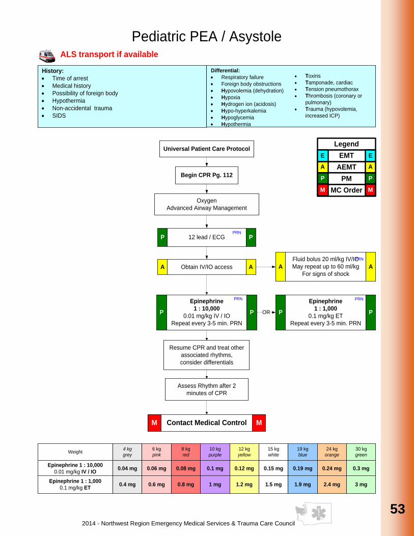
2014 - Northwest Region Emergency Medical Services & Trauma Care Council
53
53
53
53
53
53
53
53
53
53
Pediatric PEA / Asystole
Epinephrine
1 : 10,000
0.01 mg/kg IV / IO
Repeat every 3-5 min. PRN
P P
Assess Rhythm after 2
minutes of CPR
Resume CPR and treat other
associated rhythms,
consider differentials
History:
· Time of arrest
· Medical history
· Possibility of foreign body
· Hypothermia
· Non-accidental trauma
· SIDS
Oxygen
Advanced Airway Management
12 lead / ECG P P
Fluid bolus 20 ml/kg IV/IO
May repeat up to 60 ml/kg
For signs of shockA A
PRN
Differential:
· Respiratory failure
· Foreign body obstructions
· Hypovolemia (dehydration)
· Hypoxia
· Hydrogen ion (acidosis)
· Hypo-hyperkalemia
· Hypoglycemia
· Hypothermia
· Toxins
· Tamponade, cardiac
· Tension pneumothorax
· Thrombosis (coronary or
pulmonary)
· Trauma (hypovolemia,
increased ICP)
Begin CPR Pg. 112
Weight4 kg
grey
6 kg
pink
8 kg
red
10 kg
purple
12 kg
yellow
15 kg
white
19 kg
blue
30 kg
green
24 kg
orange
Epinephrine 1 : 10,000
0.01 mg/kg IV / IO0.04 mg 0.06 mg 0.08 mg 0.1 mg 0.12 mg 0.15 mg 0.19 mg 0.3 mg0.24 mg
Epinephrine 1 : 1,000
0.1 mg/kg ET0.4 mg 0.6 mg 0.8 mg 1 mg 1.2 mg 1.5 mg 1.9 mg 3 mg2.4 mg
OR
PRN
Obtain IV/IO accessA A
Epinephrine
1 : 1,000
0.1 mg/kg ET
Repeat every 3-5 min. PRN
P P
PRN
Universal Patient Care Protocol
Contact Medical ControlM M
ALS transport if available
AEMT
PM
MC Order
A
P
A
P
M M
EMT
Legend
E E
PRN

2014 - Northwest Region Emergency Medical Services & Trauma Care Council
54
54
Pediatric Post Resuscitation Management
Treat other associated signs
and symptoms per guideline
Consider temperature control
Oxygen
Advanced Airway Management
PRN
Fluid bolus 20 ml/kg IV/IO
May repeat up to 60 ml/kgA A
PRN
12 lead / ECGP PPRN
Sedation Post IntubationP P
Weight4 kg
grey
6 kg
pink
8 kg
red
10 kg
purple
12 kg
yellow
15 kg
white
19 kg
blue
30 kg
green
24 kg
orange
Midazolam 0.4 mg 0.6 mg 0.8 mg 1 mg 1.2 mg 1.5 mg 1.9 mg 2 mg2 mg
Obtain IV/IO accessA A
Universal Patient Care Protocol
Contact Medical ControlM M
Midazolam
0.1 mg/kg IV / IO
Or
Diazepam
0.1 mg/kg IV / IO max 2 mg
Or
Lorazepam
0.1 mg/kg IV / IO max 2 mg
P P
Diazepam 0.4 mg 0.6 mg 0.8 mg 1 mg 1.2 mg 1.5 mg 1.9 mg 2 mg2 mg
Lorazepam 0.4 mg 0.6 mg 0.8 mg 1 mg 1.2 mg 1.5 mg 1.9 mg 2 mg2 mg
ETCO2 If availableP PPRN
Consider Pain Management
Procedure Pg. 62
ALS transport if available
AEMT
PM
MC Order
A
P
A
P
M M
EMT
Legend
E E
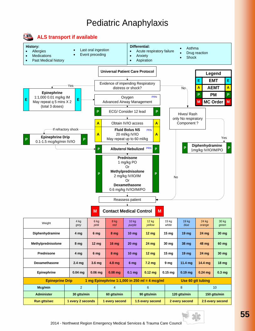
2014 - Northwest Region Emergency Medical Services & Trauma Care Council
55
55
55
55
55
55
55
55
55
55
Pediatric Anaphylaxis
Differential:
· Acute respiratory failure
· Anxiety
· Aspiration
History:
· Allergies
· Medications
· Past Medical history
Oxygen
Advanced Airway Management
PRN
Obtain IV/IO access A A
Fluid Bolus NS
20 ml/kg IV/IO
May repeat up to 60 ml/kg
A A
ECG/ Consider 12 leadP P
Weight4 kg
grey
6 kg
pink
8 kg
red
10 kg
purple
12 kg
yellow
15 kg
white
19 kg
blue
30 kg
green
24 kg
orange
Diphenhydramine 4 mg 6 mg 8 mg 10 mg 12 mg 15 mg 19 mg 30 mg24 mg
Methylprednisolone 8 mg 12 mg 16 mg 20 mg 24 mg 30 mg 38 mg 60 mg48 mg
Evidence of impending Respiratory
distress or shock?Yes
Albuterol NebulizedP PPRN
Epineprine Drip 1 mg Epinephrine 1:1,000 in 250 ml = 4 mcg/ml Use 60 gtt tubing
Mcg/min 2 4 6 8 10
Administer 30 gtts/min 60 gtts/min 90 gtts/min 120 gtts/min 150 gtts/min
Run gtts/sec 1 every 2 seconds 1 every second 1.5 every second 2 every second 2.5 every second
· Last oral ingestion
· Event preceding
· Asthma
· Drug reaction
· Shock
PRN
Epinephrine
1:1,000 0.01 mg/kg IM
May repeat q 5 mins X 2
(total 3 doses)
E E
Epinephrine Drip
0.1-1.5 mcg/kg/min IV/IOP P
No
Universal Patient Care Protocol
Contact Medical ControlM M
Prednisone 4 mg 6 mg 8 mg 10 mg 12 mg 15 mg 19 mg 30 mg24 mg
Dexamethasone 2.4 mg 3.6 mg 4.8 mg 6 mg 7.2 mg 9 mg 11.4 mg 18 mg14.4 mg
Epinephrine 0.04 mg 0.06 mg 0.08 mg 0.1 mg 0.12 mg 0.15 mg 0.19 mg 0.3 mg0.24 mg
Hives/ Rash
only No respiratory
Component ?
Diphenhydramine
1mg/kg IV/IO/IM/POP P
Yes
ALS transport if available
AEMT
PM
MC Order
A
P
A
P
M M
EMT
Legend
E E
Prednisone
1 mg/kg PO
Or
Methylprednisolone
2 mg/kg IV/IO/IM
Or
Dexamethasone
0.6 mg/kg IV/IO/IM/PO
P P
If refractory shock
Reassess patient
No

2014 - Northwest Region Emergency Medical Services & Trauma Care Council
56
56
Pediatric Apparent Life Threatening Event (ALTE)
Meets criteria for an ALTE
Patient must be transported for evaluation
even if well appearing
Contact medical control if caregiver
refusing transport
Yes to any?
History:
· Altered Mental Status
· Cardiac
· Respiratory Failure
· Seizures
· Syncope
Oxygen
Advanced Airway Management
12 lead / ECGP PPRN
PRN
Fluid bolus 20 ml/kg IV/IO
May repeat up to 60 ml/kgA A
PRN
Differential:· Hypovolemia (dehydration)
· Hypoxia
· Hydrogen ion (acidosis)
· Hypo-hyperkalemia
· Hypoglycemia
· Hypothermia
Consider other treatment Protocols
as necessary
Obtain full history and features of event
Explore medication ingestion/toxin risk
Complete thorough history
• Specifically assess for history of apnea
(>15 seconds?), increased or decreased
tone?, change of color?, pallor or cyanosis?
· Toxins
· Tamponade, cardiac
· Tension pneumothorax
· Thrombosis (coronary or
pulmonary)
· Trauma (hypovolemia,
increased ICP)
No?
Obtain IV/IO accessA A
Universal Patient Care Protocol
Contact Medical ControlM M
ALS transport if available:
AEMT
PM
MC Order
A
P
A
P
M M
EMT
Legend
E E
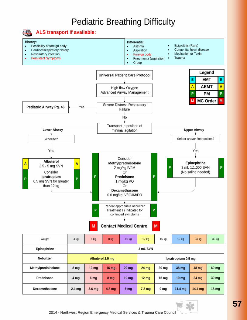
2014 - Northwest Region Emergency Medical Services & Trauma Care Council
57
57
57
57
57
57
57
57
57
57
Differential:
· Asthma
· Aspiration
· Foreign body
· Pneumonia (aspiration)
· Croup
History:
· Possibility of foreign body
· Cardiac/Respiratory history
· Respiratory infection
· Persistent Symptoms
· Epiglottitis (Rare)
· Congenital heart disease
· Medication or Toxin
· Trauma
Pediatric Breathing Difficulty
YesPediatric Airway Pg. 46
Epinephrine
3 mL 1:1,000 SVN
(No saline needed)
P P
High flow Oxygen
Advanced Airway Management
Severe Distress Respiratory
Failure
Transport in position of
minimal agitationLower Airway Upper Airway
Wheeze? Stridor and/or Retractions?
Yes
Consider
Methylprednisolone
2 mg/kg IV/IM
Or
Prednisone
1 mg/kg PO
Or
Dexamethasone
0.6 mg/kg IV/IO/IM/PO
P P
Weight 4 kg 6 kg 8 kg 10 kg 12 kg 15 kg 19 kg 30 kg24 kg
Epinephrine 3 mL SVN
Nebulizer
Methylprednisolone 8 mg 12 mg 16 mg 20 mg 24 mg 30 mg 38 mg 60 mg48 mg
No
Yes
Repeat appropriate nebulizer
Treatment as indicated for
continued symptoms
P P
Albuterol 2.5 mg Ipratropium 0.5 mg
Universal Patient Care Protocol
Contact Medical ControlM M
ALS transport if available:
Albuterol
2.5 - 5 mg SVNA A
Consider
Ipratropium
0.5 mg SVN for greater
than 12 kg
P P
AEMT
PM
MC Order
A
P
A
P
M M
EMT
Legend
E E
Prednisone 4 mg 6 mg 8 mg 10 mg 12 mg 15 mg 19 mg 30 mg24 mg
Dexamethasone 2.4 mg 3.6 mg 4.8 mg 6 mg 7.2 mg 9 mg 11.4 mg 18 mg14.4 mg

2014 - Northwest Region Emergency Medical Services & Trauma Care Council
58
58
Pediatric Diabetic Ketoacidosis / Hyperglycemia
History:
· Polyuria
· Polydipsia
· Vomiting
· Weakness
· Confusion
Fluid bolus 10 ml/kg IV/IO
Reassess, may repeatA A
Clinical Signs:
· Dehydration
· Kussmaul respirations
· Smell of ketones
· Change in mental status
No
Obtain IV/IO accessA A
Oxygen
Advanced Airway Management
PRN
Check Glucose
Clinical Signs of Dehydration
or Hyperglycemia > 250?
Clinical Signs of Intracranial
Hypertension?
Consider Rapid
Sequence Intubation
for GCS <8
Pg. 47
P P
Yes
Elevate head of bed to 45°
Ventilate patient to
maintain 30-35 mm/Hg
ETCO2
P P
Universal Patient Care Protocol
Contact Medical ControlM M
ALS transport if available
AEMT
PM
MC Order
A
P
A
P
M M
EMT
Legend
E E
Notes:
· Signs of intracranial hypertension include severe headache, lethargy, vomiting, coma.
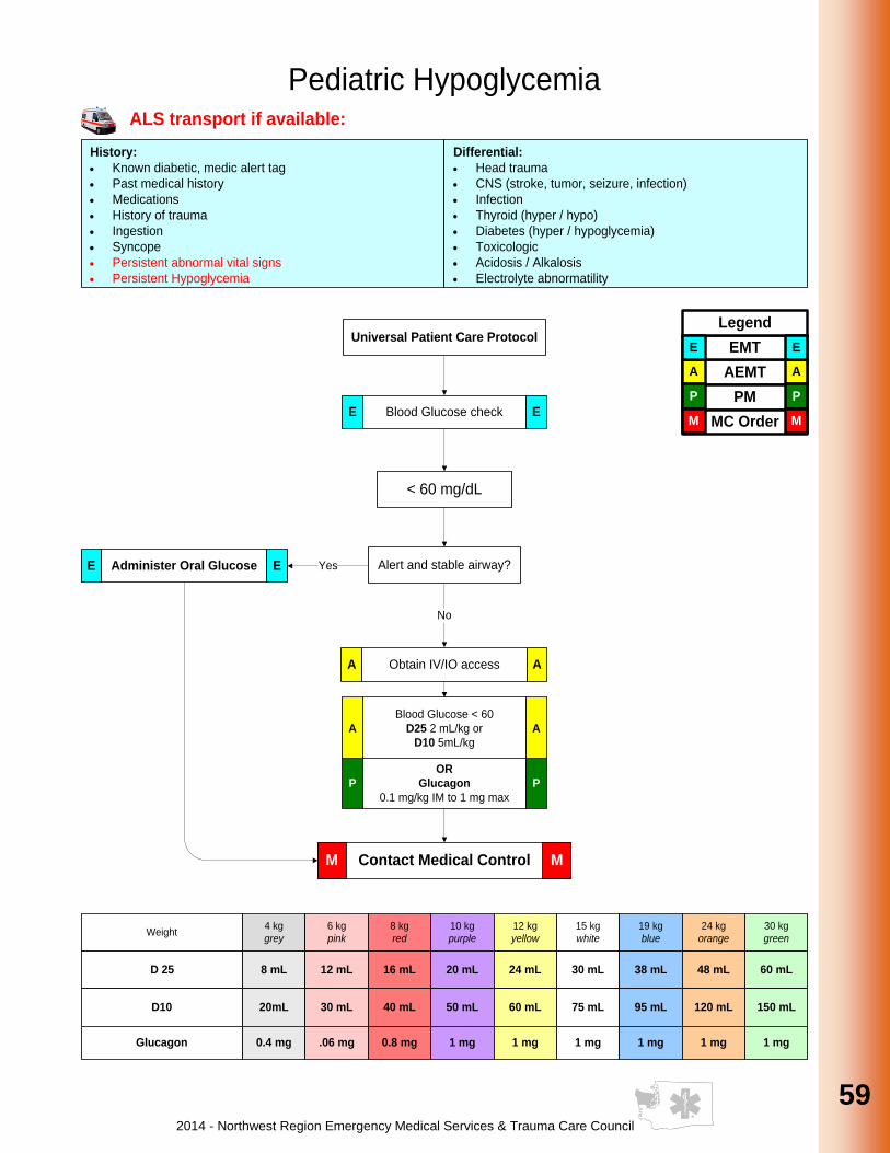
2014 - Northwest Region Emergency Medical Services & Trauma Care Council
59
59
59
59
59
59
59
59
59
59
History:
· Known diabetic, medic alert tag
· Past medical history
· Medications
· History of trauma
· Ingestion
· Syncope
· Persistent abnormal vital signs
· Persistent Hypoglycemia
Differential:
· Head trauma
· CNS (stroke, tumor, seizure, infection)
· Infection
· Thyroid (hyper / hypo)
· Diabetes (hyper / hypoglycemia)
· Toxicologic
· Acidosis / Alkalosis
· Electrolyte abnormatility
Blood Glucose checkE E
< 60 mg/dL
Pediatric Hypoglycemia
Yes
No
Weight4 kg
grey
6 kg
pink
8 kg
red
10 kg
purple
12 kg
yellow
15 kg
white
19 kg
blue
30 kg
green
24 kg
orange
D 25 8 mL 12 mL 16 mL 20 mL 24 mL 30 mL 38 mL 60 mL48 mL
D10 20mL 30 mL 40 mL 50 mL 60 mL 75 mL 95 mL 150 mL120 mL
Obtain IV/IO accessA A
Alert and stable airway?Administer Oral GlucoseE E
Blood Glucose < 60
D25 2 mL/kg or
D10 5mL/kg
A A
OR
Glucagon
0.1 mg/kg IM to 1 mg max
P P
Universal Patient Care Protocol
Contact Medical ControlM M
ALS transport if available:
AEMT
PM
MC Order
A
P
A
P
M M
EMT
Legend
E E
Glucagon 0.4 mg .06 mg 0.8 mg 1 mg 1 mg 1 mg 1 mg 1 mg1 mg
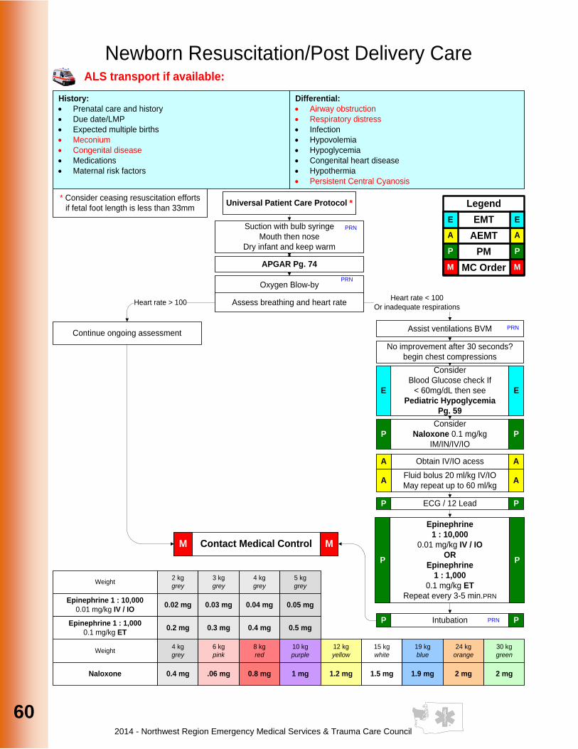
2014 - Northwest Region Emergency Medical Services & Trauma Care Council
60
60
History:
· Prenatal care and history
· Due date/LMP
· Expected multiple births
· Meconium
· Congenital disease
· Medications
· Maternal risk factors
Newborn Resuscitation/Post Delivery Care
Differential:
· Airway obstruction
· Respiratory distress
· Infection
· Hypovolemia
· Hypoglycemia
· Congenital heart disease
· Hypothermia
· Persistent Central Cyanosis
Oxygen Blow-byPRN
ECG / 12 LeadP P
Assess breathing and heart rate
Fluid bolus 20 ml/kg IV/IO
May repeat up to 60 ml/kgA A
Weight2 kg
grey
3 kg
grey
4 kg
grey
5 kg
grey
Epinephrine 1 : 10,000
0.01 mg/kg IV / IO0.02 mg 0.03 mg 0.04 mg 0.05 mg
Epinephrine 1 : 1,000
0.1 mg/kg ET0.2 mg 0.3 mg 0.4 mg 0.5 mg
Epinephrine
1 : 10,000
0.01 mg/kg IV / IO
OR
Epinephrine
1 : 1,000
0.1 mg/kg ET
Repeat every 3-5 min.PRN
P P
Continue ongoing assessment
Heart rate < 100
Or inadequate respirationsHeart rate > 100
Assist ventilations BVM PRN
Obtain IV/IO acessA A
Universal Patient Care Protocol *
Contact Medical ControlM M
Suction with bulb syringe
Mouth then nose
Dry infant and keep warm
* Consider ceasing resuscitation efforts
if fetal foot length is less than 33mm
ALS transport if available:
APGAR Pg. 74
AEMT
PM
MC Order
A
P
A
P
M M
EMT
Legend
E E
No improvement after 30 seconds?
begin chest compressions
IntubationP PPRN
Consider
Naloxone 0.1 mg/kg
IM/IN/IV/IO
P P
Consider
Blood Glucose check If
< 60mg/dL then see
Pediatric Hypoglycemia
Pg. 59
E E
PRN
Weight4 kg
grey
6 kg
pink
8 kg
red
10 kg
purple
12 kg
yellow
15 kg
white
19 kg
blue
30 kg
green
24 kg
orange
Naloxone 0.4 mg .06 mg 0.8 mg 1 mg 1.2 mg 1.5 mg 1.9 mg 2 mg2 mg

2014 - Northwest Region Emergency Medical Services & Trauma Care Council
61
61
61
61
61
61
61
61
61
61
History:
· Ingestion or suspected ingestion of a potentially toxic substance
· Substance ingested, route, quantity
· Time of ingestion
· Reason (suicidal, accidental, criminal)
· Available medications in home
Differential:
· Tricyclic antidepressants (TCAs)
· Acetaminophen (tylenol)
· Depressants
· Stimulants
· Anticholinergic
Poison Control 800-222-1222
Pediatric Toxic Exposure
Respiratory depression,
Opiate O.D
Naloxone
0.1 mg/kg IV/IO/IN/IM
A APRN
Consider
Sodium
Bicarbonate 4.2%
1 – 2 mEq/kg IV/IO
P P
Calcium Chloride
10 mg/kg IV/IOP P
Epinephrine
See Ped. Shock Non-
Traumatic Pg. 64
P P
Excess
Airway Secretions?
Atropine
0.08 mg/kg IV/IO
P P
Midazolam
0.1 mg/kg IN/IM
OR
Diazepam
0.1 mg/kg IV/IO
OR
Lorazepam
0.1 mg/kg IV/IO
P P
Glucagon
0.1 mg/kg IV/IOP P
CNS stimulantsCalcium Channel BlockersTricyclics
Consider TCPP P
Beta Blockers
PRN
Oxygen
Advanced Airway Management
Contact Medical Controlwith nature of toxic exposure
E M EM
PRN
Yes NoBLS Provider?
Consider
Activated Charcoal
1 gm/kg PO
Fluid bolus
20 ml/kg IV/IO
May repeat up to 60 ml/kg
A A
Midazolam 0.4 mg 0.6 mg 0.8 mg 1 mg 1.2 mg 1.5 mg 1.9 mg 2 mg2 mg
Diazepam 0.4 mg 0.6 mg 0.8 mg 1 mg 1.2 mg 1.5 mg 1.9 mg 2 mg2 mg
Lorazepam 0.4 mg 0.6 mg 0.8 mg 1 mg 1.2 mg 1.5 mg 1.9 mg 2 mg2 mg
Weight4 kg
grey
6 kg
pink
8 kg
red
10 kg
purple
12 kg
yellow
15 kg
white
19 kg
blue
30 kg
green
24 kg
orange
Activated Charcoal 4 gm 6 gm 8 gm 10 gm 12 gm 15 gm 19 gm 30 gm24 gm
Naloxone 0.4 mg 0.6 mg 0.8 mg 1 mg 1.2 mg 1.5 mg 1.9 mg 2 mg2 mg
Sodium Bicarbonate 4 mEq 6 mEq 8 mEq 10 mEq 12 mEq 15 mEq 19 mEq 30 mEq24 mEq
Calcium Chloride 40 mg 60 mg 80 mg 100 mg 120 mg 150 mg 190 mg 300 mg240 mg
Glucagon 0.4 mg 0.6 mg 0.8 mg 1 mg 1.2 mg 1.5 mg 1.9 mg 3 mg2.4 mg
Atropine 0.32 mg 0.48 mg 0.64 mg 0.8 mg 0.96 mg 1.2 mg 1.52 mg 2.4 mg1.92 mg
· Cardiac medications
· Solvents, Alcohols,
Cleaning agents
· Insecticides
(organophosphates)
Organophosphates
Universal Patient Care Protocol
Contact Medical ControlM M
ALS transport if available
Obtain IV accessA A
AEMT
PM
MC Order
A
P
A
P
M M
EMT
Legend
E E
ECG / 12 LeadP P

2014 - Northwest Region Emergency Medical Services & Trauma Care Council
62
62
History:
· Age
· Location
· Duration
· Severity (1 - 10)
· Past medical history
Differential:
· Per the specific protocol
· Musculoskeletal
· Visceral (abdominal)
· Cardiac
· Pleural / Respiratory
Obtain IV/IO accessA A
Pediatric Pain Management
ECG/ 12 leadP PPRN
Rest
Ice/Immobilize
Compression
Elevation
E E
PRN
High pain level?
Burns?Yes
Extremely anxious?
No
Yes
Nausea?Yes
No
No
Weight4 kg
grey
6 kg
pink
8 kg
red
10 kg
purple
12 kg
yellow
15 kg
white
19 kg
blue
30 kg
green
24 kg
orange
Morphine .04 mg .06 mg .08 mg 1 mg 1.2 mg 1.5 mg 1.9 mg 3 mg2.4 mg
Fentanyl 8 mcg 12 mcg 16 mcg 20 mcg 24 mcg 30 mcg 38 mcg 60 mcg48 mcg
0.06mg 0.09mg 0.12mg 1 mg 1mg 1mg 1mg 1mg1mgHydromorphone
0.4 mg 0.6 mg 0.8 mg 1 mg 1.2 mg 1.5 mg 1.9 mg 2 mg2 mg
Diazepam 0.4 mg 1 mg 1.2 mg 1.5 mg 1.9 mg 2 mg2 mgDiazepam
Morphine
0.1 mg/kg IV/IO/IM
OR
Fentanyl
2 mcg/kg IV/IO/IM/IN
OR
Hydromorphone
0.015 mg/kg IV/IM
P P
Ondansetron
0.1 mg/kg IV
OR
4 mg PO if
patient weight > 10kg
P P
PRN
PRN
0.4 mg 0.6 mg 0.8 mg 1 mg 1.2 mg 1.5 mg 1.9 mg 2 mg2 mgLorazepam
Contact MC 1 mg 1 mg 1 mg 2 mg 3 mg2 mg
Ondansetron PO 4 mg ODTContact MC
Nitrous Oxide
if availableP P
PRN
Universal Patient Care Protocol
Contact Medical ControlM M
· Medications
· Drug allergies
· Aggravating factors
· Alleviating factors
· Neurogenic
· Renal (colic)
Midazolam
0.1 mg/kg IV/IO/IN/IM
OR
Diazepam
0.1 mg/kg IV/IO
OR
Lorazepam
0.1 mg/kg IV/IO
P P
PRN
Midazolam
Ondansetron
0.6 mg 0.8 mg
ALS transport if patients given a sedation medication
AEMT
PM
MC Order
A
P
A
P
M M
EMT
Legend
E E
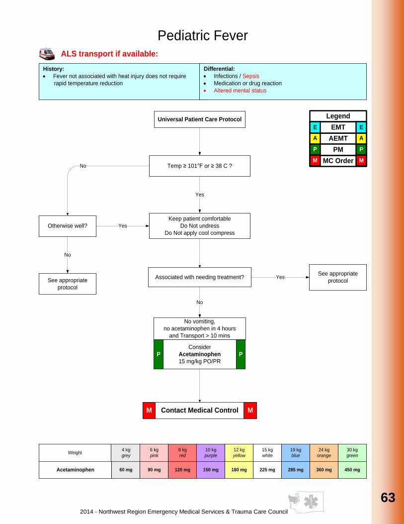
2014 - Northwest Region Emergency Medical Services & Trauma Care Council
63
63
63
63
63
63
63
63
63
63
Pediatric Fever
Yes
History:
· Fever not associated with heat injury does not require
rapid temperature reduction
Temp ≥ 101°F or ≥ 38 C ?
Differential:
· Infections / Sepsis
· Medication or drug reaction
· Altered mental status
Associated with needing treatment?
Consider
Acetaminophen
15 mg/kg PO/PR
P P
Weight4 kg
grey
6 kg
pink
8 kg
red
10 kg
purple
12 kg
yellow
15 kg
white
19 kg
blue
30 kg
green
24 kg
orange
Acetaminophen 60 mg 90 mg 120 mg 150 mg 180 mg 225 mg 285 mg 450 mg360 mg
Otherwise well?
No
See appropriate
protocol
No
Keep patient comfortable
Do Not undress
Do Not apply cool compress
Yes
See appropriate
protocolYes
No vomiting,
no acetaminophen in 4 hours
and Transport > 10 mins
No
Universal Patient Care Protocol
Contact Medical ControlM M
ALS transport if available:
AEMT
PM
MC Order
A
P
A
P
M M
EMT
Legend
E E

2014 - Northwest Region Emergency Medical Services & Trauma Care Council
64
64
Pediatric Shock Non-traumatic
Ongoing assessment
Consider other treatment Protocol
as necessary
Fluid bolus 20 ml/kg IV/IO
May repeat up to 60 ml/kgA A
PRN
Obtain IV/IO accessA A
Epinephrine Bolus
0.01 mg/kg IV/IO
Epinephrine Drip
0.1-1.5 mcg/kg/min IV/IO
P P
Epineprine Drip 1 mg Epinephrine 1:1,000 in 250 ml = 4 mcg/ml Use 60 gtt tubing
Mcg/min 2 4 6 8 10
Administer 30 gtts/min 60 gtts/min 90 gtts/min 120 gtts/min 150 gtts/min
Run gtts/sec 1 every 2 seconds 1 every second 1.5 every second 2 every second 2.5 every second
Differential:
· Respiratory effort
· Hypovolemia (dehydration)
· Hypoxia
· Hydrogen ion (acidosis)
· Hypo-hyperkalemia
· Hypoglycemia
· Hypothermia
History:
· Medical history
· Respiratory distress or arrest
· Possible toxic or poison exposure
· Congenital disease
· Medication (maternal or infant)
· Non accidental trauma
· Toxins
· Tamponade, cardiac
· Tension pneumothorax
· Thrombosis (coronary or
pulmonary)
ECG/ 12 leadP PPRN
Universal Patient Care Protocol
Contact Medical ControlM M
ALS transport if available
AEMT
PM
MC Order
A
P
A
P
M M
EMT
Legend
E E
Weight4 kg
grey
6 kg
pink
8 kg
red
10 kg
purple
12 kg
yellow
15 kg
white
19 kg
blue
30 kg
green
24 kg
orange
Epinephrine 1 : 10,000
0.01 mg/kg IV / IO0.04 mg 0.06 mg 0.08 mg 0.1 mg 0.12 mg 0.15 mg 0.19 mg 0.3 mg0.24 mg
Oxygen
Early assisted ventilation
Advanced Airway
Management
E E
PRN
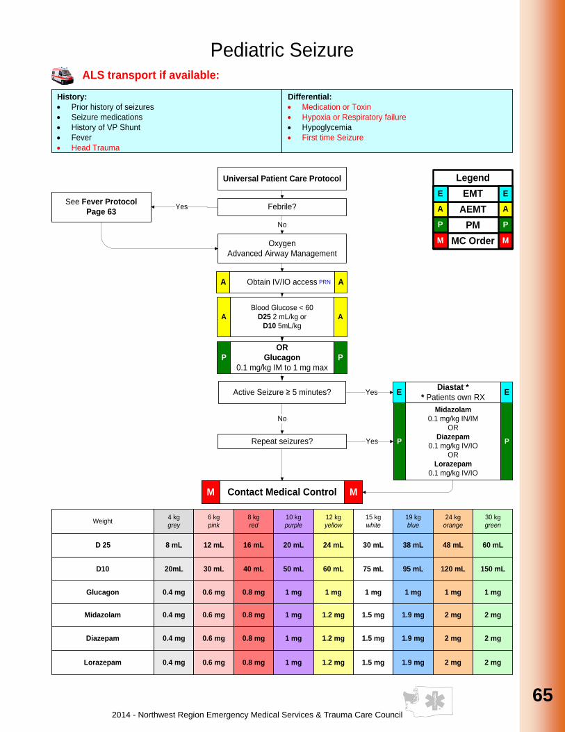
2014 - Northwest Region Emergency Medical Services & Trauma Care Council
65
65
65
65
65
65
65
65
65
65
History:
· Prior history of seizures
· Seizure medications
· History of VP Shunt
· Fever
· Head Trauma
Pediatric Seizure
Febrile?
Oxygen
Advanced Airway Management
See Fever Protocol
Page 63
Active Seizure ≥ 5 minutes?
Repeat seizures? Yes
No
Yes
No
Differential:
· Medication or Toxin
· Hypoxia or Respiratory failure
· Hypoglycemia
· First time Seizure
OR
Glucagon
0.1 mg/kg IM to 1 mg max
P P
YesDiastat *
* Patients own RXE E
Obtain IV/IO accessA APRN
Glucagon 0.4 mg 0.6 mg 0.8 mg 1 mg 1 mg 1 mg 1 mg 1 mg1 mg
Midazolam 0.4 mg 0.6 mg 0.8 mg 1 mg 1.2 mg 1.5 mg 1.9 mg 2 mg2 mg
Universal Patient Care Protocol
Contact Medical ControlM M
Midazolam
0.1 mg/kg IN/IM
OR
Diazepam
0.1 mg/kg IV/IO
OR
Lorazepam
0.1 mg/kg IV/IO
P P
Diazepam 0.4 mg 0.6 mg 0.8 mg 1 mg 1.2 mg 1.5 mg 1.9 mg 2 mg2 mg
Lorazepam 0.4 mg 0.6 mg 0.8 mg 1 mg 1.2 mg 1.5 mg 1.9 mg 2 mg2 mg
ALS transport if available:
AEMT
PM
MC Order
A
P
A
P
M M
EMT
Legend
E E
Blood Glucose < 60
D25 2 mL/kg or
D10 5mL/kg
A A
Weight4 kg
grey
6 kg
pink
8 kg
red
10 kg
purple
12 kg
yellow
15 kg
white
19 kg
blue
30 kg
green
24 kg
orange
D 25 8 mL 12 mL 16 mL 20 mL 24 mL 30 mL 38 mL 60 mL48 mL
D10 20mL 30 mL 40 mL 50 mL 60 mL 75 mL 95 mL 150 mL120 mL

2014 - Northwest Region Emergency Medical Services & Trauma Care Council
66
66
History:
· Time and Mechanism of injury
· Damage to structure or vehicle
· Location in structure or vehicle
· Others injured or dead
· Speed and details of MVA
· Restraints / protective equipment
· Symptoms preceding incident
Signs and Symptoms:
· Altered mental status or
unconscious
· Hypotension or shock
Differential:
· Chest Tension pneumothorax
Flail Chest
Pericardial Tamponade
Open chest wound
Hemothorax
· Intra-abdominal bleeding
· Pelvis/femur fracture
· Spine fracture / Cord injury
· Extremity fracture / Dislocation
· HEENT (Airway Obstruction)
· Hypothermia
Pediatric Multi-system Trauma
Universal Patient Care Protocol
Notes:
· Exam : Mental Status, Skin, HEENT, Heart, Lung, Abdomen, Extremities, Back, Neuro
· Mechanism is often a good indicator of serious injury
· If domestic violence or abuse is suspected it must be reported to Law Enforcement, receiving facility, airlift.
Arrange transport according to the CDC
Pg. 9 / Or County Operating Procedure
Signs of Shock?
Reassess Airway/Ventilation
Appropriate Protocol
Obtain IV/IO accessI I
No
Contact Medical ControlM M
Yes
Expose injuries
ALS transport if available:
LR/NS bolus: 20 mL/kg IV/IO
may repeat up to 60 mL/kgA A
Wound Care / Hemorrhage Control? Pg. 135
Pelvic Stabilization? Pg. 121
Ongoing Assesment
AEMT
PM
MC Order
A
P
A
P
M M
EMT
Legend
E E
County Operating
Procedure
Pg. 10
P P
Advanced Airway
Procedures as needed
Pediatric RSI Pg. 47
Pediatric Airway Pg. 46
Pediatric Pain Management
Pg. 62
P P
Patient with mechanism or exam concerning for
potential spinal injury
Spinal Immobilization Procedure? Pg. 126

2014 - Northwest Region Emergency Medical Services & Trauma Care Council
67
67
67
67
67
67
67
67
67
67
Pediatric Near Drowning
Consider Spinal Immobilization
Pg. 126
Consider other treatment Protocol as
necessary
Remove wet clothing
Cover with dry (warm) blankets
Warming Measures
Differential:
· Trauma
· Pre-existing medical problems
· Barotrauma
· Decompression Sickness
History:
· Submersion in water regardless of depth
· Possible history of trauma
· Duration of submersion
· Temperature of water
· Salt vs.Fresh Water
ECG / 12 leadP P
If no pulse or Symptomatic
Bradycardia begin CPR
See Appropriate Guideline
Oxygen
Advanced Airway Management
Obtain IV/IO accessA A
Universal Patient Care Protocol
Contact Medical ControlM M
Transport ALL near drowning Patients
AEMT
PM
MC Order
A
P
A
P
M M
EMT
Legend
E E
PRN
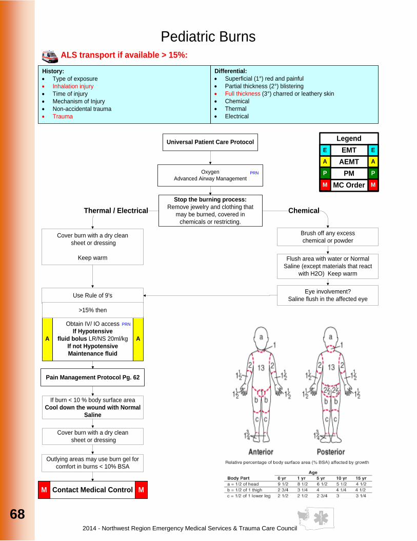
2014 - Northwest Region Emergency Medical Services & Trauma Care Council
68
68
Pediatric Burns
Stop the burning process:
Remove jewelry and clothing that
may be burned, covered in
chemicals or restricting.
Brush off any excess
chemical or powderCover burn with a dry clean
sheet or dressing
Keep warm
Eye involvement?
Saline flush in the affected eye
Flush area with water or Normal
Saline (except materials that react
with H2O) Keep warm
Thermal / Electrical Chemical
Pain Management Protocol Pg. 62
Differential:
· Superficial (1°) red and painful
· Partial thickness (2°) blistering
· Full thickness (3°) charred or leathery skin
· Chemical
· Thermal
· Electrical
History:
· Type of exposure
· Inhalation injury
· Time of injury
· Mechanism of Injury
· Non-accidental trauma
· Trauma
Obtain IV/ IO access
If Hypotensive
fluid bolus LR/NS 20ml/kg
If not Hypotensive
Maintenance fluid
A A
Oxygen
Advanced Airway ManagementPRN
PRN
Use Rule of 9's
Contact Medical ControlM M
>15% then
Universal Patient Care Protocol
ALS transport if available > 15%:
AEMT
PM
MC Order
A
P
A
P
M M
EMT
Legend
E E
If burn < 10 % body surface area
Cool down the wound with Normal
Saline
Cover burn with a dry clean
sheet or dressing
Outlying areas may use burn gel for
comfort in burns < 10% BSA
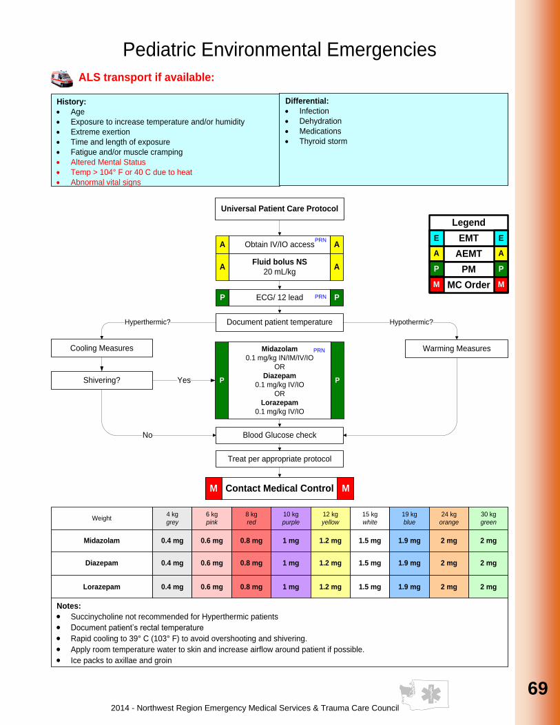
2014 - Northwest Region Emergency Medical Services & Trauma Care Council
69
69
69
69
69
69
69
69
69
69
Universal Patient Care Protocol
Pediatric Environmental Emergencies
Document patient temperature
Cooling Measures Warming Measures
Hyperthermic? Hypothermic?
Blood Glucose check
Contact Medical ControlM M
Treat per appropriate protocol
No
YesShivering?
Obtain IV/IO accessA A
Fluid bolus NS
20 mL/kgA A
ECG/ 12 leadP PPRN
PRN
ALS transport if available:
AEMT
PM
MC Order
A
P
A
P
M M
EMT
Legend
E E
Diazepam 0.4 mg 0.6 mg 0.8 mg 1 mg 1.2 mg 1.5 mg 1.9 mg 2 mg2 mg
Lorazepam 0.4 mg 0.6 mg 0.8 mg 1 mg 1.2 mg 1.5 mg 1.9 mg 2 mg2 mg
Weight4 kg
grey
6 kg
pink
8 kg
red
10 kg
purple
12 kg
yellow
15 kg
white
19 kg
blue
30 kg
green
24 kg
orange
Midazolam 0.4 mg 0.6 mg 0.8 mg 1 mg 1.2 mg 1.5 mg 1.9 mg 2 mg2 mg
Notes:
· Succinycholine not recommended for Hyperthermic patients
· Document patient’s rectal temperature
· Rapid cooling to 39° C (103° F) to avoid overshooting and shivering.
· Apply room temperature water to skin and increase airflow around patient if possible.
· Ice packs to axillae and groin
History:
· Age
· Exposure to increase temperature and/or humidity
· Extreme exertion
· Time and length of exposure
· Fatigue and/or muscle cramping
· Altered Mental Status
· Temp > 104° F or 40 C due to heat
· Abnormal vital signs
Differential:
· Infection
· Dehydration
· Medications
· Thyroid storm
Midazolam
0.1 mg/kg IN/IM/IV/IO
OR
Diazepam
0.1 mg/kg IV/IO
OR
Lorazepam
0.1 mg/kg IV/IO
P P
PRN
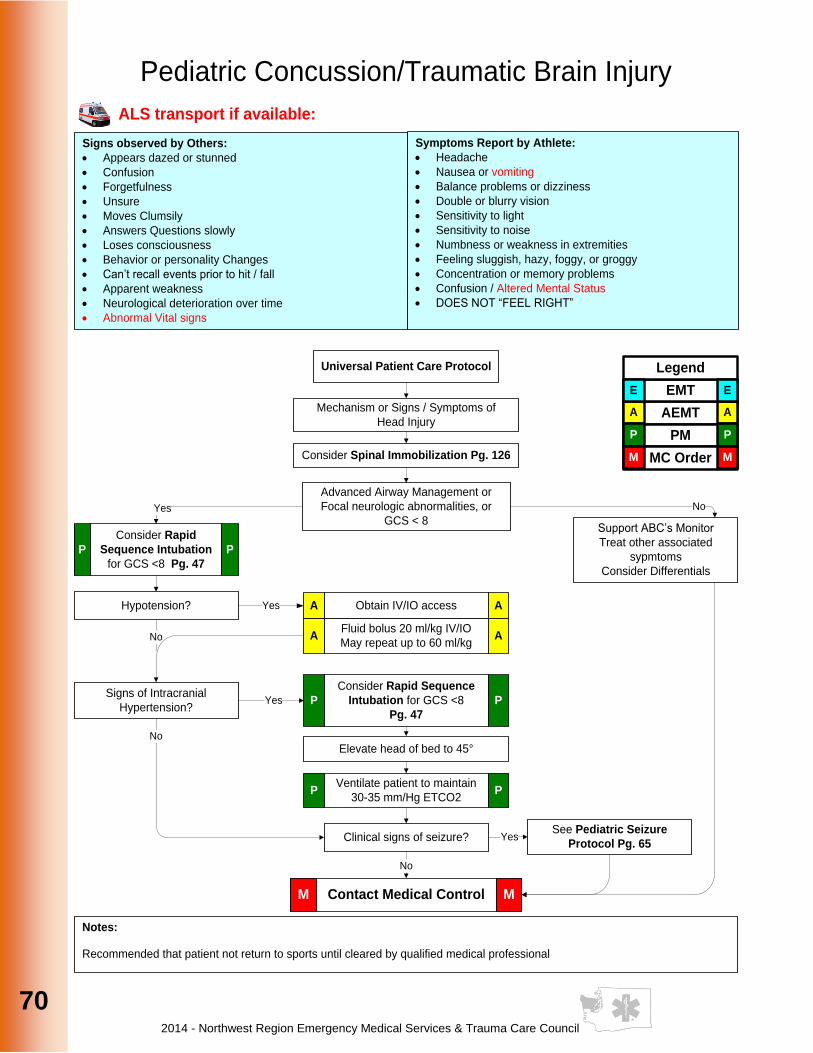
2014 - Northwest Region Emergency Medical Services & Trauma Care Council
70
70
Pediatric Concussion/Traumatic Brain Injury
Mechanism or Signs / Symptoms of
Head Injury
Advanced Airway Management or
Focal neurologic abnormalities, or
GCS < 8Yes No
No
Hypotension? Obtain IV/IO accessA A
Fluid bolus 20 ml/kg IV/IO
May repeat up to 60 ml/kgA A
Yes
Signs of Intracranial
Hypertension?
Clinical signs of seizure?
Support ABC’s Monitor
Treat other associated
sypmtoms
Consider Differentials
Yes
Yes
No
Consider Rapid Sequence
Intubation for GCS <8
Pg. 47
P P
Elevate head of bed to 45°
Ventilate patient to maintain
30-35 mm/Hg ETCO2P P
Universal Patient Care Protocol
Contact Medical ControlM M
ALS transport if available:
Consider Rapid
Sequence Intubation
for GCS <8 Pg. 47
P P
AEMT
PM
MC Order
A
P
A
P
M M
EMT
Legend
E E
Consider Spinal Immobilization Pg. 126
See Pediatric Seizure
Protocol Pg. 65
Symptoms Report by Athlete:
· Headache
· Nausea or vomiting
· Balance problems or dizziness
· Double or blurry vision
· Sensitivity to light
· Sensitivity to noise
· Numbness or weakness in extremities
· Feeling sluggish, hazy, foggy, or groggy
· Concentration or memory problems
· Confusion / Altered Mental Status
· DOES NOT “FEEL RIGHT”
Signs observed by Others:
· Appears dazed or stunned
· Confusion
· Forgetfulness
· Unsure
· Moves Clumsily
· Answers Questions slowly
· Loses consciousness
· Behavior or personality Changes
· Can’t recall events prior to hit / fall
· Apparent weakness
· Neurological deterioration over time
· Abnormal Vital signs
Notes:
Recommended that patient not return to sports until cleared by qualified medical professional
No
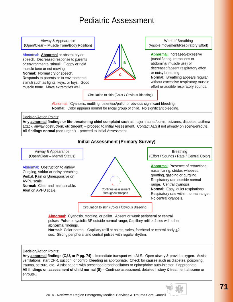
2014 - Northwest Region Emergency Medical Services & Trauma Care Council
71
71
71
71
71
71
71
71
71
71
Pediatric Assessment
BA
C
Airway & Appearance
(Open/Clear – Muscle Tone/Body Position)
Abnormal: Abnormal or absent cry or
speech. Decreased response to parents
or environmental stimuli. Floppy or rigid
muscle tone or not moving.
Normal: Normal cry or speech.
Responds to parents or to environmental
stimuli such as lights, keys, or toys. Good
muscle tome. Move extremities well.
Work of Breathing
(Visible movement/Respiratory Effort)
Abnormal: Increased/excessive
(nasal flaring, retractions or
abdominal muscle use) or
decreased/absent respiratory effort
or noisy breathing.
Normal: Breathing appears regular
without excessive respiratory muscle
effort or audible respiratory sounds.
Circulation to skin (Color / Obvious Bleeding)
Abnormal: Cyanosis, mottling, paleness/pallor or obvious significant bleeding.
Normal: Color appears normal for racial group of child. No significant bleeding.
Decision/Action Points:
Any abnormal findings or life-threatening chief complaint such as major trauma/burns, seizures, diabetes, asthma
attack, airway obstruction, etc (urgent) – proceed to Initial Assessment. Contact ALS if not already on scene/enroute.
All findings normal (non-urgent) – proceed to Initial Assessment.
Initial Assessment (Primary Survey)
Airway & Appearance
(Open/Clear – Mental Status)
Breathing
(Effort / Sounds / Rate / Central Color)
Abnormal: Obstruction to airflow.
Gurgling, stridor or noisy breathing.
Verbal, Pain or Unresponsive on
AVPU scale.
Normal: Clear and maintainable.
Alert on AVPU scale.
Abnormal: Presence of retractions,
nasal flaring, stridor, wheezes,
grunting, gasping or gurgling.
Respiratory rate outside normal
range. Central cyanosis.
Normal: Easy, quiet respirations.
Respiratory rate within normal range.
No central cyanosis.
Circulation to skin (Color / Obvious Bleeding)
Abnormal: Cyanosis, mottling, or pallor. Absent or weak peripheral or central
pulses; Pulse or systolic BP outside normal range; Capillary refill > 2 sec with other
abnormal findings.
Normal: Color normal. Capillary refill at palms, soles, forehead or central body <2
sec. Strong peripheral and central pulses with regular rhythm.
Decision/Action Points:
Any abnormal findings (C,U, or P pg. 74) – Immediate transport with ALS. Open airway & provide oxygen. Assist
ventilations, start CPR, suction, or control bleeding as appropriate. Check for causes such as diabetes, poisoning,
trauma, seizure, etc. Assist patient with prescribed bronchodilators or epinephrine auto-injector, if appropriate.
All findings on assessment of child normal (S) – Continue assessment, detailed history & treatment at scene or
enroute..
Continue assessment
throughout trasport
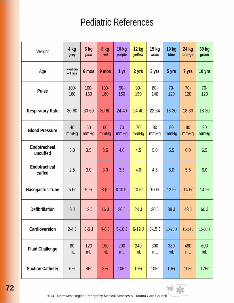
2014 - Northwest Region Emergency Medical Services & Trauma Care Council
72
72
Pediatric References
Weight4 kggrey
6 kgpink
8 kgred
10 kgpurple
12 kgyellow
15 kgwhite
19 kgblue
30 kggreen
24 kgorange
AgeNewborn
– 3 mos 6 mos 9 mos 1 yr 2 yrs 3 yrs 5 yrs 10 yrs7 yrs
Pulse100-
160
100-
160
100-
160
90-
150
90-
150
80-
140
70-
120
70-
120
70-
120
Respiratory Rate 30-60 30-60 30-60 24-40 24-40 22-34 18-30 18-3018-30
Blood Pressure40
mmHg
60
mmHg
60
mmHg
70
mmHg
70
mmHg
80
mmHg
80
mmHg
90
mmHg
80
mmHg
Endotracheal
uncuffed3.0 3.5 3.5 4.0 4.5 5.0 5.5 6.56.0
Endotracheal
cuffed2.5 3.0 3.0 3.5 4.0 4.5 5.0 6.05.5
Nasogastric Tube 5 Fr 5 Fr 8 Fr 8-10 Fr 10 Fr 10 Fr 12 Fr 14 Fr14 Fr
Defibrillation 8 J 12 J 16 J 20 J 24 J 30 J 38 J 60 J48 J
Cardioversion 2-4 J 3-6 J 4-8 J 5-10 J 6-12 J 8-15 J 10-20 J 15-30 J12-24 J
Fluid Challenge80
mL
120
mL
160
mL
200
mL
240
mL
300
mL
380
mL
600
mL
480
mL
Suction Catheter 6Fr 8Fr 8Fr 10Fr 10Fr 10Fr 10Fr 12Fr10Fr
2014 - Northwest Region Emergency Medical Services & Trauma Care Council
73
73
73
73
73
73
73
73
73
73
Airway
Needle Cricothyrotomy (Pediatric) Clinical Indications:
· Failed Airway Protocol
· Management of an airway when standard airway procedures cannot be accomplished or have failed in a patient less than or equal to 8 years of age
Procedure: 1. Have suction supplies available and ready. Collect supplies including the endotracheal
adapter of a 3.0 mm- ID ET tube.
2. Place a roll of sheets or towels under the child’s shoulders to hyperextend the neck and position the larynx as far anterior as possible.
3. Prep the area with antiseptic swab.
4. Locate the cricothyroid membrane utilizing anatomical landmarks.
5. Use the non-dominant hand to secure the membrane.
6. Use commercially prepared kit or:
a. Attach a 5-cc syringe to a 16-18-20 gauge catheter-over-needle device; insert the needle through the cricothyroid membrane at a 45 to 60 degree caudal angle.
b. Aspirate for air with the syringe throughout the procedure.
c. Once air returns easily, stop advancing the device.
d. Thread the catheter off the needle gently at a 60 degree caudal angle.
e. Maintain stabilization of the membrane remove and safely dispose of the needle.
f. Attach the previously sized ET adapter to the end of the catheter and begin ventilation with a Bag Valve Mask connected to high flow oxygen source.
7. Assess breath sounds. Make certain ample time is used not only for inspiration but expiration as well. A 1:4 ratio is not unreasonable.
8. Gentle ventilation is necessary to avoid barotrauma and subsequent pneumothorax.
9. Secure by best method available, recognizing that this method may be direct hands-on control of the device throughout the entire transport.
10. If unable to obtain an adequate airway, resume basic airway management and transport the patient as soon as possible.
11. Regardless of success or failure of needle cricothyrotomy, notify the receiving hospital at the earliest possible time of a surgical airway emergency.
12. Document time/procedure/confirmation/change in patient condition/time on the patient care record (PCR)

2014 - Northwest Region Emergency Medical Services & Trauma Care Council
74
74
AVPU Infant / Child Response Infant Child
A – Alert Curious / Recognizes
parents Alert / Aware of surroundings
V – Responds to Voice Irritable / Cries Opens eyes
P – Responds to Pain Cries in response to pain Withdraws from pain
U – Unresponsive No response No response
APGAR Scale
0 Points 1 Point 2 Points
A – Appearance (Skin Color)
Blue / Pale Normal, except for
extremities Normal over entire
body
P – Pulse Absent Below 100 Above 100
G – Grimace (Reflex Irritability)
No Response Grimace Sneeze, cough,
pulls away
A – Activity Absent Arms and Legs
Flexed Active Movement
R – Respiration Absent Slow, irregular Good, strong cry
CUPS Pediatric
C – Critical Absent airway, breathing or circulation
(cardiac or respiratory arrest or severe traumatic injury)
U – Unstable Compromised airway, breathing or circulation
(unresponsive, respiratory distress, active bleeding, shock, active seizure, significant injury, shock, near-drowning, etc.)
P – Potentially Unstable
Normal airway, breathing & circulation but significant mechanism of injury or illness
(Post-seizure, minor fractures, infant <3 months with fever, etc.)
S – Stable
Normal airway, breathing & circulation No significant mechanism of injury or illness
(small lacerations or abrasions, infant >3 months with fever)
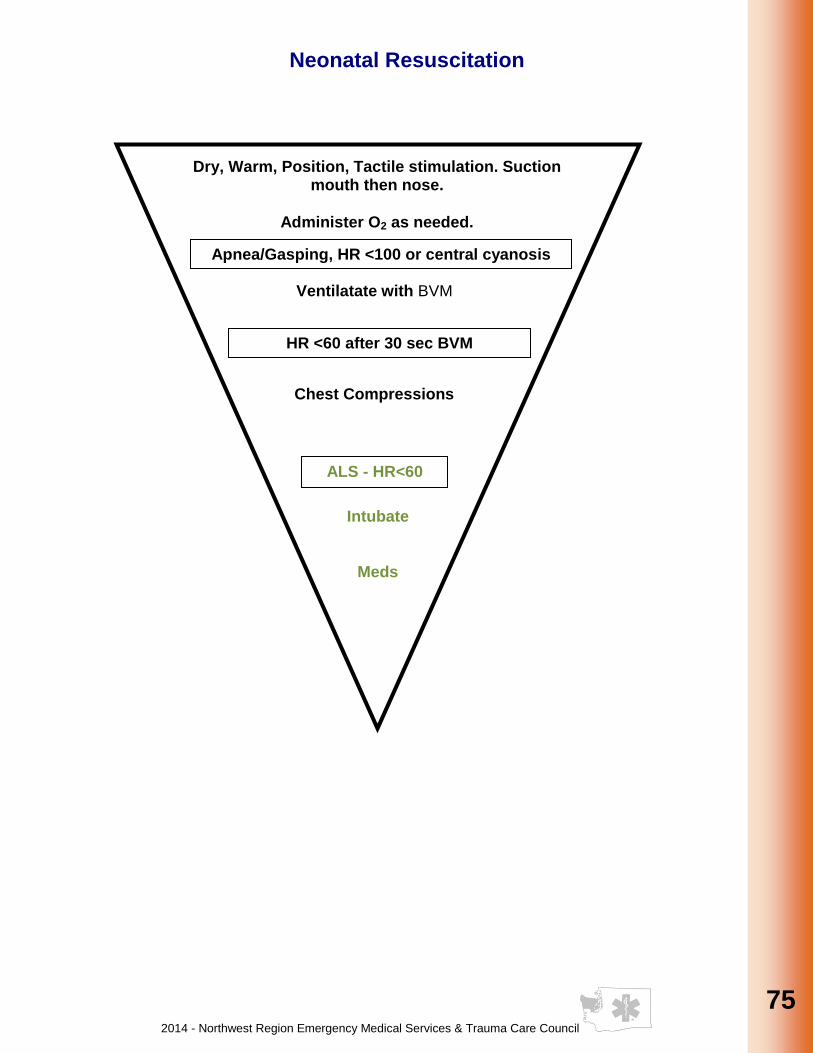
2014 - Northwest Region Emergency Medical Services & Trauma Care Council
75
75
75
75
75
75
75
75
75
75
Neonatal Resuscitation
P
Dry, Warm, Position, Tactile stimulation. Suction mouth then nose.
Administer O2 as needed.
Apnea/Gasping, HR <100 or central cyanosis
Ventilatate with BVM
ALS - HR<60
Intubate
Meds
HR <60 after 30 sec BVM
Chest Compressions
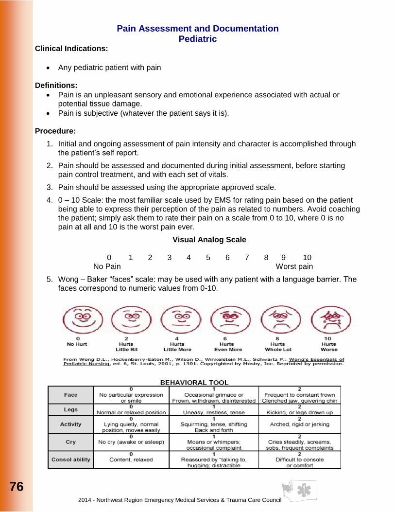
2014 - Northwest Region Emergency Medical Services & Trauma Care Council
76
76
Pain Assessment and Documentation Pediatric
Clinical Indications:
· Any pediatric patient with pain Definitions:
· Pain is an unpleasant sensory and emotional experience associated with actual or potential tissue damage.
· Pain is subjective (whatever the patient says it is). Procedure:
1. Initial and ongoing assessment of pain intensity and character is accomplished through the patient’s self report.
2. Pain should be assessed and documented during initial assessment, before starting pain control treatment, and with each set of vitals.
3. Pain should be assessed using the appropriate approved scale.
4. 0 – 10 Scale: the most familiar scale used by EMS for rating pain based on the patient being able to express their perception of the pain as related to numbers. Avoid coaching the patient; simply ask them to rate their pain on a scale from 0 to 10, where 0 is no pain at all and 10 is the worst pain ever.
Visual Analog Scale
0 1 2 3 4 5 6 7 8 9 10 No Pain Worst pain
5. Wong – Baker “faces” scale: may be used with any patient with a language barrier. The faces correspond to numeric values from 0-10.
2014 - Northwest Region Emergency Medical Services & Trauma Care Council
77
77
77
77
77
77
77
77
77
77
Venous Access Intraosseous Pediatric
Clinical Indications:
· Life threatening illness or injury in a child years of age >8 years old see Adult Venous Access Intraosseous Adult Pg. 134 (72 months) after effective ventilation is established.
Procedure:
1. Expose the lower leg.
2. Identify the tibial tubercle (bony prominence below the knee cap) on the proximal tibia. The insertion location will be 1-2 cm (2 finger widths) below this and medially.
3. Prep the site as per peripheral IV site.
4. If using a commercially prepared device follow manufacturer’s recommendation.
5. Holding the intraosseous needle perpendicular to the skin, twist the needle handle with a rotating grinding motion applying controlled downward force until a “pop” or “give” is felt indicating loss of resistance. Do not advance the needle any further.
6. Remove the trocar and attach the IV.
7. Stabilize and secure the needle.
8. Document the procedure, time, and result (success) on/with the patient care report (PCR).

2014 - Northwest Region Emergency Medical Services & Trauma Care Council
78
Airway I-gel Supraglottic Airway
Clinical Indications: Appropriate intubation is impossible due to patient access or difficult airway anatomy
· Airway, Adult; Airway Rapid Sequence Intubation; Failed Airway, Adult; & Cardiac Arrest
· Newborn Resuscitation
· Pediatric Airway; Pediatric Difficult Airway & Pediatric Rapid Sequence Intubation Caution: This airway does not prevent or protect against aspiration Clinical Contraindications:
· Pulmonary Fibrosis Procedure:
1. Select the appropriate size i-gel Compatible Endotracheal Tube Size 1.0 2 – 5 kg 3.0 mm I.D. Size 1.5 5 – 12 kg 4.0 mm I.D. Size 2 10 - 25 kg 5.0 mm I.D. Size 2.5 25 - 35 kg 5.0 mm I.D. Size 3 30 – 60 kg 6.0 mm I.D. Size 4 50 – 90 kg 7.0 mm I.D. Size 5 > 90 kg 8.0 mm I.D.
2. Lubricate with a water-soluble jelly on the middle of the smooth surface and return to the cradle 3. Pre-Oxygenate the patient 4. Grasp the lubricated i-gel along the integral bite block. Position the device so that the i-gel cuff outlet is facing towards the patient’s chin (mental region of mandible) 5. The patient should always be in the ‘sniffing position’ with the head extended and neck flexed prior to insertion unless head/neck movement is inadvisable or contraindicated 6. Introduce the leading soft tip into the mouth of the patient in the direction of the hard palate 7. Glide the i-gel downward and backward along the hard palate with a continuous but gentle push until a definitive resistance is felt 8. Connect the i-gel to a bag-valve-mask and assess for breath sounds, adequate air exchange and end tidal CO2 (EtCO2) 9. Monitor oxygen saturation with pulse oximetry, EtCO2 and heart monitor 10. Re-verify i-gel placement after every move and upon arrival in the Emergency Department 11. Secure the i-gel 12. Document the procedure, time, and result (success) on/with the patient care report (M.I.R./P.C.R.).
*When using the device after encountering a difficult intubation endotracheal intubation may be accomplished by passing a bougie through the i-gel into the trachea (see above chart for endotracheal tube compatibility). When advancing the bougie you may be able to “rail-road” the bougie to over the cartilaginous rings in the trachea to confirm proper location. Place an endotracheal tube over the bougie and advance into the trachea.
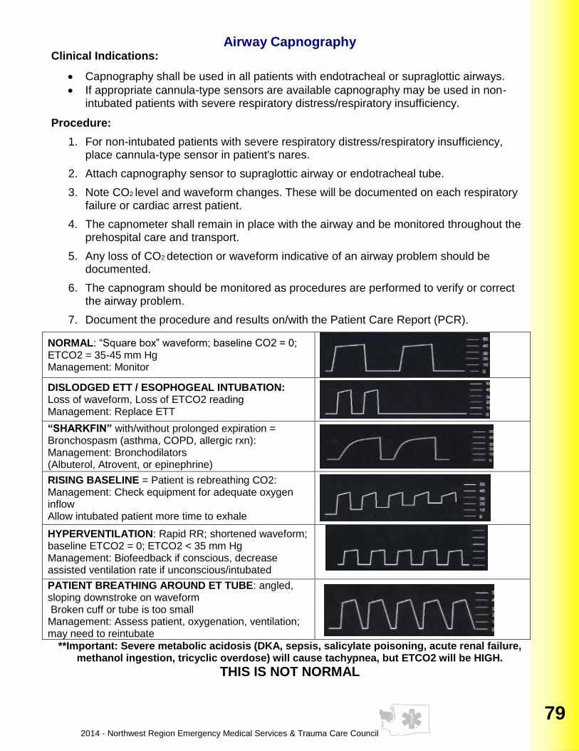
2014 - Northwest Region Emergency Medical Services & Trauma Care Council
79
79
79
79
79
79
79
79
79
79
79
79
Airway Capnography Clinical Indications:
· Capnography shall be used in all patients with endotracheal or supraglottic airways.
· If appropriate cannula-type sensors are available capnography may be used in non-intubated patients with severe respiratory distress/respiratory insufficiency.
Procedure:
1. For non-intubated patients with severe respiratory distress/respiratory insufficiency, place cannula-type sensor in patient's nares.
2. Attach capnography sensor to supraglottic airway or endotracheal tube.
3. Note CO2 level and waveform changes. These will be documented on each respiratory failure or cardiac arrest patient.
4. The capnometer shall remain in place with the airway and be monitored throughout the prehospital care and transport.
5. Any loss of CO2 detection or waveform indicative of an airway problem should be documented.
6. The capnogram should be monitored as procedures are performed to verify or correct the airway problem.
7. Document the procedure and results on/with the Patient Care Report (PCR).
NORMAL: “Square box” waveform; baseline CO2 = 0; ETCO2 = 35-45 mm Hg Management: Monitor
DISLODGED ETT / ESOPHOGEAL INTUBATION: Loss of waveform, Loss of ETCO2 reading Management: Replace ETT
“SHARKFIN” with/without prolonged expiration = Bronchospasm (asthma, COPD, allergic rxn): Management: Bronchodilators (Albuterol, Atrovent, or epinephrine)
RISING BASELINE = Patient is rebreathing CO2: Management: Check equipment for adequate oxygen inflow Allow intubated patient more time to exhale
HYPERVENTILATION: Rapid RR; shortened waveform; baseline ETCO2 = 0; ETCO2 < 35 mm Hg Management: Biofeedback if conscious, decrease assisted ventilation rate if unconscious/intubated
PATIENT BREATHING AROUND ET TUBE: angled, sloping downstroke on waveform Broken cuff or tube is too small Management: Assess patient, oxygenation, ventilation; may need to reintubate
**Important: Severe metabolic acidosis (DKA, sepsis, salicylate poisoning, acute renal failure, methanol ingestion, tricyclic overdose) will cause tachypnea, but ETCO2 will be HIGH.
THIS IS NOT NORMAL
2014 - Northwest Region Emergency Medical Services & Trauma Care Council
80
Airway Combitube Clinical Indications:
· Apneic patient when endotracheal intubation is not possible or readily available.
· Patient must be > 5 feet and >16 years of age.
· Patient must be unconscious without a gag reflex.
· No history of esophageal disease or caustic ingestion.
· Failed Airway Protocol
Procedure:
1. Preoxygenate and hyperventilate the patient.
2. Lubricate the tube.
3. Maintain the head in a neutral inline position.
4. Grasp the patient’s tongue and jaw with your gloved hand and pull forward.
5. Gently insert the tube until the teeth are between the printed rings.
6. Inflate line 1 (blue pilot balloon) leading to the pharyngeal cuff with 100 cc air.
7. Inflate line 2 (white pilot balloon) leading to the distal cuff with 15 cc of air.
8. Ventilate the patient through the longer blue tube.
9. Auscultate for breath sounds and sounds over the epigastrium.
10. Look for the chest to rise and fall.
11. If breath sounds are positive and epigastric sounds are negative, continue ventilation through the blue tube. The tube is in the esophagus.
12. In the esophageal mode, stomach contents can be aspirated through the #2, white tube relieving gastric distention.
13. If breath sounds are negative and epigastric sounds are positive, attempt ventilation through the shorter, #2 white tube and reassess for lung and epigastric sounds. If breath sounds are present and the chest rises, you have intubated the trachea and continue ventilation through the shorter tube.
14. The device is secured by the large pharyngeal balloon.
15. Confirm tube placement using end-tidal CO2 detector or esophageal bulb device.
·Endotracheal intubation with a Combitube in Place: (Not necessary if the ventilations are adequate with the Combitube.)
A. The tube must be in the esophageal mode. B. Prepare all equipment needed for endotracheal intubation. C. Decompress the stomach. D. Hyperoxygenate the patient. E. Deflate the balloons on the Combitube and remove. Suction equipment must be ready. F. Rapidly proceed with endotracheal intubation.
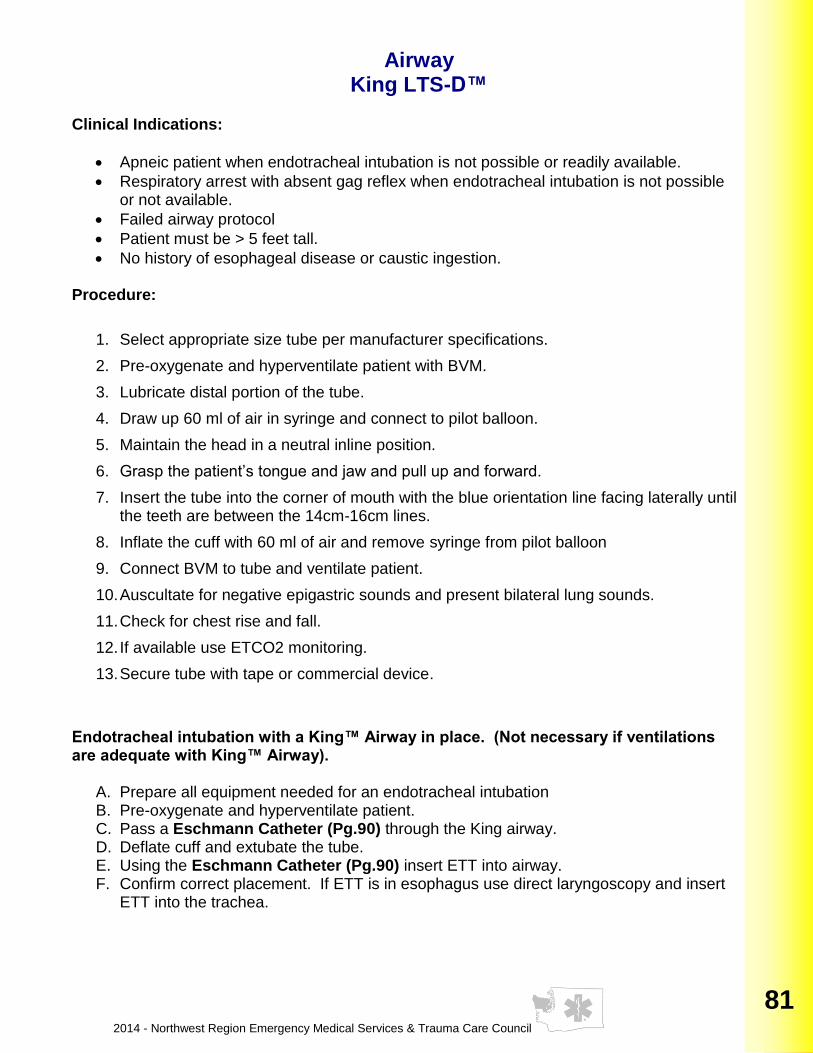
2014 - Northwest Region Emergency Medical Services & Trauma Care Council
81
81
81
81
81
81
81
81
81
81
81
81
Airway King LTS-D™
Clinical Indications:
· Apneic patient when endotracheal intubation is not possible or readily available.
· Respiratory arrest with absent gag reflex when endotracheal intubation is not possible or not available.
· Failed airway protocol
· Patient must be > 5 feet tall.
· No history of esophageal disease or caustic ingestion. Procedure:
1. Select appropriate size tube per manufacturer specifications.
2. Pre-oxygenate and hyperventilate patient with BVM.
3. Lubricate distal portion of the tube.
4. Draw up 60 ml of air in syringe and connect to pilot balloon.
5. Maintain the head in a neutral inline position.
6. Grasp the patient’s tongue and jaw and pull up and forward.
7. Insert the tube into the corner of mouth with the blue orientation line facing laterally until the teeth are between the 14cm-16cm lines.
8. Inflate the cuff with 60 ml of air and remove syringe from pilot balloon
9. Connect BVM to tube and ventilate patient.
10. Auscultate for negative epigastric sounds and present bilateral lung sounds.
11. Check for chest rise and fall.
12. If available use ETCO2 monitoring.
13. Secure tube with tape or commercial device.
Endotracheal intubation with a King™ Airway in place. (Not necessary if ventilations are adequate with King™ Airway).
A. Prepare all equipment needed for an endotracheal intubation B. Pre-oxygenate and hyperventilate patient. C. Pass a Eschmann Catheter (Pg.90) through the King airway. D. Deflate cuff and extubate the tube. E. Using the Eschmann Catheter (Pg.90) insert ETT into airway. F. Confirm correct placement. If ETT is in esophagus use direct laryngoscopy and insert
ETT into the trachea.
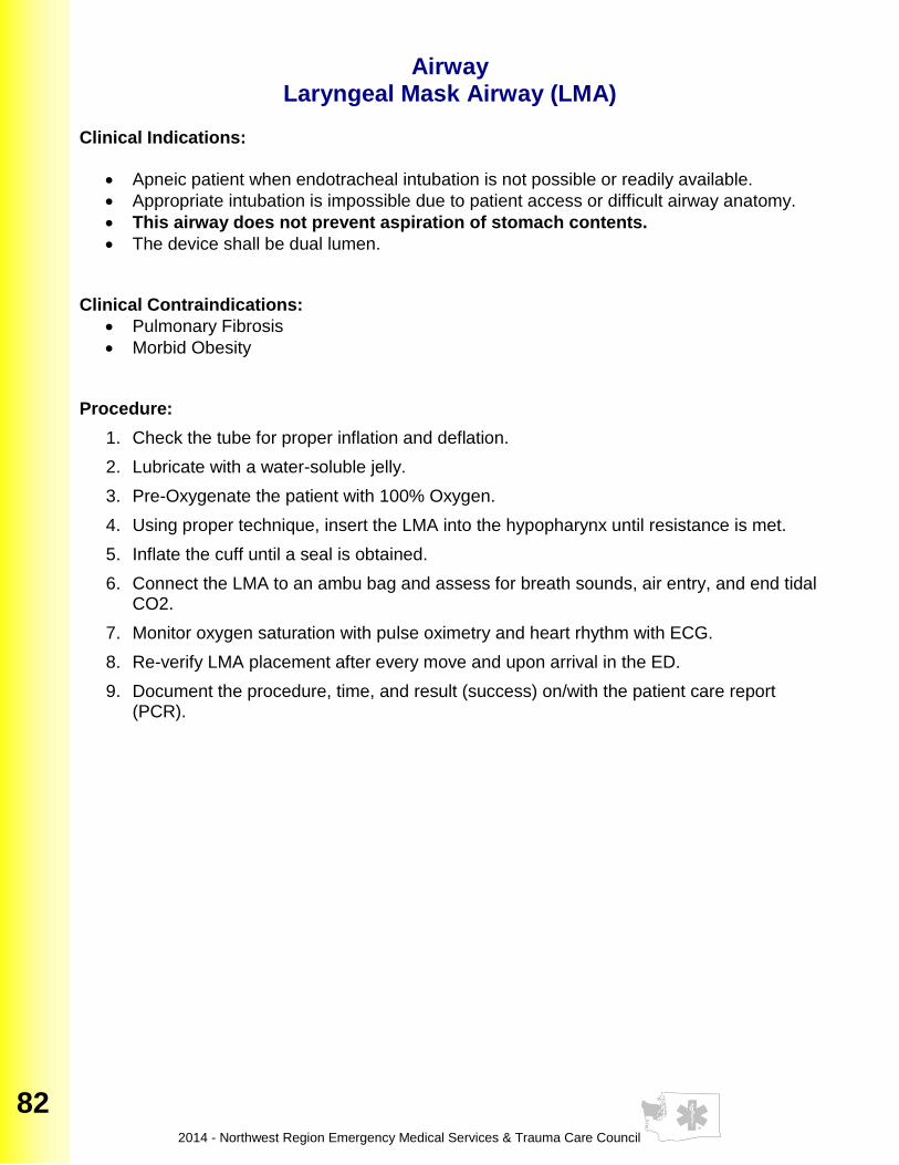
2014 - Northwest Region Emergency Medical Services & Trauma Care Council
82
Airway Laryngeal Mask Airway (LMA)
Clinical Indications:
· Apneic patient when endotracheal intubation is not possible or readily available.
· Appropriate intubation is impossible due to patient access or difficult airway anatomy.
· This airway does not prevent aspiration of stomach contents.
· The device shall be dual lumen. Clinical Contraindications:
· Pulmonary Fibrosis
· Morbid Obesity
Procedure:
1. Check the tube for proper inflation and deflation.
2. Lubricate with a water-soluble jelly.
3. Pre-Oxygenate the patient with 100% Oxygen.
4. Using proper technique, insert the LMA into the hypopharynx until resistance is met.
5. Inflate the cuff until a seal is obtained.
6. Connect the LMA to an ambu bag and assess for breath sounds, air entry, and end tidal CO2.
7. Monitor oxygen saturation with pulse oximetry and heart rhythm with ECG.
8. Re-verify LMA placement after every move and upon arrival in the ED.
9. Document the procedure, time, and result (success) on/with the patient care report (PCR).
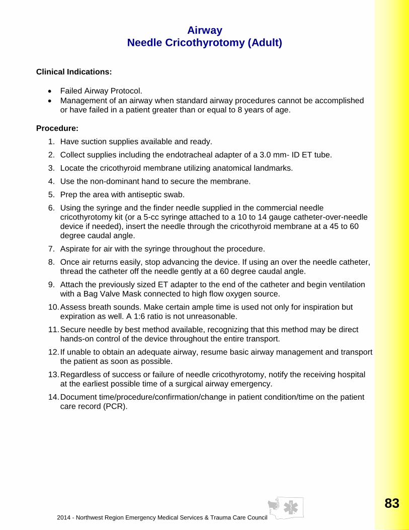
2014 - Northwest Region Emergency Medical Services & Trauma Care Council
83
83
83
83
83
83
83
83
83
83
83
83
Airway Needle Cricothyrotomy (Adult)
Clinical Indications:
· Failed Airway Protocol.
· Management of an airway when standard airway procedures cannot be accomplished or have failed in a patient greater than or equal to 8 years of age.
Procedure:
1. Have suction supplies available and ready.
2. Collect supplies including the endotracheal adapter of a 3.0 mm- ID ET tube.
3. Locate the cricothyroid membrane utilizing anatomical landmarks.
4. Use the non-dominant hand to secure the membrane.
5. Prep the area with antiseptic swab.
6. Using the syringe and the finder needle supplied in the commercial needle cricothyrotomy kit (or a 5-cc syringe attached to a 10 to 14 gauge catheter-over-needle device if needed), insert the needle through the cricothyroid membrane at a 45 to 60 degree caudal angle.
7. Aspirate for air with the syringe throughout the procedure.
8. Once air returns easily, stop advancing the device. If using an over the needle catheter, thread the catheter off the needle gently at a 60 degree caudal angle.
9. Attach the previously sized ET adapter to the end of the catheter and begin ventilation with a Bag Valve Mask connected to high flow oxygen source.
10. Assess breath sounds. Make certain ample time is used not only for inspiration but expiration as well. A 1:6 ratio is not unreasonable.
11. Secure needle by best method available, recognizing that this method may be direct hands-on control of the device throughout the entire transport.
12. If unable to obtain an adequate airway, resume basic airway management and transport the patient as soon as possible.
13. Regardless of success or failure of needle cricothyrotomy, notify the receiving hospital at the earliest possible time of a surgical airway emergency.
14. Document time/procedure/confirmation/change in patient condition/time on the patient care record (PCR).
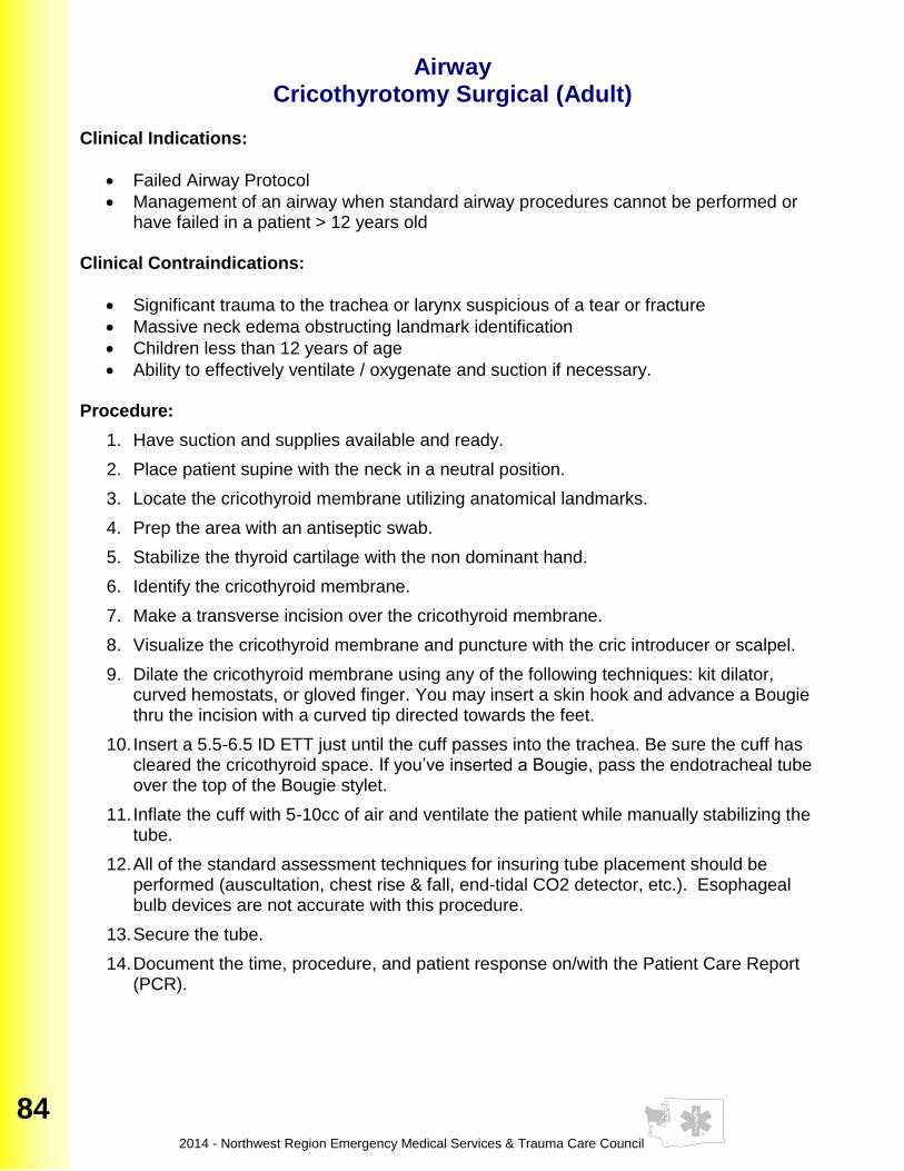
2014 - Northwest Region Emergency Medical Services & Trauma Care Council
84
Airway Cricothyrotomy Surgical (Adult)
Clinical Indications:
· Failed Airway Protocol
· Management of an airway when standard airway procedures cannot be performed or have failed in a patient > 12 years old
Clinical Contraindications:
· Significant trauma to the trachea or larynx suspicious of a tear or fracture
· Massive neck edema obstructing landmark identification
· Children less than 12 years of age
· Ability to effectively ventilate / oxygenate and suction if necessary. Procedure:
1. Have suction and supplies available and ready.
2. Place patient supine with the neck in a neutral position.
3. Locate the cricothyroid membrane utilizing anatomical landmarks.
4. Prep the area with an antiseptic swab.
5. Stabilize the thyroid cartilage with the non dominant hand.
6. Identify the cricothyroid membrane.
7. Make a transverse incision over the cricothyroid membrane.
8. Visualize the cricothyroid membrane and puncture with the cric introducer or scalpel.
9. Dilate the cricothyroid membrane using any of the following techniques: kit dilator, curved hemostats, or gloved finger. You may insert a skin hook and advance a Bougie thru the incision with a curved tip directed towards the feet.
10. Insert a 5.5-6.5 ID ETT just until the cuff passes into the trachea. Be sure the cuff has cleared the cricothyroid space. If you’ve inserted a Bougie, pass the endotracheal tube over the top of the Bougie stylet.
11. Inflate the cuff with 5-10cc of air and ventilate the patient while manually stabilizing the tube.
12. All of the standard assessment techniques for insuring tube placement should be performed (auscultation, chest rise & fall, end-tidal CO2 detector, etc.). Esophageal bulb devices are not accurate with this procedure.
13. Secure the tube.
14. Document the time, procedure, and patient response on/with the Patient Care Report (PCR).

2014 - Northwest Region Emergency Medical Services & Trauma Care Council
85
85
85
85
85
85
85
85
85
85
85
85
Airway Difficult Airway Assessment
LEMON Physical signs Less difficult airway More difficult airway
L ook externally · Normal face and neck
· No face or neck pathology
· Abnormal face shape
· Sunken cheeks
· Edentulous
· “Buck teeth”
· Narrow mouth
· Obesity
· Face or neck pathology
E valuate the
3-3-2 rule
· Mouth opening >3F
· Hyoid-chin distance >3F
· Thyroid cartilage-mouth floor distance >2F
· Mouth opening <3F
· Hyoid-chin distance <3F
· Thyroid cartilage-mouth floor distance <2F
M allampati · Class I and II (can see
the soft palate, uvula, fauces +/- faucial pillars)
· Class III and IV (can only see the hard palate +/- soft palate +/- base of uvula)
O bstruction · None
· Pathology within or surrounding the upper airway (e.g. peri-tonsillar abscess, epiglottis, retro-pharyngeal abscess)
N eck Mobility · Can flex and extend the
neck normally · Limited ROM of the neck

2014 - Northwest Region Emergency Medical Services & Trauma Care Council
86
Airway Passive Pre-oxygenation Procedure
Clinical indications:
● To support and maintain oxygen saturation throughout airway procedures
● All rapid sequence intubations
● All conscious sedations
Clinical contraindications:
● None
Procedure:
● Place a nasal cannula on all patients while preparing for RSI, with EtCO2 monitoring if
available
● For awake patients set to 4-6L/min
1. Low risk patient (96-100%): a. pre-oxygenation: non rebreather mask (NRB) w/ normal flow b. induction: NRB and nasal cannula (NC) set to 15L/min c. during intubation: NC kept at 15L/min
2. High risk (91-95%): a. pre-oxygenation: NRB or CPAP or bag valve mask (BVM) with PEEP valve b. induction: as above plus NC at 15L/min c. intubation: NC at 15L/min
3. Hypoxemic (<90%): a. pre-oxygenation: CPAP or BVM w/ PEEP b. induction: as above plus NC at 15L/min c. intubation: NC at 15L/min
4. Once tracheal intubation has been confirmed per routine the nasal cannula can be
removed
5. Document the procedure and results on/with the Patient Care Report (PCR)
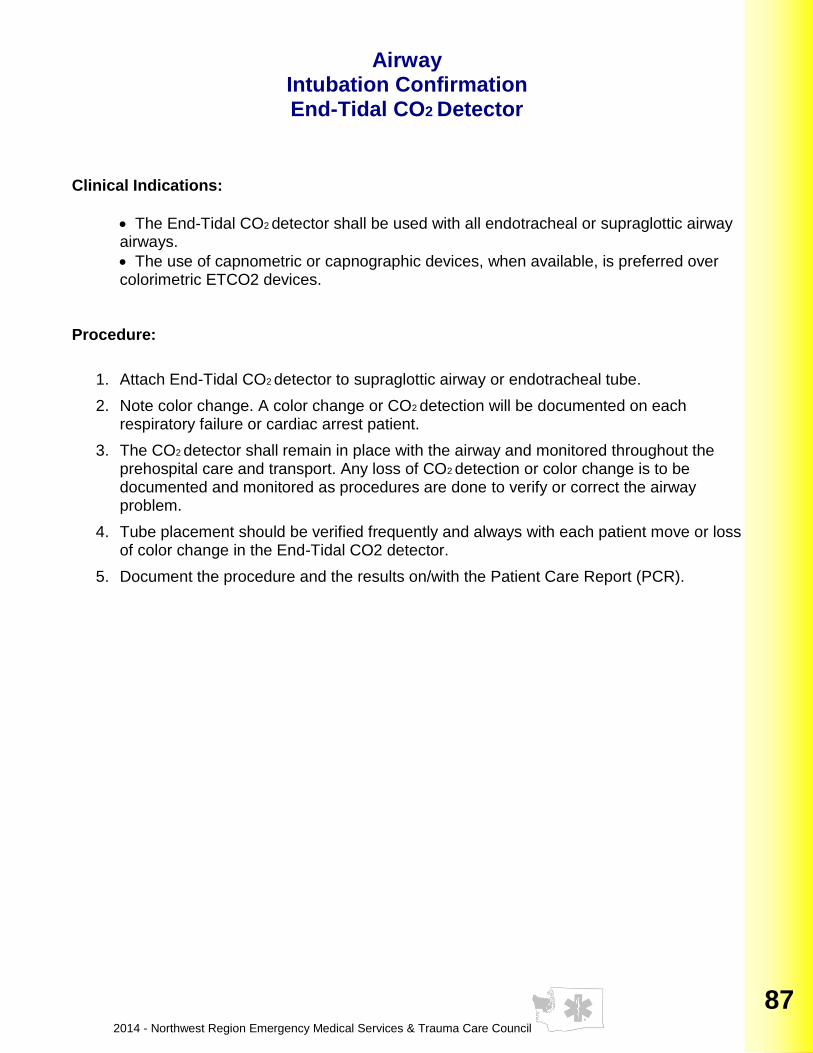
2014 - Northwest Region Emergency Medical Services & Trauma Care Council
87
87
87
87
87
87
87
87
87
87
87
87
Airway Intubation Confirmation End-Tidal CO2 Detector
Clinical Indications:
·The End-Tidal CO2 detector shall be used with all endotracheal or supraglottic airway airways.
·The use of capnometric or capnographic devices, when available, is preferred over colorimetric ETCO2 devices.
Procedure:
1. Attach End-Tidal CO2 detector to supraglottic airway or endotracheal tube.
2. Note color change. A color change or CO2 detection will be documented on each respiratory failure or cardiac arrest patient.
3. The CO2 detector shall remain in place with the airway and monitored throughout the prehospital care and transport. Any loss of CO2 detection or color change is to be documented and monitored as procedures are done to verify or correct the airway problem.
4. Tube placement should be verified frequently and always with each patient move or loss of color change in the End-Tidal CO2 detector.
5. Document the procedure and the results on/with the Patient Care Report (PCR).
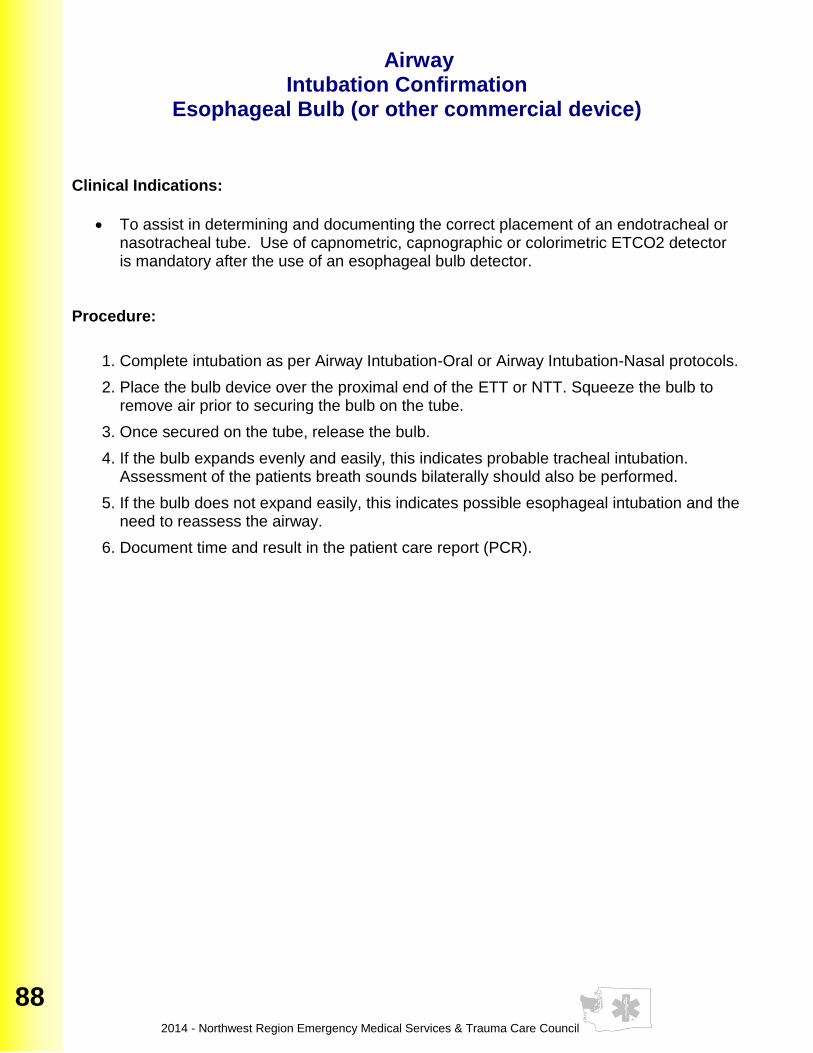
2014 - Northwest Region Emergency Medical Services & Trauma Care Council
88
Airway Intubation Confirmation
Esophageal Bulb (or other commercial device) Clinical Indications:
· To assist in determining and documenting the correct placement of an endotracheal or nasotracheal tube. Use of capnometric, capnographic or colorimetric ETCO2 detector is mandatory after the use of an esophageal bulb detector.
Procedure:
1. Complete intubation as per Airway Intubation-Oral or Airway Intubation-Nasal protocols.
2. Place the bulb device over the proximal end of the ETT or NTT. Squeeze the bulb to remove air prior to securing the bulb on the tube.
3. Once secured on the tube, release the bulb.
4. If the bulb expands evenly and easily, this indicates probable tracheal intubation. Assessment of the patients breath sounds bilaterally should also be performed.
5. If the bulb does not expand easily, this indicates possible esophageal intubation and the need to reassess the airway.
6. Document time and result in the patient care report (PCR).
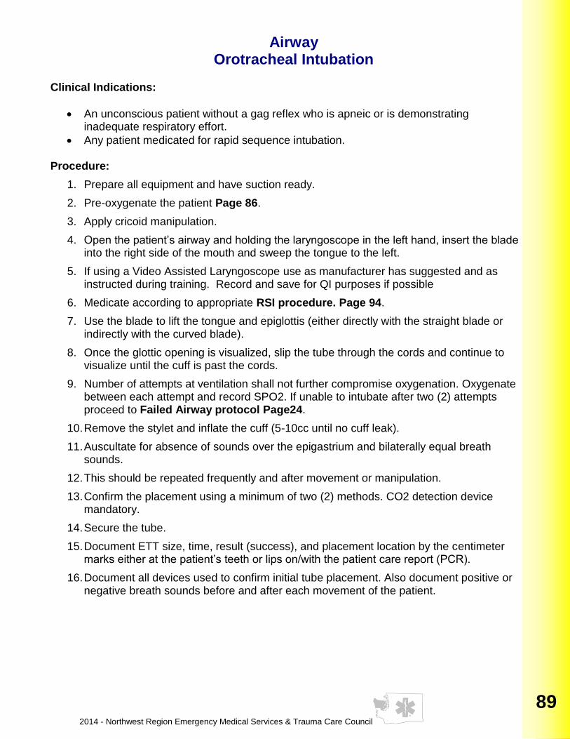
2014 - Northwest Region Emergency Medical Services & Trauma Care Council
89
89
89
89
89
89
89
89
89
89
89
89
Airway Orotracheal Intubation
Clinical Indications:
· An unconscious patient without a gag reflex who is apneic or is demonstrating inadequate respiratory effort.
· Any patient medicated for rapid sequence intubation. Procedure:
1. Prepare all equipment and have suction ready.
2. Pre-oxygenate the patient Page 86.
3. Apply cricoid manipulation.
4. Open the patient’s airway and holding the laryngoscope in the left hand, insert the blade into the right side of the mouth and sweep the tongue to the left.
5. If using a Video Assisted Laryngoscope use as manufacturer has suggested and as instructed during training. Record and save for QI purposes if possible
6. Medicate according to appropriate RSI procedure. Page 94.
7. Use the blade to lift the tongue and epiglottis (either directly with the straight blade or indirectly with the curved blade).
8. Once the glottic opening is visualized, slip the tube through the cords and continue to visualize until the cuff is past the cords.
9. Number of attempts at ventilation shall not further compromise oxygenation. Oxygenate between each attempt and record SPO2. If unable to intubate after two (2) attempts proceed to Failed Airway protocol Page24.
10. Remove the stylet and inflate the cuff (5-10cc until no cuff leak).
11. Auscultate for absence of sounds over the epigastrium and bilaterally equal breath sounds.
12. This should be repeated frequently and after movement or manipulation.
13. Confirm the placement using a minimum of two (2) methods. CO2 detection device mandatory.
14. Secure the tube.
15. Document ETT size, time, result (success), and placement location by the centimeter marks either at the patient’s teeth or lips on/with the patient care report (PCR).
16. Document all devices used to confirm initial tube placement. Also document positive or negative breath sounds before and after each movement of the patient.

2014 - Northwest Region Emergency Medical Services & Trauma Care Council
90
Airway Intubation with Eschmann Catheter,
Tracheal Tube Introducer or Gum Elastic Bougie
Technique
1. Perform direct larygoscopy after thorough pre-oxygenation. Passive Preoxygen Pg. 86
2. Insert bougie under direct visualization (grade II) or semi blind (grade III) using epiglottis as a guide. Maintain midline bent end facing anteriorly.
3. With the tip directed anteriorly guide the bougie toward the epiglottis.
4. Advance the bougie posterior to the epiglottis and into the glottic opening.
5. Cricoid pressure may facilitate correct placement (when the tip of the introducer passes the cricoid cartilage and enters the trachea it also may be palpable at the anatomic location).
6. The operator may be able to feel the bougie “click” or “bump” over the anterior tracheal rings (“wash boarding or railroading”) .
7. Use the laryngoscope to elevate the pharyngeal soft tissue.
8. Subtle maneuvering may be required to traverse the vocal cords.
9. Advance to the carina (resistance to passage) to verify placement (approximately 45 cm). Once advanced to the carina, further insertion causes the bougie to rotate on entrance into a bronchus as an additional criterion to confirm correct placement. Failure to meet resistance after inserting nearly the full length of the bougie indicates esophageal placement. Withdraw and align the black “lip-line marker” with the lips (1 cm band located 40 cm (4 stripes) from proximal end).
10. Pass endotracheal tube (larger than 6.0 mm) over the bougie.
11. If the endotracheal tube catches on the arytenoid or aryepiglottic folds, withdraw the
tube slightly and rotate it 90 counterclockwise and advance it forward (allows beveled end to pass).
12. For optimal passage of the tube over the bougie into the trachea, the laryngoscope may be left in place as the endotracheal tube is advanced with the bevel facing posteriorly.
13. Secure the tube (remove bougie) and verify tube placement.
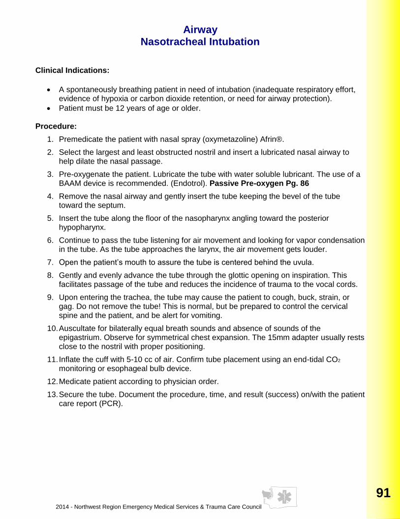
2014 - Northwest Region Emergency Medical Services & Trauma Care Council
91
91
91
91
91
91
91
91
91
91
91
91
Airway Nasotracheal Intubation
Clinical Indications:
· A spontaneously breathing patient in need of intubation (inadequate respiratory effort, evidence of hypoxia or carbon dioxide retention, or need for airway protection).
· Patient must be 12 years of age or older. Procedure:
1. Premedicate the patient with nasal spray (oxymetazoline) Afrin®.
2. Select the largest and least obstructed nostril and insert a lubricated nasal airway to help dilate the nasal passage.
3. Pre-oxygenate the patient. Lubricate the tube with water soluble lubricant. The use of a BAAM device is recommended. (Endotrol). Passive Pre-oxygen Pg. 86
4. Remove the nasal airway and gently insert the tube keeping the bevel of the tube toward the septum.
5. Insert the tube along the floor of the nasopharynx angling toward the posterior hypopharynx.
6. Continue to pass the tube listening for air movement and looking for vapor condensation in the tube. As the tube approaches the larynx, the air movement gets louder.
7. Open the patient’s mouth to assure the tube is centered behind the uvula.
8. Gently and evenly advance the tube through the glottic opening on inspiration. This facilitates passage of the tube and reduces the incidence of trauma to the vocal cords.
9. Upon entering the trachea, the tube may cause the patient to cough, buck, strain, or gag. Do not remove the tube! This is normal, but be prepared to control the cervical spine and the patient, and be alert for vomiting.
10. Auscultate for bilaterally equal breath sounds and absence of sounds of the epigastrium. Observe for symmetrical chest expansion. The 15mm adapter usually rests close to the nostril with proper positioning.
11. Inflate the cuff with 5-10 cc of air. Confirm tube placement using an end-tidal CO2
monitoring or esophageal bulb device.
12. Medicate patient according to physician order.
13. Secure the tube. Document the procedure, time, and result (success) on/with the patient care report (PCR).

2014 - Northwest Region Emergency Medical Services & Trauma Care Council
92
PAirway Nebulizer Inhalation Therapy
Clinical Indications:
· Patients experiencing bronchospasm. Procedure:
1. Gather the necessary equipment.
2. Assemble the nebulizer kit.
3. Instill appropriate medication into the reservoir well of the nebulizer.
4. Connect the nebulizer device to oxygen at 6 liters per minute or adequate flow to produce a steady, visible mist.
5. Instruct the patient to inhale normally through the mouthpiece of the nebulizer. The patient needs to have a good lip seal around the mouthpiece.
6. If the patient is unable to maintain good lip seal around the mouth piece, nebulizer may be connected to a face mask.
7. In the intubated patient, patient on NIPPV, or BVM, nebulizer should be placed in-line for effective medication delivery.
8. The treatment should last until the solution is depleted. Tapping the reservoir well near the end of the treatment will assist in utilizing all of the solution.
9. Monitor the patient for medication effects. This should include the patient’s assessment of his/her response to the treatment and reassessment of vital signs, ECG, and breath sounds.
10. Document the treatment, dose, and route on/with the patient care report (PCR).
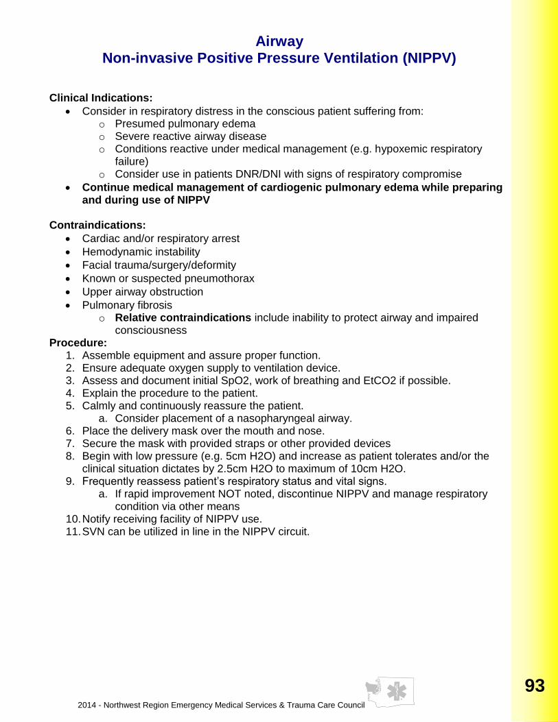
2014 - Northwest Region Emergency Medical Services & Trauma Care Council
93
93
93
93
93
93
93
93
93
93
93
93
Airway Non-invasive Positive Pressure Ventilation (NIPPV)
Clinical Indications:
· Consider in respiratory distress in the conscious patient suffering from: o Presumed pulmonary edema o Severe reactive airway disease o Conditions reactive under medical management (e.g. hypoxemic respiratory
failure) o Consider use in patients DNR/DNI with signs of respiratory compromise
· Continue medical management of cardiogenic pulmonary edema while preparing and during use of NIPPV
Contraindications:
· Cardiac and/or respiratory arrest
· Hemodynamic instability
· Facial trauma/surgery/deformity
· Known or suspected pneumothorax
· Upper airway obstruction
· Pulmonary fibrosis o Relative contraindications include inability to protect airway and impaired
consciousness Procedure:
1. Assemble equipment and assure proper function. 2. Ensure adequate oxygen supply to ventilation device. 3. Assess and document initial SpO2, work of breathing and EtCO2 if possible. 4. Explain the procedure to the patient. 5. Calmly and continuously reassure the patient.
a. Consider placement of a nasopharyngeal airway. 6. Place the delivery mask over the mouth and nose. 7. Secure the mask with provided straps or other provided devices 8. Begin with low pressure (e.g. 5cm H2O) and increase as patient tolerates and/or the
clinical situation dictates by 2.5cm H2O to maximum of 10cm H2O. 9. Frequently reassess patient’s respiratory status and vital signs.
a. If rapid improvement NOT noted, discontinue NIPPV and manage respiratory condition via other means
10. Notify receiving facility of NIPPV use. 11. SVN can be utilized in line in the NIPPV circuit.

2014 - Northwest Region Emergency Medical Services & Trauma Care Council
94
Airway Rapid Sequence Induction (RSI)
INDICATIONS Need for airway control persistent GCS ≤ 8
RELATIVE CONTRAINDICATIONS
Patients with adequate Oxygenation Patients with adequate ventilation Patients who would be difficult to intubate Patients with distorted facial or laryngeal anatomy Known neuromuscular disease Caution with Succinylcholine: Chronic renal failure and on dialysis Patient or family history of malignant hyperthermia Significant burns between 24 hours and 2 weeks old Massive crush injury / suspected rhabdomyolysis
PROCEDURE
Pre-oxygenate with 100% O2 via NRB or BVM. Pg. 86 Monitor oxygen saturation with pulse oximetry and cardiac rhythm with ECG. Ensure functioning vascular access. Evaluate for difficult airway (LEMON)-see page 85. Prepare equipment (intubation kit, BVM, suction, RSI medications, alternate Airway devices/ adjuncts: Eschmann, cric kit, supraglottic airway.
PREMEDICATE ADULT
Consider Lidocaine 1.5 mg/kg None
PREMEDICATE PEDIATRIC
Lidocaine 1.5 mg/kg Atropine 0.02 mg/kg min dose 0.1mg
Atropine 0.02 kg/kg IV/IO min dose 0.1mg
SEDATE ADULT/PEDIATRIC
Etomidate 0.3 mg/kg IV/IO over 15 sec OR
Ketamine 1-2 mg/kg IVP/IO OR
Midazolam 0.1 mg/kg IV/IO, max 10 mg
NEUROMUSCULAR BLOCKADE
Rocuronium 1 mg/kg IV/IO OR
Vecuronium 0.01 mg/kg IV/IO AND
Succinylcholine 1.5 mg/kg IV/IO/IM
Succinylcholine 1.5mg/kg IV/IO/IM OR
Rocuronium 1 mg/kg IV/IO
CONTINUED MAINTENANCE/
SEDATION/
Midazolam 2.5 – 10 mg IV/IO, max 10 mg q 5-10 min PRN
OR Lorazepam 1 – 2 mg IV/IO, max 2 mg q 5 min PRN
OR Diazepam 2 – 5 mg IV/IO, max 5 mg q 5 min
OR Propofol 1.2-5 mcg/kg per min
CONTINUED PARALYSIS
IF INDICATED
Rocuronium 1 mg/kg IV/IO
OR Vecuronium 0.1 mg/kg IV/IO
Suspected Brain Injury
Without Suspected Brain
Injury

2014 - Northwest Region Emergency Medical Services & Trauma Care Council
95
95
95
95
95
95
95
95
95
95
95
95
Airway
Video Assisted Laryngoscopy
Clinical Indications:
· Difficult airways · Routine airways · First-use intubations, replacing direct laryngoscopy (DL) · Normal or restricted oropharyngeal views / visualization and assessment of the
oropharynx · Trauma airways · Airway management in morbidly obese patients · Preterm and neonatal intubations · Patients requiring cervical spine immobilization · Supervision and documentation of the laryngoscopy · Nasal tracheal intubation · Video-guided foreign body removal · Awake intubation for difficult airway management · Insertion of double lumen tubes (DLTs)
Procedure:
1. Prepare all equipment, activate video assisted laryngoscope, and have suction ready. 2. Pre-oxygenate the patient. 3. Medicate according to appropriate RSI procedure. Page 94. 4. Record and save intubation whenever possible. 5. Instrument oropharynx as manufacturer has suggested and as instructed during training.
An endotracheal tube stylet may be recommended by the manufacturer. 6. Using the video laryngoscope, visualize the tube to pass through the vocal cords. 7. Remove the stylet and inflate the cuff (5-10 mL until no cuff leak). 8. Auscultate for absence of sounds over the epigastrium and bilaterally equal breath
sounds. 9. This should be repeated frequently and after movement or manipulation. 10. Confirm the placement using a minimum of two (2) methods. CO2 detection device
mandatory. 11. Secure the tube.
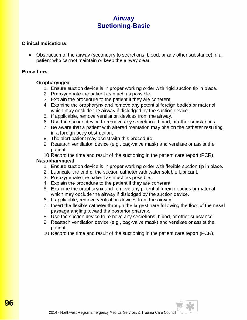
2014 - Northwest Region Emergency Medical Services & Trauma Care Council
96
Airway
Suctioning-Basic Clinical Indications:
· Obstruction of the airway (secondary to secretions, blood, or any other substance) in a patient who cannot maintain or keep the airway clear.
Procedure: Oropharyngeal
1. Ensure suction device is in proper working order with rigid suction tip in place. 2. Preoxygenate the patient as much as possible. 3. Explain the procedure to the patient if they are coherent. 4. Examine the oropharynx and remove any potential foreign bodies or material
which may occlude the airway if dislodged by the suction device. 5. If applicable, remove ventilation devices from the airway. 6. Use the suction device to remove any secretions, blood, or other substances. 7. Be aware that a patient with altered mentation may bite on the catheter resulting
in a foreign body obstruction. 8. The alert patient may assist with this procedure. 9. Reattach ventilation device (e.g., bag-valve mask) and ventilate or assist the
patient 10. Record the time and result of the suctioning in the patient care report (PCR).
Nasopharyngeal 1. Ensure suction device is in proper working order with flexible suction tip in place. 2. Lubricate the end of the suction catheter with water soluble lubricant. 3. Preoxygenate the patient as much as possible. 4. Explain the procedure to the patient if they are coherent. 5. Examine the oropharynx and remove any potential foreign bodies or material
which may occlude the airway if dislodged by the suction device. 6. If applicable, remove ventilation devices from the airway. 7. Insert the flexible catheter through the largest nare following the floor of the nasal
passage angling toward the posterior pharynx. 8. Use the suction device to remove any secretions, blood, or other substance. 9. Reattach ventilation device (e.g., bag-valve mask) and ventilate or assist the
patient. 10. Record the time and result of the suctioning in the patient care report (PCR).

2014 - Northwest Region Emergency Medical Services & Trauma Care Council
97
97
97
97
97
97
97
97
97
97
97
97
Airway Suctioning-Advanced
Clinical Indications:
· Obstruction of the airway (secondary to secretions, blood, or any other substance) in a patient currently being assisted by an airway adjunct such as:
· a naso-tracheal tube
· endotracheal tube
· supraglogic airway
· tracheostomy tube
· cricothyrotomy tube Procedure:
1. Ensure suction device is in proper working order.
2. Collect supplies including flexible suction catheter, sterile saline in container, clean gloves.
3. Pre-oxygenate the patient as much as possible. Do not over inflate the lungs.
4. Attach suction catheter to suction device, keeping end of catheter aseptic.
5. Measure length of catheter for proper depth of insertion based on the type of device in place.
6. If applicable, remove ventilation devices from the airway.
7. With the thumb port of the catheter uncovered, insert the catheter through the airway device.
8. Once the desired depth (measured in #5 above) has been reached, occlude the thumb port and remove the suction catheter slowly.
9. Interrupt ventilations for no more than 30 seconds.
10. Reattach ventilation device (e.g., bag-valve mask) and ventilate the patient.
11. Clear the suction catheter of thick secretions by aspirating sterile saline.
12. If thick secretions prevent effective suctioning instill 3-5 cc of sterile saline in the tube and ventilate the patient 3-4 breaths. Then repeat suctioning as described.
13. Document time and result including SpO2 readings before and after procedure in the patient care report (PCR).
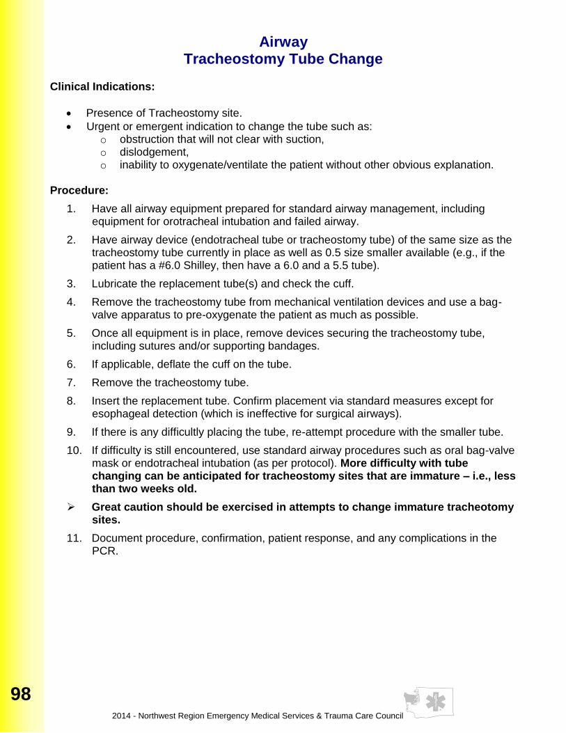
2014 - Northwest Region Emergency Medical Services & Trauma Care Council
98
Airway Tracheostomy Tube Change
Clinical Indications:
· Presence of Tracheostomy site.
· Urgent or emergent indication to change the tube such as: o obstruction that will not clear with suction, o dislodgement, o inability to oxygenate/ventilate the patient without other obvious explanation.
Procedure:
1. Have all airway equipment prepared for standard airway management, including equipment for orotracheal intubation and failed airway.
2. Have airway device (endotracheal tube or tracheostomy tube) of the same size as the tracheostomy tube currently in place as well as 0.5 size smaller available (e.g., if the patient has a #6.0 Shilley, then have a 6.0 and a 5.5 tube).
3. Lubricate the replacement tube(s) and check the cuff.
4. Remove the tracheostomy tube from mechanical ventilation devices and use a bag-valve apparatus to pre-oxygenate the patient as much as possible.
5. Once all equipment is in place, remove devices securing the tracheostomy tube, including sutures and/or supporting bandages.
6. If applicable, deflate the cuff on the tube.
7. Remove the tracheostomy tube.
8. Insert the replacement tube. Confirm placement via standard measures except for esophageal detection (which is ineffective for surgical airways).
9. If there is any difficultly placing the tube, re-attempt procedure with the smaller tube.
10. If difficulty is still encountered, use standard airway procedures such as oral bag-valve mask or endotracheal intubation (as per protocol). More difficulty with tube changing can be anticipated for tracheostomy sites that are immature – i.e., less than two weeks old.
Great caution should be exercised in attempts to change immature tracheotomy sites.
11. Document procedure, confirmation, patient response, and any complications in the PCR.

2014 - Northwest Region Emergency Medical Services & Trauma Care Council
99
99
99
99
99
99
99
99
99
99
99
99
Airway Ventilator Operation
Clinical Indications:
· Transport of an intubated patient
Procedure:
1. Confirm the placement of tube as per airway protocol.
2. Ensure adequate oxygen delivery to the ventilator device.
3. Preoxygenate the patient as much as possible with bag-valve mask.
4. Remove BVM and attach tube to ventilator device.
5. Per instructions of device, set initial values. For example, set an inspiratory / expiratory ratio of 1:4 with a rate of 12 to 20, tidal volume 6-8 mL/kg.
6. Assess breath sounds. Allow for adequate expiratory time. Adjust ventilator setting as clinically indicated.
7. If any worsening of patient condition, decrease in oxygen saturation, or any question regarding the function of the ventilator, remove the ventilator and resume bag-valve mask ventilations.
8. Document time, SPO2 / ETCO2 trends, complications, and patient response on the patient care report (PCR).
P

2014 - Northwest Region Emergency Medical Services & Trauma Care Council
100
Blood Product Administration
POLICY: For inter-facility transfer only. Only paramedics who have successfully completed MPD approved training will be monitoring the administration of blood products.
Clinical Indications:
· To replace blood loss while maintaining adequate circulating volume and oxygen during transport.
· To replace clotting factors or other life saving blood products. Complications:
· Transfusion reactions may manifest at anaphylaxis with shortness of breath, hypotension with BP<90, tachycardia, angioedema, altered mental status, rash or fever. Severe reactions are usually manifested during the initial 50cc or less of infusion.
· Too rapid an infusion producing a volume overloaded state. Equipment Use: Infusion pumps may be helpful but not required unless delivery is through a central venous catheter or pediatrics. Blood administration sets will be provided by the hospital. Procedure:
1. Initial blood administrations will be instituted at the transferring hospital. All additional units must have the transferring hospital nursing staff cross check for correct blood before accepting. (The paramedic is allowed to begin administering a new bag that has been cross checked and verified by the transferring nursing staff. )
2. Verify the physician order for blood product, blood type, rate of infusion and use of micro-aggregate or leukocyte removal filter.
3. Check for history of previous reaction to a blood product and for pre-transfusion symptoms that could be mistaken for a transfusion reaction. ( i.e fever, hypotension, tachycardia, shortness of breath, altered mental status or rash) See #4 below.
4. Assess baseline TPR and BP prior to starting transfusion and at least every 15 while blood is infusing and again when transfusion is completed (except albumin and plasma protein fraction). Vital signs must be documented.
5. Assess TPR and BP every 15 minutes x 4 during intravenous gamma globulin administration.
6. Replace blood tubing after every 2 units or after 4 hours of use. Discard tubing immediately following completion of transfusion.
7. Monitor peripheral site and infusion system at least every 15 minutes during blood product administration.
8. For any suspected reaction: a. Stop transfusion, do not clear tubing
b. Recheck labels
c. Notify base station physician
d. Remove bag and tubing; start isotonic saline
e. Monitor and treat symptoms of anaphylaxis
f. Collect a urine specimen for receiving hospital. Keep containers available.
g. Save blood bag - deliver to receiving hospital along with urine specimen for further
testing
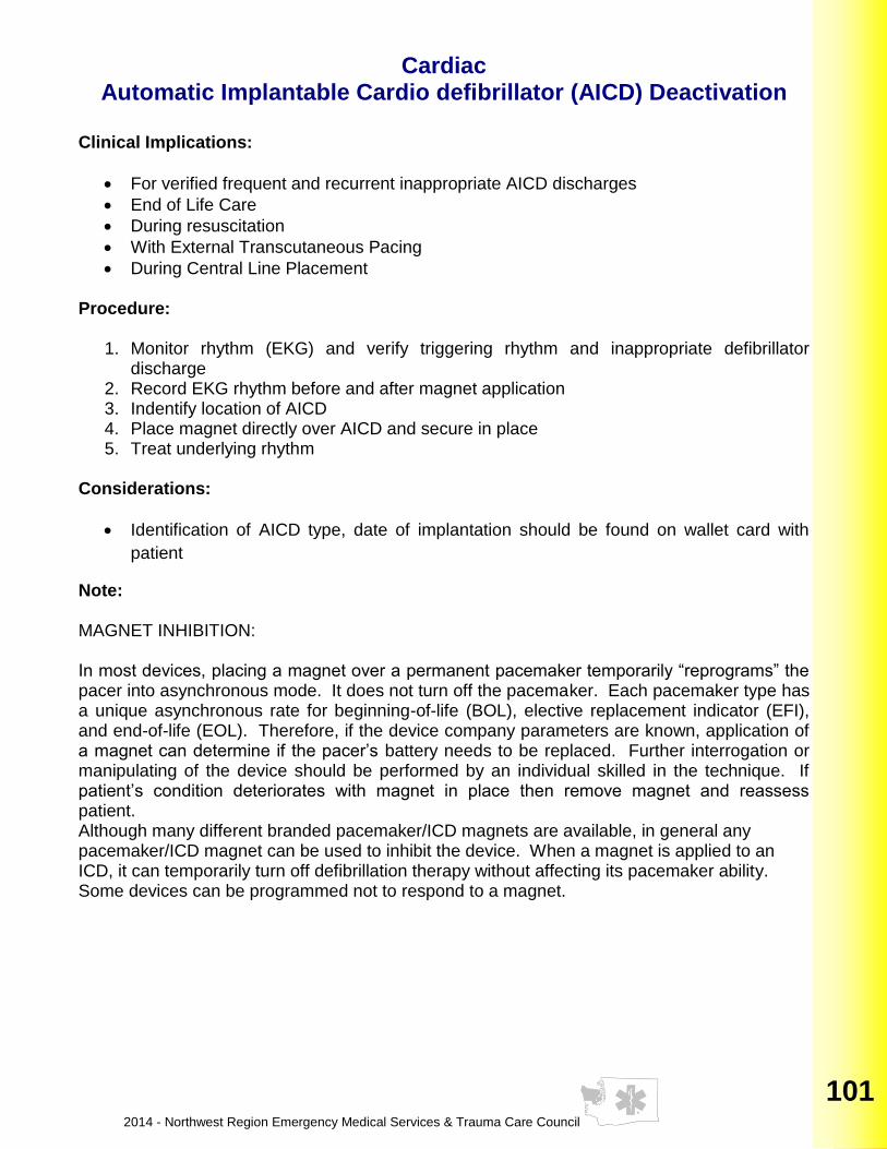
2014 - Northwest Region Emergency Medical Services & Trauma Care Council
101
101
101
101
101
10
1
10
1
101
10
1
10
1
101
101
Cardiac Automatic Implantable Cardio defibrillator (AICD) Deactivation
Clinical Implications:
· For verified frequent and recurrent inappropriate AICD discharges
· End of Life Care
· During resuscitation
· With External Transcutaneous Pacing
· During Central Line Placement Procedure:
1. Monitor rhythm (EKG) and verify triggering rhythm and inappropriate defibrillator discharge
2. Record EKG rhythm before and after magnet application 3. Indentify location of AICD 4. Place magnet directly over AICD and secure in place 5. Treat underlying rhythm
Considerations:
· Identification of AICD type, date of implantation should be found on wallet card with
patient
Note: MAGNET INHIBITION: In most devices, placing a magnet over a permanent pacemaker temporarily “reprograms” the pacer into asynchronous mode. It does not turn off the pacemaker. Each pacemaker type has a unique asynchronous rate for beginning-of-life (BOL), elective replacement indicator (EFI), and end-of-life (EOL). Therefore, if the device company parameters are known, application of a magnet can determine if the pacer’s battery needs to be replaced. Further interrogation or manipulating of the device should be performed by an individual skilled in the technique. If patient’s condition deteriorates with magnet in place then remove magnet and reassess patient. Although many different branded pacemaker/ICD magnets are available, in general any pacemaker/ICD magnet can be used to inhibit the device. When a magnet is applied to an ICD, it can temporarily turn off defibrillation therapy without affecting its pacemaker ability. Some devices can be programmed not to respond to a magnet.

2014 - Northwest Region Emergency Medical Services & Trauma Care Council
102
Cardiac 12 Lead ECG
Clinical Indications:
· Chest pain/upper abdominal pain age > 35
· Suspected overdose
· Electrical injuries
· Syncope
· Suspected arrhythmias
· Suspected electrolyte imbalance
Procedure: 1. Assess patient and monitor cardiac status. 2. Administer oxygen as patient condition warrants. 3. If patient presents with pain or complaint, suspect of a cardiac related emergency, then
perform a 12 Lead ECG. (EMT can perform ECG - read only) 4. Prepare ECG monitor and connect patient cable with electrodes. 5. Enter the required patient information (patient name, etc.) into the 12 lead ECG device. 6. Expose chest and prep as necessary. Modesty of the patient should be respected. 7. Apply chest leads and extremity leads using the following landmarks:
· RA -Right arm
· LA -Left arm
· RL -Right leg
· LL -Left leg
· V1 -4th intercostal space at right sternal border
· V2 -4th intercostal space at left sternal border
· V3 -Directly between V2 and V4
· V4 -5th intercostal space at midclavicular line
· V5 -Level with V4 at left anterior axillary line
· V6 -Level with V5 at left midaxillary line 8. Instruct patient to remain still. 9. Press the appropriate button to acquire the 12 Lead ECG. 10. If the monitor detects signal noise (such as patient motion or a disconnected electrode),
the 12 Lead acquisition will be interrupted until the noise is removed. 11. Once acquired, transmit the ECG data by fax (where available) to the appropriate
hospital. 12. Contact the receiving hospital to notify them that a 12 Lead ECG has been sent. 13. Monitor the patient while continuing with the treatment protocol. 14. Download data as per guidelines and attach a copy of the 12 lead to the Patient Care
Report. 15. Document the procedure, time, and results on/with the patient care report (PCR)
I Lateral aVR V1 Septal V4 Anterior
II Inferior aVL Lateral V2 Septal V5 Lateral
III Inferior aVF Inferior V3 Anterior V6 Lateral
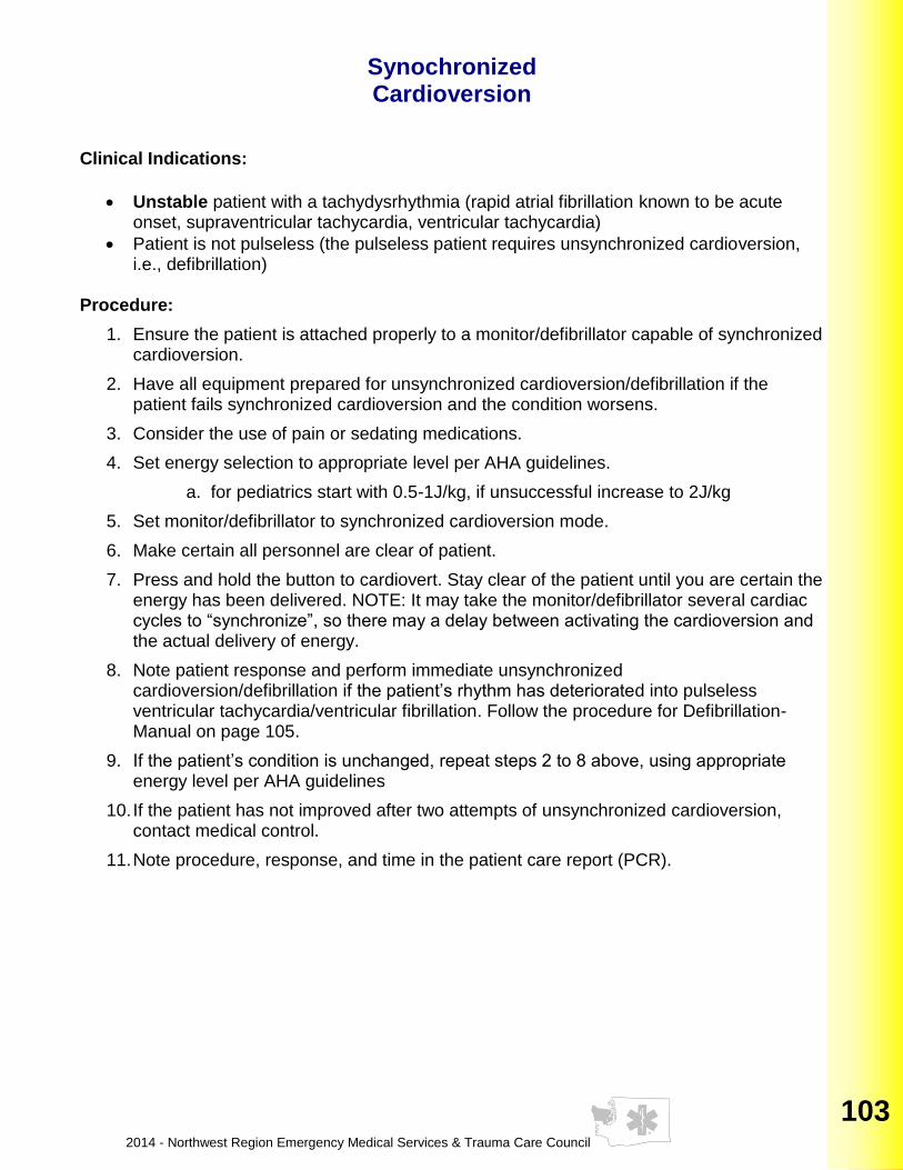
2014 - Northwest Region Emergency Medical Services & Trauma Care Council
103
103
103
103
103
10
3
10
3
103
10
3
10
3
103
103
Synochronized Cardioversion
Clinical Indications:
· Unstable patient with a tachydysrhythmia (rapid atrial fibrillation known to be acute onset, supraventricular tachycardia, ventricular tachycardia)
· Patient is not pulseless (the pulseless patient requires unsynchronized cardioversion, i.e., defibrillation)
Procedure:
1. Ensure the patient is attached properly to a monitor/defibrillator capable of synchronized cardioversion.
2. Have all equipment prepared for unsynchronized cardioversion/defibrillation if the patient fails synchronized cardioversion and the condition worsens.
3. Consider the use of pain or sedating medications.
4. Set energy selection to appropriate level per AHA guidelines.
a. for pediatrics start with 0.5-1J/kg, if unsuccessful increase to 2J/kg
5. Set monitor/defibrillator to synchronized cardioversion mode.
6. Make certain all personnel are clear of patient.
7. Press and hold the button to cardiovert. Stay clear of the patient until you are certain the energy has been delivered. NOTE: It may take the monitor/defibrillator several cardiac cycles to “synchronize”, so there may a delay between activating the cardioversion and the actual delivery of energy.
8. Note patient response and perform immediate unsynchronized cardioversion/defibrillation if the patient’s rhythm has deteriorated into pulseless ventricular tachycardia/ventricular fibrillation. Follow the procedure for Defibrillation-Manual on page 105.
9. If the patient’s condition is unchanged, repeat steps 2 to 8 above, using appropriate energy level per AHA guidelines
10. If the patient has not improved after two attempts of unsynchronized cardioversion, contact medical control.
11. Note procedure, response, and time in the patient care report (PCR).
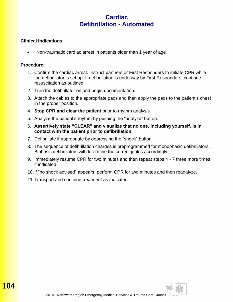
2014 - Northwest Region Emergency Medical Services & Trauma Care Council
104
Cardiac Defibrillation - Automated
Clinical Indications:
·Non-traumatic cardiac arrest in patients older than 1 year of age
Procedure:
1. Confirm the cardiac arrest. Instruct partners or First Responders to initiate CPR while the defibrillator is set up. If defibrillation is underway by First Responders, continue resuscitation as outlined.
2. Turn the defibrillator on and begin documentation.
3. Attach the cables to the appropriate pads and then apply the pads to the patient’s chest in the proper position.
4. Stop CPR and clear the patient prior to rhythm analysis.
5. Analyze the patient’s rhythm by pushing the “analyze” button.
6. Assertively state “CLEAR” and visualize that no one, including yourself, is in contact with the patient prior to defibrillation.
7. Defibrillate if appropriate by depressing the “shock” button.
8. The sequence of defibrillation charges is preprogrammed for monophasic defibrillators. Biphasic defibrillators will determine the correct joules accordingly.
9. Immediately resume CPR for two minutes and then repeat steps 4 - 7 three more times if indicated.
10. If “no shock advised” appears, perform CPR for two minutes and then reanalyze.
11. Transport and continue treatment as indicated.
P D
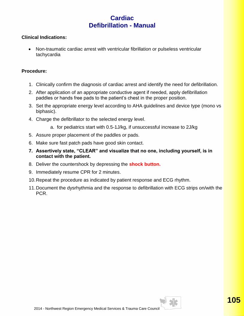
2014 - Northwest Region Emergency Medical Services & Trauma Care Council
105
105
105
105
105
10
5
10
5
105
10
5
10
5
105
105
Cardiac
Defibrillation - Manual Clinical Indications:
·Non-traumatic cardiac arrest with ventricular fibrillation or pulseless ventricular tachycardia
Procedure:
1. Clinically confirm the diagnosis of cardiac arrest and identify the need for defibrillation.
2. After application of an appropriate conductive agent if needed, apply defibrillation paddles or hands free pads to the patient’s chest in the proper position.
3. Set the appropriate energy level according to AHA guidelines and device type (mono vs biphasic).
4. Charge the defibrillator to the selected energy level.
a. for pediatrics start with 0.5-1J/kg, if unsuccessful increase to 2J/kg
5. Assure proper placement of the paddles or pads.
6. Make sure fast patch pads have good skin contact.
7. Assertively state, “CLEAR” and visualize that no one, including yourself, is in contact with the patient.
8. Deliver the countershock by depressing the shock button.
9. Immediately resume CPR for 2 minutes.
10. Repeat the procedure as indicated by patient response and ECG rhythm.
11. Document the dysrhythmia and the response to defibrillation with ECG strips on/with the PCR.
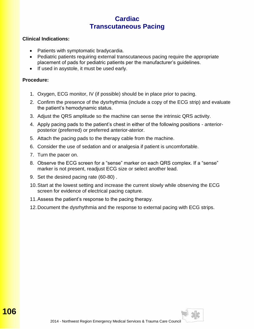
2014 - Northwest Region Emergency Medical Services & Trauma Care Council
106
Cardiac Transcutaneous Pacing
Clinical Indications:
· Patients with symptomatic bradycardia.
· Pediatric patients requiring external transcutaneous pacing require the appropriate placement of pads for pediatric patients per the manufacturer’s guidelines.
· If used in asystole, it must be used early. Procedure:
1. Oxygen, ECG monitor, IV (if possible) should be in place prior to pacing.
2. Confirm the presence of the dysrhythmia (include a copy of the ECG strip) and evaluate the patient’s hemodynamic status.
3. Adjust the QRS amplitude so the machine can sense the intrinsic QRS activity.
4. Apply pacing pads to the patient’s chest in either of the following positions - anterior-posterior (preferred) or preferred anterior-aterior.
5. Attach the pacing pads to the therapy cable from the machine.
6. Consider the use of sedation and or analgesia if patient is uncomfortable.
7. Turn the pacer on.
8. Observe the ECG screen for a “sense” marker on each QRS complex. If a “sense” marker is not present, readjust ECG size or select another lead.
9. Set the desired pacing rate (60-80) .
10. Start at the lowest setting and increase the current slowly while observing the ECG screen for evidence of electrical pacing capture.
11. Assess the patient’s response to the pacing therapy.
12. Document the dysrhythmia and the response to external pacing with ECG strips.
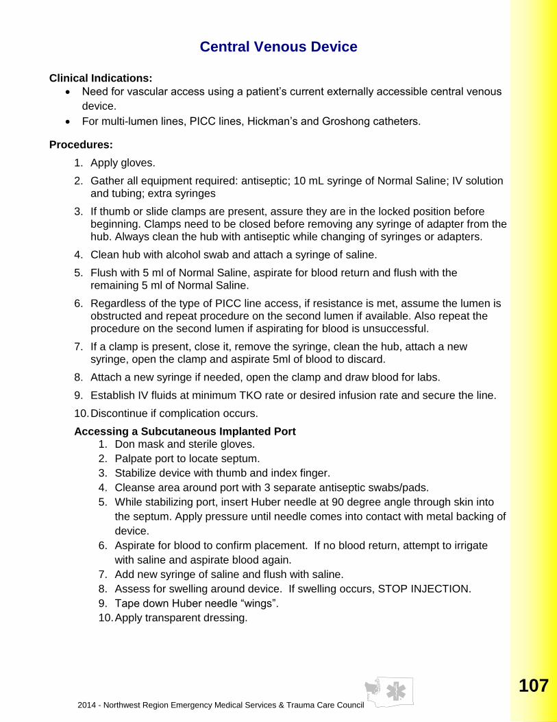
2014 - Northwest Region Emergency Medical Services & Trauma Care Council
107
107
107
107
107
10
7
10
7
107
10
7
10
7
107
107
Central Venous Device
Clinical Indications:
· Need for vascular access using a patient’s current externally accessible central venous
device.
· For multi-lumen lines, PICC lines, Hickman’s and Groshong catheters.
Procedures:
1. Apply gloves.
2. Gather all equipment required: antiseptic; 10 mL syringe of Normal Saline; IV solution and tubing; extra syringes
3. If thumb or slide clamps are present, assure they are in the locked position before beginning. Clamps need to be closed before removing any syringe of adapter from the hub. Always clean the hub with antiseptic while changing of syringes or adapters.
4. Clean hub with alcohol swab and attach a syringe of saline.
5. Flush with 5 ml of Normal Saline, aspirate for blood return and flush with the remaining 5 ml of Normal Saline.
6. Regardless of the type of PICC line access, if resistance is met, assume the lumen is obstructed and repeat procedure on the second lumen if available. Also repeat the procedure on the second lumen if aspirating for blood is unsuccessful.
7. If a clamp is present, close it, remove the syringe, clean the hub, attach a new syringe, open the clamp and aspirate 5ml of blood to discard.
8. Attach a new syringe if needed, open the clamp and draw blood for labs.
9. Establish IV fluids at minimum TKO rate or desired infusion rate and secure the line.
10. Discontinue if complication occurs.
Accessing a Subcutaneous Implanted Port 1. Don mask and sterile gloves.
2. Palpate port to locate septum.
3. Stabilize device with thumb and index finger.
4. Cleanse area around port with 3 separate antiseptic swabs/pads.
5. While stabilizing port, insert Huber needle at 90 degree angle through skin into
the septum. Apply pressure until needle comes into contact with metal backing of
device.
6. Aspirate for blood to confirm placement. If no blood return, attempt to irrigate
with saline and aspirate blood again.
7. Add new syringe of saline and flush with saline.
8. Assess for swelling around device. If swelling occurs, STOP INJECTION.
9. Tape down Huber needle “wings”.
10. Apply transparent dressing.

2014 - Northwest Region Emergency Medical Services & Trauma Care Council
108
Central Venous Device (continued)
Hickman Catheter
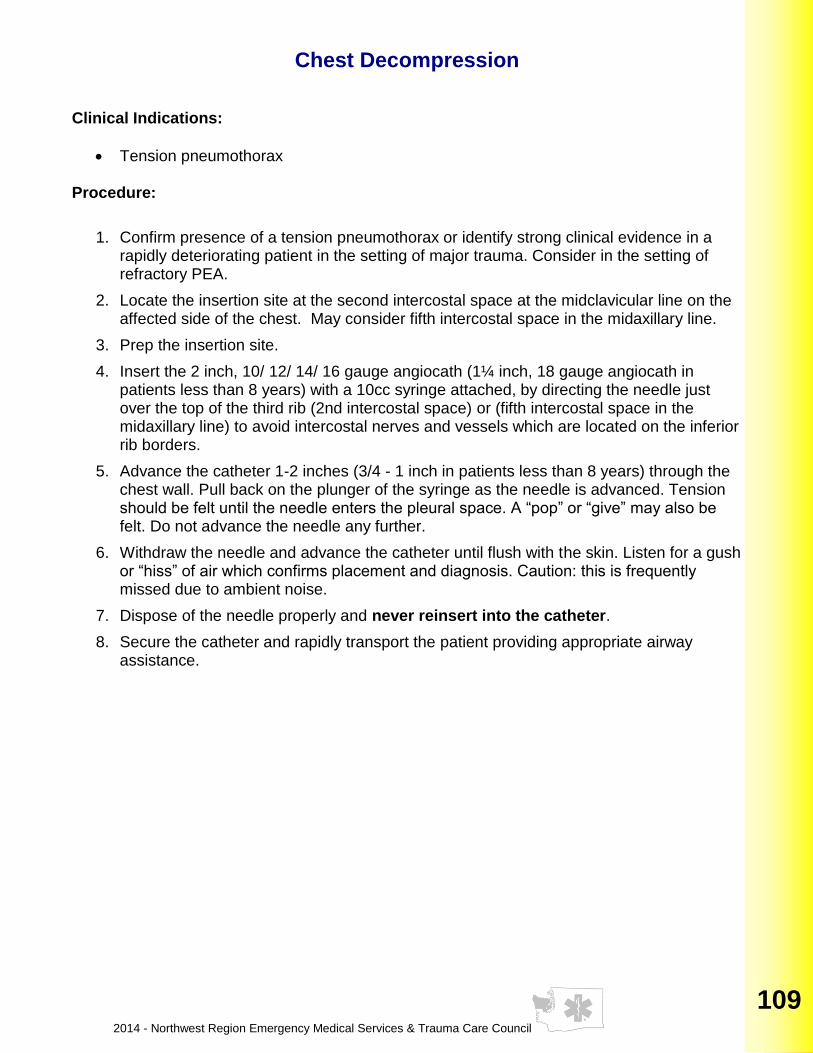
2014 - Northwest Region Emergency Medical Services & Trauma Care Council
109
109
109
109
109
10
9
10
9
109
10
9
10
9
109
109
Chest Decompression Clinical Indications:
· Tension pneumothorax Procedure:
1. Confirm presence of a tension pneumothorax or identify strong clinical evidence in a rapidly deteriorating patient in the setting of major trauma. Consider in the setting of refractory PEA.
2. Locate the insertion site at the second intercostal space at the midclavicular line on the affected side of the chest. May consider fifth intercostal space in the midaxillary line.
3. Prep the insertion site.
4. Insert the 2 inch, 10/ 12/ 14/ 16 gauge angiocath (1¼ inch, 18 gauge angiocath in patients less than 8 years) with a 10cc syringe attached, by directing the needle just over the top of the third rib (2nd intercostal space) or (fifth intercostal space in the midaxillary line) to avoid intercostal nerves and vessels which are located on the inferior rib borders.
5. Advance the catheter 1-2 inches (3/4 - 1 inch in patients less than 8 years) through the chest wall. Pull back on the plunger of the syringe as the needle is advanced. Tension should be felt until the needle enters the pleural space. A “pop” or “give” may also be felt. Do not advance the needle any further.
6. Withdraw the needle and advance the catheter until flush with the skin. Listen for a gush or “hiss” of air which confirms placement and diagnosis. Caution: this is frequently missed due to ambient noise.
7. Dispose of the needle properly and never reinsert into the catheter.
8. Secure the catheter and rapidly transport the patient providing appropriate airway assistance.
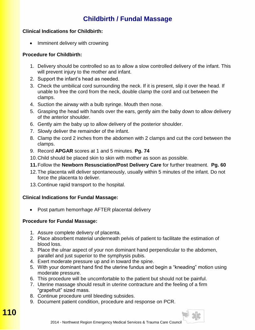
2014 - Northwest Region Emergency Medical Services & Trauma Care Council
110
Childbirth / Fundal Massage Clinical Indications for Childbirth:
· Imminent delivery with crowning Procedure for Childbirth:
1. Delivery should be controlled so as to allow a slow controlled delivery of the infant. This will prevent injury to the mother and infant.
2. Support the infant’s head as needed.
3. Check the umbilical cord surrounding the neck. If it is present, slip it over the head. If unable to free the cord from the neck, double clamp the cord and cut between the clamps.
4. Suction the airway with a bulb syringe. Mouth then nose.
5. Grasping the head with hands over the ears, gently aim the baby down to allow delivery of the anterior shoulder.
6. Gently aim the baby up to allow delivery of the posterior shoulder.
7. Slowly deliver the remainder of the infant.
8. Clamp the cord 2 inches from the abdomen with 2 clamps and cut the cord between the clamps.
9. Record APGAR scores at 1 and 5 minutes. Pg. 74
10. Child should be placed skin to skin with mother as soon as possible.
11. Follow the Newborn Resusciation/Post Delivery Care for further treatment. Pg. 60
12. The placenta will deliver spontaneously, usually within 5 minutes of the infant. Do not force the placenta to deliver.
13. Continue rapid transport to the hospital.
Clinical Indications for Fundal Massage:
· Post partum hemorrhage AFTER placental delivery Procedure for Fundal Massage:
1. Assure complete delivery of placenta. 2. Place absorbent material underneath pelvis of patient to facilitate the estimation of
blood loss. 3. Place the ulnar aspect of your non dominant hand perpendicular to the abdomen,
parallel and just superior to the symphysis pubis. 4. Exert moderate pressure up and in toward the spine. 5. With your dominant hand find the uterine fundus and begin a “kneading” motion using
moderate pressure. 6. This procedure will be uncomfortable to the patient but should not be painful. 7. Uterine massage should result in uterine contracture and the feeling of a firm
“grapefruit” sized mass. 8. Continue procedure until bleeding subsides. 9. Document patient condition, procedure and response on PCR.
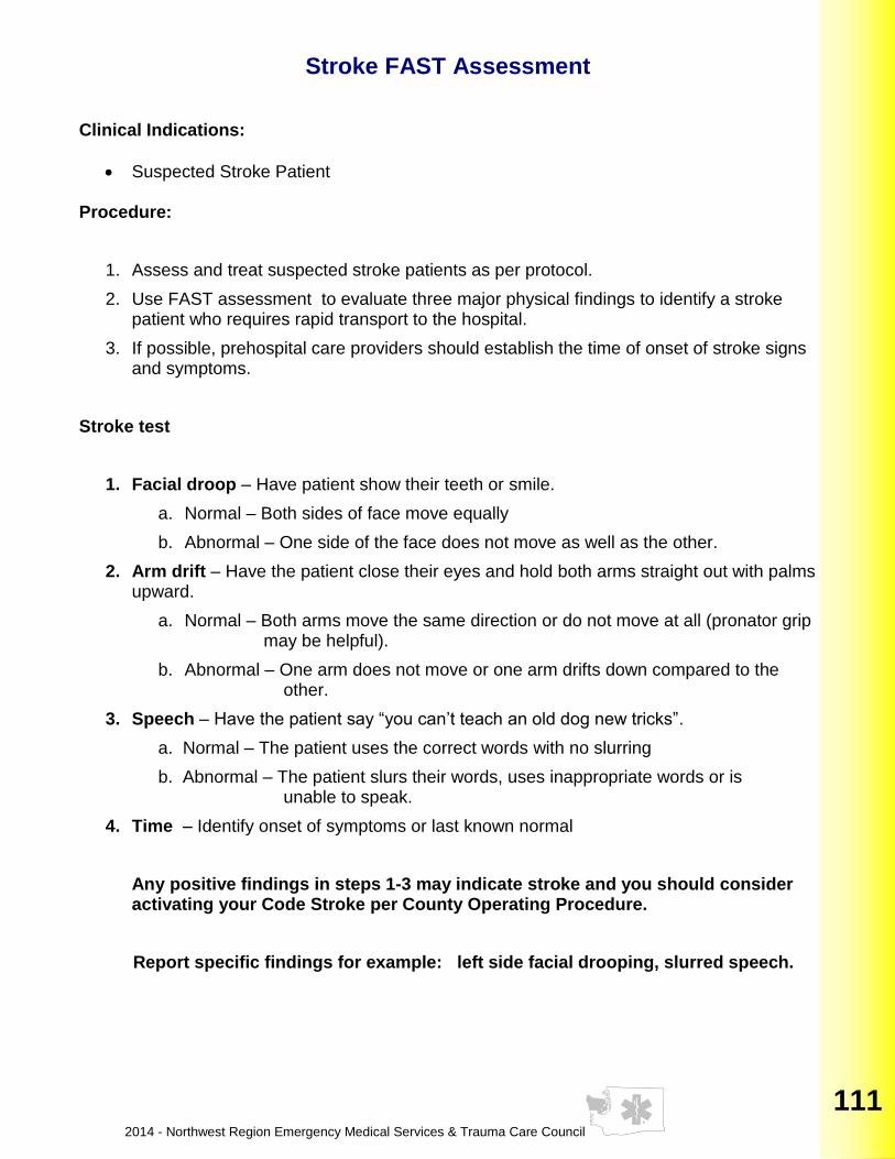
2014 - Northwest Region Emergency Medical Services & Trauma Care Council
111
111
111
111
111
11
1
11
1
111
11
1
11
1
111
111
Stroke FAST Assessment Clinical Indications:
· Suspected Stroke Patient Procedure:
1. Assess and treat suspected stroke patients as per protocol.
2. Use FAST assessment to evaluate three major physical findings to identify a stroke patient who requires rapid transport to the hospital.
3. If possible, prehospital care providers should establish the time of onset of stroke signs and symptoms.
Stroke test
1. Facial droop – Have patient show their teeth or smile.
a. Normal – Both sides of face move equally
b. Abnormal – One side of the face does not move as well as the other.
2. Arm drift – Have the patient close their eyes and hold both arms straight out with palms upward.
a. Normal – Both arms move the same direction or do not move at all (pronator grip may be helpful).
b. Abnormal – One arm does not move or one arm drifts down compared to the other.
3. Speech – Have the patient say “you can’t teach an old dog new tricks”.
a. Normal – The patient uses the correct words with no slurring
b. Abnormal – The patient slurs their words, uses inappropriate words or is unable to speak.
4. Time – Identify onset of symptoms or last known normal
Any positive findings in steps 1-3 may indicate stroke and you should consider activating your Code Stroke per County Operating Procedure.
Report specific findings for example: left side facial drooping, slurred speech.

2014 - Northwest Region Emergency Medical Services & Trauma Care Council
112
CPR (High Density)
MANEUVER Adult
HCP: Adolescent and older
Child HCP:1 year to
adolescent
Infant Under 1 year old
RECOGNITION
Unresponsive (for all ages)
No breathing or no normal breathing (ie, only gasping)
No breathing or only gasping
No pulse palpated within 10 seconds for all ages (HCP only)
ACTIVATE: Emergency Response Number (lone rescuer)
Activate when victim found unresponsive HCP: if asphyxial arrest likely, call after 5 cycles (2 minutes) of CPR
Activate after performing 5 cycles of CPR For sudden, witnessed collapse,
activate after verifying that victim unresponsive
CPR Sequence C-A-B
Compression Rate At least 100/min
Compression Depth At least 2 inches (5cm
At least 1/3 AP diameter About 2 inches (5cm)
At least 1/3 AP diameter About 1 ½ inches (5cm)
Chest Wall Recoil Allow complete recoil between compressions HCPs rotate compressors every 2 minutes
Compression Interruptions
Minimize interruptions in chest compressions Attempt to limit interruptions to <10 seconds
Airway Head tilt-chin lift (HCP suspected trauma: jaw thrust)
Compression-to-ventilation ratio (until advanced airway placed)
30:2 1 or 2 rescuers
30:2 Single rescuer
15:2 2 HCP rescuers
Ventilations: when rescuer untrained or trained and not proficient
Compressions only
Ventilations with advanced airway (HCP)
1 breath every 6-8 seconds (8-10 breaths/min) Asynchronous with chest compressions
About 1 second per breath Visible chest rise
Foreign-body airway obstruction
Responsive: Abdominal thrusts
Unresponsive: CPR with airway check
Responsive: Back slaps and chest thrusts
Unresponsive: CPR with airway check
Defibrillation
Attach and use AED as soon as available. Minimize interruptions in chest compressions before and after shock; resume CPR beginning with compressions immediately after
each shock.

2014 - Northwest Region Emergency Medical Services & Trauma Care Council
113
113
113
113
113
11
3
11
3
113
11
3
11
3
113
113
Discontinuation of CPR, Do not attempt Resuscitation Determination of Field Death
DO NOT ATTEMPT RESUSCITATION: Obvious death
· Decomposition of body tissue
· Total decapitation
· Total incineration
· Total separation or destruction of the heart or brain
· Fetus with a foot length of 33mm or less
· Traumatic Arrest (Non-breathing and pulseless)
· Rigor Mortis POLST form or DNR papers are dated and signed by the patient with appropriate witnessed signatures and there is no question they belong to the patient. The patient may be of any age.
DISCONTINUATION WITHOUT MEDICAL CONTROL: (must meet 3 requirements)
· Non-breathing
· Pulselessness
· Asystole in two leads OR No Shock Advised on AED (EMT’s can transmit Rhythm strip to hospital for convenience)
· Lividity Note: Exception – suspected hypothermia requires full resuscitation efforts
DISCONTINUATION OF EFFORTS:
1. Endotracheal intubation and drug therapy appropriate to the presenting rhythm, according to AHA guidelines, have been initiated and the patient remains apneic, pulseless, and in asystole or PEA.
2. Compelling reasons to withhold CPR/Resuscitation efforts. Such as but not limited to
· End stage of terminal condition
· Living will
· Verbal request by family Note: Prehospital care providers desiring support in the field may contact the Medical Control at any time for Determination of Death
3. DNR or POLST form has been presented after CPR was initiated.
Once death has been determined and resuscitation efforts discontinued, all ALS therapeutic modalities initiated during the resuscitation must be left in place until it has been determined that the patient will not be a Coroners’ case. This includes such equipment as endotracheal tubes, IV catheters, monitor electrodes and personal items including clothes, jewelry etc. If the coroner releases the body while the prehospital care provider is still on scene, remove all medical equipment used during the resuscitation. Time of death recorded on PCR.
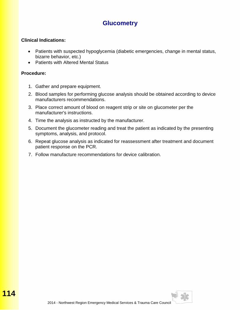
2014 - Northwest Region Emergency Medical Services & Trauma Care Council
114
Glucometry Clinical Indications:
· Patients with suspected hypoglycemia (diabetic emergencies, change in mental status, bizarre behavior, etc.)
· Patients with Altered Mental Status Procedure:
1. Gather and prepare equipment.
2. Blood samples for performing glucose analysis should be obtained according to device manufacturers recommendations.
3. Place correct amount of blood on reagent strip or site on glucometer per the manufacturer's instructions.
4. Time the analysis as instructed by the manufacturer.
5. Document the glucometer reading and treat the patient as indicated by the presenting symptoms, analysis, and protocol.
6. Repeat glucose analysis as indicated for reassessment after treatment and document patient response on the PCR.
7. Follow manufacture recommendations for device calibration.
P P
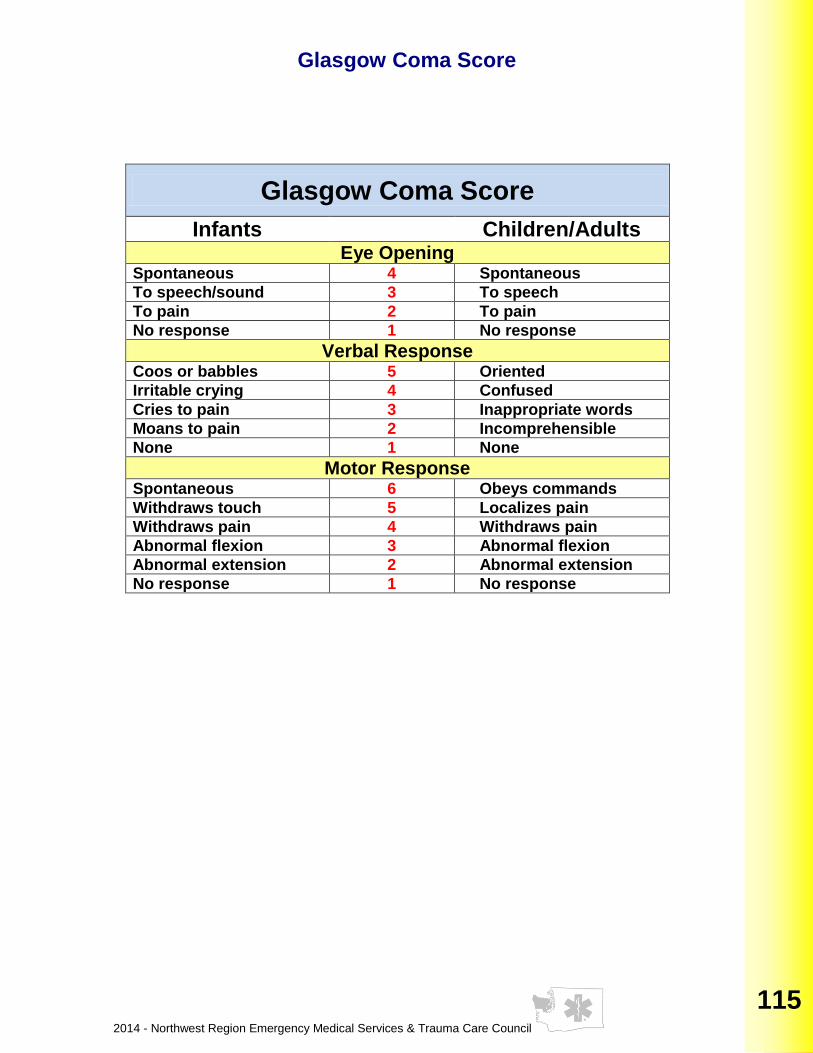
2014 - Northwest Region Emergency Medical Services & Trauma Care Council
115
115
115
115
115
11
5
11
5
115
11
5
11
5
115
115
Glasgow Coma Score
Glasgow Coma Score
Infants Children/Adults Eye Opening
Spontaneous 4 Spontaneous
To speech/sound 3 To speech
To pain 2 To pain
No response 1 No response
Verbal Response Coos or babbles 5 Oriented
Irritable crying 4 Confused
Cries to pain 3 Inappropriate words
Moans to pain 2 Incomprehensible
None 1 None
Motor Response Spontaneous 6 Obeys commands
Withdraws touch 5 Localizes pain
Withdraws pain 4 Withdraws pain
Abnormal flexion 3 Abnormal flexion
Abnormal extension 2 Abnormal extension
No response 1 No response
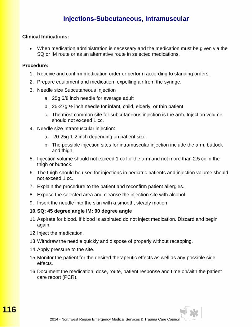
2014 - Northwest Region Emergency Medical Services & Trauma Care Council
116
Injections-Subcutaneous, Intramuscular Clinical Indications:
· When medication administration is necessary and the medication must be given via the SQ or IM route or as an alternative route in selected medications.
Procedure:
1. Receive and confirm medication order or perform according to standing orders.
2. Prepare equipment and medication, expelling air from the syringe.
3. Needle size Subcutaneous Injection
a. 25g 5/8 inch needle for average adult
b. 25-27g ½ inch needle for infant, child, elderly, or thin patient
c. The most common site for subcutaneous injection is the arm. Injection volume should not exceed 1 cc.
4. Needle size Intramuscular injection:
a. 20-25g 1-2 inch depending on patient size.
b. The possible injection sites for intramuscular injection include the arm, buttock and thigh.
5. Injection volume should not exceed 1 cc for the arm and not more than 2.5 cc in the thigh or buttock.
6. The thigh should be used for injections in pediatric patients and injection volume should not exceed 1 cc.
7. Explain the procedure to the patient and reconfirm patient allergies.
8. Expose the selected area and cleanse the injection site with alcohol.
9. Insert the needle into the skin with a smooth, steady motion
10. SQ: 45 degree angle IM: 90 degree angle
11. Aspirate for blood. If blood is aspirated do not inject medication. Discard and begin again.
12. Inject the medication.
13. Withdraw the needle quickly and dispose of properly without recapping.
14. Apply pressure to the site.
15. Monitor the patient for the desired therapeutic effects as well as any possible side effects.
16. Document the medication, dose, route, patient response and time on/with the patient care report (PCR).
P
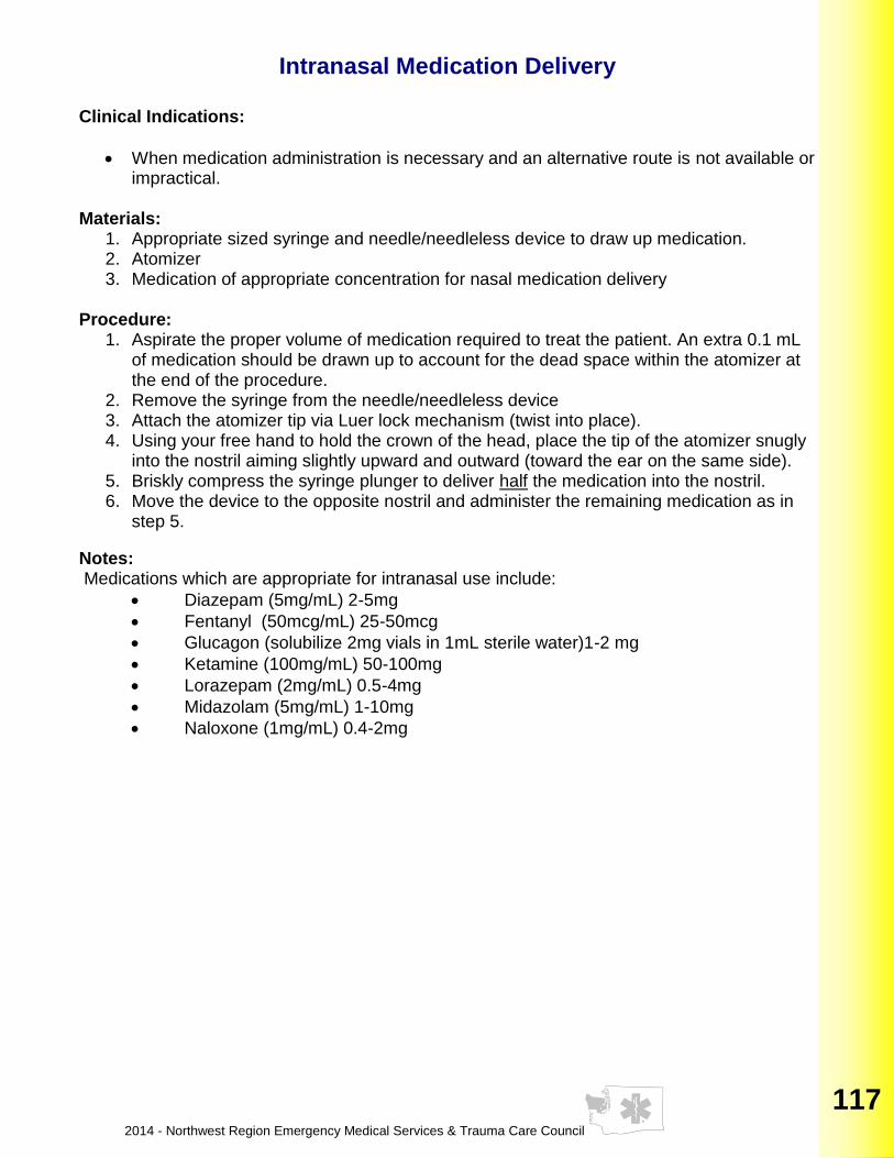
2014 - Northwest Region Emergency Medical Services & Trauma Care Council
117
117
117
117
117
11
7
11
7
117
11
7
11
7
117
117
Intranasal Medication Delivery
Clinical Indications:
· When medication administration is necessary and an alternative route is not available or impractical.
Materials:
1. Appropriate sized syringe and needle/needleless device to draw up medication. 2. Atomizer 3. Medication of appropriate concentration for nasal medication delivery
Procedure:
1. Aspirate the proper volume of medication required to treat the patient. An extra 0.1 mL of medication should be drawn up to account for the dead space within the atomizer at the end of the procedure.
2. Remove the syringe from the needle/needleless device 3. Attach the atomizer tip via Luer lock mechanism (twist into place). 4. Using your free hand to hold the crown of the head, place the tip of the atomizer snugly
into the nostril aiming slightly upward and outward (toward the ear on the same side). 5. Briskly compress the syringe plunger to deliver half the medication into the nostril. 6. Move the device to the opposite nostril and administer the remaining medication as in
step 5.
Notes: Medications which are appropriate for intranasal use include:
· Diazepam (5mg/mL) 2-5mg
· Fentanyl (50mcg/mL) 25-50mcg
· Glucagon (solubilize 2mg vials in 1mL sterile water)1-2 mg
· Ketamine (100mg/mL) 50-100mg
· Lorazepam (2mg/mL) 0.5-4mg
· Midazolam (5mg/mL) 1-10mg
· Naloxone (1mg/mL) 0.4-2mg

2014 - Northwest Region Emergency Medical Services & Trauma Care Council
118
Nasogastric Tube Insertion Clinical Indications:
· Gastric decompression in intubated patients.
· Administration of activated charcoal in patients with altered mental status with a secured airway.
Procedure:
1. Assemble all supplies. Assure functioning suction unit.
2. Estimate insertion length by superimposing the tube over the body from the nose, to the ear, to the stomach.
3. Mark the proper insertion distance with tape.
4. Flex the neck if not contraindicated to facilitate esophageal passage.
5. Liberally lubricate the distal end of the tube and pass through the patient’s nostril along the floor of the nasal passage. Do not orient the tip upward into the turbinates. This increases the difficulty of the insertion and may cause bleeding.
6. In the setting of an unconscious, intubated patient or a patient with facial trauma, oral insertion of the tube may be considered or preferred.
7. Continue to advance the tube gently until the appropriate distance is reached.
8. Confirm placement by injecting 20cc of air and auscultate for the swish or bubbling of the air over the stomach. Additionally, aspirate gastric contents to confirm proper placement.
9. Secure the tube.
10. Decompress the stomach of air and food either by connecting the tube to suction or manually aspirating with the large catheter tip syringe.
11. Mechanical suction should not reach high setting.
12. Document the procedure, time, patient response, and result (success) on/with the patient care report (PCR).

2014 - Northwest Region Emergency Medical Services & Trauma Care Council
119
119
119
119
119
11
9
11
9
119
11
9
11
9
119
119
Orthostatic Blood Pressure Measurement Clinical Indications:
· Patient situations with suspected blood / fluid loss / dehydration
· Patients larger than the Length Based tape Procedure:
1. Assess the need for orthostatic vital sign measurement.
2. Obtain patient’s pulse and blood pressure while supine.
3. Have patient stand for two minutes.
4. Obtain patient’s pulse and blood pressure while standing.
5. If pulse has increased by 30 BPM or systolic blood pressure decreased by 30 mmHg, the orthostatics are considered positive.
6. If patient is unable to stand, orthostatics may be taken while the patient is sitting with feet dangling.
7. If positive orthostatic changes occur while sitting, DO NOT continue to the standing position.
8. Patients on prolonged beta-blocker therapy will not demonstrate orthostatic vital sign changes. Provider must complete assessment and utilize clinical judgment.
9. Document the time and vital signs for supine and standing positions on/with the patient care report (PCR).
10. Determine appropriate treatment based on protocol.
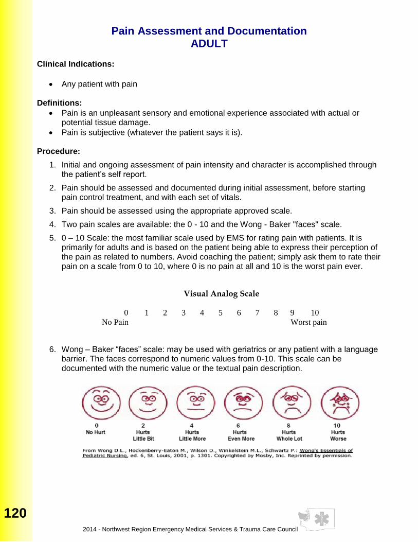
2014 - Northwest Region Emergency Medical Services & Trauma Care Council
120
Pain Assessment and Documentation ADULT
Clinical Indications:
· Any patient with pain Definitions:
· Pain is an unpleasant sensory and emotional experience associated with actual or potential tissue damage.
· Pain is subjective (whatever the patient says it is). Procedure:
1. Initial and ongoing assessment of pain intensity and character is accomplished through the patient’s self report.
2. Pain should be assessed and documented during initial assessment, before starting pain control treatment, and with each set of vitals.
3. Pain should be assessed using the appropriate approved scale.
4. Two pain scales are available: the 0 - 10 and the Wong - Baker "faces" scale.
5. 0 – 10 Scale: the most familiar scale used by EMS for rating pain with patients. It is primarily for adults and is based on the patient being able to express their perception of the pain as related to numbers. Avoid coaching the patient; simply ask them to rate their pain on a scale from 0 to 10, where 0 is no pain at all and 10 is the worst pain ever.
Visual Analog Scale
0 1 2 3 4 5 6 7 8 9 10
No Pain Worst pain
6. Wong – Baker “faces” scale: may be used with geriatrics or any patient with a language barrier. The faces correspond to numeric values from 0-10. This scale can be documented with the numeric value or the textual pain description.

2014 - Northwest Region Emergency Medical Services & Trauma Care Council
121
121
121
121
121
12
1
12
1
121
12
1
12
1
121
121
Pelvic Fracture Stabilization Clinical Indications:
· Physical exam indicates and mechanism of injury suggests a pelvic fracture Procedure:
1. Physical exam shows instability of pelvis with compression.
2. Assess the abdomen and neurovascular status of the lower extremities.
3. Assess for blood at the perineum
4. Fold a sheet lengthwise into a swathe approximately 12 to 18 inches wide.
5. Pass this swathe under the patient’s buttocks and tie circumferentially around the pelvis
covering buttocks posteriorly. The swathe should be just below the iliac crests.
6. Re-assess the abdomen and neurovascular status of the lower extremities.
OR
1. Physical exam shows instability of pelvis with compression.
2. Assess the abdomen and neurovascular status of the lower extremities.
3. Assess for blood at the perineum.
4. Utilize a commercial pelvic stabilization device following the manufacturer’s
specifications.
5. Re-assess the abdomen and neurovascular status of the lower extremities.
2014 - Northwest Region Emergency Medical Services & Trauma Care Council
122
Pulse Oximetry Clinical Indications:
· Patients with suspected hypoxemia. Procedure:
1. Turn the machine on and allow for self-tests.
2. Apply probe to patient’s finger or any other digit as recommended by the device manufacturer.
3. Allow machine to register saturation level.
4. Record time and initial saturation percent on room air if possible on/with the patient care report (PCR).
5. Verify pulse rate on machine with palpated pulse of the patient.
6. Monitor critical patients continuously until arrival at the hospital. If recording a one-time reading, monitor patients for a few minutes as oxygen saturation can vary.
7. Document percent of oxygen saturation every time vital signs are recorded and in response to therapy to correct hypoxemia.
8. In general, normal saturation is SpO2 97-99%. Below 94%, suspect a respiratory compromise.
9. Use the pulse oximetry as an added tool for patient evaluation. Treat the patient, not the data provided by the device.
10. The pulse oximeter reading should never be used to withhold oxygen from a patient in respiratory distress or when it is the standard of care to apply oxygen despite good pulse oximetry readings, such as chest pain.
11. Factors which may reduce the reliability of the pulse oximetry reading include:
(a) Poor peripheral circulation (blood volume, hypotension, hypothermia)
(b) Excessive pulse oximeter sensor motion
(c) Fingernail polish (may be removed with acetone pad)
(d) Carbon monoxide bound to hemoglobin
(e) Irregular heart rhythms (atrial fibrillation, SVT, etc.)
(f) Jaundice
(g) Excessive light entering the side of the probe causing "washout" of the signal
2014 - Northwest Region Emergency Medical Services & Trauma Care Council
123
123
123
123
123
12
3
12
3
123
12
3
12
3
123
123
Restraints
Clinical Indications:
· Patients with actual or potential threat to self or others.
· Involuntary hold
Procedure:
1. Planning:
A. Request assistance from Law Enforcement.
B. EMS personnel are not to knowingly place themselves at risk during the process of restraining a patient.
C. Obtain necessary resources to manage scene and patient.
D. Assess patient for any condition that may contribute to violent behavior. Treatment for identified conditions is to be initiated according to protocol immediately after controlling the situation and patient behavior.
E. Consult Medical Control as soon as possible regarding the application and use of restraints.
F. Verbal de-escalation techniques are to be implemented and documented. If verbal de-escalation fails, providers may need to implement physical and or chemical restraint measures.
2. Application of restraints: A. Obtaining and preparing appropriate restraints
§ Padded leather restraints § Soft restraints (i.e. posey, Velcro or seatbelt type) § Any method utilized must allow for quick release
B. Assessing the safety of the situation § Complete a visual check for potential weapons § If there is suspicion of weapon involvement request involvement of
Law Enforcement prior to engaging in patient interaction. § Providers should remove any potential weapons from their person.
(i.e., pens, flashlights, trauma shears etc.) C. Assigning a contact for the out of control person
§ Minimize the number of people speaking to the out of control person. § Continue use of verbal de-escalation
D. Designating who will direct and cue team members in the application of restraints § Assign each limb and the head to specific team members § Give the signal to go hands-on (this may be a non-verbal signal) § Supervise the application of restraints § Give the verbal signal for hands-off (RELEASE) § No team member is to release their designated limb until directed
E. Conduct a preliminary debriefing § Assess team members and patient for any injuries § Re-assess restraints for appropriate application
2014 - Northwest Region Emergency Medical Services & Trauma Care Council
124
3. Reassessment / Chemical Adjuncts:
A. Following the application of physical restraints, EMS personnel must assess the patient to determine the need for administration of an anxiolytic, or sedative to prevent continued forceful struggling against the restraint. Continued forceful struggling against the restraint can lead to hyperkalemia, rhabdomyolysis, or cardiac arrest. See Psychological/Emotional/Excited Delirium Pg. 35
B. Chemical adjuncts to physical restraints are to be administered in accordance with patient care protocols and / or on line medical direction.
C. Post restraint assessment must include hemodynamic, respiratory, and neurologic systems. Restrained extremities should be evaluated for pulse quality, capillary refill, color, nerve and motor function distal to restraints, a minimum of every fifteen minutes.
4. Documentation:
A. In addition to standard information, the Medical Incident Report must document the following:
i. Complete assessment of patient ii. Objective description of patient behavior (competence) iii. Use and effectiveness of verbal de-escalation techniques iv. Reason for physical restraint v. Explanation offered to the patient vi. Type of restraint used and time applied vii. Post restraint serial extremity evaluation viii. Post evaluation of the patient’s respiratory status ix. Condition of the patient enroute and on transfer to Emergency Department Staff.
5. Approved restraint devices / patient positioning:
A. The following forms of restraint are not to be utilized by EMS personnel:
i. “Sandwiching” patients between backboards, scoop-stretchers, or mattresses, as a restraint
ii. Restraining a patient’s hands and feet behind the patient (i.e. hog-tying) iii. Methods or other material applied in a manner that could cause respiratory,
vascular, or neurological compromise, including the use of “choke holds”. iv. Locking handcuffs v. Hard plastic ties or any restraint device requiring a key to remove
B. Patients should not be transported in the prone position. EMS personnel must
ensure that the patient’s position does not compromise the patient’s respiratory, circulatory, or neurological systems, and does not preclude any necessary medical intervention to protect the patient’s airway should vomiting occur.
C. Occasionally it is necessary for Law Enforcement to apply restraint devices that
are not approved for EMS use (e.g. handcuffs) in order to protect the safety of the patient and the public. As soon as the situation is controlled EMS personnel are to exchange these devices for those that are approved for EMS use. In the event that restraint exchange cannot safely occur, Law Enforcement must accompany patient during transport.

2014 - Northwest Region Emergency Medical Services & Trauma Care Council
125
125
125
125
125
12
5
12
5
125
12
5
12
5
125
125
Spinal Motion Restriction
Clinical Indication:
· To determine whether it is appropriate for the Certified Provider to apply spinal motion restrictions in the prehospital setting.
The following constitutes a positive spinal assessment and any ONE positive finding indicates the need for spinal motion restriction:
1. Midline bony spinal tenderness or palpable deformity on exam 2. Any neurologic complaint (numbness, weakness). 3. High energy mechanism of injury plus any of the following:
a. Altered mental status b. Drug/alcohol intoxication c. Inability to communicate (includes language barrier) d. Presence of a painful distracting injury e. Age <3
Procedure: 1. Place the patient in the appropriately sized C-collar 2. If the patient is ambulatory on scene, or if the patient can safely self-extricate:
a. Assist the patient to the EMS stretcher b. Transport the patient in a supine position or in a position of comfort if supine position
not tolerated 3. If the patient is not ambulatory or if extrication is required:
a. Use a rigid extrication device (e.g. backboard) as needed to move the patient to the EMS stretcher
b. Remove the rigid extrication device once patient is on the EMS stretcher if possible 4. The head may be supported with head blocks or similar device to prevent rotation 5. Secure the patient with seatbelts to the EMS stretcher in the supine position or a position of
comfort if supine position not tolerated. 6. If the patient has a negative spinal assessment:
a. Transport in a position of comfort b. Place in the appropriately sized C-collar if age >65
Patients who do not require spinal motion restriction must have all of the following;
1. GCS 15 2. No spine tenderness or anatomic abnormality 3. No acute neurologic impairment 4. No distracting injury 5. No evidence of intoxication
Notes: No patient shall be transported on a rigid extrication device unless removing patient from the device interferes with critical treatments or interventions. Patients who are victims of penetrating trauma without focal neurological deficits do not require spinal immobilization Patients may be transported on vacuum boards or scoop stretchers if available Upon arrival at the receiving hospital, the patient shall be transferred to the hospital gurney via a sliding board. C-collars may be removed if they interfere with airway management, or if causing extreme distress. Examples of a painful distracting injury include obvious/suspected long bone fractures, large burns or any injury producing acute functional impairment.
2014 - Northwest Region Emergency Medical Services & Trauma Care Council
126
Spinal Immobilization
Clinical Indications:
· Need for spinal immobilization as determined by protocol Assess for the following:
1. Midline bony spinal tenderness or palpable deformity on exam 2. Any neurologic complaint (numbness, weakness). 3. Blunt trauma with altered mental status 4. Consider mechanism of injury plus any of the following:
a. Drug/alcohol intoxication b. Inability to communicate (includes language barrier) c. Presence of a painful distracting injury
If any of the above findings are present, full spinal immobilization is to be implemented Procedure:
1. Gather a backboard, straps, C-collar appropriate for patient’s size, tape, and head rolls or similar commercial device to secure the head.
2. Explain the procedure to the patient.
3. Place the patient in an appropriately sized C-collar while maintaining in-line stabilization of the C-spine. This stabilization, to be provided by a second rescuer, should not involve traction or tension but rather simply maintaining the head in a neutral, midline position while the first rescuer applied the collar.
4. Once the collar is secure, the second rescuer should still maintain their position to ensure stabilization (the collar is helpful but will not do the job by itself.)
5. Place the patient on a long spine board with the log-roll technique if the patient is supine or prone. For the patient in a vehicle or otherwise unable to be placed prone or supine, place them on a backboard by the safest method available that allows maintenance of inline spinal stability.
6. Stabilize the patient with straps and head rolls/tape or other similar device. Once the head is secured to the backboard, the second rescuer may release manual in-line stabilization.
7. NOTE: Patient must be secured to stretcher. Some patients, due to size or age, will not be able to be immobilized through inline stabilization with standard backboards and C-collars. Never force a patient into a non-neutral position to immobilize them. Such situations may require a second rescuer to maintain manual stabilization throughout the transport to the hospital.
8. Document the time of the procedure in the patient care report (PCR).
PEDIATRIC PATIENTS (less than 12 yrs) should have padding under shoulders and can be placed on a papoose or stabilized in a child car seat.
2014 - Northwest Region Emergency Medical Services & Trauma Care Council
127
127
127
127
127
12
7
12
7
127
12
7
12
7
127
127
Splinting
Clinical Indications:
· Immobilization of an extremity for transport, either due to suspected fracture, sprain, or injury.
· Immobilization of an extremity for transport to secure medically necessary devices such as intravenous catheters.
Procedure:
1. Assess and document pulses, sensation, and motor function prior to placement of the splint. If no pulses are present and a fracture is suspected, consider reduction of the fracture prior to placement of the splint.
2. Remove all clothing, jewelry or restricting items from the extremity.
3. Select a site to secure the splint both proximal and distal to the area of suspected injury, or the area where the medical device will be placed.
4. Do not secure the splint directly over the injury or device.
5. Place the splint and secure with Velcro, straps, or bandage material (e.g., kling, kerlex, cloth bandage, etc.) depending on the splint manufacturer and design.
6. Document pulses, sensation, and motor function after placement of the splint. If there has been a deterioration in any of these 3 parameters, remove the splint and reassess.
7. If a femur fracture is suspected and there is no evidence of pelvic and/or knee fracture or instability, the following procedure may be followed for placement of a femoral traction splint:
a. Assess neurovascular function as in #1 above. b. Place the ankle device over the ankle. c. Place the proximal end of the traction splint on the posterior side of the
affected extremity, being careful to avoid placing too much pressure on genitalia or open wounds. Make certain the splint extends proximal to the suspected fracture. If the splint will not extend in such a manner, reassess possible involvement of the pelvis.
d. Extend the distal end of the splint at least 6 inches beyond the foot. e. Attach the ankle device to the traction crank. f. Twist until moderate resistance is met. g. Reassess alignment, pulses, sensation, and motor function. If there has been
deterioration in any of these 3 parameters, release traction and reassess.
8. Document the time, type of splint, and the pre and post assessment of pulse, sensation, and motor function in the patient care report (PCR).
2014 - Northwest Region Emergency Medical Services & Trauma Care Council
128
Taser Dart Removal Clinical Indication:
· The darts should only be removed in the field if they do not involve the eye, face, neck, breast or groin.
· Patients with retained darts in these areas should be transported to a hospital for removal by a physician.
Procedure:
1. Prior to removal, patient must be adequately restrained. 2. Body substance isolation procedures must be taken. 3. Ensure that wires are disconnected from the gun or the wires have been cut. 4. On the body part which the barbed dart (straight #8 fish hook) is imbedded, apply
pressure to either side of the dart and pull it straight out. 5. Apply alcohol or iodine to the puncture area and dress wound. 6. Treat the dart as a “contaminated sharp”. The dart should be placed in a biohazard
sharps container and turned over to law enforcement. 7. Patient must be thoroughly assessed to determine if other medical problems or injuries
are present. 8. If the individual does not have any other presenting injuries/illness, they may be left in
the custody/care of law enforcement. 9. If transported to the hospital, follow the Patient Care Procedure regarding restraints for
aggressive or violent patients. 10. Detailed documentation is very important as it is likely to become evidence.
2014 - Northwest Region Emergency Medical Services & Trauma Care Council
129
129
129
129
129
12
9
12
9
129
12
9
12
9
129
129
Temperature Measurement
Clinical Indications:
· Monitoring body temperature in a patient with suspected infection, hypothermia, hyperthermia, or to assist in evaluating resuscitation efforts.
Procedure:
1. If clinically appropriate, allow the patient to reach equilibrium with the surrounding environment. For example, the temperature of a child or infant that has been heavily bundled is often inaccurate, so “unbundle” the child for 3 to 5 minutes before obtaining temperature.
2. For adult patients that are conscious, cooperative, and in no respiratory distress, an oral temperature is preferred (steps 3 to 5 below). For infants or adults that do not meet the criteria above, a rectal temperature is preferred (steps 6 to 8 below).
3. To obtain an oral temperature, ensure the patient has no significant oral trauma and place the thermometer under the patient’s tongue with appropriate sterile covering.
4. Have the patient seal their mouth closed around thermometer.
5. If using an electric thermometer, leave the device in place until there is indication an accurate temperature has been recorded (per the “beep” or other indicator specific to the device). If using a traditional thermometer, leave it in place until there is no change in the reading for at least 30 seconds (usually 2 to 3 minutes). Proceed to step 9.
6. Prior to obtaining a rectal temperature, assess whether the patient has suffered any rectal trauma by history and/or brief examination as appropriate for patient’s complaint.
7. To obtain a rectal temperature, cover the thermometer with an appropriate sterile cover, apply lubricant, and insert into rectum no more than 1 to 2 cm beyond the external anal sphincter.
8. Follow guidelines in step 5 above to obtain temperature.
9. Record time, temperature, method, and scale (C° or F°) in Patient Care Report (PCR). P

2014 - Northwest Region Emergency Medical Services & Trauma Care Council
130
Thrombolytic Screen
Clinical Indications:
· Rapid evaluation of a patient with suspected acute stroke, acute myocardial infarction, or acute pulmonary embolus who may benefit from thrombolysis.
· OR USE YOUR COUNTY OPERATING PROCEDURE IF AVAILABLE Procedure:
1. Follow the appropriate protocol for patient’s complaint to assess need for thrombolysis (e.g., FAST assesment for suspected stroke, 12-lead EKG for suspected myocardial infarction, etc.). If the screen is positive, proceed to step 2 below.
2. By history from the patient and/or family members, obtain and record the following information:
a. history of active internal bleeding? b. history of CNS neoplasm, arteriovenous (AV) malformation, or CNS aneurysm? c. history of CNS surgery in past 2 months? d. history of severe, uncontrolled hypertension (>200/130)? e. history of bleeding disorder? f. history of aortic dissection? g. history of allergy to thrombolytic?
3. Record all findings in the Patient Care Report (PCR).
2014 - Northwest Region Emergency Medical Services & Trauma Care Council
131
131
131
131
131
13
1
13
1
131
13
1
13
1
131
131
Venous Access Blood Draw
Clinical Indications:
· Collection of a patient’s blood for laboratory analysis. Procedure:
1. Utilize universal precautions as per Infection control standards Page 11.
2. Select vein and prep with topical antiseptic as usual.
3. Select appropriate blood-drawing devices: Vacutainer blood tubes and blood tube holders with male or female adaptors shall be available and used to obtain and transfer all blood samples
4. Draw appropriate tubes of blood for lab testing per destination hospital protocol.
5. Assure that the blood samples are labeled with the correct information (a minimum of the patients name, along with the date and time the sample was collected).
6. Deliver the blood tubes to the appropriate individual at the hospital.
· Emergent procedure only – Not to be used unless transporting patients (ie: State Patrol blood draws) without contacting Medical Control.
P
2014 - Northwest Region Emergency Medical Services & Trauma Care Council
132
Venous Access External Jugular
Clinical Indications:
· External jugular vein cannulation is indicated in a critically ill patient > 8 years of age who requires intravenous access for fluid or medication administration and in whom an extremity vein is not obtainable.
· External jugular cannulation can be attempted initially in life threatening events where no obvious peripheral site is noted.
Procedure:
1. Utilize universal precautions as per Infection Control Standards Page 11.
2. Place the patient in a supine head down position. This helps distend the vein and prevents air embolism.
3. Turn the patient’s head toward the opposite side if no risk of cervical injury exists.
4. Prep the site as per peripheral IV site.
5. Align the catheter with the vein and aim toward the same side shoulder.
6. “Tourniqueting” the vein lightly with one finger above the clavicle, puncture the vein midway between the angle of the jaw and the clavicle and cannulate the vein in the usual method.
7. Attach the IV and secure the catheter avoiding circumferential dressing or taping.
8. Document the procedure, time, and result (success) on/with the patient care report (PCR).
P

2014 - Northwest Region Emergency Medical Services & Trauma Care Council
133
133
133
133
133
13
3
13
3
133
13
3
13
3
133
133
Venous Access Extremity
Clinical Indications:
· Any patient where intravenous access is indicated (significant trauma or mechanism, emergent or potentially emergent medical condition).
Procedure:
1. Utilize universal precautions as per Infection Control Standards Page 11.
2. Inspect the IV solution for expiration date, cloudiness, discoloration, leaks, or the presence of particles.
3. Connect IV tubing to the solution in a sterile manner. Fill the drip chamber half full and then flush the tubing bleeding all air bubbles from the line.
4. Place a tourniquet around the patient’s extremity to restrict venous flow only.
5. Select a vein and an appropriate gauge catheter for the vein and the patient’s condition.
6. Prep the skin with an antiseptic solution.
7. Insert the needle with the bevel up into the skin in a steady, deliberate motion until the blood flash is visualized in the catheter.
8. Advance the catheter into the vein. Never reinsert the needle through the catheter. Dispose of the needle into the proper container without recapping.
9. Draw blood samples when appropriate.
10. Remove the tourniquet and connect the IV tubing or saline lock.
11. Open the IV to assure free flow of the fluid and then adjust the flow rate as per protocol or as clinically indicated.
12. Cover the site with a sterile dressing and secure the IV and tubing.
13. Document the procedure, time and result (success) on/with the patient care report (PCR).
2014 - Northwest Region Emergency Medical Services & Trauma Care Council
134
Venous Access Intraosseous Adult
Clinical Indications:
· Inability to obtain vascular access in a patient that requires emergent access. Procedure:
1. Utilize universal precautions as per Infection Control Standards Page 11.
2. Assemble standard intravenous access equipment as well as IO needle. If a commercial kit is being used (FAST 1™, EZ-IO™, Bone Injection Gun™), or hand held Intraosseous needle, follow the procedure recommended by the manufacturer.
3. Follow manufacturers’ specifications for placement. Suggested sites: proximal humerous, proximal tibia and distal femur.
4. Holding the I/O needle perpendicular to the site, insert it with a twisting motion until you feel decreased resistance or feel a “pop”. Stop advancing the needle.
5. Remove the trochar.
6. Flush 1-3mL of 2% Lidocaine in conscious patients PRN.
7. Begin infusion of IV fluids. The fluids should flow easily. Once ease of flow has been established, the line may be used just as any other IV line.
8. Stabilize the IO needle.
9. Record the procedure, any complications, and clinical response in the patient care report (PCR).
2014 - Northwest Region Emergency Medical Services & Trauma Care Council
135
135
135
135
135
13
5
13
5
135
13
5
13
5
135
135
Wound Care and Hemorrhage Control
Clinical Indications:
· Bleeding control of open wounds, including tourniquet use.
· Protection and care for open wounds.
Procedure:
1. Utilize universal precautions as per Infection Control Standards Page 11.
2. Control Bleeding
A. Apply direct pressure to wound with clean gauze pad. If extremity wound, elevate above the level of the heart.
B. If gauze soaks through, apply additional gauze on top of original- do not remove initial dressing.
C. Use pressure points if unable to control bleeding using direct pressure, elevation, and additional gauze.
D. If prolonged extrication or transport and unable to control bleeding to extremity wound, consider use of commercially-prepared gauze impregnated with a hemostatic non-thermogenic agent (ie Quick Clot gauze). Follow manufacturers’ recommendations for packing, dressing and bandaging site.
E. Tourniquets. For partial or complete extremity amputation or uncontrolled bleeding, consider use of commercial tourniquet device. Use blood pressure cuff only if commercial device not available and continuous pressure monitoring can be assured.
i. Follow device manufacturers’ recommendations for application and monitoring.
ii. Apply tourniquet pressure only to point bleeding is controlled to preserve as much distal tissue as possible.
iii. Record application time
iv. Providers may immediately apply commercial tourniquets in the setting of severe, life-threatening bleeding to an extremity.
F. Once bleeding is controlled, bandage dressing in place – do not rely on bandage to control bleeding.
3. Once bleeding is controlled, irrigate contaminated wounds with saline as appropriate (this may have to be avoided if bleeding was difficult to control). Consider analgesia per protocol prior to irrigation.
4. Cover wounds with sterile gauze/dressings. Check distal pulses, sensation, and motor function to ensure the bandage is not too tight.
5. Monitor wounds and/or dressings throughout transport for bleeding.
6. Document the wound and assessment and care in the patient care report (PCR).
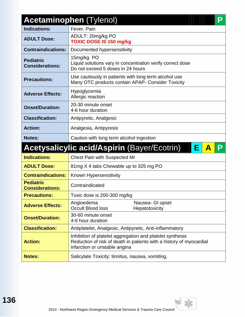
2014 - Northwest Region Emergency Medical Services & Trauma Care Council
136
Acetaminophen (Tylenol) P Indications: Fever, Pain
ADULT Dose: ADULT: 20mg/kg PO TOXIC DOSE IS 150 mg/kg
Contraindications: Documented hypersensitivity
Pediatric Considerations:
15mg/kg PO Liquid solutions vary in concentration verify correct dose Do not exceed 5 doses in 24 hours
Precautions: Use cautiously in patients with long term alcohol use Many OTC products contain APAP- Consider Toxicity
Adverse Effects: Hypoglycemia Allergic reaction
Onset/Duration: 20-30 minute onset 4-6 hour duration
Classification: Antipyretic, Analgesic
Action: Analgesia, Antipyresis
Notes: Caution with long term alcohol ingestion
Acetysalicylic acid/Aspirin (Bayer/Ecotrin) E A P Indications: Chest Pain with Suspected MI
ADULT Dose: 81mg X 4 tabs Chewable up to 325 mg PO
Contraindications: Known Hypersensitivity
Pediatric Considerations:
Contraindicated
Precautions: Toxic dose is 200-300 mg/kg
Adverse Effects: Angioedema Nausea- GI upset Occult Blood loss Hepatotoxicity
Onset/Duration: 30-60 minute onset 4-6 hour duration
Classification: Antiplatelet, Analgesic, Antipyretic, Anti-inflammatory
Action: Inhibition of platelet aggregation and platelet synthesis Reduction of risk of death in patients with a history of myocardial infarction or unstable angina
Notes: Salicylate Toxicity: tinnitus, nausea, vomiting,
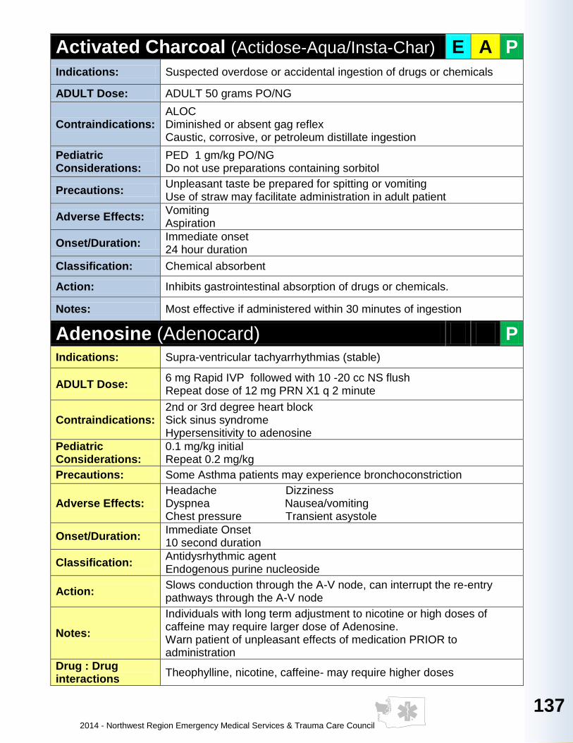
2014 - Northwest Region Emergency Medical Services & Trauma Care Council
137
137
137
137
137
13
7
13
7
137
13
7
13
7
137
137
Activated Charcoal (Actidose-Aqua/Insta-Char) E A P
Indications: Suspected overdose or accidental ingestion of drugs or chemicals
ADULT Dose: ADULT 50 grams PO/NG
Contraindications: ALOC Diminished or absent gag reflex Caustic, corrosive, or petroleum distillate ingestion
Pediatric Considerations:
PED 1 gm/kg PO/NG Do not use preparations containing sorbitol
Precautions: Unpleasant taste be prepared for spitting or vomiting Use of straw may facilitate administration in adult patient
Adverse Effects: Vomiting Aspiration
Onset/Duration: Immediate onset 24 hour duration
Classification: Chemical absorbent
Action: Inhibits gastrointestinal absorption of drugs or chemicals.
Notes: Most effective if administered within 30 minutes of ingestion
Adenosine (Adenocard) P Indications: Supra-ventricular tachyarrhythmias (stable)
ADULT Dose: 6 mg Rapid IVP followed with 10 -20 cc NS flush Repeat dose of 12 mg PRN X1 q 2 minute
Contraindications: 2nd or 3rd degree heart block Sick sinus syndrome Hypersensitivity to adenosine
Pediatric Considerations:
0.1 mg/kg initial Repeat 0.2 mg/kg
Precautions: Some Asthma patients may experience bronchoconstriction
Adverse Effects: Headache Dizziness Dyspnea Nausea/vomiting Chest pressure Transient asystole
Onset/Duration: Immediate Onset 10 second duration
Classification: Antidysrhythmic agent Endogenous purine nucleoside
Action: Slows conduction through the A-V node, can interrupt the re-entry pathways through the A-V node
Notes:
Individuals with long term adjustment to nicotine or high doses of caffeine may require larger dose of Adenosine. Warn patient of unpleasant effects of medication PRIOR to administration
Drug : Drug interactions
Theophylline, nicotine, caffeine- may require higher doses
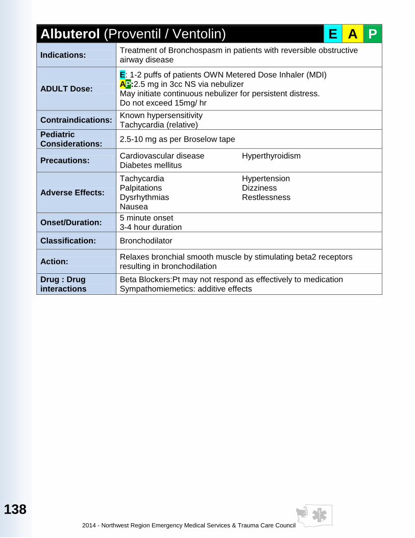
2014 - Northwest Region Emergency Medical Services & Trauma Care Council
138
Albuterol (Proventil / Ventolin) E A P
Indications: Treatment of Bronchospasm in patients with reversible obstructive airway disease
ADULT Dose:
E: 1-2 puffs of patients OWN Metered Dose Inhaler (MDI) AP:2.5 mg in 3cc NS via nebulizer May initiate continuous nebulizer for persistent distress. Do not exceed 15mg/ hr
Contraindications: Known hypersensitivity Tachycardia (relative)
Pediatric Considerations:
2.5-10 mg as per Broselow tape
Precautions: Cardiovascular disease Hyperthyroidism Diabetes mellitus
Adverse Effects:
Tachycardia Hypertension Palpitations Dizziness Dysrhythmias Restlessness Nausea
Onset/Duration: 5 minute onset 3-4 hour duration
Classification: Bronchodilator
Action: Relaxes bronchial smooth muscle by stimulating beta2 receptors resulting in bronchodilation
Drug : Drug interactions
Beta Blockers:Pt may not respond as effectively to medication Sympathomiemetics: additive effects
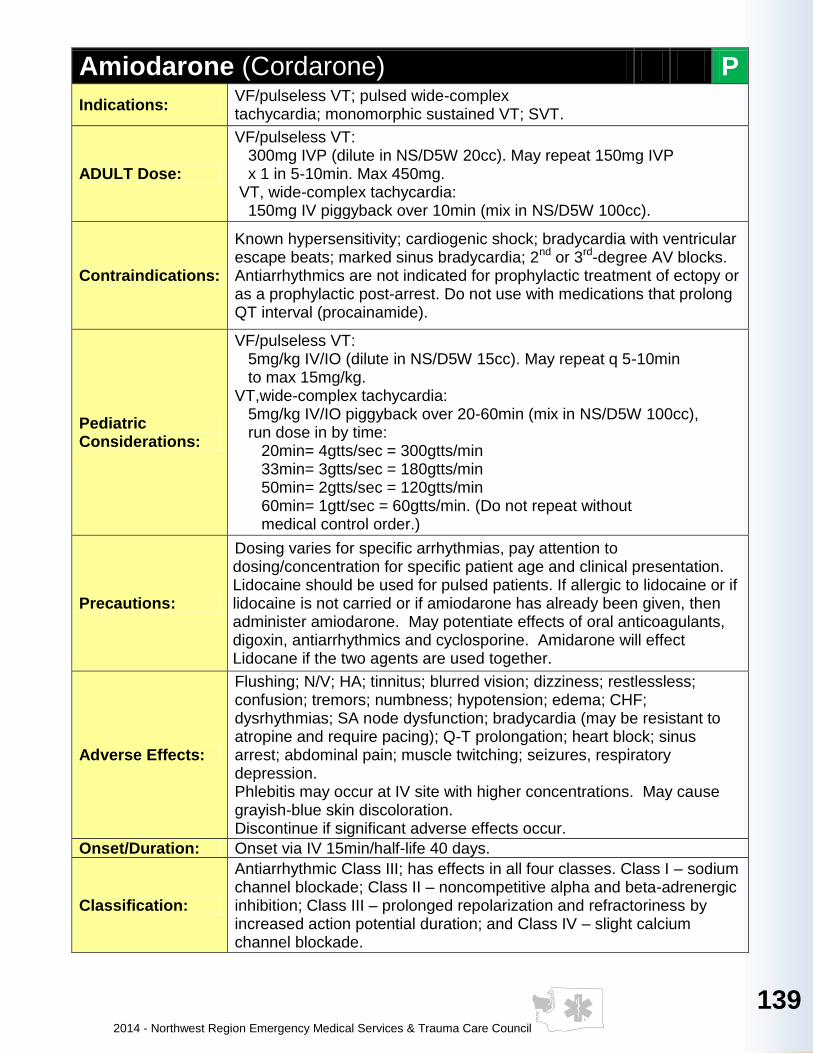
2014 - Northwest Region Emergency Medical Services & Trauma Care Council
139
139
139
139
139
13
9
13
9
139
13
9
13
9
139
139
Amiodarone (Cordarone) P
Indications: VF/pulseless VT; pulsed wide-complex tachycardia; monomorphic sustained VT; SVT.
ADULT Dose:
VF/pulseless VT: 300mg IVP (dilute in NS/D5W 20cc). May repeat 150mg IVP x 1 in 5-10min. Max 450mg. VT, wide-complex tachycardia: 150mg IV piggyback over 10min (mix in NS/D5W 100cc).
Contraindications:
Known hypersensitivity; cardiogenic shock; bradycardia with ventricular escape beats; marked sinus bradycardia; 2nd or 3rd-degree AV blocks. Antiarrhythmics are not indicated for prophylactic treatment of ectopy or as a prophylactic post-arrest. Do not use with medications that prolong QT interval (procainamide).
Pediatric Considerations:
VF/pulseless VT: 5mg/kg IV/IO (dilute in NS/D5W 15cc). May repeat q 5-10min to max 15mg/kg. VT,wide-complex tachycardia: 5mg/kg IV/IO piggyback over 20-60min (mix in NS/D5W 100cc), run dose in by time: 20min= 4gtts/sec = 300gtts/min 33min= 3gtts/sec = 180gtts/min 50min= 2gtts/sec = 120gtts/min 60min= 1gtt/sec = 60gtts/min. (Do not repeat without medical control order.)
Precautions:
Dosing varies for specific arrhythmias, pay attention to dosing/concentration for specific patient age and clinical presentation. Lidocaine should be used for pulsed patients. If allergic to lidocaine or if lidocaine is not carried or if amiodarone has already been given, then administer amiodarone. May potentiate effects of oral anticoagulants, digoxin, antiarrhythmics and cyclosporine. Amidarone will effect Lidocane if the two agents are used together.
Adverse Effects:
Flushing; N/V; HA; tinnitus; blurred vision; dizziness; restlessless; confusion; tremors; numbness; hypotension; edema; CHF; dysrhythmias; SA node dysfunction; bradycardia (may be resistant to atropine and require pacing); Q-T prolongation; heart block; sinus arrest; abdominal pain; muscle twitching; seizures, respiratory depression. Phlebitis may occur at IV site with higher concentrations. May cause grayish-blue skin discoloration. Discontinue if significant adverse effects occur.
Onset/Duration: Onset via IV 15min/half-life 40 days.
Classification:
Antiarrhythmic Class III; has effects in all four classes. Class I – sodium channel blockade; Class II – noncompetitive alpha and beta-adrenergic inhibition; Class III – prolonged repolarization and refractoriness by increased action potential duration; and Class IV – slight calcium channel blockade.
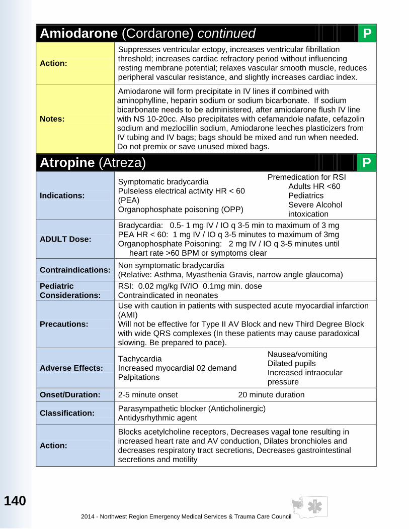
2014 - Northwest Region Emergency Medical Services & Trauma Care Council
140
Amiodarone (Cordarone) continued P
Action:
Suppresses ventricular ectopy, increases ventricular fibrillation threshold; increases cardiac refractory period without influencing resting membrane potential; relaxes vascular smooth muscle, reduces peripheral vascular resistance, and slightly increases cardiac index.
Notes:
Amiodarone will form precipitate in IV lines if combined with aminophylline, heparin sodium or sodium bicarbonate. If sodium bicarbonate needs to be administered, after amiodarone flush IV line with NS 10-20cc. Also precipitates with cefamandole nafate, cefazolin sodium and mezlocillin sodium, Amiodarone leeches plasticizers from IV tubing and IV bags; bags should be mixed and run when needed. Do not premix or save unused mixed bags.
Atropine (Atreza) P
Indications:
Symptomatic bradycardia Pulseless electrical activity HR < 60 (PEA) Organophosphate poisoning (OPP)
Premedication for RSI Adults HR <60 Pediatrics Severe Alcohol intoxication
ADULT Dose:
Bradycardia: 0.5- 1 mg IV / IO q 3-5 min to maximum of 3 mg PEA HR < 60: 1 mg IV / IO q 3-5 minutes to maximum of 3mg Organophosphate Poisoning: 2 mg IV / IO q 3-5 minutes until heart rate >60 BPM or symptoms clear
Contraindications: Non symptomatic bradycardia (Relative: Asthma, Myasthenia Gravis, narrow angle glaucoma)
Pediatric Considerations:
RSI: 0.02 mg/kg IV/IO 0.1mg min. dose Contraindicated in neonates
Precautions:
Use with caution in patients with suspected acute myocardial infarction (AMI) Will not be effective for Type II AV Block and new Third Degree Block with wide QRS complexes (In these patients may cause paradoxical slowing. Be prepared to pace).
Adverse Effects: Tachycardia Increased myocardial 02 demand Palpitations
Nausea/vomiting Dilated pupils Increased intraocular pressure
Onset/Duration: 2-5 minute onset 20 minute duration
Classification: Parasympathetic blocker (Anticholinergic) Antidysrhythmic agent
Action:
Blocks acetylcholine receptors, Decreases vagal tone resulting in increased heart rate and AV conduction, Dilates bronchioles and decreases respiratory tract secretions, Decreases gastrointestinal secretions and motility
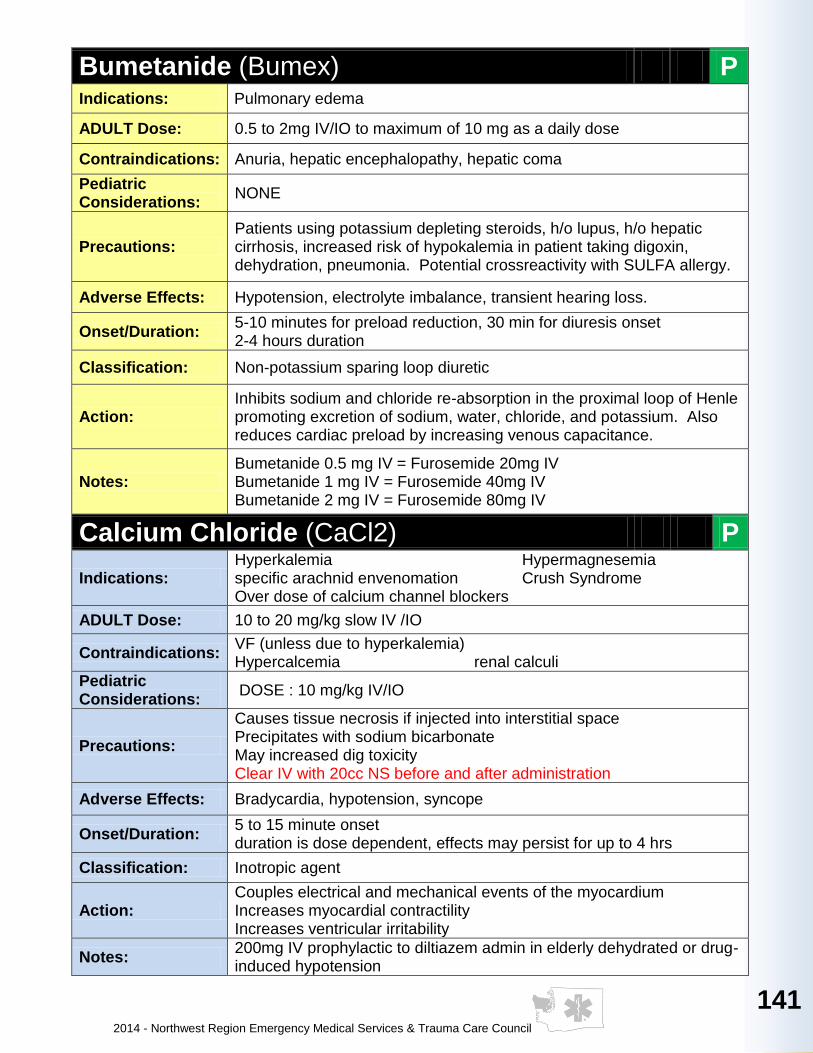
2014 - Northwest Region Emergency Medical Services & Trauma Care Council
141
141
141
141
141
14
1
14
1
141
14
1
14
1
141
141
Bumetanide (Bumex) P Indications: Pulmonary edema
ADULT Dose: 0.5 to 2mg IV/IO to maximum of 10 mg as a daily dose
Contraindications: Anuria, hepatic encephalopathy, hepatic coma
Pediatric Considerations:
NONE
Precautions: Patients using potassium depleting steroids, h/o lupus, h/o hepatic cirrhosis, increased risk of hypokalemia in patient taking digoxin, dehydration, pneumonia. Potential crossreactivity with SULFA allergy.
Adverse Effects: Hypotension, electrolyte imbalance, transient hearing loss.
Onset/Duration: 5-10 minutes for preload reduction, 30 min for diuresis onset 2-4 hours duration
Classification: Non-potassium sparing loop diuretic
Action: Inhibits sodium and chloride re-absorption in the proximal loop of Henle promoting excretion of sodium, water, chloride, and potassium. Also reduces cardiac preload by increasing venous capacitance.
Notes: Bumetanide 0.5 mg IV = Furosemide 20mg IV Bumetanide 1 mg IV = Furosemide 40mg IV Bumetanide 2 mg IV = Furosemide 80mg IV
Calcium Chloride (CaCl2) P
Indications: Hyperkalemia Hypermagnesemia specific arachnid envenomation Crush Syndrome Over dose of calcium channel blockers
ADULT Dose: 10 to 20 mg/kg slow IV /IO
Contraindications: VF (unless due to hyperkalemia) Hypercalcemia renal calculi
Pediatric Considerations:
DOSE : 10 mg/kg IV/IO
Precautions:
Causes tissue necrosis if injected into interstitial space Precipitates with sodium bicarbonate May increased dig toxicity Clear IV with 20cc NS before and after administration
Adverse Effects: Bradycardia, hypotension, syncope
Onset/Duration: 5 to 15 minute onset duration is dose dependent, effects may persist for up to 4 hrs
Classification: Inotropic agent
Action: Couples electrical and mechanical events of the myocardium Increases myocardial contractility Increases ventricular irritability
Notes: 200mg IV prophylactic to diltiazem admin in elderly dehydrated or drug-induced hypotension
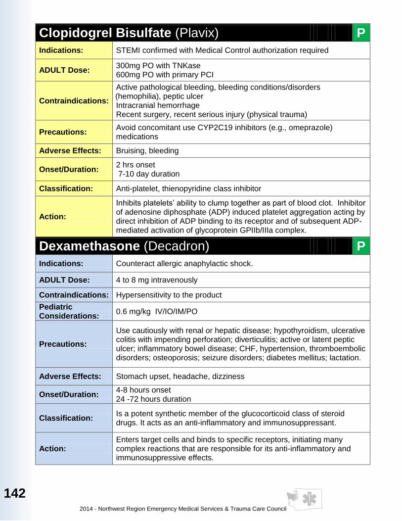
2014 - Northwest Region Emergency Medical Services & Trauma Care Council
142
Clopidogrel Bisulfate (Plavix) P
Indications: STEMI confirmed with Medical Control authorization required
ADULT Dose: 300mg PO with TNKase 600mg PO with primary PCI
Contraindications:
Active pathological bleeding, bleeding conditions/disorders (hemophilia), peptic ulcer Intracranial hemorrhage Recent surgery, recent serious injury (physical trauma)
Precautions: Avoid concomitant use CYP2C19 inhibitors (e.g., omeprazole) medications
Adverse Effects: Bruising, bleeding
Onset/Duration: 2 hrs onset 7-10 day duration
Classification: Anti-platelet, thienopyridine class inhibitor
Action:
Inhibits platelets’ ability to clump together as part of blood clot. Inhibitor of adenosine diphosphate (ADP) induced platelet aggregation acting by direct inhibition of ADP binding to its receptor and of subsequent ADP-mediated activation of glycoprotein GPIIb/IIIa complex.
Dexamethasone (Decadron) P
Indications: Counteract allergic anaphylactic shock.
ADULT Dose: 4 to 8 mg intravenously
Contraindications: Hypersensitivity to the product
Pediatric Considerations:
0.6 mg/kg IV/IO/IM/PO
Precautions:
Use cautiously with renal or hepatic disease; hypothyroidism, ulcerative colitis with impending perforation; diverticulitis; active or latent peptic ulcer; inflammatory bowel disease; CHF, hypertension, thromboembolic disorders; osteoporosis; seizure disorders; diabetes mellitus; lactation.
Adverse Effects: Stomach upset, headache, dizziness
Onset/Duration: 4-8 hours onset 24 -72 hours duration
Classification: Is a potent synthetic member of the glucocorticoid class of steroid drugs. It acts as an anti-inflammatory and immunosuppressant.
Action: Enters target cells and binds to specific receptors, initiating many complex reactions that are responsible for its anti-inflammatory and immunosuppressive effects.

2014 - Northwest Region Emergency Medical Services & Trauma Care Council
143
143
143
143
143
14
3
14
3
143
14
3
14
3
143
143
Dextrose / D50W / D25W (DGlucose) A P
Indications:
Hypoglycemia Hypoglycemia Hyperkalemia with concurrent insulin administration Altered level of consciousness due to suspected or confirmed hypoglycemia
ADULT Dose: 12.5 to 25 g IV / IO repeat dose to maximum of 50 g Consider administration with 100 mg of thiamine
Contraindications: Hyperglycemia
Pediatric Considerations:
DOSE: 0.5gm/kg D25% DILUTE D50% 1:1 to D25% with NS
Precautions: Causes tissue necrosis if injected into interstitial space May increase cerebral ischemia in CVA Caution with intracranial hemorrhage
Adverse Effects: Thrombophlebitis Osmotic Diuresis Pulmonary Edema May worsen Wernicke's encephalopathy
Onset/Duration: 30 to 60 seconds onset duration depends on severity of hypoglycemia
Classification: Hyperglycemic agent Hypotonic solution
Action: Provide immediate source of glucose for rapid utilization for cellular metabolism
Notes: Follow with complex carbohydrate if leaving patient at home

2014 - Northwest Region Emergency Medical Services & Trauma Care Council
144
Diazepam (Valium) P
Indications: Major motor seizures, status epilepticus, premedication for painful procedures, combative patients
ADULT Dose:
Sedation and pain management 2-5 mg IV for procedural Seizures 5mg IV over 2 minutes, 10 mg PR, or 2-5 mg IM Eclamptic seizures 2 mg IV q5min for effect or 10 mg PR Nerve Agent Exposure with Convulsions 2-10 mg IV or 10mg IM titrated to effect
Contraindications: Respiratory depression Hypotension
Pediatric Considerations:
Sedation and pain management 0.1mg/kg IV/IO Seizures 0.1mg/kg IV over 2 minutes, or 0.5 mg/kg PR Nerve Agent Exposure with Convulsions 0.1mg/kg IV/IM MAX DOSES: 5 mg in children and 10 mg in adolescents
Precautions: Inject slowly, do not use small veins. Should not administer to patients in shock, coma or in acute alcoholic intoxication with depression of vital signs. Use caution in elderly patients.
Adverse Effects: Hypotension Respiratory depression
Onset/Duration: IV 1-5 minute onset, 15-60 minute duration IM 15-30 minute onset, 15-60 minute duration
Classification: Benzodiazepine
Action: Suppresses spread of seizure activity through the motor cortex, skeletal muscle relaxant, reduces anxiety and causes sedation
Notes:
Intramuscular administration leads to widely variable absorption and should be avoided if possible. Diastat - EMT's man administer patients own prescription ONLY
A E
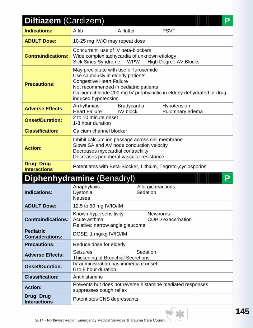
2014 - Northwest Region Emergency Medical Services & Trauma Care Council
145
145
145
145
145
14
5
14
5
145
14
5
14
5
145
145
Diltiazem (Cardizem) P Indications: A fib A flutter PSVT
ADULT Dose: 10-25 mg IV/IO may repeat dose
Contraindications: Concurrent use of IV beta-blockers Wide complex tachycardia of unknown etiology Sick Sinus Syndrome WPW High Degree AV Blocks
Precautions:
May precipitate with use of furosemide Use cautiously in elderly patients Congestive Heart Failure Not recommended in pediatric patients Calcium chloride 200 mg IV prophylactic in elderly dehydrated or drug-induced hypotension
Adverse Effects: Arrhythmias Bradycardia Hypotension Heart Failure AV block Pulomnary edema
Onset/Duration: 2 to 10 minute onset 1-3 hour duration
Classification: Calcium channel blocker
Action:
Inhibit calcium ion passage across cell membrane Slows SA and AV node conduction velocity Decreases myocardial contractility Decreases peripheral vascular resistance
Drug: Drug Interactions
Potentiates with Beta-Blocker, Lithium, Tegretol,cyclosporins
Diphenhydramine (Benadryl) P
Indications: Anaphylaxis Allergic reactions Dystonia Sedation Nausea
ADULT Dose: 12.5 to 50 mg IV/IO/IM
Contraindications: Known hypersensitivity Newborns Acute asthma COPD exacerbation Relative: narrow angle glaucoma
Pediatric Considerations:
DOSE: 1 mg/kg IV/IO/IM
Precautions: Reduce dose for elderly
Adverse Effects: Seizures Sedation Thickening of Bronchial Secretions
Onset/Duration: IV administration has immediate onset 6 to 8 hour duration
Classification: Antihistamine
Action: Prevents but does not reverse histamine mediated responses suppresses cough reflex
Drug: Drug Interactions
Potentiates CNS depressants

2014 - Northwest Region Emergency Medical Services & Trauma Care Council
146
Dopamine (Intropin) P
Indications: Cardiogenic shock Sepsis Vasogenic shock Refactory Hypotension Neurogenic shock Bradycardia
ADULT Dose: Initial 5.0 mcg/kg/min: IVD, then titrate to effect
Contraindications: Tachydysrhythmias Hypovolemic shock
Pediatric Considerations:
1-20mcg/kg/min IVD Epinephrine is pressor of choice in pediatric shock
Precautions: Titrate to blood pressure
Adverse Effects: Angina, Ectopy, Headache, Tachydysrhythmias,VT/VF, Increased myocardial ischemia, AMI, Hypertension
Onset/Duration: Less than 5min. onset Less than 10 minute duration
Classification: Sympathomimetic
Action:
Dopamine has the following dose related effects: 1-2 mcg/kg/min: dilates renal and mesenteric blood vessels (no effect on heart rate or blood pressure) 2-10 mcg/kg/min: beta effects on heart usually increase cardiac output without increasing heart rate. 10-20 mcg/kg/min: alpha peripheral effects cause peripheral vasoconstriction and increased blood pressure. Mix: 400 mg in 250 ml NS or 800 mg in 500ml NS to produce concentration of 1600 mcg/ml. Use 60gtts IV admin set only, all doses in micro drops per minute.
Concentration: 1600 mcg/kg/ml All doses in microgtts/min
Weight (kg) 5 (mcg/kg/min) 10 (mcg/kg/min) 15 (mcg/kg/min) 20 (mcg/kg/min)
50 10 20 30 40
60 10 25 35 45
70 15 25 40 50
80 15 30 45 60
90 15 35 50 70
100 20 35 55 75
110 20 40 60 85

2014 - Northwest Region Emergency Medical Services & Trauma Care Council
147
147
147
147
147
14
7
14
7
147
14
7
14
7
147
147
Droperidol (Inapsine) P
Indications: Chemical restraint, nausea and vomiting, migraine headache
ADULT Dose: 0.625-5.0 mg IV/IM/IN
Contraindications: Renal impairment, hepatic disease
Pediatric Considerations:
0.05 mg/kg IV/IM /IN max 0.1mg
Precautions: In use in elderly, debilitated, and other poor-risk patients; and patients with Parkinson’s disease, hypotension, liver, kidney, cardiac disease bradydysrhythmias.
Adverse Effects: Dystonia, hypotension, tachycardia, apnea, dizziness, anxiety
Onset/Duration: Onset 3-10 min; peak effect in 30 minutes Duration is 2-4 hours
Classification: Antiemetic, Butyrophenone derivative
Action: Produces marked sedation by directly blocking subcortical receptors. Also blocks CNS receptors at the chemoreceptor triggering zone, producing an antiemetic effect.
Notes: Use a reduced dose for elderly and debilitated patients. Consider use of Dipenhydramine with Droperidol. These medications can be given as a single injection.
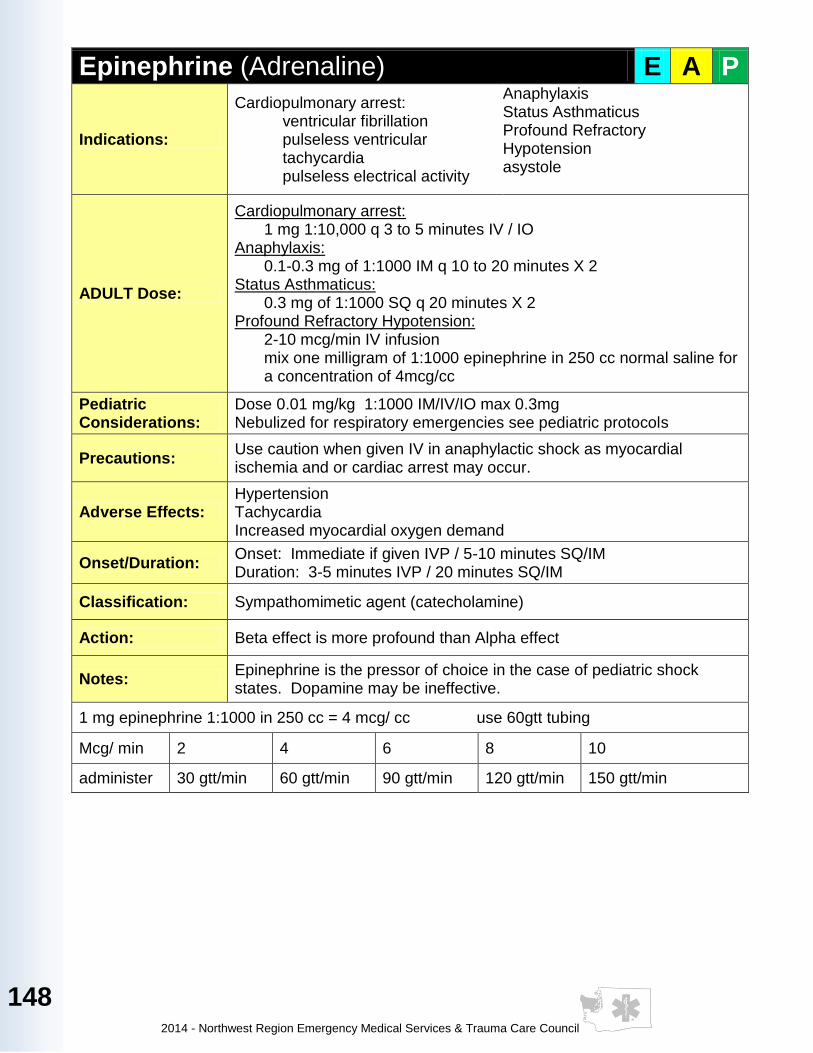
2014 - Northwest Region Emergency Medical Services & Trauma Care Council
148
Epinephrine (Adrenaline) E A P
Indications:
Cardiopulmonary arrest: ventricular fibrillation pulseless ventricular tachycardia pulseless electrical activity
Anaphylaxis Status Asthmaticus Profound Refractory Hypotension asystole
ADULT Dose:
Cardiopulmonary arrest: 1 mg 1:10,000 q 3 to 5 minutes IV / IO
Anaphylaxis: 0.1-0.3 mg of 1:1000 IM q 10 to 20 minutes X 2
Status Asthmaticus: 0.3 mg of 1:1000 SQ q 20 minutes X 2
Profound Refractory Hypotension: 2-10 mcg/min IV infusion mix one milligram of 1:1000 epinephrine in 250 cc normal saline for a concentration of 4mcg/cc
Pediatric Considerations:
Dose 0.01 mg/kg 1:1000 IM/IV/IO max 0.3mg Nebulized for respiratory emergencies see pediatric protocols
Precautions: Use caution when given IV in anaphylactic shock as myocardial ischemia and or cardiac arrest may occur.
Adverse Effects: Hypertension Tachycardia Increased myocardial oxygen demand
Onset/Duration: Onset: Immediate if given IVP / 5-10 minutes SQ/IM Duration: 3-5 minutes IVP / 20 minutes SQ/IM
Classification: Sympathomimetic agent (catecholamine)
Action: Beta effect is more profound than Alpha effect
Notes: Epinephrine is the pressor of choice in the case of pediatric shock states. Dopamine may be ineffective.
1 mg epinephrine 1:1000 in 250 cc = 4 mcg/ cc use 60gtt tubing
Mcg/ min 2 4 6 8 10
administer 30 gtt/min 60 gtt/min 90 gtt/min 120 gtt/min 150 gtt/min
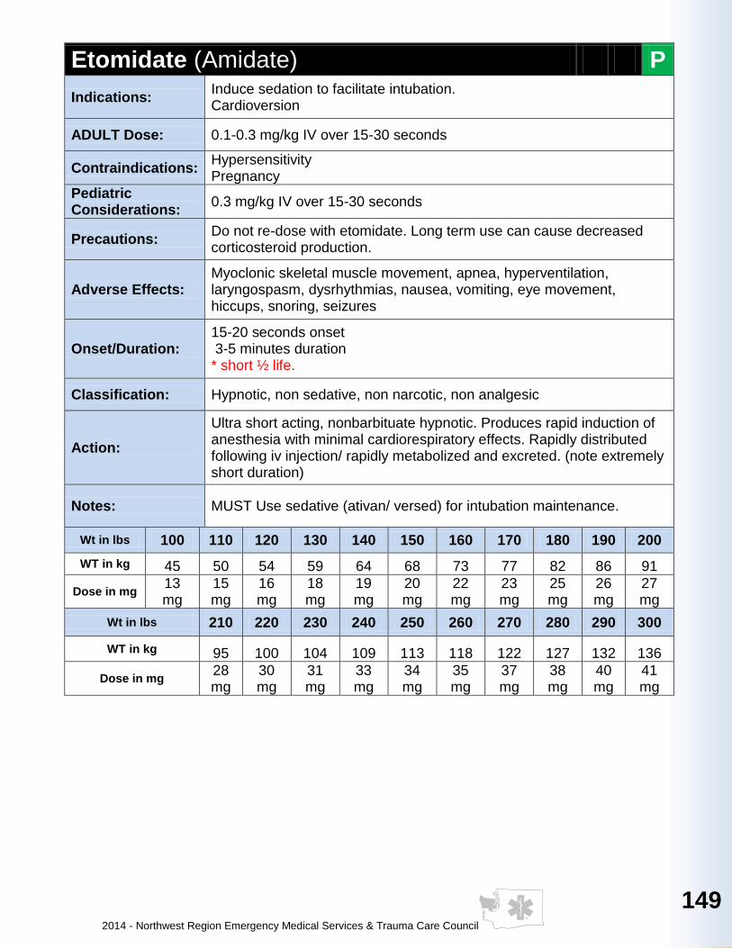
2014 - Northwest Region Emergency Medical Services & Trauma Care Council
149
149
149
149
149
14
9
14
9
149
14
9
14
9
149
149
Etomidate (Amidate) P
Indications: Induce sedation to facilitate intubation. Cardioversion
ADULT Dose: 0.1-0.3 mg/kg IV over 15-30 seconds
Contraindications: Hypersensitivity Pregnancy
Pediatric Considerations:
0.3 mg/kg IV over 15-30 seconds
Precautions: Do not re-dose with etomidate. Long term use can cause decreased corticosteroid production.
Adverse Effects: Myoclonic skeletal muscle movement, apnea, hyperventilation, laryngospasm, dysrhythmias, nausea, vomiting, eye movement, hiccups, snoring, seizures
Onset/Duration: 15-20 seconds onset 3-5 minutes duration * short ½ life.
Classification: Hypnotic, non sedative, non narcotic, non analgesic
Action:
Ultra short acting, nonbarbituate hypnotic. Produces rapid induction of anesthesia with minimal cardiorespiratory effects. Rapidly distributed following iv injection/ rapidly metabolized and excreted. (note extremely short duration)
Notes: MUST Use sedative (ativan/ versed) for intubation maintenance.
Wt in lbs 100 110 120 130 140 150 160 170 180 190 200
WT in kg 45 50 54 59 64 68 73 77 82 86 91
Dose in mg 13 mg
15 mg
16 mg
18 mg
19 mg
20 mg
22 mg
23 mg
25 mg
26 mg
27 mg
Wt in lbs 210 220 230 240 250 260 270 280 290 300
WT in kg 95 100 104 109 113 118 122 127 132 136
Dose in mg 28 mg
30 mg
31 mg
33 mg
34 mg
35 mg
37 mg
38 mg
40 mg
41 mg

2014 - Northwest Region Emergency Medical Services & Trauma Care Council
150
Fentanyl (Sublimaze) P Indications: Analgesia, Pulmonary Edema, Acute MI
ADULT Dose: 25-50 mcg IV/IO/IM
Contraindications: Known hypersensitivity
Pediatric Considerations:
DOSE: 1-2 mcg/kg IV/IM
Precautions: Head injuries, COPD, ALOC, Hypotension
Adverse Effects: CNS depression, resp. depression, hallucinations, hypotension, hypertension, arrythmias, n/v, constipation, Chest wall rigidity
Onset/Duration: Onset- 1-2min IV, 7-15min IM Duration- ½ - 1hr IV, 1-2hr IM
Classification: Opiod agonist/ narcotic analgesic
Action: Binds to opiate receptors as an agonist to alter pt’s perception of painful stimuli.
Notes: CNS and resp. depressant effects are similar to Morphine. Drug has little hypnotic activity and rarely causes histamine release.
Furosemide (Lasix) P
Indications: Pulmonary edema
ADULT Dose: 40mg-80mg (or double pt’s daily dose up to 100mg) slowly * can be dosed at 0.5-1.0 mg/kg
Contraindications: Dehydration/ hypovolemia, hypokalemia, hepatic coma
Pediatric Considerations:
2 mg/kg
Precautions: Pt’s using potassium depleting steroids, hx of lupus, hx of hepatic cirrhosis, increased risk of hypokalemia in pt’s taking digoxin, dehydration or pneunomia
Adverse Effects: Hypotension, electrolyte imbalance, transient hearing loss
Onset/Duration: Onset 5 min for preload reduction, 30 min for diuresis. Duration ~2 hours.
Classification: Non-potassium sparing loop diuretic
Action: Inhibits sodium and chloride re-absorption in the proximal loop of henle promoting excretion of sodium, water, chloride, and potassium. Also reduces cardiac preload by increasing venous capacitance.

2014 - Northwest Region Emergency Medical Services & Trauma Care Council
151
151
151
151
151
15
1
15
1
151
15
1
15
1
151
151
Glucagon P
Indications:
Hypoglycemia, Beta-blocker OD, Calcium channel blocker OD Symptoms of esophageal obstruction
ADULT Dose: Hypoglycemia- 1.0mg IM / IN Ca++ and beta-blocker OD- 3-5mg IV/ IM / IN
Contraindications: None in emergency setting
Pediatric Considerations:
Dose: 0.1mg/kg up to 1mg IM
Precautions: Do not dilute with saline solutions, will form a precipitate.
Adverse Effects: Nausea & Vomiting, hyperglycemia, hypersensitivity reactions
Onset/Duration: Onset is 5-20 minutes, peak effect at 30 minutes. Duration is 1-1.5 hours
Classification: Polypeptide hormone
Action:
Accelerates liver glycogenolysis and inhibits glycogen synthetase resulting in blood glucose elevation. Stimulates hepatic gluconeogenesis and causes an inotropic myocardial effect. Relaxes GI smooth muscle
Notes: Reconstitute powdered solution with supplied dilutent only If given IV, flush line with D-5% instead of NS solution.
Glucose Oral (Glucose Paste) E A P
Indications: Hypoglycemia in conscious pt that is able to swallow.
ADULT Dose: One tube PO- between cheek and gum
Contraindications: Unconsciousness, inability to swallow, hyperglycemia
Pediatric Considerations:
One tube PO
Precautions: Not tasty, watch for spitting
Adverse Effects: Choking if not properly administered
Classification: Carbohydrate
Action: Rapidly metabolized source of calories in pt’s with inadequate oral intake.
Notes: Perform glucose check before and after administration of Glucose. Follow with complex carbohydrate if leaving patient at home.

2014 - Northwest Region Emergency Medical Services & Trauma Care Council
152
Heparin Sodium P Indications: STEMI confirmed with Medical Control authorization required
ADULT Dose: 60U/kg IV bolus to max 4,000U (if >100kg max 5,000U); 12U/kg/hr IV drip to max 1,000U/hr
Contraindications: Allergy to heparin, thrombocytopenia, hemophilia
Precautions:
Increased risk of bleeding w/ bleeding/clotting disorders (hemophilia), GI ulceration, bacterial endocarditis Recent surgery Derived from porcine intestinal mucosa, avoid if allergic to pork
Adverse Effects: Bleeding, Heparin induced thrombocytopenia, Hyperkalemia (5-10%)
Onset/Duration: onset immediate duration 1.5 hours
Classification: Injectable anticoagulant
Action: Binds to and activates antithrombin III which binds to and inactivates thrombin. This inhibits further clot formation.
Notes: Invert infusion solution periodically to prevent pooling Do not mix in same line with droperidol
Hydromorphone (Dilaudid) P Indications: Moderate to severe pain.
ADULT Dose: 0.5mg IV q 3-5 min total of 4mg (caution in elderly) 1-2mg IM
Contraindications: Hypotension SBP <110 CNS depression Respiratory depression RR<12 Head Injury Current nausea or vomiting (prior to anti-emetics)
Pediatric Considerations:
0.015 mg/kg IV/IM
Precautions: Caution should be used in patients who have taken other central nervous depressants, narcotic analegesics, sedative/hypnotics, or tricyclic antidepressants.
Adverse Effects:
Respiratory depression Increased sedation Headache Abdominal pain Decreased LOC Impaired mental status Nausea/vomiting
Onset/Duration: Onset: IV – Immediate IM – 7-15 minutes Duration: 4-5 hours
Classification: Narcotic analgesic, Opiate
Action: Decrease sensitivity to pain, Stimulates variety of opioid receptors
Notes: This is a strong narcotic. Start with low doses given slowly and add additional low doses as needed. One mg of hydromorphone is equal to 7mg of morphine
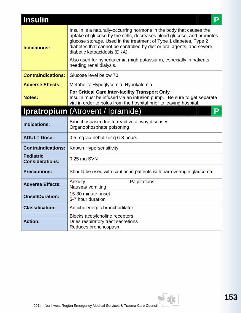
2014 - Northwest Region Emergency Medical Services & Trauma Care Council
153
153
153
153
153
15
3
15
3
153
15
3
15
3
153
153
Insulin P
Indications:
Insulin is a naturally-occurring hormone in the body that causes the uptake of glucose by the cells, decreases blood glucose, and promotes glucose storage. Used in the treatment of Type 1 diabetes, Type 2 diabetes that cannot be controlled by diet or oral agents, and severe diabetic ketoacidosis (DKA).
Also used for hyperkalemia (high potassium), especially in patients needing renal dialysis.
Contraindications: Glucose level below 70
Adverse Effects: Metabolic: Hypoglycemia, Hypokalemia
Notes: For Critical Care Inter-facility Transport Only Insulin must be infused via an infusion pump. Be sure to get separate vial in order to bolus from the hospital prior to leaving hospital.
Ipratropium (Atrovent / Ipramide) P
Indications: Bronchospasm due to reactive airway diseases Organophosphate poisoning
ADULT Dose: 0.5 mg via nebulizer q 6-8 hours
Contraindications: Known Hypersensitivity
Pediatric Considerations:
0.25 mg SVN
Precautions: Should be used with caution in patients with narrow-angle glaucoma.
Adverse Effects: Anxiety Palpitations Nausea/ vomiting
Onset/Duration: 15-30 minute onset 5-7 hour duration
Classification: Anticholenergic bronchodilator
Action: Blocks acetylcholine receptors Dries respiratory tract secretions Reduces bronchospasm
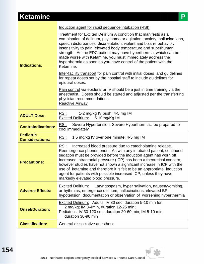
2014 - Northwest Region Emergency Medical Services & Trauma Care Council
154
Ketamine P
Indications:
Induction agent for rapid sequence intubation (RSI)
Treatment for Excited Delirium A condition that manifests as a combination of delirium, psychomotor agitation, anxiety, hallucinations, speech disturbances, disorientation, violent and bizarre behavior, insensitivity to pain, elevated body temperature and superhuman strength. As the EDC patient may have hyperthermia, which can be made worse with Ketamine, you must immediately address the hyperthermia as soon as you have control of the patient with the Ketamine.
Inter-facility transport for pain control with initial doses and guidelines for repeat doses set by the hospital staff to include guidelines for epidural doses.
Pain control via epidural or IV should be a just in time training via the anesthetist. Doses should be started and adjusted per the transferring physician recommendations. Reactive Airway
ADULT Dose: RSI: 1-2 mg/kg IV push; 4-5 mg IM Excited Delirium: 5-10mg/Kg IM
Contraindications: RSI: Severe Hypertension, Severe Hyperthermia…be prepared to cool immediately
Pediatric Considerations:
RSI: 1.5 mg/kg IV over one minute; 4-5 mg IM
Precautions:
RSI: Increased blood pressure due to catecholamine release. Reemergence phenomenon. As with any intubated patient, continued sedation must be provided before the induction agent has worn off. Increased intracranial pressure (ICP) has been a theoretical concern, however studies have not shown a significant increase in ICP with the use of ketamine and therefore it is felt to be an appropriate induction agent for patients with possible increased ICP, unless they have markedly elevated blood pressure.
Adverse Effects: Excited Delirium: Laryngospasm, hyper salivation, nausea/vomiting, arrhythmias, emergence delirium, hallucinations, elevated BP, hypotension, documentation or observation of worsening hyperthermia
Onset/Duration:
Excited Delirium: Adults: IV 30 sec; duration 5-10 min for 2 mg/kg; IM 3-4min, duration 12-25 min; Pediatrics: IV 30-120 sec; duration 20-60 min; IM 5-10 min, duration 30-90 min
Classification: General dissociative anesthetic
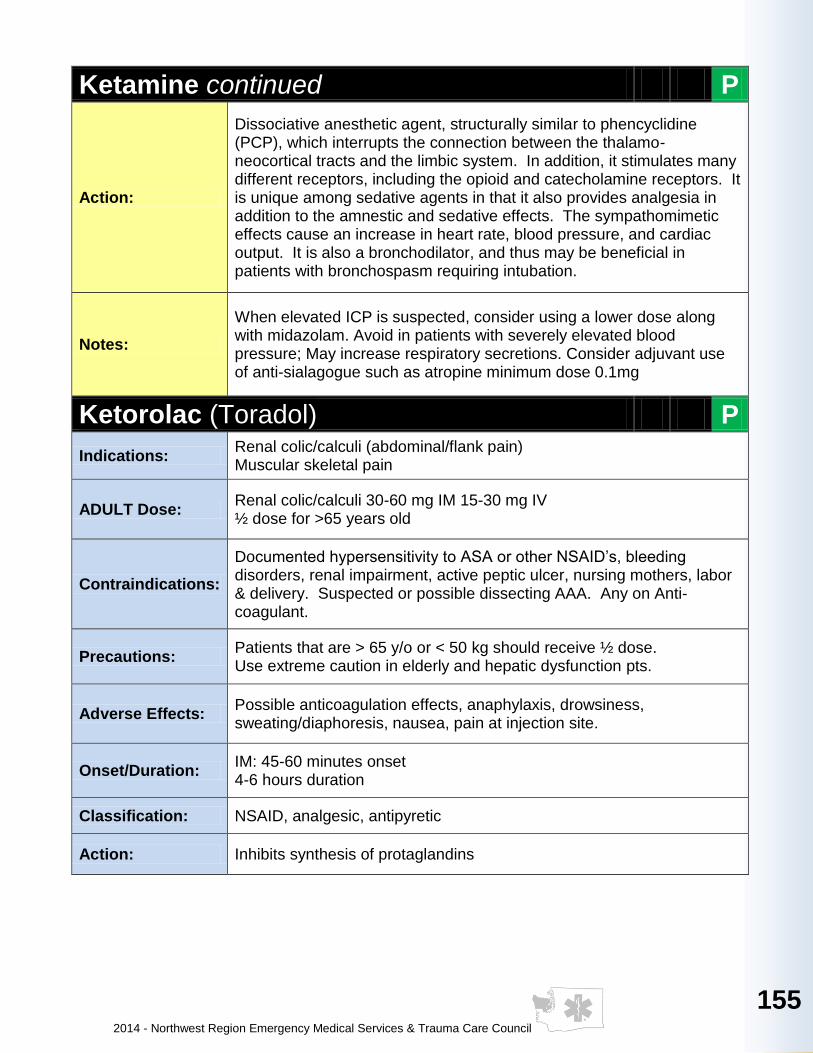
2014 - Northwest Region Emergency Medical Services & Trauma Care Council
155
155
155
155
155
15
5
15
5
155
15
5
15
5
155
155
Ketamine continued P
Action:
Dissociative anesthetic agent, structurally similar to phencyclidine (PCP), which interrupts the connection between the thalamo-neocortical tracts and the limbic system. In addition, it stimulates many different receptors, including the opioid and catecholamine receptors. It is unique among sedative agents in that it also provides analgesia in addition to the amnestic and sedative effects. The sympathomimetic effects cause an increase in heart rate, blood pressure, and cardiac output. It is also a bronchodilator, and thus may be beneficial in patients with bronchospasm requiring intubation.
Notes:
When elevated ICP is suspected, consider using a lower dose along with midazolam. Avoid in patients with severely elevated blood pressure; May increase respiratory secretions. Consider adjuvant use of anti-sialagogue such as atropine minimum dose 0.1mg
Ketorolac (Toradol) P
Indications: Renal colic/calculi (abdominal/flank pain) Muscular skeletal pain
ADULT Dose: Renal colic/calculi 30-60 mg IM 15-30 mg IV ½ dose for >65 years old
Contraindications:
Documented hypersensitivity to ASA or other NSAID’s, bleeding disorders, renal impairment, active peptic ulcer, nursing mothers, labor & delivery. Suspected or possible dissecting AAA. Any on Anti-coagulant.
Precautions: Patients that are > 65 y/o or < 50 kg should receive ½ dose. Use extreme caution in elderly and hepatic dysfunction pts.
Adverse Effects: Possible anticoagulation effects, anaphylaxis, drowsiness, sweating/diaphoresis, nausea, pain at injection site.
Onset/Duration: IM: 45-60 minutes onset 4-6 hours duration
Classification: NSAID, analgesic, antipyretic
Action: Inhibits synthesis of protaglandins
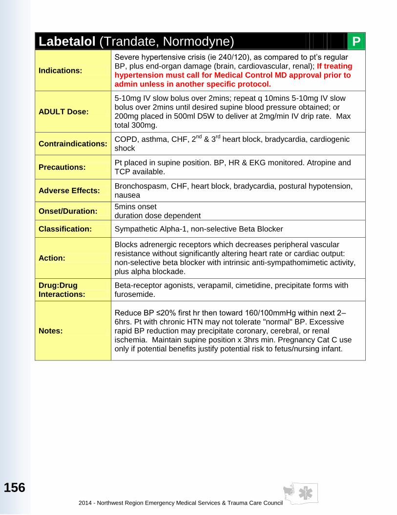
2014 - Northwest Region Emergency Medical Services & Trauma Care Council
156
Labetalol (Trandate, Normodyne) P
Indications:
Severe hypertensive crisis (ie 240/120), as compared to pt’s regular BP, plus end-organ damage (brain, cardiovascular, renal); If treating hypertension must call for Medical Control MD approval prior to admin unless in another specific protocol.
ADULT Dose:
5-10mg IV slow bolus over 2mins; repeat q 10mins 5-10mg IV slow bolus over 2mins until desired supine blood pressure obtained; or 200mg placed in 500ml D5W to deliver at 2mg/min IV drip rate. Max total 300mg.
Contraindications: COPD, asthma, CHF, 2nd & 3rd heart block, bradycardia, cardiogenic shock
Precautions: Pt placed in supine position. BP, HR & EKG monitored. Atropine and TCP available.
Adverse Effects: Bronchospasm, CHF, heart block, bradycardia, postural hypotension, nausea
Onset/Duration: 5mins onset duration dose dependent
Classification: Sympathetic Alpha-1, non-selective Beta Blocker
Action:
Blocks adrenergic receptors which decreases peripheral vascular resistance without significantly altering heart rate or cardiac output: non-selective beta blocker with intrinsic anti-sympathomimetic activity, plus alpha blockade.
Drug:Drug Interactions:
Beta-receptor agonists, verapamil, cimetidine, precipitate forms with furosemide.
Notes:
Reduce BP ≤20% first hr then toward 160/100mmHg within next 2–6hrs. Pt with chronic HTN may not tolerate "normal" BP. Excessive rapid BP reduction may precipitate coronary, cerebral, or renal ischemia. Maintain supine position x 3hrs min. Pregnancy Cat C use only if potential benefits justify potential risk to fetus/nursing infant.
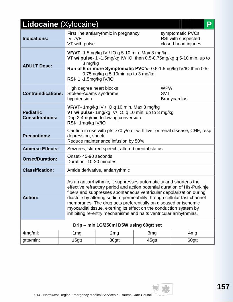
2014 - Northwest Region Emergency Medical Services & Trauma Care Council
157
157
157
157
157
15
7
15
7
157
15
7
15
7
157
157
Lidocaine (Xylocaine) P
Indications: First line antiarrythmic in pregnancy VT/VF VT with pulse
symptomatic PVCs RSI with suspected closed head injuries
ADULT Dose:
VF/VT- 1.5mg/kg IV / IO q 5-10 min. Max 3 mg/kg. VT w/ pulse- 1 -1.5mg/kg IV/ IO, then 0.5-0.75mg/kg q 5-10 min. up to
3 mg/kg Run of 6 or more Symptomatic PVC’s- 0.5-1.5mg/kg IV/IO then 0.5-
0.75mg/kg q 5-10min up to 3 mg/kg. RSI- 1 -1.5mg/kg IV/IO
Contraindications: High degree heart blocks Stokes-Adams syndrome hypotension
WPW SVT Bradycardias
Pediatric Considerations:
VF/VT- 1mg/kg IV / IO q 10 min. Max 3 mg/kg VT w/ pulse- 1mg/kg IV/ IO, q 10 min. up to 3 mg/kg Drip 2-4mg/min following conversion RSI- 1mg/kg IV/IO
Precautions: Caution in use with pts >70 y/o or with liver or renal disease, CHF, resp depression, shock. Reduce maintenance infusion by 50%
Adverse Effects: Seizures, slurred speech, altered mental status
Onset/Duration: Onset- 45-90 seconds Duration- 10-20 minutes
Classification: Amide derivative, antiarrythmic
Action:
As an antiarrhythmic, it suppresses automaticity and shortens the effective refractory period and action potential duration of His-Purkinje fibers and suppresses spontaneous ventricular depolarization during diastole by altering sodium permeability through cellular fast channel membranes. The drug acts preferentially on diseased or ischemic myocardial tissue, exerting its effect on the conduction system by inhibiting re-entry mechanisms and halts ventricular arrhythmias.
Drip – mix 1G/250ml D5W using 60gtt set
4mg/ml: 1mg 2mg 3mg 4mg
gtts/min: 15gtt 30gtt 45gtt 60gtt
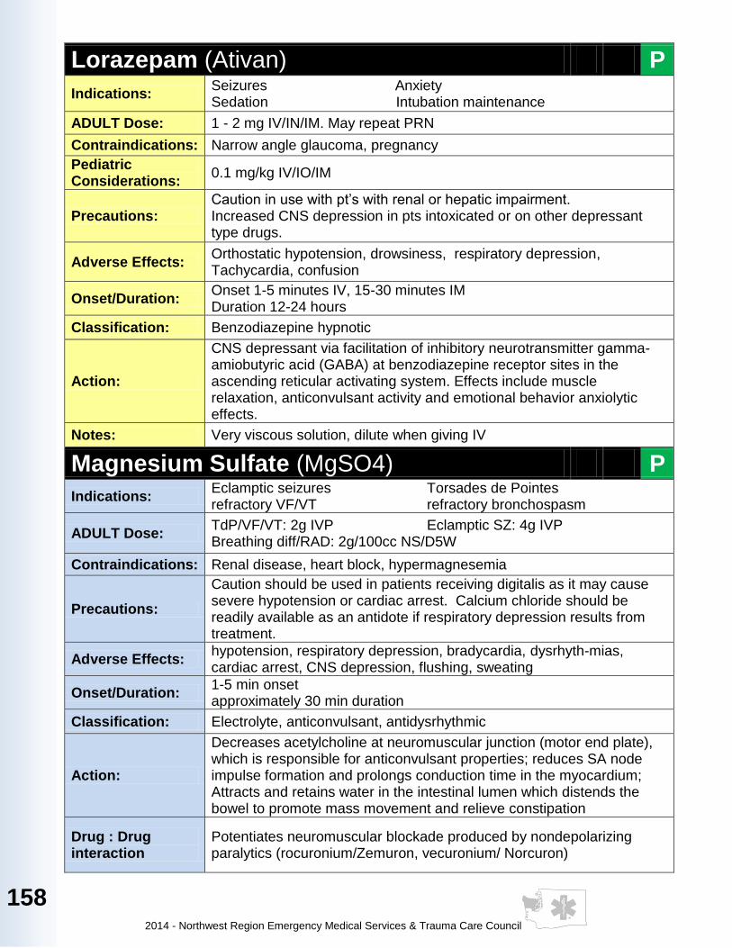
2014 - Northwest Region Emergency Medical Services & Trauma Care Council
158
Lorazepam (Ativan) P
Indications: Seizures Anxiety Sedation Intubation maintenance
ADULT Dose: 1 - 2 mg IV/IN/IM. May repeat PRN
Contraindications: Narrow angle glaucoma, pregnancy
Pediatric Considerations:
0.1 mg/kg IV/IO/IM
Precautions: Caution in use with pt’s with renal or hepatic impairment. Increased CNS depression in pts intoxicated or on other depressant type drugs.
Adverse Effects: Orthostatic hypotension, drowsiness, respiratory depression, Tachycardia, confusion
Onset/Duration: Onset 1-5 minutes IV, 15-30 minutes IM Duration 12-24 hours
Classification: Benzodiazepine hypnotic
Action:
CNS depressant via facilitation of inhibitory neurotransmitter gamma-amiobutyric acid (GABA) at benzodiazepine receptor sites in the ascending reticular activating system. Effects include muscle relaxation, anticonvulsant activity and emotional behavior anxiolytic effects.
Notes: Very viscous solution, dilute when giving IV
Magnesium Sulfate (MgSO4) P
Indications: Eclamptic seizures Torsades de Pointes refractory VF/VT refractory bronchospasm
ADULT Dose: TdP/VF/VT: 2g IVP Eclamptic SZ: 4g IVP Breathing diff/RAD: 2g/100cc NS/D5W
Contraindications: Renal disease, heart block, hypermagnesemia
Precautions:
Caution should be used in patients receiving digitalis as it may cause severe hypotension or cardiac arrest. Calcium chloride should be readily available as an antidote if respiratory depression results from treatment.
Adverse Effects: hypotension, respiratory depression, bradycardia, dysrhyth-mias, cardiac arrest, CNS depression, flushing, sweating
Onset/Duration: 1-5 min onset approximately 30 min duration
Classification: Electrolyte, anticonvulsant, antidysrhythmic
Action:
Decreases acetylcholine at neuromuscular junction (motor end plate), which is responsible for anticonvulsant properties; reduces SA node impulse formation and prolongs conduction time in the myocardium; Attracts and retains water in the intestinal lumen which distends the bowel to promote mass movement and relieve constipation
Drug : Drug interaction
Potentiates neuromuscular blockade produced by nondepolarizing paralytics (rocuronium/Zemuron, vecuronium/ Norcuron)

2014 - Northwest Region Emergency Medical Services & Trauma Care Council
159
159
159
159
159
15
9
15
9
159
15
9
15
9
159
159
Methylprednisolone (Solu-Medrol/Amethapred) P
Indications:
Allergic reaction Anaphylaxis Upper airway burns Reactive airway disease COPD exacerbations History of adrenal insufficiency associated with either serious trauma/illness or shock unresponsive to conventional therapy
ADULT Dose: 125 mg IV / IO / IM
Contraindications: Preterm infants, Newborn, systemic fungal infections
Pediatric Considerations:
2 mg/kg IV / IO / IM
Precautions: Use with caution in patients with G.I. bleeding, diabetes mellitus & severe infection
Adverse Effects: Alkalosis, CHF, headache, hypertension, hypokalemia, seizures, nausea and vomiting
Onset/Duration: Onset: 20 minutes-2 hours Duration: 18-36 hours
Classification: Corticosteroid, glucocorticoid steroid, anti-inflammatory
Action:
Decreases inflammation by depressing migration of polymophonuclear leukocytes and activity of endogenous mediators of inflammation. Potentiates vascular smooth muscle relaxation by beta adrenergic agonists.
Notes: Hypoglycemic responses to insulin and oral hypoglycemic agents may be blunted. Potassium depleting agents may potentiate hypokalemia induced by corticosteroids.
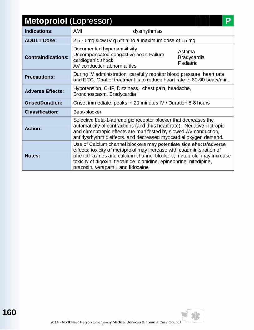
2014 - Northwest Region Emergency Medical Services & Trauma Care Council
160
Metoprolol (Lopressor) P Indications: AMI dysrhythmias
ADULT Dose: 2.5 - 5mg slow IV q 5min; to a maximum dose of 15 mg
Contraindications:
Documented hypersensitivity Uncompensated congestive heart Failure cardiogenic shock AV conduction abnormalities
Asthma Bradycardia Pediatric
Precautions: During IV administration, carefully monitor blood pressure, heart rate, and ECG. Goal of treatment is to reduce heart rate to 60-90 beats/min.
Adverse Effects: Hypotension, CHF, Dizziness, chest pain, headache, Bronchospasm, Bradycardia
Onset/Duration: Onset immediate, peaks in 20 minutes IV / Duration 5-8 hours
Classification: Beta-blocker
Action:
Selective beta-1-adrenergic receptor blocker that decreases the automaticity of contractions (and thus heart rate). Negative inotropic and chronotropic effects are manifested by slowed AV conduction, antidysrhythmic effects, and decreased myocardial oxygen demand.
Notes:
Use of Calcium channel blockers may potentiate side effects/adverse effects; toxicity of metoprolol may increase with coadministration of phenothiazines and calcium channel blockers; metoprolol may increase toxicity of digoxin, flecainide, clonidine, epinephrine, nifedipine, prazosin, verapamil, and lidocaine
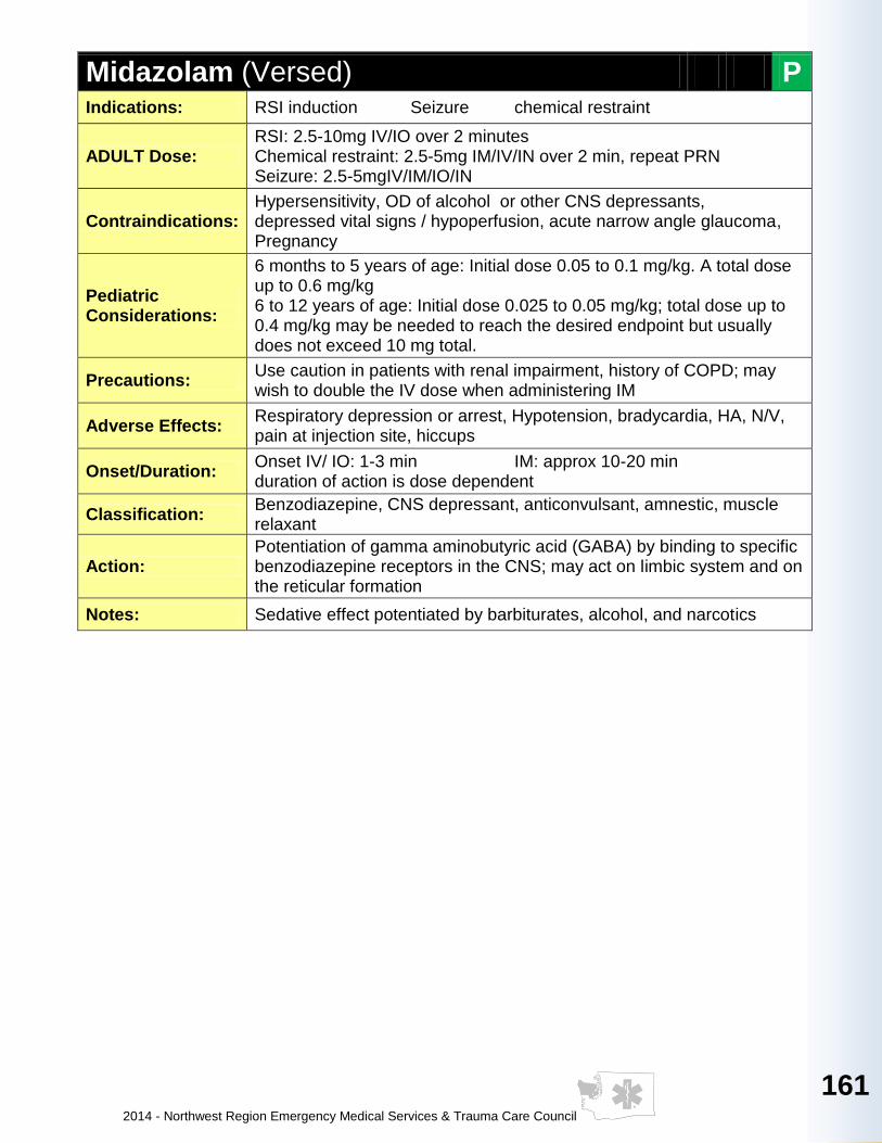
2014 - Northwest Region Emergency Medical Services & Trauma Care Council
161
161
161
161
161
16
1
16
1
161
16
1
16
1
161
161
Midazolam (Versed) P Indications: RSI induction Seizure chemical restraint
ADULT Dose: RSI: 2.5-10mg IV/IO over 2 minutes Chemical restraint: 2.5-5mg IM/IV/IN over 2 min, repeat PRN Seizure: 2.5-5mgIV/IM/IO/IN
Contraindications: Hypersensitivity, OD of alcohol or other CNS depressants, depressed vital signs / hypoperfusion, acute narrow angle glaucoma, Pregnancy
Pediatric Considerations:
6 months to 5 years of age: Initial dose 0.05 to 0.1 mg/kg. A total dose up to 0.6 mg/kg 6 to 12 years of age: Initial dose 0.025 to 0.05 mg/kg; total dose up to 0.4 mg/kg may be needed to reach the desired endpoint but usually does not exceed 10 mg total.
Precautions: Use caution in patients with renal impairment, history of COPD; may wish to double the IV dose when administering IM
Adverse Effects: Respiratory depression or arrest, Hypotension, bradycardia, HA, N/V, pain at injection site, hiccups
Onset/Duration: Onset IV/ IO: 1-3 min IM: approx 10-20 min duration of action is dose dependent
Classification: Benzodiazepine, CNS depressant, anticonvulsant, amnestic, muscle relaxant
Action: Potentiation of gamma aminobutyric acid (GABA) by binding to specific benzodiazepine receptors in the CNS; may act on limbic system and on the reticular formation
Notes: Sedative effect potentiated by barbiturates, alcohol, and narcotics

2014 - Northwest Region Emergency Medical Services & Trauma Care Council
162
Morphine P
Indications: Pain management, Pulmonary edema, Procedural sedation, Analgesia, Acute Myocardial Infarction
ADULT Dose: 2-4 mg IV/In/IM titrated, 1-3 mg q 2 min to 20 mg max
Contraindications: Head injury, exacerbated COPD, depressed respiratory drive, hypotension, ALOC
Pediatric Considerations:
0.1 mg/kg IV/IN/IO
Precautions: Patients with acute bronchial asthma, chronic pulmonary diseases, severe respiratory depression, and pulmonary edema induced by chemical irritants.
Adverse Effects: Respiratory depression, hypotension, ALOC, nausea & vomiting
Onset/Duration: IV immediate onset, peak effect 20 min. IM/SQ 15-30 min. , peak effect 30-60 min. Duration 2-7 hours
Classification: Narcotic analgesic
Action:
Narcotic agonist with activity at u-recptors (supraspinal analgesia, euphoria, respiratory and physical depression), K-receptors (sedation and myosis), and delta-receptors (dysphonia, hallucinations, respiratory and vasomotor stimulation)
Notes: Naloxone and respiratory equipment should be immediately accessible.
Naloxone (Narcan) A P
Indications: Suspected or Known narcotic overdose Altered level of consciousness
ADULT Dose: 0.4 - 2mg IM/IV/IO/IN/SQ/SVN
Contraindications: None in the emergent setting
Pediatric considerations:
Dose : 0.1 mg/kg Max dose 2 mg Use caution in newborns
Precautions:
Rapid reversal of narcotic effects may lead to combative behavior and vomiting May not reverse hypotension For patients with chronic pain issues administer 0.4 mg increments until respirations improve
Adverse Effects: Hypertension , Nausea, Vomiting, Tremors, Dysrhythmias
Onset/Duration: IV/IO immediate SQ/ IM 5-10 minutes 20-30 minute duration
Classification: Narcotic Antagonist
Action: Competitively binds with opiate receptor sites in the CNS
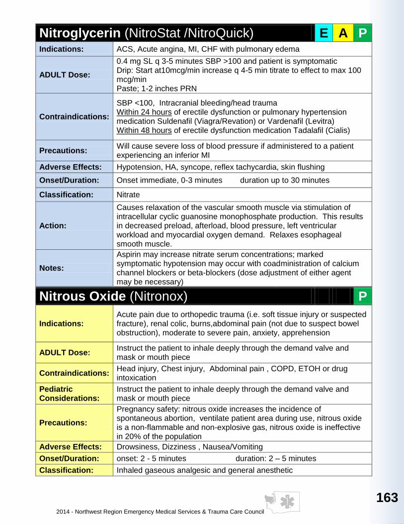
2014 - Northwest Region Emergency Medical Services & Trauma Care Council
163
163
163
163
163
16
3
16
3
163
16
3
16
3
163
163
Nitroglycerin (NitroStat /NitroQuick) E A P Indications: ACS, Acute angina, MI, CHF with pulmonary edema
ADULT Dose:
0.4 mg SL q 3-5 minutes SBP >100 and patient is symptomatic Drip: Start at10mcg/min increase q 4-5 min titrate to effect to max 100 mcg/min Paste; 1-2 inches PRN
Contraindications:
SBP <100, Intracranial bleeding/head trauma Within 24 hours of erectile dysfunction or pulmonary hypertension medication Suldenafil (Viagra/Revation) or Vardenafil (Levitra) Within 48 hours of erectile dysfunction medication Tadalafil (Cialis)
Precautions: Will cause severe loss of blood pressure if administered to a patient experiencing an inferior MI
Adverse Effects: Hypotension, HA, syncope, reflex tachycardia, skin flushing
Onset/Duration: Onset immediate, 0-3 minutes duration up to 30 minutes
Classification: Nitrate
Action:
Causes relaxation of the vascular smooth muscle via stimulation of intracellular cyclic guanosine monophosphate production. This results in decreased preload, afterload, blood pressure, left ventricular workload and myocardial oxygen demand. Relaxes esophageal smooth muscle.
Notes:
Aspirin may increase nitrate serum concentrations; marked symptomatic hypotension may occur with coadministration of calcium channel blockers or beta-blockers (dose adjustment of either agent may be necessary)
Nitrous Oxide (Nitronox) P
Indications: Acute pain due to orthopedic trauma (i.e. soft tissue injury or suspected fracture), renal colic, burns,abdominal pain (not due to suspect bowel obstruction), moderate to severe pain, anxiety, apprehension
ADULT Dose: Instruct the patient to inhale deeply through the demand valve and mask or mouth piece
Contraindications: Head injury, Chest injury, Abdominal pain , COPD, ETOH or drug intoxication
Pediatric Considerations:
Instruct the patient to inhale deeply through the demand valve and mask or mouth piece
Precautions:
Pregnancy safety: nitrous oxide increases the incidence of spontaneous abortion, ventilate patient area during use, nitrous oxide is a non-flammable and non-explosive gas, nitrous oxide is ineffective in 20% of the population
Adverse Effects: Drowsiness, Dizziness , Nausea/Vomiting
Onset/Duration: onset: 2 - 5 minutes duration: 2 – 5 minutes
Classification: Inhaled gaseous analgesic and general anesthetic

2014 - Northwest Region Emergency Medical Services & Trauma Care Council
164
Norepinephrine Bitartrate (Levophed) P
Indications:
BP control in certain acute hypotensive states (MI, septicemia, blood transfusion, drug reaction). As an adjunct in treatment of cardiac arrest and profound hypotension not related to volume loss. Hypotensive refractory to other sympathomimetics. Neuogenic shock.
ADULT Dose:
Mix 2 AMPS or 8mg in 250cc of D5W then titrate to effect. Adjust rate of flow to establish and maintain low normal BP (80-100mmHg systolic) sufficient to maintain vital organ circulation. In previously HTN pt, recommend BP rise no higher than 40mmHg below preexisting systolic BP. Turn drip off if blood pressure maintains at normal levels. Monitor BP q 2min until reach desired BP, then q 5 min with continued infusion. Rate of flow watched constantly; pt never left unattended.
Contraindications:
Sulfite allergy. Hypotensive states due to hypovolemia from blood volume deficits except emergency measure to maintain coronary and cerebral artery perfusion until blood volume replacement therapy completed. Mesenteric or peripheral vascular thrombosis.
Pediatric Considerations:
0.01-0.5mcg/kg/minute IV drip only (rarely used).
Precautions:
Can be deactivated by alkaline solutions. Infusion site in upper extremity large vein, AC if possible. Extravasation can cause tissue necrosis. Caution with occlusive vascular disease, elderly. Infusion site checked frequently for free flow. Blanching along course of infused vein, sometimes without obvious extravasation, attributed to vasa vasorum constriction with increased permeability of vein wall, permitting leakage. Extreme caution with MAOI or antidepressant triptyline or imipramine types per severe, prolonged hypertension. If continuous admin to maintain BP in absence of blood volume replacement, following may occur: severe peripheral, visceral vasoconstriction; decreased renal perfusion, urine output; poor systemic blood flow despite “normal” BP; tissue hypoxia; lactate acidosis. Avoid abrupt withdrawal.
Adverse Effects:
Anxiety, palpitations, hypertension, reflex bradycardia. VT/VF in pts with profound hypoxia or hypercarbia. Conventional dose with hypersensitive pt (hyperthyroid) or overdose may cause severe HTN, violent HA, photophobia, stabbing retrosternal px, pallor, intense sweating, vomiting.
Onset/Duration: Rapid/1-2min following discontinuation of infusion.
Classification: Sympathomimetic
Action: Peripheral vasoconstrictor (alpha-adrenergic). Inotropic stimulator of heart and coronary artery dilator (beta-adrenergic).
Notes: Elderly pt dose start at lower end, reflecting greater frequency of decreased hepatic, renal, and cardiac function. Admin in saline solution alone not recommended.
Mix 8mg into 250mLD5W Final Concentration 32mcg/mL Usual dose range 2-16mcg/min Maximum dose 20mcg/min
Dose (mcg/min)
2 3 4 5 6 7 8 9 10 11 12 13 14 15 16 17 18
Dose in mg
3.8 6 8 9 11 13 15 17 19 21 23 24 26 28 30 32 34

2014 - Northwest Region Emergency Medical Services & Trauma Care Council
165
165
165
165
165
16
5
16
5
165
16
5
16
5
165
165
Ondansetron (Zofran) P
Indications: Nausea/ vomiting
ADULT Dose: 4-8mg IV/IM/PO/SL Slow IV or IM
Contraindications: Hypersensitivity, liver disease (reduce dose)
Pediatric Considerations:
0.15 mg/kg IV/IM/PO Recommended for use in children greater than 2 years of age
Precautions: Maintain lower dose with amiodarone Maintain lower dose with liver disease
Adverse Effects: Rare hypersensitivity, fatigue, pyrexia, dizziness, headache, constipation, unirary retention.
Onset/Duration: Rapid onset duration 5 hours
Classification: Antiemetic
Action: Selective serotonin blocking agent
Notes: May precipitate with Sodium bicarbonate.
Oxymetazoline (Afrin) P Indications: Pre-medication for nasal intubation, Epistaxis
ADULT Dose: 2-3 puffs each nostril (on inhalation)
Contraindications: Known hypersensitivity
Pediatric Considerations:
Children under 12 require diluted concentration
Precautions: Hyperthyroidism, Cardiac Disease Hypertension, Diabetes mellitus, Simultaneous use of MAOI and ephedrine may result in Hypertensive crisis
Adverse Effects: Cardiovascular collapse Hypertension palpitations
Onset/Duration: Immediate onset 30min-4hour duration
Classification: vasoconstrictor
Action: Local vasoconstriction of dilated arterioles causing reduction of blood flow and reduction of nasal congestion
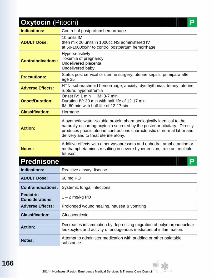
2014 - Northwest Region Emergency Medical Services & Trauma Care Council
166
Oxytocin (Pitocin) P Indications: Control of postpartum hemorrhage
ADULT Dose: 10 units IM then mix 20 units in 1000cc NS administered IV at 50-1000cc/hr to control postpartum hemorrhage
Contraindications:
Hypersensitivty Toxemia of pregnancy Undelivered placenta Undelivered baby
Precautions: Status post cervical or uterine surgery, uterine sepsis, primipara after age 35
Adverse Effects: HTN, subarachnoid hemorrhage, anxiety, dysrhythmias, tetany, uterine rupture, hyponatremia
Onset/Duration: Onset IV: 1 min IM: 3-7 min Duration IV: 30 min with half-life of 12-17 min IM: 60 min with half-life of 12-17min
Classification: Hormone
Action:
A synthetic water-soluble protein pharmacologically identical to the naturally-occurring oxytocin secreted by the posterior pituitary. Directly produces phasic uterine contractions characteristic of normal labor and delivery and to treat uterine atony.
Notes: Additive effects with other vasopressors and ephedra, amphetamine or methamphetamines resulting in severe hypertension; rule out multiple fetuses.
Prednisone P Indications: Reactive airway disease
ADULT Dose: 60 mg PO
Contraindications: Systemic fungal infections
Pediatric Considerations:
1 – 2 mg/kg PO
Adverse Effects: Prolonged wound healing, nausea & vomiting
Classification: Glucocorticoid
Action: Decreases inflammation by depressing migration of polymorphonuclear leukocytes and activity of endogenous mediators of inflammation.
Notes: Attempt to administer medication with pudding or other palatable substance

2014 - Northwest Region Emergency Medical Services & Trauma Care Council
167
167
167
167
167
16
7
16
7
167
16
7
16
7
167
167
Procainamide (Pronestyl) P
Indications: Consider as adjunct to other anti-arrhythmic (amiodarone, Lidocaine) for perfusing VT, or wide-complex tachycardia of unknown origin, WPW
ADULT Dose: Perfusing rhythm: loading dose 20mg/min IV infusion up to 17mg/kg followed by drip of 1-4mg/min (mix 2Grams/250cc NS for 8mg/cc). Stop if hypotension occurs or if QRS widens by 50%
Contraindications:
2nd & 3rd AV block Bradycardias Torsades Prolonged QT Lupus
Pediatric Considerations:
2-6mg/kg slow IV at 25 to 50mg/min
Precautions: May exacerbate arrhythmias or produce paradoxical VT in Afib/Aflutter patients
Adverse Effects: Anxiety, nausea, seizures, widening QRS, hypotension, CNS toxicity
Onset/Duration: 30 min – 2 hours onset 18-36 hours duration
Classification: Anti-arrhythmic
Action: Class 1A membrane stabilizer inhibits recovery after repolarization resulting in decreasing myocardial excitability and conduction velocity.
Notes: Caution with concomitant use of other class 1A antiarrhythmics (Quinidine, TCA’s), digoxin
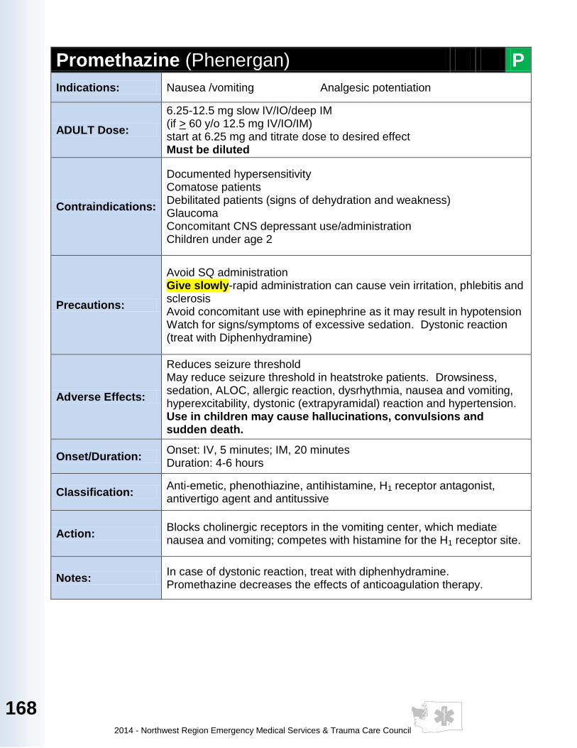
2014 - Northwest Region Emergency Medical Services & Trauma Care Council
168
Promethazine (Phenergan) P
Indications: Nausea /vomiting Analgesic potentiation
ADULT Dose:
6.25-12.5 mg slow IV/IO/deep IM (if > 60 y/o 12.5 mg IV/IO/IM) start at 6.25 mg and titrate dose to desired effect Must be diluted
Contraindications:
Documented hypersensitivity Comatose patients Debilitated patients (signs of dehydration and weakness) Glaucoma Concomitant CNS depressant use/administration Children under age 2
Precautions:
Avoid SQ administration Give slowly-rapid administration can cause vein irritation, phlebitis and sclerosis Avoid concomitant use with epinephrine as it may result in hypotension Watch for signs/symptoms of excessive sedation. Dystonic reaction (treat with Diphenhydramine)
Adverse Effects:
Reduces seizure threshold May reduce seizure threshold in heatstroke patients. Drowsiness, sedation, ALOC, allergic reaction, dysrhythmia, nausea and vomiting, hyperexcitability, dystonic (extrapyramidal) reaction and hypertension. Use in children may cause hallucinations, convulsions and sudden death.
Onset/Duration: Onset: IV, 5 minutes; IM, 20 minutes Duration: 4-6 hours
Classification: Anti-emetic, phenothiazine, antihistamine, H1 receptor antagonist, antivertigo agent and antitussive
Action: Blocks cholinergic receptors in the vomiting center, which mediate nausea and vomiting; competes with histamine for the H1 receptor site.
Notes: In case of dystonic reaction, treat with diphenhydramine. Promethazine decreases the effects of anticoagulation therapy.

2014 - Northwest Region Emergency Medical Services & Trauma Care Council
169
169
169
169
169
16
9
16
9
169
16
9
16
9
169
169
Propofol P
Indications:
FOR INTERFACILITY TRANSPORT The infusion must be initiated by transferring hospital, Verify infusion rate prior to departing hospital. Insure that drip chart comes with medication, verify CXR, verify arterial blood gas and VS are within norms
PATIENTS IN CRISIS ( EXCITED DELIRIUM CONDITION)- After initial control obtained, i.e. by police use of tasers or other means to include droperidol, midazolam or Ketamine, intubate the patient and then start an IV infusion with maintenance doses. See doses below. Do not discontinue until the EMERGENCY DEPARTMENT PHYSICIAN TAKES OVER. Decrease the infusion if hypotension, respiratory depression occur.
ADULT Dose:
20-50mg IV q 10 seconds until onset. (1-2.5mcg/kg/min ) to desired sedation level. Maintenance BP >90 Normal maintenance range: 10-20mg incremental IV bolus Avoid rapid bolus. Do not abruptly stop drug Use dedicated line. Dilute only with normal saline to a concentration not less than 2mg/ml. Rate of administration-slow IVP
Contraindications: Known hypersensitivity, known allergy to eggs or soy or propofol or peanuts
Pediatric Considerations:
2 mg/kg IV/IO additional doses of 1 mg/kg may be administered at two minute intervals
Adverse Effects:
Hypotension, respiratory depression, respiratory acidosis Most common adverse reaction: hypotension. If SBP< 90, give 500cc NS bolus (provided patient does not have pulmonary compromise) and turn drip rate down by 50%. Call Medical Control. Bradycardia, hypertension, Torsades de Pointes all respond to MgSO4. Other potential side effects include: Injection site pain, involuntary muscle movement, nausea and vomiting, anaphylaxis (rare) to soy and peanuts
Classification: Sedative/hypnotic/anesthetic adjunct
Action: CNS Depressant
Notes:
Monitoring: Continuous cardiac and pulse oximetry monitoring and ETC02 with blood pressure every 15 minutes; watch for hypotension, apnea, airway obstruction, oxygen desaturation, document ETCO2 wave form

2014 - Northwest Region Emergency Medical Services & Trauma Care Council
170
Rocuronium (Zemuron) P
Indications: Need for aggressive airway control and maintenance using RSI
ADULT Dose: 0.6-1.2 mg/kg IV
Contraindications: Muscular disorders Known hypersensitivity
Pediatric Considerations:
1 mg/kg IV
Precautions: Not recommended for RSI in Caesarean patients or those over 65 years of age.
Adverse Effects: Hypotension Altered mental status Increases pulmonary resistance
Onset/Duration: Onset: 60-70 seconds Duration: 20+ minutes
Classification: Nondepolarizing neuromuscular blocker
Action: Neuromuscular blockade (Paralysis)
Notes: Airway control equipment must be readily available. Intubation conditions expected in 1-2 minutes after injection. Consider lower doses in extremely debilitated patients.
Sodium Bicarbonate P
Indications: Cardiac arrest 20 to preexisting hyperkalemia or TCA OD with ECG changes of prolonged QT or QRS, or with seizures with VF/VT arrest in setting of meth/cocaine overdose. Consider in prolonged arrest.
ADULT Dose: 8.4% - 1 mEq/kg IV, then 25 mEq in 250ml NS and run 250 ml/hr
Pediatric Considerations:
4.2% - 1 mEq/kg IV/IO
Precautions: Do not administer in the same IV with calcium chloride Prepare to ventilate patient.
Adverse Effects: Metabolic alkalosis, electrolyte imbalance, fluid overload
Onset/Duration: Immediate if IV, onset is less than 15 min Duration 1-2 hours
Classification: Alkalizing agent
Action:
Agent that dissociates to provide bicarbonate ion to buffer hydrogen ions in order to raise the pH level to reverse acidosis. It has also been found beneficial in the event of drug overdose in order to force urine alkalinization/divresis, membrane stabilization of cardiac cells as well, and electrolyte balance restoration.
Notes:
Most catecholamines and vasopressors (dopamine, epinephrine) can be deactivated by alkaline solutions like sodium bicarbonate. When administered with calcium chloride, a precipitate may form that will clog the IV line.

2014 - Northwest Region Emergency Medical Services & Trauma Care Council
171
171
171
171
171
17
1
17
1
171
17
1
17
1
171
171
Succinylcholine (Anectine) P
Indications: An adjunct to general anesthesia, to facilitate tracheal intubation, and to provide skeletal muscle relaxation.
ADULT Dose: 1.5 mg/kg IV
Contraindications: Hyperkalemia
Pediatric Considerations:
1-2 mg/kg IV/IM
Precautions:
Caution should be observed if succinylcholine is administered to patients during the acute phase of injury following major burns, multiple trauma, extensive denervation of skeletal muscle or upper motor neuron injury.
Adverse Effects:
Respiratory depression Apnea Anaphylaxis Hypertension Hypotension Renal Failure Hyperkalemia Increased intraocular pressure Dysrhythmias Malignant hyperthermia
Onset/Duration: Onset: 1 minute Duration: 4-6 minutes
Classification: Depolorizing neuromuscular blocking agent
Action: Short-acting depolarizing-type, skeletal muscle relaxant
Notes: Should not be mixed with alkaline solutions.
Tenecteplase (TNKase) P
Indications: STEMI confirmed with Medical Control authorization required
ADULT Dose:
Single IV bolus over 5sec; if <60kg/132lbs then 30mg; if 60-70kg/132-154lbs then 35mg; if 70-80kg/154-176lbs then 40mg; if 80-90kg/176-198lbs then 45mg; and, if >90kg/198lbs then 50mg
Contraindications:
Active internal bleeding History of CVA, intracranial/intraspinal surgery or trauma past 2 months, intracranial neoplasm/arteriovenous malformation or aneurysm Severe uncontrolled HTN
Precautions: Blood vessel punctures should be minimized, especially non-compressible sites.
Adverse Effects: Bleeding
Onset/Duration: Rapid/90-130min half-life
Classification: Thrombolytic, tissue plasminogen activator
Action: Binds to fibrin and converts plasminogen to plasmin; decreases systemic activation of plasminogen and the resulting degradation of circulating fibrinogen.
Notes: Do not give with glucose-containing solution as may precipitate. Do not use in-line filter.
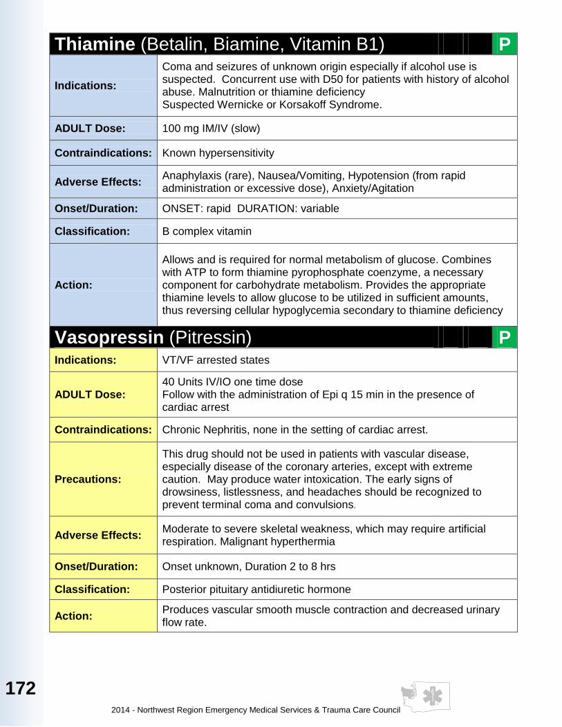
2014 - Northwest Region Emergency Medical Services & Trauma Care Council
172
Thiamine (Betalin, Biamine, Vitamin B1) P
Indications:
Coma and seizures of unknown origin especially if alcohol use is suspected. Concurrent use with D50 for patients with history of alcohol abuse. Malnutrition or thiamine deficiency Suspected Wernicke or Korsakoff Syndrome.
ADULT Dose: 100 mg IM/IV (slow)
Contraindications: Known hypersensitivity
Adverse Effects: Anaphylaxis (rare), Nausea/Vomiting, Hypotension (from rapid administration or excessive dose), Anxiety/Agitation
Onset/Duration: ONSET: rapid DURATION: variable
Classification: B complex vitamin
Action:
Allows and is required for normal metabolism of glucose. Combines with ATP to form thiamine pyrophosphate coenzyme, a necessary component for carbohydrate metabolism. Provides the appropriate thiamine levels to allow glucose to be utilized in sufficient amounts, thus reversing cellular hypoglycemia secondary to thiamine deficiency
Vasopressin (Pitressin) P Indications: VT/VF arrested states
ADULT Dose: 40 Units IV/IO one time dose Follow with the administration of Epi q 15 min in the presence of cardiac arrest
Contraindications: Chronic Nephritis, none in the setting of cardiac arrest.
Precautions:
This drug should not be used in patients with vascular disease, especially disease of the coronary arteries, except with extreme caution. May produce water intoxication. The early signs of drowsiness, listlessness, and headaches should be recognized to prevent terminal coma and convulsions.
Adverse Effects: Moderate to severe skeletal weakness, which may require artificial respiration. Malignant hyperthermia
Onset/Duration: Onset unknown, Duration 2 to 8 hrs
Classification: Posterior pituitary antidiuretic hormone
Action: Produces vascular smooth muscle contraction and decreased urinary flow rate.
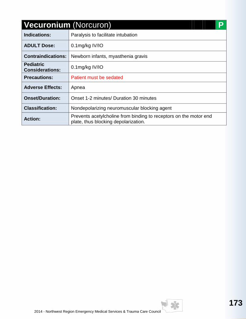
2014 - Northwest Region Emergency Medical Services & Trauma Care Council
173
173
173
173
173
17
3
17
3
173
17
3
17
3
173
173
Vecuronium (Norcuron) P Indications: Paralysis to facilitate intubation
ADULT Dose: 0.1mg/kg IV/IO
Contraindications: Newborn infants, myasthenia gravis
Pediatric Considerations:
0.1mg/kg IV/IO
Precautions: Patient must be sedated
Adverse Effects: Apnea
Onset/Duration: Onset 1-2 minutes/ Duration 30 minutes
Classification: Nondepolarizing neuromuscular blocking agent
Action: Prevents acetylcholine from binding to receptors on the motor end plate, thus blocking depolarization.
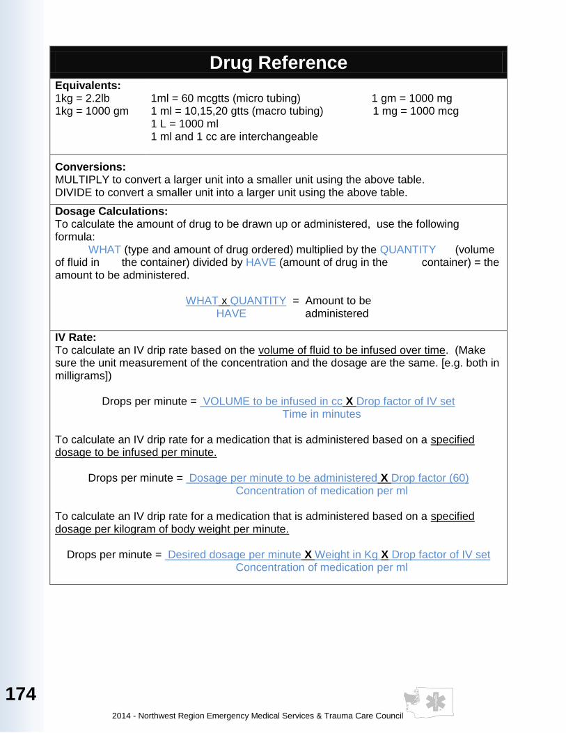
2014 - Northwest Region Emergency Medical Services & Trauma Care Council
174
Drug Reference Equivalents: 1kg = 2.2lb 1kg = 1000 gm
1ml = 60 mcgtts (micro tubing) 1 gm = 1000 mg 1 ml = 10,15,20 gtts (macro tubing) 1 mg = 1000 mcg 1 L = 1000 ml 1 ml and 1 cc are interchangeable
Conversions: MULTIPLY to convert a larger unit into a smaller unit using the above table. DIVIDE to convert a smaller unit into a larger unit using the above table.
Dosage Calculations: To calculate the amount of drug to be drawn up or administered, use the following formula: WHAT (type and amount of drug ordered) multiplied by the QUANTITY (volume of fluid in the container) divided by HAVE (amount of drug in the container) = the amount to be administered.
WHAT x QUANTITY = Amount to be HAVE administered
IV Rate: To calculate an IV drip rate based on the volume of fluid to be infused over time. (Make sure the unit measurement of the concentration and the dosage are the same. [e.g. both in milligrams])
Drops per minute = VOLUME to be infused in cc X Drop factor of IV set Time in minutes
To calculate an IV drip rate for a medication that is administered based on a specified dosage to be infused per minute.
Drops per minute = Dosage per minute to be administered X Drop factor (60) Concentration of medication per ml
To calculate an IV drip rate for a medication that is administered based on a specified dosage per kilogram of body weight per minute.
Drops per minute = Desired dosage per minute X Weight in Kg X Drop factor of IV set Concentration of medication per ml
2014 - Northwest Region Emergency Medical Services & Trauma Care Council
175
175
175
175
175
175
175
175
175
175
175
175
175
Air Ambulance Transports
An air ambulance will be activated based on the Washington State Trauma Triage Procedures by the on scene EMS provider or Incident Commander. Whenever possible, providers will contact Medical Control prior to activating an air ambulance. The decision process as to when to mobilize an air ambulance should take into consideration: site resuscitation, stabilization capabilities and ground transport time. Dispatch may assist in contacting an air ambulance service for activation as soon as the need for air transport is identified. Every attempt should be made to stabilize the patient prior to transport, including IV, airway, chest decompression or stabilization, control of external hemorrhage, and spine immobilization. Trauma associated cardiac arrest patients should not be transferred by air ambulance. Transfer of care to air ambulance personnel will optimally occur at designated landing sites. Deviation from designated landing sites should be briefly discussed with Medical Control.
Information to have available regarding airlift transport:
· Map coordinates - township, range and section
· Location of nearest landing zone
· Capability to transport to landing zone
· How landing zone is marked
· Any obstructions near landing zone
· Relevant weather information
Physician Present at the Scene The prehospital care provider functions under the direction of the on-duty Medical Control physician. With Medical Control permission, a physician, physician's assistant or nurse practitioner on scene may participate in the care of a patient at the scene of any emergency in one of the following ways:
1. Take total responsibility for management of the patient(s). If so he/she must accompany the patient(s) to the hospital. The physician, physician's assistant or nurse practitioner on scene must supply proof of being a MD or DO prior to initiation of any patient care direction or treatment.
2. Offer assistance in caring for the patient(s), allowing the prehospital care provider to remain under the control of the Medical Control Physician and within the prehospital provider’s scope of practice.
In all cases, the Medical Control Physician must be contacted to specifically delegate authority to any on-scene physician. Access to communication with the Medical Control should be provided to any on-scene physician on request. Notation of Physician’s identification and directive from Medical Control must be documented on the Medical Incident Report.
2014 - Northwest Region Emergency Medical Services & Trauma Care Council
176
Emergency at a Physician’s office
At a private Physician’s, physician's assistant or nurse practitioner's office, the individual physician maintains the responsibility for the treatment and management decisions for the patient. During transport, treatment rendered by the prehospital provider must remain within the provider’s scope of practice.
Patient Care Reports (PCR)
A copy of the ECG tracing MUST be attached to all copies of the PCR when ANY dysrhythmia or ischemic ST segment changes are encountered in the field. Per WAC requirements all PCR must be available at the Emergency Department within 24 hours of patient arrival.
The prehospital contact report is to include:
a. Unit identification b. Age and sex of patient c. Severity d. Chief complaint e. Relevant medical history f. Vital signs g. Treatment given, and response to treatment h. ETA i. Request for additional information or treatment

2014 - Northwest Region Emergency Medical Services & Trauma Care Council
177
177
177
177
177
177
177
177
177
177
177
177
177
Ten Critical Steps for Handling Possible Bioterrorism Events
1 – Maintain an index of suspicion.
In an otherwise healthy population, some associations are very suggestive, especially when seen in clusters, high numbers, or unusual presentations.
“Clustered” Symptoms Potential Bioagents
Hemoptysis Plague
Flaccid Paralysis Botulism
Purpura Viral Hemorrhagic Fevers (VHF)
Wide mediastinum Anthrax
Centripetal rash Smallpox
2 – Protect yourself and your patients.
Use appropriate personal protection equipment (PPE). Prophylaxis; vaccines, if available; or antibiotics, if risks are known.
3 – Adequately assess the patient.
Review and assess the patient’s history. Also, ask:
· Are others ill?
· Were there any unusual events?
· Was there an uncontrolled food source or other environmental factor?
· Was there vector exposure?
· Has the patient been traveling?
· What is the patient’s immunization record? Perform a physical examination with special attention to the respiratory system, nervous system, skin condition and hematologic and vascular status.
4 – Decontaminate as appropriate.
Do not use bleach on exposed people. Soap, water and shampoo are perfectly adequate for all biological and most chemical agents. Chemically contaminated clothes should be removed and discarded safely. Biologically contaminated clothes can be laundered with soap, water and perhaps, bleach.
5 – Establish a diagnosis.
Think clinically and epidemiologically; always send specimens for culture.
Symptom (individuals) Possible Diagnosis
Pulmonary Tularemia, plague, staph enterotoxin B (SEB)
Neuromuscular Botulism, Venezuelan equine encephalitis (VEE)
Bleeding/purpura VHF, ricin, plague (late)
Rash (various types) VHF, T2 mycotoxin, smallpox, plague
Flu-like symptoms Varies
Immediate Symptoms (large numbers)
Possible Diagnosis
Pulmonary SEB, mustard, Lewisite, phosgene, cyanide
Neurologic Nerve gases, cyanide
Delayed Symptoms (large numbers)
Possible Diagnosis
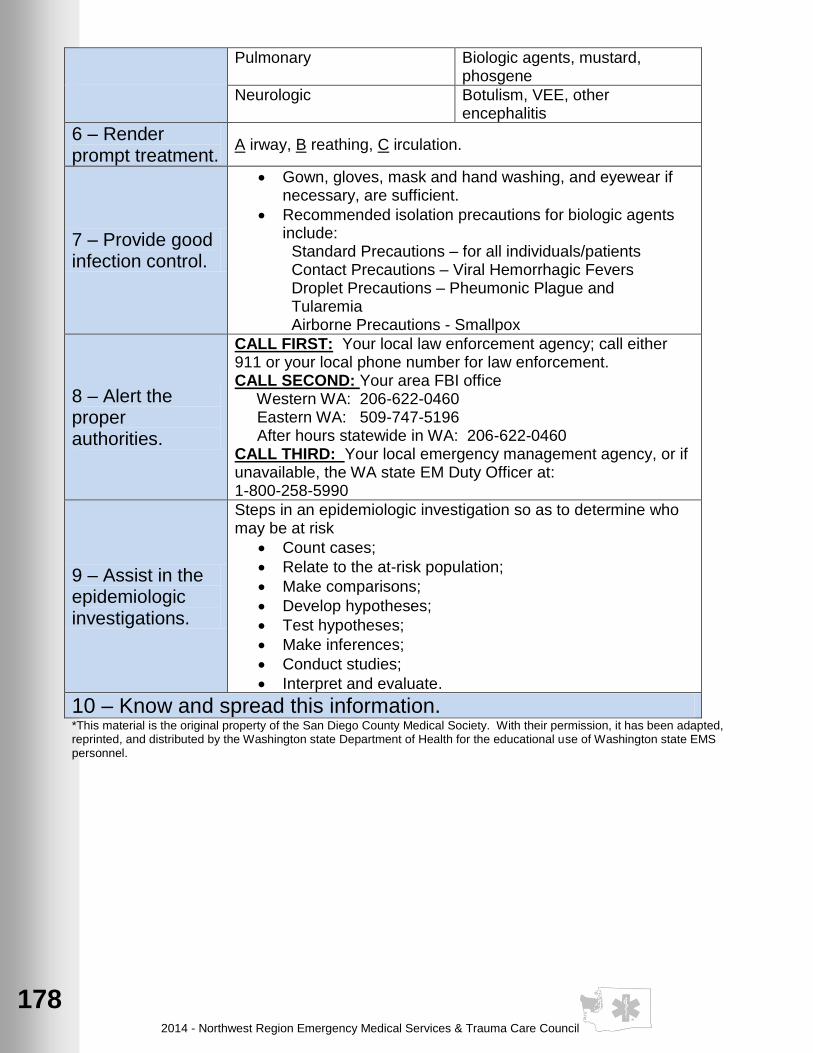
2014 - Northwest Region Emergency Medical Services & Trauma Care Council
178
Pulmonary Biologic agents, mustard, phosgene
Neurologic Botulism, VEE, other encephalitis
6 – Render prompt treatment.
A irway, B reathing, C irculation.
7 – Provide good infection control.
· Gown, gloves, mask and hand washing, and eyewear if necessary, are sufficient.
· Recommended isolation precautions for biologic agents include:
Standard Precautions – for all individuals/patients Contact Precautions – Viral Hemorrhagic Fevers
Droplet Precautions – Pheumonic Plague and Tularemia
Airborne Precautions - Smallpox
8 – Alert the proper authorities.
CALL FIRST: Your local law enforcement agency; call either 911 or your local phone number for law enforcement. CALL SECOND: Your area FBI office Western WA: 206-622-0460 Eastern WA: 509-747-5196 After hours statewide in WA: 206-622-0460 CALL THIRD: Your local emergency management agency, or if unavailable, the WA state EM Duty Officer at: 1-800-258-5990
9 – Assist in the epidemiologic investigations.
Steps in an epidemiologic investigation so as to determine who may be at risk
· Count cases;
· Relate to the at-risk population;
· Make comparisons;
· Develop hypotheses;
· Test hypotheses;
· Make inferences;
· Conduct studies;
· Interpret and evaluate.
10 – Know and spread this information. *This material is the original property of the San Diego County Medical Society. With their permission, it has been adapted, reprinted, and distributed by the Washington state Department of Health for the educational use of Washington state EMS personnel.

2014 - Northwest Region Emergency Medical Services & Trauma Care Council
179
179
179
179
179
179
179
179
179
179
179
179
179
Medical Spanish
Initial questioning
Is there someone with you who speaks English? ¿Hay alguien con usted que hable ingles? Ah-ee ahl-gee-ehn hohn oss-tehd keh ah-bleh enn-glehs?
I speak a little Spanish. Please answer yes or no to the following questions. Hablo un poco de español. Por favor conteste si o no a las siguientes preguntas. Ah-bloh oon pohr-fah-borg kokn-tehs-the see oh noh ah lahs see-gee-ehn-tehs preh-goon tahs.
Speak slowly, please. Hable despacia, por favor. Ah-bleh dehs-pah-see-oh, pohr fah-bohr.
What is your name? ¿Cómo se llama? Koh-moh she yah-mah?
How old are you? ¿Cuántos años tiene? Kwahn-tohs ah-nyohs tee-eh-neh?
When did the problem start? ¿Cuándo empezó el problema? Kwahn-doh ehm-peh-soh ehl prog-bleh-mah?
What medicine do you take? ¿Qué medicina torna? Keh meh-dee-see-nah toh-mah?
Numbers
1. uno 2. dos 3. tres 4. cuatro 5. cinco 6. seis 7. siete 8. ocho 9. neueve 10. diez
11. once 12. doce 13. trece 14. catorce 15. quince 16. dieciséis 17. diecisiete 18. dieciocho 19. diecinueve 20. viente
21. vientiuno 22. vientidós 23. veintritrés 24. veinticuatro 25. veinticinco 26. veintiséis 27. veintisiete 28. veintiocho 29. veintineuve 30. treinta
Days of the week
Lunes: Monday
Viernes: Friday Martes: Tuesday Sábado: Saturday Miércoles: Wednesday Domingo: Sunday Jueves: Thursday
Common Medical Questions/Terms
How do you feel? ¿Cómo se siente? Koh-moh she see-ehn-the?
What is the problem? ¿Cuál es el problema? Kwahl ehs ehl proh-bleh-mah?
Have you had this problem before? ¿Ha tenido este problema antes? Ah the-nee-doh ehs-the proh-bleh-mah ahn-tehs?
Do you have nausea or vomiting? ¿Tiene nausea o vómito? Tee-eh-neh nah-oo-she-ah oh boh-meh-toh?
Don’t move No se meuva Noh she mweh-bah
We are going to give you an IV Vamos a ponerie suero intravenoso. Bah-mohs ah poh-nehr-leg soo-eh-roh enn-trah-beh-noh-soh.
Do you have a fever? ¿Tiene fiebre? Tee-eh-neh fee-eh-breh?
Calm down Cálmese Kahl-meh-sah

2014 - Northwest Region Emergency Medical Services & Trauma Care Council
180
Common Medical Questions/Terms (continued)
Where does it hurt? ¿Donde le duele? Dohn-deh leh dweh-leh?
Show me Enséñeme Ehn-she-nyeh-meh.
When? ¿Cuándo? Kwahn-doh?
How? ¿Cómo? Koh-moh?
For how long? ¿Por cuánto tiempo? Pohr kwahn-toh tee-ehm-poh?
Why? ¿Por qué? Pohr keh?
Relax, please Por favor, relájese Pohr fah-bohr, reh-lah-heh-she.
High Blood Pressure? Alta presion de la sangre? Ahl-tah preh-see-ohn deh lah sahn-greh?
Diabetes? Diabetes? Dee-ah-beh-tehs?
Asthma? Asma? Ahs-mah?
Epilepsy? Epilepsia? Eh-pee-lep-see-ah?
Heart disease? Ehfermedad del corazón? Ehn-fehr-meh-dad dehl koh-rah-sohn?
Stomach ulcers? Ulceras del estomago? Ool-she-rahs dehl ehs-toh-mah-goh?
Do you take medicine? ¿Tomas usted medicina? Toh-mah oos-tehd lah meh-dee-see-nah?
Pain
When did the pain start? ¿Cuándo empezó el dolor? Kwahn-doh ehm-peh-soh ehl doh-lohr?
Where did the pain start? ¿Donde empezó el dolor? Dohn-deh ehm-peh-soh ehl doh-lohr?
Does the pain travel to another place? ¿Le viaja el dolor a otro lugar? Leh vee-ah-hah ehl doh-lohr ah oh-troh loo-gahr?
How long does the pain last? ¿Cuánto tiempo le dura el dolor? Kwahn-toh tee-ehm-poh leg doo-rah ehl doh-lohr?
Is it severe? ¿Es severo? Ehs she-beh-roh?
Does it ache? ¿Es adolorido? Ehs ah-doh-loh-ree-doh?
Is it like pressure? ¿Es opresivo? Ehs oh-preh-see-boh?
Is the pain the same since it started? ¿Es el dolor igual desde que empezó? Ehs ehl doh-lor ee-gwahl dehs-deh keh ehm-peh-soh?
Chest Pain Pain in the chest? ¿Dolor del Pecho? Doh=lohr dehl peh-choh?
Point to where the pain is, please. Apunte dónde tiene el dolor, por favor. Ah-poon-the dohn-deh tee-eh-neh ehl doh-lohr.
Does the pain travel to your left shoulder (arm)? ¿Le viaja el dolor al hombre (brazo) izquierdo? Leh bee-ah-hah ehl doh-lohr ahl ohm-broh (brah-soh) ees-kee-her-doh?
Is it piercing? ¿Es punzante? Ehs poon-sahn-the?
OB / GYN
Are you having contractions? ¿Tiene contracciones? Tee-eh-neh kohn-track-see-ohn-ehs?
(Don’t) push. (No) Empuje. (Noh) Ehm-poo-heh
How many minutes do the contractions last? ¿Cuántos minutes le duran las contracciones? Kwahn-tohs mee-noo-tohs leh doo-rahn lahs kohn-trahk-see-ohn-ehs?

2014 - Northwest Region Emergency Medical Services & Trauma Care Council
181
181
181
181
181
181
181
181
181
181
181
181
181
MNEMONIC’s
Patient Assessment: Newborn Assessment: Medical:
A: Airway A: Appearance M: Morphine
B: Breathing P: Pulse Rate O: Oxygen C: Circulation G: Grimace (facial actions) N: Nitrates D: Disability A: Activity A: Aspirin E: Expose R: Respirations
History:
S: Signs and symptoms P: Progression of symptoms
A: Allergies A: Associated chest pain M: Medications S: Sputum productions, speech, P: Pertinent past medical word sentences history T: Temperature, tiredness
L: Last oral intake M: Medications the patient is E: Events leading to injury or currently taking illness E: Exercise/Exertion normally
tolerated D: Diagnosis (previous)
Trauma Assessment: Trauma:
Scene safety V: Vitals T: Tracks, Tags, Tattoos Spinal Stabilization O: Oxygen I: Instability LOC M: Monitor C: Crepitus Airway I: IV/Information S: Scars Breathing T: Transport decision Oxygen H: History Circulation A: Allergies Arterial Bleeds M: Medications Bare the Body
Trauma or Pain Questions: Trauma:
O: Onset D: Deformities P: Provocation, progression C: Contusions Q: Quality, pain type? A: Abrasions R: Radiation P: Punctures S: Severity T: Time, duration B: Burns T: Tenderness L: Lacerations S: Swelling

2014 - Northwest Region Emergency Medical Services & Trauma Care Council
182
Causes of Pulseless electrical Activity (PEA) – “5” H’s and T’s:
H: Hypovolemia T: Toxins H: Hypoxia T: Tamponade, cardiac H: Hydrogen ion – acidosis T: Tension Pneumothorax
H: H: H:
Hypo- / Hypekalemia Hypoglycemia Hypothermia
T:
T:
Thrombosis, (Coronary or Pulmonary) Thrombosis, (hypovolemia increased ICP)
Altered Mental Status (ALOC):
A: Alcohol, Drugs T: Trauma E: Endocrine (glands) I: Infection I: Insulin, Infection P: Pyschiatric O: Overdose S: Shock U: Uremia (20 kidney insufficiency)
Triage: Charting:
A: Alert S: Subjective P: Responsive to Verbal O: Objective V: Responsive to Pain A: Assessment U: Unresponsive P: Plan
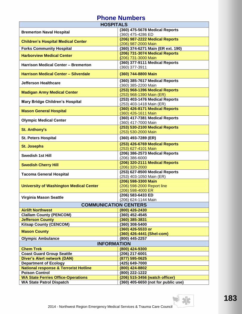
2014 - Northwest Region Emergency Medical Services & Trauma Care Council
183
183
183
183
183
183
183
183
183
183
183
183
183
Phone Numbers HOSPITALS
Bremerton Naval Hospital (360) 475-5678 Medical Reports (360) 475-4286 ED
Children’s Hospital Medical Center (206) 987-2222 Medical Reports (206) 987-2000 Main
Forks Community Hospital (360) 374-6271 Main (ER ext. 190)
Harborview Medical Center (206) 731-3074 Medical Reports (206) 731-3000 Main
Harrison Medical Center – Bremerton (360) 377-9111 Medical Reports (360) 377-3911
Harrison Medical Center – Silverdale (360) 744-8800 Main
Jefferson Healthcare (360) 385-7617 Medical Reports (360) 385-2200 Main
Madigan Army Medical Center (253) 968-1396 Medical Reports (253) 968-1390 Main (ER)
Mary Bridge Children’s Hospital (253) 403-1476 Medical Reports (253) 403-1418 Main (ER)
Mason General Hospital (360) 426-8171 Medical Reports (360) 426-1611 Main
Olympic Medical Center (360) 417-7381 Medical Reports (360) 417-7000 Main
St. Anthony's (253) 530-2100 Medical Reports (253) 530-2000 Main
St. Peters Hospital (360) 493-7289 (ER)
St. Josephs (253) 426-6769 Medical Reports (253) 627-4101 Main
Swedish 1st Hill (206) 386-2573 Medical Reports (206) 386-6000
Swedish Cherry Hill (206) 320-2111 Medical Reports (206) 320-2000
Tacoma General Hospital (253) 627-8500 Medical Reports (253) 403-1050 Main (ER)
University of Washington Medical Center (206) 598-3300 Main (206) 598-2000 Report line (206) 598-4000 ER
Virginia Mason Seattle (206) 583-6433 ED (206) 624-1144 Main
COMMUNICATION CENTERS Airlift Northwest (800) 426-2430
Clallam County (PENCOM) (360) 452-4545
Jefferson County (360) 385-3831
Kitsap County (CENCOM) (360) 308-5400
Mason County (360) 426-5533 or (360) 426-4441 (Shel-com)
Olympic Ambulance (800) 445-2257
INFORMATION Chem Trek (800) 424-9300
Coast Guard Group Seattle (206) 217-6001
Diver’s Alert network (DAN) (877) 595-0625
Department of Ecology (425) 649-7000
National response & Terrorist Hotline (800) 424-8802
Poison Control (800) 222-1222
WA State Ferries Office-Operations (206) 515-3456 (watch officer)
WA State Patrol Dispatch (360) 405-6650 (not for public use)
2014 - Northwest Region Emergency Medical Services & Trauma Care Council
184
Intentionally left blank for future
additions
2014 - Northwest Region Emergency Medical Services & Trauma Care Council
185
185
185
185
185
185
185
185
185
185
185
185
185
Intentionally left
blank for future additions

2014 - Northwest Region Emergency Medical Services & Trauma Care Council
186
Intentionally left blank for future
additions
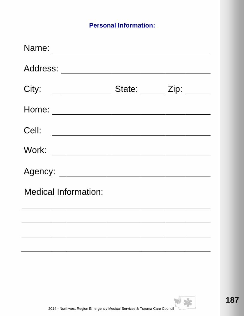
2014 - Northwest Region Emergency Medical Services & Trauma Care Council
187
187
187
187
187
187
187
187
187
187
187
187
187
Personal Information:
Name:
Address:
City: State: Zip:
Home:
Cell:
Work:
Agency:
Medical Information: