Embed Size (px)
Citation preview
Normalizing translation through 4E-BP preventsmTOR-driven cortical mislamination and amelioratesaberrant neuron integrationTiffany V. Lina,b, Lawrence Hsieha,b, Tomoki Kimuraa,b, Taylor J. Malonea,b, and Angélique Bordeya,b,1
aDepartment of Neurosurgery, Yale University School of Medicine, New Haven, CT 06520; and bDepartment of Cellular and Molecular Physiology, YaleUniversity School of Medicine, New Haven, CT 06520
Edited by Nicholas Spitzer, University of California, San Diego, La Jolla, CA, and approved August 8, 2016 (received for review April 12, 2016)
Hyperactive mammalian target of rapamycin complex 1 (mTORC1) isa shared molecular hallmark in several neurodevelopmental disor-ders characterized by abnormal brain cytoarchitecture. The mecha-nisms downstream of mTORC1 that are responsible for these defectsremain unclear. We show that focally increasing mTORC1 activityduring late corticogenesis leads to ectopic placement of upper-layercortical neurons that does not require altered signaling in radial gliaand is accompanied by changes in layer-specific molecular identity.Importantly, we found that decreasing cap-dependent translationby expressing a constitutively active mutant of the translationalrepressor eukaryotic initiation factor 4E-binding protein 1 (4E-BP1)prevents neuronal misplacement and soma enlargement, while par-tially rescuing dendritic hypertrophy induced by hyperactive mTORC1.Furthermore, overactivation of translation alone through knockdownof 4E-BP2 was sufficient to induce neuronal misplacement. These datashow that many aspects of abnormal brain cytoarchitecture can beprevented by manipulating a single intracellular process downstreamof mTORC1, cap-dependent translation.
mTOR | autism | spine | dendrite | cortical development
Overactive mammalian target of rapamycin complex 1(mTORC1) signaling is a signature of many disorders with
cortical malformations (1), ranging from tuberous sclerosis complexwith focal dysplasias to hemimegalencephaly with more diffuse,hemispheric aberrations. The high incidence of negative outcomes inindividuals with such malformations (2), which are often associatedwith intractable childhood seizures, underscores the need to betterunderstand the molecular etiology of these developmental lesions.Animal models have demonstrated the causative effect of increasedmTORC1 signaling on mislamination (3–7). The pharmacologicalmTORC1 blocker rapamycin has also been shown to reverse someof the developmental abnormalities and associated seizure activity inseveral of these mouse models (5, 6, 8–10), further emphasizing theimportance of mTORC1 in the disease pathogenesis.Despite the demonstrated relevance of mTORC1 signaling,
there is less known about the molecular mechanisms by whichmTORC1 alters cortical development. Addressing this question iscomplicated by the wide range of cellular processes regulated bymTORC1 through independent downstream targets. Among theseregulated processes are autophagy, lysosomal function, lipid syn-thesis, and, one of the best-studied functions, cap-dependenttranslation (11). Because current drugs that suppress mTORC1activity can have serious side effects (12, 13) and do not fully blocksome of mTORC1’s functions (14), a more specific understandingof how mTORC1 contributes to cortical mislamination could yieldbetter targets for treatment.This study therefore aimed to more closely characterize the
cytoarchitectural aberrations generated by hyperactive mTORC1and to examine the contribution of translational regulation tothese cortical malformations. Using in utero electroporation, wegenerated and characterized focal mislamination and morpho-logical changes driven by mTORC1 signaling. We first examinedthe contribution of radial glia to mislamination and the molecular
identity of the ectopic neurons. We then asked whether up-regu-lation of cap-dependent translation through eukaryotic initiationfactor 4E-binding proteins (4E-BPs) is both necessary formTORC1-induced misplacement and sufficient to cause ectopicneurons. Our data show that, despite the many other cellularfunctions regulated by mTORC1, up-regulation of cap-dependenttranslation through 4E-BPs is both necessary and sufficient formisplacement of cortical projection neurons.
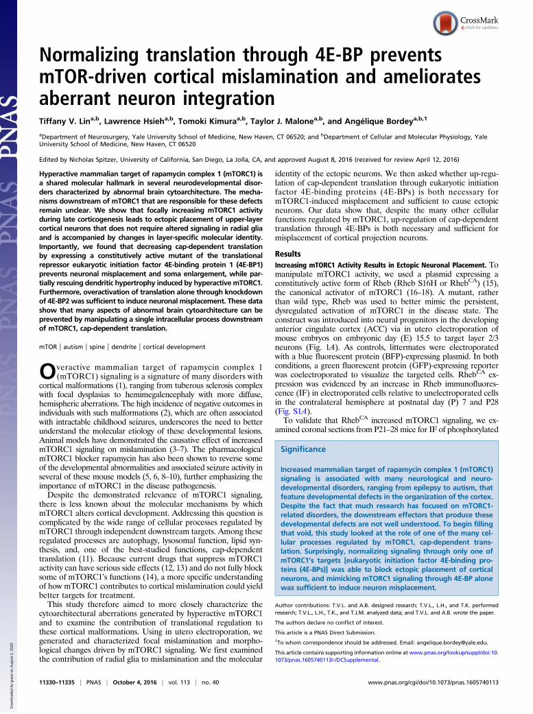
ResultsIncreasing mTORC1 Activity Results in Ectopic Neuronal Placement. Tomanipulate mTORC1 activity, we used a plasmid expressing aconstitutively active form of Rheb (Rheb S16H or RhebCA) (15),the canonical activator of mTORC1 (16–18). A mutant, ratherthan wild type, Rheb was used to better mimic the persistent,dysregulated activation of mTORC1 in the disease state. Theconstruct was introduced into neural progenitors in the developinganterior cingulate cortex (ACC) via in utero electroporation ofmouse embryos on embryonic day (E) 15.5 to target layer 2/3neurons (Fig. 1A). As controls, littermates were electroporatedwith a blue fluorescent protein (BFP)-expressing plasmid. In bothconditions, a green fluorescent protein (GFP)-expressing reporterwas coelectroporated to visualize the targeted cells. RhebCA ex-pression was evidenced by an increase in Rheb immunofluores-cence (IF) in electroporated cells relative to unelectroporated cellsin the contralateral hemisphere at postnatal day (P) 7 and P28(Fig. S1A).To validate that RhebCA increased mTORC1 signaling, we ex-
amined coronal sections from P21–28 mice for IF of phosphorylated
Significance
Increased mammalian target of rapamycin complex 1 (mTORC1)signaling is associated with many neurological and neuro-developmental disorders, ranging from epilepsy to autism, thatfeature developmental defects in the organization of the cortex.Despite the fact that much research has focused on mTORC1-related disorders, the downstream effectors that produce thesedevelopmental defects are not well understood. To begin fillingthat void, this study looked at the role of one of the many cel-lular processes regulated by mTORC1, cap-dependent trans-lation. Surprisingly, normalizing signaling through only one ofmTORC1’s targets [eukaryotic initiation factor 4E-binding pro-teins (4E-BPs)] was able to block ectopic placement of corticalneurons, and mimicking mTORC1 signaling through 4E-BP alonewas sufficient to induce neuron misplacement.
Author contributions: T.V.L. and A.B. designed research; T.V.L., L.H., and T.K. performedresearch; T.V.L., L.H., T.K., and T.J.M. analyzed data; and T.V.L. and A.B. wrote the paper.
The authors declare no conflict of interest.
This article is a PNAS Direct Submission.1To whom correspondence should be addressed. Email: [email protected].
This article contains supporting information online at www.pnas.org/lookup/suppl/doi:10.1073/pnas.1605740113/-/DCSupplemental.
11330–11335 | PNAS | October 4, 2016 | vol. 113 | no. 40 www.pnas.org/cgi/doi/10.1073/pnas.1605740113
Dow
nloa
ded
by g
uest
on
Aug
ust 2
, 202
0
S6 (pS6; Ser-240/244), a downstream target of mTORC1 signaling. A2.3-fold increase in pS6 IF was observed in RhebCA-electroporatedneurons relative to unelectroporated neurons from the contralateralside of the same section (Fig. S1B). This increase was not seen in thecontrol condition. We also attempted to analyze p4E-BP1/2 IF, butno signal was detected in the cortex despite staining in the adultneurogenic zone (Fig. S1C). However, RhebCA increased p4E-BP1(Thr-37/46) levels 2.2-fold in vitro (Fig. S1D). Other pathways knownto be regulated by mTORC1 signaling were also altered by RhebCA.Autophagy was decreased at both P7 and P21–28 as evidenced byincreased IF for p62, whose levels are negatively regulated byautophagy (19) (Fig. S2). Staining using a KDEL-peptide antibody,which recognizes endoplasmic reticulum (ER) stress markersGrp78, Grp94, and PDI (20), indicated elevated ER stress atboth time points (Fig. S3). Finally, we examined soma size becauseincreased mTORC1 activity causes cellular hypertrophy (6, 21–23). As expected, a 2.1-fold increase in soma area was observed inRhebCA-electroporated cells relative to controls (Fig. 1B).Consistent with other animal models with focal mTORC1
hyperactivation (3–7), RhebCA produced a striking mislaminationeffect. This effect was visible from P0 to P28 (Fig. 1C). The dis-tribution of neurons was analyzed by dividing the cortex into 10bins running parallel to the midline from pia to white matter (Fig.1D) and quantifying the proportion of total GFP-positive (GFP+)cells found in each bin. Analysis of tissue from P21–28 micerevealed a significant change in the distribution of the neuronsacross the cortex in the RhebCA condition relative to control (Fig.1E). We also found that that only 35% of GFP+ neurons in theRhebCA condition reached layer 2/3 (delineated by Cux1 or Er81staining), compared with 93% in controls (Fig. 1F).
mTORC1 Signaling Causes Mislamination Independently of Radial Glia.During cortical development, newborn neurons migrate alongradial glia processes to their final positions in the cortex (24).Therefore, the RhebCA mislamination phenotype could be causedby altered mTORC1 signaling in the radial glia (i.e., the scaf-folding) rather than in the neurons themselves. To dissociate thecontribution of radial glia, we used RhebCA packaged in a con-ditional vector (pCALNL-RhebCA) and coelectroporated it with aplasmid expressing Cre under the Dcx promoter (Dcx-Cre) (Fig.2A). As a control, the same conditional vector encoding dsRed2(pCALNL-dsRed) was used. In both conditions, pCALNL-GFP
was coelectroporated as a reporter. Because DCX is present inimmature migrating neurons, but not in radial glia (25, 26), ourconditional constructs should have only begun expressing in themigrating neurons (Fig. 2B). As expected, at E18, conditional GFPwas expressed outside of the ventricular zone where the radial gliareside (Fig. S4). Analysis at P7 showed that conditional RhebCA
Fig. 1. Increasing mTORC1 activity leads to ectopic neuron placement. (A) Schematic of in utero electroporation and constructs used. (B) Images of layer 2/3 GFP+
cells in coronal sections from P28 control and RhebCA-electroporated mice and quantification of soma areas. (Scale bars: 50 μm.) Areas were normalized to controlsoma area (n = 25 cells, 3 mice per condition, t test). (C) Images of GFP+ cells in ACC from control and RhebCA electroporated mice at P0, P7, and P28. (Scale bars:150 μm.) (D) Schematic of binning system used to quantify cell distribution. (E) RhebCA-electroporated neurons were more widely distributed relative to controls[n = 3 or 4 mice per condition, repeated-measures (RM) two-way ANOVA, Bonferroni post hoc]. (F) Quantification of the percent of electroporated neurons thatintegrate into layer 2/3 (n = 3 mice per condition, t test). *P < 0.05; **P < 0.005; ****P < 0.0001. All data are presented as mean ± SEM.
Fig. 2. RhebCA expression causes mislamination independent of radial glia.(A) Summary of constructs used. (B) Schematic of conditional plasmid ex-pression in the radial glia and migrating neurons. (C) Cells expressing con-ditional RhebCA are more widely distributed than control electroporatedcells (n = 3 mice per condition, RM two-way ANOVA, Bonferroni post hoc).(D) Images of GFP+ cells in the ACC of P7 control and RhebCA-electroporatedmice. (E) Quantification of the percent of electroporated neurons that in-tegrate into layer 2/3 (n = 3 mice per condition, t test). *P < 0.05; **P < 0.01;***P < 0.001; ****P < 0.0001. All data are presented as mean ± SEM.
Lin et al. PNAS | October 4, 2016 | vol. 113 | no. 40 | 11331
NEU
ROSC
IENCE
Dow
nloa
ded
by g
uest
on
Aug
ust 2
, 202
0
expression in migrating neurons resulted in a significant change inlamination, with 50% of cells failing to reach layer 2/3 (Fig. 2 C–E),indicating that RhebCA expression in the migrating neurons aloneis sufficient to induce ectopic integration.
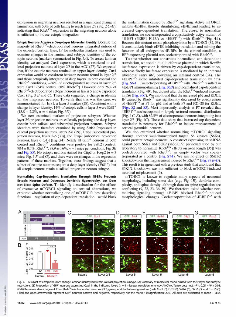
Ectopic Neurons Acquire a Deep-Layer Molecular Identity. Because themajority of RhebCA-electroporated neurons integrated outside ofthe expected cortical layer, IF for molecular markers was used toexamine changes in the laminar and subtype identities of the ec-topic neurons (markers summarized in Fig. 3A). To assess laminaridentity, we analyzed Cux1 expression, which is restricted to cal-losal projection neurons of layer 2/3 in the ACC (27). We expectedthat, if the ectopic neurons retained an upper-layer identity, Cux1expression would be consistent between neurons found in layer 2/3and those ectopically integrated in deep layers. In both control andRhebCA conditions, ∼66% of electroporated neurons in layer 2/3were Cux1+ (64% control, 68% RhebCA). However, only 26% ofRhebCA-electroporated ectopic neurons in layers 5 and 6 expressedCux1 (Fig. 3 B and C). These data suggested a change in identityfor the misplaced neurons. To verify that this was the case, weimmunostained for Er81, a layer 5 marker (28). Consistent with achange in layer identity, 14% of ectopic cells in layer 5 were Er81+
(13.8 ± 2.2%, n = 4 mice; Fig. 3D).We next examined markers of projection subtypes. Whereas
layer 2/3 projection neurons are callosally projecting, the deep layerscontain both callosal and subcortical projection neurons. Subtypeidentities were therefore examined by using Satb2 [expressed incallosal projection neurons, layers 2–6 (29)], Ctip2 [subcortical pro-jection neurons, layers 5–6 (30)], and Foxp2 [subcortical projectionneurons, layer 6 (31)] (Fig. 3A). Nearly all GFP+ neurons in bothcontrol and RhebCA conditions were positive for Satb2 (control:98.4± 0.5%, RhebCA: 98.9± 0.6%, n = 3 mice per condition; Fig. 3Eand Fig. S5). No ectopic neurons stained for Ctip2 or Foxp2 (n = 3mice; Fig. 3 F and G), and there were no changes in the expressionpatterns of these markers. Together, these findings suggest that asubset of ectopic neurons acquire a deep layer identity (Er81+), butall ectopic neurons retain a callosal projection neuron subtype.
Normalizing Cap-Dependent Translation Through 4E-BPs PreventsEctopic Neurons and Decreases Dendritic Hypertrophy, but DoesNot Block Spine Defects. To identify a mechanism for the effectsof overactive mTORC1 signaling on cortical aberrations, weexplored whether normalizing one of mTORC1’s best describedfunctions—regulation of cap-dependent translation—would block
the mislamination caused by RhebCA signaling. Active mTORC1inhibits 4E-BPs, thereby disinhibiting eIF4E and leading to in-creased cap-dependent translation. Therefore, to normalizetranslation, we coelectroporated a constitutively active mutant of4E-BP1 (4EBP1 F113A or 4EBP1CA) with RhebCA (Fig. 4A).Because this mutant resists phosphorylation by mTORC1 (32, 33),it constitutively binds eIF4E, inhibiting translation and miming thefunction of all endogenous 4E-BPs. In the control condition, aBFP-expressing plasmid was coelectroporated with RhebCA.To test whether our constructs normalized cap-dependent
translation, we used a dual luciferase plasmid in which Renillaluciferase expression is driven by cap-dependent translation,whereas firefly luciferase expression is mediated by an internalribosomal entry site, providing an internal control (34). The4EBP1CA alone inhibited cap-dependent translation by 63%(Fig. S6A). Coelectroporating 4EBP1CA with RhebCA resulted in4E-BP1 immunostaining (Fig. S6B) and normalized cap-dependenttranslation (Fig. 4B), but did not alter the RhebCA-induced increasein pS6 (Fig. S6C). We also found that the increased p62 and KDELIF caused by RhebCA was significantly decreased by coexpressionof 4EBP1CA at P7 for p62 and at both P7 and P21–28 for KDEL(Figs. S2 and S3). Most importantly, analysis at P7 revealed that4EBP1CA coelectroporation largely restored neuronal distribution(Fig. 4 C–E), with 82.5% of electroporated neurons integrating intolayer 2/3 (Fig. 4C). These data show that increased cap-dependenttranslation is necessary for RhebCA to induce misplacement ofcortical pyramidal neurons.We also examined whether normalizing mTORC1 signaling
through another well-characterized target, S6 kinases (S6Ks),could prevent ectopic neurons. A construct expressing an shRNAagainst both S6K1 and S6K2 (shS6K1/2, previously used by ourlaboratory to normalize RhebCA effects on axon length [35]) wascoelectroporated with RhebCA; an empty vector was coelec-troporated as a control (Fig. S7A). We saw no effect of S6K1/2knockdown on the misplacement induced by RhebCA (Fig. S7 B–D).This result is in agreement with a previous study that also found thatS6K1/2 knockdown was not sufficient to block mTORC1-inducedneuronal misplacement (6).mTORC1 is known to regulate many aspects of neuronal
morphology, including soma size (e.g., Fig. 1B), dendrite com-plexity, and spine density, although data on spine regulation areconflicting (9, 22, 23, 36–39). We therefore asked whether nor-malizing signaling through 4E-BP1 blocked RhebCA-inducedmorphological changes. Coelectroporation of 4EBP1CA with
Fig. 3. A subset of ectopic neurons change laminar identity but retain callosal projection subtype. (A) Summary of molecular markers used with their layer and subtyperestrictions. (B) Proportion of GFP+ neurons expressing Cux1 in the indicated layers (n = 4 mice per condition, one-way ANOVA, Tukey post hoc). *P < 0.05; **P < 0.01.(C–G) Representative images of IF for RhebCA-electroporated neurons (GFP; green) and the followingmarkers (red): Cux1 (C), Er81 (D), Satb2 (E), Ctip2 (F), and Foxp2 (G).Filled and open arrowheads represent GFP+ neurons positive and negative, respectively, for the marker. (Magnification: 20×.) All data are presented as mean ± SEM.
11332 | www.pnas.org/cgi/doi/10.1073/pnas.1605740113 Lin et al.
Dow
nloa
ded
by g
uest
on
Aug
ust 2
, 202
0
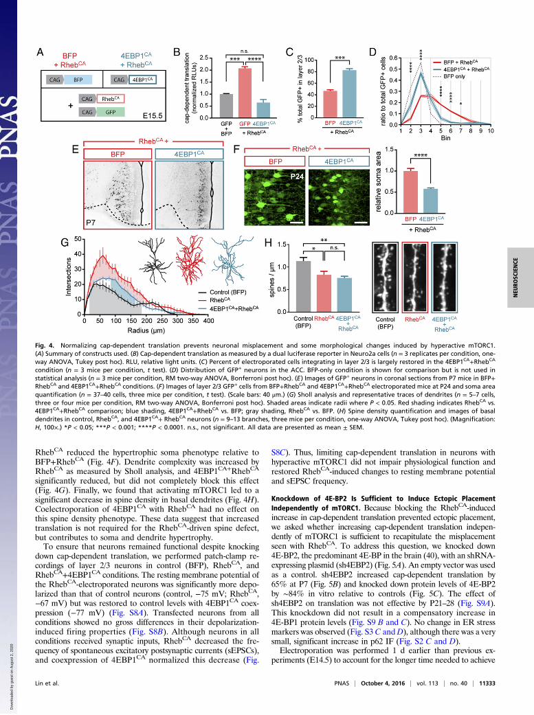
RhebCA reduced the hypertrophic soma phenotype relative toBFP+RhebCA (Fig. 4F). Dendrite complexity was increased byRhebCA as measured by Sholl analysis, and 4EBP1CA+RhebCA
significantly reduced, but did not completely block this effect(Fig. 4G). Finally, we found that activating mTORC1 led to asignificant decrease in spine density in basal dendrites (Fig. 4H).Coelectroporation of 4EBP1CA with RhebCA had no effect onthis spine density phenotype. These data suggest that increasedtranslation is not required for the RhebCA-driven spine defect,but contributes to soma and dendrite hypertrophy.To ensure that neurons remained functional despite knocking
down cap-dependent translation, we performed patch-clamp re-cordings of layer 2/3 neurons in control (BFP), RhebCA, andRhebCA+4EBP1CA conditions. The resting membrane potential ofthe RhebCA-electroporated neurons was significantly more depo-larized than that of control neurons (control, −75 mV; RhebCA,−67 mV) but was restored to control levels with 4EBP1CA coex-pression (−77 mV) (Fig. S8A). Transfected neurons from allconditions showed no gross differences in their depolarization-induced firing properties (Fig. S8B). Although neurons in allconditions received synaptic inputs, RhebCA decreased the fre-quency of spontaneous excitatory postsynaptic currents (sEPSCs),and coexpression of 4EBP1CA normalized this decrease (Fig.
S8C). Thus, limiting cap-dependent translation in neurons withhyperactive mTORC1 did not impair physiological function andrestored RhebCA-induced changes to resting membrane potentialand sEPSC frequency.
Knockdown of 4E-BP2 Is Sufficient to Induce Ectopic PlacementIndependently of mTORC1. Because blocking the RhebCA-inducedincrease in cap-dependent translation prevented ectopic placement,we asked whether increasing cap-dependent translation indepen-dently of mTORC1 is sufficient to recapitulate the misplacementseen with RhebCA. To address this question, we knocked down4E-BP2, the predominant 4E-BP in the brain (40), with an shRNA-expressing plasmid (sh4EBP2) (Fig. 5A). An empty vector was usedas a control. sh4EBP2 increased cap-dependent translation by65% at P7 (Fig. 5B) and knocked down protein levels of 4E-BP2by ∼84% in vitro relative to controls (Fig. 5C). The effect ofsh4EBP2 on translation was not effective by P21–28 (Fig. S9A).This knockdown did not result in a compensatory increase in4E-BP1 protein levels (Fig. S9 B and C). No change in ER stressmarkers was observed (Fig. S3 C andD), although there was a verysmall, significant increase in p62 IF (Fig. S2 C and D).Electroporation was performed 1 d earlier than previous ex-
periments (E14.5) to account for the longer time needed to achieve
Fig. 4. Normalizing cap-dependent translation prevents neuronal misplacement and some morphological changes induced by hyperactive mTORC1.(A) Summary of constructs used. (B) Cap-dependent translation as measured by a dual luciferase reporter in Neuro2a cells (n = 3 replicates per condition, one-way ANOVA, Tukey post hoc). RLU, relative light units. (C) Percent of electroporated cells integrating in layer 2/3 is largely restored in the 4EBP1CA+RhebCA
condition (n = 3 mice per condition, t test). (D) Distribution of GFP+ neurons in the ACC. BFP-only condition is shown for comparison but is not used instatistical analysis (n = 3 mice per condition, RM two-way ANOVA, Bonferroni post hoc). (E) Images of GFP+ neurons in coronal sections from P7 mice in BFP+RhebCA and 4EBP1CA+RhebCA conditions. (F) Images of layer 2/3 GFP+ cells from BFP+RhebCA and 4EBP1CA+RhebCA electroporated mice at P24 and soma areaquantification (n = 37–40 cells, three mice per condition, t test). (Scale bars: 40 μm.) (G) Sholl analysis and representative traces of dendrites (n = 5–7 cells,three or four mice per condition, RM two-way ANOVA, Bonferroni post hoc). Shaded areas indicate radii where P < 0.05. Red shading indicates RhebCA vs.4EBP1CA+RhebCA comparison; blue shading, 4EBP1CA+RhebCA vs. BFP; gray shading, RhebCA vs. BFP. (H) Spine density quantification and images of basaldendrites in control, RhebCA, and 4EBP1CA+ RhebCA neurons (n = 9–13 branches, three mice per condition, one-way ANOVA, Tukey post hoc). (Magnification:H, 100×.) *P < 0.05; ***P < 0.001; ****P < 0.0001. n.s., not significant. All data are presented as mean ± SEM.
Lin et al. PNAS | October 4, 2016 | vol. 113 | no. 40 | 11333
NEU
ROSC
IENCE
Dow
nloa
ded
by g
uest
on
Aug
ust 2
, 202
0
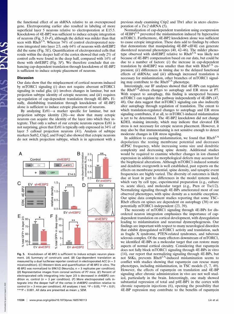
the functional effect of an shRNA relative to an overexpressedgene. Electroporating earlier also resulted in labeling of moresuperficial layer 5 neurons relative to electroporation at E15.5.Knockdown of 4E-BP2 was sufficient to induce ectopic integrationof neurons (Fig. 5 D–F), although the deficit was milder than thatseen with RhebCA. Whereas 74% of control electroporated neu-rons integrated into layer 2/3, only 64% of neurons with sh4EBP2did the same (Fig. 5E). Quantification of electroporated cells thatreside within the deeper half of the cortex showed that only 2% ofcontrol cells were found in the deep half, compared with 14% ofthose with sh4EBP2 (Fig. 5F). We therefore conclude that en-hancing cap-dependent translation through knockdown of 4E-BP2is sufficient to induce ectopic placement of neurons.
DiscussionOur data show that the misplacement of cortical neurons inducedby mTORC1 signaling (i) does not require aberrant mTORC1signaling in radial glia; (ii) involves changes in laminar, but notprojection subtype identity of ectopic neurons; and (iii) requiresup-regulation of cap-dependent translation through 4E-BPs. Fi-nally, disinhibiting translation through knockdown of 4E-BP2alone is sufficient to induce ectopic placement of neurons.By analyzing Er81—a marker specific for laminar, but not
projection subtype identity (28)—we show that many ectopicneurons can acquire the identity of the layer into which they in-tegrate. That only a subset of our ectopic neurons express Er81 isnot surprising, given that Er81 is typically only expressed in 34% oflayer 5 callosal projection neurons (41). Analysis of subtypemarkers Satb2, Ctip2, and Foxp2 also showed that ectopic neuronsdo not switch projection subtype, which is in agreement with a
previous study examining Ctip2 and Tbr1 after in utero electro-poration of a Tsc2 shRNA (5).Normalization of cap-dependent translation using coexpression
of 4EBP1CA prevented the mislamination induced by hyperactivemTORC1. Furthermore, 4E-BP2 knockdown alone was sufficientto cause ectopic integration. These data add to findings by othersthat demonstrate that manipulating 4E-BP–eIF4E can generatedisordered neuronal phenotypes (40, 42–46). The milder pheno-type observed with sh4EBP2 relative to RhebCA was likely notbecause of 4E-BP1 compensation based on our data, but could bedue to a number of factors: (i) the increase in cap-dependenttranslation by sh4EBP2 was smaller than that with RhebCA ex-pression; (ii) differences in timing due to the delayed functionaleffects of shRNAs; and (iii) although increased translation isnecessary for mislamination, other branches of mTORC1 signal-ing may contribute to the RhebCA phenotype.Interestingly, our IF analyses showed that 4E-BPs can regulate
the RhebCA-driven changes to autophagy and ER stress at P7.With respect to autophagy, this finding is unexpected becausemTORC1 can regulate this pathway independently of 4E-BPs (47,48). Our data suggest that mTORC1 signaling can also indirectlyalter autophagy through regulation of translation. The extent towhich translation-regulated changes to autophagy and ER stresssignaling contributes, if at all, to mTORC1-induced mislaminationis yet to be determined. The 4E-BP2 knockdown did not changeKDEL staining intensity, which may indicate that elevated ERstress is not necessary for ectopic neuron placement. However, itmay also be that immunostaining is not sensitive enough to detectmoderate changes in ER stress signaling.In addition to causing mislamination, we found that RhebCA
depolarizes the resting membrane potential and decreasessEPSC frequency, while increasing soma size and dendriticcomplexity and decreasing spine density. Additional studieswould be required to examine whether changes in ion channelexpression in addition to morphological defects may account forthe biophysical alterations. Although mTORC1-induced somaticand dendritic overgrowth is well established, past reports on theeffects on membrane potential, spine density, and synaptic eventfrequencies are highly varied. The diversity of outcomes is likelydue at least in part to differences in the model systems used,which vary by cell type, experimental preparation (e.g., culturevs. acute slice), and molecular target (e.g., Pten or Tsc1/2).Normalizing signaling through 4E-BPs ameliorated most of ourobserved phenotypes, with spine density as a notable exception.Our spine data complement studies reporting that some TSC–Rheb effects on spines are dependent on autophagy (38) or arepotentially mTORC1-independent (23, 39).The necessity of mTORC1 signaling through 4E-BPs for dis-
ordered neuron integration emphasizes the importance of cap-dependent translation on cortical development, with dysregulationleading to mislamination and neuronal dysmorphogenesis. Ourfindings are important with respect to many neurological disordersthat exhibit dysregulated mTORC1 activity and translation, suchas fragile X syndrome, PTEN-related syndromes, and tuberoussclerosis complex. Of the many effectors downstream of mTORC1,we identified 4E-BPs as a molecular target that can restore manyaspects of normal cortical circuitry. Considering that rapamycindoes not fully block mTORC1 signaling through 4E-BPs in vitro(14), our report that normalizing signaling through 4E-BPs, butnot S6Ks, prevents RhebCA-induced mislamination seems toconflict with studies showing that rapamycin can rescue manyphenotypes, including mislamination, in TSC models (5, 7, 49).However, the effects of rapamycin on translation and 4E-BPsignaling after chronic administration in vivo are not well stud-ied, particularly in the brain. Interestingly, one study showedsubstantial repression of total and p4E-BP1 in the cortex withchronic rapamycin injections (6), opening the possibility that4E-BP repression may contribute to the benefits of rapamycin
Fig. 5. Knockdown of 4E-BP2 is sufficient to induce ectopic neuron place-ment. (A) Summary of constructs used. (B) Cap-dependent translation asmeasured by a dual luciferase reporter construct in electroporated ACC (n = 3mice/condition). (C) Western blots and quantification of 4E-BP2 in vitro. The4E-BP2 was normalized to ERK1/2 (Neuro2a; n = 3 replicates per condition).(D) Representative images from coronal sections of P7 mice. (E) Percent ofelectroporated cells integrating into layer 2/3 is decreased in sh4EBP2 con-dition vs. control (n = 3 per condition). (F) More electroporated cells in-tegrate into the deeper half of the cortex in sh4EBP2 condition relative tocontrol (n = 3 mice per condition). All analyses: t test. *P < 0.05; **P < 0.01;***P < 0.001. All data are presented as mean ± SEM.
11334 | www.pnas.org/cgi/doi/10.1073/pnas.1605740113 Lin et al.
Dow
nloa
ded
by g
uest
on
Aug
ust 2
, 202
0
treatment. Given that rapamycin affects many processes and cancause serious side effects, understanding which effectors ofmTORC1 signaling contribute to specific phenotypes couldinform and improve options for treatment. Our data point to4E-BPs and their downstream targets as promising candidates forfurther study and therapeutic targeting.
Materials and MethodsAll procedures and protocols were approved by the Yale University InstitutionalAnimal Care and Use Committee. All mice usedwere timed pregnant CD-1mice
(Charles River) and their pups. Detailed methods can be found in SI Materialsand Methods and Table S1.
ACKNOWLEDGMENTS. We thank Drs. Hanada and Maehama (NationalInstitute of Infectious Diseases) for the RhebCA vector; Dr. Mueller(Scripps) for the Dcx-Cre construct; Dr. Blenis (Weill Cornell) for the lucif-erase reporter; and Drs. Caplan and Kim (Yale University) for use of theirluminometers. This work was supported by NIH Grant R01 NS086329, aMcKnight Endowment Fund for Neuroscience award (to A.B.), the PfizerPatricia Goldman-Rakic Fellowship (to T.V.L.), and NIH Grant GM007324(to T.J.M.).
1. Crino PB (2011) mTOR: A pathogenic signaling pathway in developmental brainmalformations. Trends Mol Med 17(12):734–742.
2. Leventer RJ, et al. (1999) Clinical and imaging features of cortical malformations inchildhood. Neurology 53(4):715–722.
3. Orlova KA, et al. (2010) STRADalpha deficiency results in aberrant mTORC1 signalingduring corticogenesis in humans and mice. J Clin Invest 120(5):1591–1602.
4. Feliciano DM, Su T, Lopez J, Platel J-C, Bordey A (2011) Single-cell Tsc1 knockoutduring corticogenesis generates tuber-like lesions and reduces seizure threshold inmice. J Clin Invest 121(4):1596–1607.
5. Tsai V, et al. (2014) Fetal brain mTOR signaling activation in tuberous sclerosis com-plex. Cereb Cortex 24(2):315–327.
6. Kassai H, et al. (2014) Selective activation of mTORC1 signaling recapitulates micro-cephaly, tuberous sclerosis, and neurodegenerative diseases. Cell Reports 7(5):1626–1639.
7. Moon UY, et al. (2015) Impaired Reelin-Dab1 signaling contributes to neuronal mi-gration deficits of tuberous sclerosis complex. Cell Reports 12(6):965–978.
8. Ehninger D, et al. (2008) Reversal of learning deficits in a Tsc2+/- mouse model oftuberous sclerosis. Nat Med 14(8):843–848.
9. Meikle L, et al. (2008) Response of a neuronal model of tuberous sclerosis to mam-malian target of rapamycin (mTOR) inhibitors: Effects on mTORC1 and Akt signalinglead to improved survival and function. J Neurosci 28(21):5422–5432.
10. Parker WE, et al. (2013) Rapamycin prevents seizures after depletion of STRADA in arare neurodevelopmental disorder. Sci Transl Med 5(182):182ra53.
11. Laplante M, Sabatini DM (2012) mTOR signaling in growth control and disease. Cell149(2):274–293.
12. Tsai PT, et al. (2013) Prenatal rapamycin results in early and late behavioral abnor-malities in wildtype C57BL/6 mice. Behav Genet 43(1):51–59.
13. Sadowski K, Kotulska K, Jó�zwiak S (2016) Management of side effects of mTOR in-hibitors in tuberous sclerosis patients. Pharmacol Rep 68(3):536–542.
14. Kang SA, et al. (2013) mTORC1 phosphorylation sites encode their sensitivity tostarvation and rapamycin. Science 341(6144):1236566.
15. Yan L, et al. (2006) Hyperactivation of mammalian target of rapamycin (mTOR) sig-naling by a gain-of-function mutant of the Rheb GTPase. J Biol Chem 281(29):19793–19797.
16. Garami A, et al. (2003) Insulin activation of Rheb, a mediator of mTOR/S6K/4E-BPsignaling, is inhibited by TSC1 and 2. Mol Cell 11(6):1457–1466.
17. Inoki K, Li Y, Xu T, Guan K-L (2003) Rheb GTPase is a direct target of TSC2 GAP activityand regulates mTOR signaling. Genes Dev 17(15):1829–1834.
18. Tee AR, Manning BD, Roux PP, Cantley LC, Blenis J (2003) Tuberous sclerosis complexgene products, Tuberin and Hamartin, control mTOR signaling by acting as a GTPase-activating protein complex toward Rheb. Curr Biol 13(15):1259–1268.
19. Bjørkøy G, et al. (2005) p62/SQSTM1 forms protein aggregates degraded by auto-phagy and has a protective effect on huntingtin-induced cell death. J Cell Biol 171(4):603–614.
20. Lee AS (2001) The glucose-regulated proteins: Stress induction and clinical applica-tions. Trends Biochem Sci 26(8):504–510.
21. Kwon C-H, Zhu X, Zhang J, Baker SJ (2003) mTor is required for hypertrophy of Pten-deficient neuronal soma in vivo. Proc Natl Acad Sci USA 100(22):12923–12928.
22. Kumar V, Zhang M-X, Swank MW, Kunz J, Wu G-Y (2005) Regulation of dendriticmorphogenesis by Ras-PI3K-Akt-mTOR and Ras-MAPK signaling pathways. J Neurosci25(49):11288–11299.
23. Tavazoie SF, Alvarez VA, Ridenour DA, Kwiatkowski DJ, Sabatini BL (2005) Regulationof neuronal morphology and function by the tumor suppressors Tsc1 and Tsc2. NatNeurosci 8(12):1727–1734.
24. Rakic P (1972) Mode of cell migration to the superficial layers of fetal monkey neo-cortex. J Comp Neurol 145(1):61–83.
25. Francis F, et al. (1999) Doublecortin is a developmentally regulated, microtubule-as-sociated protein expressed in migrating and differentiating neurons. Neuron 23(2):247–256.
26. Gleeson JG, Lin PT, Flanagan LA, Walsh CA (1999) Doublecortin is a microtubule-associated protein and is expressed widely by migrating neurons. Neuron 23(2):257–271.
27. Nieto M, et al. (2004) Expression of Cux-1 and Cux-2 in the subventricular zone andupper layers II-IV of the cerebral cortex. J Comp Neurol 479(2):168–180.
28. Hevner RF, et al. (2003) Beyond laminar fate: Toward a molecular classification ofcortical projection/pyramidal neurons. Dev Neurosci 25(2-4):139–151.
29. Alcamo EA, et al. (2008) Satb2 regulates callosal projection neuron identity in thedeveloping cerebral cortex. Neuron 57(3):364–377.
30. Arlotta P, et al. (2005) Neuronal subtype-specific genes that control corticospinalmotor neuron development in vivo. Neuron 45(2):207–221.
31. Ferland RJ, Cherry TJ, Preware PO, Morrisey EE, Walsh CA (2003) Characterization ofFoxp2 and Foxp1 mRNA and protein in the developing and mature brain. J CompNeurol 460(2):266–279.
32. Choi KM, McMahon LP, Lawrence JC, Jr (2003) Two motifs in the translational re-pressor PHAS-I required for efficient phosphorylation by mammalian target of ra-pamycin and for recognition by raptor. J Biol Chem 278(22):19667–19673.
33. Schalm SS, Fingar DC, Sabatini DM, Blenis J (2003) TOS motif-mediated raptor bindingregulates 4E-BP1 multisite phosphorylation and function. Curr Biol 13(10):797–806.
34. Li S, et al. (2002) Translational control of cell fate: Availability of phosphorylation siteson translational repressor 4E-BP1 governs its proapoptotic potency. Mol Cell Biol22(8):2853–2861.
35. Gong X, et al. (2015) Activating the translational repressor 4E-BP or reducing S6K-GSK3β activity prevents accelerated axon growth induced by hyperactive mTORin vivo. Hum Mol Genet 24(20):5746–5758.
36. Jaworski J, Spangler S, Seeburg DP, Hoogenraad CC, Sheng M (2005) Control ofdendritic arborization by the phosphoinositide-3′-kinase-Akt-mammalian target ofrapamycin pathway. J Neurosci 25(49):11300–11312.
37. Bateup HS, Takasaki KT, Saulnier JL, Denefrio CL, Sabatini BL (2011) Loss of Tsc1in vivo impairs hippocampal mGluR-LTD and increases excitatory synaptic function.J Neurosci 31(24):8862–8869.
38. Tang G, et al. (2014) Loss of mTOR-dependent macroautophagy causes autistic-likesynaptic pruning deficits. Neuron 83(5):1131–1143.
39. Yasuda S, et al. (2014) Activation of Rheb, but not of mTORC1, impairs spine synapsemorphogenesis in tuberous sclerosis complex. Sci Rep 4:5155.
40. Banko JL, et al. (2005) The translation repressor 4E-BP2 is critical for eIF4F complexformation, synaptic plasticity, and memory in the hippocampus. J Neurosci 25(42):9581–9590.
41. Yoneshima H, et al. (2006) Er81 is expressed in a subpopulation of layer 5 neurons inrodent and primate neocortices. Neuroscience 137(2):401–412.
42. Banko JL, Hou L, Poulin F, Sonenberg N, Klann E (2006) Regulation of eukaryoticinitiation factor 4E by converging signaling pathways during metabotropic glutamatereceptor-dependent long-term depression. J Neurosci 26(8):2167–2173.
43. Banko JL, et al. (2007) Behavioral alterations in mice lacking the translation repressor4E-BP2. Neurobiol Learn Mem 87(2):248–256.
44. Ran I, et al. (2013) Selective regulation of GluA subunit synthesis and AMPA receptor-mediated synaptic function and plasticity by the translation repressor 4E-BP2 inhippocampal pyramidal cells. J Neurosci 33(5):1872–1886.
45. Gkogkas CG, et al. (2013) Autism-related deficits via dysregulated eIF4E-dependenttranslational control. Nature 493(7432):371–377.
46. Santini E, et al. (2013) Exaggerated translation causes synaptic and behavioural ab-errations associated with autism. Nature 493(7432):411–415.
47. Hosokawa N, et al. (2009) Nutrient-dependent mTORC1 association with the ULK1-Atg13-FIP200 complex required for autophagy. Mol Biol Cell 20(7):1981–1991.
48. Jung CH, et al. (2009) ULK-Atg13-FIP200 complexes mediate mTOR signaling to theautophagy machinery. Mol Biol Cell 20(7):1992–2003.
49. Ehninger D (2013) From genes to cognition in tuberous sclerosis: Implications formTOR inhibitor-based treatment approaches. Neuropharmacology 68:97–105.
50. Niwa H, Yamamura K, Miyazaki J (1991) Efficient selection for high-expressiontransfectants with a novel eukaryotic vector. Gene 108(2):193–199.
51. Matsuda T, Cepko CL (2004) Electroporation and RNA interference in the rodentretina in vivo and in vitro. Proc Natl Acad Sci USA 101(1):16–22.
52. Ventura A, et al. (2004) Cre-lox-regulated conditional RNA interference from trans-genes. Proc Natl Acad Sci USA 101(28):10380–10385.
53. Rodriguez A, Ehlenberger DB, Dickstein DL, Hof PR, Wearne SL (2008) Automatedthree-dimensional detection and shape classification of dendritic spines from fluo-rescence microscopy images. PLoS One 3(4):e1997.
54. Choo AY, Yoon S-O, Kim SG, Roux PP, Blenis J (2008) Rapamycin differentially inhibitsS6Ks and 4E-BP1 to mediate cell-type-specific repression of mRNA translation. ProcNatl Acad Sci USA 105(45):17414–17419.
55. Maehama T, et al. (2008) RalA functions as an indispensable signal mediator for thenutrient-sensing system. J Biol Chem 283(50):35053–35059.
56. Bae EJ, et al. (2012) Liver-specific p70 S6 kinase depletion protects against hepaticsteatosis and systemic insulin resistance. J Biol Chem 287(22):18769–18780.
57. Franco SJ, Martinez-Garay I, Gil-Sanz C, Harkins-Perry SR, Müller U (2011) Reelinregulates cadherin function via Dab1/Rap1 to control neuronal migration and lami-nation in the neocortex. Neuron 69(3):482–497.
Lin et al. PNAS | October 4, 2016 | vol. 113 | no. 40 | 11335
NEU
ROSC
IENCE
Dow
nloa
ded
by g
uest
on
Aug
ust 2
, 202
0


![Graph Normalizing Flows · 2.2 Normalizing Flows Normalizing flows (NFs) [22, 3, 4] are a class of generative models that use invertible mappings to transform an observed vector](https://img.pdfslide.us/doc/110x75/5f37164f015bfa67bd3ee458/graph-normalizing-flows-22-normalizing-flows-normalizing-iows-nfs-22-3-4.jpg)