Embed Size (px)
Citation preview

Neuron
Article
Normalization Regulates Competitionfor Visual AwarenessSam Ling1,* and Randolph Blake1,21Vanderbilt Vision Research Center, Vanderbilt University, Nashville, TN 37204, USA2Department of Brain and Cognitive Sciences, Seoul National University, Seoul 151-746, South Korea
*Correspondence: [email protected]://dx.doi.org/10.1016/j.neuron.2012.05.032
SUMMARY
Signals in our brain are in a constant state of compe-tition, including those that vie for motor control,sensory dominance, and awareness. To shed lighton the mechanisms underlying neural competition,we exploit binocular rivalry, a phenomenon thatallows us to probe the competitive process thatordinarily transpires outside of our awareness. Bymeasuring psychometric functions under differentstates of rivalry, we discovered a pattern of gainchanges that are consistent with a model of compe-tition in which attention interacts with normalizationprocesses, thereby driving the ebb and flow betweenstates of awareness. Moreover, we reveal that atten-tion plays a crucial role in modulating competition;without attention, rivalry suppression for high-contrast stimuli is negligible. We propose a frame-work whereby our visual awareness of competingsensory representations is governed by a commonneural computation: normalization.
INTRODUCTION
Our visual environment is brimming with information, but the high
bioenergetic costs of cortical computations limit how much
of that information can be effectively processed at any given
moment (Lennie, 2003). Because of this limitation, the brain is
chronically dealing with competition among neural representa-
tions of objects and events. One prominent mechanism for regu-
lating competing neural signals is attention, which allows us to
selectively process relevant information (Reynolds and Chelazzi,
2004). A recent model proposes that attention shapes percep-
tion bymeans of a normalization framework, whereby attentional
modulation hinges on three critical factors: the locus of atten-
tional modulation, the size of the attended stimulus, and the
size of the attentional window (Reynolds and Heeger, 2009;
Herrmann et al., 2010). Changes in any of these factors can tip
the balance between neuronal excitatory and inhibitory
processes, thereby impacting how attention affects perception
(Reynolds and Heeger, 2009).
Is this normalization framework a general property of visual
competition? Although attention can deftly regulate neural repre-
sentations, the process of conflict resolution is not always so
seamless. In some instances, the visual system struggles to
reconcile competing sensory information, a compelling example
being when dissimilar images are presented to the two eyes.
In this case, visual awareness alternates between the two
images, creating the phenomenon known as binocular rivalry
(Blake and Logothetis, 2002; Leopold and Logothetis, 1999;
Tong et al., 2006; Wheatstone, 1838). Binocular rivalry offers
a unique opportunity to probe competitive processes within
the brain, by allowing us to see with our own eyes a process
that ordinarily transpires outside of our awareness, namely
dynamic competition between neural representations (Blake
and Logothetis, 2002; Leopold and Logothetis, 1999). Here, we
explore whether a common neural computation may mediate
competitive processes embodied in rivalry and in attention.
The notion that attention and rivalry are intertwined has been
debated for over a century (von Helmholtz, 1909; James, 1890;
Lack, 1978; Zhang et al., 2011; Watanabe et al., 2011), and
some have gone so far as to directly attribute the alternations
in visual awareness to switches in attention (von Helmholtz,
1909; Lack, 1978). Moreover, a growing body of research
suggests that modulation of visual awareness through rivalry in
early visual cortices depends on attentional state (Lee et al.,
2007; Zhang et al., 2011; Watanabe et al., 2011). However, the
mechanisms subserving these interactions between attention
and rivalry remain unknown. Here, we develop and test the
idea that attention and rivalry reconcile competing visual infor-
mation via a common framework, one in which modulation
of awareness through rivalry interacts with attention. We
propose a computation model for visual competition, whereby
modulation of competing neural signals relies on interactions
between normalization and attention: gain modulation depends
on the size of the competitor stimulus and the attentional state.
Finally, we empirically test a core prediction of this com-
putational model, revealing that the degree of suppression
between competing neural representations is regulated by
attentional state.
Experiment 1: Contrast Response and Visual AwarenessThe normalization model of attention makes a very clear predic-
tion: changing the size of the ‘‘attentional field’’ relative to
the stimulus will differentially modulate the signal’s contrast
response, causing either contrast gain or response gain modula-
tion depending on the configuration (Reynolds and Heeger,
2009; Herrmann et al., 2010). Under binocular rivalry, the stim-
ulus presented in one eye typically abolishes the visibility of a rival
stimulus in the other eye. For the moment, we propose that the
Neuron 75, 531–540, August 9, 2012 ª2012 Elsevier Inc. 531

A B Figure 1. Examples of Competing Stimuli
Used in the Experiment and Their Predicted
Impact on Contrast Response Functions
(A) In some trials, the probe stimulus was dichop-
tically suppressed by a large stimulus. Under this
configuration, the normalization model predicts
a contrast gain shift, with the largest effects
occurring at mid-contrasts, and little-to-no effect
at low and high contrasts.
(B) In other trials, the probe stimulus was the same
size as the stimulus in the competing eye. Under
this configuration, the normalization framework
predicts both a shift in the contrast gain, as well as
an attenuation of the response gain, with the
largest effects occurring at high contrasts.
Neuron
Normalization Regulates Visual Awareness
‘‘attentional field’’ bemore generally conceptualized as a ‘‘modu-
latory field’’ that can either boost the response to a stimulus, as is
the case with attention, or attenuate the response to a stimulus,
as is the case with rivalry suppression.
With rivalry, the size of this modulatory field can be directly
controlled by changing the size of a stimulus in one eye relative
to the other. With standard models of binocular normalization,
introducing a stimulus in a competing eye should contribute to
the pooled inhibitory component of normalization (Ding and
Sperling, 2006; Moradi and Heeger, 2009), which predicts shifts
in contrast gain (strongest effects at mid-contrasts), but not in
response gain (strongest effects at high contrasts), regardless
of size (Supplemental Information). However, if rivalry also
includes a process that behaves like attention, the shape of
contrast response functions for attenuated signals should
differ depending on the size of the dominant stimulus in the
other eye—a manipulation that would alter the size of the modu-
latory field. Specifically, when the dominant stimulus is substan-
tially larger than the stimulus in the other eye, thereby evoking
a large modulatory field, the normalization framework of
attention predicts a reduction in contrast gain for the probe
stimulus (Figure 1A). However, when the dominant stimulus
evokes a small modulatory field, the contrast response functions
should transition toward a reduction in the response gain
(Figure 1B).
To explore whether normalization modulates visual competi-
tion, we examined how psychometric functions change for an
attenuated stimulus under rivalry, and whether those changes
depend on the size of the putative modulatory field. We
measured observers’ ability to discriminate fine changes in the
orientation of a probe stimulus (4� clockwise or counterclock-
wise) that was either presented monocularly, or was suppressed
under binocular rivalry (Figure 2). To control the size of themodu-
latory field in the rivalry conditions, we manipulated the size of
the dominant competing stimulus such that in some trials, it
was either the same size as the probe (small: 1.5�), somewhat
larger than the probe (medium: 2.5�), or substantially larger
532 Neuron 75, 531–540, August 9, 2012 ª2012 Elsevier Inc.
(large: 8�). The rms contrast of the probe
stimuli ranged from 0.8%–23%, allowing
us to measure the entire psychometric
function, a behavioral measure that
scales proportionally to the signal-to-
noise ratio of the underlying contrast response function
(Herrmann et al., 2010; Pestilli et al., 2009). Specifically, changes
in the neural contrast response function under this framework
directly impacts an observer’s ability to discriminate orienta-
tion changes in the probe, which would, in turn, be reflected
in corresponding changes to the behavioral psychometric
functions.
RESULTS
Experiment 1Rivalry had a substantial impact on psychometric functions
(Figure 3A). Specifically, the size of the dominant stimulus
evoked notable qualitative differences in implied contrast
response functions: whereas a large, dominant stimulus solely
shifted the contrast gain, a intermediary dominant stimulus
reduced both the response gain and the contrast gain, and a
small dominant stimulus further reduced the response gain. To
quantify these effects, we fit the data for each observer with
Naka-Rushton functions (Herrmann et al., 2010; Pestilli et al.,
2009; Naka and Rushton, 1966; Ling et al., 2010), for which
two key parameters are predicted to change under the normali-
zation framework: C50 and d0max. These parameters have been
used in previous psychophysics studies as metrics for changes
in contrast gain and response gain. The C50 parameter corre-
sponds to the semi-saturation constant, and changes in this
parameter with rivalry suppression indicate a contrast gain shift.
The d0max parameter corresponds to the asymptotic response
at high contrasts, and changes in this parameter indicate a
response gain reduction. Parameter estimates revealed a pattern
consistent with predictions of the normalization model of atten-
tion: C50 shifted toward higher contrasts for dominant stimuli
regardless of their size, whereas d0max was attenuated the
most when the dominant stimulus was the same size as the
probe stimulus. Consistent with these results, response gain-
like modulation has previously been found with rivalry when
similar-sized stimuli are pit against each other, both in single-unit

Figure 2. Example Trial Sequence in Experiment 1
Two competing band-pass-filtered noise stimuli were viewed dichoptically. To
control rivalry state, we used the flash suppression technique. Following flash-
induced suppression, the probe in the suppressed eye changed orientation
slightly (4� clockwise or counterclockwise), and observers reported the
direction of tilt of that probe (two-alternative, forced-choice task).
Neuron
Normalization Regulates Visual Awareness
(Sengpiel and Blakemore, 1994) and behavioral studies (Ling
et al., 2010; Watanabe et al., 2004).
Fitting the data separately for each individual yielded a similar
pattern of results (Figures 3B and 3C; Figure S1 available online).
When the dominant stimulus was large, there was solely a
change in C50 for all observers (Figure 3B), with no change in
d0max (Figure 3C). However, as the size of the competitor
approached that of the probe, changes in both C50 and d0max
emerged. While standard normalization models would only
predict a contrast gain shift (Moradi and Heeger, 2009), our
results indicate that an additional mechanism is needed to
account for our results; indeed, the conjoint reduction in both
contrast gain (C50) and response gain (d0max) when the dominant
stimulus is small is a prediction borne from the normalization
model of attention for scenarios where the probe is small and
the modulatory field is roughly the same size (Reynolds and
Heeger, 2009).
One alternative explanation for the large competitor’s inability
to suppress high contrast probes is center-surround interactions
that plausibly could weaken the strength of the center region of
the competing stimulus. Although center-surround inhibition
has been shown to be least effective in the fovea (Petrov et al.,
2005), the retinal region targeted by our stimuli, we sought to
rule out this alternative explanation explicitly by performing an
additional control experiment, where we measured the degree
to which the surround region of the large stimulus attenuated
its center portion (Figure S2).
In a given trial of the control experiment, observers were
shown two consecutive displays: one with a surround, and one
without a surround (test stimulus). The size of the surround
matched the size of the large suppressor stimulus in our main
study (8�). Observers performed a 2-interval forced choice
task, indicating which one of the two intervals contained a center
stimulus with higher perceived contrast. We used an adaptive
staircase procedure to estimate the contrast the Test needed
to match the perceived contrast of the stimulus in the presence
of a surround. Using this task, we found that the surround
reduced perceived contrast of the target by only�0.08 log-units,
implying the involvement of very weak surround suppression at
best; with the surround, a 23%contrast stimulus appeared to ob-
servers as if it were between 18%–19% rms contrast (S1 =
18.2%, S2 = 18.9%, S3 = 19.2%, S4 = 19%).
To verify that thisminor reduction in apparent contrast near the
center region could not account for our rivalry results with the
large competitor, we measured contrast psychometric functions
for the same observers with the small rivalry suppressor, after
dropping the physical contrast of this competitor down to 15%
rms contrast. With this small, lower-contrast competitor, we still
found a substantial reduction in the response gain for psycho-
metric functions (Figure 4; Figure S3), thereby ruling out
center-surround suppression as a possible explanation for our
results.
Could the large competitor’s inability to suppress high con-
trast probes result from impaired fusion between the eyes,
arising from the size disparity (Ooi and He, 2006) between the
large competitor and the smaller probe? To explicitly rule out
this explanation, we conducted an additional control experiment
in which the large competitor was once again pitted against the
smaller probe, but with additional circular fusion markers pre-
sented to both eyes, surrounding the probe region (1.75�). Ifthe effects we observed were due to differences in fusion
between size conditions, the contrast psychometric functions
should now resemble that of the smaller competitor: a response
gain reduction. But that was not the case, because the additional
fusion markers failed to alter the contrast gain-like pattern
of suppression evoked by the large competitor (Figure 5;
Figure S4).
Normalization Model of Visual CompetitionTaken together, the results presented above can be construed to
mean that the regulation of visual competition, whether through
attention or through rivalry, relies on normalization. A central idea
behind the normalization model for attention is that the modula-
tory field augments the strength of a stimulus prior to divisive
normalization (Reynolds and Heeger, 2009); were that not the
case, then themodulatory signaturewould always be one of con-
trast gain. Moreover, standard models of normalization (Ding
and Sperling, 2006; Moradi and Heeger, 2009), when applied
to binocular representation, only predict a pure contrast gain
shift. Our results, however, clearly show that the response func-
tion can be modulated under rivalry by both contrast gain and
response gain, implying that there must be an additional modu-
latory field at play during visual competition—a mechanism with
the same signature of effects as those associated with the
normalization model of attention.
What is the source of this additional modulatory field compo-
nent in rivalry? One plausible candidate is, in fact, attention as
embodied in a recently proposed normalization model (Reynolds
and Heeger, 2009): when a stimulus is suppressed from
awareness during rivalry, attention may be directed toward the
competing, dominant stimulus, rather than the suppressed
probe. This dominant stimulus may thus act much like
Neuron 75, 531–540, August 9, 2012 ª2012 Elsevier Inc. 533

A B
C
Figure 3. The Effect of Rivalry on Contrast
Psychometric Functions
(A) Example psychometric functions from one
observer. When the competing stimulus was the
same size as the probe, we found both a reduction
in the asymptote, or response gain, as well as
a shift in the probe’s psychometric function, or
contrast gain. However, as the size of the
competitor increased, the response gain modula-
tion decreased, and when the competing stimulus
was much larger than the probe, the psychometric
functions only shifted in their contrast gain.
(B) C50 estimates for each observer, indexing
changes in contrast gain. We found decreased
contrast gain (higher C50) under suppression
relative to an unsuppressed stimulus, regardless
of the size of the competing stimulus.
(C) d0max estimates for each observer, indexing
changes in response gain. We found the strongest
decrease in the response gain (lower d0max) when
the competing stimulus was the same size as the
probe stimulus, and this reduction in response
gain diminished as the competitor size increased.
Error bars are bootstrapped 95% confidence
intervals.
See Figure S1.
Neuron
Normalization Regulates Visual Awareness
a modulatory attentional field, withdrawing attentional resources
from the suppressed probe across a spatial extent that spans the
size of the dominant stimulus. The impact of this withdrawal of
attention would depend on the size of the modulatory field. A
small modulatory field would solely decrease the response in
the center region of a suppressed probe stimulus, tipping the
balance between excitation and inhibition (Sundberg et al.,
2009) in favor of the inhibitory component and thus causing
both a reduction in both contrast gain and response gain. A large
modulatory field, however, would decrease the response to the
probe across a much larger spatial extent, thus maintaining the
balance between excitation and inhibition and causing only
a contrast gain shift.
This relationship between attention and awareness, and their
combined impact on a probe stimulus, can be formalized in the
normalization framework (Figure 6). The normalization model
proposes that the response to a stimulus is comprised of an
excitatory component that is divided by an inhibitory component
(Heeger, 1992). The neural response to a stimulus, Rp, can thus
be expressed as,
Rp =agPCP
gPCP +ubPCP +gSbSCS + s(Equation 1)
where CP is the contrast of the probe stimulus in one eye
(between 0 and 1), CS represents the contrast of the competing
stimulus in the other eye, s determines the contrast gain
(contrast at which neural response reaches half its maximum),
a is the maximum attainable response, gP and gS represent
the peak attentional gain for the suppressed probe stimulus
ðgPÞ and the competitor ðgSÞ, and u determines the relative
impact of the modulatory field on the surround region of the
probe. Note that an additional exponent parameter, n, would
need to be added to account for nonlinearities in signal transduc-
tion (i.e., Cn). However, for simplicity we have left that out of the
534 Neuron 75, 531–540, August 9, 2012 ª2012 Elsevier Inc.
models; the model predictions would be qualitatively similar with
or without this nonlinearity.
To model stimuli of varying sizes, the stimulus in each eye is
broken down into two components: the center and the surround.
First, consider the probe stimulus. We represent the strength of
the center region as CP, and the surround as bPCP, where bPis a scaling factor on the suppression driven by the surround
region. A large probe would encompass a large surround
ðbPz1Þ, whichwould increase the suppressive drive, attenuating
its maximum attainable contrast response (lowered asymptote).
Next, consider the competing stimulus in the other eye.
This stimulus is treated identically to the probe, with CS corre-
sponding to the center region of the competitor, and bSCS corre-
sponding to the surround, where bS is a scaling factor on the
suppression driven by the surround region of the competitor.
Much like the probe, a large competitor stimulus will encompass
a large surround ðbSz1Þ, which will also increase the suppres-
sive drive, lowering the asymptotic response. We can model
the condition where the probe was viewed in the absence of
any competitor in the other eye by simply setting the contrast
of the other eye’s stimulus to zero ðCS = 1Þ.We modeled the response to the probe stimulus assuming
that attention plays a critical role in visual awareness. Specifi-
cally, we assume that the dominant stimulus during rivalry
receives more attentional resources than the suppressed stim-
ulus: there is high attentional gain directed toward the features
of the probe when it is dominant, which we denote with gP>1,
but when the competitor in the other eye is dominant, that domi-
nant competitor receives the lion’s share of attentional
resources instead, leaving only a small portion of attentional
resources directed toward the representation of the suppressed
probe stimulus, which we denote with gP>gS. While this modu-
latory field could be the result of feature-based attention and
spatial attention, note that recent evidence suggests that
A B
C
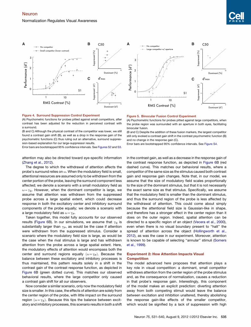
Figure 4. Surround Suppression Control Experiment
(A) Psychometric functions for probes pitted against small competitors, after
contrast has been adjusted for the reduction in perceived contrast with
a surround.
(B and C) Although the physical contrast of the competitor was lower, we still
found a contrast gain shift (B), as well as a drop in the response gain of the
psychometric functions (C) thus ruling out an alternative, surround suppres-
sion-based explanation for our large-suppressor results.
Error bars are bootstrapped 95% confidence intervals. See Figures S2 and S3.
A B
C
Figure 5. Binocular Fusion Control Experiment
(A) Psychometric functions for probes pitted against large competitors, when
the probe region was surrounded with an aperture in both eyes, facilitating
binocular fusion.
(B and C) Despite the addition of these fusion markers, the largest competitor
still only evoked a contrast gain shift in the contrast psychometric function (B)
and no change in the response gain (C).
Error bars are bootstrapped 95% confidence intervals. See Figure S4.
Neuron
Normalization Regulates Visual Awareness
attention may also be directed toward eye-specific information
(Zhang et al., 2012).
The degree to which the withdrawal of attention affects the
probe’s surround relies on u. When the modulatory field is small,
attentional resources are assumed only to bewithdrawn from the
center portion of the probe, leaving the surround component less
affected; we denote a scenario with a small modulatory field as
u=gS. However, when the dominant competitor is large, we
assume that attention was withdrawn from the suppressed
probe across a large spatial extent, which could decrease
response in both the excitatory center and inhibitory surround
components of the probe equally; we denote a scenario with
a large modulatory field as u=gP.
Taken together, this model fully accounts for our observed
results (Figure 6B). In our simulations, we assume that gS is
substantially larger than gP, as would be the case if attention
were withdrawn from the suppressed stimulus. Consider a
scenario where the modulatory field size is large, as would be
the case when the rival stimulus is large and has withdrawn
attention from the probe across a large spatial extent. Here,
the modulatory effects of attention would encompass both the
center and surround regions equally ðu=gPÞ. Because the
balance between these excitatory and inhibitory processes is
thus maintained, this pattern results solely in a shift in the
contrast gain of the contrast response function, as depicted in
Figure 6B (green dotted curve). This matches our observed
behavioral results, where the large competitor only caused
a contrast gain shift for all our observers.
Now consider a similar scenario, only now themodulatory field
size is smaller. In this case, the effects of attention are solely from
the center region of the probe, with little impact on the surround
region ðu=gSÞ. Because this tips the balance between excit-
atory and inhibitory processes, this scenario results in both a shift
in the contrast gain, as well as a decrease in the response gain of
the contrast response function, as depicted in Figure 6B (red
dashed curve). This matches our behavioral results, where a
competitor of the same size as the stimulus caused both contrast
gain and response gain changes. Note that, in our model, we
assume that the size of modulatory field scales proportionally
to the size of the dominant stimulus, but that it is not necessarily
the exact same size as that stimulus. Specifically, we assume
that the modulatory field is smaller than the dominant stimulus,
and thus the surround region of the probe is less affected by
the withdrawal of attention. This could come about simply
because the attentional field size is Gaussian-like in shape,
and therefore has a stronger effect in the center region than it
does on the outer region. Indeed, spatial attention can be
directed to a specific region of an object (Vecera et al., 2000),
even when there is no visual boundary present to ‘‘halt’’ the
spread of attention across the object (Hollingworth et al.,
2012), as was the case in our experiment. Moreover, attention
is known to be capable of selecting ‘‘annular’’ stimuli (Somers
et al., 1999).
Experiment 2: How Attention Impacts VisualCompetitionThe model advanced here proposes that attention plays a
key role in visual competition: a dominant, small competitor
withdraws attention from the center region of the probe stimulus
and, as the consequence of normalization, causes a reduction
in that probe’s response gain. Interestingly, this component
of the model makes an explicit prediction: diverting attention
away from both competing stimuli would leave the balance
between excitation and inhibition unaltered, thereby abolishing
the response gain-like effects of the smaller competitor,
which would be signified by a lack of suppression with high
Neuron 75, 531–540, August 9, 2012 ª2012 Elsevier Inc. 535
A
B C
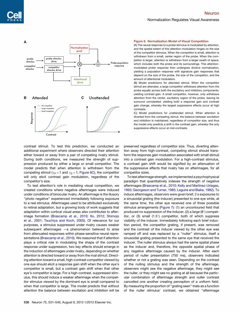
Figure 6. Normalization Model of Visual Competition
(A) The neural response to a probe stimulus is modulated by attention,
and the spatial extent of this attention modulation hinges on the size
of the competitor stimulus. When the competitor is small, attention is
withdrawn from a small, center region of the probe. When the com-
petitor is larger, attention is withdrawn from a larger swath of space,
which includes both the probe and its surroundings. This attention-
modulated probe response then undergoes divisive normalization,
yielding a population response with signature gain responses that
depend on the size of the probe, the size of the competitor, and the
amount of attentional modulation.
(B) Model predictions for attended stimuli. When the competitor
stimuli are attended, a large competitor withdraws attention from the
probe equally across both the excitatory and inhibitory components:
yielding contrast gain. A small competitor, however, only withdraws
attention from the center, excitatory region of the probe, leaving its
surround unmolested: yielding both a response gain and contrast
gain change, whereby the largest suppressive effects occur at high
contrasts.
(C) Model predictions for unattended stimuli. When attention is
diverted from the competing stimuli, the balance between excitation
and inhibition is maintained, regardless of competitor size, and thus
the model only predicts a shift in the contrast gain, whereby the only
suppressive effects occur at mid-contrasts.
Neuron
Normalization Regulates Visual Awareness
contrast stimuli. To test this prediction, we conducted an
additional experiment where observers directed their attention
either toward or away from a pair of competing rivalry stimuli.
During both conditions, we measured the strength of sup-
pression produced by either a large or small competitor. The
model predicts that when attention is withdrawn from the
competing stimuli (gP = 1 and gS = 1; Figure 6C), the competitor
will only elicit contrast gain modulation, regardless of the
competitor’s size.
To test attention’s role in mediating visual competition, we
created conditions where negative afterimages were induced
under conditions of binocular rivalry. An afterimage is the illusory
‘‘photo negative’’ experienced immediately following exposure
to a real stimulus. Afterimages used to be attributed exclusively
to retinal adaptation, but a growing body of work suggests that
adaptation within cortical visual areas also contributes to after-
image formation (Brascamp et al., 2010; Ito, 2012; Shimojo
et al., 2001; Tsuchiya and Koch, 2005). Of relevance for our
purposes, a stimulus suppressed under rivalry causes weaker
subsequent afterimages —a phenomenon believed to arise
from attenuated responses within phase-sensitive neural repre-
sentations (Brascamp et al., 2010). We reasoned that if attention
plays a critical role in modulating the shape of the contrast
response under suppression, two key effects should emerge in
the induction of afterimages under rivalry, depending on whether
attention is directed toward or away from the rival stimuli. Direct-
ing attention toward a small, high-contrast competitor viewed by
one eye should elicit a response gain shift when the other eye’s
competitor is small, but a contrast gain shift when that other
eye’s competitor is large. For a high-contrast, suppressed stim-
ulus, this should induce a weaker afterimage when the compet-
itor stimulus viewed by the dominant eye is small compared to
when that competitor is large. The model predicts that without
attention the balance between excitation and inhibition will be
536 Neuron 75, 531–540, August 9, 2012 ª2012 Elsevier Inc.
preserved regardless of competitor size. Thus, diverting atten-
tion away from high-contrast, competing stimuli should trans-
form the response gain modulation associated with small stimuli
into a contrast gain modulation. For a high-contrast stimulus,
a contrast gain shift would be signified by an attenuation of
the suppressive effects that rivalry has on afterimages, for all
competitor sizes.
To test afterimage strength,we implemented apsychophysical
paradigm that quantitatively indexes the strength of negative
afterimages (Brascamp et al., 2010; Kelly and Martinez-Uriegas,
1993; Georgeson and Turner, 1985; Leguire and Blake, 1982). To
induce afterimages, observers were given brief, 2 s exposures to
a sinusoidal grating (the inducer) presented to one eye while, at
the same time, the other eye received one of three possible
stimulus arrangements (Figure 7): (1) an uncontoured field that
produced no suppression of the inducer, (2) a large (8�) compet-
itor, or (3) small (1.5�) competitor, both of which suppress
visibility of the inducer. Immediately following each brief induc-
tion period, the competitor grating, if present, was removed
and the contrast of the inducer viewed by the other eye was
ramped off and was replaced by a ‘‘nuller’’ stimulus, itself a
sinusoidal grating presented to the same eye that received the
inducer. The nuller stimulus always had the same spatial phase
as the inducer and, therefore, the opposite spatial phase of
any negative afterimage caused by the inducer. After each
period of nuller presentation (750 ms), observers indicated
whether or not a grating was seen. Depending on the contrast
of the nulling stimulus and the strength of the afterimage,
observers might see the negative afterimage, they might see
the nuller, or they might see no grating at all because the partic-
ular combination of afterimage strength and nuller contrast
cancelled one another creating perception of a uniform field.
Bymeasuring the proportion of ‘‘grating seen’’ trials as a function
of the nuller stimulus’ contrast, we obtained ‘‘afterimage

Figure 7. Example Trial Sequence in Experiment 2
A phase-reversing noise stimulus in one eye (Competitor; 10 Hz) was pitted
against a sinusoidal grating in the other eye (Inducer). The ‘‘Dominant eye’’
received one of three possible stimulus arrangements: (1) an uncontoured field
that produced no suppression of the inducer, (2) a large (8�) competitor, or (3)
small (1.5�) competitor; the latter two suppress visibility of the inducer. After
viewing this display for 2 s, these stimuli disappeared, and a Nuller stimulus
appeared in the same eye that the inducer had been in. This was followed
by a mask stimulus, during which observers indicated whether or not a grating
was seen. To direct attention toward the competing stimuli, observers de-
tected orientation changes that occurred stochastically while the competitor
stimulus was dominant. To divert attention away from the competing stimuli,
observers performed a letter identification task (RSVP task), detecting target
letters within a rapid stream of distractor letters appearing in the periphery.
Neuron
Normalization Regulates Visual Awareness
functions’’ that approximated invertedGaussian curves. The nul-
ler contrast at which afterimage functions reach their troughs
(the mean of an inverted Gaussian function) corresponds to the
physical contrast required to nullify the negative afterimage,
thus providing a quantitative measure of the afterimage strength.
To direct attention toward the competing stimuli, we had
observers detect orientation changes that occurred stochasti-
cally while the competitor stimulus was dominant. To divert
attention away from the competing stimuli, we required
observers to perform a letter identification task (RSVP task), de-
tecting target letters within a stream of distractor letters appear-
ing in the periphery.
Experiment 2 ResultsTurning first to the condition in which attention was directed
toward the visual competition, we observed the typical
U-shaped afterimage function, regardless whether there was
a competitor or not, and regardless of the competitor’s size.
However, the troughs of these functions differed, implying that
afterimage strength depended on stimulus size. We observed
no difference in afterimage strength between the large compet-
itor and no-competitor conditions (Figure 8A; Figure S5A). This is
consistent with the contrast gain shift observed in the first
experiment, whereby the modulatory effects of suppression
are weak-to-nonexistent at high stimulus contrasts. However,
we discovered significantly weakened afterimages when the
small competitor was pitted against the inducer (Figure 8A;
Figure S5A). This pattern of results is consistent with the
response gain reduction we observed in the first experiment,
whereby the modulatory effects of suppression are greatest at
high stimulus contrasts. This is also consistent with previous
reports showing that rivalry between similarly sized small
competitors can attenuate afterimage formation (Brascamp
et al., 2010). To quantify the impact of suppression on afterimage
strength, we fit the data for each observer with inverted modified
Gaussian functions, where the estimated mean provides the
index of afterimage strength. The afterimage strength indices
reveal the same pattern of effects for all observers: while after-
image strength was unaltered by a large competitor, afterimage
strength was diminished by a small competitor (Figure 8B;
Figure S5A).
We measured a distinctly different pattern of effects when
attention was diverted away from the visual competition. While
unattended stimuli still evoked negative afterimages, we found
that without attention the competitor had no effect on afterimage
strength, and this was true for both the large competitor and the
small competitor (Figure 8C; Figure S5B). Fits with the after-
image functions revealed a similar pattern of effects across all
observers: for none did afterimage strength differ across condi-
tions (Figure 8D; Figure S5B). This is consistent with the model
predictions: the response gain reduction brought about by the
small competitor is the byproduct of attentional modulation of
normalization, and without attention, the gain change consists
of only a contrast gain shift—just like what we observed with
the large suppressor. These results suggest that the type of
modulation of awareness through rivalry hinges critically on
attention. Without attention, the suppression of competing
stimuli is substantially weakened at high contrasts.
DISCUSSION
We propose a computation model, under the normalization
framework, whereby attention plays a pivotal role in modulating
competition for visual awareness. Previous studies have re-
ported that, without attention, rivalry is weakened or altogether
abolished in visual area V1 (Zhang et al., 2011; Watanabe
et al., 2011) and in other, extrastriate cortical areas (Lee et al.,
2007). The model proposed by us can accommodate the results
from these studies, because in this model attentional modulation
is a driving force behind the suppression of awareness typically
observed under rivalry. The present results, however, do not
compel us to conclude that rivalry suppression simply does
not occur at all without attention. Rather, the model proposes
that the interaction between attention and awareness is more
nuanced, with the magnitude of suppression relying on a variety
of factors that include stimulus size, attentional state, and
contrast of the competing stimuli. It is possible, for instance,
that previous failures to find evidence for suppression without
attention were working in a high-contrast regimen where
suppression may not reveal itself when under the influence of
contrast gain modulation.
While the effects of binocular rivalry suppression have been
observed throughout the visual hierarchy (Tong et al., 2006),
the results from our experiments hint at a very early cortical
locus for the effects suppression, due to the small size (1.5�) ofthe probe stimulus used in our study. Under the normalization
framework, reductions in the response gain of a stimulus would
Neuron 75, 531–540, August 9, 2012 ª2012 Elsevier Inc. 537
A B
C D
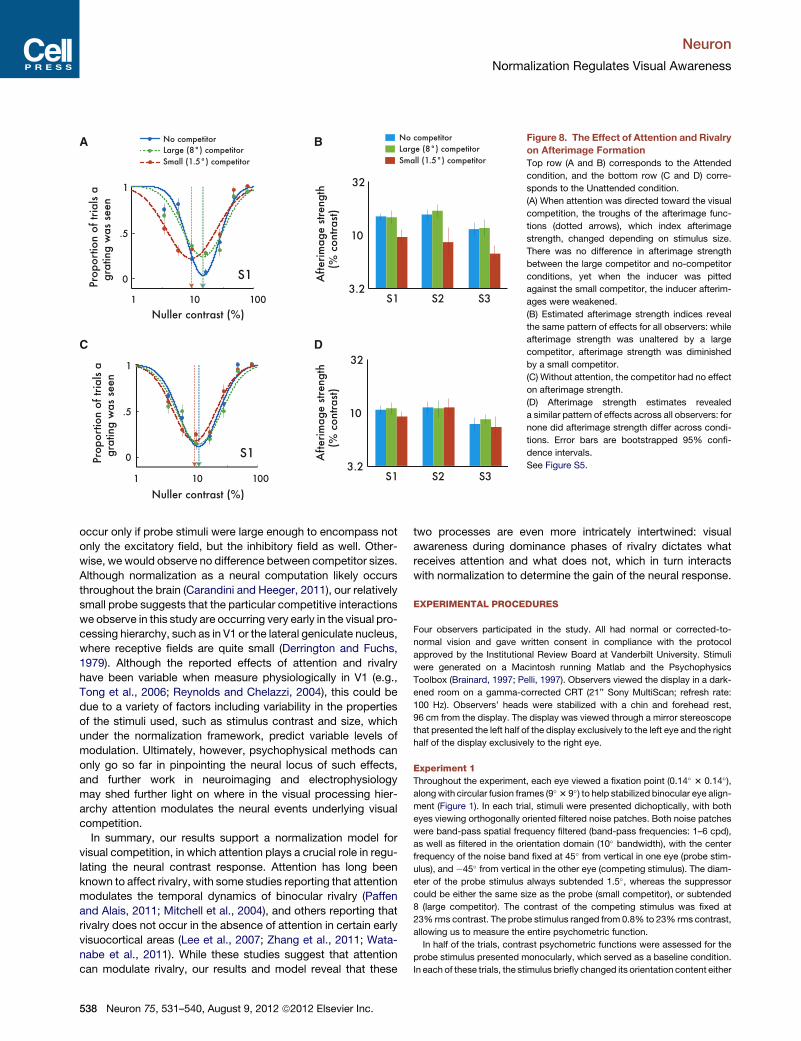
Figure 8. The Effect of Attention and Rivalry
on Afterimage Formation
Top row (A and B) corresponds to the Attended
condition, and the bottom row (C and D) corre-
sponds to the Unattended condition.
(A) When attention was directed toward the visual
competition, the troughs of the afterimage func-
tions (dotted arrows), which index afterimage
strength, changed depending on stimulus size.
There was no difference in afterimage strength
between the large competitor and no-competitor
conditions, yet when the inducer was pitted
against the small competitor, the inducer afterim-
ages were weakened.
(B) Estimated afterimage strength indices reveal
the same pattern of effects for all observers: while
afterimage strength was unaltered by a large
competitor, afterimage strength was diminished
by a small competitor.
(C) Without attention, the competitor had no effect
on afterimage strength.
(D) Afterimage strength estimates revealed
a similar pattern of effects across all observers: for
none did afterimage strength differ across condi-
tions. Error bars are bootstrapped 95% confi-
dence intervals.
See Figure S5.
Neuron
Normalization Regulates Visual Awareness
occur only if probe stimuli were large enough to encompass not
only the excitatory field, but the inhibitory field as well. Other-
wise, we would observe no difference between competitor sizes.
Although normalization as a neural computation likely occurs
throughout the brain (Carandini and Heeger, 2011), our relatively
small probe suggests that the particular competitive interactions
we observe in this study are occurring very early in the visual pro-
cessing hierarchy, such as in V1 or the lateral geniculate nucleus,
where receptive fields are quite small (Derrington and Fuchs,
1979). Although the reported effects of attention and rivalry
have been variable when measure physiologically in V1 (e.g.,
Tong et al., 2006; Reynolds and Chelazzi, 2004), this could be
due to a variety of factors including variability in the properties
of the stimuli used, such as stimulus contrast and size, which
under the normalization framework, predict variable levels of
modulation. Ultimately, however, psychophysical methods can
only go so far in pinpointing the neural locus of such effects,
and further work in neuroimaging and electrophysiology
may shed further light on where in the visual processing hier-
archy attention modulates the neural events underlying visual
competition.
In summary, our results support a normalization model for
visual competition, in which attention plays a crucial role in regu-
lating the neural contrast response. Attention has long been
known to affect rivalry, with some studies reporting that attention
modulates the temporal dynamics of binocular rivalry (Paffen
and Alais, 2011; Mitchell et al., 2004), and others reporting that
rivalry does not occur in the absence of attention in certain early
visuocortical areas (Lee et al., 2007; Zhang et al., 2011; Wata-
nabe et al., 2011). While these studies suggest that attention
can modulate rivalry, our results and model reveal that these
538 Neuron 75, 531–540, August 9, 2012 ª2012 Elsevier Inc.
two processes are even more intricately intertwined: visual
awareness during dominance phases of rivalry dictates what
receives attention and what does not, which in turn interacts
with normalization to determine the gain of the neural response.
EXPERIMENTAL PROCEDURES
Four observers participated in the study. All had normal or corrected-to-
normal vision and gave written consent in compliance with the protocol
approved by the Institutional Review Board at Vanderbilt University. Stimuli
were generated on a Macintosh running Matlab and the Psychophysics
Toolbox (Brainard, 1997; Pelli, 1997). Observers viewed the display in a dark-
ened room on a gamma-corrected CRT (21’’ Sony MultiScan; refresh rate:
100 Hz). Observers’ heads were stabilized with a chin and forehead rest,
96 cm from the display. The display was viewed through a mirror stereoscope
that presented the left half of the display exclusively to the left eye and the right
half of the display exclusively to the right eye.
Experiment 1
Throughout the experiment, each eye viewed a fixation point (0.14� 3 0.14�),alongwith circular fusion frames (9� 3 9�) to help stabilized binocular eye align-
ment (Figure 1). In each trial, stimuli were presented dichoptically, with both
eyes viewing orthogonally oriented filtered noise patches. Both noise patches
were band-pass spatial frequency filtered (band-pass frequencies: 1–6 cpd),
as well as filtered in the orientation domain (10� bandwidth), with the center
frequency of the noise band fixed at 45� from vertical in one eye (probe stim-
ulus), and �45� from vertical in the other eye (competing stimulus). The diam-
eter of the probe stimulus always subtended 1.5�, whereas the suppressor
could be either the same size as the probe (small competitor), or subtended
8 (large competitor). The contrast of the competing stimulus was fixed at
23% rms contrast. The probe stimulus ranged from 0.8% to 23% rms contrast,
allowing us to measure the entire psychometric function.
In half of the trials, contrast psychometric functions were assessed for the
probe stimulus presented monocularly, which served as a baseline condition.
In each of these trials, the stimulus briefly changed its orientation content either

Neuron
Normalization Regulates Visual Awareness
clockwise or counterclockwise (4�), and observers reported which direction
that stimulus had rotated. In the other half of the trials, observers viewed stimuli
dichoptically, with each eye viewing a different orientation band-pass-filtered
noise display. The orientation content of the display in one eye was always
orthogonal to that of the other eye—a stimulus mismatch that provokes visual
competition. To manipulate the suppression of these stimuli, we used the flash
suppression technique (Wolfe, 1984): on each trial, the to-be-suppressed
probe stimulus was presented monocularly for 3,000 ms, after which time
the competing stimulus (flash suppression competitor) abruptly appeared in
the other eye, thereby suppressing perception of the initially presented stim-
ulus in favor of the newly presented image. The timing and relatively small
size of the stimulus were specifically chosen to maximize flash suppression
duration, and to minimize instances of piecemeal rivalry within the probe dura-
tion. Each observer participated in a practice block of 50 trials and 30 exper-
imental blocks of 50 trials each, for a total of 50 data points per condition.
Experiment 2
Throughout the experiment, each eye viewed a fixation point (0.14� 3 0.14�),along with circular fusion frames (9� 3 9�). To induce afterimages in each trial,
observers were shown brief, 2 s exposures of a sinusoidal grating (the inducer;
1.5� 3 1.5�; 80% contrast; 1 cpd) in one eye while, at the same time, the other
eye viewed one of three possible stimulus arrangements (Figure 7): (1) an un-
contoured field that produced no suppression of the inducer, (2) a large (8�)competitor, or (3) small (1.5�) competitor. The large and small competitors
were identical to the competitors used in the Experiment 1, with the exception
that the stimuli counterphase flickered at 10 Hz, which suppressed the sinu-
soid during that 2 s exposure duration (Tsuchiya and Koch, 2005). Immediately
following each brief induction period, the competitor grating, if present, was
removed and the contrast of the inducer viewed by the other eye was ramped
off and was replaced by a ‘‘nuller’’ stimulus (750 ms), itself a sinusoidal grating
presented to the same eye that received the inducer. An auditory tone was
played coincident with the nuller onset, which helped distinguish the switch
from the inducer to the nuller. The nuller stimulus contrast ranged from
2%–80% contrast, and always had the same spatial phase as the inducer.
After each period of nuller presentation (750 ms), a spatial mask was
presented, which was a band-pass spatial frequency filtered noise patch
(3�; band-pass frequencies: 1–6 cpd; 23% rms contrast). Once this mask
appeared, observers indicated whether or not a grating had been seen.
To direct attention toward the competing stimuli (attended condition), we
had observers detect orientation changes (10�) that occurred stochastically
(0.3 probability of occurrence) to the dominant competitor stimulus (175 ms).
To divert attention away from the competing stimuli (unattended condition),
we required observers to perform a letter identification task (RSVP task),
detecting target letters (‘‘J’’ or ‘‘K’’; 1.5� 3 1.5�; 0.3 probability of occurrence)
within a stream of distractor letters (‘‘X’’ ‘‘L’’ ‘‘V’’ ‘‘H’’ ‘‘B’’ ‘‘A’’ ‘‘C’’ ‘‘F’’ ‘‘Z’’ ‘‘Y’’
‘‘O’’ ‘‘U’’ ‘‘N’’ ‘‘W’’ ‘‘E’’), appearing in the periphery of the inducer eye
(3.35� eccentricity) every 175 ms. Prior to each block of trials, observers
were told which task to perform throughout the block. In both the Attended
and Unattended conditions, the RSVP stream ended, and the fixation point
changed color 750 ms prior to the onset of the nuller, providing ample time
for observers to prepare for the task in which they would report whether a
grating was seen or not.
SUPPLEMENTAL INFORMATION
Supplemental Information includes five figures and can be found with this
article online at http://dx.doi.org/10.1016/j.neuron.2012.05.032.
ACKNOWLEDGMENTS
We thank David Heeger, Frank Tong, and the reviewers of this manuscript
for valuable comments and discussion. Supported by NIH grants EY13358
and P30-EY008126, and by a grant (R31-10089) from the National Research
Foundation of Korea funded by the Ministry of Education, Science and
Technology.
Accepted: May 29, 2012
Published: August 8, 2012
REFERENCES
Blake, R., and Logothetis, N.K. (2002). Visual competition. Nat. Rev. Neurosci.
3, 13–21.
Brainard, D.H. (1997). The Psychophysics Toolbox. Spat. Vis. 10, 433–436.
Brascamp, J.W., van Boxtel, J.J.A., Knapen, T.H.J., and Blake, R. (2010). A
dissociation of attention and awareness in phase-sensitive but not phase-
insensitive visual channels. J. Cogn. Neurosci. 22, 2326–2344.
Carandini, M., and Heeger, D.J. (2011). Normalization as a canonical neural
computation. Nat. Rev. Neurosci. 13, 51–62.
Derrington, A.M., and Fuchs, A.F. (1979). Spatial and temporal properties of
X and Y cells in the cat lateral geniculate nucleus. J. Physiol. 293, 347–364.
Ding, J., and Sperling, G. (2006). A gain-control theory of binocular combina-
tion. Proc. Natl. Acad. Sci. USA 103, 1141–1146.
Georgeson, M.A., and Turner, R.S. (1985). Afterimages of sinusoidal, square-
wave and compound gratings. Vision Res. 25, 1709–1720.
Heeger, D.J. (1992). Normalization of cell responses in cat striate cortex.
Vis. Neurosci. 9, 181–197.
Herrmann, K., Montaser-Kouhsari, L., Carrasco, M., and Heeger, D.J. (2010).
When size matters: attention affects performance by contrast or response
gain. Nat. Neurosci. 13, 1554–1559.
Hollingworth, A., Maxcey-Richard, A.M., and Vecera, S.P. (2012). The spatial
distribution of attention within and across objects. J. Exp. Psychol. Hum.
Percept. Perform. 38, 135–151.
Ito, H. (2012). Cortical shape adaptation transforms a circle into a hexagon:
a novel afterimage illusion. Psychol. Sci. 23, 126–132.
James, W. (1890). The Principles of Psychology, Volume 1 (New York: Holt).
Kelly, D.H., and Martinez-Uriegas, E. (1993). Measurements of chromatic and
achromatic afterimages. J. Opt. Soc. Am. A 10, 29–37.
Lack, L. (1978). Selective Attention and the Control of Binocular Rivalry
(The Hague: Mouton).
Lee, S.H., Blake, R., and Heeger, D.J. (2007). Hierarchy of cortical responses
underlying binocular rivalry. Nat. Neurosci. 10, 1048–1054.
Leguire, L.E., and Blake, R. (1982). Role of threshold in afterimage visibility.
J. Opt. Soc. Am. 72, 1232–1237.
Leopold, D.A., and Logothetis, N.K. (1999). Multistable phenomena: changing
views in perception. Trends Cogn. Sci. 3, 254–264.
Lennie, P. (2003). The cost of cortical computation. Curr. Biol. 13, 493–497.
Ling, S., Hubert-Wallander, B., and Blake, R. (2010). Detecting contrast
changes in invisible patterns during binocular rivalry. Vision Res. 50, 2421–
2429.
Mitchell, J.F., Stoner, G.R., and Reynolds, J.H. (2004). Object-based attention
determines dominance in binocular rivalry. Nature 429, 410–413.
Moradi, F., and Heeger, D.J. (2009). Inter-ocular contrast normalization in
human visual cortex. J. Vis. 9, 13.1–22.
Naka, K.I., and Rushton, W.A. (1966). S-potentials from colour units in the
retina of fish (Cyprinidae). J. Physiol. 185, 536–555.
Ooi, T.L., and He, Z.J. (2006). Binocular rivalry and surface-boundary process-
ing. Perception 35, 581–603.
Paffen, C.L.E., and Alais, D. (2011). Attentional modulation of binocular rivalry.
Front. Hum. Neurosci. 5, 105.
Pelli, D.G. (1997). The VideoToolbox software for visual psychophysics:
transforming numbers into movies. Spat. Vis. 10, 437–442.
Pestilli, F., Ling, S., and Carrasco, M. (2009). A population-coding model of
attention’s influence on contrast response: estimating neural effects from
psychophysical data. Vision Res. 49, 1144–1153.
Petrov, Y., Carandini, M., andMcKee, S.P. (2005). Two distinct mechanisms of
suppression in human vision. J. Neurosci. 25, 8704–8707.
Neuron 75, 531–540, August 9, 2012 ª2012 Elsevier Inc. 539

Neuron
Normalization Regulates Visual Awareness
Reynolds, J.H., and Chelazzi, L. (2004). Attentional modulation of visual
processing. Annu. Rev. Neurosci. 27, 611–647.
Reynolds, J.H., and Heeger, D.J. (2009). The normalization model of attention.
Neuron 61, 168–185.
Sengpiel, F., and Blakemore, C. (1994). Interocular control of neuronal respon-
siveness in cat visual cortex. Nature 368, 847–850.
Shimojo, S., Kamitani, Y., and Nishida, S. (2001). Afterimage of perceptually
filled-in surface. Science 293, 1677–1680.
Somers, D.C., Dale, A.M., Seiffert, A.E., and Tootell, R.B. (1999). Functional
MRI reveals spatially specific attentional modulation in human primary visual
cortex. Proc. Natl. Acad. Sci. USA 96, 1663–1668.
Sundberg, K.A., Mitchell, J.F., and Reynolds, J.H. (2009). Spatial attention
modulates center-surround interactions in macaque visual area v4. Neuron
61, 952–963.
Tong, F., Meng, M., and Blake, R. (2006). Neural bases of binocular rivalry.
Trends Cogn. Sci. 10, 502–511.
Tsuchiya, N., and Koch, C. (2005). Continuous flash suppression reduces
negative afterimages. Nat. Neurosci. 8, 1096–1101.
540 Neuron 75, 531–540, August 9, 2012 ª2012 Elsevier Inc.
Vecera, S.P., Behrmann, M., and McGoldrick, J. (2000). Selective attention to
the parts of an object. Psychon. Bull. Rev. 7, 301–308.
von Helmholtz, H. (1909). Handbuch der Physiologischen Optik, Volume 1, 3rd
ed. (Hamburg: Voss).
Watanabe, K., Paik, Y., and Blake, R. (2004). Preserved gain control for
luminance contrast during binocular rivalry suppression. Vision Res. 44,
3065–3071.
Watanabe, M., Cheng, K., Murayama, Y., Ueno, K., Asamizuya, T., Tanaka, K.,
and Logothetis, N. (2011). Attention but not awareness modulates the BOLD
signal in the human V1 during binocular suppression. Science 334, 829–831.
Wheatstone, C. (1838). Contributions to the physiology of vision: part the first.
On some remarkable, and hitherto unobserved, phenomena of binocular
vision. Philos. Trans. R. Soc. Lond. B Biol. Sci. 128, 371–394.
Wolfe, J.M. (1984). Reversing ocular dominance and suppression in a single
flash. Vision Res. 24, 471–478.
Zhang, P., Jamison, K., Engel, S., He, B., and He, S. (2011). Binocular rivalry
requires visual attention. Neuron 71, 362–369.
Zhang, P., Jiang, Y., and He, S. (2012). Voluntary attentionmodulates process-
ing of eye-specific visual information. Psychol. Sci. 23, 254–260.