Embed Size (px)
Citation preview
Normal Postpartum Involution of the Uterus in the Dog
M.A. Al-Bassam, R.G. Thomson and L. O'Donnell*
ABSTRACT
Ninety-eight reproductivetracts from dogs at differentpostpartum time periods wereused to investigate stages ofnormal involution. Seventy-eight reproductive tracts wereobtained from the field, and 20obtained surgically for grossand microscopic examination.Plasma progesterone was mea-sured in 22 dogs at various timespostpartum.The uterine horns during the
first week postpartum weredilated and edematous. The pla-cental sites were 1.5-3 cm inwidth, rough, granular andcovered with mucus and a fewblood clots. By the fourth weekthe placental sites were thick,grayish-tan and nodular with afew blood clots within nodules.The uterine horns during theseventh week were greatly con-tracted and the placental siteswere narrow and light in color.A few nodules were still presenton the surface. By the ninthweek the uterine horns wereuniform in shape and con-tracted with a narrow lumen.The placental sites appeared asa narrow brown band.Histologically the placental
sites during the first week post-partum were covered by aneosinophilic staining necroticmass and a few intact epithelialcells scattered on the surface asan interrupted single layer.Under the necrotic mass, largeeosinophilic staining cells inmoderate number were scat-tered throughout the laminapropria of the placental site.
These cells were considered tobe decidual cells. By the fourthweek the placental sites werecovered by a large lobulatedmass of collagen fibers. The ute-rine glands were greatly dilatedand degenerate, and mononu-clear cell infiltration in the lam-ina propria was pronounced. Bythe seventh week, large massesof collagen fibers were detachedfrom the surface, and endomet-rial glands were normal in sizeand shape. By the ninth weeksurface sloughing was com-pleted. However, regenerationand replacement of the endo-metrial lining from the mouth ofthe uterine glands continueduntil the end of the twelfth weekwhen the involution process wascompleted.The progesterone levels were
very low for eight weeks post-partum.
R:SUMPCette experience consistait a
etudier les aspects macrosco-piques et microscopiques desstades de l'involution uterinecanine normale, a differentesperiodes ulterieures a la partu-rition. On utilisa a cette fin lesorganes genitaux internes de 98chiennes; 78 de ces echantillonsprovenaient de cliniques veteri-naires et 20, de cas de chirurgieexperimentale du College VWt-rinaire de Guelph. On mesuraaussi la teneur en progesteronedu plasma de 22 chiennes, a dif-ferentes periodes ulterieures ala parturition.Au cours de la semaine qui
suivit la parturition, les cornesuterines s'avererent dilatees etoedemateuses. Les sites placen-taires mesuraient de 1,5 a 3 cmde largeur; ils etaient rugueux,granuleux et recouverts demucus et de quelques caillotssanguins. Au cours de la qua-trieme semaine, les sites placen-taires pr6sentaient une couleurgris-brun et des nodules quirecelaient quelques caillots san-guins. Au cours de la septiemesemaine, les cornes uterinesaffichaient une contractionappreciable et les sites placen-taires etaient etroits et pales;leur surface presentait encorequelques nodules. Au cours de laneuvieme semaine, les cornesuterines affichaient un retour ala normale, tandis que les sitesplacentaires ne correspondaientplus qu'a une bande brune etetroite.L'examen microscopique des
sites placentaires, au cours de lasemaine ulterieure a la parturi-tion, revela qu'ils etaient recou-verts par une masse necrotiqueeosinophile et par quelques cel-lules 6pith6liales intactes, epar-pillees superficiellement commeune couche simple, mais incom-plete. Sous la masse necrotique,le chorion des sites placentairescontenait un nombre modere decellules volumineuses et eosino-philes; ces cellules eparpilleessemblaient correspondre a descellules deciduales. Au cours dela quatrieme semaine, les sitesplacentaires etaient recouvertsd'une masse volumineuse etlobulee de fibres collagenes; lesglandes uterines affichaient parailleurs une dilatation marquee
Can. J. comp. Med. 45: 217-232 (July 1981)
*Department of Pathology and Department of Clinical Studies, Ontario Veterinary College, University of Guelph, Guelph, OntarioN1G 2W1Submitted November 26, 1980.
217

et de la d6g6n6rescence; la cho-rion pr6sentait une infiltrationimportante de mononucleaires.Au cours de la septieme semaine,des masses importantes de fibrescollagenes se detachaient de lasurface des sites placentaires etles glandes endometriales retrou-vaient progressivement leursdimensions et leur forme nor-males. Au cours de la neuviemesemaine, la desquamation de lasurface des sites placentaires secompleta; la r6gen6ration et leremplacement de l'epitheliumde la muqueuse uterine, A partirde 1'embouchure des glandesuterines, continua cependantjusqu'& la fin de la 12e semainequi marqua le completement duprocessus d'involution uterine.La teneur du plasma en proges-terone se revela tr&s basse, toutau long des huit semainesulterieures a la parturition.
INTRODUCTIONIn spite of the abundant litera-
ture regarding the canine pla-centa, only a few reports describenormal uterine postpartum invo-lution in the bitch. Involution hasbeen described briefly as a part ofthe estrous cycle in the dog (3, 13).Both reports indicate that twelveweeks were necessary for the com-pletion of involution. During thefirst week postpartum, the placen-tal sites were difficult to recognizegrossly due to marked contractionof the myometrium (3), and by thefourth week postpartum theendometrium at the interplacentalsites incurred extensive sloughingand desquamation, whereas thedesquamation at the placental sitestarted later, and was morepronounced.This paper describes in some
detail the gross and microscopicfeatures during postpartum invo-lution of the placental and inter-placental areas of the uterus in thedog.
MATERIALSAND METHODSIn order to obtain as many sam-
ples as possible, reproductivetracts were obtained from two
sources, local veterinarians andexperimental surgical cases fromthe college. The intention was tosecure as many samples as possiblein order to have a broad coverageof the postpartum period.Seventy-eight reproductive
tracts from different breeds andages of dogs with or without his-tory of uterine diseases fixed in10% formalin solution, wereobtained from local practices.Fifty-three samples were fromdogs up to three months postpar-tum, ten more than three months,12 samples at different stages ofpregnancy, and three had pyome-tra. The gross and microscopic-examinations were mostly con-cerned with the samples from dogsup to three months postpartum(Table I).Twenty pregnant or recently
postpartum bitches of differentages and breeds were obtained anduterine samples were removedsurgically at specific time periods.From four dogs, four to six uterinesamples were taken surgically atdifferent times postpartum. In theremainder, only two surgicalprocedures were performed; oneuterine horn was removed first,and later the other horn and thetwo ovaries were removed. Forty-six uterine samples were obtainedsurgically at intervals over eightweeks postpartum.
GROSS EXAMINATION
The number of placental sites ineach sample and the size and grosschanges ofeach placental site wererecorded. The number and size of
TABLE I. Postpartum Time Period Dis-tribution in Specimens from Field andSurgical Cases
Week Surgical Cases Field Cases123456789101112Totals
96774462
3
2231681043
115345
corpora lutea from each dog werealso recorded.
MICROSCOPIC EXAMINATION
Three placental sites wereselected from each uterine sample(except the first four surgicaldogs), and transverse or longitudi-nal incisions were made throughthe sites and interplacental uterinetissues. One of the ovaries fromeach case was examined micro-scopically. Tissues were embeddedin paraffin, sectioned at 5,um andstained with hematoxylin andeosin.Masson's trichrome staining
method (15), was used to distin-guish the presence of collagenfrom other components. Perl'sstaining method (16), was used todetermine the presence of hemosi-derin. In order to detect differen-ces in the microscopic structurebetween different areas of the pla-cental site, semiserial 5,um sec-tioning from 15 selected blockswas carried out, and the sectionsstained with hematoxylin andeosin. From ten selected blocks,5,um serial sections extendingfrom the myometrium to theendometrial lumen were stainedwith hematoxylin and eosin. Thisprocedure was carried out todetermine if there was closure ornarrowing in the glandular ducts.HORMONAL ASSAY
A single 10mL blood sample wascollected into evacuated tubes con-taining 143 USP units of sodiumheparin solution at the time of thesurgical procedures by cephalicvenipuncture. Plasma wasobtained by centrifugation andstored frozen until assayed.Plasma progesterone levels weredetermined in ten samples byusing the competitive proteinbinding assay (18, 19), utilizing arapid method of sample prepara-tion (14). A radioimmunoassaymethod (5) was used in the remain-ing 12 samples for the determina-tion of the plasma progesteronelevels. Circumstances were suchthat the samples had to be pro-cessed in two different laborato-ries and consequently two differ-ent methods.
218

RESULTSGROSS FINDINGS
During the first week postpar-tum, the uterus was still dilated,edematous, and signs of contrac-tion were seen on the serosal sur-face as small longitudinal folds(Fig. 1). The mucosal surface at theinterplacental site was folded andcovered with dark brown mucus.The placental sites were 1.5-2 cmin width and their surface wasrough, granular and covered withdark brown mucus and a few bloodclots. The edges of the placentalsites were long, folded inward andcovered part of the placental site(Fig. 2).During the second and third
week the uterus was smaller. Theplacental sites were visible fromthe serosal surface as swellings inthe uterine wall. The entire muc-osal surface was still covered by adark brown mucus but less incomparison to the first week. Theplacental sites were thicker andtheir surface was granular,grayish-tan and mixed with bloodclots. The edges of the placentalsites were smaller in size.By the fourth week the uterus
was much smaller and the placen-
Fig. 1. Gross changes in a uterine hornone week postpartum. The horn isdilated, and signs of contraction appearon the serosal surface as small longitudi-nal folds.
Fig. 2. Endometrium one week postpar-tum. The mucosal surface between theplacental sites is folded and covered withdark brown mucus. Placental site(arrows) are rough, 1-1.5 cm in width,and large mucosal folds cover part ofthem.
tal sites appeared on the serosalsurface as ellipsoid swellings (Fig.3). The mucosal surface of theinterplacental sites was lessfolded, and was covered with clearmucus. The placental sites weregrayish-tan and contained eight to12 small grayish nodules (0.2-
Fig. 3. Uterine horn, serosal surface fourweeks postpartum. Placental site is vis-ible clearly from serosal surface as anellipsoidal swelling.
Fig. 4. Uterine horn, mucosal surfacefour weeks postpartum. The surface isless folded and covered with clearmucus. Placental sites (arrows) aregrayish-tan. Eight to twelve smallgrayish nodules are spread over eachsite.
0.4 cm diameter) spread randomlyover the placental site (Fig. 4). Afew pinpoint hemorrhagic foci andblood clots were seen within thenodules.By the fifth week the uterine
horn had become much smaller indiameter. The placental sitesappeared as small ellipsoid swel-lings on the serosal surface, and onthe mucosal surface the placentalsites were smaller in size. Duringthe sixth and seventh weeks themucosal surface of the placentalsites were light brown and narrow(0.8-1.5 cm in width). About six toeight grayish nodules were stillpresent on the surface. No hemor-rhagic foci or blood clots werepresent. By the eighth week, thediameter of the uterine hornsreached its smallest size. Thehorns were uniform in shape withsmall ellipsoid shaped placentalsites visible from the serosal sur-face (Fig. 5). On the mucosal sur-face, the number of grayishnodules were fewer (three or four)at the placental sites (Fig. 6). Bythe ninth week the uterine hornswere uniform in shape with a nar-row lumen. The placental siteswere only differentiated from the
219

...
S.
VS'!... .. ...............t
^...<. .....
Fig. 5. Uterine horn, serosal surfaceeight weeks postpartum. The uterinehorn is greatly contracted and narrowerin diameter.
interplacental endometrium bythe brown color.MICROSCOPIC FINDINGS
Placental Site Endometrium - Atparturition the plane of separationof the placenta usually lay in thedistal portion of the spongy layerwhere there were only thin walledpartitions between the greatlydilated glands (Fig. 7).The histological structure of the
placental site from parturition toseven days postpartum was as fol-lows. After the separation of theplacenta, the basal glandular zonewas the only viable and intact layer
remaining (Figs. 8 and 9). At theplane of placental separation overthe basal glandular zone a necroticmass was observed with a fewintact, large and foamy epithelialcells scattered on the surface as aninterrupted single layer (Figs. 8and 9). These cells were consideredto be remnants from the basal partof the separated spongy layer. Theeosinophilic staining necrotic masswas composed of remnants of thefetal placenta, fragments of theseparated spongy layer, fibrin,erythrocytes, necrotic thrombosedblood vessels and nuclear debris(Figs. 8 and 9).
isiit. -,I 0
-* A --l
--------- -
t
.. .... . .' . : .' . ...... .. _... ..
...-"
.:..
...... ..
:. ..*:. .:.. .: .:.. .... ' :: ..
.'..
*:..
....
...: :.
:.
'._
...Fig. 6. Uterine horn mucosal surfaceeight weeks postpartum. Placental sitesare small and three to four grayishnodules are present per site.
Fig. 7. A schematic drawing of partially separated placenta at parturition. Theplane of separation of the fetal membrane lies in the spongy layer (arrow). Themarginal hematoma (HEM) is at the upper right and the deep glandular zone (GZ) isjust above the myometrium (M). The subplacental layer (SB) is between the deepglandular zone and the spongy layer.
220

Fig. 8. A schematic drawing of uterus showing the microscopic changes in a uterusone week postpartum. The necrotic mass (arrow) is at the top and the deep glandularzone (GZ) below. Decidual cells (arrowheads) are between the necrotic mass and thedeep glandular zone.
;5P*- ~- .; r. W
~~~~~~~~~~~~~~~~~~~Ss,Fig. 9. Microscopic changes in theendometrium at the placentalsite attachment during the first week postpartum. The necroticmass on the surface (arrow) is composed of remnants of fetalplacenta, fragments of the separated spongy layer, fibrin, ery-throcytes and nuclear debris. Decidual cells (arrowheads) are inthe upper endometrial lamina propria. H & E. X58.
Large eosinophilic cells in mod-erate numbers were scattered inthe upper loose connective tissue ofthe basal glandular zone of thelamina propria and under thenecrotic mass. The cells werepolygonal with an epithelioidappearance and abundant eosino-philic granular cytoplasm. Themajority had one large, sphericalnucleus although binucleate cellswere not uncommon (Figs. 9 and10). Many of these cells weredegenerate with pyknotic orirregular nuclei and fully baso-philic or vacuolated cytoplasm.These cells were considered to beeither decidual cells (maternal) ortrophoblastic cells (fetal), but dueto their greater similarity todecidual cells have been desig-nated as decidual cells.During the second week collagen
proliferation formed lobulatedmasses at the placental sites. Thecollagen appeared to be producedby proliferating fibroblasts in thelamina propria (Fig. 11). Smallhemorrhages and scattered mono-nuclear cells were noted in thelamina propria. Decidual cellswere seen in four out of six samplesexamined at this time and most ofthem were degenerate (Fig. 12).During the third week there was
an increase in the amount of colla-* r.... .
A
Fig. 10. Higher magnification of Fig. 9 to show the decidualcells (arrowheads). The decidual cells are large, polygonal,with an epitheliod appearance. Many of these cells are degen-erated with a vacuolated cytoplasm and pyknotic nuclei.H & E. X232.
221
%.~-
---j

- if. 3 1. 9
j4.V I .
V
. '.i.-.
Fig. 11. Cross section of the uterine wall at the placental site during the second weekpostpartum. The myometrium is contracted. Collagen fibers (arrowheads) are de-posited on the surface of the endometrial attachment of the placental site. H & E. X7.
gen covering the endometrium inthe placental sites (Fig. 13).Mononuclear cell infiltration wasgreatly increased in the laminapropria. These cells were mainlymacrophages, lymphocytes and afew plasma cells (Fig. 13). Theendometrial glands were moder-ately dilated and their lumina con-tained cellular debris and erythro-
4;
cytes. A few degenerate decidualcells were distinguished in onlytwo out of eight samples.By the fourth week the collagen
deposition at the placental sitesreached a maximum (Figs. 14 and15). It was present in the form oflarge lobulated masses. The sur-face over most of the thicker por-tions of the masses was necroticand hemorrhagic whereas thejunction of these masses with theendometrium had more cellularand immature collagen. These col-lagen masses were covered by asingle layer of foamy to eosino-philic staining columnar epithelialcells. This endometrial lining wasinterrupted over superficial partsof the masses where collagen was
GlN Ni;iFA' qW r-N MW|, -L
Fig. 13. Microscopic changes in theendometrium at the placental site dur-ing the third week. There is a greatincrease in the number of mononuclearcells (arrow) in the lamina propria. Thecollagen mass (arrows) which is att-ached to the endometrium is very cellu-lar. H & E. X150.
necrotic and sloughing. The endo-metrial glands were greatlydilated and their lumina werefilled with mucus and sparsenecrotic debris (Figs. 14 and 15).The mononuclear cell infiltrationin the lamina propria waspronounced.By the beginning of the fifth
week the last and most important
Fig. 12. Microscopic changes in theendometrium to show the decidual cells(arrowheads). Most of the decidual cellsare degenerate and vacuolated. Thereare a few mononuclear cells in the lam-ina propria (left). H & E. X96.
Fig. 14. Cross section of uterine wall at the site of placental attachment during thefourth week postpartum. Large amounts of collagen (arrows) have been depositedon the endometrial surface. The myometrium is thin and contracted. H & E. X5.
222
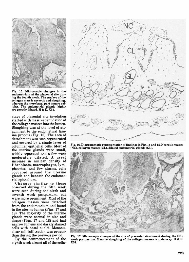
Fig. 15. Microscopic changes in theendometrium at the placental site dur-ing the fourth week. The surface of thecollagen mass is necrotic and sloughing,whereas the more basal part is more cel-lular. The endometrial glands (right)are greatly dilated. H & E. X35.
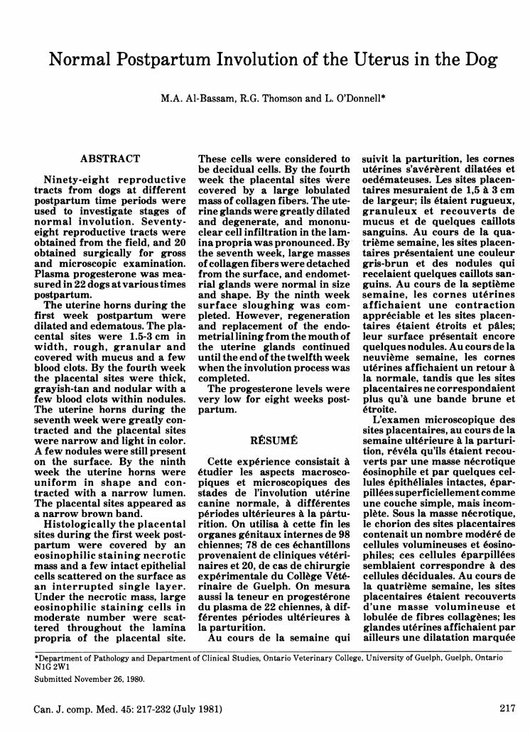
stage of placental site involutionstarted with massive denudation ofthe collagen masses into the lumen.Sloughing was at the level of att-achment to the endometrial lam-ina propria (Fig. 16). The area ofdetachment was soon regeneratedand covered by a single layer ofcolumnar epithelial cells. Most ofthe uterine glands were small,widely separated and a few weremoderately dilated. A greatincrease in nuclear density offibroblasts, macrophages, lym-phocytes, and few plasma cellsoccurred around the uterineglands and beneath the endomet-rial epithelium.Changes similar to those
observed during the fifth weekwere seen during the sixth andseventh week postpartum, butwere more prominent. Most of thecollagen masses were detachedfrom the endometrium and foundin the uterine lumen (Figs. 17 and18). The majority of the uterineglands were normal in size andshape (Figs. 17 and 18) and hadnarrow lumens and darkly stainedcells with basal nuclei. Mononu-clear cell infiltration was greaterthan during the previous period.By the commencement of the
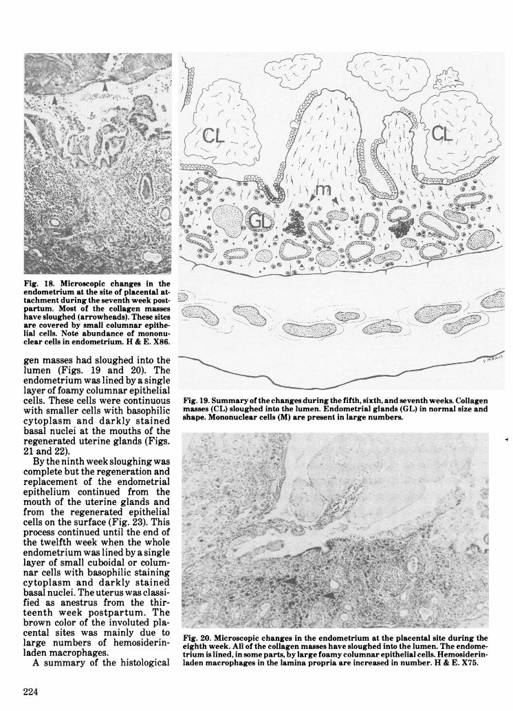
eighth week almost all of the colla-
ita.
,Zl)6v~ ~~~~_ ~!'_
Figw_.1.Darm ai ersntto ffnig nFg 1 n 5 ertcmse
Fig. 16. Diagrammatic representation of findings in Fig. 14 and 15. Necrotic masses(NC), collagen masses (CL), dilated endometrial glands (GL).
A' 1 _ I l
Fig. 17. Microscopic changes at the site of placental attachment during the fifthweek postpartum. Massive sloughing of the collagen masses is underway. H & E.X51.
223
v y^>X Ax
W.su.,*... s~~~~~~~~';E..S'iK"'&^ #\x :~
Fig. 18. Microscopic changes in theendometrium at the site of placental at-tachment during the seventh week post-partum. Most of the collagen masseshave sloughed (arrowheads). These sitesare covered by small columnar epithe-lial cells. Note abundance of mononu-clear cells in endometrium. H & E. X86.
gen masses had sloughed into thelumen (Figs. 19 and 20). Theendometrium was lined by a singlelayer of foamy columnar epithelialcells. These cells were continuouswith smaller cells with basophiliccytoplasm and darkly stainedbasal nuclei at the mouths of theregenerated uterine glands (Figs.21 and 22).By the ninth week sloughing was
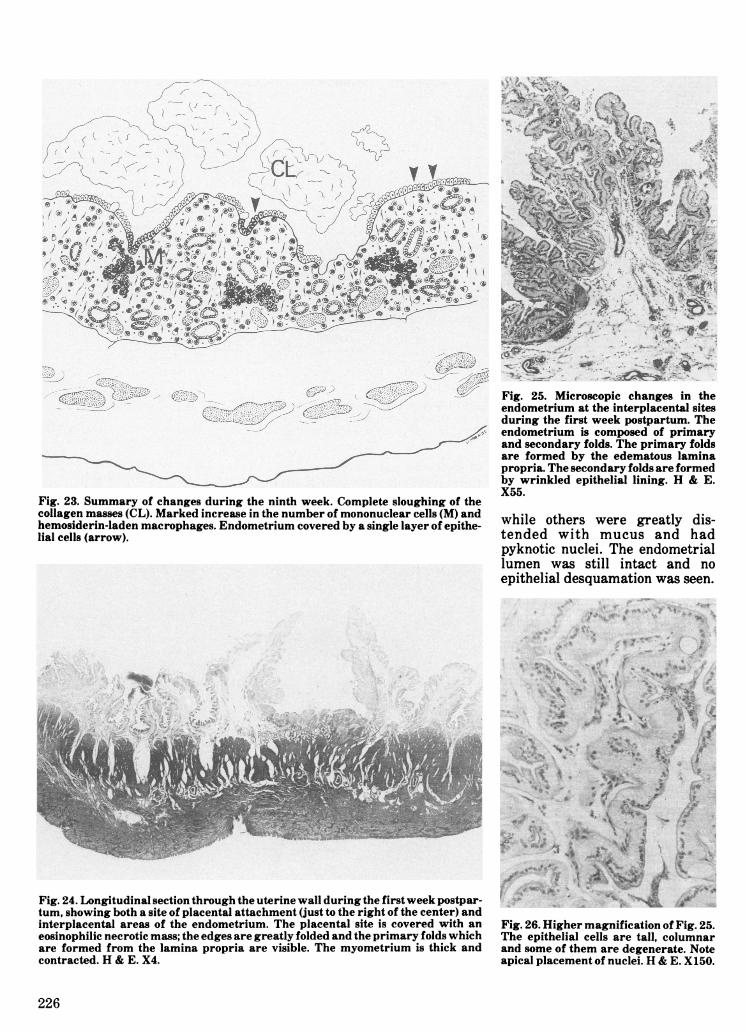
complete but the regeneration andreplacement of the endometrialepithelium continued from themouth of the uterine glands andfrom the regenerated epithelialcells on the surface (Fig. 23). Thisprocess continued until the end ofthe twelfth week when the wholeendometrium was lined by a singlelayer of small cuboidal or colum-nar cells with basophilic stainingcytoplasm and darkly stainedbasal nuclei. The uterus was classi-fied as anestrus from the thir-teenth week postpartum. Thebrown color of the involuted pla-cental sites was mainly due tolarge numbers of hemosiderin-laden macrophages.A summary of the histological
Fig. 19. Summary of the changes during the fifth, sixth, and seventh weeks. Collagenmasses (CL) sloughed into the lumen. Endometrial glands (GL) in normal size andshape. Mononuclear cells (M) are present in large numbers.
Fig. 20. Microscopic changes in the endometrium at the placental site during theeighth week. All of the collagen masses have sloughed into the lumen. The endome-trium is lined, in some parts, by large foamy columnar epithelial cells. Hemosiderin-laden macrophages in the lamina propria are increased in number. H & E. X75.
224

Fig. 21. Higher magnification of Fig. 20 to show the replacement of the tall foamycolumnar epithelial cells by small epithelial cells with basophilic staining cytoplasm,emanating from the mouths of the glands. Note hemosiderin-laden macrophages(arrows). H & E. X240.
changes is given in Table II. Theapplication of the trichromemethod of staining was very usefulto determine the amount of colla-gen deposited on the placental sitesat different time periods postpar-tum. Semiserial sections examinedfrom selected cases revealed thatthe glands opened to the lumen
without any narrowing or block-age of the ducts and no substantialmicroscopic differences were seenin different areas of the same pla-cental site.
Interplacental Site Endometrium- During the first week postpar-tum the interplacental endome-
Fig. 22. Higher magnification of Fig. 21. Small cells with basophilic cytoplasm anddarkly stained nuclei from the mouth of the gland are replacing the tall foamycolumnar epithelial cells lining the endometrium. H & E. X630.
trium was composed of primaryand secondary folds. The primaryfolds were formed by the greatlyedematous lamina propria whichcontained a few lymphocytes andplasma cells (Figs. 24 and 25). Thesecondary epithelial folds werewrinkled and consisted of longcolumnar epithelial cells (Fig. 26).The epithelial cells varied in sizeand had a foamy cytoplasm inwhich an abundance of lipid dro-plets were seen. Signs of degenera-tion were present in some cellsTABLE II. Microscopic Changes in thePlacental Sites at Different Postpar-turient Time Periods in the Dog
Week Appearance1 Eosinophilic necrotic layer
covered the placental sitesFew decidual like cells betweenthe necrotic layer and basalendometrial glands
2 Collagen fiber proliferation fromthe endometrial laminapropria
Slight mononuclear cellinfilitration in the laminapropria
A few degenerate decidual cells
3 and 4 Increased amount of collagenlining the endometrium asirregular lobulated masses
Increased mononuclear cellinfiltration
Dilated endometrial glandsAbsence of decidual cells
5 Sloughing of collagen massesGreat increase in mononuclear
cell infiltrationGlands regenerating andreturning to normal size
6 and 7 Continued sloughing of collagenmasses
Extensive mononuclear cellinfiltration
Endometrial glands normal insize
8 Sloughing of collagen massescompleted
Replacement of endometriallining from the mouth of theregenerated glands
Endometrial epithelial cellssmall with basophilic stainingcytoplasm
9-11
12
13
Continued replacement of theendometrial lining
Endometrium completely linedby a single layer of small cellswith basophilic stainingcytoplasm
Uterus at anestrus stage
225
MW ...7 ."w -.,:,.- "-io,
Vle"IO
mar 2;2%tsv!
Fig. 23. Summary of changes during the ninth week. Complete sloughing of thecollagen masses (CL). Marked increase in the number of mononuclear cells (M) andhemosiderin-laden macrophages. Endometrium covered by a single layer of epithe-lial cells (arrow).
IS ^;
Fig. 25. Microscopic changes in theendometrium at the interplacental sitesduring the first week postpartum. Theendometrium is composed of primaryand secondary folds. The primary foldsare formed by the edematous laminapropria. The secondary folds are formedby wrinkled epithelial lining. H & E.X55.
while others were greatly dis-tended with mucus and hadpyknotic nuclei. The endometriallumen was still intact and noepithelial desquamation was seen.
4....
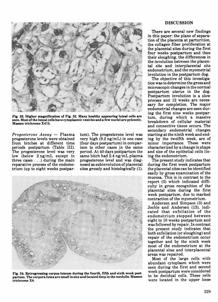
Fig. 24. Longitudinal section through the uterine wall during the firstweek postpar-tum, showing both a site of placental attachment (just to the right of the center) andinterplacental areas of the endometrium. The placental site is covered with aneosinophilic necrotic mass; the edges are greatly folded and the primary folds whichare formed from the lamina propria are visible. The myometrium is thick andcontracted. H & E. X4.
Fig. 26. Higher magnification of Fig. 25.The epithelial cells are tall, columnarand some of them are degenerate. Noteapical placement of nuclei. H & E. X150.
226
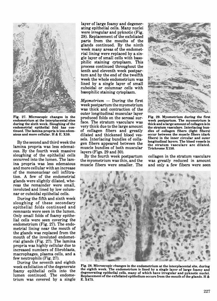
Fig. 27. Microscopic changes in theendometrium at the interplacental sitesduring the sixth week. Sloughing of theendometrial epithelial fold has con-tinued. The lamina propria is less edem-atous and more cellular. H & E. X59.
By the second and third week thelamina propria was less edemat-ous. By the fourth week massivesloughing of the epithelial cellsoccurred into the lumen. The lam-ina propria was less edematousand more cellular with an increaseof the mononuclear cell infiltra-tion. A few of the endometrialglands were slightly dilated, whe-reas the remainder were small,involuted and lined by low colum-nar or cuboidal epithelial cells.During the fifth and sixth week
sloughing of these secondaryepithelial folds continued andremnants were seen in the lumen.Only small folds of foamy epithe-lial cells were seen covering theendometrium (Fig. 27). The endo-metrial lining near the mouth ofthe glands was replaced from themouth of the involuted endomet-rial glands (Fig. 27). The laminapropria was highly cellular due toincreased numbers of fibroblasts,macrophages, plasma cells, and afew neutrophils (Fig. 27).During the seventh and eighth
week exfoliation of the degeneratefoamy epithelial cells into thelumen continued. The endome-trium was covered by a single
layer of large foamy and degener-ating epithelial cells. Many nucleiwere irregular and pyknotic (Fig.28). Replacement of the exfoliatedparts from the mouths of theglands continued. By the ninthweek many areas of the endomet-rial lining were replaced by a sin-gle layer of small cells with baso-philic staining cytoplasm. Thisprocess continued throughout thetenth and eleventh week postpar-tum and by the end of the twelfthweek the whole endometrium waslined by a single layer of smallcuboidal or columnar cells withbasophilic staining cytoplasm.
Myometrium - During the firstweek postpartum the myometriumwas thick and contraction of theouter longitudinal muscular layerproduced folds on the serosal sur-face. The stratum vasculare wasvery thick due to the large amountof collagen fibers and greatlydilated and thickened blood ves-sels. Interlacing bundles of colla-gen fibers appeared between themuscle bundles of both muscularlayers (Figs. 29 and 30).By the fourth week postpartum
the myometrium was thin, and themuscle fibers were smaller. The
Fig. 29. Myometrium during the firstweek postpartum. The myometrium isthick and a large amount of collagen is inthe stratum vasculare. Interlacing bun-dles of collagen fibers (light fibers)occur between the muscle fibers (darkfibers) in the inner circular and outerlongitudinal layers. The blood vessels inthe stratum vasculare are dilated.Trichrome X150.
collagen in the stratum vascularewas greatly reduced in amountand only a few fibers were seen
-WfW...
*.tq....t r r
Fig. 28. Microscopic changes in the endometrium at the interplacental site, duringthe eighth week. The endometrium is lined by a single layer of large foamy anddegenerating epithelial cells, many of which have irregular and pyknotic nuclei.Replacement of the exfoliated epithelium occurs from the mouth of the glands. H &E. X475.
227

Fig. 30. Portion of the longitudinal mus- Fig. 32. Retrogressing corpus luteum during the firstweek postpartum. The corporacle layer during the first week postpar- lutea are large and easily recognizable. The radiating clear spaces are less evident.tum showing the large amount of colla- Masson trichrome X10.gen fibers (light fibers) interlacedbetween the muscle fibers (dark fibers). I _ , ,Masson trichrome X150. lar due to tne aecrease in tnebetween the muscle bundles. Thearteries in the stratum vascularewere greatly contracted. Theintima and media layers werethickened, but the collagen in theadventitia was decreased in theamount. By the sixth week themyometrium appeared more cellu-
volume of the cells (Fig. 31).Luteal Regression - During thefirst week postpartum, the corporalutea were still large (Fig. 32).However, signs of cellular degen-eration were evident within thecorpus luteum. Many of the lutealcells contained numerous vacuolesand eccentric pyknotic nuclei (Fig.
Fig. 31. Myometrium during the sixth week postpartum. The myometrium is thin,contracted and more cellular. The blood vessels are thickened and contracted(arrows). Masson trichrome X75.
33). The radiating spaces in thecenter were less evident (Fig. 32)in comparison to the corpus luteumof pregnancy. By the fourth weekthe corpora lutea were in themedulla, smaller and degenerativechanges were more evident. Mostluteal cells contained variablesized vacuoles and the nuclei wereirregular in shape and many werepyknotic (Figs. 34 and 35). A fewcollagen fibers were noticedbetween the luteal cells. The bloodvessels at the periphery weregreatly thickened and many of thecapillaries within the corpusluteum had disappeared. The mostsalient change during the sixthweek was a great increase in colla-gen fibers in the corpus luteum.Very few capillaries within thecorpus luteum were seen. By 12weeks, corpora lutea were verysmall and located deeply in themedulla (Fig. 36). Most luteal cellswere filled with one clear vacuoleand their nuclei were degenerate(Figs. 37 and 38). Collagen fibersbetween the luteal cells wereincreased. By 20 weeks, the corpusluteum was greatly shrunken andcontained large amounts of colla-gen fibers and fibroblasts. Lutealcells appeared as irregular massesof vacuolated pigmented cells.
228
DISCUSSION
T.~~~~~~~'
Fig. 33. Higher magnification of Fig. 32. Many healthy appearing luteal cells areseen. Most of the luteal cells have cytoplasmic vesicles and a few nuclei are pyknotic.Masson trichrome X475.
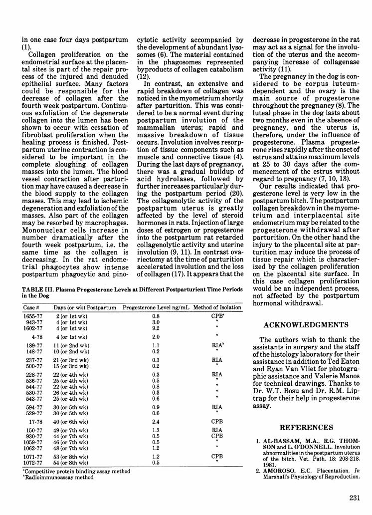
Progesterone Assay - Plasmaprogesterone levels were obtainedfrom bitches at different timeperiods postpartum (Table III).The progesterone level was verylow (below 2 ng/mL except inthree cases . . .) during the mainreparative process of the endome-trium (up to eight weeks postpar-
tum). The progesterone level wasvery high (9.2 ng/mL) in one case(four days postpartum) in compar-ison to other cases in the sameperiod. At 40 days postpartum thesame bitch had 2.4 ng/mL plasmaprogesterone level and was diag-nosed as subinvolution of placentalsites grossly and histologically (1).
Fig. 34. Retrogressing corpus luteum during the fourth, fifth and sixth week post-partum. The corpora lutea are small in size and located deep in the medulla. Massontrichrome X8.
There are several new findingsin this paper: the plane of separa-tion of the placenta at parturition,the collagen fiber proliferation atthe placental sites during the firstfour weeks postpartum and thentheir sloughing, the differences inthe involution between the placen-tal site and interplacental siteendometrium, and the myometrialinvolution in the postpartum dog.The objective of this investiga-
tion was to determine the gross andmicroscopic changes in the normalpostpartum uterus in the dog.Postpartum involution is a slowprocess and 12 weeks are neces-sary for completion. The majorendometrial changes are seen dur-ing the first nine weeks postpar-tum, during which a massivebreakdown of cellular materialand connective tissue occurs. Thesecondary endometrial changesstarting at the ninth week and end-ing by the twelfth week, are ofminor importance. These werecharacterized by a change in shapeand size of the epithelial cells lin-ing the endometrium.The present study indicates that
during the first week postpartumthe placental sites can be identifiedeasily by gross examination of themucosa. This is in contrast to thereport (3) which indicated diffi-culty in gross recognition of theplacental sites during the firstweek postpartum, due to markedcontraction of the myometrium.Anderson and Simpson (3) and
Jochle and Andersen (13), indi-cated that exfoliation of theendometrium stopped betweeneight to 10 weeks postpartum andwas followed by repair. In contrastthe present study indicates thatboth exfoliation (or sloughing) andrepair of the endometrium occurtogether and by the ninth weekmost of the endometrium at theplacental sites and interplacentalareas was repaired.Most of the large cells with
abundant cytoplasm which wereseen during the first and secondweek postpartum were consideredto be decidual cells. These cellswere located in the upper loose
229
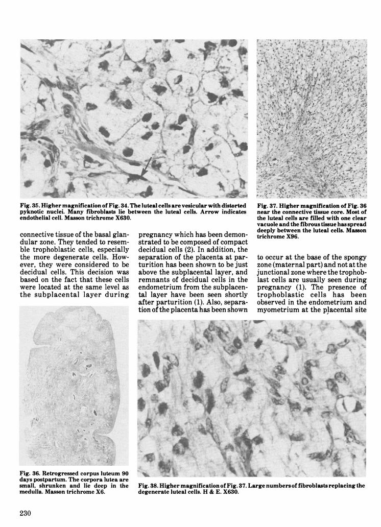
Fig. 35. Higher magnification of Fig. 34. The luteal cells are vesicular with distorted Fig. 37. Higher magnification of Fig. 36pyknotic nuclei. Many fibroblasts lie between the luteal cells. Arrow indicates near the connective tissue core. Most ofendothelial cell. Masson trichrome X630. the luteal cells are filled with one clear
vacuole and the fibrous tissue has spreaddeeply between the luteal cells. Massonconnective tissue of the basal glan- pregnancy which has been demon- trichrome X96.
dular zone. They tended to resem-ble trophoblastic cells, especiallythe more degenerate cells. How-ever, they were considered to bedecidual cells. This decision wasbased on the fact that these cellswere located at the same level asthe subplacental layer during
strated to be composed of compactdecidual cells (2). In addition, theseparation of the placenta at par-turition has been shown to be justabove the subplacental layer, andremnants of decidual cells in theendometrium from the subplacen-tal layer have been seen shortlyafter parturition (1). Also, separa-tion of the placenta has been shown
to occur at the base of the spongyzone (maternal part) and not at thejunctional zone where the trophob-last cells are usually seen duringpregnancy (1). The presence oftrophoblastic cells has beenobserved in the endometrium andmyometrium at the placental site
Fig. 36. Retrogressed corpus luteum 90days postpartum. The corpora lutea aresmall, shrunken and lie deep in themedulla. Masson trichrome X6.
Fig. 38. Higher magnification of Fig. 37. Large numbers of fibroblasts replacing thedegenerate luteal cells. H & E. X630.
230
in one case four days postpartum(1).
Collagen proliferation on theendometrial surface at the placen-tal sites is part of the repair pro-cess of the injured and denudedepithelial surface. Many factorscould be responsible for thedecrease of collagen after thefourth week postpartum. Continu-ous exfoliation of the degeneratecollagen into the lumen has beenshown to occur with cessation offibroblast proliferation when thehealing process is finished. Post-partum uterine contraction is con-sidered to be important in thecomplete sloughing of collagenmasses into the lumen. The bloodvessel contraction after parturi-tion may have caused a decrease inthe blood supply to the collagenmasses. This may lead to ischemicdegeneration and exfoliation of themasses. Also part of the collagenmay be resorbed by macrophages.Mononuclear cells increase innumber dramatically after thefourth week postpartum, i.e. thesame time as the collagen isdecreasing. In the rat endome-trial phagocytes show intensepostpartum phagocytic and pino-
cytotic activity accompanied bythe development of abundant lyso-somes (6). The material containedin the phagosomes representedbyproducts of collagen catabolism(12).
In contrast, an extensive andrapid breakdown of collagen wasnoticed in the myometrium shortlyafter parturition. This was consi-dered to be a normal event duringpostpartum involution of themammalian uterus; rapid andmassive breakdown of tissueoccurs. Involution involves resorp-tion of tissue components such asmuscle and connective tissue (4).During the last days of pregnancy,there was a gradual buildup ofacid hydrolases, followed byfurther increases particularly dur-ing the postpartum period (20).The collagenolytic activity of thepostpartum uterus is greatlyaffected by the level of steroidhormones in rats. Injection of largedoses of estrogen or progesteroneinto the postpartum rat retardedcollagenolytic activity and uterineinvolution (9, 11). In contrast ova-riectomy at the time of parturitionaccelerated involution and the lossof collagen (17). It appears that the
decrease in progesterone in the ratmay act as a signal for the involu-tion of the uterus and the accom-panying increase of collagenaseactivity (11).The pregnancy in the dog is con-
sidered to be corpus luteum-dependent and the ovary is themain source of progesteronethroughout the pregnancy (8). Theluteal phase in the dog lasts abouttwo months even in the absence ofpregnancy, and the uterus is,therefore, under the influence ofprogesterone. Plasma progeste-rone rises rapidly after the onset ofestrus and attains maximum levelsat 25 to 30 days after the com-mencement of the estrus withoutregard to pregnancy (7, 10, 13).Our results indicated that pro-
gesterone level is very low in thepostpartum bitch. The postpartumcollagen breakdown in the myome-trium and interplacental siteendometrium may be related to theprogesterone withdrawal afterparturition. On the other hand theinjury to the placental site at par-turition may induce the process oftissue repair which is character-ized by the collagen proliferationon the placental site surface. Inthis case collagen proliferationwould be an independent process,not affected by the postpartumhormonal withdrawal.
ACKNOWLEDGMENTS
The authors wish to thank theassistants in surgery and the staffof the histology laboratory for theirassistance in addition to Ted Eatonand Ryan Van Vliet for photogra-phic assistance and Valerie Manosfor technical drawings. Thanks toDr. W.T. Bosu and Dr. R.M. Lip-trap for their help in progesteroneassay.
REFERENCES
1. AL-BASSAM, M.A., R.G. THOM-SON and L. O'DONNELL. Involutionabnormalities in the postpartum uterusof the bitch. Vet. Path. 18: 208-218.1981.
2. AMOROSO, E.C. Placentation. InMarshall's Physiology of Reproduction.
231
TABLE III. Plasma Progesterone Levels at Different Postparturient Time Periodsin the Dog
Case # Days (or wk) Postpartum Progesterone Level ng/mL Method of Isolation1655-77 2 (or 1st wk) 0.8 CPBa943-77 4 (or lst wk) 3.0 "1602-77 4 (or 1st wk) 9.2
4-78 4 (or lst wk) 2.0189-77 11 (or 2nd wk) 1.1 RIAb148-77 10 (or 2nd wk) 0.2 "237-77 21 (or 3rd wk) 0.3 RIA500-77 15 (or 3rd wk) 0.2 "228-77 22 (or 4th wk) 0.3 RIA536-77 25 (or 4th wk) 0.5 "544-77 22 (or 4th wk) 0.8 "530-77 26 (or 4th wk) 0.3 "543-77 25 (or 4th wk) 0.6 "594-77 30 (or 5th wk) 0.9 RIA529-77 30 (or 5th wk) 0.6 "17-78 40 (or 6th wk) 2.4 CPB
150-77 49 (or 7th wk) 1.3 RIA930-77 44 (or 7th wk) 0.5 CPB
1059-77 46 (or 7th wk) 0.5 "1062-77 48 (or 7th wk) 1.2 "1071-77 53 (or 8th wk) 1.2 CPB1072-77 54 (or 8th wk) 0.5 "aCompetitive protein binding assay methodbRadioimmunoassay method
Volume 2, pp. 127-311. London, NewYork, Toronto: Parkes, Longmans,Green and Co. 1952.
3. ANDERSEN, A.C. and M.E. SIMP-SON. The Ovary and ReproductiveCycle of the Dog (Beagle). p. 290. LosAltos, California: Geron Inc. 1973.
4. ANDERSON, L.L. Uterine control ofovarian function. In Biology of the Ute-rus. R. Wynn, Editor. pp. 587-651. NewYork, London: Plenum Press. 1977.
5. BOSU, W.T.K., L.E. EDQVIST, P.LINDBERG, K. MARTINSOON andE.D.B. JOHANSSON. The effect ofvarious dosages of lynestrenol on theplasma levels of estrogens and proges-terone during the menstrual cycle inthe rhesus monkey. Contraception 13:677-684. 1976.
6. BRANDES, D. and E. ANTON. Anelectron microscopic cytochemicalstudy of macrophages during uterineinvolution. J. Cell Biol. 41: 450-461.1969.
7. CONCANNON, P.W., W. HANSELand W.J. VISEK. The ovarian cycle ofthe bitch: plasma estrogen, LH andprogesterone. Biol. Reprod. 13: 112-121. 1975.
8. DAVIES, J. and K.J. RYAN. Com-parative endocrinology of gestation.
Vitam. Horm. 30: 223-272. 1972.9. GRANT, R.A. Chemical changes in the
uterus of the rat during pregnancy andpostpartum involution effects of lacta-tion and hormone treatment. J. Reprod.Fert. 9:285-299. 1965.
10. HADLEY, J.C. Total unconjugatedestrogen and progesterone concentra-tion in peripheral blood during preg-nancy in the dog. J. Reprod. Fert. 44:453-460. 1975.
11. HALME, J. and J.F. WOESSNERJR. Effect of progesterone on collagenbreakdown and tissue collagenolyticactivity in the involuting rat uterus. J.Endocr. 66: 357-362. 1974.
12. JACKSON, D.S. Connective Tissue. p.62. Oxford: Blackwell Scientific Publi-cations Ltd. 1957.
13. JOCHLE, W. and A.C. ANDERSEN.The estrus cycle in the dog: A review.Theriogenology 7: 113-130. 1977.
14. JOHANNSON, E.D.B. Progesteronelevels in peripheral plasma during theluteal phase of the normal human men-strual cycle measured by a rapid com-petitive protein binding technique.Actaendocr. Copenh. 61: 592-606. 1969.
15. McMANUS, J.F.A. and R.W.MOWRY. The trichrome techniques ofpierre masson. In The Staining
Methods, Histologic and Histochemi-cal. pp. 234-236. New York, Evanstonand ILondon: Harper and Row. 1965.
16. McMANUS, J.F.A. and R.W.MOWRY. The Perl's stain for ferriciron. In The Staining Methods, Histo-logic and Histochemical. pp. 195-196.New York, Evanston and London:Haeber Medical Division, Harper andRow. 1965.
17. MORRIONE, T.G. and M.Z. RU.Injury influence on resorption of ute-rine collagen. Archs Path. 78: 591-600.1964.
18. MURPHY, B.E.P. Application of theproperty of protein-binding to the assayof minute quantities of hormones andother substances. Nature 201: 697-682.1964.
19. MURPHY, B.E.P. Some studies of theprotein-binding steroids and theirapplication to the routine micro andultramicro measurement of varioussteroids in body fluids by competitiveprotein-binding radioassay. J. clin.Endocr. Metab. 27: 973-990. 1967.
20. WOESSNER, J.F. JR. Acid hydroly-sis of the rat uterus in relation to preg-nancy, postpartum involution and col-lagen breakdown. Biochem J. 97:855-866. 1965.
232


![Title On Galois extension with involution of rings … a Galois extension with involution LlDK of fields, Ll^K is an odd type Galois extension with involution if and olnly if [L:K]](https://img.pdfslide.us/doc/110x75/5c838c4f09d3f2ca448be61a/title-on-galois-extension-with-involution-of-rings-a-galois-extension-with-involution.jpg)


![Integrated analysis of the local and systemic changes ... · are required for normal involution, tissue remodelling and return of the uterus to a pregnancy-receptive state [3, 4]](https://img.pdfslide.us/doc/110x75/5d1e992088c9933c558bf489/integrated-analysis-of-the-local-and-systemic-changes-are-required-for-normal.jpg)