Embed Size (px)
Citation preview
Metabolic Clearance Rates and
Interconversions of Estrone and 17fl-Estradiol in
Normal Males and Females
CHRISTOPHERLONGCOPE,DONALDS. LAYNE, and JAMEs F. TAIT
From the Worcester Foundation for Experimental Biology,Shrewsbury, Massachusetts
A B S T R A C T The continuous infusion of 3H-6,7-estrone and 3H-6,7-estradiol has been used tostudy the metabolic clearance rate (MCR), theinterconversions, and the red cell uptake of thesesteroids in normal males and females. The wholeblood MCRof estrone is 1,990 + 120 liters per day/m2 (SE) in males and 1,910 + 100 liters per day/M2in females. The whole blood MCRof estradiol is1,600 + 80 liters per day/M2 in males and 1,360 +
40 liters per day/M2 in females. The values infemales do not vary significantly when studied inthe follicular or luteal phase of the cycle. At least35%o of the total estrone metabolism in both sexesis extrasplanchnic and at least 25%o of the totalestradiol metabolism in males, and 15 %o in- femalesis extrasplanchnic. The [p] BB21 [transfer constantof estradiol. to estrone, which is equivalent to thefraction of the precursor (estradiol) converted tothe product (estrone) when both the infusion ofthe precursor and the measurement of the productaire in peripheral blood] is 15%o; and the [p]BB" 2[transfer constant of estrone to estradiol, which isequivalent to the fraction of the precursor (estrone)converted to product (estradiol) when both the in-fusion of the precusor and the measurement of theproduct are in peripheral blood] is 5%o in both
A portion of this work was presented before theAmerican Federation for Clinical Research, Atlantic City,N. J. 1967.
Dr. Layne's present address is Food and Drug Labora-tories, Department of National Health and Welfare, Ot-tawa 3, Ontario, Canada.
Received for publication 16 June 1967 and in revisedform 18 September 1967.
males and females. Our findings concerning the ra-dioactivity in whole blood, as measured by our pro-cedure, were the following: 15-20% of estrone inboth sexes and 15% of estradiol in males is asso-ciated with red cells. Only 2% of the whole bloodradioactivity of estradiol in females is associatedwith red cells. Changes in the distribution of radio-activity between plasma and red cells will influencethe MCRas calculated from plasma, but not ascalculated from whole blood.
INTRODUCTION
The metabolic clearance rate (MCR) of a sub-stance, defined as that volume of blood from whichit is totally and irreversibly cleared in unit time, isa concept which has been applied to studies of themetabolism of aldosterone, cortisol, progesterone,and the androgens (1-5). In this manner, infor-mation has been obtained concerning splanchnicand extrasplanchnic metabolism. In addition, theuse of the continuous infusion technique hasprovided data on the interconversion of andros-tenedione and testosterone (3, 4, 6) and on theuptake of steroid hormones into red cells (2).
Previous studies of the metabolism of estroneand 17,8-estradiol in nonpregnant humans haveprovided some data concerning the half-lives ofthese steroids in plasma (7) and the red cell bind-ing of estrone (8). The major studies have beenconcerned with urinary metabolites and with theradioactivity appearing in these metabolites afteradministration of the labeled hormone (9-11). In-
The Journal of Clinical Investigation Volume 47 1968 93
formation obtained in this manner however doesnot always reflect the dynamics of the circulatinghormone (4, 12, 13). Therefore, we felt that astudy of the metabolic clearance rates and theinterconversion of estrone and 17fl-estradiol inblood would provide a fuller picture of the metab-olism of these steriods.
The plasma MCRof testosterone is much lowerthan the hepatic plasma flow. This difference in-dicates incomplete splanchnic extraction for thissteroid (3, 4, 6). However, the plasma MCRofandrostenedione is greater than hepatic plasma flow(3, 4, 6). Bardin and Lipsett (13) have reportedthat the plasma MCRof testosterone is greater formales than for females, and the difference is sig-nificant after correction for body surface areas.There is, however, no sex difference in the MCRper square meters of body surface area of andros-tenedione. These data could be explained by bind-ing of testosterone to plasma proteins other thanalbumin, provided that such binding was greater inplasma from females than from males, and rela-tively greater than the protein binding of an-drostenedione in plasma from both sexes. Wewere, therefore, particularly interested in the pos-sibility of an analogous situation existing forestradiol and estrone.
METHODSNormal adults aged 21-40 yr who were informed andgave their consent were used for all studies. All sub-jects were in good health, and were receiving no medi-cations; the females all gave a history of normal men-strual cycles.
Methylene chloride, acetone, chloroform, and benzenewere of spectral quality. All other solvents were of tech-nical grade and were redistilled prior to use. Pyridinewas prepared as described (14). Acetic anhydride wasdistilled, and the fraction boiling at 1380C was collected.
'H-6,7-estrone and 'H-6,7-17#-estradiol (34 c/mmole)were obtained from the New England Nuclear Corp., and4-'4C-estrone (33.7 mc/mmole) and 4-14C-17f8-estradiol(31.7 mc/mmole) were obtained from the Nuclear-Chi-cago Corporation. They were separately purified priorto use by partition column chromatography on Celite us-ing the E-1 and E-2 systems of Siiteri (15). The tubescontaining the radioactive peak from each column werepooled and the contents were chromatographed on What-man No. 2 paper in a Bush-type system (stationary phase:methanol, water, 3: 1; mobile phase: toluene). The radio-active peak was located by a Vanguard 880 strip scanner,cut out, and eluted with acetone. The steroids were storedat - 15'C in MeOH-benzene, 1: 9, v/v. Tritium-labeledsteroids were purified every 3-4 wk, the "C-labeled
steroids every 4 months. The purity of the radioactivesteroids was checked as follows: 'H-estrone and 4-"C-estrone were mixed, and an aliquot was removed forcounting; another aliquot was chromatographed on What-man No. 2 paper in the Bush-type system (toluene,methanol, water, 4: 3: 1), and a third aliquot was acetyl-ated with pyridine and acetic anhydride, and the resultantestrone acetate was chromatographed on thin-layer chro-matography (TLC) in two dimensions in system II (seebelow). The 'H/"C ratios of the three samples were27.52, 27.61, and 27.69, respectively. Similarly, the'H-estradiol and the "4C-estradiol were mixed and an ali-quot counted; another aliquot was acetylated with pyri-dine and acetic anhydride and the resultant estradiol-3-monoacetate chromatographed on TLC in two dimensionsin systems III and IV (see below). The 8H/"C ratiosobtained were 53.26 and 53.11. These ratios indicate thepurity of infused steroids.
Experiments were begun between 6 and 8 a.m. withthe subjects in a fasting state and supine for 12 hr be-fore and throughout the experiment. A priming dose of4 /Ac of one of the 'H-labeled steroids in 10 ml of 8%ethanol in an isotonic saline solution was injected intoan arm vein at 0 min. The continuous infusion was begun30 min later with 4 luc of the same 8H-steroid in 25 mlof 8% ethanol in an isotonic saline solution infused into anarm vein at 0.190 ml/min by a 50 ml Yale glass syringein a Harvard infusion pump.' The tubing used wasTeflon,2 and adsorption was negligible. Samples weretaken from the priming dose and from the end of theinfusion tubing both before and after the infusion. Ali-quots of these samples were counted as described byFlood and his coworkers (16), and internal standardswere used to correct for quenching.
Estradiol was infused for 105 min, and blood sampleswere drawn at 105, 120, and 135 min from time of prim-ing dose. Estrone was infused for 120 min, and bloodsamples were drawn at 120, 135, and 150 min from timeof priming dose. When it was established that equilibriumhad been reached at these times (see below), only twoblood samples were drawn: 20 min from the end of theinfusion and at its end.
All blood samples were drawn into 50-ml heparinized,disposable syringes from a vein in the arm opposite fromthe infusion. The blood was transferred to chilled tubesand centrifuged, usually at 4'C, for 30 min. 10 ml of plasmawas analyzed as described below. When whole bloodclearance rates were determined, an aliquot of wholeblood was removed initially from the tube for estimationof the hematocrit, and 10 ml of whole blood was removedfor analysis as described below.
Thin-layer chromatography. All TLC was carried outon 20 X 20 cm glass plates using HF2uz Silica Gel 8 0.250mmthick. Carrier estrone4 and estradiol 4 were located
1 Harvard Apparatus Co., Inc., Dover, Mass.2 Becton-Dickinson & Co., Rutherford, N. J.3 Distributed by Brinkmann Instruments Inc., West-
bury, L. I.4 Obtained from Steraloids, Inc., Pawling, N. Y.
94 C. Longcope, D. S. Layne, and J. F. Tait
by absorption at 277 mtn using a mercury lamp and acombination liquid cell (17). Carrier estrogen acetates 4
were located by absorption at 254 m/A using a standard254 mIA source. Elution of the steroids from TLC wascarried out in the same manner as described by Hortonand Tait (3), except that we used 6 ml of acetone.The solvent systems used for TLC were as follows:
I. chloroform: acetoneII. chloroform: acetone
III. chloroform: acetoneIV. benzene: ethanolV. cyclohexane: ethyl acetate
85: 1592: 897: 398: 275: 25
Extraction fromn plasma. 10 ml of plasma was addedto flasks containing 4-14C-estrone (120 dpm) and 4-"4C-estradiol (120 dpm) in 0.2 ml EtOH. 1 ml of 1.25 NNaOHand 2.5 ml of saturated NaHCOawere added tothese flasks. The contents were swirled and then added toa 500 ml separatory funnel and extracted twice with 60 mlvolumes of ice-cold methylene chloride (CH2CI2). TheCH2Cl2 extracts were pooled and washed twice with 20ml of distilled water. The CH2Cl2 was removed undervacuum, and the residue was transferred to 10-ml glass-stoppered tubes and partitioned between 3.0 ml 1 N NaOHand 0.5 ml benzene. The benzene was discarded, and thealkaline extract was transferred to 60-ml separatoryfunnels, and 6.0 ml of saturated NaHCOs was added.This solution was extracted twice with 10-ml portionsof CH2Cl2 which were pooled and washed once with 2.0ml H20 and three times with 1.0 ml volumes of H20.The CH2Cl2 was removed under a stream of nitrogen, andthe residue was spotted on a thin-layer plate with 20 /Agof nonradioactive carrier estrone and 17,8-estradiol.System I was used for this chromatography, after whichthe steroids were eluted and concentrated in the bottomof 10-ml glass-stoppered tubes.
To each tube pyridine 0.2 ml and 0.005 ml of 10%acetic anhydride in benzene were added. The tubes werestoppered and placed overnight at room temperature ina desiccator. Under these conditions estrone was con-
verted to estrone acetate, and estradiol was converted toestradiol-3-monoacetate in 80-90% yield (18). The reac-tions were stopped with 0.05 ml of ethanol, and the tubesdried under a stream of nitrogen. The acetates wereseparately analyzed in two dimensions by TLC: estroneacetate in system III and estradiol-3-monoacetate in sys-tem II. 20 lig of the appropriate unlabeled estrogenacetate was added to each plate as carrier. The steroidswere located and eluted into counting vials. The vialswere dried in a vacuum oven, and 0.2 ml of methanol and10 ml of scintillation fluid (84 nil of Liquifluor,5 20 ml ofethanol, 1 liter of toluene) were added to each vial. Thevials were counted for 5 X 50 min. Using a treatmentsimilar to that described by Horton and Tait (19),we can calculate, for minimal total counting times of 250min, that the maximal errors for the determination of theconcentrations of radioactivity as the precursor and theproduct steroid would be 2 and 15%, respectively.
Extraction of whole blood. 10 ml of whole blood wasadded to a flask containing 4-14C-estrone (120 dpim)and 4-14C-estradiol (120 dpm) in 0.2 ml of ethanol. Thecontents of the flask were transferred to a 500 ml sepa-ratory funnel and the flask rinsed successively with 10ml distilled water and 10 ml diethyl ether. The rinseswere added to the funnel, and the contents were ex-tracted twice with 100-mIl volumes of ice-cold CH2Cb.The CH2CI2 extracts were pooled and washed three timeswith 30-nil volumes of water. The CHCI2 was removedunder vacuum, and the same steps followed as werepreviously described under Plasma.
Recoveries. Over-all recoveries of 4-14C-estrone addedto plasma or whole blood were 45-55%, and recoveriesof 4-14C-estradiol were 50-60%.
Conversion in the method. To check the possibility ofconversion of the estrogen occurring after the blood wasdrawn, 'H-estradiol was added to whole blood andprocessed as described above. In these experiments radio-activity was recovered as estradiol, but the area corre-
5 Liquifluor obtained from Pilot Chemicals, Inc., Water-town, Mass.
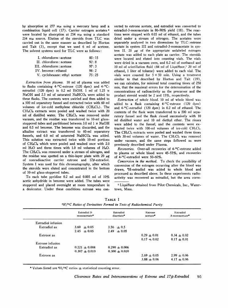
TABLE I3H/I4C Ratios of Derivatives Formed in Tests of Radiochemical Purity
Estradiol 3- Estradiol Estrone Eistradiolmonoacetate* diacetate* acetate* 3-monoacetate*
Estradiol infusionEstradiol as 3.60 i 0.05 3.56 ± 0.5
2.45 ± 0.05 2.49 ± 0.05Estrone as 0.29 4t 0.01 0.34 ± 0.02
0.17 + 0.02 0.17 ± 0.01Estrone infusion
Estradiol as 0.221 0.004 0.290 ± 0.0060.307 i 0.010 0.300 i± 0.010
Estrone as 2.69 i 0.05 2.99 ±i( 0.063.88 ± 0.06 4.17 ± 0. 06
* Values listed are 3H/14C ratios 4 statistical counting error.
Clearance Rates and Interconversions of Estrone and 1 7/3-E-stradiol (fir
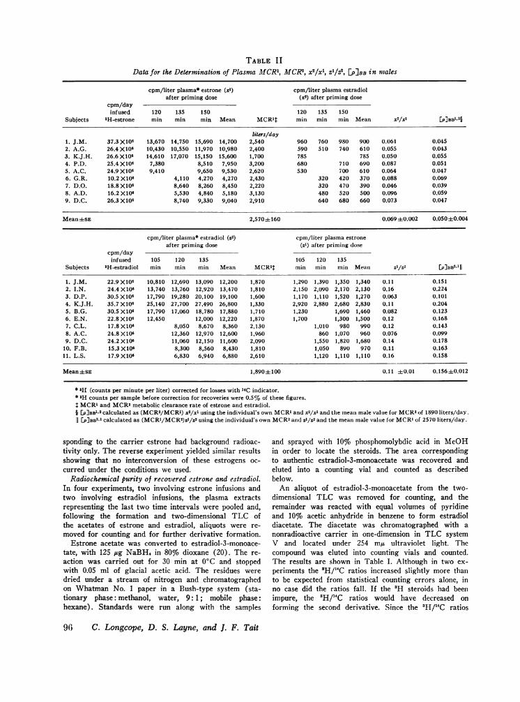
TABLE IIData for the Determination of Plasma M1CR1, MCR2, x2/xl, Z1/z2, []PBB in males
cpm/liter plasma* estrone (xl) cpm/liter plasma estradiolafter priming dose (x2) after priming dose
cpm/dayinfused 120 135 150 120 135 150
Subjects 3H-estrone min min min Mean MCR1t min min min Mean x2/xl [P]BB'.2§
liters/day1. J.M. 37.3 X10⁶ 13,670 14,750 15,690 14,700 2,540 960 760 980 900 0.061 0.0452. A.G. 26.4 X10⁶ 10,430 10,550 11,970 10,980 2,400 590 510 740 610 0.055 0.0433. K.J.H. 26.6 X10⁶ 14,610 17,070 15,150 15,600 1,700 785 785 0.050 0.0554. P.D. 25.4 X10⁶ 7,380 8,510 7,950 3,200 680 710 690 0.087 0.0515. A.C. 24.9 X10⁶ 9,410 9,650 9,530 2,620 530 700 610 0.064 0.0476. G.R. 10.2 X10⁶ 4,110 4,270 4,270 2,430 320 420 370 0.088 0.0697. D.O. 18.8 X10⁶ 8,640 8,260 8,450 2,220 320 470 390 0.046 0.0398. A.D. 16.2 X10⁶ 5,530 4,840 5,180 3,130 480 520 500 0.096 0.0599. D.C. 26.3 X10⁶ 8,740 9,330 9,040 2,910 640 680 660 0.073 0.047
Mean±SE 2,570±160 0.069 ±0.002 0.0504±0.004
cpm/liter plasma* estradiol (Z2) cpm/liter plasma estroneafter priming dose (zl) after priming dose
cpm/dayinfused 105 120 135 105 120 135
Subjects 3H-estradiol min min min Mean MCR2T min inin min Mean z1/Z2 [p]BB2j 1
1. J.M. 22.9 X10⁶ 10,810 12,690 13,090 12,200 1,870 1,290 1,390 1,350 1,340 0.11 0.1512. I.N. 24.4 X10⁶ 13,740 13,760 12,920 13,470 1,810 2,150 2,090 2,170 2,130 0.16 0.2243. D.P. 30.5 X10⁶ 17,790 19,280 20,100 19,100 1,600 1,170 1,110 1,520 1,270 0.063 0.1014. K.J.H. 35.7 X10⁶ 25,140 27,700 27,490 26,800 1,330 2,920 2,880 2,680 2,830 0.11 0.2045. B.G. 30.5 X10⁶ 17,790 17,060 18,780 17,880 1,710 1,230 1,690 1,460 0.082 0.1236. E.N. 22.8 X10⁶ 12,450 12,000 12,220 1,870 1,700 1,300 1,500 0.12 0.1687. C.L. 17.8 X10⁶ 8,050 8,670 8,360 2,130 1,010 980 990 0.12 0.1438. A.C. 24.8 X10⁶ 12,360 12,970 12,600 1,960 860 1,070 960 0.076 0.0999. D.C. 24.2 X10' 11,060 12,150 11,600 2,090 1,550 1,820 1,680 0.14 0.178
10. F.B. 15.3 X10⁶ 8,300 8,560 8,430 1,810 1,050 890 970 0.11 0.16311. L.S. 17.9X10⁶ 6,830 6,940 6,880 2,610 1,120 1,110 1,110 0.16 0.158
Mean±sE 1,890±fi100 0.11 ±)0.01 0.156±0.012
* aH (counts per minute per liter) corrected for losses with 14C indicator.* 3H counts per sample before correction for recoveries were 0.5% of these figures.t MCRl and MCR2metabolic clearance rate of estrone and estradiol.§ EP]BB1a2 calculated as (MCR2/MCRl) x2/xl using the individual's own MCRI and x5/xl and the mean male value for MCR2of 189(1 liters/day.11 [P]BB2.1 calculated as (MCRI/MCR2)z'/z2 using the individual's own MCR2and Z'/Z2 and the mean male value for MCR1of 257(1 liters/day.
sponding to the carrier estrone had background radioac-tivity only. The reverse experiment yielded similar resultsshowing that no interconversion of these estrogens oc-curred under the conditions we used.
Radiochemical purity of recovered estrone and estradiol.In four experiments, two involving estrone infusions andtwo involving estradiol infusions, the plasma extractsrepresenting the last two time intervals were pooled and,following the formation and two-dimensional TLC ofthe acetates of estrone and estradiol, aliquots were re-moved for counting and for further derivative formation.
Estrone acetate was converted to estradiol-3-monoace-tate, with 125 /ig NaBH4 in 80% dioxane (20). The re-action was carried out for 30 min at 0C and stoppedwith 0.05 ml of glacial acetic acid. The residues weredried under a stream of nitrogen and chromatographedon Whatman No. 1 paper in a Bush-type system (sta-tionary phase: methanol, water, 9:1; mobile phase:hexane). Standards were run along with the samples
and sprayed with 10% phosphomolybdic acid in MeOHin order to locate the steroids. The area correspondingto authentic estradiol-3-monoacetate was recovered andeluted into a counting vial and counted as describedbelow.
An aliquot of estradiol-3-monoacetate from the two-dimensional TLC was removed for counting, and theremainder was reacted with equal volumes of pyridineand 10% acetic anhydride in benzene to form estradioldiacetate. The diacetate was chromatographed with anonradioactive carrier in one-dimension in TLC systemV and located under 254 my ultraviolet light. Thecompound was eluted into counting vials and counted.The results are shown in Table I. Although in two ex-periments the 'H/'4C ratios increased slightly more thanto be expected from statistical counting errors alone, inno case did the ratios fall. If the 'H steroids had beenimpure, the 'H/14C ratios would have decreased onforming the second derivative. Since the 'H/'4C ratios
96 C. Longcope, D. S. Layne, and J. F. Tait
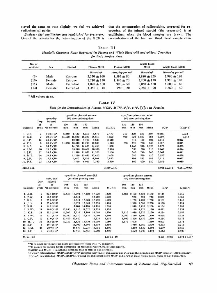
stayed the same or rose slightly, we feel we achieved that the concentration of radioactivity, corrected for re-radiochemical purity. coveries, of the infused steroid (the precursor) is at
Evidence that equilibrium was established for precursor. equilibrium when the blood samples are drawn. TheOtie of the criteria for tlhe detcrminiation of the MCRis concenitrationis of the first alid third blood sample com-
TABLE IlIMetabolic Clearance Rates Expressed in Plasma and Whole Blood with and without Correction
for Body Surface Area
No. of Whole bloodsubjects Sex Steriod Plasma MCR Plasma MCR MCR Whole blood MCR
liters/day* liters/day per m2* liters/day* liters/day per m2*
(9) Male Estrone 2,570 i 160 1,310 ±4 80 3,880 i 220 1,990 i 120(10) Female Estrone 2,210 + 120 1,320 ± 70 3,200 i 170 1,910 i 100(11) Male Estradiol 1,890 ± 100 990 ± 50 3,060 i 160 1,600 ± 80(13) Female Estradiol 1,350 ± 40 790 ± 20 2,280 ± 90 1,360 + 40
* All values ± SE.
TABLE IVData for the Determination of Plasma MCR1, MCR2, x2/xl, z1/W2, [PIBB in Females
cpm/liter plasma* estrone cpm/liter plasma estradiolcpm/day (xl) after priming dose (x2) after priming dose
Day infusedof as 120 135 150 120 135 150
Subjects cycle 3H-estrone min min min Mean MCRlJ min min min Mean X2/X1 [P]BBe.2§
1. C.R. 7 14.0 X10⁶ 6,240 5,430 5,350 5,670 2,470 310 370 320 330 0.058 0.0322. K.B. 2 30.1 X10⁶ 15,520 16,280 16,280 16,150 1,860 990 820 1,080 960 0.059 0.0433. S.P. 12 24.1 X10⁶ 10,010 9,750 9,880 2,440 310 490 400 0.040 0.0224. P.B. 8 20.1 X10⁶ 11,030 10,310 11,290 10,880 1,840 750 690 760 730 0.067 0.0495. A.B. 6 23.2 X10⁶ 14,710 14,680 14,690 1,580 1,260 990 1,120 0.076 0.0656. S.M. 10 21.8 X10⁶ 10,010 10,660 10,380 2,100 700 720 710 0.068 0.0447. J.D. 27 24.5 X10⁶ 11,570 11,070 11,320 2,160 740 740 740 0.065 0.0418. M.T. 24 29.8 X10⁶ 11,220 13,630 12,420 2,400 780 570 620 0.050 0.0289. J.P. 24 17.7 X10⁶ 6,640 5,650 6,140 2,880 790 580 680 0.111 0.052
10. P.R. 23 17.4 X10⁶ 7,770 6,960 7.360 2,360 360 400 380 0.052 0.030
Mean 1SE 2,2104120 0.065+0.016 0.04140.004
cpm/liter plasma* estradiol cpm/liter plasma estronecpm/day (Z2) after priming dose (zl) after priming dose
Day infusedof as 105 120 135 105 120 135
Subjects cycle 3H-estradiol min min min Mean MCR2I min min min Mean Z1/52 [P]BB2.lU
1. A.B. 6 22.4 X10⁶ 17,520 17,790 17,400 17,570 1,270 2,680 2,450 2,320 2,480 0.141 0.2452. P.W. 4 19.9 X10⁶ 12,540 12,540 1,590 980 570 770 0.061 0.0853. S.B. 7 25.8 X10⁶ 17,340 17,020 17,180 1,500 1,770 1,700 1,730 0.101 0.1484. J.D. 4 25.6 X10⁶ 16,870 17,640 17,250 1,481) 1,920 2,580 2,250 0.130 0.1945. S.M. 4 16.9 X10⁶ 13,100 13,390 13,390 1,260 1,940 2,470 2,200 0.164 0.2876. E.Wo. 24 16.8 X10⁶ 13,93(0 14,610 14,570 14,370 1,170 1,180 1,170 1,170 0.081 0.1537. K.B. 24 25.1 X10⁶ 16,360 15,910 15,840 16,04(1 1,570 2,110 1,98.0 2,370 2,150 0.134 0.1898. E.W. 16 22.7 X10⁶ 19,260 18,270 19,470 19,000 1,200 1,260 1,140 1,500 1,300 0.068 0.1259. C.F. 17 17.9 X10⁶ 12,400 12,640 12,520 1,430 1,600 1,260 1,430 1,430 0.114 0.175
10. M.T. 22 18.9 X10⁶ 17,720 14,420 15,970 16,040 1,180 1,370 1,690 1,530 0.093 0.17411. S.P. 6 19.7 X10⁶ 15,900 13,290 14,590 1,350 1,910 1,960 1,930 0.132 0.21612. E.M. 21 20.9 X10⁶ 18,670 19,230 18,950 1,100 1,480 1,530 1,500 0.079 0.15913. J.P. 3 24.8X1(06 17,950 17,461) 17,700 1,400 1,020 1,200 1,110 0.063 0.100
Mean-ksE 1,350± 40 0.105±0.009 0.173±0.017
* 3H (counts per minute per liter) corrected for losses witl 14C indicator.* 3H counts per sample before correction for recoveries were 0.5% of these figures.t MCRl and MCR2= metabolic clearance rate of estrone and estradiol.§ [p]sBIO calculated as (MCR2/MCRl) x2/xl using the individuals' own MCR' and x2/xl and the mean female MCR2value of 1,350 liters/day.I! [P]BB2 1calculated as (MCR'/MCR2) Zt/Z2 using the individual's own MCRSand Z'/Z2 and mean female MCRI value of 2,210 liters/day.
Clearance Rates and Interconversions of Estrone and 1 7fl-Estradiol97
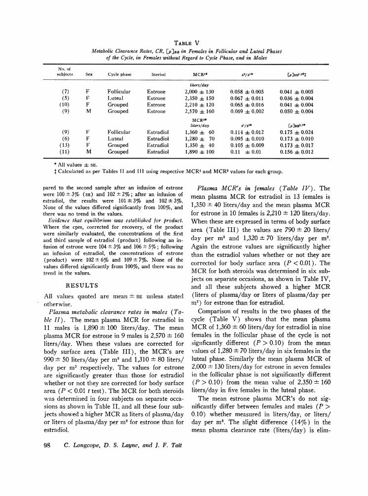
TABLE VMetabolic Clearance Rates, CR, [PIBB in Females in Follicular and Luteal Phases
of the Cycle, in Females without Regard to Cycle Phase, and in Males
No. ofsubjects Sex Cycle phase Steriod MCRxx* [p]BBl.2*t
liters/day(7) F Follicular Estrone 2,000 i 130 0.058 i- 0.005 0.041 ± 0.005(5) F Luteal Estrone 2,350 i 150 0.067 ± 0.011 0.036 ± 0.004
(10) F Grouped Estrone 2,210 : 120 0.065 1 0.016 0.041 + 0.004(9) M Grouped Estrone 2,570 i 160 0.069 i 0.002 0.050 1 0.004
MCR2*liters/day Z[/p2* [P]BB2,1*
(9) F Follicular Estradiol 1,360 60 0.114 i 0.012 0.175 ± 0.024(6) F Luteal Estradiol 1,280 i 70 0.095 ± 0.010 0.173 ± 0.010
(13) F Grouped Estradiol 1,350 ± 40 0.105 :1 0.009 0.173 ±t 0.017(11) M Grouped Estradiol 1,890 ± 100 0.11 i 0.01 0.156 ± 0.012
* All values ± SE.t Calculated as per Tables II and III using respective MCR1and MCR2values for each group.
pared to the second sample after an infusion of estronewere 100 + 3% (SE) and 102 ± 2%o; after an infusion ofestradiol, the results were 101 ± 3%o and 102 + 3%.None of the values differed significantly from 100%o, andthere was no trend in the values.
Evidence that equilibrium was established for product.Where the cpm, corrected for recovery, of the productwere similarly evaluated, the concentrations of the firstand third sample of estradiol (product) following an in-fusion of estrone were 104 ± 3%o and 106 + 5%o; followingan infusion of estradiol, the concentrations of estrone(product) were 102 + 6%o and 109 ± 7%o. None of thevalues differed significantly from 100%, and there was notrend in the values.
RESULTS
All values quoted are mean + SE unless statedotherwise.
Plasma metabolic clearance rates in males (Ta-ble II). The mean plasma MCRfor estradiol in11 males is 1,890 + 100 liters/day. The meanplasma MCRfor estrone in 9 males is 2,570 + 160liters/day. When these values are corrected forbody surface area (Table III), the MCR's are990 + 50 liters/day per m2 and 1,310 + 80 liters/day per m2 respectively. The values for estroneare significantly greater than those for estradiolwhether or not they are corrected for body surfacearea (P < 0.01 t test). The MCRfor both steroidswas determined in four subjects on separate occa-sions as shown in Table II, and all these four sub-jects showed a higher MCRas liters of plasma/dayor liters of plasma/day per m2 for estrone than forestradiol.
Plasma MCR's in females (Table IV). Themean plasma MCRfor estradiol in 13 females is1,350 ± 40 liters/day and the mean plasma MCRfor estrone in 10 females is 2,210 ± 120 liters/day.When these are expressed in terms of body surfacearea (Table III) the values are 790 ± 20 liters/day per m2 and 1,320 + 70 liters/day per M2.Again the estrone values are significantly higherthan the estradiol values whether or not they arecorrected for body surface area (P < 0.01). TheMCRfor both steroids was determined in six sub-jects on separate occasions, as shown in Table IV,and all these subjects showed a higher MCR(liters of plasma/day or liters of plasma/day perm2) for estrone than for estradiol.
Comparison of results in the two phases of thecycle (Table V) shows that the mean plasmaMCRof 1,360 + 60 liters/day for estradiol in ninefemales in the follicular phase of the cycle is notsignficantly different (P > 0.10) from the meanvalues of 1,280 + 70 liters/day in six females in theluteal phase. Similarly the mean plasma MCRof2,000 + 130 liters/day for estrone in seven femalesin the follicular phase is not significantly different(P > 0.10) from the mean value of 2,350 ± 160liters/day in five females in the luteal phase.
The mean estrone plasma MCR's do not sig-nificantly differ between females and males (P >0.10) whether measured in liters/day, or liters/day per M2. The slight difference (14%) in themean plasma clearance rate (liters/day) is elim-
98 C. Longcope, D. S. Layne, and J. F. Tait
inated when expressed in liters/day per m2 (TableIII). The mean estradiol plasma MCRvalues infemales is significantly lower than the mean valuesin males (P < 0.01). This significant difference ismaintained when the values are corrected for bodysurface area (P < 0.01 ) even though the differencebetween the mean values drops after correctionfrom 30 to 20% (expressed in terms of the maleclearance rate).
Whole blood MCR. The mean ratios betweenthe whole blood and plasma MICR's were deter-mined in four males for estradiol and in five malesfor estrone. These ratios were found to be 1.62 ±0.05 and 1.53 ± 0.05 respectively. Similarly themean ratios between whole blood and plasmaMCR's determined for estradiol (eight females)and for estrone (seven females) were 1.72 ± 0.05and 1.45 + 0.05 respectively. The difference in theratios between estrone and estradiol was signifi-cant in females but not in males. All the plasmaMCR's were then multiplied by the appropriateratio in order to give whole blood clearance rates.
Males. The mean whole blood MCRin 11males is 3,060 ± 160 liters/day for estradiol and3,880 + 220 liters/day in nine males for estrone.When expressed in terms of body surface area, themean values are 1,600 ± 80 liters/day per m2 and1,990 ± 120 liters/day per m2 respectively. Thevalues for estrone and estradiol are significantlydifferent (P < 0.01) however they are expressed.
Females. The mean whole blood MCRin 13females is 2,280 + 90 liters/day for estradiol and3,200 ± 170 liters/day in 11 females for estrone.When expressed in terms of body surface area, themean values are 1,360 ± 40 liters/day per m2 and1,910 + 100 liters/day per M2. The values forestrone and estradiol are significantly different(P < 0.01) however they are expressed.
When the mean estrone values for males andfemales are compared, their difference calculatedon the basis of liters/day is about 17% of the malevalue, which is just significant (0.05 > P > 0.02).The difference between the mean values expressedin liters/day per m2 becomes much smaller, 4% ofthe male value, and is not significant (P > 0.10).
The mean estradiol MCRmeasured in liters/dayis significantly lower (P < 0.01) in females thanmales, and the difference is 25% of the male value.When the mean values are expressed in liters/dayper m2, the difference is only 15% of the male
value. This difference, however, is significant(P < 0.01).
Conlversion ratio. The conversion ratio ofestrone to estra(liol is obtained as follows: CBBt -=.v2/x-1 [xl = plasma radioactive concentration ofestrone as precursor; .r2 plasmia radioactive con-centration. of estradiol as product]. The conversionratio of estradiol to estrone is obtained as follows:C16BB1 = 1/Z2 [Z°= )lasma radioactive concentra-tionl of estradiol as precursor; Z1 = plasma radio-active concentration of estrone as product . Threefemales and one male were infused with bothestrone and estradiol. Following these infusions,both whole blood and plasma concentration ofradioactivity in precursor and product were deter-mined. The ratios between whole blood and plasmavalues were 1.01 ± 0.10 and 1.04 ± 0.10 forCBB1,2 and CBB21 respectively. Neither of theseratios differs significantly from 1.0 (P > 0.10),although theoretically they could differsG Plasmavalues which were determined in all subjects willbe used throughout.
The mean conversion ratios of estrone to estra-diol (Tables II and IV) after infusion of estroneCB111"2 are 6.9 + 0.2%o (nine males) and 6.5 ±0.6% (10 females), which are not significantlydifferent. For females there is no statistical differ-ence between the mean values measured in thefollicular and those in the luteal plhases of the cycle(5.8 ± 0.5 % vs. 6.7 ± 1.1%, P > 0.10).
The mean conversion ratios of estradiol toestrone (Tables II and IV) after infusion ofestradiol are 11.3 ± 0.9% (1 1 males) and 1 1.0 +
1.0% (13 females). These values are not sig-nificantly different in the two sexes. In femalesthere is no statistical difference between the valuesfor the follicular and luteal phases of the cycle(11.9 ± 1.3 % vs. 9.8 ± 1.2%, P > 0.10).
[p] RB values (transfer constants) measured inplasma or blood after intravenous infusion. The
2 MCR2 x2 x2/z2
x2/z2 and x'lf' will he equal for whole blood and plasma,if the radioactivity is in equilibrium, since the measure-ment is the ratio of isotopes in the same steroid. There-fore, the [P]BB value measured in whole blood or plasmashould be equivalent, theoretically.
However, the conversion ratio e.g., Ciu,2" = ztf9, willnot necessarily be equal when measure(l in whole bloodor plasma if the distribution of estradiol and estrone intore(l cells is different.
Clearance Rates and Interconversions of Estrone and 17/3-Estradiol 99
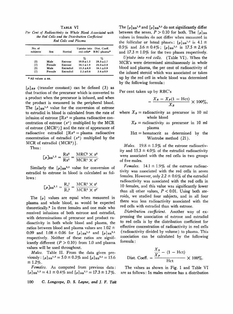
TABLE VIPer Cent of Radioactivity in Whole Blood A ssociated with
the Red Cells and the Distribution CoefficientRed Cells and Plasma
No. of Uptake into Dist. Coeff.subjects Sex Steriod red cells* RBCplasma*
(5) Male Estrone 19.84-1.5 28.3 42.7(7) Female Estrone 14.1±1.9 24.0±+3.8(5) Male Estradiol 15.3±+4.0 16.1±3.8(7) Female Estradiol 2.2 ±0.6 3.4 i0.9
* All values ± SE.
[p] BB (transfer constant) can be defined (3) asthat fraction of the precursor which is converted toa product when the precursor is infused, and whenthe product is measured in the peripheral blood.The [P]BB1'2 value for the conversion of estroneto estradiol in blood is calculated from the rate ofinfusion of estrone [Rxl = plasma radioactive con-centration of estrone (x1) multiplied by the MCRof estrone (MCR') ] and the rate of appearance ofradioactive estradiol [Rx2 = plasma radioactiveconcentration of estradiol (x2) multiplied by theMCRof estradiol (MCR2) ].
Thus:
RX2 M11RC2 X X2[P]BB' = A/Rx1MCRX X1
Similarly the [p] BB2'1 value for conversion ofestradiol to estrone in blood is calculated as fol-lows:
R2,1 \Rz ICR' X z'[p]BB1 = = \I CR2 X z2
The [p] values are equal when measured inplasma and whole blood, as would be expectedtheoretically." In three females and one male whoreceived infusions of both estrone and estradiol,with determinations of precursor and product ra-dioactivity in both whole blood and plasma, theratios between blood and plasma values are 1.02 ±0.09 and 1.08 + 0.06 for [PIBB" 2 and [p]B2"1respectively. Neither of these ratios are signif-icantly different (P > 0.10) from 1.0 and plasmavalues will be used throughout.
Males. Table II. From the data given pre-viously: [pI]BB1 2 = 5.0 ± 0.3%o and [p]"BB21 = 15.6± 1.2 %.
Females. As computed from previous data:[PI BB12 = 4.1 + 0.4%o and [PI 1B2B' = 17.3 + 1.7%.
The [pIBB1'2 and [p] BB2'1 do not significantly differbetween the sexes, P > 0.10 for both. The [P1 BB
values in females (lo not differ when measured illthe follicular or hlteal phases: [pIlll' is 4.1 +
0.5% and 3.6 + 0.47o; [pJ Bol is 17.5 + 2.4%7oand 17.3 ± 1.0% for the two phases respectively.
Uptake into red cells. (Table VI). When theMCR's were determined simultaneously in wholeblood and plasma, the per cent of radioactivity ofthe infused steroid which was associated or takenup by the red cell in whole blood was determinedby the following formula:
Per cent taken up by RBC's
X -Xp (I - Hct)X 100%
XB
where XB = radioactivity as precursor in 10 mlwhole blood
Xp= radioactivity as precursor in 10 mlplasma
Hct = hematocrit as determined by theWintrobe method (21).
Males. 19.8 + 1.5% of the estrone radioactiv-ity and 15.3 ± 4.0%o of the estradiol radioactivitywere associated with the red cells in two groupsof five males.
Females. 14.1 + 1.9% of the estrone radioac-tivity was associated with the red cells in sevenfemales. However, only 2.2 + 0.6% of the estradiolradioactivity was associated with the red cells in10 females, and this value was significantly lowerthan all other values, P < 0.01. Using both ste-roids, we studied four subjects, and in all fourthere was less radioactivity associated with thered cells with estradiol than with estrone.
Distribution coefficient. Another way of ex-pressing the association of estrone and estradiolto red cells is by the distribution coefficient foreffective concentration of radioactivity in red cells(radioactivity divided by volume) to plasma. Thisassociation can be calculated by the followingformula:
x,- (1 - Hct)Dist. Coeff. = P X lOO%.Hct X10%
The values as shown in Fig. 1 and Table VIare as follows: In males estrone has a distribution
100 C. Longcope, D. S. Layne, and J. F. Tait
ESTRONEMales Females
ESTRADIOLMales Females
50.
40.
O,> 3 Oa,30.
afi 20.
10.
0A
[:][:1-
Mean + I 12xSE %A-0 I
FIGURE 1 The distribution coefficient (effective concentration ofsteroid in RBC: Plasma) for estrone and estradiol in males andfemales.
coefficient of 28.3 + 2.7%,, and estradiol 16.1 +
4.0%. These values are significantly different(0.02 > P > 0.01). In females the distributioncoefficient of estrone is 24.0 ± 3.8%/o and for estra-diol it is 3.4 ± 0.9%. The last value is significantlylower than all the others (P < 0.01)
Following estradiol infusions into three females,blood was collected and centrifuged in the usualfashion at 40C, and samples were also collectedand centrifuged at room temperature. The red celluptakes at 4° and 250C were 0.0 and 0.0; 1.5 and1.0; and 3.8 and 6.2% respectively for the threeexperiments and the corresponding distribution co-efficients were 0.0 and 0.0; 2.3 and 1.5; and 6.0 and10.8%(,. There was therefore no consistent differ-ence between the values measured at 40C and250C.
DISCUSSION
Two methods have been described in order tomeasure the metabolic clearance rate: the singleinjection and the continuous infusion techniques(22). Wehave used the continuous infusion tech-nique throughout the study in order to determineMCR's and interconversions of estrone and 17,3-estra(liol in the circulation. While the single injec-tion technique would have given us informationconcerning the volumes of distribution, it wouldhave yielded imprecise information concerning theinterconversions that occur between these twosteroids.
Metabolic clearance rates have generally beenexpressed in terms of liters of plasma cleared per
day (1-4). A correlation between metabolic clear-ance rate and body surface area has been notedhowever (13), and Horton and Frasier haveshown that androstenedione clearances are similarin both adults and children (23) when expressed interms of body surface area.
The mean values for plasma MCRof estrone inmales and females are not significantly differentwhether or not they are expressed in terms of bodysurface. When the MCRis expressed in terms ofwhole blood, the difference between the meanestrone MCRin males and females is just sig-nificant (0.05 > P > 0.02) but when expressed interms of body surface area the difference dis-appears.
The mean plasma and whole blood MCR's forestradiol in males are significantly greater thanthe respective MCR's in females (P < 0.01 forboth), and these differences persist when theMCR's are expressed in terms of body surfacearea, although the differences become smaller whenthis correction is used.
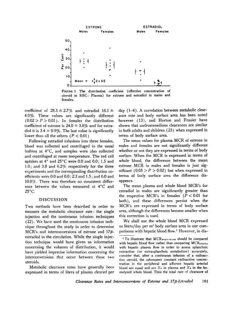
We shall use the whole blood MCRexpressedas liters/day per m2 body surface area in our com-parisons with hepatic blood flow.7 However, in dis-
7 To illustrate that MCRwto0e blood should be comparedwith hepatic blood flow rather than comparing MCRpiasmawith hepatic plasma flow in order to assess splanchnicextraction (or extrasplanchnic metabolism) accurately,consider that, after a continuous infusion of a radioac-tive steroid, the subsequent constant radioactive concen-tration in the peripheral and afferent hepatic arterialblood are equal and are X,' in plasma and X* in the he-molyzed whole blood. Then the total rate of clearance of
Clearance Rates and Interconversions of Estrone and 17,8-A,.str(diol 10
ESTRONEMales Females
ESTRADIOLMales Females
2400.
EIV
0.
0.
i
0 FF: I
Splanchnic BloodFlow
I_
Mean+ A- 2 x SE
FIGURE 2 Comparison between metabolic clearance rates expressedin liters of whole blood/day per m2 and estimated splanchnic bloodflow in liters of whole blood/day per m2.
cussing transfer constants, [p] values, we shallcontinue to use plasma values which are equivalentboth theoretically and experimentally to wholeblood values."
As shown in Fig. 2, the whole blood MCR's ofestrone expressed as liters/day per m2 in bothmales and females (1990 and 1910 liters/day permi2) are well above the estimated splanchnic bloodflow of 1200 liters/day per m2 (24). Since thesplainchnic tissue can clear only that volume ofblood which flows through it per day, it is ap-parent that at least 700 liters/day per m2 must becleared by extrasplanchnic tissue. Thus, at least35 %o of the total estrone metabolism is extra-splanclhnic.
radioactive steroid will l)e an equation: MCRIOlastIO X Xp =MCRWB.X Xn. Consider also a situation in which thesplanchnic extraction of steroid is 100% and the extra-splanchnic metabolism zero. Then hepatic blood flow(HBF) X Xi, will be the total rate of clearance of radio-active steroid. This rate will equal MCRIanRa X Xi, orMCRw,,.... ,,4,X Xi, but will not necessarily equal hepaticplasma flow (HPF) X XP. Then MCRwlloe blood X XB-HBFw,1ole blood X XR, and MCRwhol. blood = HBF. In gen-eral if there is an uptake of steroids into the red cells,MCR),.:,0 will not ec(lual HPF. Similarly it can be shownthat, in general,
MCR,,ololt...l = HBF x HE+ ESCwi,.ie loootiin which HE is the hepatic extraction of steroid as mea-sured in whole blood, and ESCwlneie ooui/MCRwitoiebluoodis the proportion of the total metabolism which is extra-splanchnic. In previous estimates of the splanchnic ex-traction and the extrasplanchnic metabolism of andros-tenedione, testosterone (3), and aldosterone (22), theMCRpin..mn Wvas compared with HPF, andi the HE and theESC were slightly overestimated because of these con-siderations.
The clearance rate of estradiol in both sexes isalso above the estimated splanchnic blood flow,but the portion of metabolism which is extra-
slplanchnic is less for estradiol than for estrone.At least 25% of the total estradiol metabolism inmales, and at least 157o% of the total in females isextrasplanchnic. It must be realized, however,that these estimates of extrasplanchnic metabolismare minimal values and presuppose an hepatic ex-traction of 100%7 for the respective estrogen. If thehepatic extraction of either steroid is less than100%, then the estimated amount of extrasplanch-nic metabolism will increase proportionately.
When the MCRis below splanchnic blood flow,as for testosterone and cortisol, it is evident thatthe estimated maximal splanchnic extraction of thesteroid will be less than 100%o (splanchnic ex-traction < MCR+ splanchnic blood flow X 100).When the MCRis above splanchnic blood flow, asin our estrone and estradiol studies, no real esti-mate of the splanchnic extraction of the infusedsteroid can be made.
In females the phase of the cycle does not seemto influence the MCR's of estrone or estradiol.Wedid not specifically study the time of ovulation,when estrogen secretion is assumed to be at a peaklevel (25), but if ovulation does have an effect onthe clearance rate, it is apparently not sustainedthroughout the latter part of the cycle.
In both sexes the clearance rate for estrone issignificantly above that of estradiol. Similarly, the17-ketone, A4-androstenedione has a higher clear-ance rate than the 17-hydroxy compound, testos-terone. Testosterone is bound to a plasma protein
102 C. Loungcope, D. S. Layne, and J. F. Tait
other than albumin (26), presumably a globulin,whereas androstenedione is bound less strongly(27), Recently Rosenbaum, Christy, and Kelly(28) have reported that estradiol may also bebound to a globulin and estrone less strongly. Thegreater binding to a plasma protein other thanalbumin (22) may then be responsible for thelower clearance rates of the 17-hydroxy compoundsas compared to the 17-ketone compounds. On theother hand, apart from considerations of differen-tial binding, the 17-ketone group or an unhinderedketone group as in progesterone (2) may renderthe steroid more liable for metabolism, particularlyextrasplanchnic metabolism. Cortisol in the hepa-tectomized dog is metabolized mainly by reductionof the 20-ketone position (29).
Bardin and Lipsett (13) and Southren and co-wvorkers (30) have reported that the plasmaclearance rate of testosterone is lower in femalesthan in males. Although, in the light of the con-siderations discussed in this paper, it would bemore meaningful to compare whole blood clearancerates, the magnitude of the effect is such that a sig-nificant difference in the whole blood values be-tween sexes for testosterone would also be ex-pected. The binding of testosterone to proteins inplasma of females is rather higher than the bindingto proteins in plasma of males, according to thedata of Pearlman and Crepy (26). Similarly, ourdata could be explained by the fact that estradiolis more strongly bound to proteins in the plasmaof females than males, as suggested by Tavernettietal. (31).
Uptake of estrone by red cells has been notedand studied by several groups (7, 8). Migeon,Wall, and Bertrand (8) reported that 15-20%o ofradioactive estrone in the blood was associatedwith the red cells after injection of 14C-estrone.This same distribution between plasma and redcells was also demonstrated in vitro again using14C-estrone (32, 33). Wall and Migeon noted thatthe isotopic estrone could be washed off the redcells with saline solution as well as with solutionsof plasma proteins (32), and they also believedthat estrone was adsorbed on the surface of the cell.
In both sexes we find that, following an infusionof 3H-estrone, about 15-20%o of the estrone radio-activity in whole blood is associated with the redcells. In males, similarly, 15% of the estradiolradioactivity in whole blood is found in association
with the red cells. In females, however, only 2%of the estradiol radioactivity in the whole blood isassociated with the red cells. It should be notedthat, in the majority of our experiments, the bloodwas drawn into syringes, transferred to chilledtubes and then centrifuged at a cold temperature.This was done to minimize the possible intercon-versions that might occur in vitro (33) and causeerrors in determinations of [p] BB values. Thistreatment might have had an effect on the distribu-tion of 3H-estrogen, since the affinity of some pro-teins for steroids is greater at 4VC than at 370C(34). However, in the three experiments in whichcomparison was made between blood processed at40C and at 250C, no trend in the uptake valuesor distribution coefficients could be shown. Thiswould suggest that these values are not markedlyaltered by the exposure to 4VC. Neither does suchexposure influence the MCR's, CBI as determinedin whole blood or [p]I B, or production rates de-termined in plasma or whole blood.
The low level of estradiol radioactivity in femalesassociated with the red cells is reflected by its verylow distribution coefficient between red cells andplasma (Fig. 1). This fact suggests that there maybe some degree of binding of estradiol, at least infemales, to a protein other than albumin in plasma,although the possibility that this binding might bedue to red cell characteristics cannot be excludedfrom the data presented here.
Beer and Gallagher (35) showed that estroneand estradiol were interconvertible in the body,and that the same pattern of urinary metabolitesappeared after injection or ingestion of either ste-roid. Fishman, Bradlow, and Gallagher (36)showed that the pathway of estradiol to estriol,a major urinary metaholite, was through estrone,and that interconversion of estradiol and estronewas strongly in favor of estrone, since the reactionof estrone to estradiol occurred less readily.
As defined by Gurpide, MacDonald, VandeWiele, and Lieberman (37), the [PI value, ortransfer constant is the fraction of the infusedsteroid which is converted to another steroid. Theyalso devised experimental means by which thistransfer constant could be measured, using theisotopic ratios that appeared in urinary metabolites.When this technique was applied to estrone andestradiol, values of 83 and 66% were obtained for[p]2"1 and [p]"12 respectively (9). Barlow, ( 1)
Clearance Rate.s and Interconversions of Estrone and 17/3-Estradiol 103
using similar techniques, obtained values of 90 and35% for the same conversions. Our [p]BB2'1 and[p] BB12 values are far lower, 15 and 5% respec-
tively. Weused calculations similar to those whichwere used by Horton and Tait (3) to derive theinterconversion rates of androstenedione and tes-tosterone when measured in blood. This markeddiscrepancy between the values obtained from uri-nary metabolites and from blood radioactivity canoccur because the conversions measured in urinelargely take place in a compartment, or compart-ments, not in equilibrium with the blood pool ofthe free steroids. Thus, the precursor is convertedto the product steroid which is then, to an appreci-able extent, further metabolized without contrib-uting to the circulating blood pool of the product.Such a conversion would contribute to the [p]value as determined from a urinary metabolite, butnot necessarily to the [PI BR value as determined byus in blood. Similar conclusions about the com-partments in which these conversions occur werealso reached by Lipsett and coworkers (4), andHembree, Bardin, and Lipsett (38).
This same difference between [p] values deter-mined in plasma and in urine has also been notedfor androstenedione and testosterone (3, 4, 6).It was further observed that the conversions con-tributing to the [P1 BB values in plasma occurredmainly extrasplanchnically (3), and that the prod-ucts of the conversions occurring in the splanchnicarea were metabolized before entering the blood(3). A similar situation might exist for estroneand estradiol, and the extrasplanchnic metabolismnoted for each steroid could be responsible for theconversion measured in the blood. In females,at least, the [p] RB21 value of 17% agrees closelywith the minimal figure for extrasplanchnic metab-olism, which suggests that most of the extra-splanchnic metabolism can be accounted for by thisreaction, if the splanchnic extraction is 100%.Side reactions of estradiol in males and of estronein males and females must occur, other than thosereactions to the respective products. These sidereactions would explain the high degree of extra-splanchnic metabolism, unless the product is ex-tracted in extrahepatic tissues.
We stated earlier that when the MCRof asteroid was greater than estimated splanchiiic bloodflow, no estimate of the splanchnic extraction of
that steroid could be made. This statement appliesto secreted steroid entering the splanchnic tissuefrom its afferent circulation. But, using the [p]values obtained from urinary metabolites, and the[p] BB values obtained in blood, it is possible toestimate the minimum splanchnic extraction forsteroids formed from the precursor directly in thesplanchnic tissues. If the splanchnic tissues wereresponsible for the entire conversion of estronie toestradiol, and if the [p1 value from the urinarymetabolites were 50% Ithe mean of values quotedby Vande Wiele (8) and Barlow (9) ], then only10% of this value would escape into the blood,since the [p] BR1'2 value is 57%. Thus the hepaticextraction for estradiol formed from estrone in thesplanchnic tissues is at least 90%. If some part ofthe [p] BB1 2 value results from extras)lanchllicconversion, then the calculated splaiichniic extrac-tion for estradiol formed inl the sl)laInclm11ic l)e(l willrise proportionately. Similarly, the splanclhnic ex-traction for estrone formed from estradiol in thesplanchnic bed must be at least 85%7. (meanurinary [p]2'1 = 85%; 1 5%) Again,this is a minimal figure which assumes no extra-splanchnic conversion. If the situation on the siteof the interconversion of estra(liol alnd estrone issimilar to that of androsteniedione and testosterone.the correct value of splanchnic extraction will bemuch greater. It should be nlote(d that in the caseof testosterone it has beeii concluded that the extrac-tion of testosterone formed from anidrostelnedioniemay be greater than the testosterone entering thesplanchnic circulation (3).
ACKNOWLEDGMENTSThis work was supported by U. S. Public Health ServiceGrants AM-11252 and ST 4 CA 5001. Dr. Longcope is arecipient of a Special Fellowship from the Institute ofChild Health and Development HD-6645. Dr. Tait is a
'recipient of the U. S. Public Health Service ResearchCareer Program Award GM-K6-18322.
REFERENCES
1. Tait, J. F., S. A. S. Tait, B. Little, and K. B. Laumas.1961. The disappearance of 7-H3-d-aldosterone in theplasma of normal subjects. J. Clin. Invest. 40: 72.
2. Little, B., J. F. Tait, S. A. S. Tait, and F. Erlenmeyer.1966. The metabolic clearance rate of progesterone inmales and ovariectomized females. J. Clin. Invest. 45:901.
3. Horton, R., and J. F. Tait. 1966. Androstenedione pro-duction and interconversion rates nmcasured in Pe-
104 C. Longcope, D. S. Layne, and J. F. Tait
ripheral blood and studies on the possible site of itsconversion to testosterone. J. Clin. Invest. 45: 301.
4. Lipsett, M. B., H. Wilson, M. A. Kirschner, S. G.Koreniman, L. M. F ishlnian, G. A. Sarfaty, and C. W.Bardin. 1966. Studics on Leydig cell physiology andpathology: Secretion and metabolism of testosterone.Recent Progr. Hormone Res. 22: 245.
5. Segre, E. J., E. H. Friedrich, 0. I. Dodek, Jr., C. W.Lloyd, J. Lobotsky, J. Levin, and E. L. Klaiber. 1966.Effects of epinephrine on the production and metabolicclearance of cortisol in normal men and women andin women with idiopathic hirsutism. Acta Endocrinol.53: 561.
6. Rivarola, M. A., J. M. Saez, W. J. Meyer, M. E.Jenkins, and C. J. Migeon. 1966. Metabolic clearancerate and blood production rate of testosterone andandrost-4-ene-3,17-dione under basal conditions,ACTH and HCG stimulation. Comparison withurinary production rate of testosterone. J. Clin.Endocrinol. Metab. 26: 1208.
7. Sandberg, A. A., and W. R. Slaunwhite, Jr. 1957.Studies on phenolic steroids in human subjects. II.The metabolic fate and hepato-biliary-enteric circula-tion of C'4-estrone and C'4-estradiol in women. J. Clin.Invest. 36: 1266.
8. Migeon, C. J., P. E. Wall, and J. Bertrand. 1959.Some aspects of the metabolism of 16-C14-estrone innormal individuals. J. Clin. Invest. 38: 619.
9. Gurpide, E., M. Angers, R. L. Vande Wiele, and S.Lieberman. 1962. Determination of secretory rates ofestrogens in pregnant and nonpregnant women fromthe specific activities of urinary metabolites. J. Clin.Endocrinol. Metab. 22: 935.
10. Vande Wiele, R. L. 1965. Discussion. In EstrogenAssays in Clinical Medicine, Basis and Methodology;a Workshop Conference. C. A. Paulsen, editor. Uni-versity of Washington Press, Seattle, Washy 151.
11. Barlow, J. J., and C. M. Logan. 1966. Estrogensecretion, biosynthesis and metabolism: their relation-ship to the menstrual cycle. Steroids. 7: 309.
12. Tait, J. F., and R. Horton. 1966. The in vivo estima-tion of blood production and interconversion rates ofandrostenedione and testosterone and the calculationof their secretion rates. In Steroid Dynamics. Proceed-ings of a Symposium on the Dynamics of Steriod Hor-mones. Tokyo. 1965. G. Pincus, T. Nakao, and J. F.Tait, editors. Academic Press, Inc., N. Y. 393.
13. Bardin, C. W., and M. B. Lipsett. 1967. Testosteroneand androstenedione blood production rates in normalwomen and women with idiopathic hirsutism or poly-cystic ovaries. J. Cliii. Invest. 46: 891.
14. Riondel, A., J. F. Tait, M. Gut, S. A. S. Tait, E.Joachim, and B. Little. 1963. Estimation of testosteronein human peripheral blood using S3-thiosemicarbazide.J. Clin. Endocrinol. Aletab. 23: 620.
15. Siiteri, P. K. 1963. The isolation of urinary estrogensand determination of their specific activities followingthe administration of radioactive precursors to humans.Steroids. 2: 687.
16. Flood, C., D. S. Layne, S. Ramcharan, E. Rossipal,J. F. Tait, and S. A. S. Tait. 1961. An investigationof the urinary metabolites and secretion rates ofaldosterone and cortisol in man and a description ofmethods for their measurement. Acta Endocrinol.36: 237.
17. Van Es, W. L., and J. H. Wisse. 1963. Narrow band-pass ultraviolet filters for continuous determination ofprotein. Anal. Biochem. 6: 135.
18. Dominguez, 0. V., J. R. Seely, and J. Gorski. 1963.Studies of the acetylation of steroids using 1-C"-aceticanhydride. Anal. Chem. 35: 1243.
19. Horton, R., and J. F. Tait. 1967. In vivo conversion ofdehydroisoandrosterone to plasma androstenedioneand testosterone in man. J. Clin. Endocrinol. Metab.27: 79.
20. Hirschmann, H., and F. B. Hirschmann. 1956. Thepreparation of 16-oxygenated etianates and their rela-tion to gitoxigenin. J. Am. Chem. Soc. 78: 3755.
21. Wintrobe, M. M. 1956. Clinical Hematology. Lea &Febiger, Philadelphia. 4th edition. 366.
22. Tait, J. F., and S. Burstein. 1964. In vivo studies ofsteroid dynamics in man. In The Hormones. G. Pincus,K. V. Thimann, and E. B. Astwood, editors. AcademicPress, Inc., N. Y. 5: 441.
23. Horton, R., and S. D. Frasier. 1966. Studies of viril-ization in congenital adrenal hyperplasia (CAH).The Endocrine Society Program of the 48th Meeting.Chicago, Ill. 44.
24. Bradley, S. E., F. J. Ingelfinger, G. P. Bradley, andJ. J. Curry. 1945. The estimation of hepatic blood flowin man. J. Clin. Invest. 24: 890.
25. Brown, J. B., and G. D. Matthew. 1962. The ap-plication of urinary estrogen measurements to prob-lems in gynecology. Recent Progr. Hormone Res. 18:337.
26. Pearlman, W. H., and 0. Crepy. 1967. Steroid-proteininteraction with particular reference to testosteronebinding by human serum. J. Biol. Chem. 242: 182.
27. Mercier, C. 1966. Specificity of a testosterone-bindingglobulin. In Second International Congress on Hor-monal Steroids, Milan, 1966. Excerpta Medica Foun-dation. International Congress Series 111: 269.(Abstr.)
28. Rosenbaum, W., N. P. Christy, and W. G. Kelly.1966. Electrophoretic evidence for the presence of anestrogen-binding 8-globulin in human plasma. J. Clin.Endocrinol. Metab. 26: 1399. (Preliminary com-munication.)
29. Gold, N. I. 1961. Partial characterization of themetabolites of cortisol-4-C" in the dog. II. Thetotally hepatectomized dog. J. Biol. Chem. 236: 1930.
30. Southren, A. L., G. G. Gordon, S. Tochimoto, G.Pinzon, D. R. Lane, and W. Stypulkowski. 1967.Mean plasma concentration, metabolic clearance andbasal production rates of testosterone in normal youngmen and women using a constant infusion procedure:
Clearance Rates and Interconversions of Estrone and 17/3-Estradiol 105
Effect of time of day and plasma concentration onthe metabolic clearance rate of testosterone. J. Clin.Endocrinol. Metab. 27: 686.
31. Tavernetti, R. R., W. Rosenbaum, W. G. Kelly, N. P.Christy, and M. S. Roginsky. 1967. Evidence forthe presence in human plasma of an estrogen bindingfactor other than albumin: Abnormal binding ofestradiol in men with hepatic cirrhosis. J. Clin. Enzdo-crinol. Metab. 27: 920.
32. Wall, P. E., and C. J. Migeon. 1959. In vitro studieswith 16-C14-estrone. Distribution between plasma andred blood cells of man. J. Clin. Invest. 38: 611.
33. Migeon, C. J., 0. L. Lescure, W. H. Zinkham, andJ. B. Sidbury, Jr. 1962. In vitro interconversion of16-C"-estrone and 16-C"-estradiol-17p8 by erythro-cytes from normal subjects and from subjects with adeficiency of red cell glucose-6-phosphate dehydro-genase activity. J. Clin. Invest. 41: 2025.
34. Sandberg, A. A., H. Rosenthal, S. L. Schneider, andW. R. Slaunwhite, Jr. 1966. Protein-steroid interac-tions and their role in the transport and metabolism
of steroids. In Steroid Dynamics. Proceedings of aSymposium on the Dynamics of Steroid Hormones.Tokyo. 1965. G. Pincus, T. Nakao, and J. F. Tait,editors. Academic Press, Inc., N. Y. 1.
35. Beer, C. T., and T. F. Gallagher. 1955. Excretion ofestrogen metabolites by humans. I. The fate of smalldoses of estrone and estradiol-17j#. J. Biol. Chem.214: 335.
36. Fishman, J., H. L. Bradlow, and T. F. Gallagher.1960. Oxidative metabolism of estradiol. J. Biol.Chern. 235: 3104.
37. Gurpide, E., P. C. MacDonald, R. L. Vande Wiele,and S. Lieberman. 1963. Measurement of the rates ofsecretion and of peripheral metabolism of two inter-convertible compounds: Dehydroisoandrosterone-de-hydroisoandrosterone sulfate. J. Clin. Endocrintol.Metab. 23: 346.
38. Hembree, W. C., C. W. Bardin, and M. B. Lipsett.1967.. Metabolic clearance and interconversion rates ofestrone (E1) and estradiol (E2). Clin. Res. 15: 259.(Abstr.)
106 C. Longcope, D. S. Layne, and J. F. Tait