Embed Size (px)
Citation preview

Normal Heart VT

Syndromes
• Long QT
• Short QT
• Brugada
• Catecholaminergic Polymorphic VT-CPVT
• Idiopathic VF
• Short coupled TdP
• Lev-Lenegre Syndrome

ChannelopathiesECG and the Action Potential
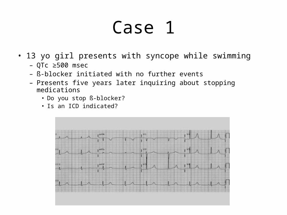
Case 1
• 13 yo girl presents with syncope while swimming– QTc ≥500 msec– ß-blocker initiated with no further events– Presents five years later inquiring about stopping medications
• Do you stop ß-blocker?• Is an ICD indicated?
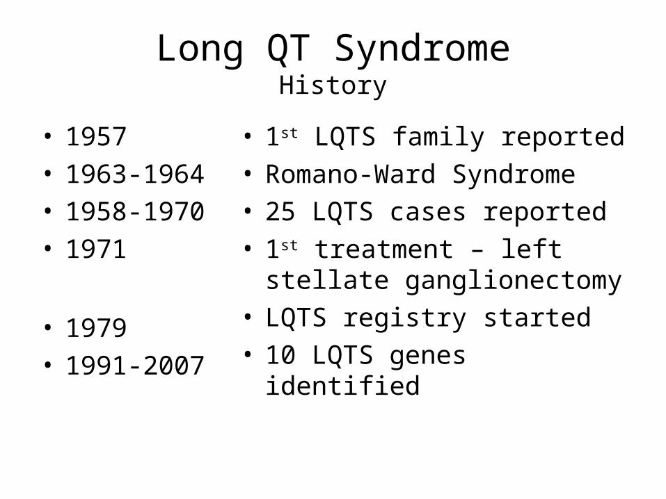
Long QT SyndromeHistory
• 1957• 1963-1964• 1958-1970• 1971
• 1979• 1991-2007
• 1st LQTS family reported• Romano-Ward Syndrome• 25 LQTS cases reported• 1st treatment – left stellate
ganglionectomy• LQTS registry started• 10 LQTS genes identified
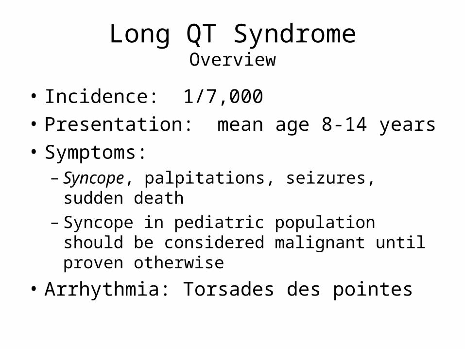
Long QT SyndromeOverview
• Incidence: 1/7,000
• Presentation: mean age 8-14 years
• Symptoms:– Syncope, palpitations, seizures, sudden death– Syncope in pediatric population should be
considered malignant until proven otherwise
• Arrhythmia: Torsades des pointes
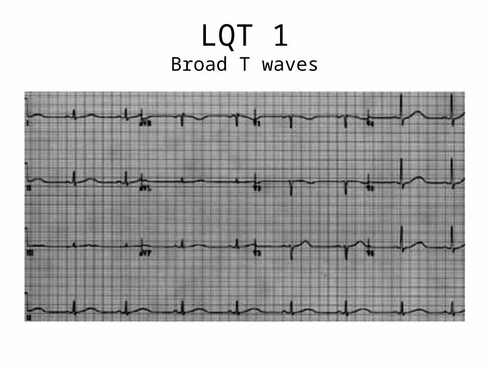
LQT 1Broad T waves
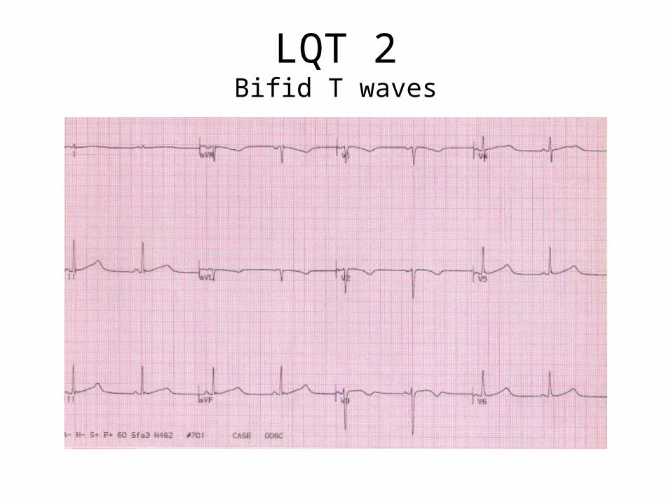
LQT 2Bifid T waves
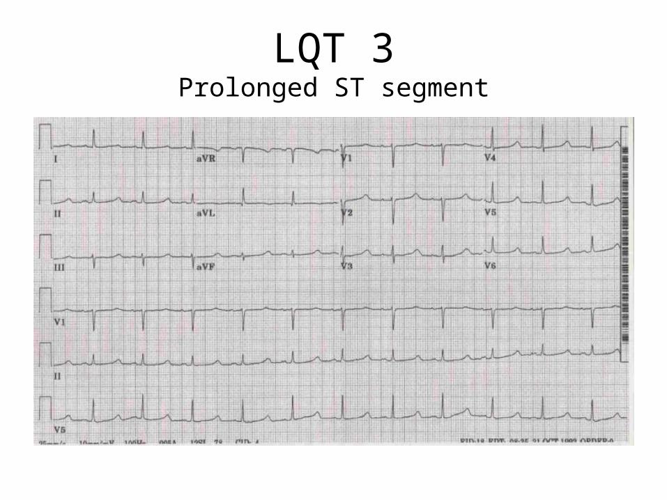
LQT 3Prolonged ST segment
Long QT SyndromeDiagnosis
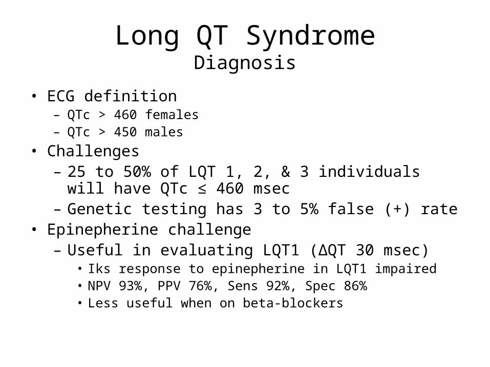
• ECG definition– QTc > 460 females– QTc > 450 males
• Challenges– 25 to 50% of LQT 1, 2, & 3 individuals will have QTc ≤
460 msec– Genetic testing has 3 to 5% false (+) rate
• Epinepherine challenge– Useful in evaluating LQT1 (∆QT 30 msec)
• Iks response to epinepherine in LQT1 impaired• NPV 93%, PPV 76%, Sens 92%, Spec 86%• Less useful when on beta-blockers

LQTSGene Specific Triggers
68
28
4
15
51
34
18
27
55
0
10
20
30
40
50
60
70
LQT1 LQT2 LQT3
ExerciseEmotionSleep
Schwartz PJ et al. Circulation 103:89-95, 2001
Lethal and Non-lethal CV Events
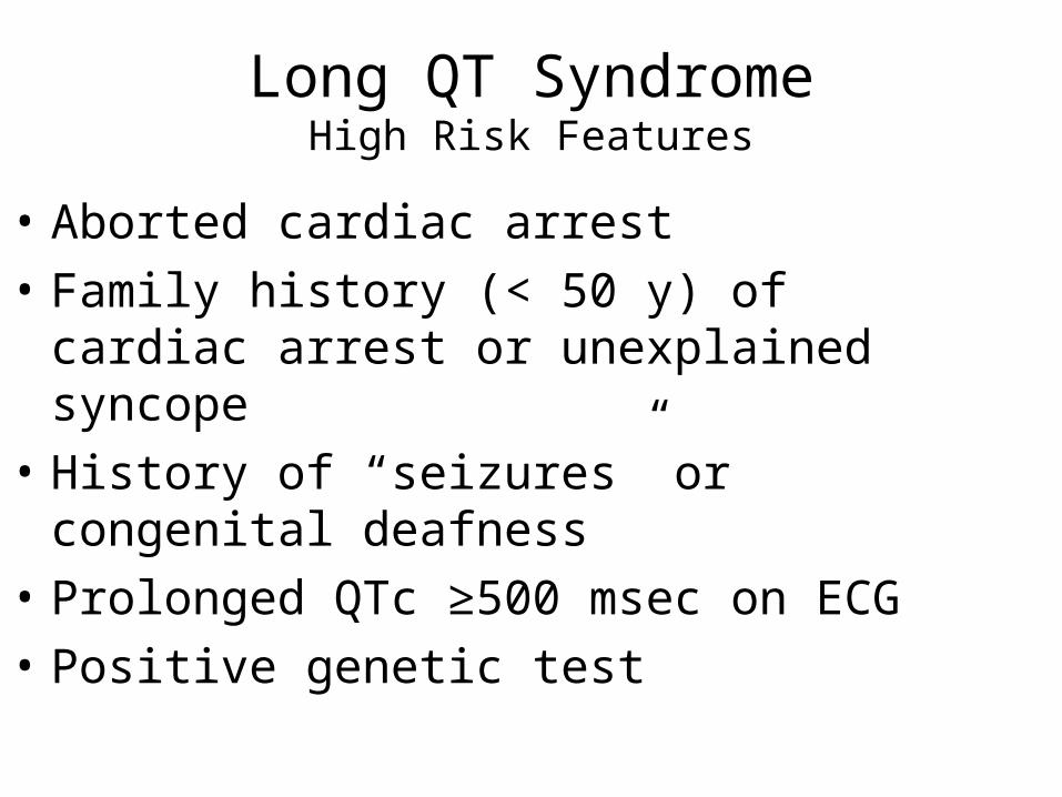
Long QT SyndromeHigh Risk Features
• Aborted cardiac arrest
• Family history (< 50 y) of cardiac arrest or unexplained syncope
• History of “seizures” or congenital deafness
• Prolonged QTc ≥500 msec on ECG
• Positive genetic test
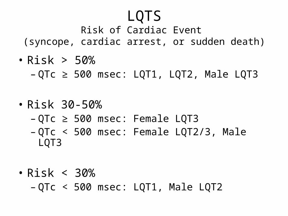
LQTSRisk of Cardiac Event
(syncope, cardiac arrest, or sudden death)
• Risk > 50%– QTc ≥ 500 msec: LQT1, LQT2, Male LQT3
• Risk 30-50%– QTc ≥ 500 msec: Female LQT3– QTc < 500 msec: Female LQT2/3, Male LQT3
• Risk < 30%– QTc < 500 msec: LQT1, Male LQT2
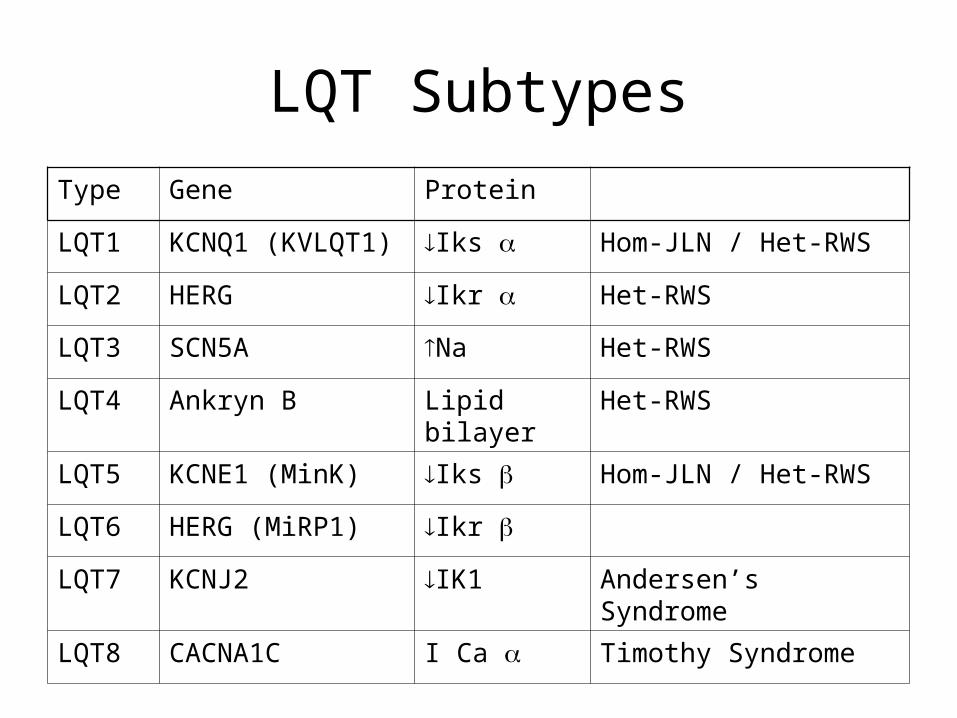
LQT Subtypes
Type Gene Protein
LQT1 KCNQ1 (KVLQT1) Iks Hom-JLN / Het-RWS
LQT2 HERG Ikr Het-RWS
LQT3 SCN5A Na Het-RWS
LQT4 Ankryn B Lipid bilayer Het-RWS
LQT5 KCNE1 (MinK) Iks Hom-JLN / Het-RWS
LQT6 HERG (MiRP1) Ikr
LQT7 KCNJ2 IK1 Andersen’s Syndrome
LQT8 CACNA1C I Ca Timothy Syndrome

LQTSManagement Options
• Lifestyle modification (IB)• Beta-blockers (IB)
– Very effective LQT1, Moderate LQT2– Minimal effect LQT3
• ICD plus BB– Cardiac arrest (IA)– Syncope / VT (IB)– Prophylactic in LQT2 or LQT3 (IIB)
• Left stellate ganglionectomy (IIB)

LQTResources
• Cardiac Arrhythmias Research & Education (CARE)– www.longqt.org
• Cardiac Arrest Survivors Network (CASN)– www.casn-network.org
• International Registry for Drug Induced Arrhythmias– www.qtdrugs.com

Case 1 Review
• 13 yo girl with syncope during swimming and QTc ≥500 msec– Asymptomatic for 5 y on BB– Swimming…suggests LQT1– High risk subgroup based LQT1 and QTc ≥500 msec– Recommendation
• Continue BB given very effective in LQT1• Consider ICD if has arrest, syncope, or VT
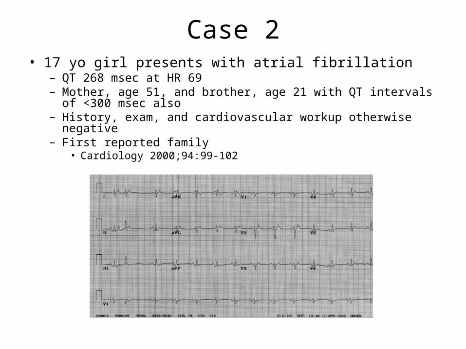
Case 2• 17 yo girl presents with atrial fibrillation
– QT 268 msec at HR 69– Mother, age 51, and brother, age 21 with QT intervals of <300
msec also– History, exam, and cardiovascular workup otherwise negative– First reported family
• Cardiology 2000;94:99-102
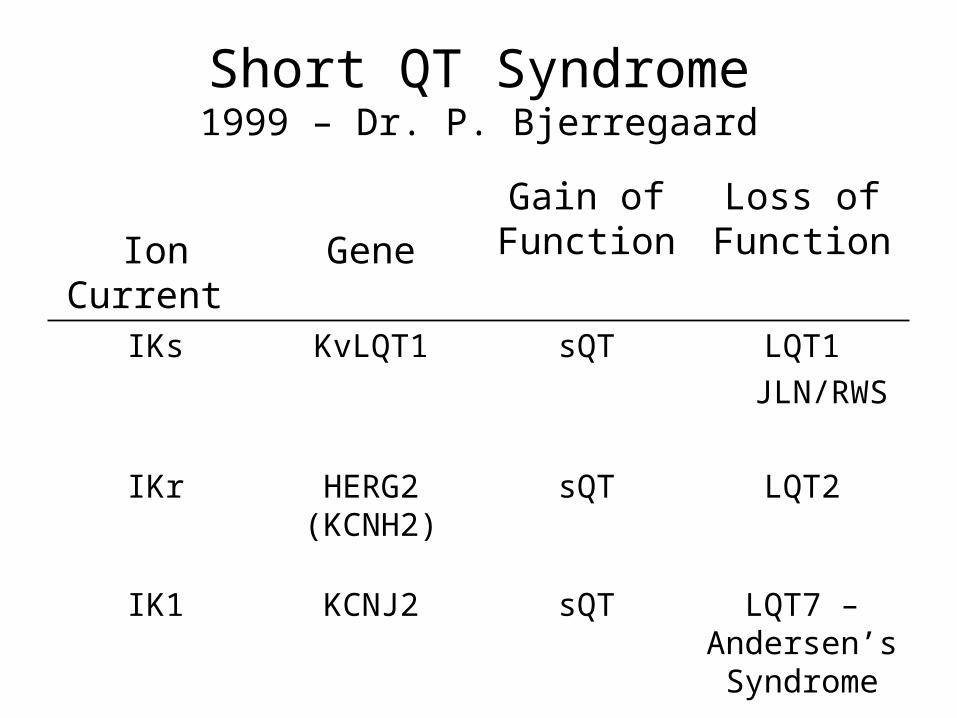
Short QT Syndrome1999 – Dr. P. Bjerregaard
Ion Current Gene
Gain of Function
Loss of Function
IKs KvLQT1 sQT LQT1
JLN/RWS
IKr HERG2 (KCNH2)
sQT LQT2
IK1 KCNJ2 sQT LQT7 – Andersen’s Syndrome
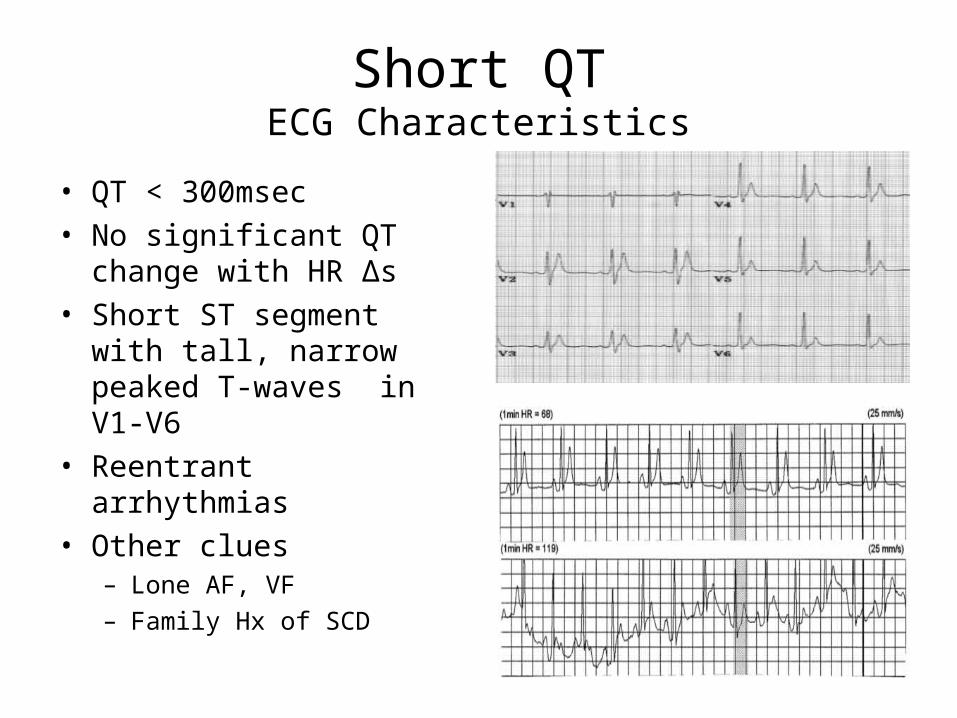
Short QTECG Characteristics
• QT < 300msec• No significant QT change
with HR ∆s• Short ST segment with
tall, narrow peaked T-waves in V1-V6
• Reentrant arrhythmias• Other clues
– Lone AF, VF– Family Hx of SCD

Short QT
• EP testing– Short atrial and ventricular refractory periods
• Management– Pharmacological (small studies)
• Only hydroquinidine effective in increasing QT• Fleicanide, sotalol, ibutilide ineffective
– ICD experience (limited)• T wave oversensing/inappropriate shocks• Device selection (St.Jude – delay/decay)

Case 2 Review
• 17 yo girl with AF and short QT. Mother and brother with short QT.– Treated with quinidine
• For atrial fibrillation suppression
• QT prolongation via K+ channel blockade
• Long-term follow-up unavailable

Case 3
• 39y man c/o cp, palpitations, and presyncope– PMH: none– SH: married, carpenter, occasional beer– FH: (-) sudden death, arrhythmias, premature CVD– Normal cardiac markers, echo

Brugada SyndromeOverview
• Identified 1992• Age spectrum - 2d to 84y• Mean age sudden death 40 ± 15y• Men > 5x risk of arrhythmic events• Prevalence
– 5/10,000 - overall – #2 cause of death SE Asian men <40y
• Dynamic but characteristic ECG changes• 1 in 5 have Na channel mutation (SCN5A)

Brugada SyndromeDefinition
• Type 1 pattern ECG in V1-V3 plus 1 of following:– Pharm conversion to Type 1 from Type 2/3 ECG
• Na channel blocker (procainamide, fleicanide, ajmaline)
– Documented VF/polymorphic VT– Family history of SCD < 45y– Inducible VT at EP study– Syncope– Nocturnal agonal respirations
• ECG pattern only = Brugada pattern ECG but not Brugada Syndrome
• Exclude other heart conditions
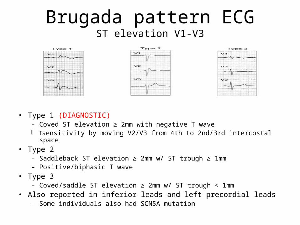
Brugada pattern ECGST elevation V1-V3
• Type 1 (DIAGNOSTIC)– Coved ST elevation ≥ 2mm with negative T wave sensitivity by moving V2/V3 from 4th to 2nd/3rd intercostal space
• Type 2– Saddleback ST elevation ≥ 2mm w/ ST trough ≥ 1mm – Positive/biphasic T wave
• Type 3– Coved/saddle ST elevation ≥ 2mm w/ ST trough < 1mm
• Also reported in inferior leads and left precordial leads– Some individuals also had SCN5A mutation

Brugada Syndrome
• Utility of EP study – Controversial– 6-9% of healthy
individuals of induced VF at EPS
– Brugada, +EPS associated w/ 8x risk
• Other conduction abnormalities– QT prolongation
(R > L precordial)• Prolonged action
potential duration in RV epicardium
– P, PR, & QRS• PR prolongation
associated with His-purkinje delay

Brugada Consensus Conference
Spontaneous Type 1 ECG
ICD (I)
Aborted SCD
ICD (I)
ExtracardiacCauseabsent
Close f/u
ExtracardiacCausepresent
SyncopeSeizure
NAR
Symptomatic
ICD (IIA)
EPS (IIA)positive
Close f/u
EPS (IIA)negative
Family Hxpositive
ICD (IIA)
EPS (IIA)positive
Close f/u
EPS (IIA)negative
Famil Hxnegative
Asymptomatic
SpontaneousType 1 ECG
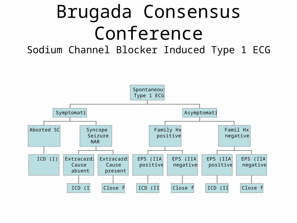
Brugada Consensus Conference
Sodium Channel Blocker Induced Type 1 ECG
ICD (I)
Aborted SCD
ICD (I)
ExtracardiacCauseabsent
Close f/u
ExtracardiacCausepresent
SyncopeSeizure
NAR
Symptomatic
ICD (IIA)
EPS (IIA)positive
Close f/u
EPS (IIA)negative
Family Hxpositive
ICD (IIA)
EPS (IIA)positive
Close f/u
EPS (IIA)negative
Famil Hxnegative
Asymptomatic
SpontaneousType 1 ECG

Case 3 Review• 39y man c/o cp, palpitations, and presyncope
– Spontaneous type 1 ECG– “Asymptomatic”– Negative family hx– EP study (IIA indication)
• Sustained VT with DES at 500 from RVA• No supraventricular arrhythmias induced• Normal AV node and His-Purkinje function
– ICD was implanted (IIA indication)• Asymptomatic without events at 32 mo f/u• ** Most events occur at night - autonomic role?
– Other tx options: ablation, quinidine

Case 4
• 16 yo girl suddenly arrests running into store• History of exertional palpitations and syncope• Successful resuscitation by bystander nurse

Catecholaminergic Polymorphic Ventricular Tachycardia (CPVT)
• Clinical Features– Direct correlation with adrenergic stimulation (physical/emotional)
• Threshold heart rate 120-130 bpm• Abnormal automaticity or triggered activity
– Bidirectional VT– Symptom onset in childhood– Genetic mutations – Ryanodine / Calsequestrin
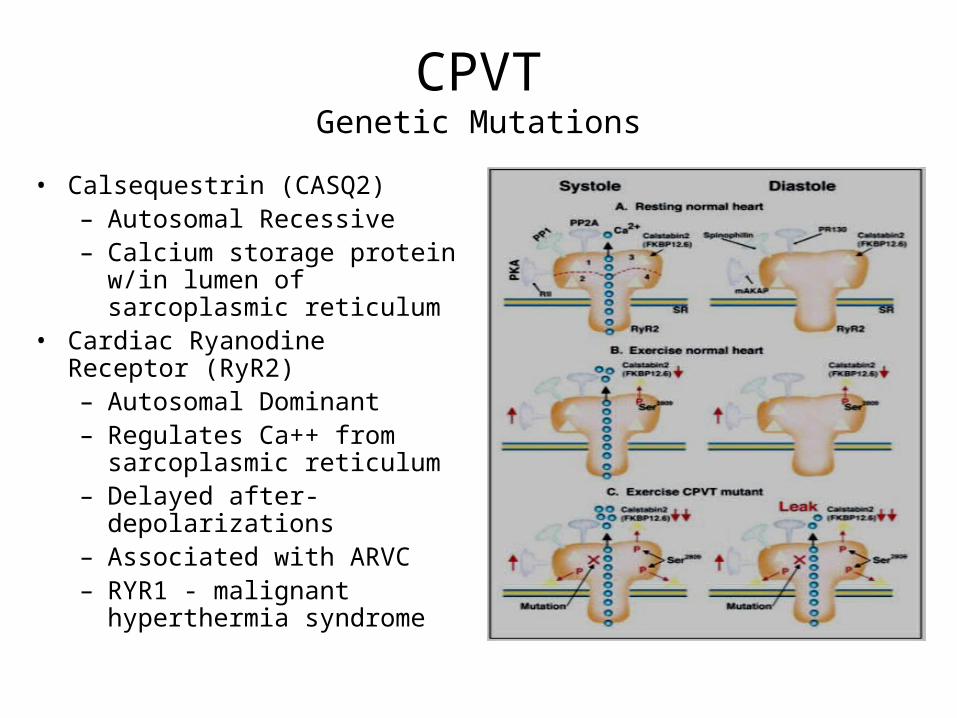
CPVTGenetic Mutations
• Calsequestrin (CASQ2)– Autosomal Recessive– Calcium storage protein w/in
lumen of sarcoplasmic reticulum
• Cardiac Ryanodine Receptor (RyR2) – Autosomal Dominant– Regulates Ca++ from
sarcoplasmic reticulum– Delayed after-depolarizations– Associated with ARVC– RYR1 - malignant
hyperthermia syndrome

CPVTManagement
• Anti-adrenergic treatment– Beta blockers are the mainstay of treatment
• ICDs– B-blockers not always effective

Case 4 Review
• 16 yo with history of palpitations and syncope who collapses in store– Arrested 3 times en route to hospital– ICD implanted and atenolol started
• 3 ICD revision procedures• 2 lead dislodgements resulting in inappropriate
ICD therapies

Case 5• 24 yo man with recurrent syncope
– Signs and symptoms• Recent decrease in exercise tolerance• Lower extremity edema• Mild elevation in liver transaminases
– Family hx + for sudden death – paternal uncle– Telemetry strip below
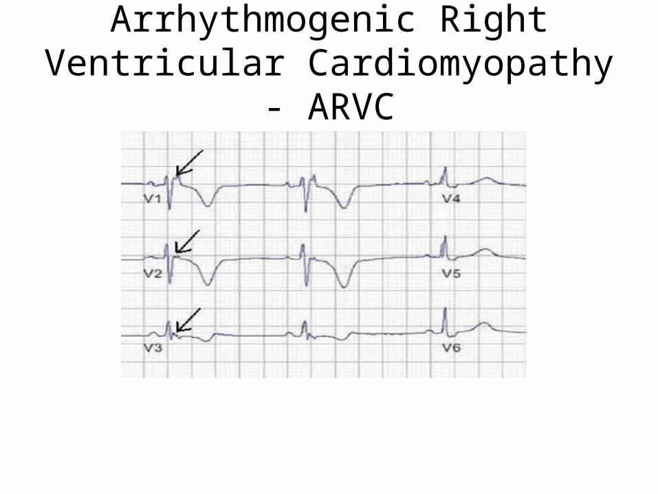
Arrhythmogenic Right Ventricular Cardiomyopathy - ARVC

ARVCIndik JH et al. Indian Pacing Electrophys J. 2003:3:148
• Top picture:– Fibro-fatty replacement of
the myocardium – Thin and enlarged RV wall.
• Bottom picture:
– Trichrome stain– Areas of mature fibrosis (F)
and adipose tissue (A) within the epicardial (Epi) and mid-myocardial zones
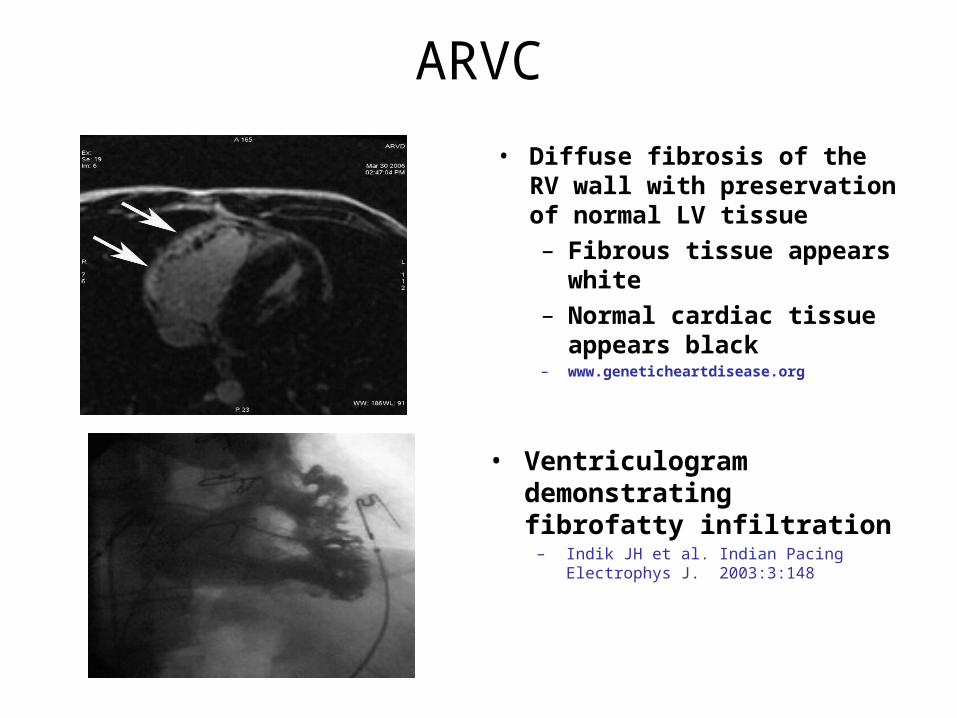
ARVC
• Diffuse fibrosis of the RV wall with preservation of normal LV tissue – Fibrous tissue appears
white – Normal cardiac tissue
appears black– www.geneticheartdisease.org
• Ventriculogram demonstrating fibrofatty infiltration
– Indik JH et al. Indian Pacing Electrophys J. 2003:3:148
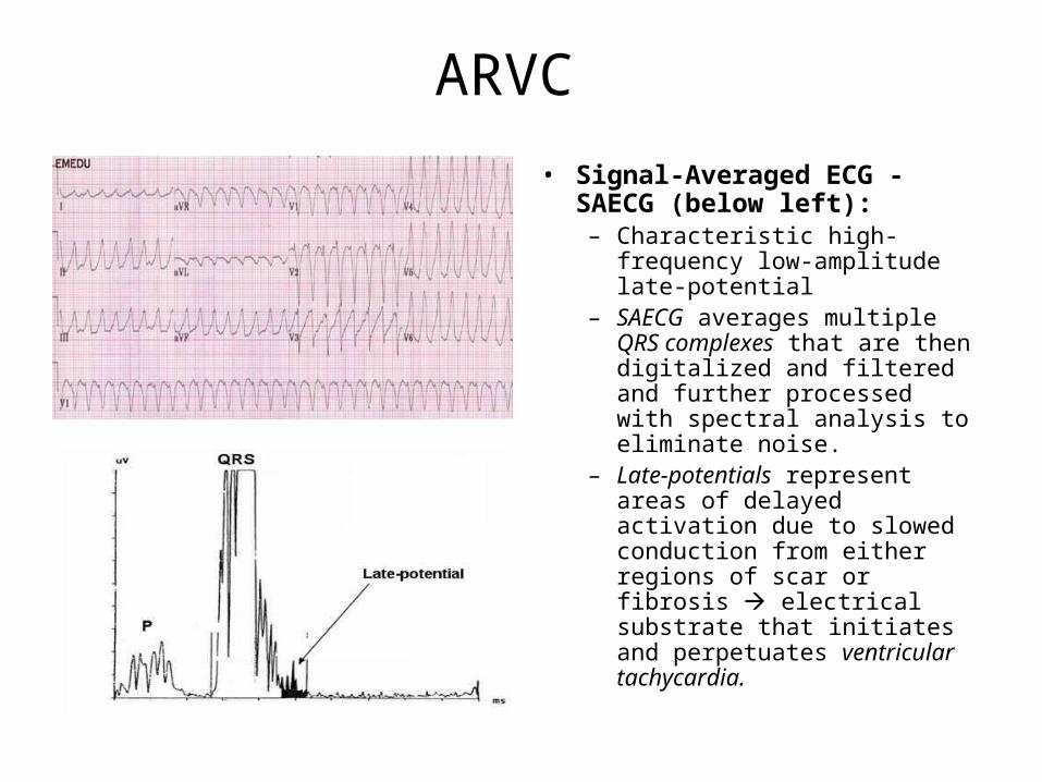
ARVC
• Signal-Averaged ECG -SAECG (below left):– Characteristic high-frequency
low-amplitude late-potential– SAECG averages multiple
QRS complexes that are then digitalized and filtered and further processed with spectral analysis to eliminate noise.
– Late-potentials represent areas of delayed activation due to slowed conduction from either regions of scar or fibrosis electrical substrate that initiates and perpetuates ventricular tachycardia.

ARVC High Risk Features
• Younger patients • Recurrent syncope • History of cardiac arrest or sustained VT • Clinical signs of RV failure or LV involvement• Patients with or having a family member with the
high risk ARVD gene (ARVD2) • Increase in QRS dispersion ≥ 40 msec
– QRS dispersion = max measured QRS minus min measured QRS
• Naxos disease
Case 5 Review
• Diagnosis– Rhythm strip and ECG notable for epsilon waves and
T wave inversion in right precordial leads
• Risk– High risk features present – young age, recurrent
syncope, signs of RV failure, family history of sudden cardiac arrest
• Management– ICD implantation

• Idiopathic Ventricular Fibrillation– Sodium channel mutation
• Short-coupled Torsades des Pointes– Normal QT interval with coupling interval of 1st ectopic beat < 300
msec– Prognosis poor with unproven tx (BB or CCB); ablation?
• Lev-Lenegre Syndrome– Progressive cardiac conduction defect associated with
bradyarrhythmias although tachyarrhythmias may also occur– Sodium channel defect

Idiopathic Ventricular Fibrillation

Lev-Lenegre Syndrome
• Progressive Cardiac Conduction Defect– Acquired complete heart block– Idiopathic fibrosis and calcification of cardiac conduction system
• Very rare• Sodium channel mutations (subtype-SCN 5A)• Often result in bradyarrhythmias although
tachyarrhythmias may also result
• Lev M. Anatomic basis for atrioventricular block. Am J Med 1964;37:742-8. • Lenegre J. Etiology and pathology of bilateral bundle branch block in relation to complete heart block. Prog
Cardiovasc Dis 1964;6:409-444

Hypertrophic Cardiomyopathy
• #1 cause of SCA in athletes – > 1/3 of deaths– Often associated with physical activity– 60% high school age– >90% males
• Genetic disorder left ventricular hypertrophy
• First symptom often sudden death

HCM - ECG
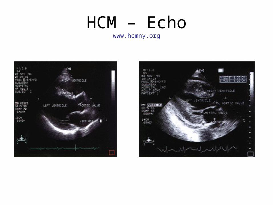
HCM – Echowww.hcmny.org
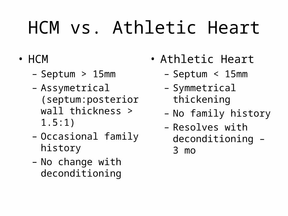
HCM vs. Athletic Heart
• HCM– Septum > 15mm– Assymetrical
(septum:posterior wall thickness > 1.5:1)
– Occasional family history
– No change with deconditioning
• Athletic Heart– Septum < 15mm– Symmetrical
thickening– No family history– Resolves with
deconditioning – 3 mo

Thank You
Questions?