Embed Size (px)
Citation preview
lable at ScienceDirect
Theriogenology 159 (2021) 176e183
Contents lists avai
Theriogenology
journal homepage: www.theriojournal .com
Normal and abnormal response to sperm deposition in female dogs: Areview and new hypotheses for endometritis
G.C.W. England a, *, T. Rijsselaere b, A. Campbell c, R. Moxon a, c, S.L. Freeman a
a School of Veterinary Medicine and Science, University of Nottingham, Sutton Bonington, Leicestershire, UKb Faculty of Veterinary Medicine, Ghent University, Merelbeke, Belgiumc Guide Dogs, National Breeding Centre, Bishops Tachbrook, Warwickshire, UK
a r t i c l e i n f o
Article history:Received 7 October 2020Accepted 7 October 2020Available online 24 October 2020
Keywords:DogSpermTransportReservoirEndometritis
Part of the Proceedings of 19th International Congr(other articles appeared in Theriogenology, volume 1* Corresponding author.
E-mail address: [email protected] (G
https://doi.org/10.1016/j.theriogenology.2020.10.0130093-691X/© 2020 The Authors. Published by Elsevier
a b s t r a c t
In mammalian species there are significant physiological responses of the female reproductive tract tothe deposition of sperm. These are particularly notable in species where sperm are deposited directlyinto the uterus, and function both to facilitate sperm transport to the sperm reservoir, and to eliminateintroduced contaminants.
In the bitch, sperm are deposited into the vagina and are rapidly transported through the open cervix.Sperm are then distributed around the uterus by uterine contractions such that transportation to the tipof the uterine horns occurs within 1 min of the start of mating.
The main sperm reservoir appears to be the distal part of the utero-tubal junction which forms a pre-uterine tube reservoir. Sperm remain attached here by their heads to uterine epithelium and remainviable. In non-capacitating conditions sperm slowly detach from this site and this seems important toreplenish the uterine tube reservoir, where sperm may re-attach to the epithelium. Post-ovulatory sig-nals trigger capacitation changes and subsequent hyperactivated motility that is associated withdetachment of sperm from both reservoirs; thus facilitating fertilization.
After mating, a physiological post-mating uterine inflammatory response occurs, evidenced by aninflux of polymorphonuclear neutrophils, increased uterine contractions, an increased uterine arteryblood flow and a decrease of the resistance index indicating a short-duration vasodilation.
Disturbance of this tightly regulated system has the potential to impact fertility by a failure of elim-ination of the introduced contaminants (such that a clinically-significant post-breeding endometritisensues) but also by impairing sperm transport.© 2020 The Authors. Published by Elsevier Inc. This is an open access article under the CC BY-NC-ND
license (http://creativecommons.org/licenses/by-nc-nd/4.0/).
1. Introduction
Inherent female reproductive tract factors are thought to beresponsible for the ‘passive transport’ of sperm [1,2]. Species dif-ferences occur during the journey that sperm take followingdeposition in the female tract [3]. Vaginal deposition is oftenassociated with contractions of the vaginawhich quickly propel theejaculate to the cervix. In species, such as humans, where there isan initial harmful low vaginal pH and a significant cervical mucusbarrier, the ejaculate coagulates close to the cervix and then liq-uefies later after vaginal pH has increased (and is no longer hostile),thus allowing sperm to swim actively and penetrate the cervical
ess on Animal Reproduction50, 1 July 2020).
.C.W. England).
Inc. This is an open access article u
mucus. In several other species, such as the horse and pig, thecervix does not form a barrier to sperm transport as the ejaculate isdeposited into the uterus [1,2,4].
Uterine contractions are involved in passive transport such thatsperm are found within the uterine tube shortly after mating [5,6].Uterine contractions may be influenced by a release of oxytocin atcoitus [7e10] or the presence of prostaglandin or oestrogen withinthe ejaculate [11,12]; especially in species where there is uterinedeposition of sperm. Additional physiological factors, such aspresence of sperm in the uterus, can also contribute to the increasein uterine contractions noted at mating [13,14].
The rapid transport of sperm to the uterine tube, appearsimportant because this site is often considered to form the func-tional sperm reservoir. Sperm that do not reach the reservoir areeliminated from the female tract (see later). Once within the uter-ine tube, sperm are rapidly distributed by active peristalticcontraction of the uterine tube itself [15]. This distribution allows
nder the CC BY-NC-ND license (http://creativecommons.org/licenses/by-nc-nd/4.0/).
G.C.W. England, T. Rijsselaere, A. Campbell et al. Theriogenology 159 (2021) 176e183
sperm to contact regions of the uterine tube epithelium in order toestablish the uterine tube sperm reservoir, which is maintained byan intimate interaction of sperm heads with the epithelium. It hasbeen known for some time that this transient epithelial contact isimportant for maintaining sperm viability [16] and fertilizingability [17]. Whilst ‘attached’ by their heads, sperm maintain slowflagellar activity, however following an appropriate capacitationsignal, sperm membrane destabilisation processes commence,fertilizing ability is achieved, and a proportion of the sperm becomehyper-activated [17]. This change in motility may assist the spermin ‘detaching’ from the epithelium, enabling the location of oocytes,following which binding to the zona pellucida, the acrosome re-action and oocyte penetration occur [16].
In many species there is a transient physiological uterine in-flammatory response which occurs after sperm enter the uterus[18,19]. This is most prominent in those species in which sperm aredeposited directly into the uterus and is characterised by increaseduterine contractions, luminal influx of polymorphonuclear leuco-cytes (PMNs), increased uterine artery blood flow and uterinevasodilation [20]. The response is thought to remove excess anddead sperm, as well as bacterial, cellular and fluid contaminants.Importantly, mitigation against the effects of this inflammatoryresponse is required to ensure that it does not negatively impactupon sperm and thus fertility. In horses, for example, seminalplasma from the stallion reduces sperm binding to PMNs and im-proves fertility [21].
Disturbance of the female response to the deposition of spermmay have an impact upon fertility. This may occur because (1)sperm transport is impeded, for example, purported abnormalcervical mucus quality preventing sperm entering the uterinelumen and leading to a failure of fertilization, or (2) becauseelimination of contaminants is not fully effective, leading to thedevelopment of a post-breeding clinical endometritis.
In veterinary clinical practice perhaps the most common ab-normality is noted where ejaculation occurs directly into theuterus, for example in the mare, sow and camel. In these species,significant numbers of sperm, cellular material and bacteriadirectly enter the uterus when compared with species where thereis a functional cervical barrier (such as primates and ruminants).Whilst uterine deposition of sperm results in rapid transportationto the utero-tubal junction, failure of elimination of the introducedcellular contaminants and, in particular, any micro-organisms mayresult in a clinical endometritis [22]. This differs from the previ-ously described ‘physiological’ endometritis described above.Persistence of such a post-breeding (also called mating-induced)clinical endometritis [23] may create a hostile uterine environ-ment that prevents survival of the developing embryos when they,at a later time, enter the uterus.
It is not surprising that the species in which uterine depositionof semen occurs are those in which post-breeding endometritis is acommon cause of infertility. It has been suspected for some timethat in the female dog there is vaginal deposition of semen butrapid transport through an open cervix [24], and knowledge ofsperm transport has improved over recent years. Alongside thisinformation is a growing body of evidence which suggests thatperturbation of the normal physiological response of the bitch mayoccur (particularly in bitches with endometrial hyperplasia) whichmay impact the elimination of uterine contaminants and alsopotentially be detrimental to sperm transport, both of which wouldresult in impaired fertility.
The aim of this review is to describe normal sperm transport inthe female dog, to discuss the consequences when this is disturbed,and to propose new hypotheses for the development ofendometritis.
177
1.1. Normal response to sperm deposition in the female dog
The female dog has an interesting biology compared with manyother species. Unlike many mammals, dog ova are released atovulation as immature primary oocytes [25]. Between one to threedays after ovulation they have reached metaphase of the secondmeiotic division (when the first polar body is extruded), and theyare considered fertilizable from this point onwards. Interestingly,oocytes may remain fertilizable for up to five or more days afterovulation [26]. The time over which mating may result in fertil-ization is further increased as sperm can survive for up to 11 dayswithin the female tract [27], such that mating many days beforeovulation may result in a pregnancy. The phenomena of prolongedsurvival of sperm within the female tract clearly points to thepresence of a sperm reservoir.
1.2. Transport of sperm to the sperm reservoir
The ejaculate of the dog has three fractions of which the first andthird originate from the prostate [28], and the second fraction is theepididymal sperm-rich portion. The sperm-rich fraction of theejaculate is deposited into the cranial vagina and is flushed throughthe cervix by the large volume of the prostatic fluid during theprotracted copulatory tie. Interestingly, an early study showed thatwhen only a small volume of radiographic contrast medium wasintroduced into the cranial vagina this was rapidly transported intothe uterine lumen without the requirement for flushing withprostatic fluid [29], perhaps indicating that the flushing action ofthe prostatic fluid is not essential. However, vaginal contractionsare present during normal coitus, and can be stimulated in theestrous bitch by digital palpation/dilation of the caudal vagina.These contractions might function similar to those noted in otherspecies, which force the ejaculate to, or through, the cervix. Con-tractions of the abdominal musculature occur in some bitches andmay also contribute to passive sperm transport [24], although thiscontention is not proven.
It is clear that the bitch cervix opens approximately four daysbefore ovulation allowing the passage of fluid, largely unob-structed, into the uterus [30]. The cervix closes approximately fivedays after ovulation once progesterone concentrations haveincreased [30e32]. Overall, during the fertilization period it wouldappear that the cervix of the bitch does not form a barrier to thepassage of sperm. Indeed, Evans [24] found sperm at the fistulatedtips of the uterine horns within 1 min of mating. That author alsoobserved that during mating active abdominal contractions resul-ted in semen being forced in small jets up to 25 cm from thefistulae.
Basal uterine contractions have been identified in the bitch [33],and we were able (with some difficulty) to identify uterine con-tractions using M-Mode ultrasound [4]. Two specific types of con-tractions were evident; (1) spontaneous basal contractions whichincreased in frequency from approximately five days before untilfive days after ovulation, and (2) specific mating-induced contrac-tions which had the greatest frequency from the day of ovulationuntil four days later. The variation in contraction frequencyappeared to have some relationship with plasma hormone con-centration; most noticeably an inhibitory effect of progesteronethat coincided with the end of the fertile period [4] (Fig. 1). Theincreased number of contractions caused by mating could not bemimicked by teasing with a male, nor by tactile stimulation of thevagina or cervix during vaginal or trans-cervical insemination,however distension of the vestibule and manual stimulation of thedorsal wall of the vagina produced contractions that were similar tothose noted during mating [34]. It is likely that these contractionsare responsible for the rapid distribution of sperm throughout the
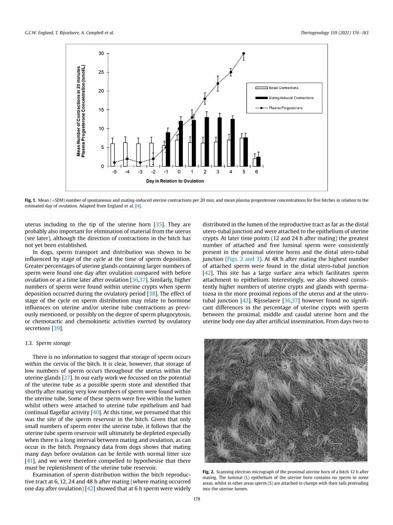
Fig. 1. Mean (þSEM) number of spontaneous and mating-induced uterine contractions per 20 min, and mean plasma progesterone concentrations for five bitches in relation to theestimated day of ovulation. Adapted from England et al. [4].
Fig. 2. Scanning electron micrograph of the proximal uterine horn of a bitch 12 h aftermating. The luminal (L) epithelium of the uterine horn contains no sperm in someareas, whilst in other areas sperm (S) are attached in clumps with their tails protrudinginto the uterine lumen.
G.C.W. England, T. Rijsselaere, A. Campbell et al. Theriogenology 159 (2021) 176e183
uterus including to the tip of the uterine horn [35]. They areprobably also important for elimination of material from the uterus(see later), although the direction of contractions in the bitch hasnot yet been established.
In dogs, sperm transport and distribution was shown to beinfluenced by stage of the cycle at the time of sperm deposition.Greater percentages of uterine glands containing larger numbers ofsperm were found one day after ovulation compared with beforeovulation or at a time later after ovulation [36,37]. Similarly, highernumbers of sperm were found within uterine crypts when spermdeposition occurred during the ovulatory period [38]. The effect ofstage of the cycle on sperm distribution may relate to hormoneinfluences on uterine and/or uterine tube contractions as previ-ously mentioned, or possibly on the degree of sperm phagocytosis,or chemotactic and chemokinetic activities exerted by ovulatorysecretions [39].
1.3. Sperm storage
There is no information to suggest that storage of sperm occurswithin the cervix of the bitch. It is clear, however, that storage oflow numbers of sperm occurs throughout the uterus within theuterine glands [27]. In our early work we focussed on the potentialof the uterine tube as a possible sperm store and identified thatshortly after mating very low numbers of spermwere found withinthe uterine tube. Some of these sperm were free within the lumenwhilst others were attached to uterine tube epithelium and hadcontinual flagellar activity [40]. At this time, we presumed that thiswas the site of the sperm reservoir in the bitch. Given that onlysmall numbers of sperm enter the uterine tube, it follows that theuterine tube sperm reservoir will ultimately be depleted especiallywhen there is a long interval between mating and ovulation, as canoccur in the bitch. Pregnancy data from dogs shows that matingmany days before ovulation can be fertile with normal litter size[41], and we were therefore compelled to hypothesise that theremust be replenishment of the uterine tube reservoir.
Examination of sperm distribution within the bitch reproduc-tive tract at 6, 12, 24 and 48 h after mating (where mating occurredone day after ovulation) [42] showed that at 6 h spermwere widely
178
distributed in the lumen of the reproductive tract as far as the distalutero-tubal junction andwere attached to the epithelium of uterinecrypts. At later time points (12 and 24 h after mating) the greatestnumber of attached and free luminal sperm were consistentlypresent in the proximal uterine horns and the distal utero-tubaljunction (Figs. 2 and 3). At 48 h after mating the highest numberof attached sperm were found in the distal utero-tubal junction[42]. This site has a large surface area which facilitates spermattachment to epithelium. Interestingly, we also showed consis-tently higher numbers of uterine crypts and glands with sperma-tozoa in the more proximal regions of the uterus and at the utero-tubal junction [42]. Rijsselaere [36,37] however found no signifi-cant differences in the percentage of uterine crypts with spermbetween the proximal, middle and caudal uterine horn and theuterine body one day after artificial insemination. From days two to
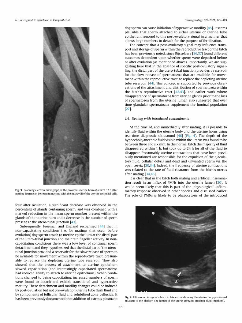
Fig. 3. Scanning electron micrograph of the proximal uterine horn of a bitch 12 h aftermating. Sperm can be seen interacting with the microvilli of the uterine epithelial cells.
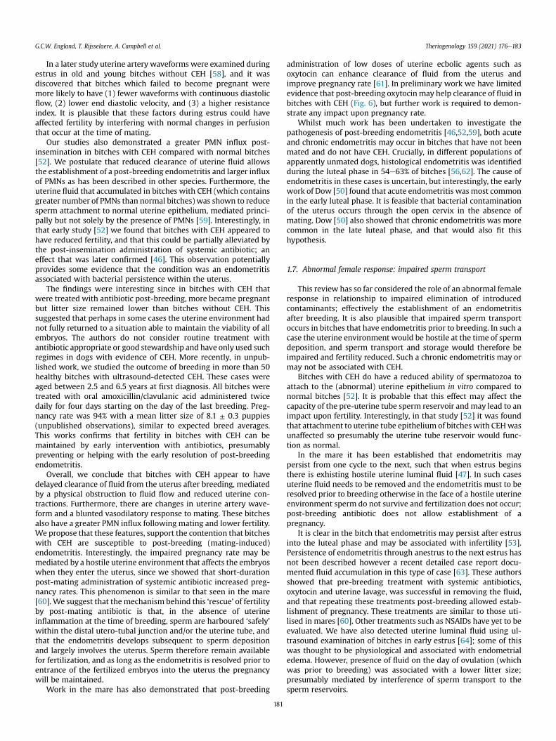
Fig. 4. Ultrasound image of a bitch in late estrus showing the uterine body positionedadjacent to the bladder. The lumen of the uterus contains anechoic fluid (markers).
G.C.W. England, T. Rijsselaere, A. Campbell et al. Theriogenology 159 (2021) 176e183
four after ovulation, a significant decrease was observed in thepercentage of glands containing sperm, and was combined with amarked reduction in the mean sperm number present within theglands of the uterine horn and a decrease in the number of spermpresent at the utero-tubal junction [43].
Subsequently, Freeman and England recognised [44] that innon-capacitating conditions (i.e. for matings that occur beforeovulation) dog sperm attach to uterine epithelium at the distal partof the utero-tubal junction and maintain flagellar activity. In non-capacitating conditions there was a low level of continual spermdetachment and they hypothesized that the distal part of the utero-tubal junction provided a reservoir for the slow release of sperm tobe available for movement within the reproductive tract, presum-ably to replace the depleting uterine tube reservoir. They alsoshowed that the process of attachment to uterine epitheliumslowed capacitation (and interestingly capacitated spermatozoahad reduced ability to attach to uterine epithelium). When condi-tions changed to being capacitating, increased numbers of spermwere found to detach and exhibit transitional and hyperactivemotility. These detachment and motility changes could be inducedby post-ovulation but not pre-ovulation uterine tube flush fluid andby components of follicular fluid and solubilised zona pellucida. Ithas been previously documented that addition of estrous plasma to
179
dog sperm can cause initiation of hyperactivemotility [45]. It seemsplausible that sperm attached to either uterine or uterine tubeepithelium respond to this post-ovulatory signal in a manner thatallows large numbers to detach for the purpose of fertilization.
The concept that a post-ovulatory signal may influence trans-port and storage of spermwithin the reproductive tract of the bitchhas been previously noted, since Rijsselaere [36,37] found differentoutcomes dependent upon whether sperm were deposited beforeor after ovulation (as mentioned above). Importantly, we are sug-gesting here that in the absence of specific post-ovulatory signal-ling, the distal part of the utero-tubal junction provides a reservoirfor the slow release of spermatozoa that are available for move-ment within the reproductive tract, to replace the depleting uterinetube reservoir [44]. This concept is supported by previous obser-vations of the attachment and distribution of spermatozoa withinthe bitch’s reproductive tract [42,43], and earlier work wheredisappearance of spermatozoa from uterine glands prior to the lossof spermatozoa from the uterine lumen also suggested that overtime glandular spermatozoa supplement the luminal population[27].
1.4. Dealing with introduced contaminants
At the time of, and immediately after mating, it is possible toidentify fluid within the uterine body and the uterine horns usingreal-time diagnostic ultrasound [46] (Fig. 4). The depth of thehypoechoic/anechoic fluid visible within the uterus was found to bebetween three and six mm. In the normal bitch themajority of fluiddisappeared within 1 h, but took up to 24 h for all of the fluid todisappear. Presumably uterine contractions that have been previ-ously mentioned are responsible for the expulsion of the ejacula-tory fluid, cellular debris and dead and unwanted sperm via theopen cervix [20,34]. Indeed, the frequency of uterine contractionswas related to the rate of fluid clearance from the bitch’s uterusafter mating [34,46].
It is clear that in the bitch both mating and artificial insemina-tion result in an influx of PMNs into the uterine lumen [20]. Itwould seem likely that this is part of the ‘physiological’ inflam-matory response observed in other species and discussed earlier.The role of PMNs is likely to be phagocytosis of the introduced
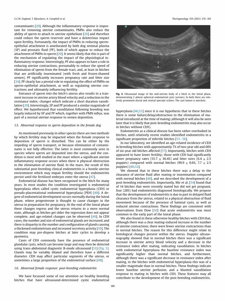
Fig. 5. Ultrasound image of the mid-uterine body of a bitch in the luteal phase,demonstrating 2 almost spherical endometrial cysts (arrows). In both there are rela-tively prominent dorsal and ventral specular echoes. The cyst lumen is anechoic.
G.C.W. England, T. Rijsselaere, A. Campbell et al. Theriogenology 159 (2021) 176e183
contaminants [20]. Although the inflammatory response is impor-tant for removing uterine contaminants, PMNs also reduce theability of sperm to attach to uterine epithelium [20] and thereforecould reduce the sperm reservoir and have a deleterious impactupon fertility. Fortunately, the impact of PMNs in reducing sperm-epithelial attachment is ameliorated by both dog seminal plasma(SP) and prostatic fluid (PF), both of which appear to reduce theattachment of PMNs to sperm [20]. It seems likely that this is part ofthe mechanism of regulating the impact of the physiological in-flammatory response. Interestingly, PF also appears to have a role inreducing uterine contractions, presumably to reduce the speed ofelimination of sperm from the female tract, and, at least in bitchesthat are artificially inseminated (with fresh and frozen-thawedsemen), PF significantly increases pregnancy rate and litter size[34]. PF clearly has a pivotal role in regulating the effect of PMNs onsperm-epithelial attachment, as well as regulating uterine con-tractions and ultimately influencing fertility.
Entrance of sperm into the bitch’s uterus also results in a tran-sient increase in uterine artery blood velocity and a reduction in theresistance index; changes which indicate a short duration vasodi-lation [20]. Interestingly, SP and PF produced a similar magnitude ofeffect. We hypothesized that vasodilation following breeding waslargely induced by SP and PF which, together with PMN influx, waspart of a normal uterine response to semen deposition.
1.5. Abnormal response to sperm deposition in the female dog
Asmentioned previously in other species there are twomethodsby which fertility may be impacted when the female response todeposition of sperm is disturbed. This can be either by theimpeding of sperm transport, or because elimination of contami-nants is not fully effective. The latter is most commonly seen inspecies where sperm are deposited into the uterus, and the con-dition is most well studied in the mare where a significant uterineinflammatory response occurs when there is physical obstructionto the elimination of uterine fluid. In the mare, the result of thissubstantial post-breeding clinical endometritis is a hostile uterineenvironment which may impair fertility should the endometritispersist until the fertilized embryos enter the uterus [47].
Endometrial disease has been recognised in the bitch for manyyears. In most studies the condition investigated is endometrialhyperplasia often called cystic endometrial hyperplasia (CEH) orpseudo-placentational endometrial hyperplasia (PEH) [48]. A de-gree of endometrial development occurs normally during the lutealphase, where progesterone is thought to cause changes in theuterus in preparation for pregnancy. At the end of the luteal phasethese changes regress and the uterus returns to a more normalstate, although as bitches get older the regression does not appearcomplete, and age-related changes can be observed [49]. In CEHcases, the number and size of endometrial glands are increased, andthere is disparity in the number and configuration of glands causinga thickened endometrium and increased secretory activity [50]. Thecondition may pre-dispose bitches at later cycles to develop apyometra.
Cases of CEH commonly have the presence of endometrialglandular cysts, which can become large and may then be detectedusing trans-abdominal diagnostic B-mode ultrasound [51] (Fig. 5).Glands may be variably dilated and therefore have a differentdiameter. CEH may affect particular segments of the uterus, orsometimes a large proportion of the endometrial surface [48].
1.6. Abnormal female response: post-breeding endometritis
We have focussed some of our attention on healthy breedingbitches that have ultrasound-determined cystic endometrial
180
hyperplasia [46,52] since it is our hypothesis that in these bitchesthere is some failure/delay/obstruction to the elimination of ma-terial introduced at the time of mating (although it will also be seenlater that it is likely that post-breeding endometritis may also occurin bitches without CEH).
Endometritis as a clinical disease has been rather overlooked inbitches, until relatively recent studies identified endometritis in asignificant proportion of infertile bitches [53e56].
In our laboratory, we identified an age-related incidence of CEHin breeding bitches with approximately 7% of two-year old and 60%of six-year old bitches affected [57]. Importantly, bitches with CEHappeared to have lower fertility; those with CEH had significantlylower pregnancy rates (55.7 ± 36.4%) and litter sizes (6.4 ± 2.5puppies) compared with normal bitches (90.9 ± 0.6%, 7.7 ± 2.5puppies) [46,52].
We showed that in these bitches there was a delay in theclearance of uterine fluid after mating or insemination comparedwith normal bitches [46], and we described the development of apost-breeding endometritis. Importantly, Mir et al. [54] found thatof 14 bitches that were recently mated but did not get pregnant,four (28%) had endometritis diagnosed histologically. We proposethat the development of endometritis is due in part to delayed fluidclearance from the uterus, related to a physical obstruction of fluidmovement because of the presence of luminal cysts, as well asreduced uterine contractions. These findings are consistent withobservations from Dow [50] that acute endometritis was mostcommon in the early part of the luteal phase.
We also found in these otherwise healthy bitches with CEH that,although there was a clear mating-induced increase in the numberof uterine contractions, there were fewer uterine contractions thanin normal bitches. The reason for this difference might relate tohistological changes present within the uterus. Doppler ultraso-nography showed that in normal bitches there was a significantincrease in uterine artery blood velocity and a decrease in theresistance index after mating, indicating vasodilation. In bitcheswith endometrial hyperplasia the baseline resistance index wassignificantly higher than normal bitches, and furthermore,although there was a significant decrease in resistance index aftermating, in the bitches with endometrial hyperplasia this was of asmaller magnitude than in normal bitches. These findings indicatelower baseline uterine perfusion, and a blunted vasodilationresponse to mating in bitches with CEH. These features may allcontribute to the development of the post-breeding endometritis.
G.C.W. England, T. Rijsselaere, A. Campbell et al. Theriogenology 159 (2021) 176e183
In a later study uterine artery waveforms were examined duringestrus in old and young bitches without CEH [58], and it wasdiscovered that bitches which failed to become pregnant weremore likely to have (1) fewer waveforms with continuous diastolicflow, (2) lower end diastolic velocity, and (3) a higher resistanceindex. It is plausible that these factors during estrus could haveaffected fertility by interfering with normal changes in perfusionthat occur at the time of mating.
Our studies also demonstrated a greater PMN influx post-insemination in bitches with CEH compared with normal bitches[52]. We postulate that reduced clearance of uterine fluid allowsthe establishment of a post-breeding endometritis and larger influxof PMNs as has been described in other species. Furthermore, theuterine fluid that accumulated in bitches with CEH (which containsgreater number of PMNs than normal bitches) was shown to reducesperm attachment to normal uterine epithelium, mediated princi-pally but not solely by the presence of PMNs [59]. Interestingly, inthat early study [52] we found that bitches with CEH appeared tohave reduced fertility, and that this could be partially alleviated bythe post-insemination administration of systemic antibiotic; aneffect that was later confirmed [46]. This observation potentiallyprovides some evidence that the condition was an endometritisassociated with bacterial persistence within the uterus.
The findings were interesting since in bitches with CEH thatwere treated with antibiotic post-breeding, more became pregnantbut litter size remained lower than bitches without CEH. Thissuggested that perhaps in some cases the uterine environment hadnot fully returned to a situation able to maintain the viability of allembryos. The authors do not consider routine treatment withantibiotic appropriate or good stewardship and have only used suchregimes in dogs with evidence of CEH. More recently, in unpub-lished work, we studied the outcome of breeding in more than 50healthy bitches with ultrasound-detected CEH. These cases wereaged between 2.5 and 6.5 years at first diagnosis. All bitches weretreated with oral amoxicillin/clavulanic acid administered twicedaily for four days starting on the day of the last breeding. Preg-nancy rate was 94% with a mean litter size of 8.1 ± 0.3 puppies(unpublished observations), similar to expected breed averages.This works confirms that fertility in bitches with CEH can bemaintained by early intervention with antibiotics, presumablypreventing or helping with the early resolution of post-breedingendometritis.
Overall, we conclude that bitches with CEH appear to havedelayed clearance of fluid from the uterus after breeding, mediatedby a physical obstruction to fluid flow and reduced uterine con-tractions. Furthermore, there are changes in uterine artery wave-form and a blunted vasodilatory response to mating. These bitchesalso have a greater PMN influx following mating and lower fertility.We propose that these features, support the contention that bitcheswith CEH are susceptible to post-breeding (mating-induced)endometritis. Interestingly, the impaired pregnancy rate may bemediated by a hostile uterine environment that affects the embryoswhen they enter the uterus, since we showed that short-durationpost-mating administration of systemic antibiotic increased preg-nancy rates. This phenomenon is similar to that seen in the mare[60]. We suggest that themechanism behind this ‘rescue’ of fertilityby post-mating antibiotic is that, in the absence of uterineinflammation at the time of breeding, sperm are harboured ‘safely’within the distal utero-tubal junction and/or the uterine tube, andthat the endometritis develops subsequent to sperm depositionand largely involves the uterus. Sperm therefore remain availablefor fertilization, and as long as the endometritis is resolved prior toentrance of the fertilized embryos into the uterus the pregnancywill be maintained.
Work in the mare has also demonstrated that post-breeding
181
administration of low doses of uterine ecbolic agents such asoxytocin can enhance clearance of fluid from the uterus andimprove pregnancy rate [61]. In preliminary work we have limitedevidence that post-breeding oxytocin may help clearance of fluid inbitches with CEH (Fig. 6), but further work is required to demon-strate any impact upon pregnancy rate.
Whilst much work has been undertaken to investigate thepathogenesis of post-breeding endometritis [46,52,59], both acuteand chronic endometritis may occur in bitches that have not beenmated and do not have CEH. Crucially, in different populations ofapparently unmated dogs, histological endometritis was identifiedduring the luteal phase in 54e63% of bitches [56,62]. The cause ofendometritis in these cases is uncertain, but interestingly, the earlywork of Dow [50] found that acute endometritis was most commonin the early luteal phase. It is feasible that bacterial contaminationof the uterus occurs through the open cervix in the absence ofmating. Dow [50] also showed that chronic endometritis was morecommon in the late luteal phase, and that would also fit thishypothesis.
1.7. Abnormal female response: impaired sperm transport
This review has so far considered the role of an abnormal femaleresponse in relationship to impaired elimination of introducedcontaminants; effectively the establishment of an endometritisafter breeding. It is also plausible that impaired sperm transportoccurs in bitches that have endometritis prior to breeding. In such acase the uterine environment would be hostile at the time of spermdeposition, and sperm transport and storage would therefore beimpaired and fertility reduced. Such a chronic endometritis may ormay not be associated with CEH.
Bitches with CEH do have a reduced ability of spermatozoa toattach to the (abnormal) uterine epithelium in vitro compared tonormal bitches [52]. It is probable that this effect may affect thecapacity of the pre-uterine tube sperm reservoir andmay lead to animpact upon fertility. Interestingly, in that study [52] it was foundthat attachment to uterine tube epithelium of bitches with CEHwasunaffected so presumably the uterine tube reservoir would func-tion as normal.
In the mare it has been established that endometritis maypersist from one cycle to the next, such that when estrus beginsthere is exhisting hostile uterine luminal fluid [47]. In such casesuterine fluid needs to be removed and the endometritis must to beresolved prior to breeding otherwise in the face of a hostile uterineenvironment sperm do not survive and fertilization does not occur;post-breeding antibiotic does not allow establishment of apregnancy.
It is clear in the bitch that endometritis may persist after estrusinto the luteal phase and may be associated with infertility [53].Persistence of endometritis through anestrus to the next estrus hasnot been described however a recent detailed case report docu-mented fluid accumulation in this type of case [63]. These authorsshowed that pre-breeding treatment with systemic antibiotics,oxytocin and uterine lavage, was successful in removing the fluid,and that repeating these treatments post-breeding allowed estab-lishment of pregnancy. These treatments are similar to those uti-lised in mares [60]. Other treatments such as NSAIDs have yet to beevaluated. We have also detected uterine luminal fluid using ul-trasound examination of bitches in early estrus [64]; some of thiswas thought to be physiological and associated with endometrialedema. However, presence of fluid on the day of ovulation (whichwas prior to breeding) was associated with a lower litter size;presumably mediated by interference of sperm transport to thesperm reservoirs.
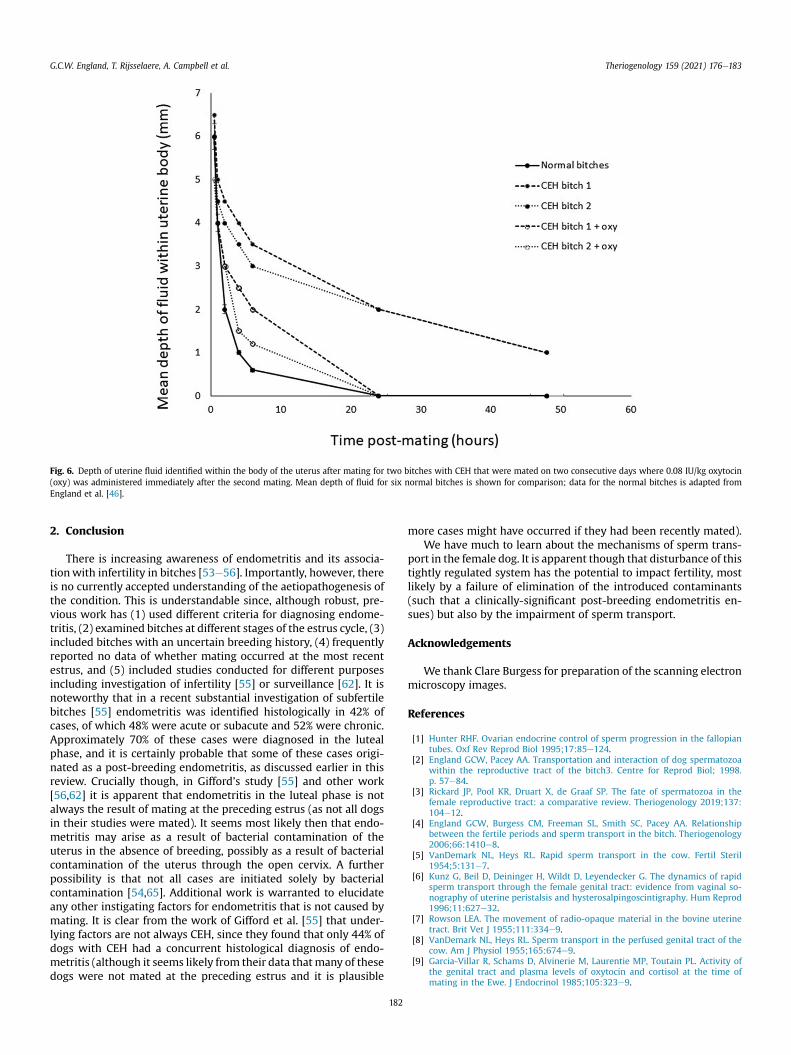
Fig. 6. Depth of uterine fluid identified within the body of the uterus after mating for two bitches with CEH that were mated on two consecutive days where 0.08 IU/kg oxytocin(oxy) was administered immediately after the second mating. Mean depth of fluid for six normal bitches is shown for comparison; data for the normal bitches is adapted fromEngland et al. [46].
G.C.W. England, T. Rijsselaere, A. Campbell et al. Theriogenology 159 (2021) 176e183
2. Conclusion
There is increasing awareness of endometritis and its associa-tionwith infertility in bitches [53e56]. Importantly, however, thereis no currently accepted understanding of the aetiopathogenesis ofthe condition. This is understandable since, although robust, pre-vious work has (1) used different criteria for diagnosing endome-tritis, (2) examined bitches at different stages of the estrus cycle, (3)included bitches with an uncertain breeding history, (4) frequentlyreported no data of whether mating occurred at the most recentestrus, and (5) included studies conducted for different purposesincluding investigation of infertility [55] or surveillance [62]. It isnoteworthy that in a recent substantial investigation of subfertilebitches [55] endometritis was identified histologically in 42% ofcases, of which 48% were acute or subacute and 52% were chronic.Approximately 70% of these cases were diagnosed in the lutealphase, and it is certainly probable that some of these cases origi-nated as a post-breeding endometritis, as discussed earlier in thisreview. Crucially though, in Gifford’s study [55] and other work[56,62] it is apparent that endometritis in the luteal phase is notalways the result of mating at the preceding estrus (as not all dogsin their studies were mated). It seems most likely then that endo-metritis may arise as a result of bacterial contamination of theuterus in the absence of breeding, possibly as a result of bacterialcontamination of the uterus through the open cervix. A furtherpossibility is that not all cases are initiated solely by bacterialcontamination [54,65]. Additional work is warranted to elucidateany other instigating factors for endometritis that is not caused bymating. It is clear from the work of Gifford et al. [55] that under-lying factors are not always CEH, since they found that only 44% ofdogs with CEH had a concurrent histological diagnosis of endo-metritis (although it seems likely from their data that many of thesedogs were not mated at the preceding estrus and it is plausible
182
more cases might have occurred if they had been recently mated).We have much to learn about the mechanisms of sperm trans-
port in the female dog. It is apparent though that disturbance of thistightly regulated system has the potential to impact fertility, mostlikely by a failure of elimination of the introduced contaminants(such that a clinically-significant post-breeding endometritis en-sues) but also by the impairment of sperm transport.
Acknowledgements
We thank Clare Burgess for preparation of the scanning electronmicroscopy images.
References
[1] Hunter RHF. Ovarian endocrine control of sperm progression in the fallopiantubes. Oxf Rev Reprod Biol 1995;17:85e124.
[2] England GCW, Pacey AA. Transportation and interaction of dog spermatozoawithin the reproductive tract of the bitch3. Centre for Reprod Biol; 1998.p. 57e84.
[3] Rickard JP, Pool KR, Druart X, de Graaf SP. The fate of spermatozoa in thefemale reproductive tract: a comparative review. Theriogenology 2019;137:104e12.
[4] England GCW, Burgess CM, Freeman SL, Smith SC, Pacey AA. Relationshipbetween the fertile periods and sperm transport in the bitch. Theriogenology2006;66:1410e8.
[5] VanDemark NL, Heys RL. Rapid sperm transport in the cow. Fertil Steril1954;5:131e7.
[6] Kunz G, Beil D, Deininger H, Wildt D, Leyendecker G. The dynamics of rapidsperm transport through the female genital tract: evidence from vaginal so-nography of uterine peristalsis and hysterosalpingoscintigraphy. Hum Reprod1996;11:627e32.
[7] Rowson LEA. The movement of radio-opaque material in the bovine uterinetract. Brit Vet J 1955;111:334e9.
[8] VanDemark NL, Heys RL. Sperm transport in the perfused genital tract of thecow. Am J Physiol 1955;165:674e9.
[9] Garcia-Villar R, Schams D, Alvinerie M, Laurentie MP, Toutain PL. Activity ofthe genital tract and plasma levels of oxytocin and cortisol at the time ofmating in the Ewe. J Endocrinol 1985;105:323e9.
G.C.W. England, T. Rijsselaere, A. Campbell et al. Theriogenology 159 (2021) 176e183
[10] Carmichael MS, Warburton VL, Dixen J, Davidson JM. Relationships amongcardiovascular, muscular, and oxytocin responses during human sexual ac-tivity. Arch Sex Behav 1994;23:59e79.
[11] Bygdeman M. Effects of prostaglandins on the genital tract. Acta Vet ScandSuppl 1981;77:47e54.
[12] Claus R, Meyer HD, Gimenez T, Hoang-Vu C, Munster E. Effect of seminaloestrogens on prostaglandin F2alpha release from the uterus of the sow. AnimReprod Sci 1990;23:145e56.
[13] Campbell MLH, England GCW. Effect of teasing, mechanical stimulation andthe intrauterine infusion of saline on uterine contractions in mares. Vet Rec2004;155:103e10.
[14] Campbell MLH, England GCW. Effect coitus and the artificial insemination ofdifferent volume of fresh semen on uterine contractions in the mare. Vet Rec2006;159:843e9.
[15] Hino T, Yanagimachi R. Active peristaltic movement and fluid production onthe mouse oviduct: their role in fluid and sperm transport and fertilization.Biol Reprod 2019;101:40e9.
[16] Smith TT, Yanagimachi R. Attachment and release of spermatozoa from thecaudal isthmus of the hamster oviduct. J Reprod Fertil 1991;91:567e73.
[17] Suarez SS. Formation of a reservoir of sperm in the oviduct. Reprod DomestAnim 2002;37:140e3.
[18] Cohen J. Immunological aspects of sperm collection and transport. In:Crighton DB, editor. Immunological aspects of reproduction in mammals.Kent: Butterworths; 1984. p. 77e89.
[19] Troedsson MTH. Uterine clearance and resistance to persistent endometritisin the mare. Theriogenology 1999;52:461e71.
[20] England GCW, Russo M, Freeman SL. The bitch uterine response to semendeposition and its modification by male accessory gland secretions. Vet J2013;195:179e84.
[21] Alghamdi AS, Foster DN, Troedsson MTH. Equine seminal plasma reducessperm binding to polymorphonuclear neutrophil (PMNs) and improves thefertility of fresh semen inseminated into inflamed uteri. Reprod127 2004:593e600.
[22] Brook D. Cytological and bacteriological examination of the mare’s endome-trium. Eq Vet Sci 1985;5:16e22.
[23] Hughes JP, Loy RG. Investigations on the effect of intrauterine inoculation ofStreptococcus zooepidemicus in the mare. Proc Am Assoc Eq Pract 1969;15:289e92.
[24] Evans EI. The transport of spermatozoa in the dog. Am J Physiol 1933;105:287e93.
[25] Holst PA, Phemister RD. The prenatal development of the dog: preimplanta-tion events. Biol Reprod 1971;5:194e206.
[26] Concannon PW, McCann JP, Temple M. Biology and endocrinology of ovula-tion, pregnancy and parturition in the dog. J Reprod Fertil Suppl 1989;39:3e25.
[27] Doak RL, Hall A, Dale HE. Longevity of spermatozoa in the reproductive tractof the bitch. J Reprod Fertil 1967;13:51e8.
[28] England GCW, Allen WE, Middleton DJ. An investigation into the origin of thefirst fraction of the canine ejaculate. Res Vet Sci 1990;49:66e70.
[29] Linde C. Transport of radiopaque fluid into the uterus after vaginal depositionin the oestrous bitch. Acta Vet Scand 1978;19:463e5.
[30] Silva LDM, Onclin K, Verstegen JP. Cervical opening in relation to progesteroneand oestradiol during heat in beagle bitches. J Reprod Fertil 1995;104:85e90.
[31] Allen WE, France C. A contrast radiographic study of the vagina and uterus ofthe normal bitch. J Small Anim Pract 1985;26:153e66.
[32] Tsutsui T, Kawakami E, Murao I, Ogossa A. Transport of spermatozoa in thereproductive tract of the bitch: observations through uterine fistula. Jpn J VetSci 1989;3:560e5.
[33] Wheaton LG, Pijanowski GJ, Weston PG, Burke TJ. Uterine motility during theestrous cycle; studies in healthy bitches. Am J Vet Res 1988;49:82e6.
[34] England GCW, Moxon R, Freeman SL. Stimulation of mating-induced uterinecontractions in the bitch and their modification and enhancement of fertilityby prostatic fluid. Reprod Domest Anim 2012;6:1e5.
[35] Fukushima FB, Malm C, Henry M, Gheller VA, Serakidis R, Neves MM,Macedo SP, Figueiredo MS, Andrade MEJ, Chaves MS, Silva MX, Rezende CMF,Melo EG. Site of intra-uterine artificial insemination in the bitch does notaffect sperm distribution within the uterus. Reprod Domest Anim 2010;45:1059e64.
[36] Rijsselaere T, Van Soom A, Van Cruchten S, Coryn M, G€ortz K, Maes D, deKruif A. Sperm distribution in the genital tract of the bitch following artificialinsemination in relation to the time of ovulation. Reproduction 2004;128:801e11.
[37] Rijsselaere T. New techniques for canine semen assessment and character-ization of the sperm reservoir in the bitch. PhD thesis. Faculty of VeterinaryMedicine, Ghent University; 2004. p. 189e212.
[38] Rijsselaere T, Van Soom A, Van Cruchten S, Coryn M, G€ortz K, Maes D, deKruif A. Effect of the timing of artificial insemination on the number ofspermatozoa discovered in the uterine crypts of the bitch. Reprod Fertil Dev2005;17:152 [abstract)].
183
[39] Rijsselaere T, England GCW, Freeman SL, Maes D, Van Soom A. Currentknowledge on the transport and fate of spermatozoa in the reproductive tractof the bitch. Reprod Domest Anim 2014;49:2e7.
[40] Pacey AA, Freeman SL, England GCW. Contact of dog spermatozoa with ho-mologous uterine tube epithelium prolongs flagellar activity in relation to thestage of the estrus cycle. Theriogenology 2000;54:109e18.
[41] England GCW, Allen WE, Blythe SA. Variability of the time of calculatedluteinising hormone release in 218 canine pregnancies. Vet Rec 1989;125:624e5.
[42] England GCW, Burgess CM, Clutterbuck AL, Freeman SL. Epithelial surfacechanges and spermatozoa storage in the reproductive tract of the bitch. Vet J2013;195:185e91.
[43] Karre I, Meyer-Lindenberg A, Urhausen C, Beineke A, Meinecke B, Piechotta M,Beyerbach M, Günzel-Apel AR. Distribution and viability of spermatozoa inthe canine female genital tract during post-ovulatory oocyte maturation. ActaVet Scand 2012;54. 49.
[44] Freeman SL, England GCW. Storage and release of spermatozoa from the pre-uterine tube reservoir. PLoS One 2013;8(2):e57006. https://doi.org/10.1371/journal.pone.0057006.
[45] Iguer-ouada M. Oestrus cycle stage dependent effects of plasma and vaginalfluid on dog semen motility parameters. Medically assisted procreation incanine species: analysis and 4�C preservation of semen, PhD-thesis, Faculty ofVeterinary Medicine. Li�ege University; 2000pp187e202.
[46] England GCW, Moxon R, Freeman SL. Delayed uterine fluid clearance andreduced uterine perfusion in bitches with endometrial hyperplasia and clin-ical management with post-mating antibiotic. Theriogenology 2012;78:1611e7.
[47] Hurtgen JP. Pathogenesis and treatment of endometritis in the mare: a review.Theriogenology 2006;66:560e6.
[48] Schlafer DH, Gifford AT. Cystic endometrial hyperplasia, pseudo-placentational endometrial hyperplasia, and other cystic conditions of thecanine and feline uterus. Theriogenology 2008;70:349e58.
[49] Hadley JC. The development of cystic endometrial hyperplasia in the bitchfollowing serial uterine biopsies. J Small Anim Pract 1975;16:249e57.
[50] Dow C. The cystic hyperplasia-pyometra complex in the bitch. J comp Path1959;69:237e50.
[51] Bigliardi E, Parmigiani E, Cavirani S, Luppi A, Bonati L, Corradi A. Ultraso-nography and cystic hyperplasia-pyometra complex in the bitch. ReprodDomest Anim 2004;39:136e40.
[52] England GCW, Burgess CM, Freeman SL. Perturbed sperm-epithelial interac-tion in bitches with mating-induced endometritis. Vet J 2012;194:314e8.
[53] Fontaine E, Levy X, Grellet A, Luc A, Bernex F, Boulouis HJ, Fontbonne A.Diagnosis of endometritis in the bitch: a new approach. Reprod Domest Anim2009;44:196e9.
[54] Mir F, Fontaine E, Albaric O, Greer M, Vannier F, Schlafer DH, Fontbonne A.Findings in uterine biopsies obtained by laparotomy from bitches with un-explained infertility or pregnancy loss: an observational study. Theriogenol-ogy 2013;79:312e22.
[55] Gifford AT, Scarlett JM, Schlafer DH. Histopathologic findings in uterine biopsysamples from subfertile bitches: 399 cases (1990-2005). J Am Vet Med Assoc2014;244:180e6.
[56] García Mitacek MC, Praderio RG, Stornelli MC, de la Sota RL, Stornelli MA.Prostaglandin synthesis enzymes’ gene transcription in bitches with endo-metritis. Reprod Domest Anim 2017;52:298e302.
[57] Moxon R, Whiteside H, England GCW. Prevalence of ultrasound-determinedcystic endometrial hyperplasia and the relationship with age in dogs. Ther-iogenology 2016;86:976e80.
[58] Freeman SL, Russo M, England GCW. Uterine artery blood flow characteristicsduring oestrus in pregnant and non-pregnant bitches. Vet J 2013;197:205e10.
[59] Freeman SL, Green MJ, England GCW. Uterine fluid from bitches with mating-induced endometritis reduces the attachment of spermatozoa to the uterineepithelium. Vet J 2013;198:76e80.
[60] Pycock JF, Newcombe JR. Assessment of the effect of three treatments toremove intrauterine fluid on pregnancy rate in the mare. Vet Rec 1996;138:320e3.
[61] Pycock JF, Newcombe JR. The relationship between intraluminal uterine fluid,endometritis and pregnancy rate in the mare. Equine Pract 1996;18:19e22.
[62] Praderio RG, García Mitacek MC, Nú~nez Favre R, Rearte R, de la Sota RL,Stornelli MA. Uterine endometrial cytology, biopsy, bacteriology, and serumC-reactive protein in clinically healthy diestrus bitches. Theriogenology2019;131:153e61.
[63] Lyman CC, Hornberger KT, Hallman RM, Holyoak GR. Theriogenology questionof the month. J Am Vet Med Assoc 2018;253:1267e70.
[64] Freeman SL1, Green MJ, England GCW. Prevalence and effect of uterineluminal free fluid on pregnancy and litter size in bitches. Theriogenology2013;80:73e6.
[65] Dhaliwal GK, England GC, Noakes DE. The effects of endometrial scarificationon uterine steroid receptors, bacterial flora and histological structure in thebitch. Anim Reprod Sci 2002;69:239e49.