Embed Size (px)
Citation preview
Expression of an Engineered Heterologous AntimicrobialPeptide in Potato Alters Plant Development and MitigatesNormal Abiotic and Biotic ResponsesRavinder K. Goyal1¤a, Robert E. W. Hancock2, Autar K. Mattoo3*, Santosh Misra1
1 Department of Biochemistry and Microbiology, University of Victoria, Victoria, British Columbia, Canada, 2 Centre for Microbial Diseases and ImmunityResearch, University of British Columbia, Vancouver, Canada, 3 The Henry A. Wallace Beltsville Agricultural Research Center, United States Department ofAgriculture, Agricultural Research Service, Sustainable Agricultural Systems Laboratory, Beltsville, Maryland, United States of America
Abstract
Antimicrobial cationic peptides (AMPs) are ubiquitous small proteins used by living cells to defend against a widespectrum of pathogens. Their amphipathic property helps their interaction with negatively charged cellular membraneof the pathogen causing cell lysis and death. AMPs also modulate signaling pathway(s) and cellular processes inanimal models; however, little is known of cellular processes other than the pathogen-lysis phenomenon modulatedby AMPs in plants. An engineered heterologous AMP, msrA3, expressed in potato was previously shown to causeresistance of the transgenic plants against selected fungal and bacterial pathogens. These lines together with thewild type were studied for growth habits, and for inducible defense responses during challenge with biotic (necrotrophFusarium solani) and abiotic stressors (dark-induced senescence, wounding and temperature stress). msrA3-expression not only conferred protection against F. solani but also delayed development of floral buds and prolongedvegetative phase. Analysis of select gene transcript profiles showed that the transgenic potato plants weresuppressed in the hypersensitive (HR) and reactive oxygen species (ROS) responses to both biotic and abioticstressors. Also, the transgenic leaves accumulated lesser amounts of the defense hormone jasmonic acid uponwounding with only a slight change in salicylic acid as compared to the wild type. Thus, normal host defenseresponses to the pathogen and abiotic stressors were mitigated by msrA3 expression suggesting MSRA3 regulates acommon step(s) of these response pathways. The stemming of the pathogen growth and mitigating stress responsepathways likely contributes to resource reallocation for higher tuber yield.
Citation: Goyal RK, Hancock REW, Mattoo AK, Misra S (2013) Expression of an Engineered Heterologous Antimicrobial Peptide in Potato Alters PlantDevelopment and Mitigates Normal Abiotic and Biotic Responses. PLoS ONE 8(10): e77505. doi:10.1371/journal.pone.0077505
Editor: Ashlesh K Murthy, Midwestern University, United States of America
Received June 25, 2013; Accepted September 4, 2013; Published October 16, 2013
This is an open-access article, free of all copyright, and may be freely reproduced, distributed, transmitted, modified, built upon, or otherwise used byanyone for any lawful purpose. The work is made available under the Creative Commons CC0 public domain dedication.
Funding: This study was funded by the NCE-AFMNet, Canada grant (to S.M.). Mention of trade names or commercial products in this publication is solelyfor the purpose of providing specific information and does not imply recommendation or endorsement by the U.S. Department of Agriculture. The fundershad no role in study design, data collection and analysis, decision to publish, or preparation of the manuscript.
Competing interests: The authors have declared that no competing interests exist.
* E-mail: [email protected]
¤a Current address: AAFC Lethbridge Research Centre, Lethbridge, Alberta, Canada
Introduction
Sustained plant losses due to microbial diseases cause cropyield reduction and are of major economical concern to farmersand agriculture industry [1,2]. Throughout the world, therefore,there is an ongoing effort to develop crops resistant to differentdiseases. Understanding host plant-microbe interactions andelucidating mechanisms that enable some plants to defendagainst one or more pathogens are currently dynamic researchareas [3]. The dynamics of plant response to a disease(s)change with environmental interactions [4], thus requiring an in-depth understanding of the molecular mechanisms involved.Plants that are able to resist a pathogen are more capable thanthe susceptible ones in creating physical barriers like
thickening and lignification of the cell wall [5,6], deposit callose[7], release phenolics or toxic substances (phytoalexins,proteinases, proteinase inhibitors) that inhibit the pathogengrowth or detoxify pathogen-derived toxins [8], and releasechemicals that inactivate the hydrolytic enzymes secreted bythe pathogen [6].
Plants are known to harbor a unique systemic immunologicalresponse, which is activated upon recognition of a pathogen.One of the extensively studied inducible plant defenseresponses is a hypersensitive response (HR). Cells displayingHR undergo localized programmed cell death (PCD) to limit thedamage, and the host plant may get immunized againstsubsequent pathogen attack, a phenomenon named systemicacquired resistance (SAR) [3,9]. HR is accompanied by an
PLOS ONE | www.plosone.org 1 October 2013 | Volume 8 | Issue 10 | e77505
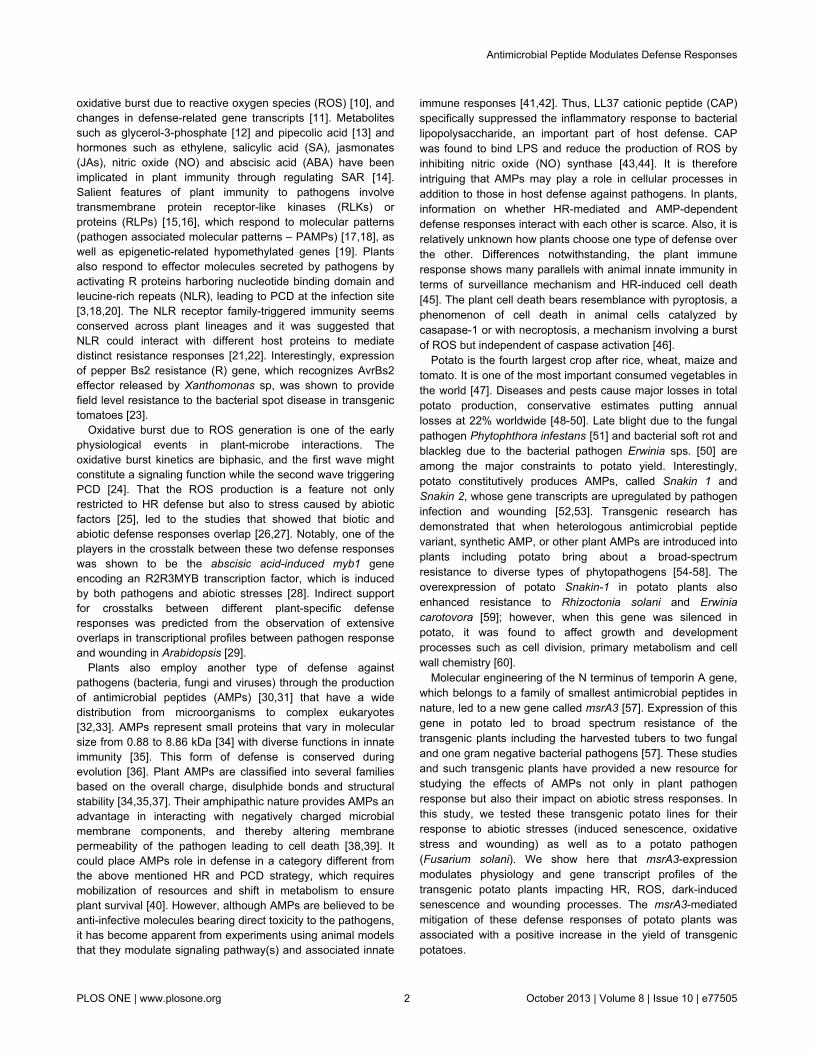
oxidative burst due to reactive oxygen species (ROS) [10], andchanges in defense-related gene transcripts [11]. Metabolitessuch as glycerol-3-phosphate [12] and pipecolic acid [13] andhormones such as ethylene, salicylic acid (SA), jasmonates(JAs), nitric oxide (NO) and abscisic acid (ABA) have beenimplicated in plant immunity through regulating SAR [14].Salient features of plant immunity to pathogens involvetransmembrane protein receptor-like kinases (RLKs) orproteins (RLPs) [15,16], which respond to molecular patterns(pathogen associated molecular patterns – PAMPs) [17,18], aswell as epigenetic-related hypomethylated genes [19]. Plantsalso respond to effector molecules secreted by pathogens byactivating R proteins harboring nucleotide binding domain andleucine-rich repeats (NLR), leading to PCD at the infection site[3,18,20]. The NLR receptor family-triggered immunity seemsconserved across plant lineages and it was suggested thatNLR could interact with different host proteins to mediatedistinct resistance responses [21,22]. Interestingly, expressionof pepper Bs2 resistance (R) gene, which recognizes AvrBs2effector released by Xanthomonas sp, was shown to providefield level resistance to the bacterial spot disease in transgenictomatoes [23].
Oxidative burst due to ROS generation is one of the earlyphysiological events in plant-microbe interactions. Theoxidative burst kinetics are biphasic, and the first wave mightconstitute a signaling function while the second wave triggeringPCD [24]. That the ROS production is a feature not onlyrestricted to HR defense but also to stress caused by abioticfactors [25], led to the studies that showed that biotic andabiotic defense responses overlap [26,27]. Notably, one of theplayers in the crosstalk between these two defense responseswas shown to be the abscisic acid-induced myb1 geneencoding an R2R3MYB transcription factor, which is inducedby both pathogens and abiotic stresses [28]. Indirect supportfor crosstalks between different plant-specific defenseresponses was predicted from the observation of extensiveoverlaps in transcriptional profiles between pathogen responseand wounding in Arabidopsis [29].
Plants also employ another type of defense againstpathogens (bacteria, fungi and viruses) through the productionof antimicrobial peptides (AMPs) [30,31] that have a widedistribution from microorganisms to complex eukaryotes[32,33]. AMPs represent small proteins that vary in molecularsize from 0.88 to 8.86 kDa [34] with diverse functions in innateimmunity [35]. This form of defense is conserved duringevolution [36]. Plant AMPs are classified into several familiesbased on the overall charge, disulphide bonds and structuralstability [34,35,37]. Their amphipathic nature provides AMPs anadvantage in interacting with negatively charged microbialmembrane components, and thereby altering membranepermeability of the pathogen leading to cell death [38,39]. Itcould place AMPs role in defense in a category different fromthe above mentioned HR and PCD strategy, which requiresmobilization of resources and shift in metabolism to ensureplant survival [40]. However, although AMPs are believed to beanti-infective molecules bearing direct toxicity to the pathogens,it has become apparent from experiments using animal modelsthat they modulate signaling pathway(s) and associated innate
immune responses [41,42]. Thus, LL37 cationic peptide (CAP)specifically suppressed the inflammatory response to bacteriallipopolysaccharide, an important part of host defense. CAPwas found to bind LPS and reduce the production of ROS byinhibiting nitric oxide (NO) synthase [43,44]. It is thereforeintriguing that AMPs may play a role in cellular processes inaddition to those in host defense against pathogens. In plants,information on whether HR-mediated and AMP-dependentdefense responses interact with each other is scarce. Also, it isrelatively unknown how plants choose one type of defense overthe other. Differences notwithstanding, the plant immuneresponse shows many parallels with animal innate immunity interms of surveillance mechanism and HR-induced cell death[45]. The plant cell death bears resemblance with pyroptosis, aphenomenon of cell death in animal cells catalyzed bycasapase-1 or with necroptosis, a mechanism involving a burstof ROS but independent of caspase activation [46].
Potato is the fourth largest crop after rice, wheat, maize andtomato. It is one of the most important consumed vegetables inthe world [47]. Diseases and pests cause major losses in totalpotato production, conservative estimates putting annuallosses at 22% worldwide [48-50]. Late blight due to the fungalpathogen Phytophthora infestans [51] and bacterial soft rot andblackleg due to the bacterial pathogen Erwinia sps. [50] areamong the major constraints to potato yield. Interestingly,potato constitutively produces AMPs, called Snakin 1 andSnakin 2, whose gene transcripts are upregulated by pathogeninfection and wounding [52,53]. Transgenic research hasdemonstrated that when heterologous antimicrobial peptidevariant, synthetic AMP, or other plant AMPs are introduced intoplants including potato bring about a broad-spectrumresistance to diverse types of phytopathogens [54-58]. Theoverexpression of potato Snakin-1 in potato plants alsoenhanced resistance to Rhizoctonia solani and Erwiniacarotovora [59]; however, when this gene was silenced inpotato, it was found to affect growth and developmentprocesses such as cell division, primary metabolism and cellwall chemistry [60].
Molecular engineering of the N terminus of temporin A gene,which belongs to a family of smallest antimicrobial peptides innature, led to a new gene called msrA3 [57]. Expression of thisgene in potato led to broad spectrum resistance of thetransgenic plants including the harvested tubers to two fungaland one gram negative bacterial pathogens [57]. These studiesand such transgenic plants have provided a new resource forstudying the effects of AMPs not only in plant pathogenresponse but also their impact on abiotic stress responses. Inthis study, we tested these transgenic potato lines for theirresponse to abiotic stresses (induced senescence, oxidativestress and wounding) as well as to a potato pathogen(Fusarium solani). We show here that msrA3-expressionmodulates physiology and gene transcript profiles of thetransgenic potato plants impacting HR, ROS, dark-inducedsenescence and wounding processes. The msrA3-mediatedmitigation of these defense responses of potato plants wasassociated with a positive increase in the yield of transgenicpotatoes.
Antimicrobial Peptide Modulates Defense Responses
PLOS ONE | www.plosone.org 2 October 2013 | Volume 8 | Issue 10 | e77505
Materials and Methods
Plant materialPotato (Solanum tuberosum L.) cultivar Desiree (WT) and
two transgenic lines (T3 and T26) expressing antimicrobialpeptide msrA3 gene [57] were grown in the greenhouse facility,University of Victoria, Victoria, B.C., Canada. The T3 and T26transgenic lines represent two independent insertion eventsand contain a single copy of msrA3 [57]. For brevity, leaflets ofa compound leaf are referred to as leaves.
Growth conditions and tuber yield determinationWT and the T3 and T26 transgenic plants were grown from
seed tubers. Tubers with a mean weight of 56 g (50-63 g) wereplanted in 11-L and 15-L pots for growth chamber andgreenhouse experiments, respectively. Number of plants perpot was three for growth chamber and four for greenhouse, ifnot specified otherwise. After 2 weeks of germination, one dose(75 g) of 6-8-6 (nitrogen/phosphorus/potash) fertilizer (EvergoCanada Inc., Delta, B.C., Canada) was applied. In growthchamber, the plants were raised under 16/8h photoperiod at21°/18°C day/night temperature unless otherwise stated. Theplants were watered as needed. At each time of planting, thepots were triplicated in a randomized clear block design in achamber. In the greenhouse, plants were grown in 6replications under 16/8h daylight and irrigated throughautomated drip irrigation. Following 16 weeks of growth, thefresh tuber yield was recorded.
RNA extraction and northern-blot analysisTotal RNA was isolated from frozen leaf tissue with Trizol as
per the manufacturer’s protocol. RNA was fractionated on 1%agarose-formaldehyde gels and blotted onto nylon membrane(Schleicher & Schull, Germany). Gene probes were labeledwith [α-32P] dCTP using High Prime random priming kit (Roche)and purified on ProbeQuant G-50 Micro Columns (GEHealthcare). The membranes after hybridization with therespective probes for 16h at 65°C were washed twice with2xSSC, 0.1% SDS at 65°C for 20 min each, once in 1xSSC,0.1% SDS and twice in 0.2xSSC, 0.1% SDS for 20 min each at60°C. The hybridized blots were exposed to X-ray films withintensifying screens at -75°C. Following genes were analyzed:Pathogenesis-related protein (pr-1) (AJ250136), osmotin (osm)(AY256439), ascorbate peroxidese (apx) (AB041343), catalase(cat) (Z37106), γ-vacuolar processing enzyme (vpe) (D61395),senescence associated gene 12 (sag12) (AI776170), 13-lipoxygenase (13-lox) (X96406), potato peroxidase2 (Stprx2)(AJ401150), Cu/Zn superoxide dismutase (sod) (AF355460),longevity assurance gene1 (lag1) (AF198177), rbcL (AI486088)and glutamine synthetase-1 (gs-1) (AW626325). (Table S1 inFile S1) lists primer sequences used for amplifying the geneprobes. The genes, pr-1, vpe, lag1and cat, were PCR amplifiedfrom potato genomic DNA. For apx and Cu/Zn sod, the cDNAwas prepared to RNA isolated from untreated leaves. ForStprx2 and13-lox, the cDNA was prepared to RNA isolatedfrom wounded leaves. Amplification and primer sequences forosm, sag12, rbcL and gs-1 were the same as previouslydescribed [61]. cDNA was synthesized using SuperScriptTmII
RNaseH reverse transcriptase (Invitrogen) followingmanufacturer’s protocol. The Qiagen MasterMix kit was usedfor 25 µl PCR reactions as follows: 94°C for 10 min, and 35cycles of 94°C for 30 sec, temp (1°C below Tm of the primersequence) for 30 sec, and 72°C for 1 min followed by 15 minextension at the end.
In situ detection and determination of H2O2
H2O2 was visualized in leaves using 3, 3’-diaminobenzidine(DAB) staining [62]. The cut end of each detached leaf wasincubated with 1mg mL-1 DAB, pH 4.5 for 3h. After leaf de-colorization in hot ethanol (95%), the intensity of brown colorstain was monitored.
For quantifying H2O2, leaf tissue (400 mg) was powdered inliquid nitrogen and then homogenized in 1 mL 10%trichloroacetic acid (TCA). The homogenate was centrifuged at16,000 g for 15 min and supernatant collected. The content ofH2O2 in the supernatant was determined by slight modificationof a previously described method [63]. Briefly, the supernatant(40μL) was mixed with10 μL of 1N NaOH, and then 50μl ofxylene-orange reagent (500 μM ferrous ammonium sulfate, 50mM H2SO4, 200 μM xylene orange and 200 mM sorbitol) wasadded. The color was allowed to develop for 2 h and theabsorbance determined at 560 nm. To ascertain that TCAaddition had disabled the H2O2-metabolizing enzymes and thatit was compatible with the xylene-orange assay, a knownquantity of H2O2 was added during tissue homogenization fordetermining % recovery. The recovery was 99.2% and noactivity of peroxidase or catalase was detected in the TCA-extract. The assay generated linear curves using differentconcentrations of H2O2 in 10% TCA. Only the relativeabundance of H2O2 rather than absolute values are reportedbecause any unknown component in the plant extract with apotential to affect the A560 values was not tested.
Enzyme assaysMethods used for preparing cell-free extracts and assaying
guaiacol- or pyrogallol-peroxidases activities were the same aspreviously described [64].
Chlorophyll analysisTotal chlorophyll was extracted from leaves by grinding 0.5 g
of tissue with 5 mL of pure acetone in a mortar with pestlefollowed by several extractions with 80% acetone to a finalvolume of 15 mL. The clarified extract was diluted, andabsorbance at λ 646 nm and λ 663 nm was determined. Thecontents of total chlorophyll, chlorophyll a, and chlorophyll bwere calculated as described [65].
Determination of lipid peroxidationPeroxidated lipids were measured as thiobarbituric acid
reactive species (TBARS) [66]. Frozen leaf tissue (50 mg) washomogenized in 125 μl of 50 mM 2-morpholinoethanesulphonicacid (MES), pH 7.1, containing 2% SDS and 2 μl of 1% 2,6-di-tert-butyl-4-methylphenol (butylated hydroxytoluene). To thehomogenate, 700 μl of 0.8% (w/v) thiobarbituric acid in 10%TCA was added and the contents vortexed for 1 min. The
Antimicrobial Peptide Modulates Defense Responses
PLOS ONE | www.plosone.org 3 October 2013 | Volume 8 | Issue 10 | e77505
samples were heated at 95°C for 15 min, vortexed for 1 minand re-heated for 15 min. After cooling on ice, TBARS wereextracted in 500 μl of n-butanol by vigorous mixing. Thecontents were centrifuged at 5,000 g for 10 min andabsorbance of the supernatant was measured at 532 nm and600 nm. TBARS content was determined after subtracting thenonspecific background absorbance at 600 nm.
Jasmonic acid and salicylic acid analysesJasmonic acid (JA) and salicylic acid (SA) were analyzed at
the Plant Biotechnology Institute, National Research Council,Saskatoon, Canada. The details of JA/SA extraction andanalysis by High Performance Liquid ChromatographyElectrospray tandem Mass Spectrometry (HPLC/ES-MS/MS)were the same as previously described [67].
Statistical analysis
Data were statistically analyzed using analysis of variance(ANOVA), and mean separation (Tukey’s) test was performedusing the SPSS statistical program.
Results
Delayed emergence of floral buds in transgenic potatoplants expressing Msra3 gene
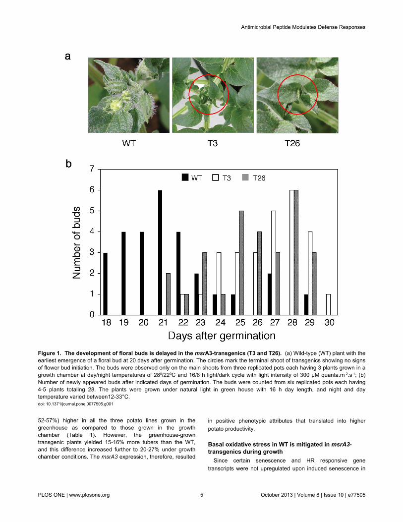
One of the earliest phenotypic differences observed betweenthe wild-type (WT) and transgenic plants (T3 and T26)expressing msrA3 (msrA3-transgenics) was in the developmentof floral buds (Figure 1a). In the growth chamber with a day/night temperature regime of 28°/22°C, floral buds in WT plantsemerged at 20 days after germination while bud initiation in thetwo transgenic lines was not apparent by this time (Figure 1a).Fifty percent or more of the WT plants had visible buds by day26 after germination, at least 3 days earlier than seen intransgenic plants. More validation of the delayed emergence offloral buds in the transgenic plants compared to the WT wasobtained from experiments carried out in the greenhouse undernatural lighting conditions with a day/night temperaturevariation of 33°/12°C. Under these conditions of greaterfluctuations in day and night temperatures than in thecontrolled growth chamber, emergence of buds in the WT wasaccelerated (Figure 2b). In the transgenic plants, buds initiatedon day 21 (T3) and day 22 (T26) of germination, by which time60-75% of the WT plants had already developed the buds.Thus, both the transgenic lines tended to flower later than theWT plants.
msrA3-transgenics have delayed dark-inducedsenescence
The delay in the emergence of floral buds (flowering) in thetransgenic lines indicated that msrA3 expression may impactleaf senescence since a slower initiation of reproductivedevelopment seems associated with longer vegetative growthin some plants [68]. Therefore, we tested this possibility usingthe model of dark-induced senescence [69]. Detached fullydeveloped leaves from the WT and the T3 and T26 transgenic
lines were incubated at room temperature in the dark and leafchlorophyll content was analyzed on day 1, 3 and 5. By day 5in the dark, WT plants showed visible symptoms of senescencebut the two transgenic lines were still robust and greener(Figure 2a). This difference in the physical condition of WTversus transgenics correlated with the steady loss ofchlorophyll in WT leaves starting day 1 in the dark anddecreasing thereafter to 50% of the original content by day 5while in the transgenic leaves the chlorophyll content remainedmore or less similar until day 3 and registered a slight declineby day 5 (Figure 2b).
Differential expression of a select class of genesbetween WT and transgenic leaves during inducedsenescence
Delayed floral development associated with delayed dark-induced senescence in the msrA3-transgenics suggested thatmsrA3 expression influences plant development. We thereforequantified changes in the expression of a medley of genetranscripts in the leaves of the WT and the two msrA3-transgenic lines including anti-senescence gene marker (largesubunit of Rubisco which promotes growth) [70-72] versus pro-senescence gene markers [senescence associated gene 12(sag12) and glutamine synthetase-1 (gs-1)] [73]. To that end,expression of genes not only associated with senescence[(sag12), γ-vacuolar processing enzyme (vpe), (gs-1)] andcarbon fixation [rubisco large subunit (rbcL)] but also thoseassociated with oxidative stress [ascorbate peroxidese (apx),catalase (cat), Cu/Zn superoxide dismutase (sod)] and HR[pathogenesis-related protein (pr-1), osmotin (osm), longevityassurance gene1 (lag1)] were analyzed by northern analysis ofRNA from leaves exposed to dark induced senescence (Figure2c). Dark-induced senescence in WT was associated withincreases in the levels of pr-1, osm, apx, cat, sod, sag12, vpe,lag1 and gs1 concomitant with a substantial decrease in thelarge subunit of rubisco (rbcL) (panel WT). In contrast to thesepatterns found in WT leaves, response to dark incubation of theleaves from the two transgenics (panels T3 and T26) wasdifferent. In fact, the expression of pr-1, osm, apx, cat, lag1 andgs1 transcripts was mitigated in the two transgenic lines whilesag12 and vpe transcripts were barely observed except sag12in T26 after 5 days of dark incubation (Figure 2c). Notably, inthe two transgenic lines, expression of rbcL remained at asteady level and that of sod fluctuated, remaining at a lowerlevel than the WT. Thus, distinctly different patterns in thesteady state levels of transcripts for genes associated with HRand senescence between WT and msrA3-transgenics wereapparent and indicated that msrA3 expression dampens theresponse seen in the WT leaves, particularly during the earlyphase of induced senescence (Figure 2c).
Higher tuber yield in msrA3-transgenicsIn order to determine the long-term effects of msrA3
expression in terms of the tuber yield, WT and the two msrA3-expressing transgenic lines were grown in three differentseasons to full maturity in the greenhouse as well as in acontrolled growth chamber and their tuber yield was quantified.Tuber yield was consistently and significantly (between
Antimicrobial Peptide Modulates Defense Responses
PLOS ONE | www.plosone.org 4 October 2013 | Volume 8 | Issue 10 | e77505
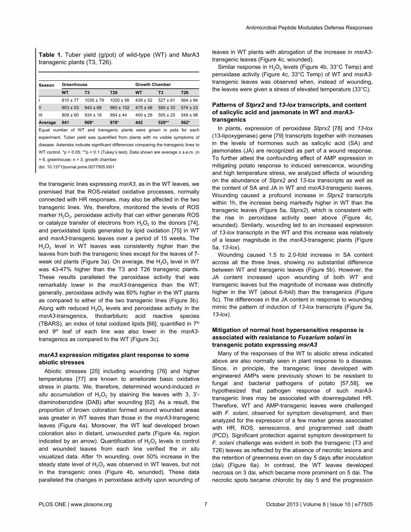
52-57%) higher in all the three potato lines grown in thegreenhouse as compared to those grown in the growthchamber (Table 1). However, the greenhouse-growntransgenic plants yielded 15-16% more tubers than the WT,and this difference increased further to 20-27% under growthchamber conditions. The msrA3 expression, therefore, resulted
in positive phenotypic attributes that translated into higherpotato productivity.
Basal oxidative stress in WT is mitigated in msrA3-transgenics during growth
Since certain senescence and HR responsive genetranscripts were not upregulated upon induced senescence in
Figure 1. The development of floral buds is delayed in the msrA3-transgenics (T3 and T26). (a) Wild-type (WT) plant with theearliest emergence of a floral bud at 20 days after germination. The circles mark the terminal shoot of transgenics showing no signsof flower bud initiation. The buds were observed only on the main shoots from three replicated pots each having 3 plants grown in agrowth chamber at day/night temperatures of 280/220C and 16/8 h light/dark cycle with light intensity of 300 µM quanta.m-2.s-1; (b)Number of newly appeared buds after indicated days of germination. The buds were counted from six replicated pots each having4-5 plants totaling 28. The plants were grown under natural light in green house with 16 h day length, and night and daytemperature varied between12-33°C.doi: 10.1371/journal.pone.0077505.g001
Antimicrobial Peptide Modulates Defense Responses
PLOS ONE | www.plosone.org 5 October 2013 | Volume 8 | Issue 10 | e77505
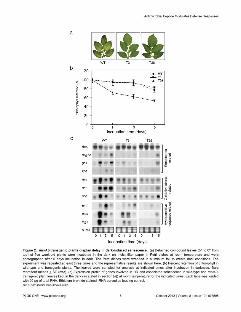
Figure 2. msrA3-transgenic plants display delay in dark-induced senescence. (a) Detached compound leaves (5th to 6th fromtop) of five week-old plants were incubated in the dark on moist filter paper in Petri dishes at room temperature and werephotographed after 5 days incubation in dark. The Petri dishes were wrapped in aluminum foil to create dark conditions. Theexperiment was repeated at least three times and the representative results are shown here. (b) Percent retention of chlorophyll inwild-type and transgenic plants. The leaves were sampled for analysis at indicated times after incubation in darkness. Barsrepresent means + SE (n=3). (c) Expression profile of genes involved in HR and associated senescence in wild-type and msrA3-transgenic plant leaves kept in the dark (as stated in section [a]) at room temperature for the indicated times. Each lane was loadedwith 20 μg of total RNA. Ethidium bromide stained rRNA served as loading control.doi: 10.1371/journal.pone.0077505.g002
Antimicrobial Peptide Modulates Defense Responses
PLOS ONE | www.plosone.org 6 October 2013 | Volume 8 | Issue 10 | e77505
the transgenic lines expressing msrA3, as in the WT leaves, wepremised that the ROS-related oxidative processes, normallyconnected with HR responses, may also be affected in the twotransgenic lines. We, therefore, monitored the levels of ROSmarker H2O2, peroxidase activity that can either generate ROSor catalyze transfer of electrons from H2O2 to the donors [74],and peroxidated lipids generated by lipid oxidation [75] in WTand msrA3-transgenic leaves over a period of 15 weeks. TheH2O2 level in WT leaves was consistently higher than theleaves from both the transgenic lines except for the leaves of 7-week old plants (Figure 3a). On average, the H2O2 level in WTwas 43-47% higher than the T3 and T26 transgenic plants.These results paralleled the peroxidase activity that wasremarkably lower in the msrA3-transgenics than the WT;generally, peroxidase activity was 60% higher in the WT plantsas compared to either of the two transgenic lines (Figure 3b).Along with reduced H2O2 levels and peroxidase activity in themsrA3-transgenics, thiobarbituric acid reactive species(TBARS), an index of total oxidized lipids [66], quantified in 7th
and 9th leaf of each line was also lower in the msrA3-transgenics as compared to the WT (Figure 3c).
msrA3 expression mitigates plant response to someabiotic stresses
Abiotic stresses [25] including wounding [76] and highertemperatures [77] are known to ameliorate basic oxidativestress in plants. We, therefore, determined wound-induced insitu accumulation of H2O2 by staining the leaves with 3, 3’-diaminobenzidine (DAB) after wounding [62]. As a result, theproportion of brown coloration formed around wounded areaswas greater in WT leaves than those in the msrA3-transgenicleaves (Figure 4a). Moreover, the WT leaf developed browncoloration also in distant, unwounded parts (Figure 4a, regionindicated by an arrow). Quantification of H2O2 levels in controland wounded leaves from each line verified the in situvisualized data. After 1h wounding, over 50% increase in thesteady state level of H2O2 was observed in WT leaves, but notin the transgenic ones (Figure 4b, wounded). These dataparalleled the changes in peroxidase activity upon wounding of
Table 1. Tuber yield (g/pot) of wild-type (WT) and MsrA3transgenic plants (T3, T26).
Season Greenhouse Growth Chamber
WT T3 T26 WT T3 T26I 810 ± 77 1030 ± 79 1020 ± 56 439 ± 52 527 ± 61 564 ± 94II 903 ± 53 943 ± 68 960 ± 102 475 ± 48 550 ± 33 574 ± 23III 809 ± 60 934 ± 18 954 ± 44 400 ± 29 505 ± 25 549 ± 98
Average 841 969* 978* 442 529** 562*Equal number of WT and transgenic plants were grown in pots for eachexperiment. Tuber yield was quantified from plants with no visible symptoms ofdisease. Asterisks indicate significant differences comparing the transgenic lines toWT control. *p < 0.05; **p < 0.1 (Tukey’s test). Data shown are average ± s.e.m. (n= 6, greenhouse; n > 3, growth chamber.doi: 10.1371/journal.pone.0077505.t001
leaves in WT plants with abrogation of the increase in msrA3-transgenic leaves (Figure 4c, wounded).
Similar response in H2O2 levels (Figure 4b, 33°C Temp) andperoxidase activity (Figure 4c, 33°C Temp) of WT and msrA3-transgenic leaves was observed when, instead of wounding,the leaves were given a stress of elevated temperature (33°C).
Patterns of Stprx2 and 13-lox transcripts, and contentof salicylic acid and jasmonate in WT and msrA3-transgenics
In plants, expression of peroxidase Stprx2 [78] and 13-lox(13-lipoxygenase) gene [79] transcripts together with increasesin the levels of hormones such as salicylic acid (SA) andjasmonates (JA) are recognized as part of a wound response.To further attest the confounding effect of AMP expression inmitigating potato response to induced senescence, woundingand high temperature stress, we analyzed effects of woundingon the abundance of Stprx2 and 13-lox transcripts as well asthe content of SA and JA in WT and msrA3-transgenic leaves.Wounding caused a profound increase in Stprx2 transcriptswithin 1h, the increase being markedly higher in WT than thetransgenic leaves (Figure 5a, Stprx2), which is consistent withthe rise in peroxidase activity seen above (Figure 4c,wounded). Similarly, wounding led to an increased expressionof 13-lox transcripts in the WT and this increase was relativelyof a lesser magnitude in the msrA3-transgenic plants (Figure5a, 13-lox).
Wounding caused 1.5 to 2.0-fold increase in SA contentacross all the three lines, showing no substantial differencebetween WT and transgenic leaves (Figure 5b). However, theJA content increased upon wounding of both WT andtransgenic leaves but the magnitude of increase was distinctlyhigher in the WT (about 6-fold) than the transgenics (Figure5c). The differences in the JA content in response to woundingmimic the pattern of induction of 13-lox transcripts (Figure 5a,13-lox).
Mitigation of normal host hypersensitive response isassociated with resistance to Fusarium solani intransgenic potato expressing msrA3
Many of the responses of the WT to abiotic stress indicatedabove are also normally seen in plant response to a disease.Since, in principle, the transgenic lines developed withengineered AMPs were previously shown to be resistant tofungal and bacterial pathogens of potato [57,58], wehypothesized that pathogen response of such msrA3-transgenic lines may be associated with downregulated HR.Therefore, WT and AMP-transgenic leaves were challengedwith F. solani, observed for symptom development, and thenanalyzed for the expression of a few marker genes associatedwith HR, ROS, senescence, and programmed cell death(PCD). Significant protection against symptom development toF. solani challenge was evident in both the transgenic (T3 andT26) leaves as reflected by the absence of necrotic lesions andthe retention of greenness even on day 5 days after inoculation(dai) (Figure 6a). In contrast, the WT leaves developednecrosis on 3 dai, which became more prominent on 5 dai. Thenecrotic spots became chlorotic by day 5 and the progression
Antimicrobial Peptide Modulates Defense Responses
PLOS ONE | www.plosone.org 7 October 2013 | Volume 8 | Issue 10 | e77505
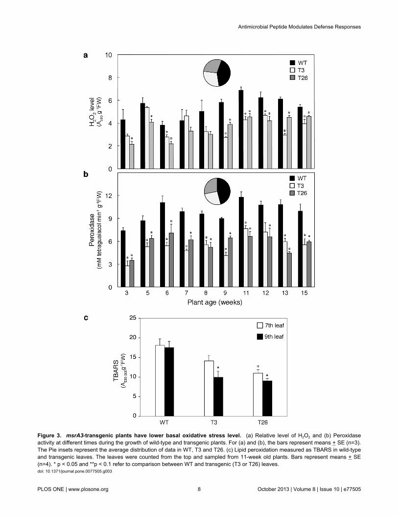
Figure 3. msrA3-transgenic plants have lower basal oxidative stress level. (a) Relative level of H2O2 and (b) Peroxidaseactivity at different times during the growth of wild-type and transgenic plants. For (a) and (b), the bars represent means + SE (n=3).The Pie insets represent the average distribution of data in WT, T3 and T26. (c) Lipid peroxidation measured as TBARS in wild-typeand transgenic leaves. The leaves were counted from the top and sampled from 11-week old plants. Bars represent means + SE(n=4). * p < 0.05 and **p < 0.1 refer to comparison between WT and transgenic (T3 or T26) leaves.doi: 10.1371/journal.pone.0077505.g003
Antimicrobial Peptide Modulates Defense Responses
PLOS ONE | www.plosone.org 8 October 2013 | Volume 8 | Issue 10 | e77505
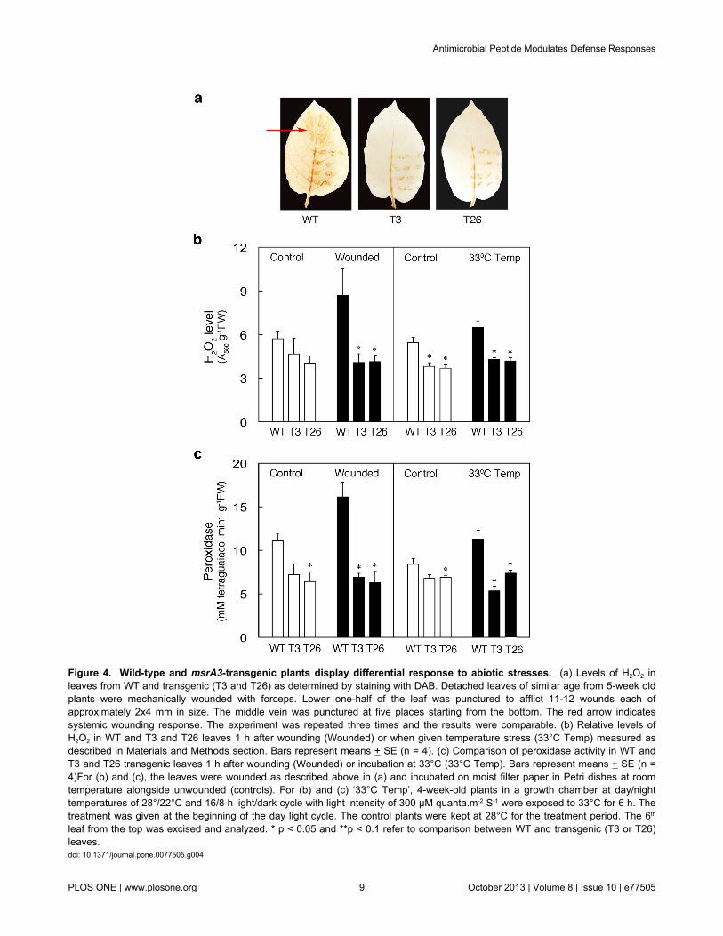
Figure 4. Wild-type and msrA3-transgenic plants display differential response to abiotic stresses. (a) Levels of H2O2 inleaves from WT and transgenic (T3 and T26) as determined by staining with DAB. Detached leaves of similar age from 5-week oldplants were mechanically wounded with forceps. Lower one-half of the leaf was punctured to afflict 11-12 wounds each ofapproximately 2x4 mm in size. The middle vein was punctured at five places starting from the bottom. The red arrow indicatessystemic wounding response. The experiment was repeated three times and the results were comparable. (b) Relative levels ofH2O2 in WT and T3 and T26 leaves 1 h after wounding (Wounded) or when given temperature stress (33°C Temp) measured asdescribed in Materials and Methods section. Bars represent means + SE (n = 4). (c) Comparison of peroxidase activity in WT andT3 and T26 transgenic leaves 1 h after wounding (Wounded) or incubation at 33°C (33°C Temp). Bars represent means + SE (n =4)For (b) and (c), the leaves were wounded as described above in (a) and incubated on moist filter paper in Petri dishes at roomtemperature alongside unwounded (controls). For (b) and (c) ‘33°C Temp’, 4-week-old plants in a growth chamber at day/nighttemperatures of 28°/22°C and 16/8 h light/dark cycle with light intensity of 300 µM quanta.m-2 S-1 were exposed to 33°C for 6 h. Thetreatment was given at the beginning of the day light cycle. The control plants were kept at 28°C for the treatment period. The 6th
leaf from the top was excised and analyzed. * p < 0.05 and **p < 0.1 refer to comparison between WT and transgenic (T3 or T26)leaves.doi: 10.1371/journal.pone.0077505.g004
Antimicrobial Peptide Modulates Defense Responses
PLOS ONE | www.plosone.org 9 October 2013 | Volume 8 | Issue 10 | e77505
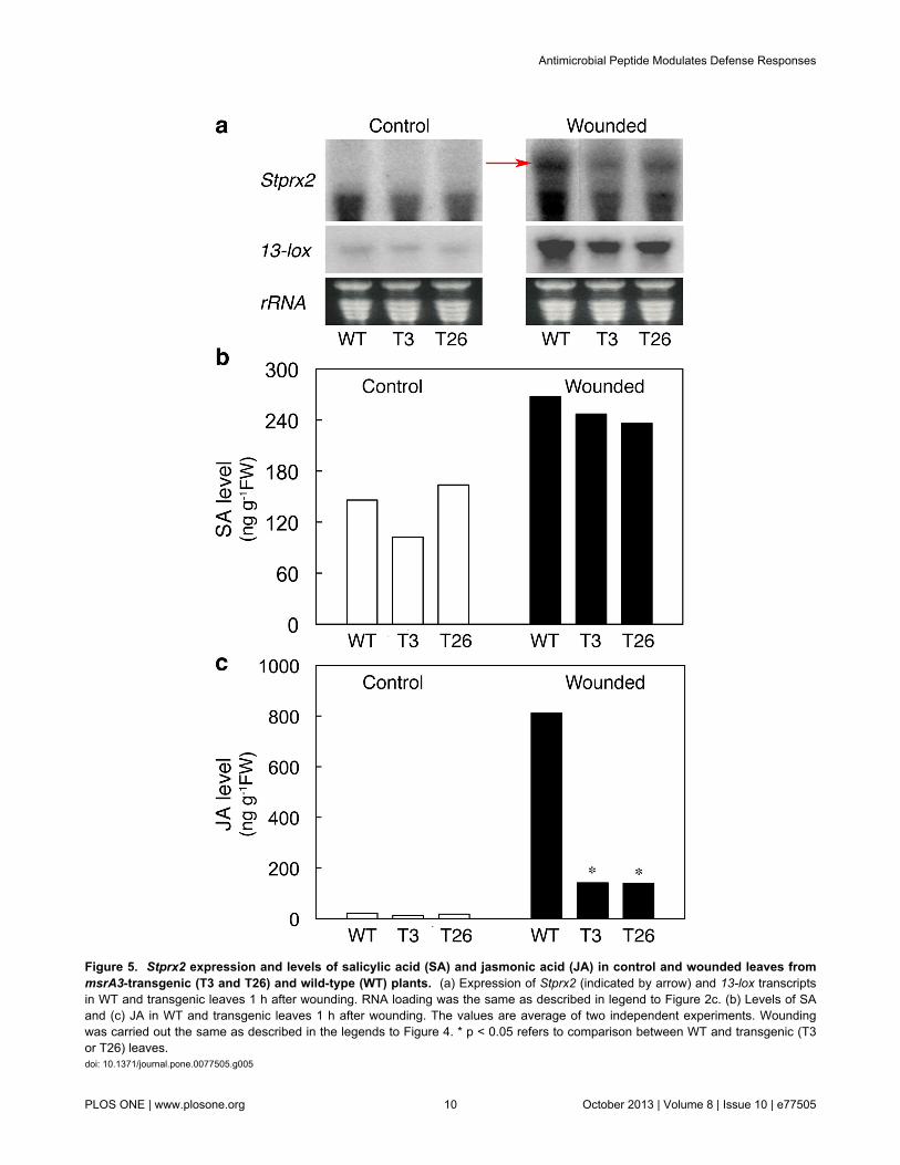
Figure 5. Stprx2 expression and levels of salicylic acid (SA) and jasmonic acid (JA) in control and wounded leaves frommsrA3-transgenic (T3 and T26) and wild-type (WT) plants. (a) Expression of Stprx2 (indicated by arrow) and 13-lox transcriptsin WT and transgenic leaves 1 h after wounding. RNA loading was the same as described in legend to Figure 2c. (b) Levels of SAand (c) JA in WT and transgenic leaves 1 h after wounding. The values are average of two independent experiments. Woundingwas carried out the same as described in the legends to Figure 4. * p < 0.05 refers to comparison between WT and transgenic (T3or T26) leaves.doi: 10.1371/journal.pone.0077505.g005
Antimicrobial Peptide Modulates Defense Responses
PLOS ONE | www.plosone.org 10 October 2013 | Volume 8 | Issue 10 | e77505
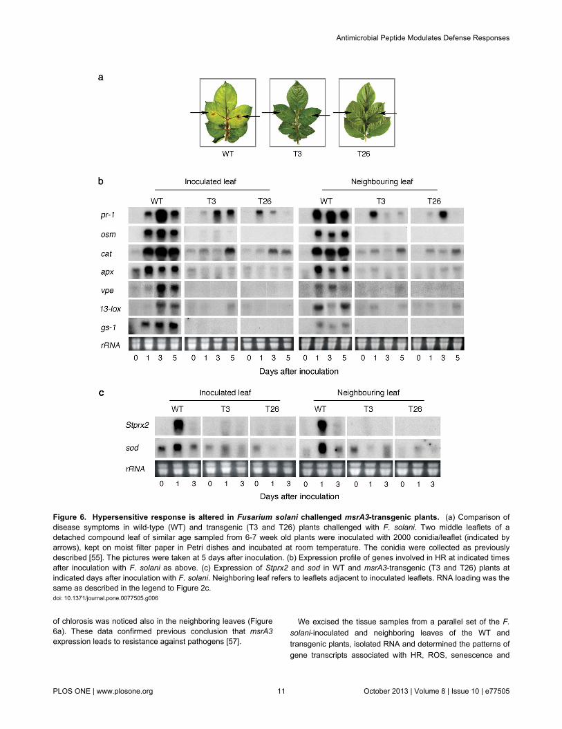
of chlorosis was noticed also in the neighboring leaves (Figure6a). These data confirmed previous conclusion that msrA3expression leads to resistance against pathogens [57].
We excised the tissue samples from a parallel set of the F.solani-inoculated and neighboring leaves of the WT andtransgenic plants, isolated RNA and determined the patterns ofgene transcripts associated with HR, ROS, senescence and
Figure 6. Hypersensitive response is altered in Fusarium solani challenged msrA3-transgenic plants. (a) Comparison ofdisease symptoms in wild-type (WT) and transgenic (T3 and T26) plants challenged with F. solani. Two middle leaflets of adetached compound leaf of similar age sampled from 6-7 week old plants were inoculated with 2000 conidia/leaflet (indicated byarrows), kept on moist filter paper in Petri dishes and incubated at room temperature. The conidia were collected as previouslydescribed [55]. The pictures were taken at 5 days after inoculation. (b) Expression profile of genes involved in HR at indicated timesafter inoculation with F. solani as above. (c) Expression of Stprx2 and sod in WT and msrA3-transgenic (T3 and T26) plants atindicated days after inoculation with F. solani. Neighboring leaf refers to leaflets adjacent to inoculated leaflets. RNA loading was thesame as described in the legend to Figure 2c.doi: 10.1371/journal.pone.0077505.g006
Antimicrobial Peptide Modulates Defense Responses
PLOS ONE | www.plosone.org 11 October 2013 | Volume 8 | Issue 10 | e77505
PCD. Already by 1 dai, the expression of HR-related genes(pr-1, osm), ROS-related genes (cat, apx), and senescence-PCD markers (vpe, gs-1) were greatly upregulated in theinoculated WT but not in the transgenic leaves except for pr-1and cat transcripts whose induction, though small, wasapparent also in the transgenic leaves (Figure 6b, compare leftpanel WT with T3 and T26). This early induction of thecandidate genes was intensified by 3 dai and slightly declinedby 5 dai for some transcripts in the inoculated WT leaves(Figure 6b, left panel WT), which corresponded to intensenecrosis in such leaves. The expression of 13-lox transcripts,responsible for the induction of defense hormone jasmonate,was evident on 3 dai in the inoculated WT leaves. In theinoculated transgenic leaves, pr-1 and cat gene transcriptswere present on 3 and 5 dai but their intensity was much lowerthan that seen in WT leaves (Figure 6b, compare WT with T3and T26). Notably, osm, apx, vpe, 13-lox, and gs-1 expressionin the inoculated msrA3 transgenic leaves (T3 and T26) wasnearly absent (Figure 6b, compare lane 0 dai with lanes 1, 3and 5 dai), excepting for a sudden appearance in 13-loxtranscript in T3 line on 5 dai but this was not reproduced withT26 line.
A systemic response of F. solani challenge apparent bychlorosis of the neighboring leaves of inoculated WT plantswas not seen in the msrA3-transgenics (Figure 6a, WT). At thelevel of gene transcripts, there was hardly any signal apparentfor the examined genes in the neighboring leaves on 0 dai butby 1 dai all of them were induced, albeit to different extents(Figure 6b, Neighboring leaf, WT). In fact, the systemicincrease in lox-13 and vpe gene transcripts in WT plantsoccurred on 1 dai while in the inoculated WT leaves theirrobust expression was delayed until 3 dai. Overall, whentranscript profiles of the two msrA3-transgenic plants (T3 andT26) were compared with WT, there was clearly a distinctabsence of induction except for pr-1 and cat genes (Figure 6b).
Suppression of cat and apx gene transcripts in the twotransgenics suggested a low and un-sustained HR response.This was further confirmed by analyzing the expressionpatterns of ROS/oxidative burst associated genes, Stprx2 andsod in pathogen-inoculated and their neighboring leaves. Anintense signal for their transcripts was apparent in inoculatedWT and the neighboring leaves on 1 dai (Figure 6c). In themsrA3-transgenic lines, Stprx2 expression was undetectedwhile that of sod was shadowy.
Discussion
We demonstrate here that expression of an antimicrobialpeptide, MsrA3, in potato provides resistance against thepathogen F. solani, mitigates plant defense responsesincluding HR, ROS, leaf senescence and wounding, and alterstiming of bud development, which finally culminates inincreased yield of the two transgenic potato lines. Thus, whileAMPs are known to be directly toxic to plant pathogens[54,57-59], as was evident here for MsrA3 potato - F. solaniinteraction, we show that msrA3 expression also causesdelayed floral development and suppresses the normal
defense pathways of plants in response to a few abiotic-typestressors.
During normal growth conditions, ROS reflected by the levelsof endogenous H2O2 were generally higher in the WT than themsrA3 transgenics and these data paralleled the total leaf lipidperoxidation status (TBARS) in WT versus transgenics. ROSlevels in the WT leaves were further stimulated upon woundingas well as when the leaves were subjected to a temperaturestress at 33°C. Temperature-induced stress is known toelevate H2O2 content [77]. Under both wounding andtemperature stress, msrA3 transgenics did not respond byelevating ROS levels compared to untreated samples.Elevation of ROS (measured as H2O2 content and DABstaining) in WT plants and its mitigation in the msrA3transgenics was associated with a parallel trend in peroxidaseactivity during aging, wounding and temperature stress. Thus,msrA3 expression mitigates the WT plant ROS response toaging, wounding, and high temperature stress.
Dark-induced senescence led to chlorophyll loss in the WTline starting at day 1 of darkness but the transgenic plants wereable to retain the chlorophyll content for up to day 3 to thelevels that in day 0 control. Associated with these changes wasa differential accumulation of transcripts of gene markers forHR (pr-1, osm), ROS (cat, apx, sod), and senescence-PCD(sag12, vpe, lag1, rbcL, gs-1) in WT versus msrA3 transgenics.A substantial up-regulation of apx, cat, and sod on day 1 ofdarkness in WT leaves is indicative of the onset of oxidativeburst, which was associated with induction of pr-1 and osmgenes suggesting that HR was triggered. Relative to this WTresponse, the transgenics expressing msrA3 had a subduedHR and ROS response, more subdued in T26 line than T3 line,indicating a lower oxidative stress in them. Further, sag12, vpe,lag1 and gs-1 transcripts were less abundant in the transgenicsas compared to the WT, but opposite trends of accumulationwere apparent for rbcL transcripts. These results together withdifferential loss of chlorophyll content and visual observationssuggest that msrA3 expression antagonizes or delaysapoptosis (PCD, senescence) in transgenics compared to theWT.
The dampening effect of msrA3 expression on gene markersfor HR, ROS and PCD-senescence in the transgenic lines wasalso evident during challenge with the necrotrophic pathogen F.solani. MsrA3 as an antimicrobial agent effectively preventednecrosis in the leaves of transgenic potato plants in responseto the pathogen challenge compared to the WT leaves.Consistent with the phenotypic observations, the transgenicleaves had subdued induction of pr-1 and osm gene transcriptscompared to their robust induction in the WT leaves within day1 of pathogen inoculation. Since these genes in potato tubersform a part of hypersensitive defense response against thisfungus [80], it is evident that msrA3 expression interferes withthe pathogen-mediated HR. The suppressive effect of themsrA3 expression on HR induction was further supported bythe pattern of induction or lack thereof of cat and apx genetranscripts in the transgenics. In this regard, selective activationof vpe and gs-1 only in the leaves of WT plants highlights F.solani-mediated cell death pathway, which is clearly mitigatedin msrA3-expressing transgenic plants. In addition to its role in
Antimicrobial Peptide Modulates Defense Responses
PLOS ONE | www.plosone.org 12 October 2013 | Volume 8 | Issue 10 | e77505
senescence, vpe is considered as one of the architects ofvirus-induced HR and cell death [81]. It is worth noting that vpeexpression was more enhanced in F. solani challenged WTleaves than during their dark-induced senescence. Cell deathis a culmination of defense response, which is relatively morerapid and intense in response to a pathogen than duringsenescence. Similar trend in the activation of gene transcriptswas evident in the neighboring non-inoculated leaves, which isreminiscent of the systemic response. Again, except for asubdued induction of pr-1 and cat gene transcripts, expressionof the remainder of the tested genes was nearly absent in theinoculated and neighboring leaves of the msrA3 transgenicplants.
The synthetic activity of peroxidases produces O2●-,
dismutated by SOD to H2O2 [82]. Induction of the potatoperoxidase, Stprx2, which is more of an anabolic peroxidaserather than H2O2 catabolizing enzyme due to its similarity withperoxidases involved in oxidative burst [83,84] (see Figure S1in File S1), in conjunction with sod transcripts during woundingand pathogen challenge in leaves of WT plants is suggestive ofits involvement in oxidative burst in potato. These results alsofavor the possibility that msrA3 expression intercepts normalplant defense response including ROS, HR and senescence,which in turn may contribute to the lower threshold of ROShomeostasis in the growing plants.
Independent or co-induction of salicylic acid (SA), JA and/orethylene is considered a common defense response of plantsagainst pathogen attack or abiotic stressors [85-88], and likelyculminates in cell death processes involving ROS. SAincreases in response to biotrophs and JA in response tonecrotrophs and insects [89]. Wounding induces synthesis ofJA [90,91], ethylene [90,92] and SA [93]. Also, a selectiveinvolvement of JA and SA has been indicated based on thewounding agent employed [94,95]. The content of SA and JA inthe unwounded and wounded leaves of WT and msrA3transgenic plants showed a differential pattern. SA levels wereinduced upon wounding to the same extent in the WT andtransgenics while the JA content was considerably increasedupon wounding and the wounded WT leaves containedseveral-fold higher JA in contrast to the wounded msrA3transgenic leaves. These data parallel the extent ofcorresponding induction of the 13-lox gene transcripts, whichare known to be involved in JA biosynthesis pathway [96]. Theobserved differences in the intensity of DAB-H2O2 staining indistal leaf tissue of WT and transgenics are consistent with therole of JA in systemic accumulation of H2O2 in potato, and itsmitigation in plants expressing msrA3.
The findings that msrA3 expression suppresses 13-loxtranscripts during pathogen-induced HR and antagonizeswounding response of the transgenic potato plants, except maybe for the induction of SA, indicate that MsrA3 interferes withJA/H2O2 signaling. Involvement of JA and SA in defenseresponse and resistance against pathogens depend on the lifestyle of a pathogen [97,98]. Interestingly, an increase in SA andsuppression of JA, as seen here in msrA3-expressing potatoplants, is a phenomenon known to discourage hemibiotrophicpathogens [99-101]. However, assuming that wounding duringthe challenge with F. solani would activate JA synthesis in the
WT leaves as was found here upon normal wounding, wewould have expected more resistance of the WT to thisnecrotroph, which was not found to be the case. Instead, themsrA3 expression in the transgenics was sufficient to triggerresistance to F. solani even though the JA content was 1/8th thelevel of the WT. JA and ROS are the part of a signalingnetwork responsible for the induction of HR and, subsequently,when the cell undergoes PCD it benefits the fungus because itcan feed on the dead cells and proliferate. These resultsdemonstrate that the msrA3 expression introduces facets ofpathogen defense based on its mechanism of pathogen cell-membrane lysis while using still to be determinedmechanism(s) to mitigate a number of normal host plantdefense responses including wounding, high temperature andsenescence. This, in turn, likely modifies bud development,prolongs vegetative phase, and tuber yield.
The mechanism by which an antimicrobial peptide mitigatesa plant’s normal response to different stresses or developmentis unknown. Previously, cationic antimicrobial peptides withdirect microbicidal property were found to also have the abilityto modify host innate immune response [41]. Nitric oxide, whichmediates S-nitrosation of cellular proteins, was found tomitigate sensitivity of melanoma cells to cisplatin [102]. Inanother instance, negative effects of excessive N on tomatogrowth were mitigated by a chemical cocktail provided by alegume cover residue [103].
A stress environment induces a higher threshold of ROS,which in plants modulates development, signaling the stressedplant to grow rapidly, flower early and even shorten the grainfilling period in field crops to complete the life cycle [104-107].Such a redirection of nutrient flow from vegetative organs toreproductive growth seems to be the norm during a plant’stransition from vegetative to reproductive growth [68]. It is alsoknown that generation of ROS-mediated HR (as a response toa stress or a pathogen attack) causes a shift in cellularmetabolism for resource re-allocation [40,108], involving globalchanges in gene expression [109,110]. Thus, a heighteneddefense response of a plant contributes to the fitness cost, asseen during JA-dependent defense against herbivores [111]and pathogenesis [112,113]. In our study, the expression ofmsrA3 in potato suppressed ROS (and HR) and prevented theinduction of a number of gene transcripts analyzed,characteristics that were associated with an extendedvegetative growth, delayed floral development, and highertuber yield. By extrapolation to studies in the literature, wesuggest that the delayed allocation of resources forreproductive growth translated into an increased tuber yield inthe transgenics. Therefore, a dual action of MsrA3 involvingstemming of the pathogen growth and maintaining a lowerbasal oxidative stress may contribute to enhanced productivityin plants. Since resource reallocation involves a global shift inthe levels of hormones IAA and GA and/or nutrient balance[68], we suggest that MsrA3 function may influence theseprocesses.
Based on the literature on plant defense responses and thefindings here on the suppression of these responses by anectopically expressed AMP, MsrA3, a working model isproposed (Figure 7). Plants respond to biotic and abiotic
Antimicrobial Peptide Modulates Defense Responses
PLOS ONE | www.plosone.org 13 October 2013 | Volume 8 | Issue 10 | e77505
challenges by causing a burst of ROS that marks the inductionof HR [24]. These species through a network of signalinginvolving NO, ethylene, JA and SA lead to comprehensivechanges in gene expression responsible for the synthesis of amultitude of defense-related compounds utilizing plantresources [109]. The lack of oxidative burst, lower levels ofH2O2, and early suppression of gene transcription, shown herefor msrA3 transgenics, in response to different stressorsindicate that MsrA3 functions upstream of these processes.
This is consistent with the suggestion that downstream theonset of stress recognition patterns the two types of stressresponse pathways converge [27]. Future research in thisarena should throw light on the mechanisms and factorsinvolved. Finally, the data presented here show thatantimicrobial peptide-based defense (immunity) is associatedwith longevity of potato plants via mechanisms that bypassROS and HR signaling.
Antimicrobial Peptide Modulates Defense Responses
PLOS ONE | www.plosone.org 14 October 2013 | Volume 8 | Issue 10 | e77505
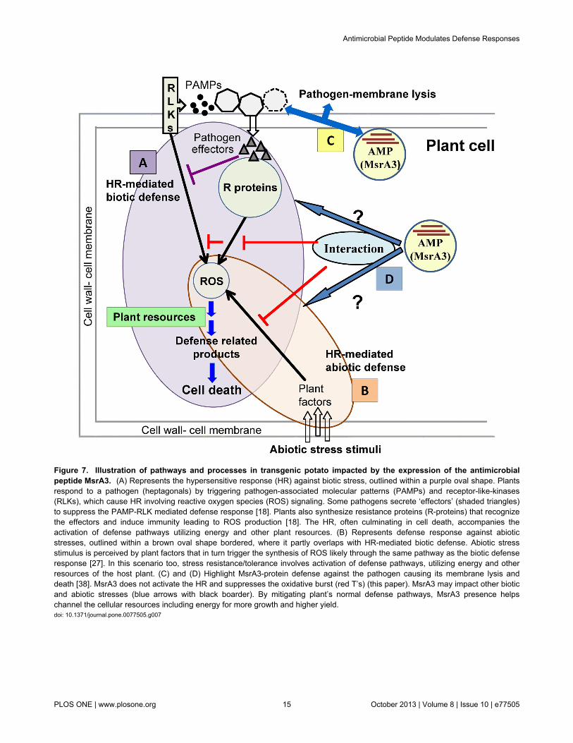
Figure 7. Illustration of pathways and processes in transgenic potato impacted by the expression of the antimicrobialpeptide MsrA3. (A) Represents the hypersensitive response (HR) against biotic stress, outlined within a purple oval shape. Plantsrespond to a pathogen (heptagonals) by triggering pathogen-associated molecular patterns (PAMPs) and receptor-like-kinases(RLKs), which cause HR involving reactive oxygen species (ROS) signaling. Some pathogens secrete ‘effectors’ (shaded triangles)to suppress the PAMP-RLK mediated defense response [18]. Plants also synthesize resistance proteins (R-proteins) that recognizethe effectors and induce immunity leading to ROS production [18]. The HR, often culminating in cell death, accompanies theactivation of defense pathways utilizing energy and other plant resources. (B) Represents defense response against abioticstresses, outlined within a brown oval shape bordered, where it partly overlaps with HR-mediated biotic defense. Abiotic stressstimulus is perceived by plant factors that in turn trigger the synthesis of ROS likely through the same pathway as the biotic defenseresponse [27]. In this scenario too, stress resistance/tolerance involves activation of defense pathways, utilizing energy and otherresources of the host plant. (C) and (D) Highlight MsrA3-protein defense against the pathogen causing its membrane lysis anddeath [38]. MsrA3 does not activate the HR and suppresses the oxidative burst (red T’s) (this paper). MsrA3 may impact other bioticand abiotic stresses (blue arrows with black boarder). By mitigating plant’s normal defense pathways, MsrA3 presence helpschannel the cellular resources including energy for more growth and higher yield.doi: 10.1371/journal.pone.0077505.g007
Antimicrobial Peptide Modulates Defense Responses
PLOS ONE | www.plosone.org 15 October 2013 | Volume 8 | Issue 10 | e77505
Supporting Information
File S1. Supporting Figure and Table.Figure S1. Amino acid sequence alignment of the potatoperoxidase, StPrx2 (GenBank No. AJ401150) with peroxidasesfrom French bean peroxidase1, FBP1 (GenBank No. 149277)and pepper, CaPO2 (GenBank No. DQ489711). Table S1.Primer sequences for gene probes.(PDF)
Acknowledgements
We thank Prof. Robert Fluhr (Weizmann Institute of Science,Israel), Drs. G. Selvaraj (Plant Biotechnology Institute,
Saskatoon, Canada) and W.J. Kay and B. Hawkins (Universityof Victoria, Canada) for their critical comments on themanuscript. We thank Dr. V. Kumar for preparing the tomatoprobes for osm, sag12, rbcL and gs-1 gene and the Centre forForest Biology for use of greenhouse and other facilities.
Author Contributions
Conceived and designed the experiments: RG AKM SM.Performed the experiments: RG. Analyzed the data: RG AKM.Contributed reagents/materials/analysis tools: RH AKM SM.Wrote the manuscript: RG AKM.
References
1. Teng PS, Shane WW, MacKenzie DR (1984) Crop losses due to plantpathogens. Crit Rev Plant Sci 2: 21-47. doi:10.1080/07352688409382187.
2. Oerke E-C (2006) Crop losses to pests. J Agric Sci 144: 31-43. doi:10.1017/S0021859605005708.
3. Spoel SH, Dong X (2012) How do plants achieve immunity? Defensewithout specialized immune cells. Nat Rev Immunol 12: 89-100. doi:10.1038/nri3141. PubMed: 22273771.
4. Atkinson NJ, Urwin PE (2012) The interaction of plant biotic and abioticstresses: from genes to the field. J Exp Bot 63: 3523-3543. doi:10.1093/jxb/ers100. PubMed: 22467407.
5. Graham TL, Sequeira T, Huang TSR (1977) Bacteriallipopolysaccharides as inducers of disease resistance in tobacco. ApplEnviron Microbiol 34: 424–432. PubMed: 21613.
6. Hückelhoven R (2007) Cell wall–associated mechanisms of diseaseresistance and susceptibility. Annu Rev Phytopathol 45: 101–127. doi:10.1146/annurev.phyto.45.062806.094325. PubMed: 17352660.
7. Ellinger D, Naumann M, Falter C, Zwikowics C, Jamrow T et al. (2013)Elevated early callose deposition results in complete penetrationresistance to powdery mildew in Arabidopsis. Plant Physiol 161:1433-1444. doi:10.1104/pp.112.211011. PubMed: 23335625.
8. Ahuja I, Kissen R, Bones AM (2012) Phytoalexins in defense againstpathogens. Trends Plant Sci 17: 73-90. doi:10.1016/j.tplants.2011.11.002. PubMed: 22209038.
9. Ross AF (1961) Systemic acquired resistance induced by localizedvirus infections in plants. Virology 14: 340-358. doi:10.1016/0042-6822(61)90319-1. PubMed: 13743578.
10. Apel K, Hirt H (2004) Reactive oxygen species: metabolism, oxidativestress, and signal transduction. Annu Rev Plant Biol 55: 373-399. doi:10.1146/annurev.arplant.55.031903.141701. PubMed: 15377225.
11. Gadjev I, Stone JM, Gechev TS (2008) Programmed cell death inplants: new insights into redox regulation and the role of hydrogenperoxide. Int Rev Cell Mol Biol 270: 87-144. doi:10.1016/S1937-6448(08)01403-2. PubMed: 19081535.
12. Chanda B, Xia Y, Mandal MK, Yu K, Sekine K-T et al. (2011)Glycerol-3-phosphate is a critical mobile inducer of systemic immunityin plants. Nat Genet, 43: 421–7. doi:10.1038/ng.798. PubMed:21441932.
13. Návarová H, Bernsdorff F, Döring A-C, Zeier J (2012) Pipecolic acid, anendogenous mediator of defense amplification and priming, is a criticalregulator of inducible plant immunity. Plant Cell 24: 5123-5141. doi:10.1105/tpc.112.103564. PubMed: 23221596.
14. Robert-Seilaniantz A, Grant M, Jones JDG (2011) Hormone crosstalk inplant disease and defense: more than just jasmonate-salicylateantagonism. Annu Rev Phytopathol 49: 317–343. doi:10.1146/annurev-phyto-073009-114447. PubMed: 21663438.
15. Zipfel C (2009) Early molecular events in PAMP-triggered immunity.Curr Opin Plant Biol 12: 414–420. doi:10.1016/j.pbi.2009.06.003.PubMed: 19608450.
16. Lukasik E, Takken FL (2009) STANDing strong, resistance proteinsinstigators of plant defence. Curr Opin Plant Biol 12: 427–436. doi:10.1016/j.pbi.2009.03.001. PubMed: 19394891.
17. Song WY, Wang GL, Chen LL, Kim HS, Pi LY et al. (1995) A receptorkinase-like protein encoded by the rice disease resistance gene, Xa21.
Science 270: 1804-1806. doi:10.1126/science.270.5243.1804.PubMed: 8525370.
18. Chisholm ST, Coaker G, Day B, Staskawicz BJ (2006) Host-microbeinteractions: shaping the evolution of the plant immune response. Cell124: 803-814. doi:10.1016/j.cell.2006.02.008. PubMed: 16497589.
19. Luna E, Toby JAB, Roberts MR, Flors V, Ton J (2012) Next-generationsystemic acquired resistance. Plant Physiol 158: 844-853. doi:10.1104/pp.111.187468. PubMed: 22147520.
20. Klingler J, Creasy R, Gao L, Nair RM, Calix AS et al. (2005) Aphidresistance in Medicago truncatula involves antixenosis and phloem-specific, inducible antibiosis, and maps to a single locus flanked byNBS-LRR resistance gene analogs. Plant Physiol 137: 1445–1455. doi:10.1104/pp.104.051243. PubMed: 15778464.
21. Mukhtar MS, Carvunis AR, Dreze M, Epple P, Steinbrenner J et al.(2011) Independently evolved virulence effectors converge onto hubs ina plant immunity system network. Science 333: 596-601. doi:10.1126/science.1203659. PubMed: 21798943.
22. Maekawa T, Kracher B, Vernaldi S, Ver Loren van Themaat E,Schulze-Lefert P (2012) Conservation of NLR-triggered immunityacross plant lineages. Proc Natl Acad Sci U_S_A 49: 20119-20123.PubMed: 23175786.
23. Horvath DM, Stall RE, Jones JB, Pauly MH, Vallad GE et al. (2012)Transgenic resistance confers effective field level control of bacterialspot disease in tomato. PLOS ONE 7: e42036. doi:10.1371/journal.pone.0042036. PubMed: 22870280.
24. Lamb C, Dixon RA (1997) The oxidative burst in plant diseaseresistance. Annu Rev Plant Physiol Plant Mol Biol 48: 251–275. doi:10.1146/annurev.arplant.48.1.251. PubMed: 15012264.
25. Bowler C, Fluhr R (2000) The role of calcium and activated oxygens assignals for controlling cross-tolerance. Trends Plant Sci 5: 241–246.doi:10.1016/S1360-1385(00)01628-9. PubMed: 10838614.
26. Torres MA, Dangl JL (2005) Functions of the respiratory burst oxidasein biotic interactions, abiotic stress and development. Curr Opin PlantBiol 8: 397-403. doi:10.1016/j.pbi.2005.05.014. PubMed: 15939662.
27. Fujita M, Fujita Y, Noutoshi Y, Takahashi F, Narusaka Y et al. (2006)Crosstalk between abiotic and biotic stress responses: a current viewfrom the point of convergence in the stress signaling networks. CurrOpin Plant Biol 9: 436-442. doi:10.1016/j.pbi.2006.05.014. PubMed:16759898.
28. AbuQamar S, Luo H, Laluk K, Mickelbart MV, Mengiste T (2009)Crosstalk between biotic and abiotic stress responses in tomato ismediated by the AIM1 transcription factor. Plant J 58: 347-360. doi:10.1111/j.1365-313X.2008.03783.x. PubMed: 19143995.
29. Cheong YH, Chang H-S, Gupta R, Wang X, Zhu T et al. (2002)Transcriptional profiling reveals novel interactions between wounding,pathogen, abiotic stress, and hormonal responses in Arabidopsis. PlantPhysiol 129: 661-677. doi:10.1104/pp.002857. PubMed: 12068110.
30. Lehrer RI (2004) Primate defensins. Nat Rev Microbiol 2: 727-738. doi:10.1038/nrmicro976. PubMed: 15372083.
31. Lay FT, Anderson MA (2005) Defensins – components of the innateimmune system in plants. Curr Prot. J Pept Sci 6: 85-101.
32. Wang Z, Wang G (2004) APD: the antimicrobial peptide database.Nucleic Acids Res 32: D590-D592. doi:10.1093/nar/gkh025. PubMed:14681488.
Antimicrobial Peptide Modulates Defense Responses
PLOS ONE | www.plosone.org 16 October 2013 | Volume 8 | Issue 10 | e77505
33. Wong JH, Xia L, Ng TB (2007) A review of defensins of diverse origins.Curr Protein Pept Sci 8: 446-459. doi:10.2174/138920307782411446.PubMed: 17979760.
34. Pelegrini PB, del Sarto RP, Silva ON, Franc LZ, Grossi-de-Sa MF(2011) Antibacterial peptides from plants: what they are and how theyprobably work. Biochem Res Intl: 1-9.
35. Robinson MW, Hutchinson AT, Donnelly S (2012) Antimicrobialpeptides: utility players in innate immunity. Front Immunol 3: 325.PubMed: 23112800.
36. Brogden KA (2005) Antimicrobial peptides: pore formers or metabolicinhibitors in bacteria. Nat Rev Microbiol 3: 238-249. doi:10.1038/nrmicro1098. PubMed: 15703760.
37. García-Olmedo F, Rodríguez-Palenzuela P, Molina A, Alamillo JM,López-Solanilla E et al. (2001) Antibiotic activities of peptides,hydrogen peroxide and peroxynitrite in plant defence. FEBS Lett 498:219-222. doi:10.1016/S0014-5793(01)02456-5. PubMed: 11412861.
38. Zasloff M (2002) Antimicrobial peptides of multicellular organisms.Nature 415: 389-394. doi:10.1038/415389a. PubMed: 11807545.
39. Wiesner J, Vilcinskas A (2011) Antimicrobial peptides. The ancient armof the human immune system. Virulence 1: 440-464.
40. Bolton MD (2009) Primary metabolism and plant defense—Fuel for thefire. Mol Plants Microb Intl 22: 487-497. doi:10.1094/MPMI-22-5-0487.
41. Bowdish DM, Davidson DJ, Scott MG, Hancock REW (2005)Immunomodulatory activities of small host defense peptides. AntimicroAgents Chemother 49: 1727-1732. doi:10.1128/AAC.49.5.1727-1732.2005. PubMed: 15855488.
42. Scott MG, Dullaghan E, Mookherjee N, Glavas N, Waldbrook M et al.(2007) An anti-infective peptide that selectively modulates the innateimmune response. Nat Biotechnol 25: 465-472. doi:10.1038/nbt1288.PubMed: 17384586.
43. Zanetti M (2004) Cathelicidins, multifunctional peptides of the innateimmunity. J Leukoc Biol 75: 39-48. PubMed: 12960280.
44. Ciornei CD, Sigurdardóttir T, Schmidtchen A, Bodelsson M (2005)Antimicrobial and chemoattractant activity, lipopolysaccharideneutralization, cytotoxicity, and inhibition by serum of analogs of humancathelicidin LL-37. Antimicrob Agents Chemother 49: 2845–2850. doi:10.1128/AAC.49.7.2845-2850.2005. PubMed: 15980359.
45. Nürnberger T, Brunner F, Kemmerling B, Piater L (2004) Innateimmunity in plants and animals: striking similarities and obviousdifferences. Immunol Rev 198: 249–266. doi:10.1111/j.0105-2896.2004.0119.x. PubMed: 15199967.
46. Coll NS, Epple P, Dangl JL (2011) Programmed cell death in the plantimmune system. Cell Death Differen 18: 1247–1256. doi:10.1038/cdd.2011.37. PubMed: 21475301.
47. Razdan MK, Mattoo AK (2005) Genetic Improvement of SolanaceousCrops. Volume I: Potato. Enfield (NH): Science and Publishing HousePublishers, Inc..
48. Ross H (1986) Potato Breeding – Problems and Perspectives. AdvPlant Breed (suppl 13). Berlin: Verlag Paul Parey. 132 pp.
49. Jansky S (2000) Breeding for disease resistance in potato. In: J Janick,Plant Breeding Reviews. New York: John Wiley and Sons, Inc. 19:69-155
50. Zimnoch-Guzowska E, Lojkowska E, Perombelon M (2005) Resistanceto bacterial pathogens. In: MK RazdanAK Mattoo. GeneticImprovement of Solanaceous Crops. Volume I: Potato. Enfield (NH):Science and Publishing House Publishers, Inc.. pp. 339-395.
51. Jones RW, Simko I (2005) Resistance to late blight and other fungi. In:MK RazdanAK Mattoo. Genetic Improvement of Solanaceous Crops.Volume I: Potato. Enfield (NH): Science and Publishing HousePublishers, Inc.. pp. 397-417.
52. Segura A, Moreno M, Madueño F, Molina A, García-Olmedo F (1999)Snakin-1, a peptide from potato that is active against plant pathogens.Mol Plant Microbe Interact 12: 16-23. doi:10.1094/MPMI.1999.12.1.16.PubMed: 9885189.
53. Berrocal-Lobo M, Segura A, Moreno M, López G, García-Olmedo F etal. (2002) Snakin-2, an antimicrobial peptide from potato whose gene islocally induced by wounding and responds to pathogen infection. PlantPhysiol 128: 951-961. doi:10.1104/pp.010685. PubMed: 11891250.
54. Gao AG, Hakimi SM, Mittanck CA, Wu Y, Woerner BM et al. (2000)Fungal pathogen protection in potato by expression of a plant defensinpeptide. Nat Biotechnol 18: 1307-1310. doi:10.1038/82436. PubMed:11101813.
55. Osusky M, Zhou G, Osuska L, Hancock RE, Kay WW et al. (2000)Transgenic plants expressing cationic peptide chimeras exhibit broad-spectrum resistance to phytopathogens. Nat Biotechnol 18: 1162-1166.doi:10.1038/81145. PubMed: 11062434.
56. Ponti D, Mangoni ML, Mignogna G, Simmaco M, Barra D (2003) Anamphibian antimicrobial peptide variant expressed in Nicotiana
tabacum confers resistance to phytopathogens. Biochem J 371:121-127. PubMed: 12435273.
57. Osusky M, Osuska L, Hancock RE, Kay WW, Misra S (2004)Transgenic potatoes expressing a novel cationic peptide are resistantto late blight and pink rot. Transgenic Res 13: 181-190. doi:10.1023/B:TRAG.0000026076.72779.60. PubMed: 15198205.
58. Osusky M, Osuska L, Kay W, Misra S (2005) Genetic modification ofpotato against microbial diseases: in vitro and in planta activity of adermaseptin B1 derivative, Msra2. Theor Appl Genet 111: 711-722. doi:10.1007/s00122-005-2056-y. PubMed: 15947906.
59. Almasia NI, Bazzini AA, Hopp HE, Vazquez-Rovere C (2008) Over-expression of snakin-1 gene enhances resistance to Rhizoctonia solaniand Erwinia carotovora in transgenic potato plants. Mol Plant Pathol 9:329–338. doi:10.1111/j.1364-3703.2008.00469.x. PubMed: 18705874.
60. Nahirñak V, Almasia NI, Fernandez PV, Hopp HE, Estevez JM et al.(2012) Potato Snakin-1 gene silencing affects cell division, primarymetabolism, and cell wall composition. Plant Physiol 158: 252-263. doi:10.1104/pp.111.186544. PubMed: 22080603.
61. Kumar V, Mills DJ, Anderson JD, Mattoo AK (2004) An alternativeagriculture system is defined by a distinct expression profile of selectgene transcripts and proteins. Proc Natl Acad Sci U_S_A 101: 10535–10540. doi:10.1073/pnas.0403496101. PubMed: 15249656.
62. Thordal-Christenen H, Zhang Z, Wei Y, Collinge DB (1997) Subcellularlocalization of H2O2 accumulation in papillae and hypersensitiveresponse during the barley-powdery mildew interaction. Plant J 11:1187-1194. doi:10.1046/j.1365-313X.1997.11061187.x.
63. Bellincampi D, Dipierro N, Salvi G, Cervone F, Lorenzo G (2000)Extracellular H2O2 induced by oligogalaturonides is not involved in theinhibition of auxin-regulated rolB gene expression in tobacco leafexplants. Plant Physiol 122: 1379-1385. doi:10.1104/pp.122.4.1379.PubMed: 10759534.
64. Knorzer OC, Durner J, Boger P (1996) Alterations in the antioxidativesystem of suspension-cultured soybean cells (Glycine max) induced byoxidative stress. Physiol Plant 97: 388-396. doi:10.1034/j.1399-3054.1996.970225.x.
65. Porra RJ (2002) The chequered history of the development and use ofsimultaneous equations for the accurate determination of chlorophylls aand b. Photosyn Res 73: 149-156. doi:10.1023/A:1020470224740.PubMed: 16245116.
66. Murphy AS, Eisinger WR, Shaff JE, Kochian LV, Taiz L (1999) Earlycopper-induced leakage of K+ from Arabidopsis seedlings is mediatedby ion channel and coupled to citrate efflux. Plant Physiol 121:1375-1382. doi:10.1104/pp.121.4.1375. PubMed: 10594125.
67. Weech M-H, Chapleau M, Pan L, Ide C, Bede JC (2008) Caterpillarsaliva interferes with induced Arabidopsis thaliana defence responsesvia the systemic acquired resistance pathway. J Exp Bot 59:2437-2448. doi:10.1093/jxb/ern108. PubMed: 18487634.
68. Sklensky DE, Davies PJ (2011) Resource partitioning to male andfemale flowers of Spinicia oleracea L. in relation to whole-plantmonocarpic senescence. J Exp Bot 62: 4323-4336. doi:10.1093/jxb/err148. PubMed: 21565983.
69. Weaver LM, Amasino RM (2001) Senescence is induced in individuallydarkened Arabidopsis leaves, but inhibited in whole darkened plants.Plant Physiol 127: 876-886. doi:10.1104/pp.010312. PubMed:11706170.
70. Mehta RA, Fawcett TW, Porath D, Mattoo AK (1992) Oxidative stresscauses rapid membrane translocation and in vivo degradation ofribulose-1,5-bisphosphate carboxylase/oxygenase. J Biol Chem 267:2810-2816. PubMed: 1733975.
71. Vicente R, Morcuende R, Babiano J (2011) Differences in rubisco andchlorophyll content among tissues and growth stages in two tomato(Lycopersicon esculentum Mill.) varieties. Agron Res 9: 501–507.
72. Suzuki Y, Makino A (2013) Translational downregulation of rbcL isoperative in the coordinated expression of rubisco genes in senescentleaves in rice. J Exp Bot, 64: 1145–52. doi:10.1093/jxb/ers398.PubMed: 23349140.
73. Gepstein S, Sabehi G, Carp MJ, Hajouj T, Nesher MF et al. (2003)Large-scale identification of leaf senescence-associated genes. Plant J36: 629-642. doi:10.1046/j.1365-313X.2003.01908.x. PubMed:14617064.
74. Bernards MA, Fleming WD, Llewellyn DB, Priefer R, Yang X et al.(1999) Biochemical characterization of the suberization-associatedanionic peroxidase of potato. Plant Physiol 121: 135–146. doi:10.1104/pp.121.1.135. PubMed: 10482668.
75. Blokhina O, Virolainen E, Fagerstedt KV (2003) Antioxidants, oxidativedamage and oxygen deprivation stress: a review. Ann Bot 91: 179-194.doi:10.1093/aob/mcf118. PubMed: 12509339.
76. Orozco-Cardenas ML, Ryan CA (1999) Hydrogen peroxide isgenerated systemically in plant leaves by wounding and systemin via
Antimicrobial Peptide Modulates Defense Responses
PLOS ONE | www.plosone.org 17 October 2013 | Volume 8 | Issue 10 | e77505
the octadecanoid pathway. Proc Natl Acad Sci U_S_A 96: 6553-6557.doi:10.1073/pnas.96.11.6553. PubMed: 10339626.
77. Volkov RA, Panchuk II, Mullineaux PM, Schöffl F (2006) Heat stress-induced H2O2 is required for effective expression of heat shock genesin Arabidopsis. Plant Mol Biol 61: 733-746. doi:10.1007/s11103-006-0045-4. PubMed: 16897488.
78. Collinge M, Boller T (2001) Differential induction of two potato genes,Stprx2 and StNAC, in response to infection by Phytophthora infestansand to wounding. Plant Mol Biol 46: 521-529. doi:10.1023/A:1010639225091. PubMed: 11516145.
79. Royo J, Vancanneyt G, Pérez AG, Sanz C, Störmann K et al. (1996)Characterization of three potato lipoxygenases with distinct enzymaticactivities and different organ-specific and wound-regulated expressionpatterns. J Biol Chem 271: 21012-21019. doi:10.1074/jbc.271.35.21012. PubMed: 8702864.
80. D’Ippólito S, Martín ML, Salcedo MF, Atencio HM, Casalongué CA etal. (2010) Transcriptome profiling of Fusarium solani f. sp. eumartii –infected potato tubers provides evidence of an inducible defenseresponse. Physiol Mol Plant Pathol 75: 3-12. doi:10.1016/j.pmpp.2010.09.002.
81. Hatsugai N, Kuroyanagi M, Yamada K, Meshi T, Tsuda S et al. (2004)A plant vacuolar protease, VPE, mediates virus-induced hypersensitivecell death. Science 30: 5855-5858. PubMed: 15297671.
82. Martinez C, Montillet JL, Bresson E, Agnel JP, Dai GH et al. (1998)Apoplastic peroxidase generates superoxide anions in cells of cottoncotyledons undergoing the hypersensitive reaction to Xanthomonascampestris pv. malvacearum Race 18. Mol Plant Microbe Interact 11:1038-1047. doi:10.1094/MPMI.1998.11.11.1038.
83. Bindschedler LV, Dewdney J, Blee KA, Stone JM, Asai T et al. (2006)Peroxidase-dependent apoplastic oxidative burst in Arabidopsisrequired for pathogen resistance. Plant J 47: 851-863. doi:10.1111/j.1365-313X.2006.02837.x. PubMed: 16889645.
84. Choi HW, Kim YJ, Lee SC, Hong JK, Hwang BK (2007) Hydrogenperoxide generation by the pepper extracellular peroxidase CaPO2activates local and systemic cell death and defense response tobacterial pathogens. Plant Physiol 145: 890-904. doi:10.1104/pp.107.103325. PubMed: 17905862.
85. Ton J, Pelt JAV, Van Loon LC, Pieterse CMJ (2002) Differentialeffectiveness of salicylate-dependent and jasmonate/ethylene-dependent induced resistance in Arabidopsis. Mol Plant MicrobeInteract 15: 27-34. doi:10.1094/MPMI.2002.15.1.27. PubMed:11858171.
86. Overmyer K, Brosché M, Kangasjärvi J (2003) Reactive oxygenspecies and hormonal control of cell death. Trends Plant Sci 8:335-342. doi:10.1016/S1360-1385(03)00135-3. PubMed: 12878018.
87. Scalschi L, Vicedo B, Camañes G, Fernandez-Crespo E, Lapeña L etal. (2010) Hexanoic acid is a resistance inducer that protects tomatoplants against Pseudomonas syringae by priming the jasmonic acidand salicylic acid pathways. Mol Plant Pathol, 14: 342–55. doi:10.1111/mpp.12010. PubMed: 23279078.
88. Nambeesan S, AbuQamar S, Laluk K, Mattoo AK, Mickelbart MV et al.(2012) Polyamines attenuate ethylene-mediated defense responses toabrogate resistance to Botrytis cinerea in tomato. Plant Physiol 158:1034-1045. doi:10.1104/pp.111.188698. PubMed: 22128140.
89. Beckers GJM, Spoel SH (2006) Fine-tuning plant defence signalling:salicylate versus jasmonate. Plant Biol 8: 1-10. doi:10.1055/s-2005-872705. PubMed: 16435264.
90. Rojo E, León J, Sánchez-Serrano JJ (1999) Cross-talk between woundsignalling pathways determines local versus systemic gene expressionin Arabidopsis thaliana. Plant J 20: 135–142. doi:10.1046/j.1365-313x.1999.00570.x. PubMed: 10571873.
91. Zhang Y, Turner JG (2008) Wound-induced endogenous jasmonatesstunt plant growth by inhibiting mitosis. PLOS ONE 3: e3699. doi:10.1371/journal.pone.0003699. PubMed: 19002244.
92. Mattoo AK, Suttle J (1991) The Plant Hormone Ethylene. Boca Raton,FL: CRC Press Inc..
93. Klessig DF, Durner J, Noad R, Navarre DA, Wendehenne D et al.(2000) Nitric oxide and salicylic acid signaling in plant defense. ProcNatl Acad Sci U_S_A 97: 8849-8855. doi:10.1073/pnas.97.16.8849.PubMed: 10922045.
94. Bruessow F, Gouhier-Darimont C, Buchala A, Metraux J-P, Reymond P(2010) Insect eggs suppress plant defence against chewing herbivores.
Plant J 62: 876–885. doi:10.1111/j.1365-313X.2010.04200.x. PubMed:20230509.
95. Arimura G-I, Ozawa R, Maffei ME (2011) Recent advances in plantearly signaling in response to herbivory. Int J Mol Sci 12: 3723-3739.doi:10.3390/ijms12063723. PubMed: 21747702.
96. Kausch KD, Sobolev AP, Goyal RK, Fatima T, Laila-Beevi R et al.(2012) Methyl jasmonate deficiency alters cellular metabolome,including the aminome of tomato (Solanum lycopersicum L.) fruit.Amino Acids 42: 843-856. doi:10.1007/s00726-011-1000-5. PubMed:21814797.
97. Mengiste T (2012) Plant immunity to necrotrophs. Annu RevPhytopathol 50: 267-294. doi:10.1146/annurev-phyto-081211-172955.PubMed: 22726121.
98. Prusky D, Alkan N, Mengiste T, Fluhr R (2013) Quiescent andnecrotrophic lifestyle choice during postharvest disease development.Annu Rev Phytopathol, 51: 155–76. doi:10.1146/annurev-phyto-082712-102349. PubMed: 23682917.
99. Kunkel BN, Brooks DM (2002) Cross talk between signaling pathwaysin pathogen defense. Curr Opin Plant Biol 5: 325–331. doi:10.1016/S1369-5266(02)00275-3. PubMed: 12179966.
100. Alkan N, Fluhr R, Prusky D (2012) Ammonium secretion duringColletotrichum coccodes infection modulates salicylic and jasmonicacid pathways of ripe and unripe tomato fruit. Mol Plant MicrobeInteract 25: 85-96. doi:10.1094/MPMI-01-11-0020. PubMed: 22150075.
101. Rahman TAE, Oirdi ME, Gonzalez-Lamothe R, Bouarab K (2012)Necrotrophic pathogens use the salicylic acid signaling pathway topromote disease development in tomato. Mol Plant Microbe Interact 25:1584-1593. doi:10.1094/MPMI-07-12-0187-R. PubMed: 22950753.
102. Godoy LC, Andersona CTM, Chowdhury R, Trudel LJ, Wogan GN(2012) Endogenously produced nitric oxide mitigates sensitivity ofmelanoma cells to cisplatin. Proc Natl Acad Sci U_S_A, 109: 20373–8.doi:10.1073/pnas.1218938109. PubMed: 23185001.
103. Fatima T, Teasdale JR, Bunce J, Mattoo AK (2012) Tomato responseto legume cover crop and nitrogen: differing enhancement patterns offruit yield, photosynthesis and gene expression. Funct Plant Biol 39:246-254. doi:10.1071/FP11240.
104. Foyer CH, Noctor G (2005) Oxidant and antioxidant signalling in plants:a re-evaluation of the concept of oxidative stress in a physiologicalcontext. Plant Cell Environ 28: 1056–1071. doi:10.1111/j.1365-3040.2005.01327.x.
105. Gapper C, Dolan L (2006) Control of plant development by reactiveoxygen species. Plant Physiol 141: 341-345. doi:10.1104/pp.106.079079. PubMed: 16760485.
106. Mittler R, Blumwald E (2010) Genetic engineering for modernagriculture: challenges and perspectives. Annu Rev Plant Biol 61:443-462. doi:10.1146/annurev-arplant-042809-112116. PubMed:20192746.
107. Miller G, Suzuki N, Ciftci-Yilmaz Mittler R (2010) Reactive oxygenspecies homeostasis and signalling during drought and salinity stress.Plant Cell Environ 33: 453–467. doi:10.1111/j.1365-3040.2009.02041.x. PubMed: 19712065.
108. Brown JKM (2002) Yield penalties of disease resistance in crops. CurrOpin Plant Biol 5: 1-6. doi:10.1016/S1369-5266(01)00220-5. PubMed:12179968.
109. Scheideler M, Schlaich NL, Fellenberg K, Beissbarth T, Hauser NC etal. (2002) Monitoring the switch from housekeeping to pathogendefense metabolism in Arabidopsis thaliana using cDNA arrays. J BiolChem 277: 10555-10561. doi:10.1074/jbc.M104863200. PubMed:11748215.
110. Ciarmiello LF, Woodrow P, Fuggi A, Pontecorvo G, Carillo P (2011)Plant genes for abiotic stress. In: A Shanker. Abiotic Stress in Plants -Mechanisms and Adaptations. Intech: 283-308. ISBN:978-953-307-394-1.
111. Baldwin IT (2001) An ecological motivated analysis of plant-herbivoreinteractions in native tobacco. Plant Physiol 127: 1449-1458. doi:10.1104/pp.010762. PubMed: 11743088.
112. Tian D, Traw MB, Chen JQ, Kreitman M, Bergelson J (2003) Fitnesscosts of R-gene-mediated resistance in Arabidopsis thaliana. Nature423: 74-77. doi:10.1038/nature01588. PubMed: 12721627.
113. Swarbrick PJ, Schulze-Lefert P, Scholes JD (2006) Metabolicconsequences of susceptibility and resistance (race-specific and broad-spectrum) in barley leaves challenged with powdery mildew. Plant CellEnviron 29: 1061-1076. doi:10.1111/j.1365-3040.2005.01472.x.PubMed: 17080933.
Antimicrobial Peptide Modulates Defense Responses
PLOS ONE | www.plosone.org 18 October 2013 | Volume 8 | Issue 10 | e77505