Embed Size (px)
Citation preview
Nordic Guidelines on von Willebrand disease. Version: April 23, 2008
1
Nordic guidelines for diagnosis and management of von Willebrand disease Guidelines of the Nordic Hemophilia Council
Contents Nordic guidelines for diagnosis and management of von Willebrand disease Guidelines of the Nordic Hemophilia Council ________________________________ 1
Contents __________________________________________________________________ 1 The Nordic working group on von Willebrand disease ____________________________ 2 The Nordic Hemophilia Council ______________________________________________ 2
Nordic Hemophilia Centers _________________________________________________________ 2 Diagnostic guidelines________________________________________________________ 3
Background to von Willebrand factor and von Willebrand disease. __________________________ 3 Diagnosis of VWD and it’s subtypes__________________________________________________ 6 Acquired VWD _________________________________________________________________ 17
Guidelines on treatment and management of VWD _____________________________ 18 Introduction ____________________________________________________________________ 18 Description of hemostatic agents____________________________________________________ 19 Desmopressin __________________________________________________________________ 19 Tranexamic acid ________________________________________________________________ 22 Oral contraceptive pills or progesteron IUD ___________________________________________ 22 VWF concentrates _______________________________________________________________ 23 Management of specified bleeds or invasive procedures__________________________________ 24 Management of outpatients ________________________________________________________ 27 Prophylactic treatment with VWF concentrates ________________________________________ 28 Management of patients with allo-antibodies to vWF ____________________________________ 28 Acquired VWD _________________________________________________________________ 29
References _______________________________________________________________ 30
Nordic Guidelines on von Willebrand disease. Version: April 23, 2008
2
The Nordic working group on von Willebrand disease
Stefan Lethagen, Copenhagen, Denmark (Chairman)
Jørgen Ingerslev, Århus, Denmark
Pål André Holme, Oslo, Norway
Pia Petrini, Stockholm, Sweden
Riitta Lassilla, Helsinki, Finland
Páll Torfi Önundarson, Reykjavik, Iceland
The Nordic Hemophilia Council The Nordic Hemophilia Council (NHC) is a society of physicians from the Nordic Hemophilia Centers. The NHC meets twice a year and forms a base for co-operation between the Nordic centers.
N O R D I C H E M O P H I L I A C E N T E R S
Country Centers
Denmark Copenhagen Århus
Finland Helsinki
Iceland Reykavijk
Norway Oslo
Sweden Gothenburg Malmö Stockholm
Nordic Guidelines on von Willebrand disease. Version: April 23, 2008
3
Diagnostic guidelines
BACKGROU ND TO VO N W I L L E B R A N D F A C T O R A N D V O N W I L L E B R A N D D I S E A S E .
D E F I N I T I O N O F V O N W I L L E B R A N D D I S E A S E A N D T H E V O N W I L L E B R A N D F A C T O R
von Willebrand disease (VWD) is a bleeding disorder caused by deficiency of von Willebrand factor (VWF). VWD is usually inherited, but rare acquired forms exist. Congenital VWD is divided into type 1, characterized by quantitative deficiency of VWF, type 2, by qualitative VWF deficiency, and type 3, by total lack of VWF. Type 2 is further subdivided into subtypes 2A, 2B, 2M and 2N, depending on the type of functional disorder.
The von Willebrand factor (VWF) is a large multimeric protein with two main functions in hemostasis. It is crucial for the flow-dependent bridging of platelets to the subendothelium (adhesion) and to other platelets (aggregation) in the formation of the platelet plug in primary hemostasis. Furthermore it is a carrier protein for coagulation factor VIII, protecting FVIII from degradation in plasma.
V O N W I L L E B R A N D F A C T O R – I N T R O D U C T I O N T O I T S B I O C H E M I S T R Y
Von Willebrand factor (VWF) is a large glycoprotein circulating in plasma where its concentration is close to 10 milligram/L. VWF is synthesized in endothelial cells (EC) and megakaryocytes. The VWF protein is secreted into plasma from the endothelial cells (EC) by a continuous constitutive secretion mechanism, while VWF in platelets is not communicated to plasma and is only released upon platelet activation. Endothelial VWF may also be released acutely from stores in the Weibel Palade bodies when EC’s are exposed to various pertubating stimuli.
The plasma form of VWF is a multimeric protein constructed of between two and 40 dimer subunits of the protomer giving a range of differently sized multimers having molecular weights ranging from 500 kD to 20.000 kD. The mature VWF protomer hosts several well-characterised binding sites. Most importantly, in the context of von Willebrand disease (VWD), one binding site interacts with collagen and a second binding site with glycoprotein Ib of the platelet surface contributing to platelet adhesion at the wound site. This particular function in primary hemostasis is dependent upon the molecular weight of VWF multimers, and subsets of multimers of low molecular weight are regarded too small to provide a sufficient spacer function. In addition, the VWF subunit holds binding sites for factor VIII, a RGD motif recognizing the platelet GpIIb/IIIa in platelet aggregation, and a site that interacts with heparins and heparin like molecules. The binding site for factor VIII exerts a protective non-covalent binding to factor VIII, hereby limiting its random proteolytic breakdown. With our current understanding, VWF
Nordic Guidelines on von Willebrand disease. Version: April 23, 2008
4
multimers are assembled in Golgi of endothelial cells and if not directly exported, retained in the Weibel Palade bodies. It has been suggested that endothelial cells may also store factor VIII.
Following release of VWF from ECs, enzymatic modifications of VWF take place catalyzed by a metalloprotease by the name of ADAMTS13, whereby the largest VWF multimers are somewhat reduced in size and discrete changes occur in the oligomer sub-bands indicating limited proteolysis. The protective effect of VWF towards factor VIII is important for the survival of factor VIII in circulation, since complete lack of VWF as well as mutations in the factor VIII binding sequences of VWF can cause a significant reduction in the plasma level of factor VIII, and values may be as low as 2-10% in individuals having an entirely normal secretion rate of factor VIII.
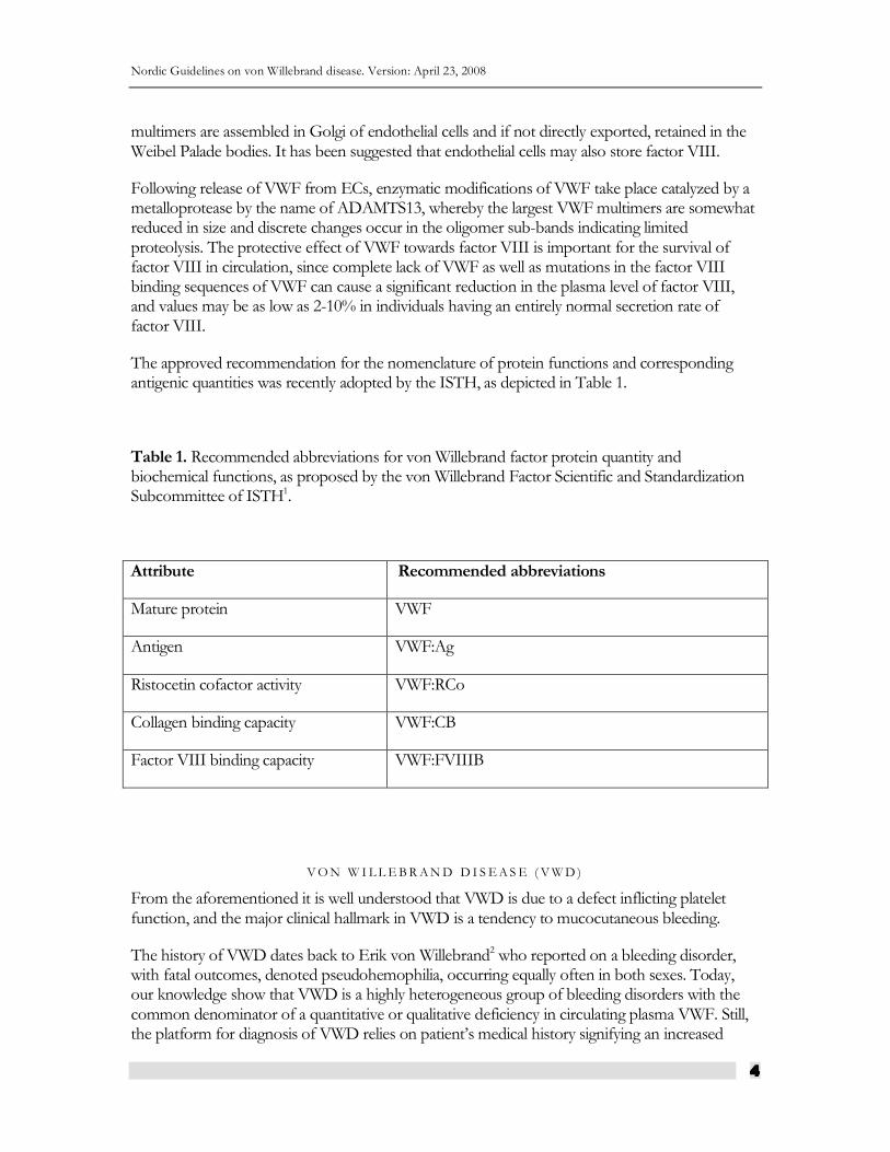
The approved recommendation for the nomenclature of protein functions and corresponding antigenic quantities was recently adopted by the ISTH, as depicted in Table 1.
Table 1. Recommended abbreviations for von Willebrand factor protein quantity and biochemical functions, as proposed by the von Willebrand Factor Scientific and Standardization Subcommittee of ISTH1.
Attribute Recommended abbreviations
Mature protein VWF
Antigen VWF:Ag
Ristocetin cofactor activity VWF:RCo
Collagen binding capacity VWF:CB
Factor VIII binding capacity VWF:FVIIIB
V O N W I L L E B R A N D D I S E A S E ( V W D )
From the aforementioned it is well understood that VWD is due to a defect inflicting platelet function, and the major clinical hallmark in VWD is a tendency to mucocutaneous bleeding.
The history of VWD dates back to Erik von Willebrand2 who reported on a bleeding disorder, with fatal outcomes, denoted pseudohemophilia, occurring equally often in both sexes. Today, our knowledge show that VWD is a highly heterogeneous group of bleeding disorders with the common denominator of a quantitative or qualitative deficiency in circulating plasma VWF. Still, the platform for diagnosis of VWD relies on patient’s medical history signifying an increased
Nordic Guidelines on von Willebrand disease. Version: April 23, 2008
5
bleeding tendency together with phenotypic characteristics indicative of a defect in primary hemostasis.
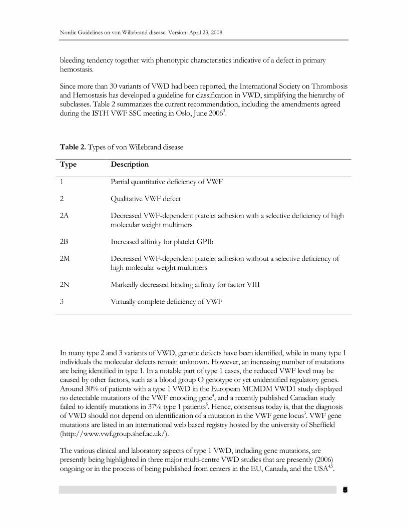
Since more than 30 variants of VWD had been reported, the International Society on Thrombosis and Hemostasis has developed a guideline for classification in VWD, simplifying the hierarchy of subclasses. Table 2 summarizes the current recommendation, including the amendments agreed during the ISTH VWF SSC meeting in Oslo, June 20063.
Table 2. Types of von Willebrand disease
Type Description
1 Partial quantitative deficiency of VWF
2 Qualitative VWF defect
2A Decreased VWF-dependent platelet adhesion with a selective deficiency of high molecular weight multimers
2B Increased affinity for platelet GPIb
2M Decreased VWF-dependent platelet adhesion without a selective deficiency of high molecular weight multimers
2N Markedly decreased binding affinity for factor VIII
3 Virtually complete deficiency of VWF
In many type 2 and 3 variants of VWD, genetic defects have been identified, while in many type 1 individuals the molecular defects remain unknown. However, an increasing number of mutations are being identified in type 1. In a notable part of type 1 cases, the reduced VWF level may be caused by other factors, such as a blood group O genotype or yet unidentified regulatory genes. Around 30% of patients with a type 1 VWD in the European MCMDM VWD1 study displayed no detectable mutations of the VWF encoding gene4, and a recently published Canadian study failed to identify mutations in 37% type 1 patients5. Hence, consensus today is, that the diagnosis of VWD should not depend on identification of a mutation in the VWF gene locus3. VWF gene mutations are listed in an international web based registry hosted by the university of Sheffield (http://www.vwf.group.shef.ac.uk/).
The various clinical and laboratory aspects of type 1 VWD, including gene mutations, are presently being highlighted in three major multi-centre VWD studies that are presently (2006) ongoing or in the process of being published from centers in the EU, Canada, and the USA4,5.
Nordic Guidelines on von Willebrand disease. Version: April 23, 2008
6
D I A G N O S I S O F V W D A N D I T ’ S S U B T Y P E S
The diagnosis of VWD is based on three main criteria: the patient should have significant bleeding symptoms, there should be a family history of VWD or significant bleeding symptoms, except in recessively inherited subtypes, and VWF levels should be significantly decreased. While the detection of VWD is relatively simple, its classification by best standards requires a quite extensive laboratory armamentarium.
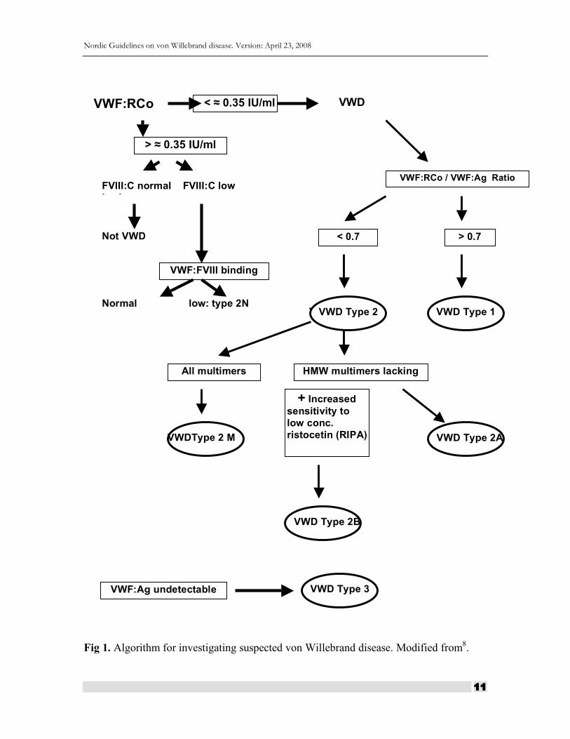
As shown by the algorithm in Fig. 1, the diagnosis of von Willebrand disease is dependent on a significantly reduced ristocetin cofactor (VWF:RCo) level, with the exception of type 2N. The Nordic Hemophilia Council recommends that the diagnosis of VWD should not be applied unless repeated VWF:RCo levels below 0.35 kIU/L are demonstrated. However, the exact level remains a matter of debate, and some individuals will be diagnosed with VWD also with somewhat higher VWF:RCo levels, dependent on symptoms and family history. Recording of the factor VIII:C level is important as a prerequisite to approach a subtype 2N diagnosis. Additionally, Factor VIII plays an important role in assessment of the bleeding risk in VWD. In all subclasses of VWD the level of factor VIII is often somewhat decreased, and particularly low in type 3.
Nordic Guidelines on von Willebrand disease. Version: April 23, 2008
7
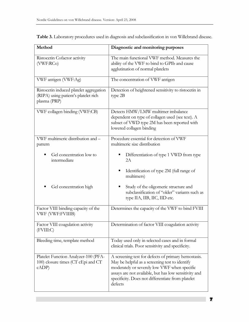
Table 3. Laboratory procedures used in diagnosis and subclassification in von Willebrand disease.
Method Diagnostic and monitoring purposes
Ristocetin Cofactor activity (VWF:RCo)
The main functional VWF method. Measures the ability of the VWF to bind to GPIb and cause agglutination of normal platelets
VWF antigen (VWF:Ag) The concentration of VWF antigen
Ristocetin induced platelet aggregation (RIPA) using patient’s platelet rich plasma (PRP)
Detection of heightened sensitivity to ristocetin in type 2B
VWF collagen binding (VWF:CB) Detects HMW/LMW multimer imbalance dependent on type of collagen used (see text). A subset of VWD type 2M has been reported with lowered collagen binding
VWF multimeric distribution and –pattern
Procedure essential for detection of VWF multimeric size distribution
Gel concentration low to intermediate
Differentiation of type 1 VWD from type 2A
Identification of type 2M (full range of multimers)
Gel concentration high Study of the oligomeric structure and subclassification of “older” variants such as type IIA, IIB, IIC, IID etc.
Factor VIII binding capacity of the VWF (VWF:FVIIIB)
Determines the capacity of the VWF to bind FVIII
Factor VIII coagulation activity (FVIII:C)
Determination of factor VIII coagulation activity
Bleeding time, template method Today used only in selected cases and in formal clinical trials. Poor sensitivity and specificity.
Platelet Function Analyzer-100 (PFA-100) closure times (CT cEpi and CT cADP)
A screening test for defects of primary hemostasis. May be helpful as a screening test to identify moderately or severely low VWF when specific assays are not available, but has low sensitivity and specificity. Does not differentiate from platelet defects
Nordic Guidelines on von Willebrand disease. Version: April 23, 2008
8
C O M M E N T S T O L A B O R A T O R Y M E T H O D S
Ristocetin cofactor activity (VWF:RCo)
This functional method is the key diagnostic test in VWD. Patient´s plasma is mixed with freeze dried (non-activatable) platelets, and platelet agglutination is recorded following addition of ristocetin. Since the ristocetin cofactor result illustrates a biological function, its determination is operational for the diagnosis of most of VWD variants. The method is quite laborious, its performance being highly dependent on the quality of the platelets, and its accuracy and reproducibility varies. Recently, automated kit-methods have become available with improved analytical precision characteristics6.
von Willebrand factor antigen (VWF:Ag)
Various methods are available based on immunometric principles. ELISA methods are used widespread. Automated immunoturbidometric (nefelometry) assays are also available.
Normal amounts of VWF:Ag may be found in patients with VWD caused by qualitative defects in VWF. For example most type 2N (if not compound heterozygous) and some patients with other type 2 variants may present with quite normal quantities of (dysfunctional) antigen.
Ristocetin induced platelet aggregation (RIPA)
This test determines the platelet agglutination as recorded in patient’s platelet rich plasma (PRP) in the presence of ristocetin. This method is relatively insensitive to quantitative deficiencies of VWF, but serves an important diagnostic role since type 2B variants display increased platelet agglutination to low concentrations of ristocetin. Consensus on the use of this test: increased sensitivity to ristocetin at 0.5 mg/ml or lower, indicate the presence of type 2B VWD. Platelet aggregation with other agonists, such as collagen and ADP are usually normal in VWD.
von Willebrand factor collagen binding (VWF:CB)
VWF displays a high binding affinity towards collagen. Methods have been developed that quantify VWF based on its interaction with collagen. These tests are extremely sensitive and generally quite reproducible.
Nordic Guidelines on von Willebrand disease. Version: April 23, 2008
9
The collagen binding ELISA assay is thought to reflect a biological function of VWF, and the conformational state of VWF might be reflected in the assay result when compared to VWF:Ag mass concentration. Comparative studies have revealed, however, that VWF binding to collagen is critically dependent on the type and nature of the collagen utilized in the assay model. With some collagen preparations (i.e. type III collagens), results in type 2A variants are comparable to the VWF:Ag results., whereas in others, conformation of VWF (multimeric size) determines the degree of binding. A decreased VWF:CB/VWF:Ag ratio is thus often found in cases of type 2 VWD with lacking HMW VWF multimers.
Multimeric sizing electrophoresis techniques.
The study of VWF multimer composition is based on electrophoresis of plasma in a gel system well suited for separation of macromolecules. Following separation, VWF molecules are electroeluted onto a nitrocellulose membrane on which patterns of VWF multimeric subsets are identified by means of an immuno-enzymatic or lumographic techniques. Previously radioiodine labelled antibody detection systems were often used. The test is difficult to perform and interpret. The test should be referred to specialized laboratories.
Von Willebrand factor binding to factor VIII (VWF:FVIIIB)
This analysis is indicated if the exploration of hemostasis reveals a low factor VIII level in a patient with a negative family history of hemophilia, where a low factor VIII may result from a decreased carrier effect of VWF. In principle, an assay is performed in which patient’s VWF is bound to an ELISA microtiter plate and incubated with highly purified factor VIII. After extraction, the bound fraction of factor VIII:C is determined by a chromogenic factor VIII assay, or by an antibody to factor VIII.
Global screening tests of primary hemostasis
Global tests include the invasive template bleeding time (BT) and the non-invasive Platelet Function Analyzer-100 (PFA-100) closure times (CT), which screen non-specifically for both VWD and platelet disorders. Both tests are rather insensitive to mild lowering of VWF. The BT has a high false positive rate and is therefore not suitable as a screening test. The PFA-100 has a higher sensitivity for VWD than the BT, but the specificity is low. The PFA-100 may be used for monitoring the response to DDAVP in VWD, whereas it is not always corrected by infusion of a VWF concentrate7.
Nordic Guidelines on von Willebrand disease. Version: April 23, 2008
10
P R A C T I C A L D I A G N O S T I C C O N S I D E R A T I O N S
Pre-analytical considerations
Blood samples should preferably be collected in the coagulation laboratory. The patient should be at rest. Exercise, stress, infections and pregnancy elevate VWF and FVIII levels and may obscure the diagnosis of mild VWD type 1. Care should be taken to remove platelets from plasma by centrifuging. If not tested immediately, plasma samples should be frozen without delay at -70°C. If transportation of plasma samples is needed, samples should be kept frozen. Blood can be drawn without consideration to the menstrual cycle.
Laboratory tests in suspected VWD
Screening tests: Complete blood count with differential and platelet count, prothrombin time (PT) or INR, activated partial thromboplastin time (APTT), VWF:RCo, VWF:Ag, Factor VIII level, ABO blood type.
Defining the subtype of VWD: RIPA, VWF multimers. VWF:FVIIIB in cases with suspected type 2N. Mutational analysis. VWF:CB may add some information in the differentiation between 2A and 2M subtypes.
DDAVP test
All patients should be given a test dose to ensure that the response is sufficient for clinical use. The recommended test dose is 0.3 microgram/kg intravenously or subcutaneously. The intravenous dose can either be given as a slow injection of DDAVP dissolved in 10-15 ml saline over 15 minutes or as an infusion of DDAVP dissolved in 50-100 ml saline and infused over 30 minutes. Blood samples for VWF and FVIII measurements should be taken before and 30-60 minutes after the start of the intravenous dose and 60-120 minutes after the subcutaneous dose. A further blood sample taken after 3-6 hours is advisable to exclude that the patient has a very short half-life of released VWF and/or FVIII following DDAVP stimulation.
DNA samples
It would be advantageous for future genetic studies and family investigations if blood samples for mutation analysis were taken in patients diagnosed with VWD, and stored for possible mutation analysis at a specialized laboratory. Approval for storage of blood samples in a bio-bank must be gathered from the patient and from the appropriate authorities.
Nordic Guidelines on von Willebrand disease. Version: April 23, 2008
11
Fig 1. Algorithm for investigating suspected von Willebrand disease. Modified from8.
FVIII:C normal FVIII:C low lowlow
Not VWD
VWF:FVIII binding binding
VWF:RCo
Normal low: type 2N
VWF:Ag undetectable VWD Type 3
VWD
VWF:RCo / VWF:Ag Ratio
> 0.7
VWD Type 1
< 0.7
VWD Type 2 VWD Type 1
All multimers
> ≈ 0.35 IU/ml
HMW multimers lacking
VWDType 2 M
+ Increased sensitivity to low conc. ristocetin (RIPA)
VWD Type 2A
VWD Type 2B
< ≈ 0.35 IU/ml
Nordic Guidelines on von Willebrand disease. Version: April 23, 2008
12
B L E E D I N G S Y M P T O M S
VWD is characterized by mucocutanous bleeds, e.g. epistaxis and menorrhagia, bleeding after tooth extraction or surgery, and bleeding after minor wounds. Joint bleeds occur in severe cases. Significant mucocutaneous bleeding symptoms are defined as:
Nose bleeding, ≥ 2 episodes without a history of trauma not stopped by short compression of <10 min, or ≥ 1 episode requiring blood transfusion.
Cutaneous hemorrhage and bruisability with minimal or no apparent trauma, as a presenting symptom or requiring medical treatment.
Prolonged bleeding from trivial wounds, lasting ≥15 min or recurring spontaneously during the 7 days after wounding.
Oral cavity bleeding that requires medical attention, such as gingival bleeding, or bleeding with tooth eruption or bites to lips and tongue.
Spontaneous gastrointestinal bleeding requiring medical attention, or resulting in acute or chronic anemia, unexplained by ulceration or portal hypertension.
Heavy, prolonged, or recurrent bleeding after tooth extraction or other oral surgery such as tonsillectomy and adenoidectomy, requiring medical attention.
Menorrhagia resulting in acute or chronic anemia, or requiring medical treatment, not associated with known structural lesions of the uterus, e.g. myomas.
Bleeding from other skin or mucous membrane surfaces requiring medical treatment (e.g. eye, ear, respiratory tract, genitourinary tract other than uterus).
Criteria for mucocutaneous bleeding symptoms
A significant mucocutaneous bleeding history requires at least two symptoms in the absence of a blood transfusion history, or one symptom requiring treatment with blood transfusion, or one symptom recurring on at least three distinct occasions.
Nordic Guidelines on von Willebrand disease. Version: April 23, 2008
13
Bleeding score
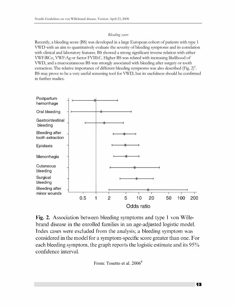
Recently, a bleeding score (BS) was developed in a large European cohort of patients with type 1 VWD with an aim to quantitatively evaluate the severity of bleeding symptoms and its correlation with clinical and laboratory features. BS showed a strong significant inverse relation with either VWF:RCo, VWF:Ag or factor FVIII:C. Higher BS was related with increasing likelihood of VWD, and a mucocutaneous BS was strongly associated with bleeding after surgery or tooth extraction. The relative importance of different bleeding symptoms was also described (Fig. 2)8. BS may prove to be a very useful screening tool for VWD, but its usefulness should be confirmed in further studies.
From: Tosetto et al. 20068
Nordic Guidelines on von Willebrand disease. Version: April 23, 2008
14
C R I T E R I A F O R F A M I L Y H I S T O R Y
A positive family history compatible with VWD (except for types 2N and 3) requires that at least one first-degree relative, or at least two second-degree relatives, have a personal history of significant mucocutaneous bleeding and laboratory tests compatible with VWD. When available, the use of VWF mutations or genetic markers linked to the VWF locus may permit the analysis of more remote relatives, and may allow asymptomatic relatives with low VWF levels to provide evidence for inheritance.
C R I T E R I A F O R V W D T Y P E 1
Laboratory test results are compatible with VWD type 1 if the levels of both VWF:RCo and VWF:Ag are < 0.35 kIU/L on ≥2 determinations. If the tests are performed, RIPA must not indicate abnormal sensitivity to low concentrations of ristocetin, and the plasma VWF multimer distribution must be normal.
Type 1 VWD: VWD type 1 is an hereditary bleeding disorder due to quantitative deficiency of VWF. In most cases type 1 is inherited as an autosomal dominant trait. The diagnosis therefore is based upon criteria for symptoms, VWF deficiency, and inheritance, all of which must be satisfied. These include: significant mucocutaneous bleeding, laboratory tests compatible with VWD type 1, and either a positive family history for VWD type 1 or an appropriate VWF mutation.
Possible type 1: VWD: Possible VWD type 1 includes persons with laboratory tests compatible with VWD type 1 and either significant mucocutaneous bleeding or a positive family history for VWD type 1.
To meet this definition, an asymptomatic person with low VWF must have a positive family history, which means that they must have at least two relatives with definite VWD type 1. Asymptomatic individuals are typically children who have not yet been challenged with trauma or invasive procedures that could cause bleeding.
In many circumstances, symptomatic patients with either VWD type 1 or possible VWD type 1 will be treated identically. Such empiric treatment may also be appropriate for selected asymptomatic patients. The distinction between possible VWD type 1 and VWD type 1 will be useful for certain clinical studies and genetic studies. Alternative diagnoses should continue to be considered for patients with possible VWD.
Special considerations on the diagnosis of type 1 von Willebrand disease
In the investigation of type 1 von Willebrand disease, the bleeding history is particularly important. The hierarchy of bleeding manifestations observed in the European study (MCMDC-VWD1) are presented in Fig. 2, giving the odds ratio of various bleeding symptoms for the risk of a von Willebrand disease based on 154 families studied8.
Nordic Guidelines on von Willebrand disease. Version: April 23, 2008
15
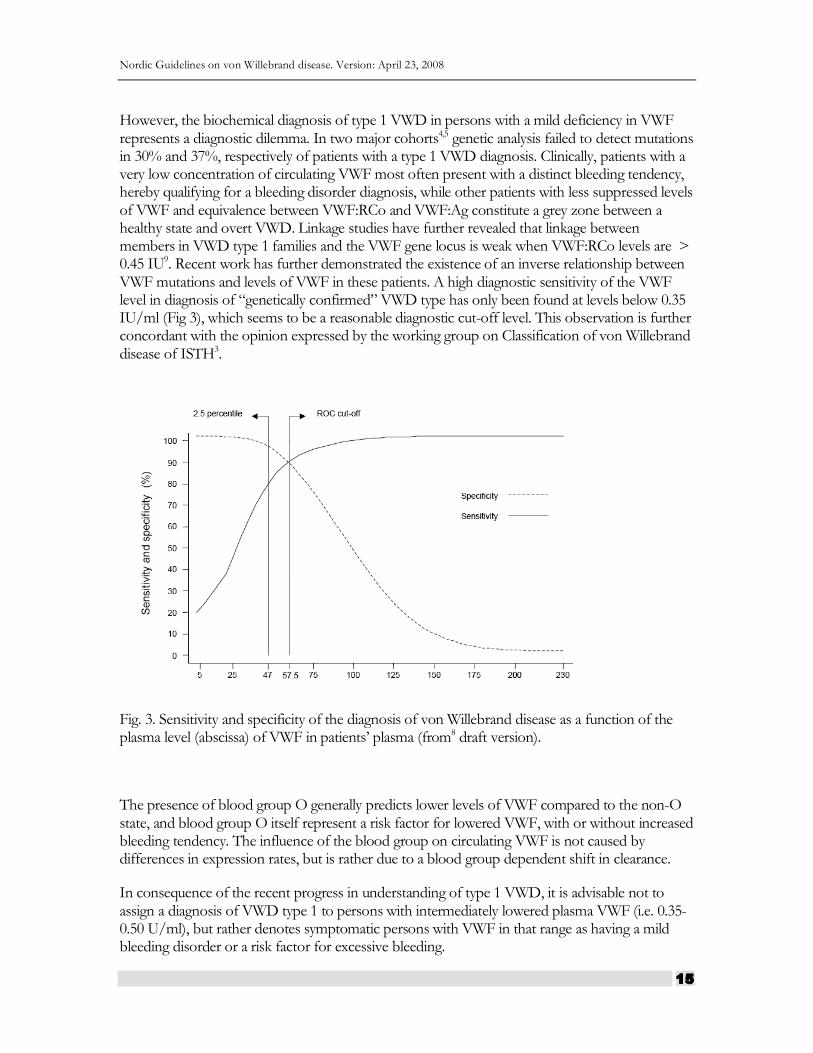
However, the biochemical diagnosis of type 1 VWD in persons with a mild deficiency in VWF represents a diagnostic dilemma. In two major cohorts4,5 genetic analysis failed to detect mutations in 30% and 37%, respectively of patients with a type 1 VWD diagnosis. Clinically, patients with a very low concentration of circulating VWF most often present with a distinct bleeding tendency, hereby qualifying for a bleeding disorder diagnosis, while other patients with less suppressed levels of VWF and equivalence between VWF:RCo and VWF:Ag constitute a grey zone between a healthy state and overt VWD. Linkage studies have further revealed that linkage between members in VWD type 1 families and the VWF gene locus is weak when VWF:RCo levels are > 0.45 IU9. Recent work has further demonstrated the existence of an inverse relationship between VWF mutations and levels of VWF in these patients. A high diagnostic sensitivity of the VWF level in diagnosis of “genetically confirmed” VWD type has only been found at levels below 0.35 IU/ml (Fig 3), which seems to be a reasonable diagnostic cut-off level. This observation is further concordant with the opinion expressed by the working group on Classification of von Willebrand disease of ISTH3.
Fig. 3. Sensitivity and specificity of the diagnosis of von Willebrand disease as a function of the plasma level (abscissa) of VWF in patients’ plasma (from8 draft version).
The presence of blood group O generally predicts lower levels of VWF compared to the non-O state, and blood group O itself represent a risk factor for lowered VWF, with or without increased bleeding tendency. The influence of the blood group on circulating VWF is not caused by differences in expression rates, but is rather due to a blood group dependent shift in clearance.
In consequence of the recent progress in understanding of type 1 VWD, it is advisable not to assign a diagnosis of VWD type 1 to persons with intermediately lowered plasma VWF (i.e. 0.35-0.50 U/ml), but rather denotes symptomatic persons with VWF in that range as having a mild bleeding disorder or a risk factor for excessive bleeding.
Nordic Guidelines on von Willebrand disease. Version: April 23, 2008
16
C R I T E R I A F O R V W D T Y P E 2
VWD type 2 is defined by low levels of functional VWF:RCo and a low VWF:RCo/VWF:Ag ratio (<0.7) (except type 2N). Type 2 is further subdivided into subtypes 2A, 2B, 2M and 2N depending on the type of functional defect (Table 2, Fig 1).
Type 2A and the classical form of type 2B lack the high molecular weight multimers, whereas the rare subtype of 2B called the type Malmö/New York has all multimers. Also type 2M have a full set of multimers, albeit the separate bands may be aberrant. The RIPA test is decreased in type 2A and 2M, but increased in type 2B.
Type 2A is inherited as a dominant trait, but a recessive inheritance has been described10. Two groups of mutations in the A2 domain of the VWF subunit cause the lack of HMWM in type 2A11. Group 1 mutations affect intracellular transport, assembly, storage and secretion of VWF multimers, and group 2 mutations cause increased susceptibility to proteolysis in plasma12.
Also, mutations in the D1 and D2 domain of the propeptide or in the D3 domain of the mature protein may cause a multimerization defect by affecting intramolecular disulfide bonding within the D3 domain13,14.
Type 2B is caused by mutations in the A1 domain, and is characterized by an increased sensitivity to ristocetin in the RIPA test. In the classical form of type 2A, there is a lack of HMWM and a tendency to thrombocytopenia. Thrombocytopenia may be worsened by pregnancy, infections, stress or administration of DDAVP. In the rare Type 2B Malmö/New York15, the multimers are normal and there is no tendency to thrombocytopenia. This variant resembles type 1, apart from the increased RIPA.
Type 2M is in similarity to type 2A characterized by a decreased binding of the VWF to platelets, but in contrast to 2A, all multimers are present. Mutations have been found in the A1 domain of the VWF. The type 2M-Vicenza subtype is characterized by the presence of multimers that are larger than normal. Mutations are in the D3 domain16.
FVIII deficiency is the characteristic feature of type 2N VWD, and ranges from 0.01 – 0.40 kIU/L, but is usually above 0.05 kIU/L17. In some patients there are discrepant levels between different FVIII:C methods (one-stage, two stage or chromogenic methods). The phenotypic diagnosis of type 2N is based on measuring the ability of VWF to bind FVIII (VWF:FVIIIB assay). The FVIII:VWF ratio is typically decreased, but may be only mildly decreased in compound heterozygotes for a type 2N mutation and a mutation causing a quantitative VWF deficiency. A low VWF:FVIIIB/VWF:Ag ratio seems to be the best predictor of type 2N18.
Nordic Guidelines on von Willebrand disease. Version: April 23, 2008
17
C R I T E R I A F O R V W D T Y P E 3
VWD type 3 is inherited as a recessive trait, and is defined by virtual absence of VWF and very low levels of FVIII (about 0.05 kIU/L). It is very rare, with a prevalence of about 3-5 cases per million. Bleeding symptoms are usually moderate to severe, and many type 3 patients require substitution therapy with VWF-containing concentrates. The patients are usually homozygotes or compound heterozygotes for mutations in the VWF gene19. Bleeding symptoms are more prevalent in obligatory carriers than in the normal population, but not as frequent as in patients with type 1 VWD20.
A C Q U I R E D V W D
Acquired VWD (aVWD) is a rare disorder characterized by low levels of VWF and FVIII, but in comparison to inherited VWD, bleeding symptoms appear later in life, and there is no family history. About 50-60% of cases are caused by lympho- or myeloproliferitve disorders. Other causes are solid tumors, immunological disorders, cardiovascular disorders, hypothyreosis, drugs and miscellaneous causes. The pathophysiology is not always clear. In hypothyreosis, synthesis of VWF is decreased. In most other conditions, synthesis seems to be normal, but clearance is increased through four different possible mechanisms, specific autoantibodies, non-specific autoantibodies forming immune complexes, adsorption to malignant cell clones or increased proteolytic degradation21. In contrast to acquired hemophilia, inhibiting antibodies are only rarly demonstrated.
It may be difficult to distinguish aVWD from congenital VWD. The symptoms are similar, ie. mostly mucocutaneous bleeds, but present later in life and there is no family history. Laboratory findings usually includes mildly decreased VWF:Ag and FVIII:C and more pronounced decrease of VWF:RCo and VWF:CB. The VWF:RCo/VWF:Ag ratio is therefore frequently decreased. The large multimers are often lacking due to increased clearance. Thus, these patients often exhibit a type 2A resembling phenotype. Mixing of patient plasma with normal plasma seems to be a relatively insensitive method to identify inhibiting antibodies. An ELISA assay appeared to be more sensitive than functional assays. VWF propeptide levels may be increased in plasma, but the assay is not generally available22. An international registry collects data about this rare disorder23.
Nordic Guidelines on von Willebrand disease. Version: April 23, 2008
18
Guidelines on treatment and management of VWD
I N T R O D U C T I O N
In von Willebrand disease, bleeding tendency is caused by decreased levels of von Willebrand factor (VWF), and sometimes low levels of coagulation factor VIII (FVIII). The deficiencies can be corrected either by stimulation of endogenous VWF and FVIII release with desmopressin (DDAVP), or by substitution with a VWF/FVIII concentrate. DDAVP may temporarily normalize hemostasis if sufficient functional levels of VWF and FVIII can be reached by endogenous release. VWF/FVIII concentrates are used when DDAVP is not an alternative24,25. Plasma derived concentrates carry a potential risk of transmission of infectious agents, even if this risk is thought to be negligible with current safety procedures. Cost is another issue, as these concentrates are expensive. On the other hand, there are also safety issues with DDAVP due to its side effects, mainly caused by the strong antidiuretic effect that may limit its use and also limit the duration of the treatment period. Antifibrinolytic treatment (tranexamic acid) is of importance as an adjuvant to DDAVP or concentrates, or sometimes as a single hemostatic agent especially in connection with mucous membrane bleeds. Oral contraceptive pills may be used as treatment of menorrhagia in females with VWD, as menstrual blood loss may be significantly diminished. Progesterone releasing IUDs are highly effective in some women26.
The choice of treatment depends on several factors:
the nature of the bleed or invasive procedure
subtype and severity of VWD – level of functional VWF and FVIII
previous bleed history and response to treatment
for how long treatment is needed – one dose or a longer period of treatment
outcome of a DDAVP test – post DDAVP level and half-life of functional VWF and FVIII
age of the patient. Restricted use of DDAVP in elderly and in small children – increased risk of fluid overload after DDAVP in children and thrombosis risk in elderly or in patients with strong cardiovascular risk factors.
presence of other diseases that may contraindicate use of a therapeutic agent
Nordic Guidelines on von Willebrand disease. Version: April 23, 2008
19
D E S C R I P T I O N O F H E M O S T A T I C A G E N T S
Desmopressin (DDAVP)
VWF concentrates
Tranexamic acid
Oral contraceptive pills and IUD
D E S M O P R E S S I N
Desmopressin (1-desamino-8-D-arginine vasopressin, DDAVP) is a synthetic analogue of vasopressin, initially used for the treatment of diabetes insipidus. Desmopressin was designed to have a longer duration than the native substance and to have no pressor effects. In the mid-1970s the first reports were published that desmopressin in high dosage stimulated the release of endogenous FVIII, VWF and tissue plasminogen activator (t-PA). The effect is immediate, with on average 2-6-fold increases in plasma concentrations of FVIII, VWF and t-PA. Optimal hemostatic effect is achieved with a dosage of 0.3 microgram/kg given intravenously. A higher dose will not improve the response27. Subcutaneous or intranasal spray administrations are both effective and suitable for home treatment. The response to subcutaneous or intranasal administration is of comparable magnitude, but somewhat slower than that of intravenous administration.
A V A I L A B L E P R O D U C T S I N T H E N O R D I C C O U N T R I E S
Table 4: Desmopressin formulations available in the Nordic Countries
PRODUCT DK FI IS NO SE
Octostim solution for injection (15 microgram/mL)
** ** ** * *
Minirin solution of injection (4 microgram/mL)
* * ** ** *
Octostim nasal spray (1.5 microgram/mL)
* * ** * *
* Licenced, **available
Nordic Guidelines on von Willebrand disease. Version: April 23, 2008
20
D O S E
0.3 microgram/kg i.v. or s.c.
300 microgram i.n. (spray) (150 microgram if BW<30 kg)
M O D E S O F A D M I N I S T R A T I O N
Intravenously (i.v.): slow injection (diluted in saline to 10 ml) over 15 minutes or infusion over 30 minutes of DDAVP diluted in 50-100 ml saline. Peak FVIII/VWF levels are seen after about 60 minutes.
Subcutaneously (s.c.): Peak FVIII/VWF levels are reached after about 120 minutes. The Octostim solution (15 microgram/mL) is most suitable for s.c. administration, being more concentrated.
Intranasal (i.n.) spray: Peak FVIII/VWF levels are reached after about 120 minutes. One spray in each nostril will provide the normal adult dose of 300 microgram. For patients with a body weight <30 kg, a dose of 150 microgram is recommended (one spray in one nostril). In small children (less than about 15 kg) the spray should not be used.
T E S T D O S E ( B I O L O G I C A L R E S P O N S E )
All patients should be given a test dose to ensure that the response is sufficient for clinical use. Blood samples for VWF and FVIII measurements should be taken before and after 30-60 minutes. A further blood sample taken after 3-6 hours is advisable to exclude that the patient has a very short half-life of VWF and/or FVIII after DDAVP.
D O S A G E I N T E R V A L S
Twelve to 24 hours is the ordinary interval, but depending on the half-life of factor levels and the nature of the bleeding, the dose may be given with 8-hour intervals in hospitalized patients. The risk of severe hyponatremia must be considered if repeated doses are given.
T R E A T M E N T W I T H D E S M O P R E S S I N I N R E L A T I O N T O T H E N A T U R E O F T H E B L E E D I N G E P I S O D E
Major bleeds
Response criteria:
DDAVP can be used for treatment of major bleeds in patients in whom VWF:RCo and FVIII:C reach normal levels (i.e. within the normal reference range as defined by the respective center) after DDAVP. The levels should remain at least above about 0.30 kIU/L during 12 hours post
Nordic Guidelines on von Willebrand disease. Version: April 23, 2008
21
DDAVP (Consensus opinion. No data). In connection with life threatening bleeds a VWF/FVIII concentrate should be considered as a first line treatment.
If response is lower or duration shorter, a VWF/FVIII concentrate should be considered.
Minor bleeds
Response criteria:
VWF:RCo and FVIII:C should reach a level of at least 0.30 kIU/L within 2 hours after DDAVP.
Type of invasive procedure
All procedures – VWF and FVIII activities should reach normal levels within the first 2 hours and stay elevated ≥ 0.30 kIU/L for at least 12 hours post DDAVP.
If treatment ≥ 3 days is required, anaphylaxis and antidiuretic effects must be taken into account, and factor levels and s-Na should be monitored.
L I M I T A T I O N S
Subtypes, age of patients, duration of treatment
Patients with classical type 2B VWD in whom DDAVP causes platelet aggregation and thrombocytopenia should not be given DDAVP. Patients with VWD type 3 should not be considered for DDAVP treatment, as they are non-responders.
Half-life of FVIII:C may be short in 2N.
Half-life of VWF:RCo may be short in certain subtypes.
DDAVP should preferably not be given to small children (< 2 years), and adult patients with cardiovascular disease (history of angina, AMI, stroke). If DDAVP is given to small children, fluids must be restricted and electrolytes monitored closely.
Duration of treatment should normally not exceed 3 days. Treatment may be prolonged if factor levels and water balance (s-Na) are monitored. Tachyphylaxis has been reported after several days of treatment with DDAVP.
A D V E R S E E F F E C T S
Tiredness, headache, nausea, temporary lowering of blood pressure with secondary tachycardia, facial flushing, fluid retention, hyponatremia and seizures

Nordic Guidelines on von Willebrand disease. Version: April 23, 2008
22
T R A N E X A M I C A C I D
Tranexamic acid is an antifibrinolytic agent. It interferes with the lysis of newly formed clots by binding to the lysine-binding sites of plasminogen thus inhibiting the binding of plasminogen to fibrin. Administration can be oral, intravenous or topical (e.g. as mouthwash). It can be used alone (e.g. in the management of epistaxis and menorrhagia) or in combination with DDAVP or VWF concentrates. To increase its effectiveness, tranexamic acid should be given prior to elective procedures.
A V A I L A B L E P R O D U C T S I N T H E N O R D I C C O U N T R I E S
Tranexamic acid solution for injection (100 mg/mL) and tablets 500 mg. In Sweden dissolvable tablets (1g) are available.
D O S E A N D M O D E S O F A D M I N I S T R A T I O N
Orally 25 mg per kg BW 3- 4 times daily for 7-10 days.
Intravenously 10 mg per kg BW 3-4 times daily for 7-10 days.
Mouthwash 10 mL of a 5% solution 4 times daily, which can be swallowed.
L I M I T A T I O N S
Contraindicated in the management of upper urinary tract bleeds.
Should be avoided in persons with a history of thromboembolism.
A D V E R S E E F F E C T S
Nausea, vomiting, diarrhea and abdominal pain.
O R A L C O N T R A C E P T I V E P I L L S O R P R O G E S T E R O N I U D
Menstrual blood loss is diminished with oral contraceptive pills and progesterone IUD, in women with VWD, even in type 3 patients. Estrogens increase the plasma level of VWF, except in patients with type 3 VWD. However, the response is variable and unpredictable. The mechanism of action is partly dependent on the increased level of VWF and partly on local effect on the endometrium.
Nordic Guidelines on von Willebrand disease. Version: April 23, 2008
23
V W F C O N C E N T R A T E S
I M P O R T A N T P R O P E R T I E S
Several properties are important when choosing a concentrate for treatment of VWD. The VWF may be more or less functionally inactivated during the manufacturing process, which may be reflected by a low ratio between VWF activity and antigen, or by an abnormal multimeric pattern. The ratio between VWF activity and FVIII:C is important to concider when dosing the concentrate. VWF activity in relation to amount of total protein (specific activity) gives information about the purity. Adequate virus inactivation is a prerequisite28.
The multimeric structure of the VWF
The VWF is a multimeric protein, the largest multimers (HMWM) being most effective for binding platelets in the formation of the platelet plug, i.e. in primary hemostasis.
An aberrant multimeric structure may indicate that the VWF is more or less dysfunctional due to proteolysis during manufacture. The multimeric structure may be visualized with electrophoretic methods, and objectified by densitometry. An indirect method is to calculate the ratio between the VWF activity (most often measured as the VWF ristocetin cofactor activity, VWF:RCo) and the VWF antigen (VWF:Ag) concentration. A low VWF:RCo/VWF:Ag ratio indicates loss of functional activity. Concentrates with a VWF:RCo/VWF:Ag ratio > 0.7 are probably to be preferred. A perfusion chamber technique with flowing blood would be preferable to measure the platelet and collagen binding functions of VWF, but such methods are not generally available, and the methods are not standardized.
The ratio between VWF activity and FVIII:C
Most concentrates used for treatment of VWD contain both VWF and FVIII. A concentrate with a high relative amount of FVIII (a low VWF:RCo/FVIII:C ratio) may cause high plasma levels of FVIII, as the infused FVIII adds to the patient’s endogenously released FVIII. Even if patients with VWD may have very low basal levels of FVIII in plasma, they do have the ability to produce and release FVIII, if VWF becomes available in plasma. Therefore the infused VWF will stimulate synthesis and release of FVIII. When repeated doses are given over a longer time, FVIII levels should be monitored and factor doses adjusted in order to avoid very high FVIII levels during longer periods. Also, the relation between the half-lives of VWF:RCo and FVIII:C should be considered. A concentrate with a short half-life of VWF:RCo in relation to FVIII:C may impose an increased risk of high FVIII levels, if it has to be dosed frequently.
Viral safety
All concentrates used for patients with VWD must be virally inactivated. The possibility of variant Creutzfeldt-Jakob Disease (vCJD) being present in the donor population should be considered. Upcoming recombinant products might be preferred in the future.
Nordic Guidelines on von Willebrand disease. Version: April 23, 2008
24
D O S A G E
Concentrates used for VWD should be dosed according to the VWF:RCo content, which therefore must be labeled on the vials. The recovery of VWF:RCo in adults is roughly 0.015 – 0.020 kIU/L per infused IU VWF:RCo/kg body weight. A dose of 50 IU/kg can be expected to increase the VWF:RCo with about 0.75 – 1.00 kIU/L. Therefore, a loading dose of 50-60 IU VWF:RCo/kg body weight is recommended for patients with very low basal levels of VWF:RCo. Children may have a lower recovery.
In general the half-life of VWF:RCo is considered to be equal to that of FVIII:C. Therefore, VWF concentrate administration usually takes place every 12-24 hours in surgery and similar conditions. VWF concentrate can also be administered as a continuous infusion.
When used for prophylaxis in outpatients, a VWF concentrate administered 2-3 times per week may be sufficient to prevent bleeds.
Levels of VWF:RCo and FVIII:C should be monitored when repeated doses are given over a longer period. Measurement of the VWF:Ag level is not sufficient as the VWF may be dysfunctional in the concentrate29.
M A N A G E M E N T O F S P E C I F I E D B L E E D S O R I N V A S I V E P R O C E D U R E S
B L E E D S F R O M N O S E A N D M O U T H
Bleeds from nose and mouth are relatively common especially in younger patients with VWD. Tranexamic acid given orally (mixture or tablets) or locally is often sufficient to stop theses bleeds. In case of oral bleeds, a mouthwash with tranexamic acid (the i.v. solution) may be effective.
If tranexamic acid is not sufficient to control the bleeds, DDAVP or a VWF-containing concentrate is indicated.
Prolonged or recurrent nasal bleeds may require local treatment, e.g. nasal cautery or laser, and treatment with tranexamic acid over a longer period. Regular local treatment with Vaseline or similar may reduce the bleeding tendency from nosebleeds.
D E N T A L E X T R A C T I O N S
In mild cases, minor dental extractions with local anesthesia may be carried out under cover of tranexamic acid only. But in more severe VWD cases, or in connection with more extensive procedures, and if regional anesthesia, or an inferior dental block is given, DDAVP or a VWF-containing concentrate should be added to tranexamic acid. A single dose of DDAVP or VWF-
Nordic Guidelines on von Willebrand disease. Version: April 23, 2008
25
concentrate is often sufficient. Tranexamic acid should be continued for about 5-7 days. The respective dosage levels are specified above.
M E N O R R H A G I A
Treatment options for menorrhagia in females with VWD include tranexamic acid, DDAVP, VWF concentrates, oral combined contraceptive pills (COC), and intra-uterine progesterone contraceptives. Tranexamic acid reduces menstrual blood loss with about 50%. Tranexamic acid is taken only during the menstrual week, in some cases only during the first days of menstruation. If tranexamic acid and COC is not sufficient, DDAVP or a VWF concentrate may be needed to control the menstrual bleed. DDAVP and concentrates are typically only taken during the menstruation days. DDAVP is usually restricted to maximum three consecutive days because of the risk of fluid retention.
G A S T R O I N T E S T I N A L B L E E D S
Gastrointestinal (GI) bleeds in general may have many different causes, both acute or chronic and benign or malignant. The source of the bleed may be located anywhere in the GI tract. In VWD, the dysfunctional primary hemostasis contributes to the tendency to bleed from a GI locus minoris. Angiodysplasia (AD) leading to GI bleed has been described in VWD since 1967. The prevalence of AD in VWD has been reported to be between 1.1-6.5%. In VWD patients older than 50 years-of-age, AD may even be as prevalent as 10%. AD occur both in patients with congenital and acquired VWD. There seem to be an association between Heyde's syndrome (aortic stenosis plus bleed GI angiodysplasia) and acquired VWD type 2A, with loss of the largest multimers of von Willebrand factor (VWF). An interesting observation is that aortic valve replacement generally cures GI bleed in these patients, and that the deficient multimeric pattern of the VWF is normalized after valve replacement. VWD patients with AD may have recurrent severe GI bleeds requiring hospital admissions, and transfusion with packed red cells, FVIII or VWF concentrates and plasma. Various local invasive procedures have been tested, e.g. surgery, electrocoagulation, laser photocoagulation, sclerotherapy, coiling, and arteriography with embolization. Often, these measures are insufficient to stop recurrence of the GI bleeds. There are some case reports of good response to oestrogens and progesterone, and beta-blocking drugs. In case of recurrent GI bleeds, secondary prophylaxis with a VWF/FVIII concentrate in a dosage of about 50 IU VWF:RCo/kg i.v. 2-3 times per week or even more frequent, in combination with oral tranexamic acid may be needed for shorter or longer periods.
S U R G E R Y A N D O T H E R I N V A S I V E P R O C E D U R E S
Surgery and other invasive procedures should be performed at a specialized hemophilia center with clinicians experienced in VWD, and with a coagulation laboratory that can measure VWF and FVIII activities 24 h per day. Both FVIII and VWF levels must be taken into account, depending on the type of procedure. FVIII activity is an important determinant for surgical and soft tissue bleeds and therapeutic levels should be reached during surgery and for a period of 7-10 days postoperatively. VWF activity is important for mucous membrane bleeds and should be
Nordic Guidelines on von Willebrand disease. Version: April 23, 2008
26
normalized in connection with invasive procedures involving mucous membranes. FVIII:C and VWF activity (VWF:RCo or VWF:CB) should be monitored in connection with all major surgical procedures. During surgery and the first post-operative day, normal levels of FVIII:C and VWF:RCo levels should be reached. Repeated infusions of a VWF/FVIII concentrate may induce high levels of FVIII:C in plasma, due to the additive effect of the endogenously released FVIII. Very high FVIII:C levels (>1.50 kIU/L) over a longer period postoperatively should be avoided, because of the risk of thromboembolic complications.
DDAVP and surgery
DDAVP can be used in responsive patients, but the risk of water retention and tachyphylaxis must be considered if prolonged treatment with repeated doses is given. This may limit the usefulness of DDAVP also in responsive patients. In some cases a combination of DDAVP and a VWF concentrate may be useful. DDAVP may be given intranasally by spray in connection with minor procedures, but in connection with major procedures, it is advisable to give DDAVP parenterally, either i.v. or s.c..
DDAVP intranasal spray in a dose of 300 microgram i.n. (150 microgram if BW <30 kg) should be taken about 60 minutes before the invasive procedure.
Intravenous (i.v.) or subcutaneous (s.c.) DDAVP should be given in a dose of 0.3 microgram/kg about 30 (i.v.) or 60 minutes (s.c.), respectively, before the procedure.
If prolonged DDAVP treatment is needed, the dose may be repeated with 12-24 h intervals. DDAVP should preferably not be given for more than 3 consecutive days because of the risk of fluid retention and hyponatremia. Fluid and electrolyte balance should be monitored, when prolonged treatment is given.
VWF/FVIIII concentrate and surgery
Patients that are unresponsive to DDAVP should be given a VWF containing concentrate approved for use in VWD. In case of major surgery in patients with severe VWD, treatment should be given for at least 1-2 weeks post surgery. As the recovery of VWF:RCo is about 0.015 – 0.020 kIU/L per infused IU VWF:RCo/kg BW, a loading dose of 50-60 IU VWF:RCo/kg i.v. is recommended for patients with very low basal levels of VWF:RCo. The ensuing doses can usually be lower, about 25-40 IU VWF:RCo/kg i.v. every 12-24 hours. After 24-48 hours a once daily dose, or a dose every other day, may be sufficient for the first post-operative week. FVIII:C should be monitored to avoid very high levels over a longer period of time.
Tranexamic acid
Tranexamic acid should be given in addition to DDAVP or VWF concentrate, especially if the procedure involves mucous membranes.
Tranexamic acid is given in a dose of 10 mg/kg i.v. about 30 minutes before surgery or 20-25 mg/kg orally about 2 h before surgery. Thereafter the tranexamic doses are repeated with 6-8 h intervals for at least a week post-operatively.
Nordic Guidelines on von Willebrand disease. Version: April 23, 2008
27
Platelet transfusions
Platelet concentrates may be considered if treatment with VWF concentrate fails to control a bleed in patients with severe VWD.
Thrombosis prophlylaxis
Thrombosis prophylaxis should not be given routinely to patients with VWD undergoing surgery. It may be considered in patients with multiple prothrombotic risk factors, given high doses of a VWF/FVIII concentrate.
P R E G N A N C Y A N D D E L I V E R Y
During pregnancy the hemostatic system shifts in a prothrombotic direction. Plasma levels of VWF and FVIII increase significantly and may even normalize in patients with mild type 1 VWD. In type 3, there is, however no increase of VWF and FVIII. If treatment is needed, tranexamic acid, DDAVP and VWF concentrates may be used during pregnancy. Tranexamic acid is the first treatment option in case of mucocutaneous bleeds.
Generally, vaginal delivery is regarded as safe if VWF:RCo is > 0.40 kIU/L whereas for caesarean section the VWF:RCo level should be > 0.50 kIU/L.
The delivery should preferably take place at an obstetrical unit close to the hemophilia center. The hemophilia center should be involved early in pregnancy and should present a plan for the hemostatic treatment in connection with delivery in cooperation with the obstetrician.
In all cases, treatment with tranexamic acid is commenced when the patient arrives to the maternity ward to give birth, and thereafter continued until two weeks post partum. The dose is either 10 mg/kg i.v., or 20-25 mg/kg orally, every 6-8 hours.
In mild cases (DDAVP responders), DDAVP is given intravenously in a dose of 0.3 microgram/kg immediately after delivery. One dose is usually sufficient in the mildest cases, but DDAVP may be repeated with 8-12 h intervals if needed. In such cases, the risk of water retention must be remembered.
In DDAVP non-responders, treatment with a VWF concentrate is initiated when the delivery starts. A dose of 50-60 IU VWF:RCo/kg i.v. is recommended for patients with very low levels of VWF:RCo. In severe cases, treatment with a VWF concentrate about 3 times weekly, is continued over a period of two weeks post partum.
M A N A G E M E N T O F O U T P A T I E N T S
Patients with VWD should be seen regularly at a hemophilia center with clinicians experienced in VWD and a coagulation laboratory that can measure VWF and FVIII activities. Patients with severe VWD (especially type 3), or those with frequent or severe bleeds should be seen once a
Nordic Guidelines on von Willebrand disease. Version: April 23, 2008
28
year or more often if required. Milder patients may be seen less regularly, e.g. with 2-3 years intervals.
All patients should be given an “identity” or “bleeder’s” card informing about the bleeding disorder, the initial treatment in case of trauma or bleed and contact information to the hemophilia center.
P R O P H Y L A C T I C T R E A T M E N T W I T H V W F C O N C E N T R A T E S
Regular prophylactic treatment with a VWF concentrate may be considered in some patients. Joint bleeds in type 3 VWD patients is a strong indication for further prophylactic treatment. If nose bleeds, menorrhagia or gastrointestinal bleeds are so severe that they cause significant anemia despite iron supplementation, cause a significant impact on social life and/or all other treatment modalities have failed prophylactic treatment should also be considered.
When used for prophylaxis in outpatients, a VWF concentrate in a dose of about 50 IU VWF:RCo/kg i.v. administered 2-3 times per week may be sufficient to prevent bleeds. Levels of VWF:RCo and FVIII:C should be monitored when repeated doses are given over a longer period.
M A N A G E M E N T O F P A T I E N T S W I T H A L L O - A N T I B O D I E S T O V W F
Some type 3 VWD patients develop anti-VWF alloantibodies after multiple transfusions. Exposure to VWF containing products might cause life-threatening postinfusion anaphylaxis besides being ineffective.
• Recombinant FVIII can be given at large doses during surgery. Continuous infusion is mandatory due to the very short half –life of FVIII.
• By-pass therapy with activated prothrombin complex concentrates aPCC (FEIBA) and rFVII (NovoSeven) may also be considered.

Nordic Guidelines on von Willebrand disease. Version: April 23, 2008
29
A C Q U I R E D V W D
Management of acquired VWD (aVWD) involves treatment both of bleeds and of the underlying condition. VWF and FVIII levels can be increased either with desmopressin or a VWF-FVIII containing concentrate, but factor levels can be expected to be short-lived due to increased clearance. Recombinant activated factor VII (rFVIIa) has been effective in some cases that were resistant to desmopresin or VWF-FVIII concentrates. Adminitration of high dose intravenous IgG (IVIG) may prolong the half-life of VWF by interfering with clearance mechanisms. IVIG has been used in connection with treatment of bleeds and for prophylactic treatment during surgery or delivery. Plasma exchange has been successful in some cases with a monoclonal protein. Extracorporeal immunoadsorption has been reported in cases with high titer inhibiting antibodies. Immunosuppressive agents and corticosteroids are effective in some patients with autoimmune disorders or monoclonal gammopathy of undetermined significance (MGUS).
Treatment of the underlying condition may result in improved or normalized VWF levels. Complete restoration has been achieved after tumor resection, chemotherapy, radiotherapy, valve replacement, or thyroxine replacement21,23.
Nordic Guidelines on von Willebrand disease. Version: April 23, 2008
30
References
1. Mazurier C, Rodeghiero F. Recommended abbreviations for von Willebrand Factor and its activities. Thromb Haemost. 2001;86:712. 2. von Willebrand EA. Hereditär pseudohemofili. Finska läkaresällskapets handlingar. 1926;68:87-112. 3. Sadler JE, Budde U, Eikenboom JC, et al. Update on the pathophysiology and classification of von Willebrand disease: a report of the Subcommittee on von Willebrand Factor. J Thromb Haemost. 2006;4:2103-2114. 4. Goodeve A, Eikenboom J, Castaman G, et al. Phenotype and genotype of a cohort of families historically diagnosed with type 1 von Willebrand disease in the European study, Molecular and Clinical Markers for the Diagnosis and Management of Type 1 von Willebrand Disease (MCMDM-1VWD). Blood. 2007;109:112-121. 5. James PD, Notley C, Hegadorn C, et al. The mutational spectrum of type 1 von Willebrand disease: Results from a Canadian cohort study. Blood. 2007;109:145-154. 6. Redaelli R, Corno AR, Borroni L, et al. von Willebrand factor ristocetin cofactor (VWF:RCo) assay: implementation on an automated coagulometer (ACL). J Thromb Haemost. 2005;3:2684-2688. 7. Favaloro EJ, Kershaw G, Bukuya M, Hertzberg M, Koutts J. Laboratory diagnosis of von Willebrand disorder (vWD) and monitoring of DDAVP therapy: efficacy of the PFA-100 and vWF:CBA as combined diagnostic strategies. Haemophilia. 2001;7:180-189. 8. Tosetto A, Rodeghiero F, Castaman G, et al. A quantitative analysis of bleeding symptoms in type 1 von Willebrand disease: results from a multicenter European study (MCMDM-1 VWD). J Thromb Haemost. 2006;4:766-773. 9. Eikenboom J, Van Marion V, Putter H, et al. Linkage analysis in families diagnosed with type 1 von Willebrand disease in the European study, molecular and clinical markers for the diagnosis and management of type 1 VWD. J Thromb Haemost. 2006;4:774-782. 10. Asakura A, Harrison J, Gomperts E, Abildgaard C. Type IIA von Willebrand disease with apparent recessive inheritance. Blood. 1987;69:1419-1420. 11. Meyer D, Fressinaud E, Gaucher C, et al. Gene defects in 150 unrelated French cases with type 2 von Willebrand disease: from the patient to the gene. INSERM Network on Molecular Abnormalities in von Willebrand Disease. Thromb Haemost. 1997;78:451-456. 12. Lyons SE, Bruck ME, Bowie EJ, Ginsburg D. Impaired intracellular transport produced by a subset of type IIA von Willebrand disease mutations. J Biol Chem. 1992;267:4424-4430. 13. Enayat MS, Guilliatt AM, Surdhar GK, et al. Aberrant dimerization of von Willebrand factor as the result of mutations in the carboxy-terminal region: identification
Nordic Guidelines on von Willebrand disease. Version: April 23, 2008
31
of 3 mutations in members of 3 different families with type 2A (phenotype IID) von Willebrand disease. Blood. 2001;98:674-680. 14. Schneppenheim R, Budde U, Ruggeri ZM. A molecular approach to the classification of von Willebrand disease. Best Pract Res Clin Haematol. 2001;14:281-298. 15. Holmberg L, Dent JA, Schneppenheim R, Budde U, Ware J, Ruggeri ZM. von Willebrand factor mutation enhancing interaction with platelets in patients with normal multimeric structure. J Clin Invest. 1993;91:2169-2177. 16. Schneppenheim R, Federici AB, Budde U, et al. Von Willebrand Disease type 2M "Vicenza" in Italian and German patients: identification of the first candidate mutation (G3864A; R1205H) in 8 families. Thromb Haemost. 2000;83:136-140. 17. Mazurier C, Goudemand J, Hilbert L, Caron C, Fressinaud E, Meyer D. Type 2N von Willebrand disease: clinical manifestations, pathophysiology, laboratory diagnosis and molecular biology. Best Pract Res Clin Haematol. 2001;14:337-347. 18. Casonato A, Pontara E, Sartorello F, et al. Identifying carriers of type 2N von Willebrand disease: procedures and significance. Clin Appl Thromb Hemost. 2007;13:194-200. 19. Eikenboom JC. Congenital von Willebrand disease type 3: clinical manifestations, pathophysiology and molecular biology. Best Pract Res Clin Haematol. 2001;14:365-379. 20. Castaman G, Rodeghiero F, Tosetto A, et al. Hemorrhagic symptoms and bleeding risk in obligatory carriers of type 3 von Willebrand disease: an international, multicenter study. J Thromb Haemost. 2006;4:2164-2169. 21. Franchini M, Lippi G. Acquired von Willebrand syndrome: an update. Am J Hematol. 2007;82:368-375. 22. Siaka C, Rugeri L, Caron C, Goudemand J. A new ELISA assay for diagnosis of acquired von Willebrand syndrome. Haemophilia. 2003;9:303-308. 23. Federici AB, Budde U, Rand JH. Acquired von Willebrand syndrome 2004: International Registry--diagnosis and management from online to bedside. Hamostaseologie. 2004;24:50-55. 24. Federici AB. Management of inherited von Willebrand disease in 2006. Semin Thromb Hemost. 2006;32:616-620. 25. Mannucci PM. Treatment of von Willebrand's Disease. N Engl J Med. 2004;351:683-694. 26. Federici AB, Castaman G, Mannucci PM. Guidelines for the diagnosis and management of von Willebrand disease in Italy. Haemophilia. 2002;8:607-621. 27. Lethagen S, Harris AS, Sjorin E, Nilsson IM. Intranasal and intravenous administration of desmopressin: effect on F VIII/vWF, pharmacokinetics and reproducibility. Thromb Haemost. 1987;58:1033-1036. 28. Lethagen S, Carlson M, Hillarp A. A comparative in vitro evaluation of six von Willebrand factor concentrates. Haemophilia. 2004;10:243-249. 29. Lethagen S, Kyrle PA, Castaman G, Haertel S, Mannucci PM. von Willebrand factor/factor VIII concentrate (Haemate P) dosing based on pharmacokinetics: a prospective multicenter trial in elective surgery. J Thromb Haemost. 2007;5:1420-1430.


![[Nordic Built Challenge 17.12.2013] Trine Pertou Mach, Nordic Built: Nordic Built](https://img.pdfslide.us/doc/110x75/547174dcb4af9f980a8b4ad9/nordic-built-challenge-17122013-trine-pertou-mach-nordic-built-nordic-built.jpg)