Embed Size (px)
Citation preview

66
Received:March 23, 2015, Revised:May 12, 2015, Accepted:May 13, 2015
Corresponding to:Minyoung Her, Divisions of Rheumatology, Department of Internal Medicine, Busan Paik Hospital, Inje University College of Medicine, 75 Bokji-ro, Busanjin-gu, Busan 47392, Korea. E-mail : [email protected]
pISSN: 2093-940X, eISSN: 2233-4718Copyright ⓒ 2016 by The Korean College of Rheumatology. All rights reserved.This is a Free Access article, which permits unrestricted non-commerical use, distribution, and reproduction in any medium, provided the original work is properly cited.
Case ReportJournal of Rheumatic Diseases Vol. 23, No. 1, February, 2016http://dx.doi.org/10.4078/jrd.2016.23.1.66
Nontuberculous Mycobacterium Arthritis and Spondylitis in a Patient with Lupus
Nayoung Park1, Sunjoo Lee2, Chisook Moon3, Dongyook Kim1, Heuichul Gwak4, Minyoung Her1
Divisions of 1Rheumatology and 3Infection Disease, Department of Internal Medicine, Departments of 2Radiology and 4Orthopedic Surgery, Busan Paik Hospital, Inje University College of Medicine, Busan, Korea
Approximately 90% of nontuberculous mycobacterium (NTM) infections involve the pulmonary system; NTM infections in-volving areas of the musculoskeletal system such as the joints or spine are uncommon. This report describes a case of refractory knee swelling in a patient with systemic lupus erythematosus (SLE). Indolent arthritis of the knee eventually progressed to spon-dylitis and a paraspinal abscess requiring surgical incision and drainage. The cause of the infectious arthritis and spondylitis was diagnosed as NTM infection, specifically Mycobacterium kansasii. This case emphasizes the importance of a high index of clin-ical suspicion for mycobacterial infection, as well as repeated attempts to isolate the organism, in patients with SLE who present with atypical chronic arthritis. (J Rheum Dis 2016;23:66-70)
Key Words. Nontuberculous mycobacteria, Systemic lupus erythematosus
INTRODUCTION
Arthritis is a common manifestation of systemic lupus erythematosus (SLE) and is one component of the 1982 American College of Rheumatology (ACR) SLE classi-fication criteria. It is not unusual for physicians to see SLE patients with arthritis. SLE-associated arthritis generally presents as intermittent polyarthritis involving the hands, wrists and knees [1]. If pain persists in one joint, especially the knee or hip, avascular necrosis should be suspected in SLE patients. Nontuberculous mycobacterium (NTM) species are typi-
cally environmental and poorly pathogenic for humans; they can, however, be responsible for opportunistic dis-eases in subjects with various predisposing conditions. Approximately 90% of NTM infections involve the pulmo-nary system; the rest involve the lymph nodes, skin, soft tissues, bones and joints, which are also important targets of NTM infection [2,3]. Mycobacterium kansasii, M. mar-inum, and M. avium are common forms of NTM and can in-
volve musculoskeletal tissues [4]. There is a well-known and steadily increasing risk of disseminated infections in immunocompromised patients, such as organ transplant recipients and patients with autoimmune diseases, in-cluding SLE [5-7]. In this paper, an SLE patient who presented with re-
fractory knee arthritis progressed to spondylitis and a paraspinal abscess caused by NTM infection, specifically M. kansasii. This case highlights the importance of a high index of clinical suspicion for mycobacterial infection in patients with atypical chronic arthritis.
CASE REPORT
A 73-year-old woman presented with a 1-month history of left knee pain and swelling; 4 months prior, she was di-agnosed with SLE based on the presence of leukopenia, thrombocytopenia, oral ulcers, pleuritis, high antinuclear antibody titers, and positive anti-Ro antibodies. She was treated with hydroxychloroquine, danazol and inter-
NTM Arthritis and Spondylitis in a SLE Patient
www.jrd.or.kr 67
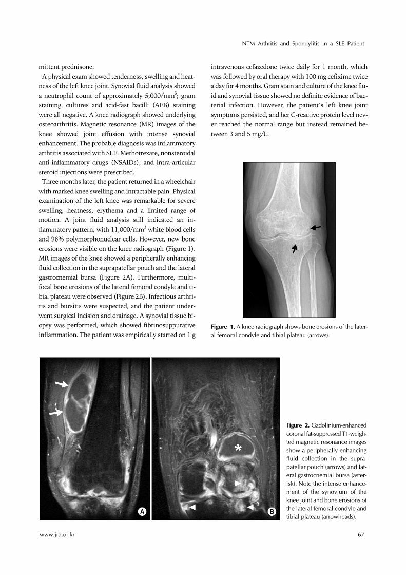
Figure 2. Gadolinium-enhanced coronal fat-suppressed T1-weigh-ted magnetic resonance images show a peripherally enhancing fluid collection in the supra-patellar pouch (arrows) and lat-eral gastrocnemial bursa (aster-isk). Note the intense enhance-ment of the synovium of the knee joint and bone erosions of the lateral femoral condyle and tibial plateau (arrowheads).
Figure 1. A knee radiograph shows bone erosions of the later-al femoral condyle and tibial plateau (arrows).
mittent prednisone. A physical exam showed tenderness, swelling and heat-
ness of the left knee joint. Synovial fluid analysis showed a neutrophil count of approximately 5,000/mm3; gram staining, cultures and acid-fast bacilli (AFB) staining were all negative. A knee radiograph showed underlying osteoarthritis. Magnetic resonance (MR) images of the knee showed joint effusion with intense synovial enhancement. The probable diagnosis was inflammatory arthritis associated with SLE. Methotrexate, nonsteroidal anti-inflammatory drugs (NSAIDs), and intra-articular steroid injections were prescribed. Three months later, the patient returned in a wheelchair
with marked knee swelling and intractable pain. Physical examination of the left knee was remarkable for severe swelling, heatness, erythema and a limited range of motion. A joint fluid analysis still indicated an in-flammatory pattern, with 11,000/mm3 white blood cells and 98% polymorphonuclear cells. However, new bone erosions were visible on the knee radiograph (Figure 1). MR images of the knee showed a peripherally enhancing fluid collection in the suprapatellar pouch and the lateral gastrocnemial bursa (Figure 2A). Furthermore, multi-focal bone erosions of the lateral femoral condyle and ti-bial plateau were observed (Figure 2B). Infectious arthri-tis and bursitis were suspected, and the patient under-went surgical incision and drainage. A synovial tissue bi-opsy was performed, which showed fibrinosuppurative inflammation. The patient was empirically started on 1 g
intravenous cefazedone twice daily for 1 month, which was followed by oral therapy with 100 mg cefixime twice a day for 4 months. Gram stain and culture of the knee flu-id and synovial tissue showed no definite evidence of bac-terial infection. However, the patient’s left knee joint symptoms persisted, and her C-reactive protein level nev-er reached the normal range but instead remained be-tween 3 and 5 mg/L.

Nayoung Park et al.
68 J Rheum Dis Vol. 23, No. 1, February, 2016
Figure 3. (A) A gadolinium-en-hanced sagittal fat-suppressed T1-weighted magnetic resonance(MR) image shows heteroge-neous enhancement of the L4 and L5 vertebral bodies, sug-gesting spondylitis (arrows). (B) A gadolinium-enhanced axial fat-suppressed T1-weighted MRimage shows peripheral en-hancement of a fluid collection in the right psoas muscle (arrow).
While she remained on antibiotics, she gradually devel-oped back pain that was refractory to analgesics. Physical examination revealed tenderness with gentle percussion in the lumbar region, a limited range of motion in the back, and marked swelling of the left popliteal fossa. An ultrasonographic examination of the left knee was per-formed, which showed a large cystic lesion in the left pop-liteal fossa with internal debris. Gram stain, cultures, tu-berculosis polymerase chain reaction (Tb-PCR), and AFB stain of the Baker’s cyst were all negative. MR images of the lumbar spine showed heterogeneous enhancement of the L4 and L5 vertebral bodies (Figure 3A). A small ab-scess (5×12 mm) was observed in the right psoas muscle at the L4-5 level (Figure 3B). Infectious spondylitis with a right psoas abscess was diagnosed, and decompression with surgical incision and drainage of the abscess were performed. Routine cultures with gram staining of the ab-scess showed negative results. AFB positive strain was cultured on egg-based solid media from the abscess and this was presumptively reported as NTM by negative re-sults for Tb-PCR. This strain was finally identified as M. kansasii using GenoType Mycobacterium assay (Hain Life science GmbH, Nehren, Germany). M. kansasii was also detected in the Baker’s cyst fluid using this assay. The final diagnosis was NTM knee arthritis, an infected
Baker’s cyst, spondylitis, and a paravertebral abscess caused by NTM infection, specifically M. kansasii. The hu-man immunodeficiency virus (HIV) antibody was neg-ative, and a chest X-ray was normal. Treatment with an-ti-mycobacterial medications including isoniazid (300 mg/d), rifampin (450 mg/d) and ethambutol (800 mg/d) was initiated, but the patient developed nausea and vomiting. She was therefore treated with clarithromycin (500 mg/d), rifampin (450 mg/d) and ethambutol (800
mg/d) for more than one year, and her symptoms finally improved with these medications.
DISCUSSION
NTM are widely distributed in nature and usually have low pathogenic potential. More than 140 NTM species have been reported in the literature; only 25 species have been strongly associated with atypical mycobacterial dis-eases, and the rest are environmental organisms [7,8]. Approximately 90% of NTM cases involve the pulmonary system, and NTM infections involving the muscu-loskeletal system are uncommon [2]. Musculoskeletal mycobacterium tuberculosis (MTB) infection mostly oc-curs via hematogenous spread from a primary focus, such as a lung, kidney, or lymph node, and rarely via direct in-oculation from adjacent tissues [9]. In contrast, muscu-loskeletal NTM infection is usually acquired through di-rect inoculation from penetrating trauma, injuries, needle injections or surgical procedures. Clinically, osteoarticular NTM infections are indis-
tinguishable from MTB infections. Signs and symptoms of both types of infections include localized pain, joint swelling, stiffness, low grade fever, sweating, chill, fa-tigue, anorexia and weight loss [2]. Shu et al. (2009) re-ported about mycobacterial arthritis of large joints. Relative to patients with MTB large-joint arthritis, pa-tients with NTM large-joint arthritis tend to be younger and have longer symptom duration and more delayed diagnosis. In cases of NTM arthritis, systemic symptoms such as fever are more common, synovial fluid analysis has a higher leukocyte counts, there is a relatively specific histological finding of non-caseating granulomatous in-flammation, and the clinical course is generally more in-
NTM Arthritis and Spondylitis in a SLE Patient
www.jrd.or.kr 69
dolent and less toxic. However these differences in clin-ical characteristics are statistically insignificant [10]. Patients with NTM-related vertebral osteomyelitis gen-
erally have various degrees of immunosuppression, such as acquired immunodeficiency syndrome, SLE, or a his-tory of organ transplantation [2,8]. M. kansasii and the M. avium complex are the most common causes of NTM dis-eases in the United States [3,4]. In cases of atypical monoarthritis in immunocompro-
mised patients, mycobacterial infections should be con-sidered [4-6]. However, due to their rarity, clinical diver-sity, the lack of specific imaging findings, and the diffi-culty in isolating and culturing the infectious organism, heightened clinical suspicion is needed to diagnose cases of NTM arthritis [11-13]. The clinical features are often indistinguishable from those of pyogenic osteomyelitis. M. kansasii infections are generally indolent, and a delay in diagnosis is common [11]. In our case, the delayed diag-nosis of knee arthritis led to spondylitis and a paraspinal abscess that required surgical treatment. Despite accu-mulating data regarding NTM, its diagnosis and treat-ment remain challenging. If mycobacteria are not present in sufficient numbers to be observed under the micro-scope, an AFB smear may be negative despite mycobacte-rial infection [3,12]. Therefore, an AFB culture must be performed in conjunction with an AFB smear to rule out the possibility of mycobacterial infection. There are no established guidelines for the treatment of
NTM infections. Antimicrobial treatment is poorly effec-tive when not combined with surgical treatment [13,14]. Once an NTM infection is diagnosed, a combination of surgical and antimicrobial therapy is often required to completely eradicate the lesions. In addition, the optimal duration of therapy has not been established because an-tibiotics penetrate more poorly into the bones than into the respiratory tract. Therefore, bone NTM infections re-quire a longer duration of antibiotic treatment (usually more than 12 months) than do respiratory NTM in-fections, and medications should not be discontinued un-til symptoms resolve and erythrocyte sedimentation rate (ESR) normalizes [11]. It is important for the physician to recognize NTM in-
fections, in addition to M. tuberculosis, in patients present-ing with musculoskeletal symptoms. Physicians should include NTM as well as M. tuberculosis in the differential diagnosis of organisms that can cause indolent infectious arthritis in patients with atypical chronic arthritis, espe-cially in the setting of immunocompromised conditions.
SUMMARY
NTM infections involving the musculoskeletal system are uncommon but need to be considered in the appro-priate circumstances, or an important diagnosis may be missed. Due to their rarity, as well as the lack of system-atic epidemiologic studies, standard case definitions, ac-curate mycobacterial identification and specific imaging findings, a definitive diagnosis of NTM diseases is often delayed or even impossible. Therefore, a heightened clin-ical suspicion for NTM in patients with arthritis is needed when routine gram stains, cultures or histopathological findings do not identify the organism. In cases of atypical monoarthritis in patients with SLE or other rheumatic diseases with recent-onset inflammation of any part of the musculoskeletal system, mycobacterial infection should be considered as a possible causative agent, espe-cially when the patient is receiving immunosuppressive drugs.
CONFLICT OF INTEREST
No potential conflict of interest relevant to this article was reported.
REFERENCES
1. Petri M, Orbai AM, Alarcón GS, Gordon C, Merrill JT, Fortin PR, et al. Derivation and validation of the Systemic Lupus International Collaborating Clinics classification criteria for systemic lupus erythematosus. Arthritis Rheum 2012;64: 2677-86.
2. Piersimoni C, Scarparo C. Extrapulmonary infections asso-ciated with nontuberculous mycobacteria in immunocom-petent persons. Emerg Infect Dis 2009;15:1351-8.
3. Nakano N, Wada R, Yajima N, Yamamoto N, Wakai Y, Otsuka H. Mycobacterial infection of the musculoskeletal tissues: the use of pathological specimens for identification of causative species by PCR-direct sequencing of 16S rDNA. Jpn J Infect Dis 2010;63:188-91.
4. Yoo JW, Jo KW, Kang BH, Kim MY, Yoo B, Lee CK, et al. Mycobacterial diseases developed during anti-tumour ne-crosis factor-α therapy. Eur Respir J 2014;44:1289-95.
5. Mok MY, Wong SS, Chan TM, Fong DY, Wong WS, Lau CS. Non-tuberculous mycobacterial infection in patients with systemic lupus erythematosus. Rheumatology (Oxford) 2007;46:280-4.
6. Shin J, Kim SM, Kim DJ, Kim YG, Oh HY, Jeon K, et al. A case of lung disease and vertebral osteomyelitis due to non-tuberculous mycobacteria in a kidney transplant recipient. Korean J Med 2013;84:299-302.
7. Rohilla M, Khan K, Raza H. Musculoskeletal manifestations of atypical mycobacterium: case reports. Surg Sci 2014;

Nayoung Park et al.
70 J Rheum Dis Vol. 23, No. 1, February, 2016
5:25-7.8. Petitjean G, Fluckiger U, Schären S, Laifer G. Vertebral os-
teomyelitis caused by non-tuberculous mycobacteria. Clin Microbiol Infect 2004;10:951-3.
9. Tseng CC, Huang RM, Chen KT. Tuberculosis arthritis: epi-demiology, diagnosis, treatment. Clin Res Foot Ankle 2014;2:131.
10. Shu CC, Wang JY, Yu CJ, Lee LN. Mycobacterial arthritis of large joints. Ann Rheum Dis 2009;68:1504-5.
11. Nakamura T, Yamamura Y, Tsuruta T, Tomoda K, Sakagu-chi M, Tsukano M. Mycobacterium kansasii arthritis of the foot in a patient with systemic lupus erythematosus. Intern
Med 2001;40:1045-9.12. Nakanaga K, Sekizuka T, Fukano H, Sakakibara Y, Takeuchi
F, Wada S, et al. Discrimination of Mycobacterium ab-scessus subsp. massiliense from Mycobacterium abscessus subsp. abscessus in clinical isolates by multiplex PCR. J Clin Microbiol 2014;52:251-9.
13. Kang GH, Kim SH, Lee HY, Seo HW, Song EH, Lee KW, et al. A case of arthritis due to mycobacterium intracellulare in a immunocompetent patient. J Rheum Dis 2011;18:122-4.
14. Kim HH, Kim SW, Chang HH, Kim HI, Jeong JY, Jin S, et al. Nine cases of soft tissue infection due to non-tuberculous mycobacterium. Korean J Med 2014;87:311-7.


![Ankylosing spondylitis and related conditions - NHS Wales1].pdf · Condition Ankylosing spondylitis Ankylosing spondylitis and related conditions This booklet provides information](https://img.pdfslide.us/doc/110x75/5d53eb2788c993a4728b841d/ankylosing-spondylitis-and-related-conditions-nhs-1pdf-condition-ankylosing.jpg)