Embed Size (px)
Citation preview
1554
Scapular function is crucial to not only the shoulder butalso the entire upper extremity. As knowledge of the shoul-der and its surrounding structures has increased over thepast decade, so has interest in the scapula. The scapula’srole is 2-fold: it is required to maintain a stable base ofsupport for the humerus; it is also required to be mobile,allowing dynamic positioning of the glenoid fossa duringglenohumeral elevation. These 2 functions require activepositioning and active motion by the periscapular mus-cles.8 Having no true synovial characteristics such asattachment by fibrous, synovial, or cartilaginous tissue,the scapula maintains its function purely through dynamicmuscular control.73 The scapula’s attachment to the axialskeleton is provided through the acromioclavicular jointand the sternoclavicular joints.39 The clavicle acts as astrut for the scapula, opposing medially directed forces ofthe axioscapular muscles, and allowing scapular rotationand translation along the thoracic cage.33 This anatomicalconfiguration allows for smooth gliding motions along thethoracic wall.16,33 Alteration of the normal position or motionof the scapulothoracic joint has been termed dyskinesisand can be caused by pain, muscle weakness, muscleinflexibility, or muscle imbalances. Although not extreme-ly common, scapulothoracic crepitus can occasionally be acontributor to shoulder pain and dysfunction in the activeathlete.
Scapulothoracic crepitus, also known as “snappingscapula” or “washboard syndrome,”11,12,14 is a disorder that
ranges from simple annoyance to a truly disabling condi-tion for the symptomatic patient. This crepitus is usuallydescribed as production of a snapping, grinding, thumping,or popping sound with scapulothoracic motion. This soundis amplified by the thoracic cavity, which acts as a reso-nance chamber as in the body of a stringed instrument.61
Historically identified initially by Boinet,5 scapular crepi-tus has been attributed to numerous causes. Bateman3,4
reported that the lesion might develop because of a chronic,forceful, and repetitive action of the shoulder mechanism.After this action, repetitive microtrauma will then inducemicrotears along the periosteum at the medial border ofthe scapula. The end result of this insult will be a tractionosteophyte or bone spur at the muscular attachment of thescapula. Codman14 felt that scapular crepitus was causedby irritation of several bursae around the scapula thatmay be the result of decreased musculature function orscapular tilting. Milch50 and Milch and Burman51 are inagreement with Codman that scapulothoracic crepitusmay originate from bursitis, but they identified that anosseous lesion, such as an osteochondroma in the scapu-lothoracic space, may become pathologic.
Regardless of the cause, symptomatic scapulothoraciccrepitus requires either nonsurgical or surgical treatment.This article will cover nonsurgical treatment techniquesas well as possible surgical treatments of the snappingscapula syndrome. Because an alteration of scapulotho-racic mechanics can be a common condition associatedwith snapping scapula, a review of anatomy of this regionis paramount.
ANATOMY
The scapula is a large triangular-shaped bone that is situ-ated on the posterior thorax. The pseudojoint between thescapula and the thorax is one of the least congruent of the
Nonoperative and Operative
Management of Snapping Scapula
Robert C. Manske,*†‡ MEd, MPT, SCS, ATC, CSCS, Michael P. Reiman,‡ MEd, PT, ATC, CSCS, and Mark L. Stovak,‡ MDFrom †Wichita State University, Wichita, Kansas, and ‡Via Christi Regional Medical Center, Wichita, Kansas
Snapping scapula is a painful crepitus of the scapulothoracic articulation. This crepitus is a grinding or snapping noise withscapulothoracic motion that may or may not accompany pain. This condition is commonly seen in overhead-throwing athletes.Treatment of patients with this syndrome begins with nonoperative methods; when nonoperative treatment fails, several surgi-cal options exist. This article will discuss both nonoperative and operative management of this common shoulder condition.
Keywords: scapulothoracic crepitus; scapulothoracic bursitis; scapular disorders; shoulder rehabilitation
*Address correspondence to Robert C. Manske, MEd, MPT, SCS, ATC,CSCS,1845 North Fairmount, Wichita, KS 67260-0043 (e-mail:[email protected]).
No potential conflict of interest declared.
The American Journal of Sports Medicine, Vol. 32, No. 6DOI: 10.1177/0363546504268790© 2004 American Orthopaedic Society for Sports Medicine
Clinical Sports Medicine Update
Vol. 32, No. 6, 2004 Management of Snapping Scapula 1555
human body. Because there is no true bony attachmentfrom the scapula to the axial skeleton, its stability isafforded mainly by the surrounding musculature. Thescapula provides a large area for the attachment of 10muscles. The only attachment of the scapula to the rest ofthe proximal skeleton is at the acromioclavicular joint.Interposed between the scapula and the thoracic wall liethe subscapularis and the serratus anterior muscles,which help to stabilize the scapula against the chest walland thus prevent scapular winging.16 The scapular costalsurface is slightly concave, which allows it to come inti-mately close to the convex posterior surface of the thorax.In its normal resting position, the scapula sits approxi-mately 2 inches laterally from the spine on the posteriorthorax. Most will find the scapula located between the sec-ond through seventh ribs or transverse processes.31
Although a large variability exists between people, thescapula generally lies 30° to 40° in the frontal plane and istipped anteriorly approximately 10° to 20° from the verti-cal.40 This static position is commonly referred to as the“plane of the scapula.”14,29,68
CAUSES
Soft tissues such as muscle tendons and bursae are locatedbetween the bony thorax and the scapula. Several bursaethat lie in or around the scapulothoracic joint can poten-tially cause scapular dysfunction and crepitus38 (see Table1). It is in and around these structures that painful scapu-lothoracic crepitus and/or bursitis can occur.
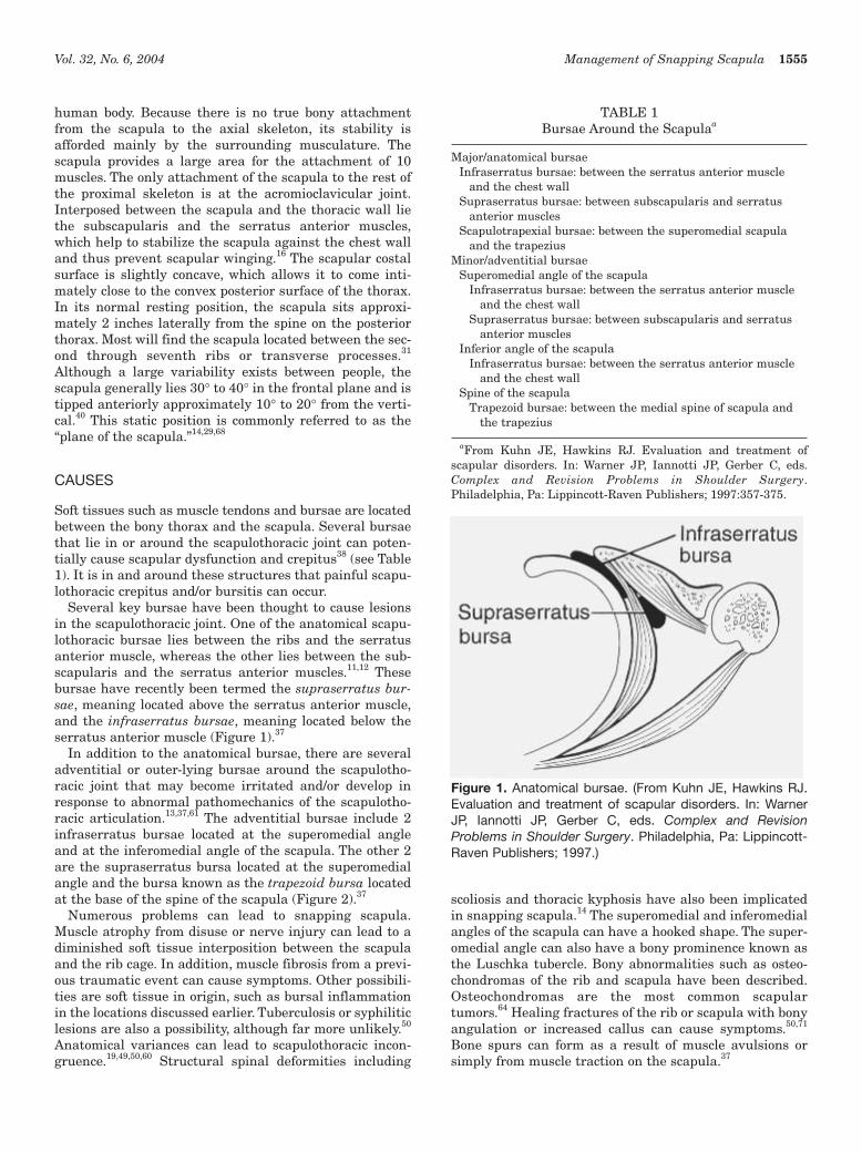
Several key bursae have been thought to cause lesionsin the scapulothoracic joint. One of the anatomical scapu-lothoracic bursae lies between the ribs and the serratusanterior muscle, whereas the other lies between the sub-scapularis and the serratus anterior muscles.11,12 Thesebursae have recently been termed the supraserratus bur-sae, meaning located above the serratus anterior muscle,and the infraserratus bursae, meaning located below theserratus anterior muscle (Figure 1).37
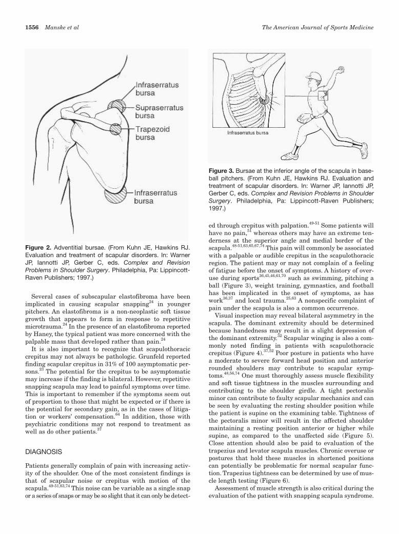
In addition to the anatomical bursae, there are severaladventitial or outer-lying bursae around the scapulotho-racic joint that may become irritated and/or develop inresponse to abnormal pathomechanics of the scapulotho-racic articulation.13,37,61 The adventitial bursae include 2infraserratus bursae located at the superomedial angleand at the inferomedial angle of the scapula. The other 2are the supraserratus bursa located at the superomedialangle and the bursa known as the trapezoid bursa locatedat the base of the spine of the scapula (Figure 2).37
Numerous problems can lead to snapping scapula.Muscle atrophy from disuse or nerve injury can lead to adiminished soft tissue interposition between the scapulaand the rib cage. In addition, muscle fibrosis from a previ-ous traumatic event can cause symptoms. Other possibili-ties are soft tissue in origin, such as bursal inflammationin the locations discussed earlier. Tuberculosis or syphiliticlesions are also a possibility, although far more unlikely.50
Anatomical variances can lead to scapulothoracic incon-gruence.19,49,50,60 Structural spinal deformities including
scoliosis and thoracic kyphosis have also been implicatedin snapping scapula.14 The superomedial and inferomedialangles of the scapula can have a hooked shape. The super-omedial angle can also have a bony prominence known asthe Luschka tubercle. Bony abnormalities such as osteo-chondromas of the rib and scapula have been described.Osteochondromas are the most common scapulartumors.64 Healing fractures of the rib or scapula with bonyangulation or increased callus can cause symptoms.50,71
Bone spurs can form as a result of muscle avulsions orsimply from muscle traction on the scapula.37
TABLE 1Bursae Around the Scapulaa
Major/anatomical bursaeInfraserratus bursae: between the serratus anterior muscle
and the chest wallSupraserratus bursae: between subscapularis and serratus
anterior musclesScapulotrapexial bursae: between the superomedial scapula
and the trapeziusMinor/adventitial bursae
Superomedial angle of the scapulaInfraserratus bursae: between the serratus anterior muscle
and the chest wallSupraserratus bursae: between subscapularis and serratus
anterior musclesInferior angle of the scapula
Infraserratus bursae: between the serratus anterior muscle and the chest wall
Spine of the scapulaTrapezoid bursae: between the medial spine of scapula and
the trapezius
aFrom Kuhn JE, Hawkins RJ. Evaluation and treatment ofscapular disorders. In: Warner JP, Iannotti JP, Gerber C, eds.Complex and Revision Problems in Shoulder Surgery.Philadelphia, Pa: Lippincott-Raven Publishers; 1997:357-375.
Figure 1. Anatomical bursae. (From Kuhn JE, Hawkins RJ.Evaluation and treatment of scapular disorders. In: WarnerJP, Iannotti JP, Gerber C, eds. Complex and RevisionProblems in Shoulder Surgery. Philadelphia, Pa: Lippincott-Raven Publishers; 1997.)
1556 Manske et al The American Journal of Sports Medicine
Several cases of subscapular elastofibroma have beenimplicated in causing scapular snapping24 in youngerpitchers. An elastofibroma is a non-neoplastic soft tissuegrowth that appears to form in response to repetitivemicrotrauma.24 In the presence of an elastofibroma reportedby Haney, the typical patient was more concerned with thepalpable mass that developed rather than pain.24
It is also important to recognize that scapulothoraciccrepitus may not always be pathologic. Grunfeld reportedfinding scapular crepitus in 31% of 100 asymptomatic per-sons.23 The potential for the crepitus to be asymptomaticmay increase if the finding is bilateral. However, repetitivesnapping scapula may lead to painful symptoms over time.This is important to remember if the symptoms seem outof proportion to those that might be expected or if there isthe potential for secondary gain, as in the cases of litiga-tion or workers’ compensation.64 In addition, those withpsychiatric conditions may not respond to treatment aswell as do other patients.37
DIAGNOSIS
Patients generally complain of pain with increasing activ-ity of the shoulder. One of the most consistent findings isthat of scapular noise or crepitus with motion of thescapula.49-51,63,74 This noise can be variable as a single snapor a series of snaps or may be so slight that it can only be detect-
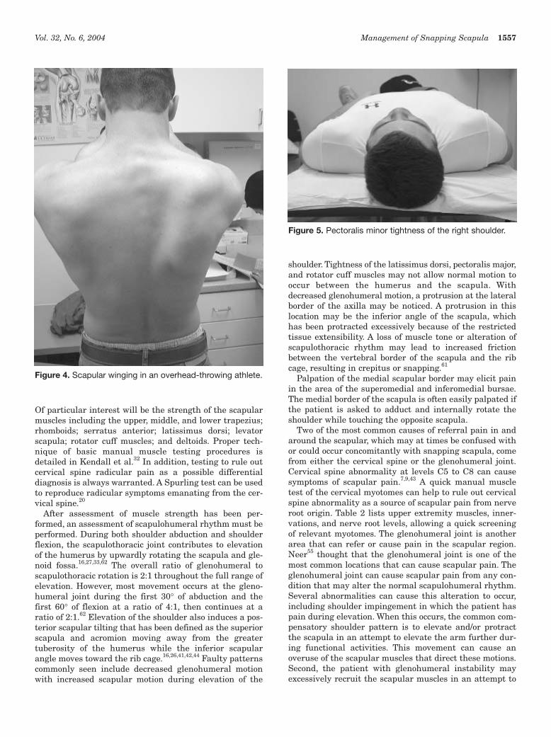
ed through crepitus with palpation.49-51 Some patients willhave no pain,51 whereas others may have an extreme ten-derness at the superior angle and medial border of thescapula.48-51,63,65,67,74 This pain will commonly be associatedwith a palpable or audible crepitus in the scapulothoracicregion. The patient may or may not complain of a feelingof fatigue before the onset of symptoms. A history of over-use during sports36,45,46,61,70 such as swimming, pitching aball (Figure 3), weight training, gymnastics, and footballhas been implicated in the onset of symptoms, as haswork36,37 and local trauma.25,63 A nonspecific complaint ofpain under the scapula is also a common occurrence.
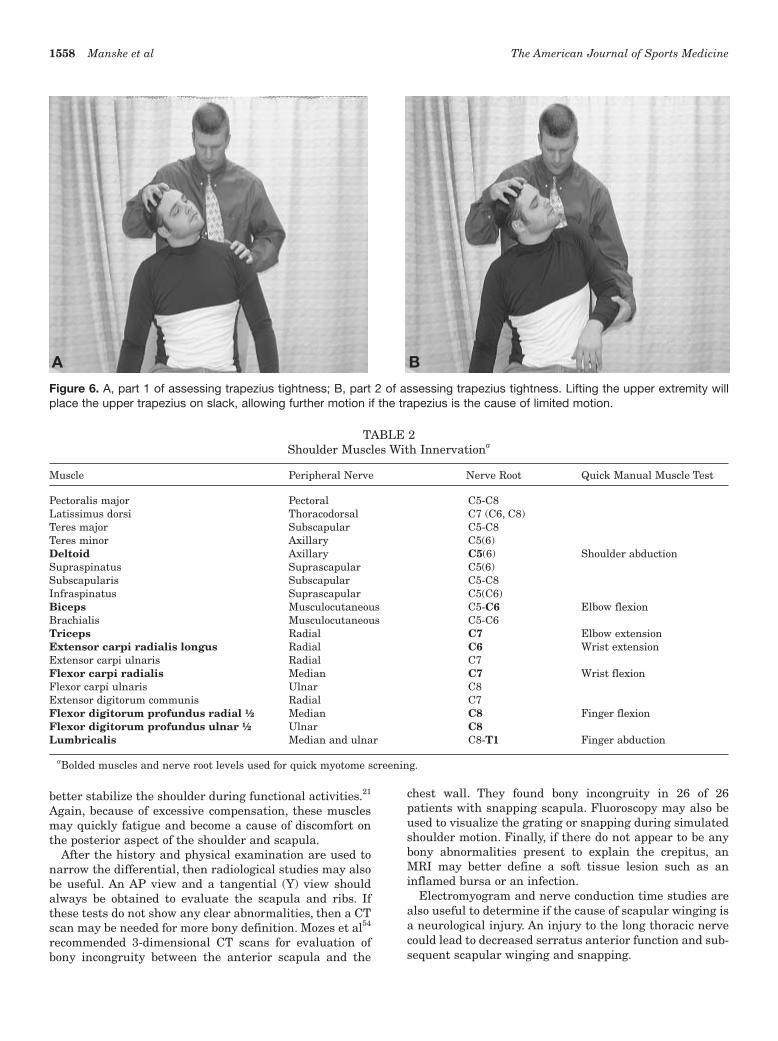
Visual inspection may reveal bilateral asymmetry in thescapula. The dominant extremity should be determinedbecause handedness may result in a slight depression ofthe dominant extremity.32 Scapular winging is also a com-monly noted finding in patients with scapulothoraciccrepitus (Figure 4).37,52 Poor posture in patients who havea moderate to severe forward head position and anteriorrounded shoulders may contribute to scapular symp-toms.48,56,74 One must thoroughly assess muscle flexibilityand soft tissue tightness in the muscles surrounding andcontributing to the shoulder girdle. A tight pectoralisminor can contribute to faulty scapular mechanics and canbe seen by evaluating the resting shoulder position whilethe patient is supine on the examining table. Tightness ofthe pectoralis minor will result in the affected shouldermaintaining a resting position anterior or higher whilesupine, as compared to the unaffected side (Figure 5).Close attention should also be paid to evaluation of thetrapezius and levator scapula muscles. Chronic overuse orpostures that hold these muscles in shortened positionscan potentially be problematic for normal scapular func-tion. Trapezius tightness can be determined by use of mus-cle length testing (Figure 6).
Assessment of muscle strength is also critical during theevaluation of the patient with snapping scapula syndrome.
Figure 2. Adventitial bursae. (From Kuhn JE, Hawkins RJ.Evaluation and treatment of scapular disorders. In: WarnerJP, Iannotti JP, Gerber C, eds. Complex and RevisionProblems in Shoulder Surgery. Philadelphia, Pa: Lippincott-Raven Publishers; 1997.)
Figure 3. Bursae at the inferior angle of the scapula in base-ball pitchers. (From Kuhn JE, Hawkins RJ. Evaluation andtreatment of scapular disorders. In: Warner JP, Iannotti JP,Gerber C, eds. Complex and Revision Problems in ShoulderSurgery. Philadelphia, Pa: Lippincott-Raven Publishers;1997.)
Vol. 32, No. 6, 2004 Management of Snapping Scapula 1557
Of particular interest will be the strength of the scapularmuscles including the upper, middle, and lower trapezius;rhomboids; serratus anterior; latissimus dorsi; levatorscapula; rotator cuff muscles; and deltoids. Proper tech-nique of basic manual muscle testing procedures isdetailed in Kendall et al.32 In addition, testing to rule outcervical spine radicular pain as a possible differentialdiagnosis is always warranted. A Spurling test can be usedto reproduce radicular symptoms emanating from the cer-vical spine.20
After assessment of muscle strength has been per-formed, an assessment of scapulohumeral rhythm must beperformed. During both shoulder abduction and shoulderflexion, the scapulothoracic joint contributes to elevationof the humerus by upwardly rotating the scapula and gle-noid fossa.16,27,33,62 The overall ratio of glenohumeral toscapulothoracic rotation is 2:1 throughout the full range ofelevation. However, most movement occurs at the gleno-humeral joint during the first 30° of abduction and thefirst 60° of flexion at a ratio of 4:1, then continues at aratio of 2:1.62 Elevation of the shoulder also induces a pos-terior scapular tilting that has been defined as the superiorscapula and acromion moving away from the greatertuberosity of the humerus while the inferior scapularangle moves toward the rib cage.16,26,41,42,44 Faulty patternscommonly seen include decreased glenohumeral motionwith increased scapular motion during elevation of the
shoulder. Tightness of the latissimus dorsi, pectoralis major,and rotator cuff muscles may not allow normal motion tooccur between the humerus and the scapula. Withdecreased glenohumeral motion, a protrusion at the lateralborder of the axilla may be noticed. A protrusion in thislocation may be the inferior angle of the scapula, whichhas been protracted excessively because of the restrictedtissue extensibility. A loss of muscle tone or alteration ofscapulothoracic rhythm may lead to increased frictionbetween the vertebral border of the scapula and the ribcage, resulting in crepitus or snapping.61
Palpation of the medial scapular border may elicit painin the area of the superomedial and inferomedial bursae.The medial border of the scapula is often easily palpated ifthe patient is asked to adduct and internally rotate theshoulder while touching the opposite scapula.
Two of the most common causes of referral pain in andaround the scapular, which may at times be confused withor could occur concomitantly with snapping scapula, comefrom either the cervical spine or the glenohumeral joint.Cervical spine abnormality at levels C5 to C8 can causesymptoms of scapular pain.7,9,43 A quick manual muscletest of the cervical myotomes can help to rule out cervicalspine abnormality as a source of scapular pain from nerveroot origin. Table 2 lists upper extremity muscles, inner-vations, and nerve root levels, allowing a quick screeningof relevant myotomes. The glenohumeral joint is anotherarea that can refer or cause pain in the scapular region.Neer55 thought that the glenohumeral joint is one of themost common locations that can cause scapular pain. Theglenohumeral joint can cause scapular pain from any con-dition that may alter the normal scapulohumeral rhythm.Several abnormalities can cause this alteration to occur,including shoulder impingement in which the patient haspain during elevation. When this occurs, the common com-pensatory shoulder pattern is to elevate and/or protractthe scapula in an attempt to elevate the arm further dur-ing functional activities. This movement can cause anoveruse of the scapular muscles that direct these motions.Second, the patient with glenohumeral instability mayexcessively recruit the scapular muscles in an attempt to
Figure 4. Scapular winging in an overhead-throwing athlete.
Figure 5. Pectoralis minor tightness of the right shoulder.
1558 Manske et al The American Journal of Sports Medicine
better stabilize the shoulder during functional activities.21
Again, because of excessive compensation, these musclesmay quickly fatigue and become a cause of discomfort onthe posterior aspect of the shoulder and scapula.
After the history and physical examination are used tonarrow the differential, then radiological studies may alsobe useful. An AP view and a tangential (Y) view shouldalways be obtained to evaluate the scapula and ribs. Ifthese tests do not show any clear abnormalities, then a CTscan may be needed for more bony definition. Mozes et al54
recommended 3-dimensional CT scans for evaluation ofbony incongruity between the anterior scapula and the
chest wall. They found bony incongruity in 26 of 26patients with snapping scapula. Fluoroscopy may also beused to visualize the grating or snapping during simulatedshoulder motion. Finally, if there do not appear to be anybony abnormalities present to explain the crepitus, anMRI may better define a soft tissue lesion such as aninflamed bursa or an infection.
Electromyogram and nerve conduction time studies arealso useful to determine if the cause of scapular winging isa neurological injury. An injury to the long thoracic nervecould lead to decreased serratus anterior function and sub-sequent scapular winging and snapping.
A B
Figure 6. A, part 1 of assessing trapezius tightness; B, part 2 of assessing trapezius tightness. Lifting the upper extremity willplace the upper trapezius on slack, allowing further motion if the trapezius is the cause of limited motion.
TABLE 2Shoulder Muscles With Innervationa
Muscle Peripheral Nerve Nerve Root Quick Manual Muscle Test
Pectoralis major Pectoral C5-C8Latissimus dorsi Thoracodorsal C7 (C6, C8)Teres major Subscapular C5-C8Teres minor Axillary C5(6)Deltoid Axillary C5(6) Shoulder abductionSupraspinatus Suprascapular C5(6)Subscapularis Subscapular C5-C8Infraspinatus Suprascapular C5(C6)Biceps Musculocutaneous C5-C6 Elbow flexionBrachialis Musculocutaneous C5-C6Triceps Radial C7 Elbow extensionExtensor carpi radialis longus Radial C6 Wrist extensionExtensor carpi ulnaris Radial C7Flexor carpi radialis Median C7 Wrist flexionFlexor carpi ulnaris Ulnar C8Extensor digitorum communis Radial C7Flexor digitorum profundus radial ½ Median C8 Finger flexionFlexor digitorum profundus ulnar ½ Ulnar C8Lumbricalis Median and ulnar C8-T1 Finger abduction
aBolded muscles and nerve root levels used for quick myotome screening.
Vol. 32, No. 6, 2004 Management of Snapping Scapula 1559
NONOPERATIVE MANAGEMENT
As with most physical ailments, an attempt at nonsurgicalrehabilitation is recommended with scapular dysfunc-tion.64 Although pain-relieving modalities may help reducepain, correction of postural faults, strengthening of weakmuscles, and stretching of tight muscles may be moreeffective in eliminating the abnormality.§ The direction ofrehabilitation will obviously depend on the factors causingthe snapping scapula. It is generally believed that if theoffending cause of scapulothoracic crepitus is soft tissueabnormalities, altered posture, scapular winging, or scapu-lothoracic dyskinesia, surgical intervention will not berequired.12,45,46
Rehabilitation for the snapping scapula syndromepatient should address not only all the functional roles ofthe scapula43,53,58 but also postural considerations. Themost common postural dysfunction involving the shoulderhas been described in the literature to include such char-acteristics as increased thoracic kyphosis, forward head,rounded shoulders, abducted and forward-tipped scapu-la,34 and suboccipital extension.18,28 Functionally tight-ened muscles include the pectoralis major and minor, lev-ator scapulae, upper trapezius, latissimus dorsi, subscapu-laris, sternocleidomastoid, rectus capitis, and scalene mus-cles. Muscles that could be inhibited or weakened func-tionally would include the rhomboids, mid and lowertrapezius, serratus anterior, teres minor, infraspinatus,posterior deltoid, and longus colli or longus capitis. Themost common of these are the lower stabilizers of thescapula (serratus anterior, middle trapezius, and lowertrapezius).21,28
Scapular weakness and abnormal rhythm have oftenbeen associated with instability and rotator cuff abnor-mality.21,58,69 Additional abnormalities, including bicepstendinitis, thoracic outlet syndrome, and headaches, arealso distinct possibilities.28,40 The rehabilitation programfor the patient with snapping scapula must address all ofthese functional issues.
Restoring scapular strength establishes static proximalstability to provide a stable base.35 Because the scapularmuscles’ primary function is static posturing of the shoul-der girdle (an activity of prolonged duration), endurancetraining should be emphasized. These muscles are not typ-ically required to produce large amounts of force,30 yetthey are required to produce this force over an extendedperiod of time. Scapular muscle fatigue can lead to com-pensatory motion.6,47 Strengthening of these musclesshould also take into account the natural force couples andthe fact that many roles of the scapula are eccentricactions. Emphasis should be on strengthening the inhibitedor functionally weakened muscles in both open and closedchain functional patterns. Gradual progression into func-tional patterns of movement that include, or closely resem-ble, the patient’s required activities would improve thechances of a successful rehabilitation outcome.
The ultimate goal of rehabilitation is for the patient tohave full, functional recovery. Weak muscles cannot beoptimally strengthened if their antagonistic counterpartsare not stretched.28 It is important to stretch the tightenedmuscles that may be inhibiting functional strength; it isalso important to maintain proper posture during thefunctional rehabilitation program. Proper postural align-ment will allow for maximal neuromuscular efficiencybecause the normal length-tension relationship, force cou-ple relationship, and arthrokinematics will be maintainedduring functional movement patterns.18,28,40 Biomechani-cal faults will be minimized, and static proximal stabilityof the shoulder girdle will be improved.
Closed kinetic chain strengthening can be advantageousin the early stages of rehabilitation because of the stabi-lization effects. Proximal joint stability can be promoted ingradually increased ranges of motion. Improved shouldergirdle stability will be promoted with less risk of overload-ing static and dynamic restraints (Figure 7).
Other means of nonsurgical treatment have includedlocal injections, nonsteroidal anti-inflammatory medica-tions, and thermal modalities.11,12,48,50,61 Ciullo11 and Ciulloand Jones12 implemented the use of diathermy, ultra-sound, and iontophoresis to the undersurface at the medialborder of the scapula, along with periscapular and rotatorcuff muscle strengthening.11,12 Thoracic spinal mobiliza-tion to promote correct posture and spinal movementshould also be a point of emphasis in the treatment plan.
Avoiding prolonged, excessive scapular protraction andelevation will limit functional scapular weakness andimprove the biomechanical advantage of the shoulder gir-dle to perform its typical endurance activities. Abductionand elevation of the scapula cause increased pressure onthe underlying soft tissues and strain on the attachedmuscles and tendons.61 Alteration of scapulothoracicrhythm and loss of muscle tone may lead to increased fric-tion between the vertebral border of the scapula and ribcage, resulting in crepitus or snapping.3,61
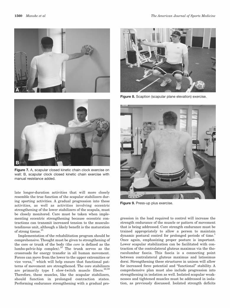
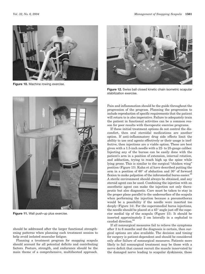
Progression along the rehabilitation continuum fromisometric and isotonic scapular and rotator cuff strength-ening to endurance eccentric strengthening of the scapularmuscles is recommended. Scapular core exercises as iden-tified by Moseley et al53 should be emphasized early on.These include scaption (Figure 8), press-up (Figure 9),rowing (Figure 10), and push-up plus (Figure 11) exercises.Advanced scapular exercises can then be progressed tofunctional patterns emphasizing eccentric scapular con-trol. Exercises of this nature include plyometric exercisessuch as Plyoback (AliMed Inc, Dedham, Mass), propriocep-tive neuromuscular facilitation (PNF) D2 patterns mim-icking triplanar upper extremity movements, and Swissball isometric-hold scapular stabilization exercises (Figure12). Endurance strengthening, by definition, consists oflow intensity (power output) with high volume (amount oftraining time from high repetitions of activity).2 Becausethese muscles are more postural in nature, higher repeti-tions with lighter loads may be more appropriate for amore functional training stimulus.30 Performing repeti-tions in the range of 12 to 20 with lighter loads will simu-§References 11, 12, 25, 37, 45, 46, 61, 70, 74.
1560 Manske et al The American Journal of Sports Medicine
late longer-duration activities that will more closelyresemble the true function of the scapular stabilizers dur-ing sporting activities. A gradual progression into theseactivities, as well as activities involving eccentricstrengthening of the lower stabilizers of the scapula, mustbe closely monitored. Care must be taken when imple-menting eccentric strengthening because eccentric con-tractions can transmit increased tension to the musculo-tendinous unit, although a likely benefit is the maturationof strong tissue.15
Implementation of the rehabilitation program should becomprehensive. Thought must be given to strengthening ofthe core or trunk of the body (the core is defined as thelumbo-pelvic-hip complex).18 The trunk serves as thecrossroads for energy transfer in all human movement.Forces can move from the lower to the upper extremities orvice versa,17 which will help ensure that functional pat-terns of movement are strengthened. The core stabilizersare primarily type I slow-twitch muscle fibers.22,59
Therefore, these muscles, like the scapular stabilizers,should function in prolonged contraction states.Performing endurance strengthening with a gradual pro-
gression in the load required to control will increase thestrength endurance of the muscle or pattern of movementthat is being addressed. Core strength endurance must betrained appropriately to allow a person to maintaindynamic postural control for prolonged periods of time.1
Once again, emphasizing proper posture is important.Lower scapular stabilization can be facilitated with con-traction of the contralateral gluteus maximus via the tho-racolumbar fascia. This fascia is a connecting pointbetween contralateral gluteus maximus and latissimusdorsi. Strengthening these structures in unison will allowfor increased force potential and “functional” stability. Acomprehensive plan must also include progression intostrengthening in isolation as well. Isolated scapular weak-nesses and tightened muscles must be addressed in isola-tion, as previously discussed. Isolated strength deficits
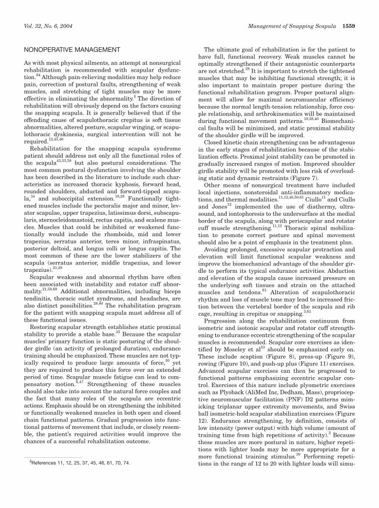
Figure 7. A, scapular closed kinetic chain clock exercise onwall; B, scapular clock closed kinetic chain exercise withmanual resistance added.
Figure 8. Scaption (scapular plane elevation) exercise.
Figure 9. Press-up plus exercise.
A
B
Vol. 32, No. 6, 2004 Management of Snapping Scapula 1561
should be addressed after the larger functional strength-ening patterns when planning each treatment session tohelp avoid isolated muscular fatigue.
Planning a treatment program for snapping scapulashould account for all potential deficits and contributingfactors. Posture, strength, and endurance should be themain theme of a comprehensive, multifaceted approach.
Pain and inflammation should be the guide throughout theprogression of the program. Planning the progression toinclude reproduction of specific requirements that the patientwill return to is also imperative. Failure to adequately trainthe patient in functional activities can be a common rea-son for poor results with therapeutic exercise programs.
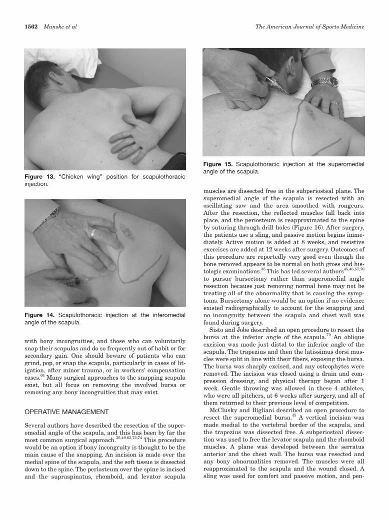
If these initial treatment options do not control the dis-comfort, then oral steroidal medications are anotheroption. If anti-inflammatory drug side effects limit theability to use oral agents effectively or their usage is inef-fective, then injections are a viable option. These are bestgiven with a 1.5-inch needle with a 22- to 25-gauge caliber.Injecting any of the bursae can be easily done with thepatient’s arm in a position of extension, internal rotation,and adduction, trying to reach high up the spine whilelying prone. This is similar to the surgical “chicken wing”position (Figure 13). Kuhn et al have described putting thearm in a position of 60° of abduction and 30° of forwardflexion to make palpation of the inferomedial bursa easier.38
A sterile environment should always be obtained, and anysteroid agent can be used. Combining the injection with ananesthetic agent can make the injection not only thera-peutic but also diagnostic. Care must be taken to stay inthe proper plane parallel to the undersurface of the scapulawhen performing the injection because a pneumothoraxwould be a possibility if the needle were inserted toodeeply (Figure 14). For the superomedial bursa injections,the needle should be placed at a 45° angle just off the supe-rior medial tip of the scapula (Figure 15). It should beinserted approximately 2 cm laterally in a cephalad tocaudad direction.64
If all nonsurgical measures fail to relieve the symptomsafter 3 to 6 months and the diagnosis is certain, then sur-gical options are also available. The decision and timingfor surgery is patient-dependent and should be consideredonly after failure of nonsurgical measures. Patients morelikely to fail nonsurgical treatment may be those with anerve deficit that cannot recruit the muscle innervated bythe damaged nerve leading to scapular dyskinesis, those
Figure 10. Machine rowing exercise.
Figure 11. Wall push-up plus exercise.
Figure 12. Swiss ball closed kinetic chain isometric scapularstabilization exercise.
1562 Manske et al The American Journal of Sports Medicine
with bony incongruities, and those who can voluntarilysnap their scapulas and do so frequently out of habit or forsecondary gain. One should beware of patients who cangrind, pop, or snap the scapula, particularly in cases of lit-igation, after minor trauma, or in workers’ compensationcases.64 Many surgical approaches to the snapping scapulaexist, but all focus on removing the involved bursa orremoving any bony incongruities that may exist.
OPERATIVE MANAGEMENT
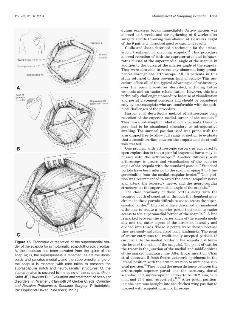
Several authors have described the resection of the super-omedial angle of the scapula, and this has been by far themost common surgical approach.36,49,63,72,74 This procedurewould be an option if bony incongruity is thought to be themain cause of the snapping. An incision is made over themedial spine of the scapula, and the soft tissue is dissecteddown to the spine. The periosteum over the spine is incisedand the supraspinatus, rhomboid, and levator scapula
muscles are dissected free in the subperiosteal plane. Thesuperomedial angle of the scapula is resected with anoscillating saw and the area smoothed with rongeurs.After the resection, the reflected muscles fall back intoplace, and the periosteum is reapproximated to the spineby suturing through drill holes (Figure 16). After surgery,the patients use a sling, and passive motion begins imme-diately. Active motion is added at 8 weeks, and resistiveexercises are added at 12 weeks after surgery. Outcomes ofthis procedure are reportedly very good even though thebone removed appears to be normal on both gross and his-tologic examinations.38 This has led several authors45,46,57,70
to pursue bursectomy rather than superomedial angleresection because just removing normal bone may not betreating all of the abnormality that is causing the symp-toms. Bursectomy alone would be an option if no evidenceexisted radiographically to account for the snapping andno incongruity between the scapula and chest wall wasfound during surgery.
Sisto and Jobe described an open procedure to resect thebursa at the inferior angle of the scapula.70 An obliqueexcision was made just distal to the inferior angle of thescapula. The trapezius and then the latissimus dorsi mus-cles were split in line with their fibers, exposing the bursa.The bursa was sharply excised, and any osteophytes wereremoved. The incision was closed using a drain and com-pression dressing, and physical therapy began after 1week. Gentle throwing was allowed in these 4 athletes,who were all pitchers, at 6 weeks after surgery, and all ofthem returned to their previous level of competition.
McClusky and Bigliani described an open procedure toresect the superomedial bursa.45 A vertical incision wasmade medial to the vertebral border of the scapula, andthe trapezius was dissected free. A subperiosteal dissec-tion was used to free the levator scapula and the rhomboidmuscles. A plane was developed between the serratusanterior and the chest wall. The bursa was resected andany bony abnormalities removed. The muscles were allreapproximated to the scapula and the wound closed. Asling was used for comfort and passive motion, and pen-
Figure 13. “Chicken wing” position for scapulothoracicinjection.
Figure 14. Scapulothoracic injection at the inferomedialangle of the scapula.
Figure 15. Scapulothoracic injection at the superomedialangle of the scapula.
Vol. 32, No. 6, 2004 Management of Snapping Scapula 1563
dulum exercises began immediately. Active motion wasallowed at 3 weeks and strengthening at 6 weeks aftersurgery. Gentle throwing was allowed at 12 weeks. Eightof the 9 patients described good or excellent results.
Ciullo and Jones described a technique for the arthro-scopic treatment of snapping scapula.12 This procedureallowed resection of both the supraserratus and infraser-ratus bursae at the superomedial angle of the scapula inaddition to the bursa at the inferior angle of the scapula.They were also able to resect any abnormal bony promi-nences through the arthroscope. All 13 patients in thisstudy returned to their previous level of activity. This pro-cedure offers all of the typical advantages of arthroscopyover the open procedures described, including bettercosmesis and an easier rehabilitation. However, this is atechnically challenging procedure because of visualizationand portal placement concerns and should be consideredonly by arthroscopists who are comfortable with the tech-nical challenges of the procedure.
Harper et al described a method of arthroscopic bonyresection of the superior medial corner of the scapula.25
They described symptom relief in 6 of 7 patients. One sur-gery had to be abandoned secondary to intraoperativeswelling. The surgical position used was prone with thearm draped free to allow full range of motion to evaluatethat a smooth surface between the scapula and chest wallwas created.
One problem with arthroscopic surgery as compared toopen exploration is that a painful trapezoid bursa may bemissed with the arthroscope.57 Another difficulty witharthroscopy is access and visualization of the superiorangle of the scapula with the standard portals.10 Standardportals have been inferior to the scapular spine 3 to 4 fin-gerbreadths from the medial scapular border.66 This posi-tion was recommended to avoid the dorsal scapular nerveand artery, the accessory nerve, and the neurovascularstructures at the superomedial angle of the scapula.66
The close proximity of these portals along with therequired depth of penetration through the rhomboid mus-cles make these portals difficult to use to access the super-omedial border.10 Chan et al have described an inside-outtechnique to create a superior portal that enables easieraccess to the superomedial border of the scapula.10 A lineis marked between the superior angle of the scapula medi-ally and the outer aspect of the acromion laterally anddivided into thirds. These 2 points were chosen becausethey are easily palpable, fixed bony landmarks. The pointof trocar entry was the traditionally accepted position (2cm medial to the medial border of the scapula just belowthe level of the spine of the scapula). The point of exit forthe trocar is the junction of the medial and middle thirdsof the marked imaginary line. After trocar insertion, Chanet al dissected 5 fresh-frozen cadaveric specimens in thelateral position with the arm in traction to mimic the sur-gical position.10 They found the mean distance between thearthroscopic superior portal and the accessory, dorsalscapular, and suprascapular nerves to be 18.2 mm, 20.2mm, and 24.6 mm, respectively.10,60 After portal position-ing, the arm was brought into the chicken wing position toproceed with scapulothoracic arthroscopy.
Figure 16. Technique of resection of the superomedial bor-der of the scapula for symptomatic scapulothoracic crepitus.A, the trapezius has been elevated from the spine of thescapula; B, the supraspinatus is reflected, as are the rhom-boids and serratus medially, and the superomedial angle ofthe scapula is resected with care taken to preserve thesuprascapular notch and neurovascular structures; C, thesupraspinatus is secured to the spine of the scapula. (FromKuhn JE, Hawkins RJ. Evaluation and treatment of scapulardisorders. In: Warner JP, Iannotti JP, Gerber C, eds. Complexand Revision Problems in Shoulder Surgery. Philadelphia,Pa: Lippincott-Raven Publishers; 1997.)
A
B
C
1564 Manske et al The American Journal of Sports Medicine
Despite the lack of agreement among orthopaedic sur-geons regarding which procedure is best for snappingscapula, most studies have resulted in good outcomes forvery high percentages of patients. A combination of bur-sectomy and bony resection may be the best option formany patients, and this must be a decision made intraop-eratively based on the patient’s given findings. Mostpatients returned to work and/or sports (including over-head sports) within about 3 to 4 months regardless of theprocedure chosen. Few studies in the literature discuss thepostoperative rehabilitation protocol in much detail, but itis clear that return to sports is not allowed until properthoracic posture, scapular control, and strength areobtained with the physical therapy techniques previouslydescribed.
CONCLUSIONS
Scapulothoracic crepitus and bursitis are common condi-tions seen by sports medicine clinicians. For most patients,the initial treatment plan for this condition is nonopera-tive and requires scapular strengthening exercises andpostural reeducation. Local modalities, oral anti-inflam-matory medications, and localized injections can also berecommended. Surgical correction should be saved forthose who do not demonstrate satisfactory clinical out-comes with nonoperative intervention.
REFERENCES
1. Ashmen KJ, Swanik CB, Lephart SM. Strength and flexibility charac-teristics of athletes with chronic low back pain. J Sport Rehabil.1996;5:275-286.
2. Baechle TR. Essentials of Strength Training and Conditioning.Champaign, Ill: Human Kinetics; 1994.
3. Bateman JE. The Shoulder and Neck. Philadelphia, Pa: WB SaundersCompany; 1972.
4. Bateman JE. Shoulder injury in the throwing sports. In: The AmericanAcademy of Orthopedic Surgeons: Symposium on Sports Medicine.St. Louis, Mo: CV Mosby; 1969:94.
5. Boinet W. Bulletin de la Societe Imperiale de Chirugie de Paris, 2ndSeries. 1867;8:458.
6. Bradley J, Tibone J. Electromyographic analysis of muscle actionabout the shoulder. Clin Sports Med. 1991;10:789-795.
7. Brown C. Compressive, invasive referred pain to the shoulder. ClinOrthop. 1983;173:55-62.
8. Burkhart SS, Morgan CD, Kibler WB. The disabled throwing shoulder:spectrum of pathology part III: the SICK scapula dyskinesis, thekinetic chain, and rehabilitation. Arthroscopy. 2003;19:641-661.
9. Cailliet R. Neck and Arm Pain. 3rd ed. Philadelphia, Pa: FA Davis;1991.
10. Chan BK, Chakrabiarti AJ, Bell SN. An alternative portal for scapu-lothoracic arthroscopy. J Shoulder Elbow Surg. 2002;11:253-258.
11. Ciullo JV. Subscapular bursitis: treatment of “snapping scapula” or“washboard syndrome.” Arthroscopy. 1992;8:412.
12. Ciullo JV, Jones E. Subscapular bursitis: conservative and endo-scopic treatment of “snapping scapula” or “washboard syndrome.”Orthop Trans. 1993;16:740.
13. Cobey MC. The rolling scapula. Clin Orthop. 1968;60:193-194.14. Codman EA. The Shoulder: Rupture of the Supraspinatus Tendon and
Other Lesions in or About the Subdeltoid Bursa. Malabar, Fla: KriegerPublishing; 1984:123-177.
15. Curwin S, Stanish WD. Tendinitis: Its Etiology and Treatment.Lexington, Mass: Heath; 1984.
16. Della Valle CJ, Rokito AS, Birdzell MG, Zuckerman JD. Biomechanicsof the shoulder. In: Norkin M, Frankel VH, eds. Basic Biomechanics ofthe Musculoskeletal System. Philadelphia, Pa: Lippincott Williams &Wilkins; 2001:318-339.
17. Dintman G, Ward B, Tellez, T. Sports Speed. Champaign, Ill: HumanKinetics; 1997.
18. Dominguez RH, Gajda R. Total Body Training. East Dundee, Ill:Moving Force Systems; 1982.
19. Edelson JG. Variations in the anatomy of the scapula with referenceto snapping scapula. Clin Orthop. 1996;11:111-115.
20. Evans RC. Illustrated Orthopedic Physical Assessment. 2nd ed. St.Louis, Mo: Mosby; 2001.
21. Glousman R, Jobe FW, Tibone JE, Moynes D, Antonelli D, Perry J.Dynamic electromyographic analysis of the throwing shoulder withglenohumeral instability. J Bone Joint Surg Am. 1988;70:220-226.
22. Gracovetsky S, Farfan H. The optimum spine. Spine. 1986;11:543-573.
23. Grunfeld G. Beitrag zur Genese des Skapularkrachens und derSkapuloargeraushe. Arch Orthop J Unfall Chir. 1927;24:610-615.
24. Haney TC. Subscapular elastofibroma in a young pitcher: a casereport. Am J Sports Med. 1990;18:642-644.
25. Harper GD, McIlroy S, Bayley JIL, Calvert PT. Arthroscopic partialresection of the scapula for snapping scapula: a new technique. JShoulder Elbow Surg. 1999;8:55-57.
26. Herbert LJ, Moffet H, MacFadyen BJ, et al. Scapular behavior inshoulder impingement syndrome. Arch Phys Med Rehabil.2002;83:60-69.
27. Inman VT, Saunders JB, Abbott LC. Observations of the function ofthe shoulder joint. Clin Orthop. 1996;330:3-12.
28. Janda V. Muscles and cervicogenic pain syndromes. In: Grant R, ed.Physical Therapy of the Cervical and Thoracic Spine. New York, NY:Churchill Livingstone; 1988:153-166.
29. Johnston TB. The movements of the shoulder joint: a plea for the useof the “plane of the scapula” as the plane of reference in movementsoccurring at the humero-scapular joint. Br J Surg. 1937;25:252.
30. Kamkar A, Irrgang JJ, Whitney SL. Nonoperative management ofsecondary shoulder impingement syndrome. J Orthop Sports PhysTher. 1993;17:212-224.
31. Kapandji I. The Physiology of Joints. Vol 1. 5th ed. London, England:Churchill Livingstone; 1982.
32. Kendall FP, McCreary EK, Provance PG. Muscles Testing andFunction. 4th ed. Baltimore, Md: Williams & Wilkins; 1993:81.
33. Kibler WB. The role of the scapula in athletic shoulder function. Am JSports Med. 1998;26:325-337.
34. Kisner C, Colby LA. Therapeutic Exercise: Foundation andTechniques. 4th ed. Philadelphia, Pa: FA Davis; 2002.
35. Knott M, Voss D. Proprioceptive Neuromuscular Facilitation. NewYork, NY: Harper and Row; 1968.
36. Kouvalchouk JF, Merat J, Durey A. Subscapular Crepitus. Clichy,France: Societe Francaise de Chirurgie Orthopedique etTraumatologique; 1985:587-588.
37. Kuhn JE. Scapulothoracic crepitus and bursitis. In: DeLee JC, DrezD, Miller MD, eds. Orthopedic Sports Medicine Principles andPractice. Vol 1. 2nd ed. Philadelphia, Pa: Elsevier Science; 2003.
38. Kuhn JE, Plancher KD, Hawkins RJ. Symptomatic scapulothoraciccrepitus and bursitis. J Am Acad Orthop Surg. 1998;6:267-273.
39. Levangie PK, Norkin CC. Joint Structure and Function: AComprehensive Analysis. 3rd ed. Philadelphia, Pa: FA Davis; 2001.
40. Lewitt K. Manipulative Therapy in Rehabilitation of the LocomotorSystem. London, England: Butterworth-Heinemann; 1985.
41. Ludewig PM, Cook TM, Nawoczenski DA. Three-dimensional scapu-lar orientation and muscle activity at selected positions of humeralelevation. J Orthop Sports Phys Ther. 1996;24:57-65.
42. Lukasiewicz AC, McClure P, Michener L, Pratt N, Sennett B.Comparison of 3-dimensional scapular position and orientationbetween subjects with and without shoulder impingement. J OrthopSports Phys Ther. 1999;29:574-583.
Vol. 32, No. 6, 2004 Management of Snapping Scapula 1565
43. Makin GJV, Brown WF, Ebers GC. C7 radiculopathy: importance ofscapular winging in clinical diagnosis. J Neurol Neurosurg Psychiatry.1986;49:640-644.
44. McClure PW, Michener LA, Sennett BJ, Karduna AR. Direct 3-dimen-sional measurement of scapular kinematics during dynamic move-ments in vivo. J Shoulder Elbow Surg. 2001;10:269-277.
45. McClusky GM, Bigliani L. Partial scapulectomy for disabling scapulo-thoracic snapping. Orthop Trans. 1990;14:252-253.
46. McClusky GM, Bigliani L. Scapulothoracic disorders. In: Andrews JR,Wilk KE, eds. The Athlete’s Shoulder. New York, NY: ChurchillLivingston; 1994:305-316.
47. McQuade K, Dawson J, Schmidt G. Scapulothoracic muscle fatigueassociated with alterations in scapulohumeral rhythm. J OrthopSports Phys Ther. 1998;28:74-80.
48. Michele AA, Eisenberg J. Scapulocostal syndrome. Arch Phys MedRehabil. 1968;49:383-387.
49. Milch H. Partial scapulectomy for snapping of the scapula. J BoneJoint Surg Am. 1968;32:561-566.
50. Milch H. Snapping scapula. Clin Orthop. 1961;20:139-150.51. Milch H, Burman MS. Snapping scapula and humerus varus: report
of six cases. Arch Surg. 1933;26:570-588.52. Morse JB, Ebraheim NA, Jackson WT. Partial scapulectomy for snap-
ping scapula syndrome. Orthop Rev. 1993;22:1141-1144.53. Moseley JB, Jobe FW, Pink M, Perry J, Tibone J. EMG analysis of the
scapular muscles during a shoulder rehabilitation program. Am JSports Med. 1992;20:128-134.
54. Mozes G, Bickels J, Ovadia D, Dekel S. The use of three-dimensionalcomputed tomography in evaluating snapping scapula syndrome.Orthopedics. 1999;22:1029-1033.
55. Neer C. Shoulder Reconstruction. Philadelphia, Pa: WB SaundersCompany; 1990.
56. Neviaser RJ. Painful conditions affecting the shoulder. Clin Orthop.1983;173:63-69.
57. Nicholson GP, Duckeworth MA. Scapulothoracic bursectomy forsnapping scapula syndrome. J Shoulder Elbow Surg. 2002;11:80-85.
58. Paine R, Voight M. The role of the scapula. J Orthop Sports PhysTher. 1993;18:386-391.
59. Panjabi MM. The stabilizing system of the spine, part I: function, dys-function, adaptation, and enhancement. J Spinal Disord. 1992;5:383-389.
60. Pavlik A, Ang K, Coghlan J, Bell S. Arthroscopic treatment of painfulsnapping of the scapula by using a new superior portal. Arthroscopy.2003;19:608-612.
61. Percy EC, Birbrager D, Pitt MJ. Snapping scapula: a review of the lit-erature and presentation of 14 patients. Can J Surg. 1988;31:248-250.
62. Poppen NK, Walker PS. Normal and abnormal motion of the shoul-der. J Bone Joint Surg Am. 1976;58:195-201.
63. Richards R, McKee MD. Treatment of painful scapulothoracic crepi-tus by resection of the supero-medial angle of the scapula. ClinOrthop. 1989;247:111-116.
64. Rockwood CA, Matsen FA. The Shoulder. 2nd ed. Philadelphia, Pa:WB Saunders Company; 2000.
65. Rose DI, Novak EJ. The painful shoulder: the scapulocostal syn-drome in shoulder pain. J Kans Med Soc. 1966;67:112-114.
66. Ruland LJ, Ruland CM, Matthews LS. Scapulothoracic anatomy forthe arthroscopist. Arthroscopy. 1995;11:52-56.
67. Russek AS. Diagnosis and treatment of scapulocostal syndrome. JAm Med Assoc. 1952;150:25-27.
68. Saha AK. Mechanism of shoulder movements and a plea for therecognition of “zero position” of glenohumeral joint. Indian J Surg.1950;12:153.
69. Scovazzo M, Browne A, Pink M, Jobe F, Kerrigan J. The painfulshoulder during the freestyle stroke. Am J Sports Med. 1991;19:577-582.
70. Sisto DJ, Jobe FW. The operative treatment of scapulothoracic bur-sitis in professional pitchers. Am J Sports Med. 1986;14:192-194.
71. Steindler A. Traumatic Deformities and Disability of the UpperExtremity. Springfield, Ill: CC Thomas Publishing; 1946.
72. Strizak AM, Cowen MH. The snapping scapula syndrome: a casereport. J Bone Joint Surg Am. 1982;64:941-942.
73. Voight ML, Thompson BC. The role of the scapula in the rehabilita-tion of shoulder injuries. J Athl Train. 2000;35:364-372.
74. Wood VE, Verska JM. The snapping scapula in association with thethoracic outlet syndrome. Arch Surg. 1989;124:1335-1337.


![Operative versus Nonoperative Treatment of Acute …...treatment following Achilles tendon rupture and reported a relativeriskofreruptureof0.32(95%confidenceinterval[CI], 0.14 to](https://img.pdfslide.us/doc/110x75/5fa052da28595e20265c2b35/operative-versus-nonoperative-treatment-of-acute-treatment-following-achilles.jpg)


![Common Challenges in Primary Care – Fracture Management · Fracture management can be divided into - nonoperative[also known as “ conservative”!] and operative techniques. The](https://img.pdfslide.us/doc/110x75/5e2e7826cf4553401272e3b9/common-challenges-in-primary-care-a-fracture-management-fracture-management-can.jpg)