Embed Size (px)
Citation preview
Chapter 2Nonmalignant Tumors of the Orbit
Eric M. Hink and Vikram Durairaj
Abstract Most orbital tumors are nonmalignant. Nonmalignant orbital tumors canarise from any of the structures within the orbit, including blood vessels, fat,nerves, lacrimal gland, and connective tissue. Nonmalignant orbital tumors can begrouped into cystic lesions, vascular tumors, lymphoproliferative lesions, inflamma-tory lesions, mesenchymal tumors, neurogenic tumors, and lacrimal gland tumors.Although most orbital tumors are benign, their location may compromise ocularhealth and function and necessitate treatment with surgery, radiation, or chemother-apy. Patient characteristics, signs, and findings on ophthalmic examination andimaging, including computed tomography and magnetic resonance imaging, guidethe clinician in formulating a differential diagnosis.
2.1 Presentation
Orbital tumors often present with a constellation of signs suggestive of a space-occupying lesion in the bony confines of the orbit. These orbital signs include lidedema or fullness; ptosis or retraction; proptosis or nonaxial globe displacement;axial hyperopia or acquired astigmatism; vascular or lymphatic congestion pro-ducing conjunctival chemosis; hyperemia or secondary glaucoma; dysmotility orpalsy of cranial nerve II, III, IV, V, or VI; chorioretinal folds; optic nerve edemaor atrophy; and double vision or loss of vision. In addition to a complete oph-thalmic examination, appropriate imaging, including ultrasonography, computedtomography (CT), and magnetic resonance imaging (MRI), provides valuable infor-mation regarding the location and radiographic characteristics of the lesion. Oftenthe patient’s age, sex, race, clinical course, and radiographic images can narrow thedifferential diagnosis. Incisional or excisional biopsy may be pursued to confirm thediagnosis.
E.M. Hink (B)Oculofacial Plastic and Orbital Surgery, Department of Ophthalmology, University of ColoradoSchool of Medicine, Denver, CO, USAe-mail: [email protected]
13B. Esmaeli (ed.), Ophthalmic Oncology, M.D. Anderson Solid TumorOncology Series 6, DOI 10.1007/978-1-4419-0374-7_2,C© Springer Science+Business Media, LLC 2010

14 E.M. Hink and V. Durairaj
2.2 Cystic Lesions
Almost all orbital cysts are benign. Cystic lesions may arise from developmentalabnormalities or from the adjacent sinuses or cranium.
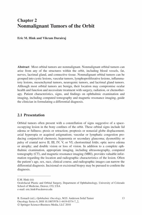
Most congenital orbital cysts are choristomas known as dermoid or epidermoidcysts. These cysts are the most common orbital tumors in children, accounting for30–46% of all excised orbital tumors in this age group [1]. These cysts arise fromnests of ectodermal cells that become trapped in the orbital bony sutures, most com-monly the frontozygomatic suture, during closure of the neural tube [2]. Dermoidor epidermoid cysts often present as firm, smooth, partially mobile masses along thesuperotemporal orbital rim. “Dumbbell” dermoids may have a deep orbital compo-nent. Deep orbital dermoids typically present later in life with orbital signs. Imagingof dermoid and epidermoid cysts demonstrates a well-demarcated cystic lesion withsurrounding bony sclerosis, erosion, and remodeling. These lesions can frequentlybe excised without difficulty (Fig. 2.1). Pathological examination will demonstratea cyst lined with stratified squamous epithelium and filled with keratin. Dermoidcysts contain dermal appendages, including hair and sebaceous glands, whereas epi-dermoid cysts are devoid of such elements. Dermoid and epidermoid cysts rarelyrupture in vivo, but when they do, often as the result of trauma, a vigorous inflam-matory reaction occurs, leading to a clinical picture similar to idiopathic orbitalinflammation or orbital cellulitis.
Another type of congenital orbital tumor is the orbital teratoma, a rare con-genital cystic tumor derived from all three embryonic germ layers that is rarelymalignant [3].
Acquired cysts include chocolate cysts, hemorrhagic cysts, and lacrimal glandcysts. Chocolate cysts are most often associated with lymphangiomas. Lacrimal
Fig. 2.1 Surgical excision of dermoid cyst in a 2-year-old patient
2 Nonmalignant Tumors of the Orbit 15
gland cysts, known as dacryops, form when the excretory ducts of the lacrimal glandbecome obstructed. In addition, bacterial abscesses or larval cysts may arise in theorbit during infection.
In adults, cystic orbital lesions often arise from the adjacent sinuses and herniateinto the orbit. Mucoceles or mucopyoceles arise from an obstructed sinus ostiumcausing chronic sinusitis. In children, a congenital nasolacrimal duct obstruction,often the result of an imperforate valve of Hasner, can form a cystic tear-filledmass known as a dacryocystocele. These present as a bluish, soft, cystic mass belowthe medial canthal tendon, and early probing is advocated to prevent dacryocysti-tis [4]. Dacryocystoceles are often associated with nasal dacryoceles that can causeupper airway obstruction and require immediate surgical marsupialization. Rarely,congenital herniations of intracranial contents known as cephaloceles, includingmeningoceles and encephaloceles, can involve the orbit via the orbital fissures orbony defects [3].
2.3 Vascular Tumors
Vascular lesions are the second most common orbital tumors in children and themost common orbital tumors in adults [1, 5]. There is some debate as to the classi-fication and naming of these tumors. The traditional nomenclature will be used inthis chapter.
Capillary hemangiomas (benign hemangioendotheliomas) are the most commonvascular orbital tumor in children. The tumor varies in location and presentation,although it generally appears within the first few months of life, grows for 6–12months, and then involutes over the next few years [1]. Superficial capillary heman-giomas involving the dermis appear as bright red lesions or “strawberry nevi.”Subdermal tumors may appear as a blue mass in the eyelid. The presence of numer-ous capillary hemangiomas may cause platelet sequestration and thrombocytopenia,a phenomenon known as Kasabach–Merritt syndrome. Because these tumors fre-quently involute, management typically involves limiting the tumors’ amblyogenic(deprivational, strabismic, and astigmatic) effects. Intralesional steroids and lim-ited surgical resection are the mainstays of therapy. Recently the systemic oraladministration of propranolol has shown promising results in the treatment of infan-tile capillary hemangioma (please see a more detailed discussion of this topicin Chapter 3).
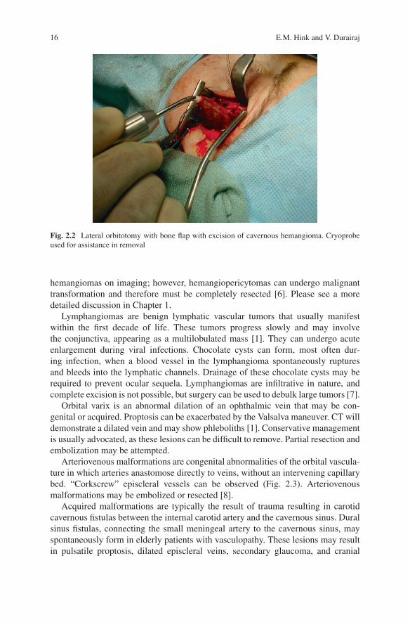
Cavernous hemangiomas are the most common benign orbital tumor in adults,with middle-aged women being the most frequently affected [5]. Cavernoushemangiomas typically appear with orbital signs. Imaging demonstrates a well-circumscribed mass with limited systemic vascular communication and poor con-trast enhancement. These tumors can be intraconal, and cautious surgical excisionmay be required if these tumors compromise ocular function (Fig. 2.2) [5].
Hemangiopericytomas are benign pericyte tumors primarily appearing duringmiddle age. They present with orbital signs and appear similar to cavernous
16 E.M. Hink and V. Durairaj
Fig. 2.2 Lateral orbitotomy with bone flap with excision of cavernous hemangioma. Cryoprobeused for assistance in removal
hemangiomas on imaging; however, hemangiopericytomas can undergo malignanttransformation and therefore must be completely resected [6]. Please see a moredetailed discussion in Chapter 1.
Lymphangiomas are benign lymphatic vascular tumors that usually manifestwithin the first decade of life. These tumors progress slowly and may involvethe conjunctiva, appearing as a multilobulated mass [1]. They can undergo acuteenlargement during viral infections. Chocolate cysts can form, most often dur-ing infection, when a blood vessel in the lymphangioma spontaneously rupturesand bleeds into the lymphatic channels. Drainage of these chocolate cysts may berequired to prevent ocular sequela. Lymphangiomas are infiltrative in nature, andcomplete excision is not possible, but surgery can be used to debulk large tumors [7].
Orbital varix is an abnormal dilation of an ophthalmic vein that may be con-genital or acquired. Proptosis can be exacerbated by the Valsalva maneuver. CT willdemonstrate a dilated vein and may show phleboliths [1]. Conservative managementis usually advocated, as these lesions can be difficult to remove. Partial resection andembolization may be attempted.
Arteriovenous malformations are congenital abnormalities of the orbital vascula-ture in which arteries anastomose directly to veins, without an intervening capillarybed. “Corkscrew” episcleral vessels can be observed (Fig. 2.3). Arteriovenousmalformations may be embolized or resected [8].
Acquired malformations are typically the result of trauma resulting in carotidcavernous fistulas between the internal carotid artery and the cavernous sinus. Duralsinus fistulas, connecting the small meningeal artery to the cavernous sinus, mayspontaneously form in elderly patients with vasculopathy. These lesions may resultin pulsatile proptosis, dilated episcleral veins, secondary glaucoma, and cranial

2 Nonmalignant Tumors of the Orbit 17
Fig. 2.3 Arteriovenous malformation of the left orbit. Note episcleral involvement
nerve VI palsy. CT demonstrates an enlarged superior ophthalmic vein and possibleenlargement of the extraocular muscles. Embolization is the treatment of choice [9].
2.4 Lymphoproliferative Masses
The clinical spectrum of histiocytic, hematopoietic, and lymphoproliferative orbitalmasses ranges from benign, reactive, inflammatory masses to malignant tumors.Often cytology, immunohistochemistry, and molecular genetic analyses are neces-sary to distinguish benign from malignant processes.
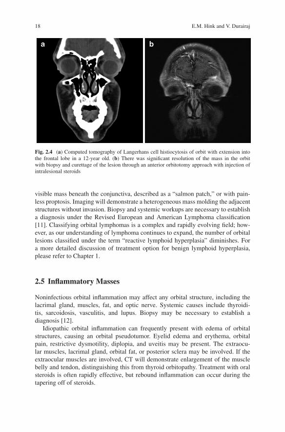
Langerhans cell histiocytosis is now the preferred term for histiocytosis X,eosinophilic granuloma, Hand–Schüller–Christian disease, and Letterer–Siwe dis-ease. On the relatively benign end of the spectrum, Langerhans cell histiocytosismay present as a unifocal bony orbital mass (Fig. 2.4). These are thought to rep-resent 1–3% of pediatric orbital tumors [1]. Presenting signs may include orbitalsigns with or without evidence of orbital inflammation. CT will demonstrate anintraosseous lytic lesion. A complete systemic workup is mandatory. Treatment iscontroversial and may involve observation, excision and curettage, steroids, radia-tion therapy, and chemotherapy. Systemic chemotherapy may be considered in aneffort to decrease the likelihood that the patient will develop diabetes insipidus.
Juvenile xanthogranuloma is a non-Langerhans cell histiocytosis. Although cuta-neous and ocular involvement is more common, isolated orbital tumors do occur[10]. There is no systemic involvement. Treatment modalities include observation,steroids, chemotherapy, and radiation therapy.
Reactive lymphoid hyperplasia is a term used to describe benign lymphopro-liferative lesions of the orbit. Lymphoproliferative orbital masses can present as a
18 E.M. Hink and V. Durairaj
Fig. 2.4 (a) Computed tomography of Langerhans cell histiocytosis of orbit with extension intothe frontal lobe in a 12-year old. (b) There was significant resolution of the mass in the orbitwith biopsy and curettage of the lesion through an anterior orbitotomy approach with injection ofintralesional steroids
visible mass beneath the conjunctiva, described as a “salmon patch,” or with pain-less proptosis. Imaging will demonstrate a heterogeneous mass molding the adjacentstructures without invasion. Biopsy and systemic workups are necessary to establisha diagnosis under the Revised European and American Lymphoma classification[11]. Classifying orbital lymphomas is a complex and rapidly evolving field; how-ever, as our understanding of lymphoma continues to expand, the number of orbitallesions classified under the term “reactive lymphoid hyperplasia” diminishes. Fora more detailed discussion of treatment option for benign lymphoid hyperplasia,please refer to Chapter 1.
2.5 Inflammatory Masses
Noninfectious orbital inflammation may affect any orbital structure, including thelacrimal gland, muscles, fat, and optic nerve. Systemic causes include thyroidi-tis, sarcoidosis, vasculitis, and lupus. Biopsy may be necessary to establish adiagnosis [12].
Idiopathic orbital inflammation can frequently present with edema of orbitalstructures, causing an orbital pseudotumor. Eyelid edema and erythema, orbitalpain, restrictive dysmotility, diplopia, and uveitis may be present. The extraocu-lar muscles, lacrimal gland, orbital fat, or posterior sclera may be involved. If theextraocular muscles are involved, CT will demonstrate enlargement of the musclebelly and tendon, distinguishing this from thyroid orbitopathy. Treatment with oralsteroids is often rapidly effective, but rebound inflammation can occur during thetapering off of steroids.
2 Nonmalignant Tumors of the Orbit 19
2.6 Mesenchymal Tumors
Benign mesenchymal tumors of the orbit include lipomas, fibrous histiocytomas,solitary fibrous tumor, fibrous dysplasia, and osteomas. Dermolipoma, or lipoder-moid, is a benign congenital tumor typically visible lateral to the globe. The tumoris typically unilateral, smooth, and yellow. Histologically, there is a mixture of col-lagenous and adipose tissue surrounded by a stratified squamous epithelium. Thesetumors do not typically require excision and may be associated with Goldenharsyndrome [1]. Fibrous histiocytomas are rare orbital tumors that may be benign ormalignant. Benign tumors can typically be resected without recurrence [13]. A soli-tary fibrous tumor is a mesenchymal tumor that rarely involves the orbit; when itdoes involve the orbit, it is typically indolent [14], is well encapsulated, and canoften be resected en bloc. Many pathologists believe that solitary fibrous tumor isclosely related to hemangiopericytoma from the standpoint of its biological andclinical behavior. Fibrous dysplasia is a genetic but nonfamilial osteodystrophy thatcan affect craniofacial bones, including the orbit [15]. Osteomas are benign, slowlyprogressive bony tumors that can invade the orbit from the paranasal sinus.
2.7 Neurogenic Tumors
Orbital neurogenic tumors arise from the optic nerve or peripheral orbital nerves.Optic nerve gliomas (also known as juvenile pilocytic astrocytomas) are benign,slow-growing optic nerve tumors. Although most often intraorbital, they can affectthe optic chiasm and tract. Typically presenting between 2 and 6 years of age, theyaccount for 2–3% of all pediatric orbital tumors. Girls are affected more often thanboys at a ratio of 3:2 [1]. Twenty-five percent of patients with optic nerve gliomashave neurofibromatosis type I, and 15% of patients with neurofibromatosis type Iwill develop an optic nerve glioma [16]. CT or MRI scans will show characteristicfusiform enlargement of the optic nerve. Management involves observation, surgicalresection, and radiation therapy or chemotherapy. These tumors are often stable andmay involute; thus, observation is often employed. Surgical resection results in aloss of vision but may be necessary when the tumor threatens the chiasm or causessignificant proptosis and corneal exposure.
Neurofibromas are benign tumors that arise from peripheral nerves and con-tain axons, Schwann cells, and fibroblasts. Plexiform neurofibromas can involvethe orbit and eyelid and may result in S-shaped ptosis (Fig. 2.5). They havebeen described as a “bag of worms” on examination and are pathognomonic forneurofibromatosis type I [1]. These tumors are difficult to excise completely.Isolated neurofibromas can usually be excised without recurrence. Malignant trans-formation is rare, but transformation to sarcoma has been observed and warrantsaggressive therapy (Fig. 2.6).
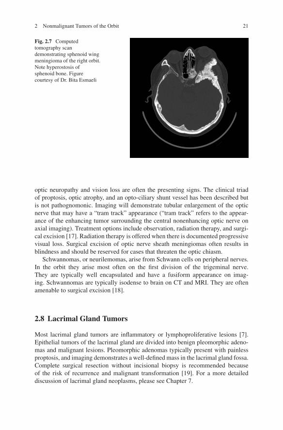
Meningiomas arise from the arachnoid villi and can be invasive. Orbital menin-giomas most often arise from the intracranial portion of the sphenoid wing (Fig. 2.7)

20 E.M. Hink and V. Durairaj
Fig. 2.5 (a) Plexiform neurofibroma of upper eyelid causing a secondary mechanical ptosis andamblyopia in a 3-year old with neurofibromatosis. (b) Appearance after debulking of the mass andcorrection of ptosis. Photos are courtesy of Dr. Bita Esmaeli
Fig. 2.6 Orbital neurofibroma transformed to sarcoma. (a) External photograph of a patient withneurofibromatosis and long-standing bilateral orbital neurofibromas with the right orbital neurofi-broma recently transformed to sarcoma. (b) MRI of a right orbital sarcoma transformed from aneurofibroma in the same patient. Photos are courtesy of Dr. Bita Esmaeli
and extend into the orbit through the bone, superior orbital fissure, or optic canal.Orbital signs including proptosis and early visual deficits can occur. CT willdemonstrate hyperostosis and may show intralesional calcifications. Primary orbitalmeningiomas are less common and arise from the optic nerve sheath. Compressive
2 Nonmalignant Tumors of the Orbit 21
Fig. 2.7 Computedtomography scandemonstrating sphenoid wingmeningioma of the right orbit.Note hyperostosis ofsphenoid bone. Figurecourtesy of Dr. Bita Esmaeli
optic neuropathy and vision loss are often the presenting signs. The clinical triadof proptosis, optic atrophy, and an opto-ciliary shunt vessel has been described butis not pathognomonic. Imaging will demonstrate tubular enlargement of the opticnerve that may have a “tram track” appearance (“tram track” refers to the appear-ance of the enhancing tumor surrounding the central nonenhancing optic nerve onaxial imaging). Treatment options include observation, radiation therapy, and surgi-cal excision [17]. Radiation therapy is offered when there is documented progressivevisual loss. Surgical excision of optic nerve sheath meningiomas often results inblindness and should be reserved for cases that threaten the optic chiasm.
Schwannomas, or neurilemomas, arise from Schwann cells on peripheral nerves.In the orbit they arise most often on the first division of the trigeminal nerve.They are typically well encapsulated and have a fusiform appearance on imag-ing. Schwannomas are typically isodense to brain on CT and MRI. They are oftenamenable to surgical excision [18].
2.8 Lacrimal Gland Tumors
Most lacrimal gland tumors are inflammatory or lymphoproliferative lesions [7].Epithelial tumors of the lacrimal gland are divided into benign pleomorphic adeno-mas and malignant lesions. Pleomorphic adenomas typically present with painlessproptosis, and imaging demonstrates a well-defined mass in the lacrimal gland fossa.Complete surgical resection without incisional biopsy is recommended becauseof the risk of recurrence and malignant transformation [19]. For a more detaileddiscussion of lacrimal gland neoplasms, please see Chapter 7.
22 E.M. Hink and V. Durairaj
References
1. Castillo BV Jr, Kaufman L. Pediatric tumors of the eye and orbit. Pediatr Clin North Am2003;50:149–72.
2. Ahuja R, Azar NF. Orbital dermoids in children. Semin Ophthalmol 2006;21:207–11.3. Shields JA, Shields CL. Orbital cysts of childhood—classification, clinical features, and
management. Surv Ophthalmol 2004;49:281–99.4. Becker BB. The treatment of congenital dacryocystocele. Am J Ophthalmol 2006;142:835–8.5. Scheuerle AF, Steiner HH, Kolling G, et al. Treatment and long-term outcome of patients with
orbital cavernomas. Am J Ophthalmol 2004;138:237–44.6. Valentini V, Nicolai G, Fabiani F, et al. Surgical treatment of recurrent orbital hemangioperi-
cytoma. J Craniofac Surg 2004;15:106–13.7. Shields JA, Shields CL, Scartozzi R. Survey of 1264 patients with orbital tumors and simulat-
ing lesions: the 2002 Montgomery lecture, part 1. Ophthalmology 2004;111:997–1008.8. Trombly R, Sandberg DI, Wolfe SA, et al. High-flow orbital arteriovenous malformation in a
child: current management and options. J Craniofac Surg 2006;17:779–82.9. Leibovitch I, Modjtahedi S, Duckwiler GR, et al. Lessons learned from difficult or unsuc-
cessful cannulations of the superior ophthalmic vein in the treatment of cavernous sinus duralfistulas. Ophthalmology 2006;113:1220–6.
10. Vick VL, Wilson MW, Fleming JC, et al. Orbital and eyelid manifestations of xanthogranulo-matous disease. Orbit 2006;25:221–5.
11. Coupland SE, Hummel M, Stein H. Ocular adnexal lymphomas: five case presentations and areview of the literature. Surv Ophthalmol 2002;47:470–90.
12. Gordon LK. Orbital inflammatory disease: a diagnostic and therapeutic challenge. Eye2006;20:1196–206.
13. Bajaj MS, Pushker N, Kashyap S, et al. Fibrous histiocytoma of the lacrimal gland. OphthalPlast Reconstr Surg 2007;23:145–7.
14. Cerda-Nicolas M, Lopez-Gines C, Gil-Beso R, et al. Solitary fibrous tumor of the orbit:morphological cytogenetic and molecular features. Neuropathology 2006;26:557–63.
15. Hullar TE, Lustig LR. Paget’s disease and fibrous dysplasia. Otolaryngol Clin North Am2003;36:707–32.
16. Liu GT. Optic gliomas of the anterior visual pathway. Curr Opin Ophthalmol 2006;17:427–31.17. Carrasco JR, Penne RB. Optic nerve sheath meningiomas and advanced treatment options.
Curr Opin Ophthalmol 2004;15:406–10.18. Cantore WA. Neural orbital tumors. Curr Opin Ophthalmol 2000;11:367–71.19. Bernardini FP, Devoto MH, Croxatto JO. Epithelial tumors of the lacrimal gland: an update.
Curr Opin Ophthalmol 2008;19:409–13.

http://www.springer.com/978-1-4419-0373-0

![Orbit type: Sun Synchronous Orbit ] Orbit height: …...Orbit type: Sun Synchronous Orbit ] PSLV - C37 Orbit height: 505km Orbit inclination: 97.46 degree Orbit period: 94.72 min ISL](https://img.pdfslide.us/doc/110x75/5f781053e671b364921403bc/orbit-type-sun-synchronous-orbit-orbit-height-orbit-type-sun-synchronous.jpg)