Embed Size (px)
Citation preview
REVIEW
Nonalcholic fatty hepatitis: An in1portant clinical condition
SAMU EL w. FRENCH. MO, LESLIE B. Emus. MD,] . FREEMAN, MD
ABSTRACT: The entity fatty hepatitis is defined and the li teratu re characterizing the clinical settings in which it d evelops is reviewed . The pathogenesis is discussed with emphasis on the common denominators shared by the various clinical conditions with which it is associated. The roles of alcohol, obesity and type II diabetes are stressed where inhib ition of fatty acid oxidation by the liver is the basic defect in metabolism lead ing to fatty change, balloon degeneration and Mallory bod y for mation. le is concluded that this important entity is more common than is generally appreciated . Can J Gastroenterol 1989;3(5): 189-197
Key Words: Diabetes type TT, Fatty hepatitis, Mallory bodies, Obesity
L'hepatite graisseuse nonalcoolique: Une conditon clinique importante
RESUME: L'hepatite graisseusc cstdefinie com me enrite morbide. La littera ture caracterisant les cadres cliniq ues ou cette affection se developpe est passee en revue. La pathogenese est examinee et !'accent est pone sur les denominateurs com muns que partagent les diverses conditions cliniques auxquelles clle est associee. Le role de l'alcool, de l'obesire et du d iabete de type 11 est souligne clans les cas ou !'inhibition d u processus d'oxydation hepatique des acides gras est la deficience principale d u merabolisme entrainant !'accumulation de graisses, la degenerescence graisseuse et la formation de corps de Mallo ry. On conclut que cette entire importance est plus frequente qu 'on ne l'estime generalemen t.
De/>artments of Pathology and S11rgery. Unrt,er.my of Ottau•a a nd Deparrmcn1 of Laboratory Medicine. 0 Hawa General Hospira/. Ot1awa. Onwrio
Correspondence and reprints· Dr Samuel \,\I French. Professor and Chairman, Departmcnl of Pathology. Farnlcy of Hellhli Sciences, Univernry of Ouawa. 451 Smyth Road, Ottawa. Ontario KIH BM.5
Received for /mhlicauon July 21. / 989 Accepted Sc/>tcmber 19, 1989
CAN) GASTROENl rnoL Vo, 1 No 5 NO\ HIBtR/Dl:ClMB~R 1989
THE TERM FATIY HEPATIT IS HAS MANY
synonyms: fatty metamorphosis of the liver in morbid obesity ( I), d iabetic hepatitis (2,3), nonalcoholic stearohepatitis (4,5), fatty live r hepa titis (6), alcohollike liver d isease in nonalcoholics (7), fasting in obesity liver injury with alcoho lic hya line (8), stcatonecrosis (9) and nonalcoholic Lacnncc's ( 10, 11 ). T he term fa tty hepatitis is preferred for its simplicity and for the concept that it conveys, one of fatty change with infla mmation in the liver. T he d isease process is impo rtant to recognize clinically because it can be mistaken for benign fa tty liver or alcoholic hepatitis. Fatty hepatitis is a pernicious disease which may progress to ci rrhosis without the physician real izing it unless a liver bio psy is performed ( l ). The clinical manifestations, associated diseases and pathology are the subject of this review.
HISTORICAL BACKGROUND For many years, fa tty live r, regard less
of its cause, was considered a benign reversib le process which d id not progress to cirrhosis. This idea was based primar-
189

FRENCH era/
Figure 1) Gross photograph of a liver cross section taken ar autopsy from an obe.1e patient diagnosed as having /atcy hepatitis on liver biopsy. The liver appeared heterogeneous and partly nodular due to an uneven d1strrbution of /at. The light areas are/atcy and the dark areas are not.* Incidental bile duct cyst
Figure 3) High power of Figure 2 showing the Junction between 4-1 faery change on the left and minimal/acty change on the right ( x 83)
Figure 2) Low power view of a histologic section from the same liver shown in Figure I. The pale areas are /atty. The dark areas are nodular analogous to nodular regenernrive hyperplasia ( X 3)
Figure 4) Same liver as Figures 1 and 2 bur stained for collagen with sinus red. Note the cencral-central bridging fibrosis (arrows) . This is incomplete cirrhosis ( X 3)
ily on the observation that the fatty liver associated with Kwashiorkor in childhood did not progress to cirrhosis. A lso, the diabetic fatty liver, unlike the alco
holic fatty liver. did not progress to cirrhosis unless the patients were also alcoholics { l2). A similar result was reported in the fatty liver of obesity {13). Fibrosis or cirrhosis was not encountered unless there was also a history of excessive alcohol intake ( 13). The severity of fatty liver in alcoholics is predictive of cirrhosis ( 14 ). However, in the study of fatty liver associated with morbid obesity, a few cases of liver fibrosis or cirrhosis were observed in which alcohol abuse was not a factor (I). Examination of the liver biopsies taken before and after a small bowel bypass operatio n for morbid obesity showed p rogression co
190
cirrhosis in some ( 15). Hepatitis of the fatty liver was first de
scribed by Thaler ( 16). but the entity was not widely appreciated until it became apparen t that a ll of the morphologic features of alcoho lic hepatitis, including Mallory body formation and progression to cirrhosis, were sometimes observed in nonalcoholic patients. It has now been found to be associated with diabetes (2-7,9, 13,17-21), obesity (3-7,13), mor
bid obesity with or without intestinal bypass or gastroplasty ( 1, 15,22-36), fasting in obese patients (8,22), massive resection of the small intestine (3 7 ,38), bulimia (39) and drug therapy (40-45) {Table 1). Fatty liver with fibrosis or cir
rhosis caused by methotrexate may be hard to distinguish morphologically from nonalcoho lic fatty he patitis ( 46). The
present authors have seen three cases of methotrexate-induced nonalcoholic fatty hepatitis with Mallory body formation,
including one case in which typical central sclerosing hyalin necrosis was found in an elderly abstainer.
TABLE 1 Nonalcoholic fatty hepatitis and fatty cirrhosis: Clinlcally associated conditions
Diobetes mellltus (2-7.9.13.17-21) Obesity(3-7.13) Morbid obesity with or without intestinal
bypass or gostroplasty ( 1.15.22-36) Fasting in obese patients (8.22) Massive resection of small intestine ( 3 7 -38) Bullmia(39) Drug therapy: Glucocorticolds. amiodarone.
perhexiline maleote ( diuretics. hypoglycemics. cardiac/hypertension. estrogens a nd thyroid)( 40-45)
CAN J G ASTROENTEROL VOL 3 N O 5 N OVEMBER/DECEMllER 1989

Fatty hepatitis
Figure 5) Lwer biopsy of a patienl wi1h morbid obesiry and /ally cirrhosis. The bridging fibrosis incorpora1es central ,,eins and portal traces (arrow). SrriitS red X I 7
Figure 7) High power view of a broJ,sy of faery hepa1icisfrom a morbidly obese pacienr. Note both 1he nucro-and macrovesicular fa1 The mrcrovesicular [a1 resembles the foamy degenera1ion seen rn alcoholic hepatitis ( x 330)
-'CV
•
Figure 6) View of a porcal rracr ( PT) and centrilob11lar area (CV I showing borh pmportal and centrilobular fibrosis. The .:eniral fibrosis is pericellular (arrows). This is/rom a biopsy of faery hepatitis from an obese patient. Sirn~1 red x 83
Figure 8) High power view of 1he same liver shown in Figure 6 showing a micro[octtS of 1he characteristic macrophage infiltrate seen in che lobule (arrows/ The macrophages are iden1i/iable beca11se chey contain periodic acid-Schiff positive {Jhagosomes. Digested penodrc acid-Schiff reagent x 330
The frequency of cirrhosis and fibrosis va ries with different series, but in a
review of 41 papers o n liver morphology of 15 15 morbidly obese patients, 29% had fibrosis and 3°lo cirrhosis ( 34). In a n ine month follow-up of 34 cases of jejunoileal bypass for morbid obesity, six developed fibrosis a nd three cirrhosis (35).
PATHOLOGY Hepatomegaly is the rule. Grossly, the
liver is pale yellow or a mottled ye llow and brown , owing co a heterogeneous fatty change (Figure I) . Fibrosis is variable and irregularly distributed unless the live r ha s progressed to cirrhosis.
Microscopically, the fatty ch ange may be diffuse early and more patchy in late r stages (Figures 2, 3 ). Scarring is
variable , being absent early and progressing to incomplete cirrhosis (Figure 4) or cirrhosis (Figure 5 ).
Fibrous spurs ex tend from the portal tracts toward zone 3 of the lobule (Figure 6) whereas scars in zone 3 form fibrous bridges between the terminal hepatic venules (central veins) (Figure 4). The earliest change is fatty change, both microvesicu lar and macrovesicular (Figure 7). The earliest infla mmatory change,
which distinguishes fatty liver from fatty hepatitis, is the development of foci of
macrophages in the parenchyma (Figure 8). This is fo llowed by balloon degeneration and Mallory body formation in the centrilobular zone (Figure 9). This lesion
is accompanied by a mo nonuclear infiltrate and fibrosis, and is associated with a loss of cytokeratin stain ing in the bal-
CAN J GASTROENTFROI Vo1 l Nn 5 N<WEMBFR/D~CFMllrn 1989
loon cells, and the in tense sta ining of cyco keratin in the Mallory bodies (Fig
ure 10), analogous to that seen in alcoholic hepatitis ( 4 7). Marked centrilobular bile ductule metaplasia occurs in centrilobular scars (Figures 11, 12), as seen in
alcoholic hepatitis ( 48-5 1). This proliferation of bile ductules induces a desmoplastic response similar to that seen in the periporcal areas (Figures 11 , 12). This phenomenon, in which liver cells express cytokera tins which are no rmally o nly expressed by bile duct epithelium, is
known as bile duct metaplasia of hepatocytes ( 49). The use of immunoperoxidase in the localization of these bile duct cytokeratins is illustrated in Figures 11
and 12. Thus, the evolution of fatty hepatitis to cirrhosis resembles that of alcoholic hepatitis except for one feature .
191

FRENCH er al
, • •
• • PT
• " '
C
• ii
• ' •
Figure 9) Centrilobular balloon degencrarion in hepawcy1es from a mor· bidly obese patient. Nore rite Mallory bodies /arrows) within rite balloon cells. Also, note the absence of polymorphonuclear lmkocytes and the foci of mononuclear inflammation and fibrosis ( x 330)
Figure 11) Same liver as in Figures 9 and 10. The bile ductulemetaplasw (closed arrows) is illustrated in bo1h the periporral (PT) area and the centrilobular (CV) area Normal bile ducts (open arrow) also stain /JOSI·
tively for cytokeratins. The bile ductules sea in /1ositively for cycokerarins like bile ducts e11en though the bile ductules are 1ransf ormed liver cells. These meraplastic duc1ules s1imula1e periducralfibrosis. lmmunoperoxidase x 83
I
Figure 10) Same liver as Figure 9 stained/or normal liver cell cyrokeratins. The balloon cells fail to swin positively like the adjacent normal hepawcytes and 1he Mallory bodies (arroU1S) which are intensely seamed. This failure to scain che cycof,lasm with antibodies to liver cell cytokeratin is typical of cells which contain Mallory bodies. 1mmunoperoxidase x 330
Figure 12) Cenrrilobular scar shown in Figure J J. It is possible ro see rhar the liver cells /irs1 develop increased cytokeratin around the edge of the cell (open arrow). These liver cells seem ro become progressively smaller and express cyrokeratin throughout 1he cell. They 1hen form small nests (solid arrows) which resemble bile ductules (bile duccular meraplasia). lmmunoperoxidase x 330
The polymorphonuclear leukocyte in· filtrate around hepatocytes that contain Mallory bodies (satellitosis) (52) is miss· ing in fatty hepatitis, although there are exceptions (3, 7).
The pathologic changes of fatty hepatitis have been compared in detail with those of alcoholic hepatitis in a study reported by Diehl et al (7). They observed no qualitative differences in histology between two groups of patients (39 nonalcoholic and 68 alcoholic hepatitis). There were some differences in the average severity of some of the features. Micro- and macrovesicular fat were more often moderate to marked in severi ty in the nonalcoholics (P< 0.05). Mallory
192
bodies were identified in 90% of the specimens in both groups. Mallory bodies were very numerous particularly in the alcoholic patients (P<0.05). lntra-acinar inflammation (predominantly neutrophilic) was more p rominent in the alcoholics (P< 0.005). Centrilobular pericellular fibrosis or cirrhosis was seen in both groups, but severe fibrosis or cirrhosis was present more often in the alcoholic group (63%) compared to the nonalcoholic group (38%). Likewise, the clinical stigmata of portal hypertension were found more often with marked fibrosis or cirrhosis in the alcoholic group.
Another study of hepatic histology involving 320 alcoholics and 348 non-
alcoholics at autopsy established a relationship between the severity of sceatosis, Mallory bodies, fibrosis and the degree ofobesity in both groups (21). The prevalence of Mallory bodies and fibrosis was higher in type ll diabetes. ln alcoholic patients who were markedly obese, there was a higher prevalence of Mallory bodies compared to nonobese a lcoholics (68 versus 38%, P<0.001).
The frequency of fatty liver in obesity was 48% in a series of 50 patients hospitalized for weight reduction ( 13). Fatty hepatitis was diagnosed in 26% of nonalcoholics. Fatty fibrosis was found in 8% and cirrhosis in 8%, but in these patients alcohol abuse was a factor. Protein defi-
CAN J GASTROENTEROL VOL 3 No 5 N0VEMB1:.Rl0tCEM6ER 1989

ciency correlated with steatosis, inflammation and fibrosis. Diabetes mellitus correlated with severe steatosis. Alcohol intake correlated with severe steatosis, fatty hepatitis, fatty fibrosis and fatty cirrhosis in this series.
Diabetes mellitus is sometimes complicated by simple fatty liver and cirrhosis. In a series of 62 patients with type II diabetes, I 7 had fatty hepatitis (3). The histology in nine biopsies studied systematically revealed fe a tures of alcoholic hepatitis. Fatty change and liver cell ballooning were constant features. Neutrophils in association with Mallory bodies were found in five; mononuclear infiltrate in four; acidophil bodies in five; fat granulomas in seven; pericellular fibrosis was mild. In six cases, there was bridging fibrosis between the portal and central ve in, but distortion of the lobular architecture was absent or slight. Cirrhosis was not encounte red , a lthough progression was discovered in one case on a subsequent biopsy.
In a series of 29 obese patients without alcohol abuse, fatty liver was seen in seven, fatty hepatitis in eight, fatty fibrosis in seven and fatty cirrhosis in seven (6). Seven cases had Mallory bodies and I 2 had central or perisi n usoidal fi brosis. There was no correlation between the severity of liver damage and the degree of fatty change. Polymorphonuclear leukocytes were numerous in all of the patients with fatty hepatitis. In these patients, diabetes and lipoprotein abnormalities (mostly type IV) were common. In a second series of 20 cases of nonalcoholic steatohepatitis (5), 90°/o were obese. Moderate to severe macrovesicular fatty change and lobular inflammation were present in all cases as these were the criteria for case selection. Fat was distributed centrally or diffusely and fat cysts were always present. The inflammatory cells were mixed lymphocytes, mononuclear cells and neutrophils located either centrilobularly or in areas of focal necrosis. Mallory bodies located in zone 3 were present in 70%, Councilman bodies in 25%, perisinusoidal centrilobular or septa! fibrosis in 70% and cirrhosis in [5%.
The histopathology of the liver in morbid obesity has been reported by numerous investigators. In a review of 4 l articles
in which the liver morphology of a combination of 1515 morbidly obese patients was summarized (34 ), the liver was normal in 12% of the cases. Fatty change was the most frequent abnormality found (80%). Portal inflammation was seen in 33% and fibrosis, mainly portal or periportal, was seen in 29%, while cirrhosis was seen in 3%. Mallory bodies were rarely found. A few reports describe central or pericellular fibrosis in nonalcoholic patients (34 ). The same authors (53) studied the livers of 61 cases of morbid obesity (average 82% overweight). The patients' alcohol intake did not exceed a moderate amount and only one patient was diabetic. Seven percent of the biopsies showed a normal liver. Fatty change was the most common abnormality (85%). The degree of fatty change correlated with the presence of lipogranuloma (54%), focal necrosis (33%) and Kupffercell prol iferation (49%). No Mallory bodies or cirrhosis were observed and alcoholic hepatitis was absent. These authors took the view that when Mallory bodies, fibrosis or cirrhosis arc encountered in obese patients, it is likely that the patient is abusing alcohol (34,53).
When patients with morbid obesity are treated with a small bowel bypass or gastroplasty, the liver morphology may worsen, implying that malnutrition plays a role in the pathogenesis of fatty hepatitis (29 .30.32.35 ). This idea is strengthened by the observation that a massive sma ll bowel resection can induce fatty hepatitis (37.38) although starvation therapy alone had no injurious effect on the live r of the morbidly obese (30); a mere reduction in fat stores in the liver was observed. Liver biopsies in 88 patients taken one to two years after jejunoileal bypass showed that an increased number of patients had increased fat stores (30). However, portal fibrosis, inflammation or biliary proliferation did not predict progression to a lcoholic hepatitis. O nly central or pericellular fibrosis predicted progression to alcoholic hepatitis. In 6.8%, central fibrosis progressed to form central-portal bridging fibrosis. One half of these patients developed regenerative cirrhotic nodules. Only one case developed in to a morphologic picture resembling alcoholic hepatitis. Mallory bodies were more frequently en-
CANJ GAS'fROENTEROI Vo1 3 No 5 N OVEMIIER/DECEMllER 1989
Fatty hepatitis
countered but were apparently periportal in location.
Peters (29) reported serious hepatic disease in I to 17% of patients who underwent jejunoileal bypass. Most common was acute hepatic fa ilure two-and-onehalf to nine months after the bypass. This failure was associated with a large fatty liver a long with fibrosis and hepatocellular necrosis. The fibrosis was both central and portal. The perivcnular hepatocytes were hydropic and surrounded by collagen. Mallory bodies were found in these swollen hepatocytes while neutrophils were sparse. The lesion was indistinguishable from alcoholic hepatitis. A sim ilar lesion was encountered after gastroplasty (32). Some of the patients continued to drink alcohol while others did not, however, the histological changes were id entical. This indicates that caution must be exercised regarding validity of a negative history of alcohol abuse as the patien t may not be candid about drinking habits.
Fatality in patients who underwent small bowel bypass su rgery was common. The second least common sequela to bypass was the insidious development of cirrhosis, discovered either accidendy or after the development of ascites. Another important but uncommon complication was the manifestation of tenuous hepatic reserve. In this condition , liver disease developed rather suddenly as a consequence of some o the r systemic insult, similar to alcoholic liver disease.
The progression of liver disease in 34 patients who underwent jejunoileal bypass for morbid obesity was studied via follow-up biopsy five to nine months postoperatively. Twelve of 15 patients with no or slight steatosis progressed to either moderate to severe steatosis, steatohepatitis or steatofibrosis. The six patients wi th initial moderate to severe stcatosis all progressed co steatohepatitis or steatofibrosis, and one developed septa! fibrosis. All of the patients with steatohepatitis (steatosis and a lobular lymphocytic inflammation) progressed to steatofibrosis. Of the five patients with steatofibrosis, three developed bridging fibrosis. Mallory bodies appeared postoperatively in 11 patients (32%), all of whom had had either severe steatosis, steatohepatitis or stcatofibrosis preoperatively.
193
FRENCH eta/
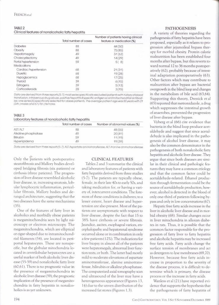
TABLE2 Clinical features of nonalcoholic fatty hepatitis
Number of patients having clinical Total number of cases feature or medication(%)
Diabetes Obesity Hepatomegoly Cholecystectomy Portal hypertension Medications
Cardiac/hypertension Diuretic Hypoglycemics Thyroid Estrogens Corticosteroids
88 88 49 49 59
68 68 68 39 39 20
44(50) 75(85) 31 (63) 14(29) 5( 8)
21 (31) 19(28) 17 (25) 6(15) 5(13) 3(15)
Doto ore derived from three reports(5-7t most senes specit,collyexctuded pot,ents with history of blood tronsfus,on. introvenous drug abuse. pos1t1ve hepatitis Bspecif1c ontlgen orontim,tochondriot ontibodies: one senes(6J specificolly selected for obese potients The overoge pot,ent oge wos 50 yeors with 21 (24":,J motes ond 67 (76'~,) temoles
TABLE 3 Laboratory features of nonalcoholic fatty hepatitis ____ _
Total number of coses Number of abnormal values(%)
AST/ALT Alkaline phosphatase Bilirubin Hyperllpldemo
88 49 59 49
48(55) 20(41) 11 (19) 19(39)
Dote ore derived from three reports (5-7) AST Asportote ominotronsferose. ALT Alonine ominotronsferose
Only the patients with postoperative steatofibrosis and Mallory bodies developed bridging fibrosis (six patients) or cirrhosis (three patients). The progression of liver disease resembled alcoholic liver disease, ie, increasing steatosis, lobular lymphocytic inflammation, pericellular fibrosis, Mallory bodies and deranged architecture, suggesting that the two diseases have the same mechanisms (35).
One of the features of fatty liver in alcoholics and morbidly obese patients is megamitochondria seen by light microscopy or electron microscopy. The megamitochondria, which are elliptical or cigar-shaped due to intramitochondria l filaments (54), are located in periportal hepatocytes. These are nonspecific, but the globular mitochondria located in centrilobular hepatocytes are a useful marker of both alcoholic liver disease (55-59) and nonalcoholic fatty liver (60,61 ). There is no prognostic value in the presence of megamitochondna in alcoholic liver disease (59); the prognostic implication of the presence of mega mitochondria in fatty hepatitis in nonalcoholics is as yet unknown.
CLINICAL FEATURES Tables 2 and 3 summarize th.e clinical
and laboratory features of patients with fatty hepatitis derived from three studies (5-7). The patients are typically obese, female. in their late 40s to early 50s, and taking medication for, or having a variety of, intercurrent conditions. The foremost of these conditions is diabetes; to a lesser extent, heart disease and hypertension are also present. Most of the patients are asymptomatic with respect to liver disease, despite the fact that 15 to 38% have cirrhosis or severe fibrosis. Jaundice, ascites, esophageal varices, encephalopathy and hcpatorenal syndrome occurred alone or in combination in only a few patients ( 12%). The indications for liver biopsy in almost all of the patients were hepatomegaly, abnormal liver function tests, or both. The latter had mostly mild to moderate elevations of asparcate aminotransferase, alanine aminotransferase, bilirubin or alkaline phosphatase. The computerized axial tomography scan and ultrasound of the liver may have a heterogeneous appearance (Figures 13, 14) due to the unven distribution of the increased fat stores (Figures 1,3 ).
PATHOGENESIS A variety of theo ries regarding the
pathogenesis of fatty hepatitis have been proposed, especially as it relates to progression after jejunoileal bypass therapy for morbid obesity. Protein calorie malnu trition has been established four months after bypass, but this reverts toward normal 12 to 36 months postoperatively (62), probably because of intestinal adapta tion postoperatively ( 63 ). Other factors which may contribute to malnutrition after bypass are bacterial overgrowth in the blind loop and changes in the metabolism of bile acid (63,64). Supporting this theory, Drenick et al ( 65) reported that metronidazole, a d rug which suppresses the intestinal growth of anaerobes, prevented the progression of liver disease after bypass.
Vy berg et al (66) ci te evidence that bacteria in the b lind loop produce acetaldehyde and suggest that since acetaldehyde is also implicated in the pathogenesis of alcohol liver d isease, it may also be the common denominator in the pathogenesis of both nonalcoholic faery hepatitis and alcoholic liver disease. They argue that since both diseases are similar in their clinical and pathologic features they may have a common etiology and that the common factor could be acetaldehyde-related. Ethanol production through fermen tation could be the source of acetaldehyde production; however, alcohol is detected in th.e blood of only one-third of patients followi ng bypass and only in low concentrations (67).
Hepatic free fatty acids increase in the liver in alcoholic liver disease and in morb id obesity (68). Similar changes occur in liver mitochondria in alloxan diabetes in rats. Thus, it is possible that the common factor responsible for the progression of fatty liver to fatty hepatitis and alcoholic hepatitis may be increased free fatty acids. Fatty acids change the surface tension of membranes and act as detergents to lyse membranes (69,70). However. because free fatty acids increase in proportion to the severity of the disease process, it is difficult to determine which is primary, the disease process or the increase in fatty acids.
Wanless et al (21) have reported evidence that supports the hypothesis that the pathogenesis of fatty hepatitis of
194 CAN J CASTROENTFROL V Ol 3 No 5 N<WFMRrR/DffEMI\FR 19119

Fatty hepatitis
Figure 13) Computenzed axial tomography scan showing heterogeneity m the liver shown m Figures I to 3 Note that the density of the lwer is
focally decreased relative to the blood vessels ( arrow)
Figure 14) Ultrasound from the liver shown in Figures I to 3 Note the typical bright echo of /atty liver (arrow)
morbid obesity with type ll diabetes,
and fatty hepatitis following jejunoileal
bypass surgery for morbid o besity is the
same, ie, increased free fatty acids in
the liver. They observed steatosis and
steatonecrosis with Mallory bodies in the
subcapsular liver in patients on perito
neal dialvsis with cype I diabetes receiv
ing intraperitoneal insulin on a regular
basis. They suggested that this localized
fatty change was due to insulin and glu
cose diffusing from the peritoneum into
the liver tissue. From this idea they rea-
REFERENCES I. Kern WH, Heger AH, PayneJH.
De Wind LT. Fatty metamorphosis of the liver in morbid obesity. Arch Pathol Lab Med 1973;96342-6.
2. Batman PA, Scheuer P. Diabetic hepatitis preceding the onset of glucose intolerance. Histopathology 1985; 9:237-43
3. Nagore N. Scheuer PJ The pathology of diabetic hepatitis.) Pathol 1988; 156: 155-60.
4. ltoh S. Matsuo S. lchinse A, Yamaba Y, Miya.awa M Nonalcoholic steatohepatit1s and cirrhosis with Mallory's hyalin. Dig DisSci 1982;27.34 1-6
5. Ludwig]. Viggiano TR. McGill DB, Oh BJ. Nonalcoholic stcatohepatitis. Mayo Clin Proc 1980;555:434-8
6. Alder M, Schaffner F. Fatty liver hepatitis and cirrhosis in obese pauents Am) Med 1979;67:8ll-6.
7. Diehl AM, Goodman Z. Ishak KG Alcohol-like liver disease m nonalcoholics. Gastroenterology J 988;95: 1056-62.
8. Capron J-P, Delamarre J, DupasJL,
soned that insulin inhibited fatty acid
oxidation in the liver, and that it caused
faery acids to be prefere~tially esterified
and stored as triglycerides, resulting in a
fatty liver. Free fatcy acids thus accumu
lated , causing membrane injury, swell
ing of hepatocytes and steatonecrosis Generally, when insulin levels are ele
vated enough to block fatty acid oxida
tion but not high enough to block free
fatty acid mobilization, farcy liver and ne
crosis may result. Such a clinical situa
tion exists in obesity associated with cype
Braillon A, Degon C. Quenum C Fasting m obesity. Another cause of liver injury with alcoholic hyaline? Dig Dis Sci 1982:27-265-8
9. Van Thiel DH. Diabetes mellitus and hepatobiliary disease. Curr Concepts Gastroenterol 1986;4:4- J 3.
10. Miller DJ , Ishimaru H, Klatskin G Non-alcoholic liver disease mimicking alcoholic hepatitis and cirrhosis. Gastroenterology 1979;77:27 . (Abst)
11 TorosisJD. Barwick KW, Miller DJ, Klatskin G, Riely CA Nonalcoholic Laennec's: Clinical characteristics and long term fo llow-up Hepatology 1986;6: l 170. (Abst)
12. Robbers H, Srrohfeldt P, Krugger CH. Differential diagnosis between diabetic and alcoholic fatty liver Germ Med Monthly 1968; 13: 124-5.
13 Braillon A, Capron JP, Herve MA, DeGott C, Quenum C. Liver in obesity. Gut 1985;26: 133-9.
14. Sorensen TJA, Bentsen KD, Eghoje K. Orholm M. Hoybye G, Christoffersen P. Prospective evaluation of alcohol abuse
CAN J GASTROENHROL VOL 3 No 5 Nov, MBERIDH:b MBtR JQ89
II diabetes and morbid obesity treated
with bypass surgery, but not in morbid
obesity controlled by diet reduction therapy. This may explain why fatcy hepati
tis progresses after bypass surgery but reverts to a normal liver when diet calo
rie restriction without surgery is successful in causing weight loss (30). It is likely
that a similar mechanism is involved in
alcoholic liver disease with obesity, since
the metabolism of alcohol blocks fatcy acid ox idation by liver mitochondria
(71)
and alcoholic liver injury in men as predictors of development of cirrhosis. Lancet 1984;ii:241-4.
15. Holzbach RT Hepatic effects of Jejunoileal bypass for morbid obesity. Am) Clin Nutr 1977;30:43-52.
16. Thaler H. Die Fettleber und ihre pathogenethische Beziehung zur Lebercirrhose. Virchows Arch 1962;335: 180-2 JO
17 Fleming KA, MorconJA, Barbacis C, Burns), Canning S, McGeeJO'D Mallory bodies in alcoholic and non-alcoholic liver disease contain a common antigenic determinant. Gut 1981,22:341-4.
18. Falchuk KR, Fiske SC, Haggitt RC, Federman M, Trey C. Pericentral hepatic fibrosis and intracellular hyalin in diabetes mellitus. Gastroenterology 1980;78:535-41.
19. Stone BG. Van Thiel DH. Diabetes mellitus and the liver. Seminars Liver Dis 19855:8-28.
20. Schaffner F, Thaler H. Non-alcoholic fatty liver disease. In: Popper H.
195

FRENCH eta/
Schaffner F, eds. Progress in Liver Disease, Vol Vlll. New York: Grune and Stratton, 1986:283-98.
21. Wanless IR, Bargman]M, Oreopoulos DO, Vas SL Subcapsular steatonecrosis in response to peritoneal insulin delivery. A clue to the pathogenesis of steatonecrosis in obesity. Mod Pathol 1989;2:69-74.
22. Drenick EJ, Simmons F, Murphy JF. Effect on hepatic morphology of treatmentofobesity by fasting, reducing d iets and small-bowel bypass. N Engl J Med l 970;282:829-34.
23. Marres JL, Tay low HB, Starkloff GB. Relationship between hepatic morphology and clinical and biochemical findings in morbidly obese patients. J Clin Pathol 1973;26:776-83.
24. Moxley RT lll , Pozefsky T, Lockwood DH. Protem nutrition and liver disease after jeJunoileal bypass for morbid obesity. N Engl J Med I 974:290:921-6.
25. Kern WH, PayncJH, DcWind LT. Hepatic changes after small-intestinal bypass for morbid obesity AmJ Clin Pathol 1974;61:763-8.
26. Peters RL, Gay T, Reynolds TB. Post-jejunoileal-bypass hepatic disease. Its similarity to alcoholic hepatic disease. Am] Clin Pathol 1975:63:3 18-31.
27. Marubbio AT Jr, Buchwald H, Schwartz MZ, Varco R. Hepatic lesions of central pericellular fibrosis in morbid obesity and after jejunoileal bypass. Am J Clin Pathol I 976;66:684-9 l.
28. Piepkorn MW, Mottet NK, Smuckler EA. Fatty metamorphosis of the liver associated with jejunoileal bypass. Arch Pathol Lab Med l977;101:4l l-5 .
29. Peters RL. Patterns of hepatic morphology in jcjunoileal bypass patients. Am J Clin Nutr 1977:30:53-7.
30. Marubbio AT, Rucker RD Jr, Schneider PD, lrorstmann JP, Varco RL, Buchwald H. The liver in morbid obesity and following bypass surgery for obesity. Surg Clin N Am 1979;59: 1079-93.
31. Kroyer JM, Talbert WM Jr. Morphologic liver changes in intestinal bypass patients. Am J Surg 1980; 139:855-9.
32. Hamilton DL, Vest TK, Brown BS, Shah AN, Menguy RB, Chey WY. Liver inju ry with alcoholic like hyalin after gasrroplasty for morbid obesity. Gastroenterology 1983;85:722-6.
33. Hocking MP. Duerson MC, O 'Leary IP, Woodward ER. Jejunoileal bypass for morbid obesity. N Engl J Med 1983;308:995-9.
34. Andersen T, Gluud C. Liver morpholo!(Y in morbid obesity. A literature study. Int J Obes 1984:8:97- 106.
35. Vyberg M, Raun V, Andersen B. Pattern of progression in liver injury following bypass for morbid obesity. Liver 1987:7:271-6.
36. Silverman JF, Dabbs DJ, O'Brien KF,
196
Norris HT, Caro J. Liver pathology in morbid obesity. L,b Invest l987;56:74A.
37. Peura DA, Stromeyer FW,Johnson LF. Liver injury with alcoholic hyaline after intestinal resection. Gastroenterology 1980;79: 128-30.
38. Craig RM, Neumann T,Jeejeebhoy KN, Yoko OH. Severe hepatocellular reaction resembling alcohol ic hepatitis with cirrhosis after massive small bowel resection and prolonged total paren teral nutrition. Gastroenterology 1980;79: 131-7.
39. Cuellar RE, Tarter R, Hays A, Van Thiel DH . The possible occurrence of 'alcoholic hepatitis' in a patient with bulimia in the absence of diagnosable alcoholism. Hepatology 1987;7:878-83.
40. Passayre D, Dichara M, Feldmann G, Degon C, Lotet F, BenhanouJP. Perhexiline maleate-induced cirrhosis. Gastroenterology 1979; 76: 170-7 .
41. ltoh S. Tsukada Y. Clinico-pathological and electron microscopical studies on a coronary dilating agent: 4,4-diethyl-aminethoxyhexestrol-induced liver injury. Acta Hepatogastroenterol 1973;20:204- l S.
42. lroh S, lgarashi M, Tsukada Y, lchinse A. Nonalcoholic fatty liver with alcoholic hyalin after long-term glucocorticoid therapy. Acta Hepatogastroenterol 1977;24:415-8.
4 l Poucell S, Ireton J, Valencia-Mayoral P, et al. Amiodarone-associated phospholipidosis and fibrosis of the liver. Light, immunohistochemical and electron microscopic studies. Gastroenterology I 984;86:926-36.
44. Seki K, Minamia Y, Nishikawa M, et al. Nonalcohol steatohepatitis induced by massive doses of synthetic estrogen. Gastroenterol Jpn 1983; 18: 197-203
45. SimonJB, Maanley PN, BrienJF, Armstrong PW. Amiodarone hepacotoxicity simulating alcoholic liver disease . N Engl) Med 1984;31 I: 167-72.
46. Podurgiel BJ. McGill DB. Ludwig], Taylor WF. Muller SA. Liver injury associated with methotrexate therapy for psoriasis. MayoClin Proc 1973;48:787-92.
47 KimoffRJ, HuangS-H. lmmunocytochemical and immunoelectron microscopic studies on Mallory bodies. Lab Invest 1981 ;45:49 1-503.
48. Ray MB. Distribution patterns of cytokeratin antigen determinants in alcoholic and nonalcoholic liver disease. Hum Pathol 1987:18:61-6.
49. Gerber MA, Popper H. Relation between central canals and portal tracts in alcoholic hepatitis. Hum Pathol 1972;3: 199-207
50. Van Eyken P, Sciot R. Desmet VJ. A cytokeratin immunohistochemical study of alcoholic liver disease: Evidence that hepatocytes can express 'bile duct type' cytokeratins. Histopathology 1988;
'
L3:605- I 7. 5 l. Lai Y-S, Thung SN, Gerber MA, Chen
M-L, Schaffner F. Expression of cytokeratins in normal and diseased livers and in p rimary liver carcinomas. Arch Pathol Lab Med 1989; 113: 134-8.
52. French SW, Davies PL. The Mallory body in the pathogenesis of alcoholic liver disease. In: KhannaJM, Israel Y. Kalant H. eds. Alcoholic Liver Disease. Toronto: Addiction Research Foundation of Ontario Publications, 1975:113-43.
53. Andersen T, Christoffersen P, Gluud C. The liver in consecu tive patients with morbid obesity: A clinical, morphological, and biochemical study. Int) Obes 1984;8: 107- 15.
54. Friedman H I, Chandler JG, Nemeth TJ. Hepatic intramitochondrial filaments in
morbidly obese patients undergoing intestinal bypass. Gastroenterology 1977;73: 1353-61.
55. French SW. Role of mitochondrial damage in alcoholic liver disease. In: Malchrowicz E, Nobel EP, eds. Biochemistry and Pharmacology of Ethanol. New York: Plenum Press, 1979:409-32.
56. Iseri OA, Gotlieb LS. Alcoholic hyalin and megamitochondria as separate and distinct entities in liver disease associated with alcoholism. Gastroenterology 1971;60:1027-35.
57. Yoko OH, Singh SK, Harvashi AH. Giant mit0chondria in alcoholic liver disease. Arch Pathol Lab Med 1978; 102:213-4.
58. Uchida T, Kronborg I, Peters RL. Giant mitochondria in alcoholic liver diseases: Their identification, frequency and pathologic sigmficance. Liver 1984; 4:29-38.
59. Chedid A, Mendenhall CL, Tosch T. et al. Significance of megamitochondria in alcoholic liver disease. Gastroenterology 1986;90: 1858-64.
60. Yoko OH. Giant mitochondria in human hepatocytes. Arch Pathol Lab Med 1979;103:365.
61 Stewart RV, Dincsoy HP. The significance of giant mitochondria in liver biopsies as observed by light microscopy. AmJ Clin Pathol 1982:78:293-8
62. Moxley RT, Pozefsky T, Lockwood DH. Protein nutrition and liver disease after jejunoileal bypass for morbid obesity. N Engl) Med 1974;290:92 1-6.
63. lversera BM, Schjonsby H, Skagen OW. Solhaug JH. Intestinal adaptation after jejunoileal bypass operation for massive obesity. Eur J C lin Invest 1976:6:355-60.
64. Powell-Jackson PR, Maudgal DP, Sharp D, Goldie A, Maxwell JD. Intestinal bacterial metabolism of protein and bile acids: Role in the pathogenesis of hepatic d isease after jejunoileal bypass surgery. Br J Surg 1979:66: 772-5.
CAN ) 0ASTROENTEROL VOL 3 No 5 N OVEMBER/DECEMBER 1989

65. Drenick EJ, Fisler J, Johnson D Hepatic stearos1s after mtcstinal hypass Prevenuon and reversal by mctronida zole, 1rrcsrect1vc of protcm-calmic malnutntion Gastmcntcrology 1982, 82 535-48
66. Vyherg M. Ravn V, Andersen B Pattl'rn of progression 111 liver mJury following JCJunoileal byra,s for morbid obcstty. Liver 1987,7:271-6
67 Mezey E. lmbcnbo AL, Potter JJ. Rent KC. Lombardo R, Holt PR. Endogenous
ethanol production and h,-pauc disease following 1e1unoileal bypas,, for morbid obesity AmJ Clin Nurr 1975.28: 1277-83
68. Mavrclis PG, Ammon HV, GleystcenJJ, Komorowski RA. CharafUK. Hepatic frel' forty acids in alcoholic liver disease and morbid obestty Hepmoloi.:y 1983.3 226 > I.
69 French SW. Norum ML, lhng TJ. TodoroffT Effect of phospholipi<l hydrolysis on the structural and functional mtcgrity of mitochondria
Fatly hepatitis
Lab lnvesi 197 1.25,427->4. 70 French SW, TodoroffT. Norum ML.
Ihrig TJ Effect nf lipid hydrolysis and phosphate swellmg on the structural and functional mtegnty of mttochondna Exp Mol Pathol 1972. 16:16-12.
71 Cedarbaum Al, Lieber CS. Beattle DS. Rubm E. Effect of chronic ethanol ingestion on fatty acid ox1dauon by hepatic m11ochondna J Biol Chem 1975,250:5 122-9

Submit your manuscripts athttp://www.hindawi.com
Stem CellsInternational
Hindawi Publishing Corporationhttp://www.hindawi.com Volume 2014
Hindawi Publishing Corporationhttp://www.hindawi.com Volume 2014
MEDIATORSINFLAMMATION
of
Hindawi Publishing Corporationhttp://www.hindawi.com Volume 2014
Behavioural Neurology
EndocrinologyInternational Journal of
Hindawi Publishing Corporationhttp://www.hindawi.com Volume 2014
Hindawi Publishing Corporationhttp://www.hindawi.com Volume 2014
Disease Markers
Hindawi Publishing Corporationhttp://www.hindawi.com Volume 2014
BioMed Research International
OncologyJournal of
Hindawi Publishing Corporationhttp://www.hindawi.com Volume 2014
Hindawi Publishing Corporationhttp://www.hindawi.com Volume 2014
Oxidative Medicine and Cellular Longevity
Hindawi Publishing Corporationhttp://www.hindawi.com Volume 2014
PPAR Research
The Scientific World JournalHindawi Publishing Corporation http://www.hindawi.com Volume 2014
Immunology ResearchHindawi Publishing Corporationhttp://www.hindawi.com Volume 2014
Journal of
ObesityJournal of
Hindawi Publishing Corporationhttp://www.hindawi.com Volume 2014
Hindawi Publishing Corporationhttp://www.hindawi.com Volume 2014
Computational and Mathematical Methods in Medicine
OphthalmologyJournal of
Hindawi Publishing Corporationhttp://www.hindawi.com Volume 2014
Diabetes ResearchJournal of
Hindawi Publishing Corporationhttp://www.hindawi.com Volume 2014
Hindawi Publishing Corporationhttp://www.hindawi.com Volume 2014
Research and TreatmentAIDS
Hindawi Publishing Corporationhttp://www.hindawi.com Volume 2014
Gastroenterology Research and Practice
Hindawi Publishing Corporationhttp://www.hindawi.com Volume 2014
Parkinson’s Disease
Evidence-Based Complementary and Alternative Medicine
Volume 2014Hindawi Publishing Corporationhttp://www.hindawi.com


![Hepatitis B virus and hepatitis C virus play different ... · alcoholic cirrhosis, hepatitis viruses, tobacco and metabolic diseases[4]. Hepatitis viruses, including hepatitis B virus](https://img.pdfslide.us/doc/110x75/60e46cab5bd9101a6f539e91/hepatitis-b-virus-and-hepatitis-c-virus-play-different-alcoholic-cirrhosis.jpg)