Embed Size (px)
Citation preview
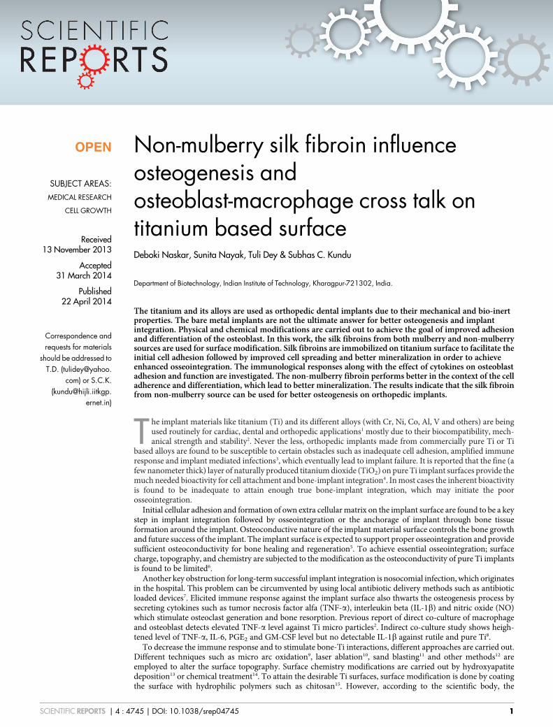
Non-mulberry silk fibroin influenceosteogenesis andosteoblast-macrophage cross talk ontitanium based surfaceDeboki Naskar, Sunita Nayak, Tuli Dey & Subhas C. Kundu
Department of Biotechnology, Indian Institute of Technology, Kharagpur-721302, India.
The titanium and its alloys are used as orthopedic dental implants due to their mechanical and bio-inertproperties. The bare metal implants are not the ultimate answer for better osteogenesis and implantintegration. Physical and chemical modifications are carried out to achieve the goal of improved adhesionand differentiation of the osteoblast. In this work, the silk fibroins from both mulberry and non-mulberrysources are used for surface modification. Silk fibroins are immobilized on titanium surface to facilitate theinitial cell adhesion followed by improved cell spreading and better mineralization in order to achieveenhanced osseointegration. The immunological responses along with the effect of cytokines on osteoblastadhesion and function are investigated. The non-mulberry fibroin performs better in the context of the celladherence and differentiation, which lead to better mineralization. The results indicate that the silk fibroinfrom non-mulberry source can be used for better osteogenesis on orthopedic implants.
The implant materials like titanium (Ti) and its different alloys (with Cr, Ni, Co, Al, V and others) are beingused routinely for cardiac, dental and orthopedic applications1 mostly due to their biocompatibility, mech-anical strength and stability2. Never the less, orthopedic implants made from commercially pure Ti or Ti
based alloys are found to be susceptible to certain obstacles such as inadequate cell adhesion, amplified immuneresponse and implant mediated infections3, which eventually lead to implant failure. It is reported that the fine (afew nanometer thick) layer of naturally produced titanium dioxide (TiO2) on pure Ti implant surfaces provide themuch needed bioactivity for cell attachment and bone-implant integration4. In most cases the inherent bioactivityis found to be inadequate to attain enough true bone-implant integration, which may initiate the poorosseointegration.
Initial cellular adhesion and formation of own extra cellular matrix on the implant surface are found to be a keystep in implant integration followed by osseointegration or the anchorage of implant through bone tissueformation around the implant. Osteoconductive nature of the implant material surface controls the bone growthand future success of the implant. The implant surface is expected to support proper osseointegration and providesufficient osteoconductivity for bone healing and regeneration5. To achieve essential osseointegration; surfacecharge, topography, and chemistry are subjected to the modification as the osteoconductivity of pure Ti implantsis found to be limited6.
Another key obstruction for long-term successful implant integration is nosocomial infection, which originatesin the hospital. This problem can be circumvented by using local antibiotic delivery methods such as antibioticloaded devices7. Elicited immune response against the implant surface also thwarts the osteogenesis process bysecreting cytokines such as tumor necrosis factor alfa (TNF-a), interleukin beta (IL-1b) and nitric oxide (NO)which stimulate osteoclast generation and bone resorption. Previous report of direct co-culture of macrophageand osteoblast detects elevated TNF-a level against Ti micro particles2. Indirect co-culture study shows heigh-tened level of TNF-a, IL-6, PGE2 and GM-CSF level but no detectable IL-1b against rutile and pure Ti8.
To decrease the immune response and to stimulate bone-Ti interactions, different approaches are carried out.Different techniques such as micro arc oxidation9, laser ablation10, sand blasting11 and other methods12 areemployed to alter the surface topography. Surface chemistry modifications are carried out by hydroxyapatitedeposition13 or chemical treatment14. To attain the desirable Ti surfaces, surface modification is done by coatingthe surface with hydrophilic polymers such as chitosan15. However, according to the scientific body, the
OPEN
SUBJECT AREAS:MEDICAL RESEARCH
CELL GROWTH
Received13 November 2013
Accepted31 March 2014
Published22 April 2014
Correspondence andrequests for materials
should be addressed toT.D. (tulidey@yahoo.
com) or S.C.K.([email protected].
ernet.in)
SCIENTIFIC REPORTS | 4 : 4745 | DOI: 10.1038/srep04745 1
biochemical characteristics of Ti surfaces may be modified by util-izing bioactive molecules such as peptides, or proteins to accomplishthe mentioned challenges16,17. This involves chemical immobilizationor physical deposition of the protein molecules such as bone mor-phogenic protein 2 (BMP2)18, fibronectin19, cyclo-DfKRG peptide20
etc. for directional cell adherence. The chemical immobilization ofprotein or peptides through covalent modifications is used to obtainstable, uniform layer on Ti or implant surfaces.
As a natural biomaterial, silk protein fibroin obtained from silk-worms of mulberry origin is found to be extremely useful in differentbiomedical applications21 such as to prepare scaffold, thin film andother different types of matrices22. The silk fibroin offers significantpromise as a biomaterial for bone tissue engineering23 thoughBombyx mori (Bm) fibroin does not contain osteogenic propertiesin itself24. Recent studies show the influence of cross linked RGD-fibroin25,26 and RGD- sericin complex27 on cellular adhesion andproliferation on Ti. The recent reports of non-mulberry fibroin fromthe species Antheraea mylitta (Am) show additional advantages dueto its higher mechanical strength28 and existence of integral RGDsequences29. Am fibroin is found to be greatly cytocompatible withlowered immune response30, which supports the hypothesis of usingit as an immobilizing agent in order to modify the chemical, physicaland biological nature of the implant surface.
The present study utilizes the silk fibroins particularly non-mul-berry fibroin as a viable material for Ti based implant surface modi-fication. The biological effects of immobilized protein over osteoblastand macrophage activation are analyzed in vitro before designing anyin vivo approach. For this reason the mulberry and non-mulberry silkprotein fibroins are immobilized on titanium surfaces to evaluatetheir effects on osseointegration. The protein deposition is confirmedby FTIR, XPS and EDX analyses. The evaluations of adherence,growth, proliferation and differentiation of osteoblast like cells arecarried out to determine the quality of osseointegration. To quantifythe immune response of fibroin immobilized surfaces, TNF-a, IL1-band nitric oxide production level are measured. The effect of cyto-kines on osteoblast adherence is analyzed by direct co-culture ofmacrophages and osteoblast. The result confirms the influence ofsilk protein fibroin particularly nonmulberry fibroin on the osteo-genesis on modified titanium surface.
ResultsExtraction of silk fibroin from mulberry (Bombyx mori/Bm) andnon-mulberry tropical tasar (Antheraea mylitta/Am) sources.Mulberry silk (Bm) cocoon pieces (15.00 gm) yield 10.37 gmdegummed fiber or fibroin (approx. 66%). Dissolution of the silkfiber in LiBr solution gives almost 100% yield of fibroin in solutionform and finally a stock of 20 mg/ml is prepared. From non-mulberry tasar (Am) silk gland, obtained fibroin was dissolved inSDS solution. This was further diluted to reach 20 mg/ml stock.
For immobilization purpose 5 ml of 20 mg/ml of stock solution foreach protein was used to reach a final concentration of 100 mg fibroinon each titanium coupon (1 cm2).
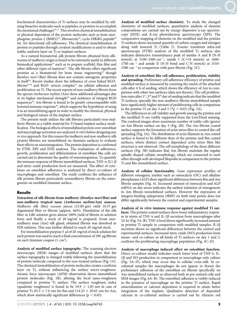
Analysis of modified surface topography. The scanning electronmicroscopic (SEM) images of modified surfaces show that thesurface topography is changed visibly following the immobilizationof protein molecule compared to the non-treated surfaces (Fig. 1A).The chemical immobilization of protein molecules creates a uniformlayer on Ti, without influencing the surface micro-roughness.Atomic force microscopic (AFM) observation shows immobilizedprotein molecules (Fig. 1B), altering the local nano-roughnesscompared to pristine Ti surface. The surface roughness index(quadratic roughness) is found to be 19.9 6 1.05 nm in case ofpristine Ti, 83.3 6 7.6 nm for Bm and 114.23 6 20.65 nm for Am,which show statistically significant differences (p , 0.05).
Analysis of modified surface chemistry. To study the changedchemistry of modified surfaces, quantitative analysis of elementcompositions are carried out by energy dispersive x-ray spectros-copy (EDX) and X-ray photoelectron spectroscopy (XPS). Thecomparative mapping of elements on the modified and the pristineTi surfaces shows increased quantity of carbon, oxygen and nitrogenalong with lowered Ti (Table 1). Fourier transform infra-redspectroscopy (FTIR) analysis of the modified Ti surfaces alsoindicates distinctive transmittance peak of amides A and B (N-Hstretch) at 3100–3300 cm21, amide I (C5O stretch) at 1600–1700 cm21 and amide II (N-H bend and C-N stretch) at 1510–1580 cm21 in comparison with pure fibroin (Fig. 1C).
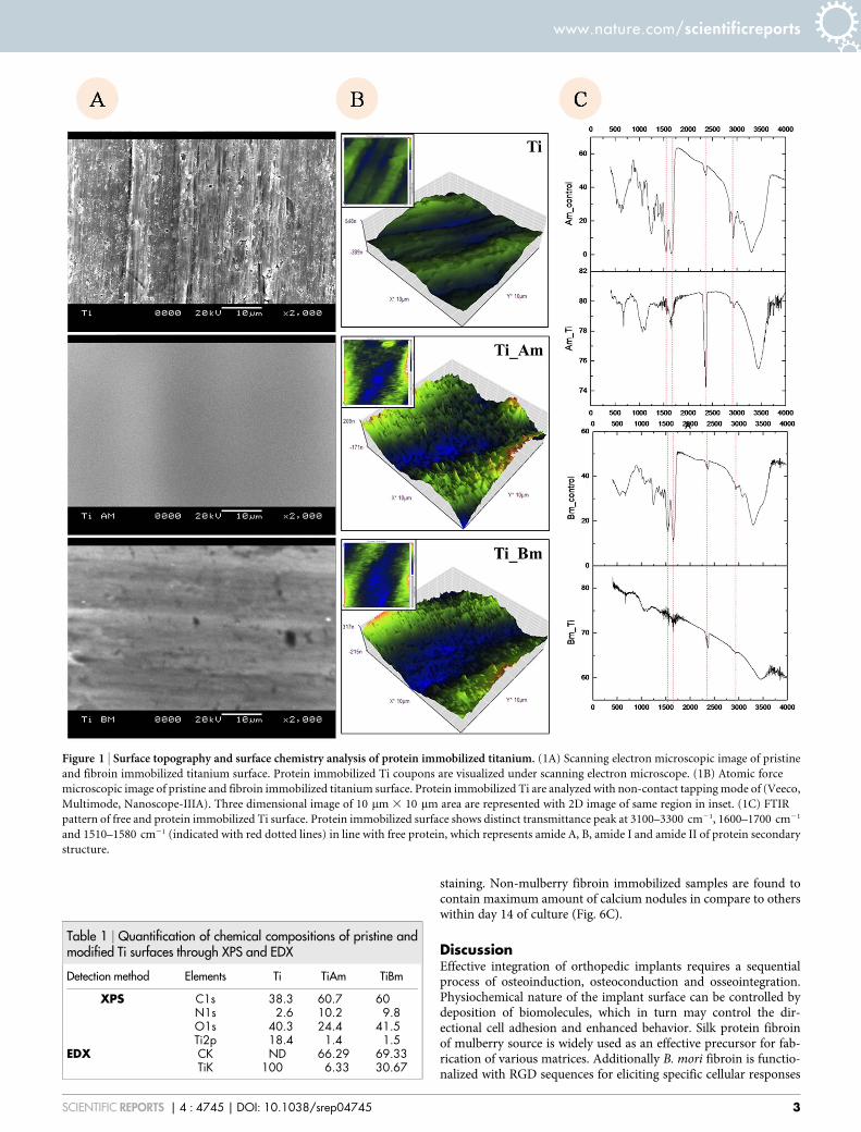
Analysis of osteoblast like cell adherence, proliferation, viabilityand spreading. Preliminary cell adherence efficiency of pristine andmodified surface is measured by counting the nuclei of the attachedcells after 4 h of seeding, which shows the efficiency of Am in com-parison with other two surfaces (data not shown). The cell prolifera-tion rates after 1st, 3rd and 5th day of seeding indicate that the modifiedTi surfaces, specially the non-mulberry fibroin immobilized samplehave significantly higher amount of proliferating cells in comparisonto the pristine Ti on day 3 and 5 (*p , 0.05) (Fig. 2A).
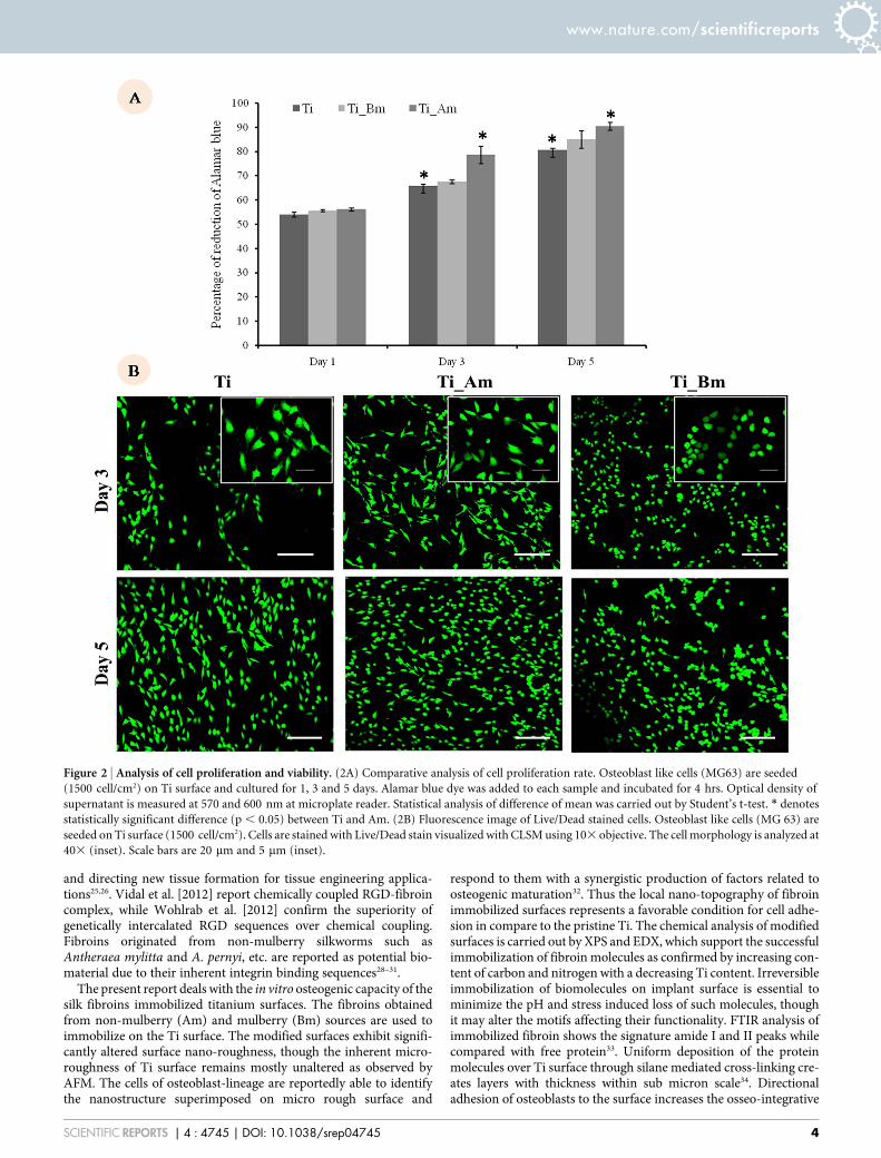
The differences in cell viability and growth among the pristine andthe modified Ti are visibly supported from the Live/Dead staining.The confocal images show maximum number of viable cells (green)on Am fibroin surface on day 3 (Fig. 2B). The Am fibroin coatedsurface supports the formation of actin stress fiber to control the cellspreading (Fig. 3A). The distribution of actin filament in Am coatedsurface is found to be different than of the pristine and Bm coatedsurfaces, where distinct contact dependent actin stress fiber likestructure is not observed. The cell morphology of the three differentsurfaces (Fig. 3B) indicates that Am fibroin maintain the regularspindle shaped cellular morphology, which are connected to eachother through well-developed filopodia in comparison to the pristineTi and Bm immobilized surface.
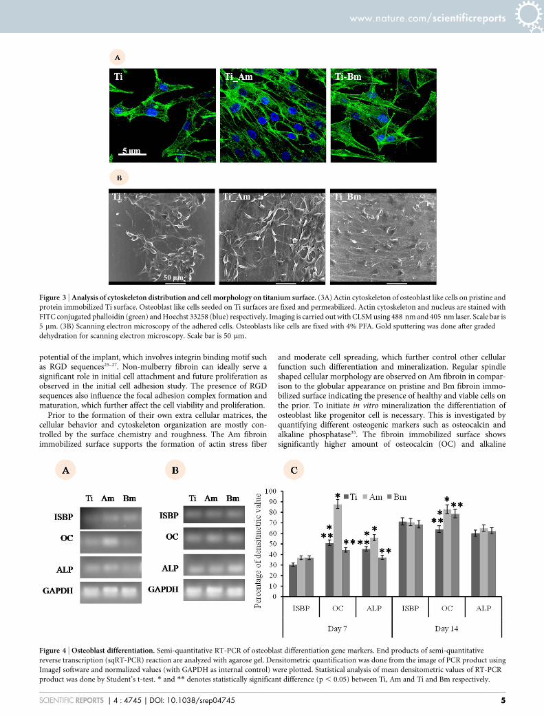
Analysis of cellular functionality. Gene expression profiles ofdifferent osteogenic marker such as osteocalcin (OC) and alkalinephosphatase (ALP) show significant differences between Bm and Amfibroin samples (Fig. 4). Increased amount of osteocalcin and ALPmRNA on day seven indicates the earliest initiation of osteogenesisin Am fibroin immobilized surfaces. However the expression ofintegrin-binding sialoprotein (IBSP) on both time points does notdiffer significantly between the control and experimental samples.
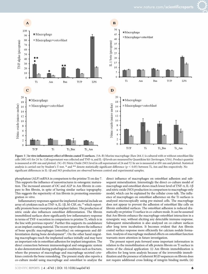
Analysis of in vitro immune response against modified Ti sur-faces. The protein coated surfaces show lower inflammatory respon-se in terms of TNF-a and IL-1b secretion from macrophages after24 hr (Fig. 5A–B). TNF-a level shows significantly increased amountin pristine Ti sample in comparison with modified samples. Il-1bsecretion shows no significant differences between the control andexperimental surfaces. Increased nitric oxide (NO) production frommono- and co-culture in all kinds of Ti surfaces on day 1 and 3,confirms the proliferating macrophage population (Fig. 5C–D).
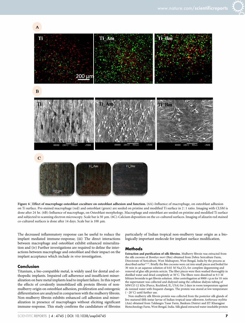
Analysis of macrophage induced effect on osteoblast function.Direct co-culture model indicates much lower level of TNF-a, IL-1b and NO production in comparison to macrophage only culture(Fig. 5A–D), which may occur due to cellular cross-talk. In co-cultured samples the macrophages do not appear to thwart thepreliminary adhesion of the osteoblast on fibroin specifically onAm immobilized surfaces as observed both in pre-stained cells andSEM images (Fig. 6A–B). The osteoblast adhesion is visibly reducedin the presence of macrophage on the pristine Ti surface. Rapidmineralization or calcium deposition is required to attain betterintegration of the implant. Qualitative detection of depositedcalcium in co-cultured surfaces is carried out by Alizarin red
www.nature.com/scientificreports
SCIENTIFIC REPORTS | 4 : 4745 | DOI: 10.1038/srep04745 2
staining. Non-mulberry fibroin immobilized samples are found tocontain maximum amount of calcium nodules in compare to otherswithin day 14 of culture (Fig. 6C).
DiscussionEffective integration of orthopedic implants requires a sequentialprocess of osteoinduction, osteoconduction and osseointegration.Physiochemical nature of the implant surface can be controlled bydeposition of biomolecules, which in turn may control the dir-ectional cell adhesion and enhanced behavior. Silk protein fibroinof mulberry source is widely used as an effective precursor for fab-rication of various matrices. Additionally B. mori fibroin is functio-nalized with RGD sequences for eliciting specific cellular responses
Figure 1 | Surface topography and surface chemistry analysis of protein immobilized titanium. (1A) Scanning electron microscopic image of pristine
and fibroin immobilized titanium surface. Protein immobilized Ti coupons are visualized under scanning electron microscope. (1B) Atomic force
microscopic image of pristine and fibroin immobilized titanium surface. Protein immobilized Ti are analyzed with non-contact tapping mode of (Veeco,
Multimode, Nanoscope-IIIA). Three dimensional image of 10 mm 3 10 mm area are represented with 2D image of same region in inset. (1C) FTIR
pattern of free and protein immobilized Ti surface. Protein immobilized surface shows distinct transmittance peak at 3100–3300 cm21, 1600–1700 cm21
and 1510–1580 cm21 (indicated with red dotted lines) in line with free protein, which represents amide A, B, amide I and amide II of protein secondary
structure.
Table 1 | Quantification of chemical compositions of pristine andmodified Ti surfaces through XPS and EDX
Detection method Elements Ti TiAm TiBm
XPS C1s 38.3 60.7 60N1s 2.6 10.2 9.8O1s 40.3 24.4 41.5Ti2p 18.4 1.4 1.5
EDX CK ND 66.29 69.33TiK 100 6.33 30.67
www.nature.com/scientificreports
SCIENTIFIC REPORTS | 4 : 4745 | DOI: 10.1038/srep04745 3
and directing new tissue formation for tissue engineering applica-tions25,26. Vidal et al. [2012] report chemically coupled RGD-fibroincomplex, while Wohlrab et al. [2012] confirm the superiority ofgenetically intercalated RGD sequences over chemical coupling.Fibroins originated from non-mulberry silkworms such asAntheraea mylitta and A. pernyi, etc. are reported as potential bio-material due to their inherent integrin binding sequences28–31.
The present report deals with the in vitro osteogenic capacity of thesilk fibroins immobilized titanium surfaces. The fibroins obtainedfrom non-mulberry (Am) and mulberry (Bm) sources are used toimmobilize on the Ti surface. The modified surfaces exhibit signifi-cantly altered surface nano-roughness, though the inherent micro-roughness of Ti surface remains mostly unaltered as observed byAFM. The cells of osteoblast-lineage are reportedly able to identifythe nanostructure superimposed on micro rough surface and
respond to them with a synergistic production of factors related toosteogenic maturation32. Thus the local nano-topography of fibroinimmobilized surfaces represents a favorable condition for cell adhe-sion in compare to the pristine Ti. The chemical analysis of modifiedsurfaces is carried out by XPS and EDX, which support the successfulimmobilization of fibroin molecules as confirmed by increasing con-tent of carbon and nitrogen with a decreasing Ti content. Irreversibleimmobilization of biomolecules on implant surface is essential tominimize the pH and stress induced loss of such molecules, thoughit may alter the motifs affecting their functionality. FTIR analysis ofimmobilized fibroin shows the signature amide I and II peaks whilecompared with free protein33. Uniform deposition of the proteinmolecules over Ti surface through silane mediated cross-linking cre-ates layers with thickness within sub micron scale34. Directionaladhesion of osteoblasts to the surface increases the osseo-integrative
Figure 2 | Analysis of cell proliferation and viability. (2A) Comparative analysis of cell proliferation rate. Osteoblast like cells (MG63) are seeded
(1500 cell/cm2) on Ti surface and cultured for 1, 3 and 5 days. Alamar blue dye was added to each sample and incubated for 4 hrs. Optical density of
supernatant is measured at 570 and 600 nm at microplate reader. Statistical analysis of difference of mean was carried out by Student’s t-test. * denotes
statistically significant difference (p , 0.05) between Ti and Am. (2B) Fluorescence image of Live/Dead stained cells. Osteoblast like cells (MG 63) are
seeded on Ti surface (1500 cell/cm2). Cells are stained with Live/Dead stain visualized with CLSM using 103 objective. The cell morphology is analyzed at
403 (inset). Scale bars are 20 mm and 5 mm (inset).
www.nature.com/scientificreports
SCIENTIFIC REPORTS | 4 : 4745 | DOI: 10.1038/srep04745 4
potential of the implant, which involves integrin binding motif suchas RGD sequences25–27. Non-mulberry fibroin can ideally serve asignificant role in initial cell attachment and future proliferation asobserved in the initial cell adhesion study. The presence of RGDsequences also influence the focal adhesion complex formation andmaturation, which further affect the cell viability and proliferation.
Prior to the formation of their own extra cellular matrices, thecellular behavior and cytoskeleton organization are mostly con-trolled by the surface chemistry and roughness. The Am fibroinimmobilized surface supports the formation of actin stress fiber
and moderate cell spreading, which further control other cellularfunction such differentiation and mineralization. Regular spindleshaped cellular morphology are observed on Am fibroin in compar-ison to the globular appearance on pristine and Bm fibroin immo-bilized surface indicating the presence of healthy and viable cells onthe prior. To initiate in vitro mineralization the differentiation ofosteoblast like progenitor cell is necessary. This is investigated byquantifying different osteogenic markers such as osteocalcin andalkaline phosphatase35. The fibroin immobilized surface showssignificantly higher amount of osteocalcin (OC) and alkaline
Figure 3 | Analysis of cytoskeleton distribution and cell morphology on titanium surface. (3A) Actin cytoskeleton of osteoblast like cells on pristine and
protein immobilized Ti surface. Osteoblast like cells seeded on Ti surfaces are fixed and permeabilized. Actin cytoskeleton and nucleus are stained with
FITC conjugated phalloidin (green) and Hoechst 33258 (blue) respectively. Imaging is carried out with CLSM using 488 nm and 405 nm laser. Scale bar is
5 mm. (3B) Scanning electron microscopy of the adhered cells. Osteoblasts like cells are fixed with 4% PFA. Gold sputtering was done after graded
dehydration for scanning electron microscopy. Scale bar is 50 mm.
Figure 4 | Osteoblast differentiation. Semi-quantitative RT-PCR of osteoblast differentiation gene markers. End products of semi-quantitative
reverse transcription (sqRT-PCR) reaction are analyzed with agarose gel. Densitometric quantification was done from the image of PCR product using
ImageJ software and normalized values (with GAPDH as internal control) were plotted. Statistical analysis of mean densitometric values of RT-PCR
product was done by Student’s t-test. * and ** denotes statistically significant difference (p , 0.05) between Ti, Am and Ti and Bm respectively.
www.nature.com/scientificreports
SCIENTIFIC REPORTS | 4 : 4745 | DOI: 10.1038/srep04745 5
phosphatase (ALP) mRNA in comparison to the pristine Ti on day 7.This supports the influence of nanostructures in osteogenic matura-tion. The increased amount of OC and ALP in Am fibroin in com-pare to Bm fibroin, in spite of having similar surface topography.This suggests the superiority of Am fibroin in promoting osseointe-gration in vitro.
Inflammatory responses against the implanted material include anarray of cytokines such as TNF-a, IL-1b, M-CSF, etc.36 which report-edly promote bone resorption and implant failure. The production ofnitric oxide also influences osteoblast differentiation. The fibroinimmobilized surfaces show significantly low inflammatory responsein terms of TNF-a secretion in comparison to pristine Ti, which is inthe line with previous reports2. Further this supports its candidatureas an implant coating material. The recent report shows the influenceof bone specific macrophages (osteoMac) on osteogenesis and dif-ferentiation during bone development37. On the other hand migrat-ing macrophages reach the implanted surface initially and may havean important role in osteoblast adhesion for implant integration. Thedirect connection between immuonological and ostegogenic systemis also demonstrated during pathological conditions such as fracture,where the presence of macrophage and macrophage secreted cyto-kines controls the bone remodeling. The present study also reports aco-culture model using macrophage and osteoblast to analyze the
direct influence of macrophages on osteoblast adhesion and sub-sequent mineralization. Interestingly the direct co-culture model ofmacrophage and osteoblast shows much lower level of TNF-a, IL-1band nitric oxide (NO) production in comparison to macrophage onlymodel, which can be explained by the cellular cross-talk. The influ-ence of macrophages on osteoblast adherence on the Ti surfaces isanalyzed microscopically using pre-stained cells. The macrophagedoes not appear to prevent the adhesion of osteoblast like cells onfibroin embedded surfaces. The osteoblast adhesion is reduced dra-matically on pristine Ti surface in co-culture mode. It can be assumedthat Am fibroin enhance the macrophage-osteoblast interaction in asynergistic way, without eliciting any detectable immune-response.Subsequent mineralization is also analyzed on co-culture surfacesafter long term incubation. It becomes evident that Am fibroincoated surface response more efficiently for calcium nodule forma-tion. Analysis of macrophage mediated effects on osteoblast functionwarrants more attention in future investigation.
The present report puts forward some important information inrelation to the immobilization of silk protein fibroin on Ti surface interms of the clinical application: (i) Am fibroin immobilization iseffective for long term analysis because of the irreversible immob-ilization and the presence of inherent RGD sequences on fibroin doesnot require additional cross linking of integrin binding motifs; (ii)
Figure 5 | In vitro inflammatory effect of fibroin coated Ti surfaces. (5A–B) Murine macrophage (Raw 264.1) is cultured with or without osteoblast like
cells (MG-63) for 24 hr. Cell supernatant was collected and TNF-a, and IL-1b levels are measured by Quantikine kit (Invitrogen, USA). Product quantity
is measured at 450 nm and plotted. (5C–D) Nitric Oxide (NO) level in cell supernatant of 24 and 72 hr are is measured at 450 nm and plotted. Statistical
analysis is carried out by Student’s T-test. * and ** denote statistically significant difference (p , 0.05) between Ti, Am and Bm respectively. No
significant differences in IL-1b and NO production are observed between control and experimental samples.
www.nature.com/scientificreports
SCIENTIFIC REPORTS | 4 : 4745 | DOI: 10.1038/srep04745 6
The decreased inflammatory response can be useful to reduce theimplant mediated immune-response; (iii) The direct interactionsbetween macrophage and osteoblast exhibit enhanced mineraliza-tion and (iv) Further investigations are required to define the inter-actions between macrophage and osteoblast and their impact on theimplant acceptance which include in vivo investigation.
ConclusionTitanium, a bio-compatible metal, is widely used for dental and or-thopedic implants. Impaired cell adherence and insufficient miner-alization on bare metal implants lead to implant failure. In this reportthe effects of covalently immobilized silk protein fibroin of non-mulberry origin on osteoblast adhesion, proliferation and osteogenicdifferentiation are analyzed in comparison with the mulberry fibroin.Non-mulberry fibroin exhibits enhanced cell adhesion and miner-alization in presence of macrophages without eliciting significantimmune-response. This study confirms the candidature of fibroins
particularly of Indian tropical non-mulberry tasar origin as a bio-logically important molecule for implant surface modification.
MethodsExtraction and purification of silk fibroins. Mulberry fibroin was extracted fromthe silk cocoons of Bombyx mori (Bm) obtained from Debra Sericulture Farm,Directorate of Sericulture, West Midnapore, West Bengal, India by the process asdescribed earlier21,22. Briefly the Bm cocoons were cut into small pieces and boiled for30 min in an aqueous solution of 0.02 M Na2CO3 for complete degumming andremoval of glue silk protein sericin. The fiber pieces were then washed thoroughly indistilled water and dried completely at 50uC. The fibers were dissolved in 9.3 Mlithium bromide to get fibroin solution. After centrifugation at 9000 r.p.m for 10 minthe supernatant was collected and dialyzed using the cellulose dialysis tubing ofMWCO 12 kDa (Pierce, Rockford, IL, USA) for 2 days in room temperature againstde-ionized water with frequent changes. The protein was stored at low temperature(220uC) until further use.
Non-mulberry silk fibroin protein was collected from the posterior silk glands oflive matured fifth instar larvae of Indian tropical tasar silkworm Antheraea mylitta(Am) obtained from Taldangra Tasar Farm, Bankura District and IIT KharagpurBiotechnology Farm, West Bengal, India. Silk gland extracted water insoluble protein
Figure 6 | Effect of macrophage-osteoblast coculture on osteoblast adhesion and function. (6A)-Influence of macrophage, on osteoblast adhesion
on Ti surface. Pre-stained macrophage (red) and osteoblast (green) are seeded on pristine and modified Ti surface in 251 ratio. Imaging with CLSM is
done after 24 hr. (6B)-Influence of macrophage, on Osteoblast morphology. Macrophage and osteoblast are seeded on pristine and modified Ti surface
and subjected to scanning electron microscopy. Scale bar is 50 mm. (6C)-Calcium deposition on the co-cultured surfaces. Imaging of alizarin red stained
co-cultured surfaces is done after 14 days. Scale bar is 100 mm.
www.nature.com/scientificreports
SCIENTIFIC REPORTS | 4 : 4745 | DOI: 10.1038/srep04745 7
fibroin was solubilized with 1% sodium lauryl sulfate (SLS)30,31. The solubilized fib-roin was dialyzed for 10–12 hrs at room temperature against deionized water withfrequent changes. The fibroin solution was stored at 220uC. Protein content wasestimated by colorimetric (Bradford) method.
Modification of titanium (Ti) surface. Silanization of Ti surface using APTES. Thetitanium sheet (0.1 mm thickness with 99.9% purity from Sigma Aldrich, St Louis,USA) was cut into 1 cm 3 1 cm pieces. The pieces were then washed thoroughly for15 min each by sonication in acetone (70% v/v) followed by ethanol and de-ionizedwater. The samples were dried to facilitate the silanization process. Theimmobilization of APTES (Sigma Aldrich, St Louis, USA) on the titanium sampleswas carried out by following the recipes mentioned earlier28 with little modifications.The cleaned pieces were submerged in a 2% (v/v) solution of APTES in toluene for10 h at room temperature. The samples were washed with pure toluene thrice (each30 min) to remove any residual APTES. To remove the toluene, the metal pieces werewashed with ethanol followed by deionized water. Gluteraldehyde solution (2%)(Sigma Aldrich, St Louis, USA) was poured over the Ti-APTES samples and left for5 h at room temperature. The samples were further rinsed thoroughly with deionizedwater and dried.
Immobilization fibroins of mulberry (Bm) and non-mulberry (Am) origins. Bm andAm fibroin solutions (each 20 mg/ml) in PBS were used for immobilization. Thesilanized Ti samples were incubated in the protein solution for 60 min at 25uC. Afterthe immobilization the metal pieces were washed thrice in distilled water to removeany unattached fibroin.
Cell culture and maintenance. Mouse osteoblast cell line (MG-63) and mousemacrophage (RAW 264.7) cells were cultured in Dulbecco’s Modified Eagle’sMedium (Invitrogen, USA) supplemented with 10% fetal bovine serum, 100 U/mlpenicillin and 100 mg/ml streptomycin (Gibco BRL, Grand Island, NY, USA). All thecell lines were incubated at 37uC in a humidified atmosphere of 5% CO2 with thegrowth media changed every 48 h. The subsequent passages were carried out from theconfluent monolayer of cells obtained by washing the cells with sterile 13 phosphatebuffered saline (PBS) pH 7.4 and trypsinization with chilled 13 trypsin EDTA (GibcoBRL Grand Island, NY, USA). The harvested cells were centrifuged and seeded on Tisurface (1500 cell/cm2) for further experimentation.
Analysis of the modified surface topography. Surface morphology analysis by SEM.The surface morphology of pristine Ti, and modified Ti samples were observed aftergold sputtering using SEM (JEOL JSM 5800).
Surface topography analysis by AFM. AFM (Veeco, Multimode, Nanoscope-IIIA) wascarried out by scanning the 10 3 10 mm2 area in non-contact mode with tips mountedon cantilevers at spring constant of 40 N/m (as specified by the manufacturer).Quadric roughness was calculated to compare the surface roughness of control andexperimental samples.
Analysis of modified surface chemistry. XPS and EDX analysis. The chemicalcomposition of pristine Ti, and modified samples were determined by XPS (PHI 5000Versa Probe III, Physical Electronics, Inc. Minnesota, USA) and EDX (INCA PentaFET 33, Oxford Instrument UK).
FTIR analysis. The infrared absorption spectra of the free protein and the proteinimmobilized samples were obtained from an FTIR spectrophotometer (ShimadzuDT-40 model 883 IR Spectrophotometer) in diffuse reflectance mode. For eachspectrum obtained, a total of 64 scans were accumulated at 4 cm21 resolution.Scanning was conducted in the range from 400 to 4000 cm21.
Analysis of osteoblast like cell adherence, proliferation/viability and morphology.Cell proliferation. MG-63 cells were seeded at a density of 1500 cells/cm2 on pristineTi and coated Ti samples and cultured at 37uC in a humidified atmosphere with 5%CO2. The cell seeded samples were washed in sterile 13 PBS and placed into freshsterile wells after 1, 3 and 5 days. Alamar blue (Resazurin) dye (Invitrogen, USA) wasused following Manufacturer’s protocol and dye reduction was measured bymeasuring optical density of the solution at 570 and 600 nm.
Cell viability. The cell seeded surfaces were rinsed three times with sterile 13 PBScarefully and incubated with live/dead stain (Molecular Probes, Invitrogen, USA) for30 min at room temperature. Live/dead stained cells were visualized under confocalmicroscope (CLSM) using 488 and 564 nm lasers (Fluoview1000, Olympus, Japan).
Analysis of cytoskeleton organization and cellular morphology. For actin filamentstaining, the cell seeded Ti coupons were fixed with 2% PFA and permeabilized with0.1% TritonX-100. FITC conjugated phalloidin (actin) and Hoechst (nucleus) wereused in 15200 dilution (in 13 PBS). The samples were analyzed using 405 nm and488 nm laser in CLSM.
To analyze the cell morphology, the cell seeded Ti samples were fixed and preparedfor SEM analysis by gradual dehydration. The samples were then gold sputtered andanalyzed using SEM.
Analysis of cellular functionality. RT-PCR analysis. The cell seeded Ti coupons weretreated with Trizol (Invitrogen, USA) followed by the isolation of total RNA usingRNA isolation kit (Qiagen, Germany). First strand cDNA was synthesized from 10 mg
of total RNA using first strand cDNA synthesis kit (Fermentas, Canada). Semi-quantitative RT-PCR reaction of integrin-binding sialoprotein (IBSP), osteocalcin(OC) and alkaline phosphatase (ALP) were carried out by using custom madeforward and reverse primers. Comparative analysis of end products (approx. 120–150 kDa) was performed using gel analysis plug-in of ImageJ software andnormalized with GAPDH.
Analysis of immune response elicited by the modified Ti surfaces. Quantification ofTNF-a and IL-1b production from macrophage culture. Macrophage (105 cells/cm2)cells were seeded on the fibroin coated samples. After 24 h of cell seeding, thecomplete medium was replaced with incomplete medium. After 24 h supernatant wascollected to analyze TNF-a and IL-1b level using TNF-a and IL-1b quantification kit(Invitrogen, USA). Nitric oxide level was analyzed with samples from 24 hr and72 hr, using Griess reagent (Sigma, St Louis, USA). The data accumulations werecarried out at 450 nm and 600 nm using a microplate reader.
Analysis of macrophage elicited influence on osteoblast activity. Osteoblastadhesion in macrophage-osteoblast co-culture. For direct co-culture the osteoblast (0.53 105/cm2) and macrophage (105 cells/cm2) cells were pre-stained withCellTrackerTM Green CMFDA (Invitrogen) and CellTrackerTM Orange CMTMR(Invitrogen) respectively for 30 min following the Manufacturer’s protocol. Cellseeded surfaces were treated as per standard protocol and fixed with 2% PFA. Imagingwas carried out using 488 and 543 nm laser in CLSM (FV1000, Olympous, Japan) insequential mode to analyze the cell adhesion efficiency.
Adhered cell morphology in co-culture model was analyzed by fixing the surfacewith 2% PFA and prepared for SEM analysis by gradual dehydration. The sampleswere then gold sputtered and analyzed using SEM (JEOL JSM 5800).
Quantification of TNF-a, IL-1b and nitric oxide (NO) production in macrophage-osteoblast co-culture. The macrophage and osteoblast cells (251 ratio) were seeded onpristine and modified Ti for 24 hr. The cell supernatants were collected and analyzedfor TNF-a, IL-1b and NO quantifications as stated before.
Calcium deposition. Alizarin red staining for mineralization was performed afterfourteen days of co-culture. The cell seeded samples were fixed with 10% formalin,and stained in 1% alizarin red S (Sigma Aldrich, St Louis, USA) solution (pH 4.0) for8–10 min. Images were taken with ECLIPSE TS100 (Nikon, Japan).
Statistical analysis. All the experiments were repeated three times (with samplenumber $3) and the mean value are plotted with standard deviations. The statisticalsignificance is calculated using Student’s t test.
1. Brunette, D. M. (ed.) Titanium in Medicine. (Springer-Verlag, Berlin &Heidelberg. 2001).
2. Horowitz, S. M. & Gonzales, J. B. Inflammatory Response to Implant Particulatesin a Macrophage/Osteoblast Coculture Model. Calcif. Tissue Int. 59, 392–396(1996).
3. Schierholz, J. M. & Beuth, J. Implant infections: a haven for opportunistic bacteria.J. Hosp. Infect. 49, 87–93 (2001).
4. Sul, Y. et al. Characteristics of the surface oxides on turned and electrochemicallyoxidized pure titanium implants up to dielectric breakdown: the oxide thickness,micropore configurations, surface roughness, crystal structure and chemicalcomposition. Biomaterials 23, 491–501 (2002).
5. Albrektsson, T. & Johanssen, C. Osteoinduction, osteoconduction andosseointegration. Eur. Spine. J. 10, 96–101 (2001).
6. Yamamoto, D. et al. Osteoconductivity and hydrophilicity of TiO2 coatings on Tisubstrates prepared by different oxidizing processes. Bioinorg. Chem. Appl. 2012,1–7 (2012).
7. Han, C. M., Lee, E. J., Kim, H. E., Koh, Y. H. & Jang, J. H. Porous TiO2 films on Tiimplants for controlled release of tetracycline-hydrochloride (TCH). Thin SolidFilms 519, 8074–8076 (2011).
8. Valles, G., Gil-Garay, E., Munuera, L. & Vilaboa, N. Modulation of the cross-talkbetween macrophages and osteoblasts by titanium-based particles. Biomaterials29, 2326–2335 (2008).
9. Chen, H. T. et al. Osteoblast growth behavior on micro-arc oxidized b-titaniumalloy. Surf. Coat. Tech. 205, 1624–1629 (2010).
10. Faeda, R. S., Tavares, H. S., Sartori, R., Guastaldi, A. C. & Marcantonio, E., Jr.Evaluation of titanium implants with surface modification by laser beam:biomechanical study in rabbit tibias. Braz. Oral. Res. 23, 137–143 (2009).
11. Piattelli, A., Scarano, A., Piattelli, M. & Calabrese, L. Direct bone formation onsand-blasted titanium implants: an experimental study. Biomaterials 17,1015–1018 (1996).
12. Guehennec, L. L., Soueidan, A., Layrolle, P. & Amouriq, Y. Surface treatments oftitanium dental implants for rapid osseointegration. Dent. Mater. 23, 844–854(2007).
13. Ball, M. D. et al. Osteoblast growth on titanium foils coated with hydroxyapatiteby pulsed laser ablation. Biomaterials 22, 337–347 (2001).
14. Rupp, F. et al. Enhancing surface free energy and hydrophilicity through chemicalmodification of microstructured titanium implant surfaces. J. Biomed. Mater. Res.A 76, 323–334 (2006).
www.nature.com/scientificreports
SCIENTIFIC REPORTS | 4 : 4745 | DOI: 10.1038/srep04745 8
15. Leedy, M. R. et al. Use of chitosan as a bioactive implant coating for bone-implantapplications. Adv. Polym. Sci. 244, 129–165 (2011).
16. Giannoni, P. et al. Osteogenic differentiation of human mesenchymal stromalcells on surface-modified titanium alloys for orthopedic and dental implants. Int.J. Artif. Organs 32, 811–820 (2009).
17. Morra, M. Biochemical modification of titanium surfaces: peptides and ECMproteins. Eur. Cell Mater. 12, 1–15 (2006).
18. Kashiwagi, K., Tsuji, T. & Shiba, K. Directional BMP-2 for functionalization oftitanium surfaces. Biomaterials 30, 1166–1175 (2009).
19. Degasne, I. et al. Effects of roughness, fibronectin and vitronectin on attachment,spreading, and proliferation of human osteoblast-like cells (Saos-2) on titaniumsurfaces. Calcif. Tissue Int. 64, 499–507 (1999).
20. Pallu, S. et al. The effect of cyclo-DfKRG peptide immobilization on titanium onthe adhesion and differentiation of human osteoprogenitor cells. Biomaterials 26,6932–6940 (2005).
21. Omenetto, F. G. & Kaplan, D. L. New Opportunities for an ancient material.Science 329, 528–531 (2010).
22. Rockwood, D. N. et al. Materials fabrication from Bombyx mori silk fibroin. Nat.Protoc. 6, 1612–1631 (2011).
23. Sofia, S., McCarthy, M. B., Gronowicz, G. & Kaplan, D. L. Functionalized silk-based biomaterials for bone formation. J. Biomed. Mater. Res. 54, 139–148 (2001).
24. Bhumiratana, S. et al. Nucleation and growth of mineralized bone matrix on silk-hydroxyapatite composite scaffolds. Biomaterials 32, 2812–2820 (2011).
25. Vidal, G. et al. Enhanced cellular adhesion on titanium by silk functionalized withtitanium binding and RGD peptides. Acta Biomater. 9, 4935–4943 (2012).
26. Wohlrab, S. et al. Cell adhesion and proliferation on RGD-modified recombinantspider silk proteins. Biomaterials 33, 6650–6659 (2012).
27. Nayak, S., Dey, T., Naskar, D. & Kundu, S. C. The promotion of osseointegrationof titanium surfaces by coating with silk protein sericin. Biomaterials 34(12),2855–2864 (2013). (DOI: 10.1016/j.biomaterials.2013.01.019. Epub 2013 Jan 26.)
28. Kundu, S. C. et al. Nonmulberry silk biopolymers. Biopolymers 97, 455–467(2012).
29. Patra, C. et al. Silk protein fibroin from Antheraea mylitta for cardiac tissueengineering. Biomaterials 33, 2673–2680 (2012).
30. Mandal, B. B. & Kundu, S. C. Osteogenic and adipogenic differentiation of ratbone marrow cells on non-mulberry and mulberry silk gland fibroin 3D scaffolds.Biomaterials 30, 5019–5030 (2009).
31. Datta, A., Ghosh, A. K. & Kundu, S. C. Purification and characterization of fibroinfrom the tropical saturniid silkworm, Antheraea mylitta. Insect Biochem. Mol.Biol. 31, 1013–1018 (2001).
32. Gittens, R. A. et al. Differential responses of osteoblast lineage cells tonanotopographically-modified, microroughened titanium-aluminum-vanadiumalloy surfaces. Biomaterials 33, 8986–8994 (2012).
33. Acharya, C., Ghosh, S. K. & Kundu, S. C. Silk fibroin film from non-mulberrytropical tasar silkworms: A novel substrate for in vitro fibroblast culture. ActaBiomaterialia. 5, 429–437 (2009).
34. Kato, K., Uchida, E., Kang, E. T., Uyama, Y. & Ikada, Y. Polymer surface with graftchains. Prog Polym Sci 28, 209–259 (2003).
35. Kim, S. E. et al. The effect of immobilization of heparin and bone morphogenicprotein-2 (BMP-2) to titanium surfaces on inflammation and osteoblast function.Biomaterials 32, 366-373 (2011).
36. Chaudhary, L. R., Spelsberg, T. C. & Riggs, B. L. Production of various cytokines bynormal human osteoblast-like cells in response to interleukin-1 beta and tumornecrosis factor-alpha: lack of regulation by 17-beta-estradiol. Endocrinology 130,2528–2534 (1992).
37. Arron, J. R. & Choi, Y. Osteoimmunology: Bone versus immune system. Nature408, 535–536 (2000).
AcknowledgmentsThis work is supported by Department of Biotechnology, and Department of Science andTechnology, Government of India. We are greatly indebted to Dr. Mahitosh Mandal andDr. Ananta K. Ghosh, Indian Institute of Technology Kharagpur for scientific inputs andsuggestions.
Author contributionsS.C.K. and T.D. design the experiments; D.N., T.D. and S.N. perform the experiments andanalyzed the data. D.N., T.D. and S.C.K. wrote the manuscript. The manuscript was writtenthrough the contributions of all authors. All authors have given approval to the final versionof the manuscript.
Additional informationSupplementary information accompanies this paper at http://www.nature.com/scientificreports
Competing financial interests: The authors declare no competing financial interests.
How to cite this article: Naskar, D., Nayak, S., Dey, T. & Kundu, S.C. Non-mulberry silkfibroin influence osteogenesis and osteoblast-macrophage cross talk on titanium basedsurface. Sci. Rep. 4, 4745; DOI:10.1038/srep04745 (2014).
This work is licensed under a Creative Commons Attribution-NonCommercial-NoDerivs 3.0 Unported License. The images in this article are included in thearticle’s Creative Commons license, unless indicated otherwise in the image credit;if the image is not included under the Creative Commons license, users will need toobtain permission from the license holder in order to reproduce the image. To viewa copy of this license, visit http://creativecommons.org/licenses/by-nc-nd/3.0/
www.nature.com/scientificreports
SCIENTIFIC REPORTS | 4 : 4745 | DOI: 10.1038/srep04745 9