Embed Size (px)
Citation preview

Non-destructive imaging of an individual protein
J.-N. Longchamp*, T. Latychevskaia, C. Escher, H.-W. Fink
Physics Institute
University of Zurich
Winterthurerstrasse 190
8057 Zurich
Switzerland
*Corresponding author:
E-mail: [email protected]
Major category: Physics
Minor category: Biophysics and Computational Biology

Abstract
The mode of action of proteins is to a large extent given by their ability to adopt
different conformations. This is why imaging single biomolecules at atomic resolution
is one of the ultimate goals of biophysics and structural biology. The existing protein
database has emerged from X-ray crystallography, NMR or cryo-TEM investigations.
However, these tools all require averaging over a large number of proteins and thus
over different conformations. This of course results in the loss of structural
information. Likewise it has been shown that even the emergent X-FEL technique will
not get away without averaging over a large quantity of molecules(1).
Here we report the first recordings of a protein at sub-nanometer resolution obtained
from one individual ferritin by means of low-energy electron holography. One single
protein could be imaged for an extended period of time without any sign of radiation
damage(2). Since ferritin exhibits an iron core, the holographic reconstructions could
also be cross-validated against TEM images of the very same molecule by imaging
the iron cluster inside the molecule while the protein shell is decomposed.

Introduction
All conventional methods for investigating the structure of biomolecules such as
proteins suffer from significant drawbacks. X-ray crystallography, NMR and cryo-
electron microscopy require averaging over a large number of molecules, usually
more than 106 similar entities(6) and as a consequence conformational details remain
undiscovered. Furthermore, these methods can only be applied to a small subset of
biological molecules that either readily crystallize to be used for X-ray studies or are
readily soluble and small enough (< 35 kDa) for NMR investigations.
Emerging X-ray coherent diffraction imaging (CDI) projects envision imaging the
molecule instantaneously before it is destroyed by radiation damage(7). Recently,
Seibert et al. succeeded in imaging a single highly symmetric 750 nm diameter
mimivirus with 32 nm resolution(8). While there was the initial believe that this
technique would provide high resolution images of a single biomolecule, it is now
evident that averaging over a large number of molecules cannot be omitted even with
the brightest source of the X-FEL projects(1, 9).
We have recently demonstrated that electrons with kinetic energies in the range of
50-200 eV do not cause radiation damage to biomolecules, enabling the investigation
of an individual molecule for an extended period of time(2). Since the electron
wavelength associated with this kinetic energy range is between 0.86 Å (for 200 eV)
and 1.7 Å (for 50 eV), low-energy electrons have the potential for non-destructive
imaging of single biomolecules and in particular individual proteins at atomic
resolution.

Our low-energy electron holography experimental scheme transcribes the original
idea of holography invented by Dennis Gabor(10). A sharp tungsten tip acts as field
emitter of a divergent coherent electron beam(11). As illustrated in Figure 1a, the
sample is brought in the path of the coherent electron wave. The electrons scattered
off the object interfere with the unscattered so-called reference wave and at a distant
detector the interference pattern is recorded(12). A hologram, in contrast to a
diffraction pattern, contains the phase information of the object wave and the object
can be unambiguously reconstructed(13).
The protein of interest needs to be free-standing in space when exposed to the low-
energy electron beam. Therefore the protein is attached to a carbon nanotube
suspended over a hole. Strictly speaking, arrays of small holes of 240 nm in diameter
are milled in a carbon coated silicon nitride membrane by means of a focused gallium
ion beam. The nanotube-ferritin complex is kept in aqueous solution of which a
droplet is applied onto the holey membrane. Once the droplet has dried out some of
the nanotubes remain across holes providing free-standing ferritin molecules. These
molecules can then be examined in our low-energy electron microscope(12).
Prior to the described preparation procedure the carbon nanotubes undergo acid
treatment in order to form carboxyl groups on the outer wall and hence disperse
efficiently in ultra highly purified water. Adding a buffered solution of proteins, the
latter eventually bind to the nanotubes by dipole forces, as schematically illustrated in
the inset of Figure 1. This preparation does not rely on specific features of ferritin and
hence is applicable to a large class of biomolecules.

Here we report the first images of ferritin at sub-nanometer resolution obtained from
one individual protein by means of low-energy electron holography. The proteins
could be imaged for an extended period of time without any sign of radiation damage.
Ferritin is a globular protein with a molecular weight of 450 kDa and is composed of
24 subunits, the outer diameter of the protein amounts to 11-12 nm and the inner
cavity exhibits a diameter of 8 nm(3-4). Up to 4500 iron atoms can be stored in the
cavity and the main function of ferritin in the animal metabolism is the regulation of
the iron level in the body(4). Recently, it has been shown that ferritin plays an
essential role in the progression of neuronal degenerative diseases like Alzheimer or
Parkinson(5). Understanding the function of ferritin in more detail by identifying its
various conformations, would be an important step towards a better comprehension
of the mechanism behind these diseases.
Results
In Figure 2a, an example of a low-energy electron hologram of ferritin molecules
attached to nanotubes is presented. There are several ferritin molecules
chemisorbed onto the nanotubes as evident from the hologram reconstruction.
Individual ferritin molecules can be resolved, as shown in Figure 2b-c. The ability to
image several molecules exhibiting different orientations and conformations in just
one single-shot is a promising feature inherent to our method. The individual ferritin
molecules have been monitored for more than 20 minutes without observing any
change in the hologram. The corresponding total dose amounts to more than 109
electrons/nm2 and is at least six orders of magnitude higher than the permissible
dose in TEM or X-ray examinations of biomolecules. The globular form and the size
are in agreement with the structure proposed by X-ray crystallography and cryo-TEM
investigations.
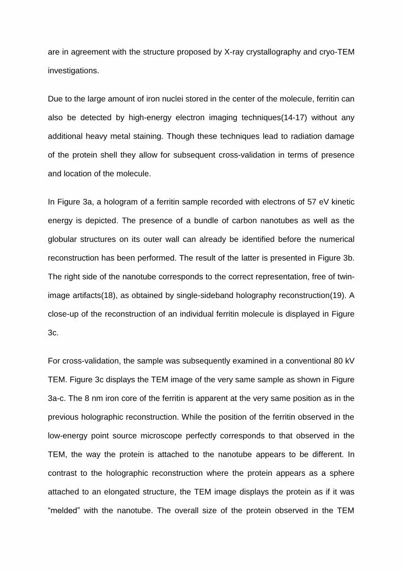
Due to the large amount of iron nuclei stored in the center of the molecule, ferritin can
also be detected by high-energy electron imaging techniques(14-17) without any
additional heavy metal staining. Though these techniques lead to radiation damage
of the protein shell they allow for subsequent cross-validation in terms of presence
and location of the molecule.
In Figure 3a, a hologram of a ferritin sample recorded with electrons of 57 eV kinetic
energy is depicted. The presence of a bundle of carbon nanotubes as well as the
globular structures on its outer wall can already be identified before the numerical
reconstruction has been performed. The result of the latter is presented in Figure 3b.
The right side of the nanotube corresponds to the correct representation, free of twin-
image artifacts(18), as obtained by single-sideband holography reconstruction(19). A
close-up of the reconstruction of an individual ferritin molecule is displayed in Figure
3c.
For cross-validation, the sample was subsequently examined in a conventional 80 kV
TEM. Figure 3c displays the TEM image of the very same sample as shown in Figure
3a-c. The 8 nm iron core of the ferritin is apparent at the very same position as in the
previous holographic reconstruction. While the position of the ferritin observed in the
low-energy point source microscope perfectly corresponds to that observed in the
TEM, the way the protein is attached to the nanotube appears to be different. In
contrast to the holographic reconstruction where the protein appears as a sphere
attached to an elongated structure, the TEM image displays the protein as if it was
“melded” with the nanotube. The overall size of the protein observed in the TEM

amounts to 9-10 nm. We associate theses differences to the radiation damage which
has occurred during TEM imaging. Following the high-energy electron investigations,
the sample was transferred back to our low-energy electron point source microscope;
the corresponding hologram and its reconstruction are displayed in Figure 3e-f
respectively. Here, the damage provoked by the 80 keV kinetic energy electrons is
evident. The way the protein is now attached to the nanotubes coincides with the
TEM results.
Discussion
We have reported the very first non-destructive investigation of an individual protein
by means of low-energy electron holography. The successful imaging of a single
protein is cross-validated by TEM examination. The sample preparation method can
be applied to a broad class of molecules.
While the spatial resolution in the holographic reconstructions presented here is
already below the nanometer scale, it is now an experimental challenge to reach a
resolution close to the employed wavelength with an advanced low-energy electron
holographic setup.
Besides this engineering challenge of improving interference resolution in
holography, another route towards higher resolution is to combine holography with
coherent diffraction imaging using low-energy electrons(20). Essential ingredients like
a micron sized electron lens(21) and the ability to record coherent diffraction
patterns(22) are already at hand for the pursuit of single molecule structural biology
at atomic resolution.
Methods
Sample preparation. COOH-functionalized double-walled carbon nanotubes from
the company NanoLab were dispersed to a concentration of 0.2 mg/ml by sonication
for 15 min in ultra highly purified water. Subsequently, ferritin proteins in a MES
buffer (pH 6.5) were added to the carbon nanotubes solution, with a final
concentration of 0.5 nM. The ferritin-nanotube solution was kept under gentle stirring
overnight.
SiN membranes (50 nm thickness) from the company Silson were first coated with a
5 nm thick carbon layer. Subsequently, holes of 200 nm in diameter were milled in
the membranes by means of a focused Ga ion beam. Thereafter, the membranes
were coated again with 20 nm carbon layers on both sides.
Next, the membranes were hydrophylized by exposition to ozone in an UV-ozone
oven for 20 min. Before 1 l of the ferritin-nanotubes solution was applied and air-
dryed on the membrane.
Holography. The detector system of our low-energy electron point source
microscope consists of a multi-channel plate (MCP) and a phosphor screen directly
mounted on a fiber optic plate (FOP). Both, MCP and Phosphor screen-FOP are 75
mm in diameter. A 8000x6000 pixels CCD camera was used to record the
holograms. The overall resolution of the detector system is around 20m.
An electro chemically etched single-crystal <111>-tungsten tip was used as point
source of coherent low-energy electrons. The holograms were recorded with a total
electron current of 50 nA.

Acknowledgements
We would like to thank Jennifer Clark for her help with sample preparation and the
Swiss National Science Foundation for its financial support.
Author Contributions
J.N.L. conducted the holographic and TEM investigations. T.L. performed the
hologram reconstructions. J.N.L. and C.E. worked on the sample preparation and
H.W.F. contributed to the design of the experimental setup. All authors discussed the
results and implications and jointly wrote the manuscript.

References
1 Neutze, R., Wouts, R., van der Spoel, D., Weckert, E. & Hajdu, J. Potential for biomolecular imaging with femtosecond X-ray pulses. Nature 406, 752-757 (2000).
2 Germann, M., Latychevskaia, T., Escher, C. & Fink, H.-W. Nondestructive imaging of individual biomolecules. Phys. Rev. Lett. 104, 095501 (2010).
3 Lawson, D. M. et al. Solving the structure of human H ferritin by genetically engineering intermolecular crystal contacts. Nature 349, 541-544 (1991).
4 Theil, E. C. Ferritin: Structure, Gene Regulation, and Cellular Function in Animals, Plants, and Microorganisms. Annual Review of Biochemistry 56, 289-315, doi:doi:10.1146/annurev.bi.56.070187.001445 (1987).
5 Jellinger, K., Paulus, W., Grundke-Iqbal, I., Riederer, P. & Youdim, M. Brain iron and ferritin in Parkinson's and Alzheimer's diseases. Journal of Neural Transmission: Parkinson's Disease and Dementia Section 2, 327-340, doi:10.1007/bf02252926 (1990).
6 Chiu, W. et al. High-resolution cryoelectron microscopy of biological specimens. Ultramicroscopy 23, 232-232 (1987).
7 Chapman, H. N. et al. Ultrafast coherent diffraction imaging with x-ray free-electron lasers. Proceedings of FEL 2006, BESSY, Berlin, Germany, 805-811 (2006).
8 Seibert, M. M. et al. Single mimivirus particles intercepted and imaged with an X-ray laser. Nature 470, 78-81, doi:10.1038/nature09748 (2011).
9 Miao, J. W., Chapman, H. N., Kirz, J., Sayre, D. & Hodgson, K. O. Taking X-ray diffraction to the limit: Macromolecular structures from femtosecond X-ray pulses and diffraction microscopy of cells with synchrotron radiation. Annu. Rev. Biophys. Biomol. Struct. 33, 157-176 (2004).
10 Fink, H. W. in Electron Microscopy 1994, Vol 1 - Interdisciplinary Developments and Tools eds B. Jouffrey & C. Colliex) 319-320 (1994).
11 Fink, H. W. Point-source for ions and electrons. Physica Scripta 38, 260-263 (1988).
12 Fink, H.-W., Stocker, W. & Schmid, H. Holography with low-energy electrons. Phys. Rev. Lett. 65, 1204-1206 (1990).
13 Latychevskaia, T. & Fink, H.-W. Simultaneous reconstruction of phase and amplitude contrast from a single holographic record. Opt. Express 17, 10697-10705 (2009).
14 Mann, S., Bannister, J. V. & Williams, R. J. P. Structure and composition of ferritin cores isolated from human spleen, limpet (Patella vulgata) hemolymph and bacterial (Pseudomonas aeruginosa) cells. Journal of Molecular Biology 188, 225-232 (1986).
15 Kawasaki, T., Endo, J., Matsuda, T., Osakabe, N. & Tonomura, A. Applications of Holographic Interference Electron Microscopy to the Observation of Biological Specimens. Journal of electron microscopy 35, 211-214 (1986).

16 Lichte, H. & Lehmann, M. in Advances in imaging and electron physics Vol. Volume 123 eds Pier Georgio Merli Gianluca Calestani Peter W. Hawkes & Vittori-Antisari Marco) 225-255 (Elsevier, 2002).
17 Quintana, C., Cowley, J. M. & Marhic, C. Electron nanodiffraction and high-resolution electron microscopy studies of the structure and composition of physiological and pathological ferritin. J. Struct. Biol. 147, 166-178 (2004).
18 Latychevskaia, T. & Fink, H.-W. Solution to the twin image problem in holography. Phys. Rev. Lett. 98, 233901 (2007).
19 Bryngdah.O & Lohmann, A. Single-sideband holography. J. Opt. Soc. Am. 58, 620-624 (1968).
20 Latychevskaia, T., Longchamp, J. N. & Fink, H.-W. When Holography Meets Coherent Diffraction Imaging arXiv, 1106.1320v1101 (2011).
21 Steinwand, E., Longchamp, J. N. & Fink, H. W. Fabrication and characterization of low aberration micrometer-sized electron lenses. Ultramicroscopy 110, 1148-1153 (2010).
22 Steinwand, E., Longchamp, J.-N. & Fink, H.-W. Coherent low-energy electron diffraction on individual nanometer sized objects. Ultramicroscopy 111, 282-284 (2011).

Figure legends
Figure 1: Scheme for recording the low-energy electron hologram of a protein.
Conduction electrons confined in a pointed W(111) single crystal wire are field
emitted into vacuum at an atomic-sized emission area providing a coherent low-
energy electron point source (PS). At the less than 1 micron distant object-plane
(OP), part of the coherent electron wave is scattered by a ferritin attached to a carbon
nanotube constituting the object wave indicated in red. At a distant detector screen
(S), the far-field interference pattern between object- (red) and reference-wave (blue)
- the hologram - is recorded and its digital record is subject to the numerical
reconstruction of the protein.

Figure 2: Low-energy electron hologram of ferritin and its numerical
reconstruction. a 53eV kinetic energy electron hologram of several ferritin
molecules attached to a bundle of carbon nanotubes. b reconstruction of a. c close-
up of b.

Figure 3: Control experiments using a TEM. a 57eV kinetic energy electron
hologram of individual ferritin molecules attached to a bundle of carbon nanotubes. b
side-band holography reconstruction of a. c close-up of b. d TEM image of the very
same ferritin molecule. e 45eV kinetic energy electron hologram of the very same
sample as in a but after the TEM investigations. f side-band holography
reconstruction of e.