Embed Size (px)
Citation preview
Noise, neural codes and cortical organization
Michael N Shadlen and William T Newsome
Stanford University School of Medicine, Stanford, USA
Cortical circuitry must facilitate information transfer in accordance with
a neural code. In this article we examine two candidate neural codes:
information is represented in the spike rate of neurons, or information is
represented in the precise timing of individual spikes. These codes can be
distinguished by examining the physiological basis of the highly irregular
interspike intervals typically observed in cerebral cortex. Recent advances in
our understanding of cortical microcircuitry suggest that the timing of neuronal
spikes conveys little, if any, information. The cortex is likely to propagate a noisy rate code through redundant, patchy interconnections.
Current Opinion in Neurobiology 1994, 4:569-579
Introduction
Although it is generally agreed that neurons signal in- formation through sequences of action potentials, the neural code by which information is transfered through the cortex remains elusive. In the cortex, the timing of successive action potentials is highly irregular [l”], and the interpretation of this irregularity has led to two di- vergent views of cortical organization. On the one hand, the irregularity might arise from stochastic forces. If so, the irregular interspike interval (ISI) reflects a random process and implies that an instantaneous estimate of spike rate can only emerge from the pooled responses of many individual neurons [2]. In keeping with this theory, one would expect that the temporal pattern of spikes conveys little information. Alternatively, the irreg- ular IS1 may result from precise coincidences of presy- naptic events. In this scenario, it is postulated that the timing of spikes, their intervals and patterns can convey information [3-131. According to this view, the irregu- larity of the IS1 reflects a rich bandwidth for information transfer.
Our understanding of cortical organization and interpre- tation of neurophysiological data depend critically on whether neurons convey a noisy rate code or a pre- cise temporal code. Is it reasonable to expect the average discharge rate of a neuron in the visual cortex to convey information about a visual stimulus [14-l 71, or should we attend to particular patterns of spikes? Is cortical cir- cuitry organized to average out noise among redundant neurons, or to provide an expansion of temporal signal- ing capacity by eliminating redundancy in the pattern of inputs to different neurons? At the heart of this con- troversy, the critical question is why neurons spike with
such irregularity. In this article, we will attempt to iden- tify the physiological factors that may lead to irregularity in the spike train. In essence, our task is to determine whether neurons within cortical circuits behave as coin- cidence detectors or as integrate-and-fire devices [3].
Coincidence detectors or integrate-and-fire
devices?
Given our current understanding of cortical physiology and biophysics, why do cortical neurons discharge so ir- regularly? This is the question recently posed by Sofiky and Koch [l”]. They examined spike trains from neu- rons in the visual cortex of the monkey, applying a clever normalization scheme that permitted them to estimate the variability of the IS1 at nominally constant firing rates. They found that spiking patterns approximate a random (Poisson) process. Using a sophisticated model of a cortical pyramidal neuron and its connections [18], they argued that such irregularity is unattainable through an integrate-and-fire mechanism [lo*]. Any neuron that integrates synaptic inputs with a membrane time con- stant of 7-20ms should spike more regularly. The in- tuitive appeal of this argument is based on the follow- ing physical analogy. Imagine a Geiger counter that is wired to click only upon integration of 40 radioac- tive decays. Although individual decays are random in time, the modified counter clicks with great regularity because the sum of random intervals becomes reliable as the number of counted intervals increases. By anal- ogy, a neuron that integrates random presynaptic events
Abbreviations EPSP-excitatory postsynaptic potential; ips-impulses per second; IPSP-inhibitory postsynaptic potential;
ISI-interspike interval; IT-inferotemporal; MST-medial superior temporal area; MT-middle temporal area; PSP-postsynaptic potential; Vl-primary visual cortex.
0 Current Biology Ltd ISSN 0959-4388 569

570 Sensorv systems
- counting to some number to reach spike threshold - cannot preserve such irregularity in its own spike output.
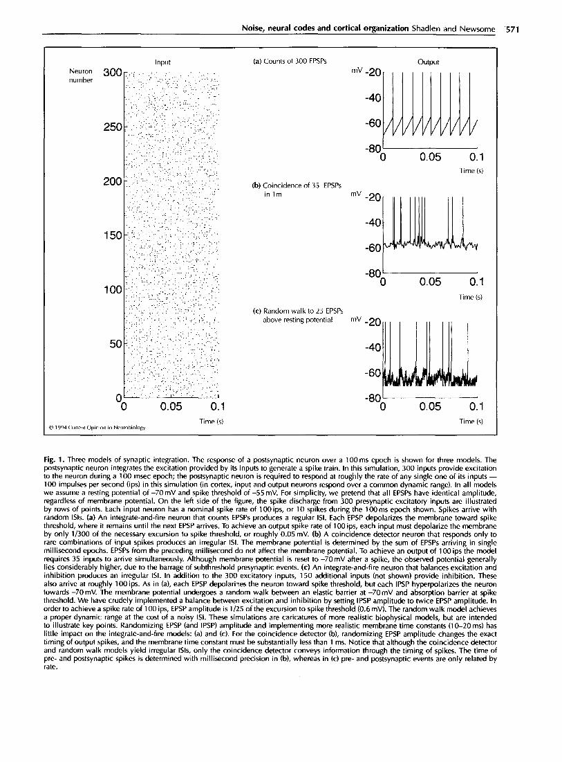
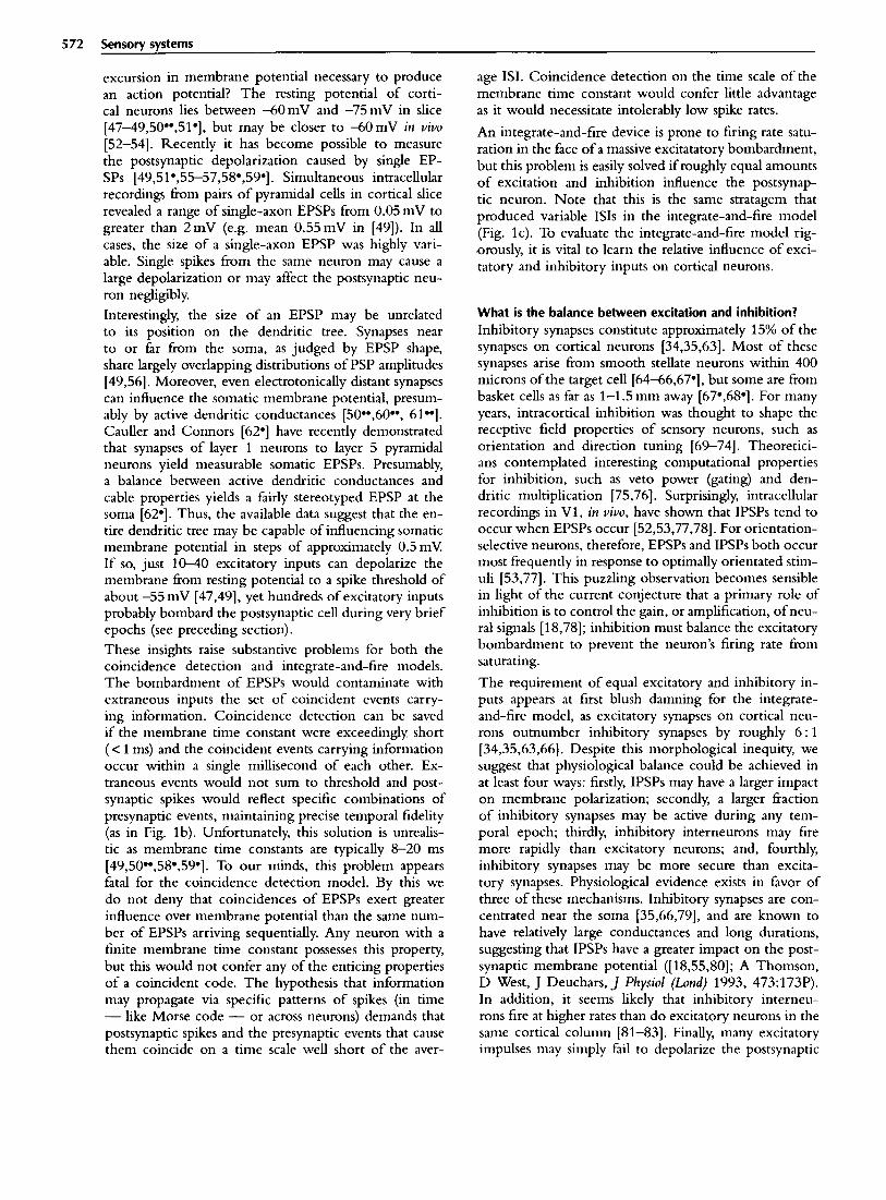
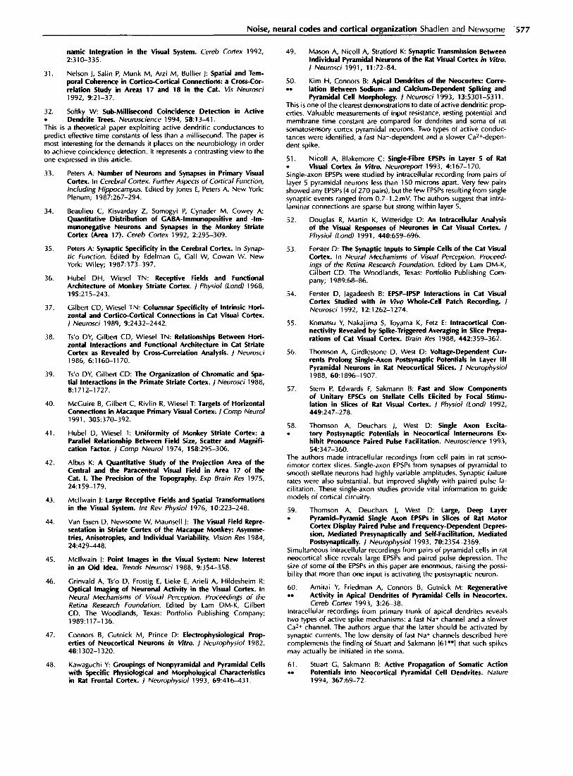
Fig. la depicts the expected output from an integrate- and-fire device; the output is nearly periodic, even though input spikes arrive at random intervals. Softky and Koch [lo*] conclude that cortical neurons must perform some sort of coincidence detection, such that a particular combination of presynaptic events leads to a postsynaptic spike. This combination would occur with sufficient irregularity to account for the variable ISI. This idea is illustrated in Fig. 1 b. Such a neuron would be capable of transmitting information in the precise timing of individual spikes or their temporal pattern.
On the other hand, it has long been known that a bal- ance of excitation and inhibition will yield an irregu- lar ISI from the standard integrate-and-fire neuron. The idea was first proposed by Gerstein and Mandelbrot [19] and subsequently developed by Calvin and Stevens [20] in their seminal work on synaptic noise in spinal mo- toneurons. These models are often referred to as ran- dom walk, or diffusion processes, and they have a rich theoretical base [21-291. The idea behind this model is that the membrane potential undergoes a random walk between resting potential and spike threshold (Fig. lc). EPSPs (excitatory postsynaptic potentials) drive the po- tential toward spike threshold and IPSPs (inhibitory postsynaptic potentials) drive the potential toward Eel (chloride reversal potential), beyond which there can be no further hyperpolarization. With an appropriate bal- ance of excitation and inhibition, the IS1 can be highly irregular.
Although the Geiger counter model (Fig. la) is clearly wrong, either the coincidence detector or random walk model can account for the irregular ISIS seen in cortical neurons. However, these two schemes sanction very dif- ferent strategies for cortical organization. If cortical neu- rons behave as coincidence detectors, then the timing of spikes can propagate through the cortex with great fi- delity to convey information and to synchronize other neurons [6,10,30,31]. If an irregular IS1 results from in- tegration of excitatory and inhibitory PSPs (postsynap- tic potentials), then the timing of postsynaptic spikes is random and no longer reflects the timing of presynaptic events. Precise patterns of spikes - their intervals and coincidences - would fail to propagate.
The validity of these two views rest ultimately on the be- havior of neurons within cortical circuits. Which synap- tic events cause a neuron to fire? The coincidence detec- tor requires the effective set of synaptic events to occur at roughly the same frequency as the neuron spike rate. Noncoincident EPSPs must either arrive infrequently or be prevented from summating to spike threshold by a very short membrane time constant (i.e. less than a few milliseconds) [1”,32*]. The random walk model in- corporates a more reasonable time constant (-10 ms or more), but the opposition of excitatory and inhibitory PSPs demands a very large number of presynaptic events to drive the neuron to threshold. Moreover, the ran-
dom walk model requires roughly equal depolarizing and hyperpolarizing influences on membrane voltage to gen- erate highly variable ISIS (MN Shadlen, WT Newsome, unpublished data). To choose between the coincidence detection and random walk models, we need answers to several critical questions. How many EPSPs arrive at the neuron during an epoch of activity, and what is their impact on the postsynaptic membrane voltage? How many IPSPs arrive during the same epoch, and what is their impact on the postsynaptic membrane? What is the balance between excitatory and inhibitory influences on membrane potential?
Evidence from synaptic physiology
How many synaptic inputs are active? The number of synaptic contacts for cortical neurons has been estimated to be between 3000 and 10 000, depend- ing on cortical area and species [33]. The most recent estimate for monkey visual cortex is 3900 synapses per neuron ]34]. Approximately 85% of these contacts are asymmetric and, therefore, are presumed to be exci- tatory. The majority of these synapses are from other cortical neurons, either within the cortical column or connected to the column via horizontal axon collaterals [l&35].
It is more difficult to estimate the fraction of these excitatory synapses that are active during an epoch of excitation. Consider a pyramidal cell in Vl (primary vi- sual cortex) that responds to an optimally oriented bar of light passing through its receptive field. How many of the neuron’s inputs are active over any 30-50ms epoch (2-3 time constants)? Many of the excitatory inputs from within a cortical column will respond under the same stimulus conditions as our pyramidal cell [36], as would most of the direct excitatory inputs fi-om the thalamus. Inputs from horizontal connections within the cortex tend to arise horn neurons with similar receptive fields [37], and cross-correlation analyses reveal that many of these neurons are active simultaneously [38,39]. Never- theless, not all of the horizontal connections would be expected to be active at the same moment, as many arise from portions of the visual map outside the classical re- ceptive field of the neuron they innervate [34,40] (but see [41-45]). The extent to which the map of activity overlaps the map of connectivity remains to be clarified, although optical imaging data suggest that the degree of overlap is substantial ([46]; DY Ts’o, personal commu- nication). We are left with the impression that a large fraction of excitatory inputs ought to be active. Ex- actly how large a fraction remains to be determined. (We chose 10% for the simulations in Fig. 1, but the following analyses apply to any fraction over 2-3X)
How large is an EPSP? More important than the number of synapses is their effectiveness. How large is an EPSP in relation to the
Noise, neural codes and cortical organization Shadlen and Newsome .57l
Neuron number
D 1994 Current Opimon in Neurobiology Time (s) Time (s)
(a) Counts of 300 EPSPs
(b) Coincidence of 35 EPSPs in lm
(c) Random walk to 25 EPSPs above resting potential
output mV-20
-40
-60
-80' 0 0.05 0.1
Time (s)
mV-20
-40
-60
-801 0 0.05 0.1
Time (s)
-60
-80' 0 0.05 0.1
Fig. 1. Three models of synaptic integration. The response of a postsynaptic neuron over a 100 ms epoch is shown for three models. The postsynaptic neuron integrates the excitation provided by its inputs to generate a spike train. In this simulation, 300 inputs provide excitation to the neuron during a 100 msec epoch; the postsynaptic neuron is required to respond at roughly the rate of any single one of its inputs - 100 impulses per second (ips) in this simulation (in cortex, input and output neurons respond over a common dynamic range). In all models we assume a resting potential of -70 mV and spike threshold of -55 mV. For simplicity, we pretend that all EPSPs have identical amplitude, regardless of membrane potential. On the left side of the figure, the spike discharge from 300 presynaptic excitatory inputs are illustrated by rows of points. Each input neuron has a nominal spike rate of 100 ips, or 10 spikes during the 100ms epoch shown. Spikes arrive with random ISIS. (a) An integrate-and-fire neuron that counts EPSPs produces a regular ISI. Each EPSP depolarizes the membrane toward spike threshold, where it remains until the next EPSP arrives. To achieve an output spike rate of 100 ips, each input must depolarize the membrane by only l/300 of the necessary excursion to spike threshold, or roughly 0.05 mV. (b) A coincidence detector neuron that responds only to rare combinations of input spikes produces an irregular ISI. The membrane potential is determined by the sum of EPSPs arriving in single millisecond epochs. EPSPs from the preceding millisecond do not affect the membrane potential. To achieve an output of 100 ips the model requires 35 inputs to arrive simultaneously. Although membrane potential is reset to -7OmV after a spike, the observed potential generally lies considerably higher, due to the barrage of subthreshold presynaptic events. (c) An integrate-and-fire neuron that balances excitation and inhibition produces an irregular ISI. In addition to the 300 excitatory inputs, 150 additional inputs (not shown) provide inhibition. These also arrive at roughly 100 ips. As in (a), each EPSP depolari:es the neuron toward spike threshold, but each IPSP hyperpolarizes the neuron towards -7OmV. The membrane potential undergoes a random walk between an elastic barrier at -7OmV and absorption barrier at spike threshold. We have crudely implemented a balance between excitation and inhibition by setting IPSP amplitude to twice EPSP amplitude. In order to achieve a spike rate of 100 ips, EPSP amplitude is l/25 of the excursion to spike threshold (0.6 mV). The random walk model achieves a proper dynamic range at the cost of a noisy ISI. These simulations are caricatures of more realistic biophysical models, but are intended to illustrate key points. Randomizing EPSP (and IPSP) amplitude and implementing more realistic membrane time constants (1 O-20 ms) has little impact on the integrate-and-fire models: (a) and (c). For the coincidence detector (b), randomizing EPSP amplitude changes the exact timing of output spikes, and the membrane time constant must be substantially less than 1 ms. Notice that although the coincidence detector and random walk models yield irregular ISIS, only the coincidence detector conveys information through the timing of spikes. The time of pre- and postsynaptic spikes is determined with millisecond precision in (b), whereas in (c) pre- and postsynaptic events are only related by rate.
572 Sensory systems
excursion in membrane potential necessary to produce an action potential ? The resting potential of corti- cal neurons lies between -60mV and -75 mV in slice [47-49,50**,51*], but may be closer to -60mV in viva [52-541. Recently it has become possible to measure the postsynaptic depolarization caused by single EP- SPs [49,51*,55-57,58*,59*]. Simultaneous intracellular recordings I?om pairs of pyramidal cells in cortical slice revealed a range of single-axon EPSPs from 0.05 mV to greater than 2mV (e.g. mean 0.55 mV in [49]). In all cases, the size of a single-axon EPSP was highly vari- able. Single spikes from the same neuron may cause a large depolarization or may affect the postsynaptic neu- ron negligibly
Interestingly, the size of an EPSP may be unrelated to its position on the dendritic tree. Synapses near to or far from the soma, as judged by EPSP shape, share largely overlapping distributions of PSP amplitudes [49,56]. Moreover, even electrotonically distant synapses can influence the somatic membrane potential, presum- ably by active dendritic conductances [50**,60”, 61**]. Cauller and Connors [62*] have recently demonstrated that synapses of layer 1 neurons to layer 5 pyramidal neurons yield measurable somatic EPSPs. Presumably, a balance between active dendritic conductances and cable properties yields a fairly stereotyped EPSP at the soma [62*]. Thus, the available data suggest that the en- tire dendritic tree may be capable of influencing somatic membrane potential in steps of approximately 0.5mV If so, just 10-40 excitatory inputs can depolarize the membrane from resting potential to a spike threshold of about -55 mV [47,49], yet hundreds of excitatory inputs probably bombard the postsynaptic cell during very brief epochs (see preceding section).
These insights raise substantive problems for both the coincidence detection and integrate-and-fire models. The bombardment of EPSPs would contaminate with extraneous inputs the set of coincident events carry- ing information. Coincidence detection can be saved if the membrane time constant were exceedingly short (< 1 ms) and the coincident events carrying information occur within a single millisecond of each other. Ex- traneous events would not sum to threshold and post- synaptic spikes would reflect specific combinations of presynaptic events, maintaining precise temporal fidelity (as in Fig. lb). Unfortunately, this solution is unrealis- tic as membrane time constants are typically 8-20 ms [49,500*,58*,59*]. To our minds, this problem appears fatal for the coincidence detection model. By this we do not deny that coincidences of EPSPs exert greater influence over membrane potential than the same num- ber of EPSPs arriving sequentially. Any neuron with a finite membrane time constant possesses this property, but this would not confer any of the enticing properties of a coincident code. The hypothesis that information may propagate via specific patterns of spikes (in time - like Morse code - or across neurons) demands that postsynaptic spikes and the presynaptic events that cause them coincide on a time scale well short of the aver-
age ISI. Coincidence detection on the time scale of the membrane time constant would confer little advantage as it would necessitate intolerably low spike rates.
An integrate-and-fire device is prone to firing rate satu- ration in the face of a massive excitatatory bombardment, but this problem is easily solved if roughly equal amounts of excitation and inhibition influence the postsynap- tic neuron. Note that this is the same stratagem that produced variable ISIS in the integrate-and-fire model (Fig. lc). To evaluate the integrate-and-fire model rig- orously, it is vital to learn the relative influence of exci- tatory and inhibitory inputs on cortical neurons.
What is the balance between excitation and inhibition? Inhibitory synapses constitute approximately 15% of the synapses on cortical neurons [34,35,63]. Most of these synapses arise from smooth stellate neurons within 400 microns of the target cell [64-66,67*], but some are from basket cells as far as l-l .5 mm away [67*,68*]. For many years, intracortical inhibition was thought to shape the receptive field properties of sensory neurons, such as orientation and direction tuning [69-741. Theoretici- ans contemplated interesting computational properties for inhibition, such as veto power (gating) and den- dritic multiplication [75,76]. Surprisingly, intracellular recordings in Vl, in vioo, have shown that IPSPs tend to occur when EPSPs occur [52,53,77,78]. For orientation- selective neurons, therefore, EPSPs and IPSPs both occur most frequently in response to optimally orientated stim- uli [53,77]. This puzzling observation becomes sensible in light of the current conjecture that a primary role of inhibition is to control the gain, or amplification, of neu- ral signals [18,78]; inhibition must balance the excitatory bombardment to prevent the neuron’s firing rate from saturating.
The requirement of equal excitatory and inhibitory in- puts appears at first blush damning for the integrate- and-tire model, as excitatory synapses on cortical neu- rons outnumber inhibitory synapses by roughly 6 : 1 [34,35,63,66]. Despite this morphological inequity, we suggest that physiological balance could be achieved in at least four ways: firstly, IPSPs may have a larger impact on membrane polarization; secondly, a larger fraction of inhibitory synapses may be active during any tem- poral epoch; thirdly, inhibitory interneurons may fire more rapidly than excitatory neurons; and, fourthly, inhibitory synapses may be more secure than excita- tory synapses. Physiological evidence exists in favor of three of these mechanisms. Inhibitory synapses are con- centrated near the soma [35,66,79], and are known to have relatively large conductances and long durations, suggesting that IPSPs have a greater impact on the post- synaptic membrane potential ([18,55,80]; A Thomson, D West, J Deuchars, J Physiol (Lund) 1993, 473:173P). In addition, it seems likely that inhibitory interneu- rons fire at higher rates than do excitatory neurons in the same cortical column [81-831. Finally, many excitatory impulses may simply fail to depolarize the postsynaptic
Noise, neural codes and cortical organization Shadlen and Newsome- 573
1 For example, the estimated spike rate from 100 neurons over any 10 ms epoch, each spiking at 100 impulses per second tips) with random ISI, is 100 ips +6?. By contrast, one of the chief disadvantages of neural codes that rely on temporal patterns of spikes is that such patterns may take a long time to propagate. To take advantage of the information capacity rendered by an irregular ISI, the nervous system may have to wait a long time for critical spikes and intervals.
neuron. Such synaptic failures have been demonstrated in hippocampal cell culture and slice [84*,85,86*], and are thought to reflect a presynaptic failure to release neuro- transmitter. Although this phenomenon is just beginning to receive attention in the neocortex ([57,58.,59*,87*]; D Smetters, S Nelson, Sot Netrrosci Abstr 1993, 19:628), preliminary evidence suggests that inhibitory inputs are more secure than excitatory synapses (A Thomson, D West, J Deuchars,I Physiol (Z..&) 1993, 473:173P). In- hibitory neurons tend to make multiple contacts with their targets [64,68*], whereas (excitatory) pyramidal neurons tend to make single synapses [40,86*]. Although the available data are inconclusive, it seems entirely plau- sible, if not probable, that excitation and inhibition on cortical neurons are much more closely balanced than the anatomy would suggest.
With these data in mind, let us return to Fig. 1. To pro- duce this figure, we assumed 300 presynaptic neurons are active (- 10% of the excitatory input to a pyrami- dal neuron) during a 1OOms period. Each input neuron provides about 10 spikes, which arrive at irregular in- tervals during this epoch. The postsynaptic neuron is not allowed to saturate, so a good rule of thumb is to find conditions that allow it to fire about 10 spikes. To do this with the simple counting device (Fig. la), we had to assume an EPSP amplitude of 0.05mV (l/300 of the excursion from reset to spike threshold), which is clearly incorrect. In any case, we can exclude this model because it produces a regular ISI. To model the coin- cidence detector (Fig. lb), we had to assume an EPSP amplitude of about 0.4mV. Unfortunately, we also had to mimic a membrane time constant of under 0.5 ms. Basically, there can be no summation of EPSPs beyond an interval of 1 ms or less. There is no plausible basis for this conjecture (but see [32*]). Finally, we consid- ered a simple random walk mechanism (Fig. lc). Here the average EPSP was 0.6mV and we mimicked a long time constant (greater than 10ms). However the model assumes a strong source of inhibition. In this rendition we added 150 inhibitory presynaptic neurons, each pro- viding roughly 10 spikes, arriving randomly. Each IPSP hyperpolarizes the membrane toward -70mV in steps twice as large as an EPSP This is a crude approxi- mation to a balance between hyperpolarizing and de- polarizing forces on the membrane. This idea seems to be most consistent with synaptic physiology. In essence, the random walk model with balanced excitatory and inhibitory inputs allows the neuron to behave as an integrate-and-fire device and maintain a reasonable re- sponse rate. The cost, however, is an irregular ISI. If this conjecture is correct, then the timing of output spikes is stochastic and can convey little, if any, infor- mation.
Implications for cortical organization
Our understanding of the sources of ISI irregularity has fundamental implications for our views of cortical or- ganization. If the variable ISI reflects a precise tempo- ral code that must be propagated through the cortex, the pattern of cortical connectivity should emphasize divergence, and redundancy should be avoided. Mov- ing downstream in a cortical pathway, therefore, we would expect fewer neurons to covary their responses under similar stimulus conditions, a view that proba- bly demands reduction, if not outright elimination, of redundancy in the form of columnar organization. In fact, we would expect to see progressively less spike rate modulation at all, as rate modulation can only mud- dle a temporal code with spurious coincidences [88*,89]. Alternatively, if synaptic integration produces a truly ran- dom ISI, the neural code consists simply of modulations in spike rate. As any one neuron provides a poor esti- mate of the instantaneous spike rate, the cortex must use ensembles of neurons to represent the same infor- mation. This view demands a reiterated organization of redundant, column-like modules, even in higher cortical areas.
Clustering of neurons with similar response properties (redundancy) is a well-established principle in primary sensory and motor areas of the cortex, and is beginning to receive attention fi-om investigators working on higher cortical areas as well. By analyzing patterns of connectiv- ity revealed by local biocytin injections, Amir et al. [90-l found a patchy organization of horizontal connections reiterated in striate, extrastriate, and parietal cortex of the macaque monkey. Similar observations have been made in inferotemporal (IT) [91-l, frontal and limbic cortex [92], suggesting common organizational principles that are consistent with a redundant coding strategy [93-J. Recent physiological data born IT cortex also support this point of view; nearby neurons, probably organized in the form of columns, appear to share preferences for similar features of visual objects [94,95] and faces [96] (but see [97-l).
Size of fundamental signaling units in cerebral
cortex
If clusters of functionally similar neurons carry informa- tion in the form of noisy, redundant rates, how many neurons are needed to estimate firing rate precisely? Simple statistical considerations reveal that the instanta- neous rate horn an ensemble of 100 or so neurons can be estimated reliably within a single 1%‘. We suggest that
574 Sensorv svstems
neuronal pools of this size may comprise the fundamental signaling units of cerebral cortex.
Interestingly, the existence of common noise within cor- tical columns suggests that pool sizes exceeding 100 neurons confer little or no signaling advantage. The noisy spike rates of adjacent cortical neurons elicited by repeated presentations of a particular stimulus are not independent, but covary weakly with an average correlation coefficient of roughly 0.12 [97*,98]. Cor- related noise (presumably arising f_?om common input) shared by all members of a neuronal pool places funda- mental limits on signaling power because the common noise cannot be eliminated by averaging among neurons within the pool. Monte Carlo simulations indicate that the signaling advantage gained by averaging asymptotes at roughly 100 neurons [17,98,99], further supporting the notion that ensembles of this size may comprise the fundamental signaling units of cortex.
Objections to the random walk model and rate-coding hypothesis
The biophysical support is weak This objection to our point of view is fair enough. The random walk model for generating variable ISIS depends critically on an approximate equality of excitatory and inhibitory influences on membrane voltage, but the ac- tual state of affiirs is simply unknown. We have cited fragmentary physiological evidence indicating that in- hibition has a more substantial impact than suggested by morphology, but little direct evidence is available on the critical issues of single IPSP size, relative firing frequencies of excitatory and inhibitory neurons, and relative failure rates for EPSPs and IPSPs. We note, how- ever, that this caveat does not argue against the random walk model (and, by extension, the rate-coding hypo- thesis), but rather clarifies the type of biophysical data that will ultimately permit a truly informed choice of models.
Reliable temporal spike patterns exist in cortex Abeles and colleagues [88*,89] have demonstrated re- peating temporal patterns of spike discharge among en- sembles of neurons in fi-ontal cortex. In visual cortex, synchronous patterns of discharge have been observed in anesthetized and awake animals under a variety of con- ditions (see [lo] for review). If the IS1 is truly random, then these observations must be attributed to co-mod- ulation of spike rate, imposed by some common input such as the thalamus. They do not necessitate coinci- dence detection; nor do they imply a precise temporal code.
In a somewhat different vein, Optican, Richmond and colleagues [8,9,100] have shown that temporal patterns of spikes convey more information about visual stim- uli than the spike rate does in single neurons of areas Vl and IT. It is unclear whether the type of tem-
poral encoding proposed by these investigators would necessitate a deterministic ISI. Modulation of the aver- age response rate might suffice. Interestingly, Tovee et al. [lOl**] have shown that brief (20-50 ms) estimates of rate from IT neurons convey nearly as much information as the 300-400 ms components of a putative temporal code.
The acid test for any theory of the neural code is to es- tablish a connection to behavior. In cortical areas MT and MST, fluctuations in spike rate have been shown to correlate with an animal’s decisions in a motion discrimination task [99,102,103”]. Examples abound in the motor cortex for a connection between neural dis- charge rate and behavior (see [104]). To our knowledge, however, there is no evidence that a temporal pattern of activity (beyond rate modulating) in cortex has any consequence for behavior.
Spike timing is critical in some neural systems Clearly, some neural structures convey information in the timing of successive spikes. The best examples are probably from brainstem auditory pathways, where spikes may be time-locked to peripheral events (e.g. primary-like neurons of the cochlear nucleus [105]). So long as synaptic contacts are multiple and, hence, secure, spikes can propagate reliably fi-om one neuron to the next, preserving a temporal code. In cortex, how- ever, we argue that many inputs affect the neuron, and a single presynaptic spike has little bearing on the exact timing of a postsynaptic spike. This is not true, however, if certain inputs have privileged contacts. For example, a single spike f?om thalamus may induce a time-locked spike in visual cortex consistent with monosynaptic ex- citation ([106]; J Alonso, R Reid, T Wiesel, Sot Neuro- sci Abstr 1993, 18:425). Within cortex, such time-locked spikes are exceptional [38,39,107,108’].
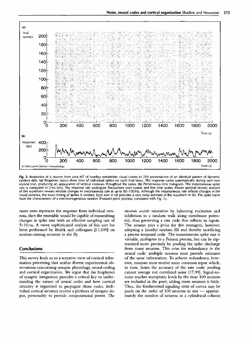
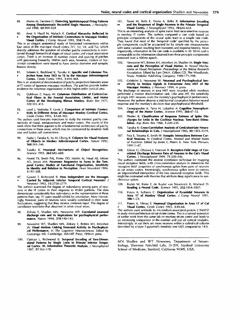
Spike rates may be time-locked to stimulus change The inability to preserve information about the time of a spike would seem to imply that the discharge rate cannot modulate in a time-locked fashion to external inputs. This is not true. In a random walk mecha- nism, the discharge rate follows the activity of inputs; but given a base rate, the time to the next spike is (nearly) random. Consider the spike train recorded from an MT neuron responding to successive presentations of an identical pattern of moving dots (Fig. 2). The average discharge rate fluctuates in a time-locked fashion to the moving dot display. Yet the exact time of any one spike within any lO-20ms epoch is nearly random from trial to trial. The neuron approximates a random (Poisson) point process with non-stationary rate. The variance of the spike count tallied for each trial actually exceeds the mean. Nevertheless, the average instantaneous discharge rate is consistent from trial to trial, as is apparent in the raster’s vertical structure (Fig. 2a). Presumably, this rate maintains temporal fidelity with changes in the stimu- lus. If instead of successive trials, we imagine that the
Noise, neural codes and cortical organization Shadlen and Newsome 575
(a)
Trial number
“0 200 400 600 800 1000 1200 1400 1600 1800 2000
Time (s)
Response 400 c (ips)
200 -
0 0 200 400 600 800 1000 1200 1400 1600 1800 2000
0 1994 Current Opinion in Neurobiology Time (5) -
Fig. 2. Responses of a neuron from area MT of monkey extrastriate visual cortex to 210 presentations of an identical pattern of dynamic random dots. (a) Response rasters show time of individual spikes on each trial (row). The response varies systematically during each two second trial, producing an appearance of vertical contours throughout the raster. (b) Peristimulus time histogram. The instantaneous spike rate is computed in 2 ms bins. The response rate undergoes fluctuations over coarse and fine time scales. Power spectral density analysis of this waveform reveals reliable changes in instantaneous rate at up to 60-l 00 Hz. Although the instantaneous rate reflects changes in the visual stimulus, the exact timing of spikes is random. Each row in (a) provides a very noisy estimate of the waveform in fb). The spike trains have the characteristic of a non-homogeneous random (Poisson) point process, consistent with Fig. lc.
raster rows represent the response from individual neu- rons, then the ensemble would be capable of transmitting changes in spike rate with an effective sampling rate of !%lOms. A more sophisticated analysis of this sort has been performed by Bialek and colleagues [11,109] on motion-sensing neurons in the fly.
Conclusions
This survey leads us to a tentative view of cortical infor- mation processing that unifies diverse experimental ob- servations concerning synaptic physiology, neural coding and cortical organization. We argue that the biophysics of synaptic integration provides a critical key to under- standing the nature of neural codes and how cortical circuitry is organized to propagate those codes. Indi- vidual cortical neurons receive a plethora of synaptic in- put, presumably to provide computational power. The
neumn avoids saturation by balancing excitation and inhibition in a random walk along membrane poten- tial, thus preserving a rate code that reflects its inputs. The neuron pays a price for this stratagem, however, adopting a (nearly) random ISI and thereby sacrificing a precise temporal code. The instantaneous spike rate is variable, analogous to a Poisson process, but can be rep- resented more precisely by pooling the spike discharge from many neurons. This cries for redundancy in the neural code: multiple neurons must provide estimates of the same information. To achieve redundancy, how- ever, neurons must receive some common input which, in turn, limits the accuracy of the rate code: pooling cannot average out correlated noise [17,99]. Signal-to- noise reaches asymptotic levels by the time 100 neurons are included in the pool; adding more neurons is futile. Thus, the fundamental signaling units of cortex may be pools on the order of 100 neurons in size - approxi- mately the number of neurons in a cylindrical column

576 Sensor systems
aligned with the dendritic field of one layer 5 pyramidal cell [llO,lll*]. On this view, pieces of cortex must exert influence over other pieces of cortex as small signaling units of redundant and weakly correlated neurons. As the fundamental motivation for this point of view rests ulti- mately in synaptic physiology, we see no reason that the same principle should not apply at any location in neo- cortex, where thousands of inputs influence a neuron’s output.
A central implication of this point of view is that the or- ganization of cortical connectivity should remain coarse. Downstream neurons are unlikely to draw input from a special neuron here and another one there, but instead probably receive redundant input from a pool (50-100) here and another pool there. If we are correct, then the search for information in temporal patterns, synchrony, and specially labeled spikes is unlikely to succeed. On the other hand, there is reason for optimism because the secrets of the brain’s messages may be revealed by the analysis of single neurons acting in concert with others of similar ilk. Thus, the activity of single neurons may be connected to behavior by virtue of redundancy, raising hopes that probing the brain with tungsten will continue to yield important knowledge.
Acknowledgements
We wish to thank W Bair, I< Horn, K Written, U Connors, R Douglas, J Groh, C Koch, 11 Prince, W Softky, C Stevens, M Stryker, D Ts’o and E Zohary for helpful discussions. W Bair suggested the analysis in Fig. 2. MN Shadlen is supported by a Howard Hughes postdoctoral fellowship for physician scien- tists. WT Newsome is supported by the National Eye Institute (EY05603).
References and recommended reading
Papers of particular interest, published within the annual period of review, have been highlighted as: . of special interest . . of outstanding interest
1. Softky WR, Koch C: The Highly Irregular Firing of Cortical . . Cells is Inconsistent with Temporal Integration of Random
EPSPs. / Neurosci 1993, 13:33&350. This theoretical paper makes two important contributions to our knowl- edge about neural signals. The authors analyzed single unit data from monkey visual cortex and found that ISIS are highly irregular. This is a tricky analysis and the authors perform it commendably. The bulk of the paper focuses on models of synaptic integration. The authors conclude that neurons cannot behave as integrate-and-fire devices, as is classically thought, but that they act as coincidence detectors. We believe they have underestimated the importance of inhibition (see text).
2. Adrian ED, Zotterman Y: The Impulses Produced by Sensory Nerve-Endings. Part 2. The Response of a Single End-Organ. 1 Physiol 1926, 61:151-171.
3. Abeles M: Role of Cortical Neuron: Integrator or Coincidence Detector? Isr 1 Med Sci 1982, 18:83-92.
4. Von der Malsburg C: Nervous Structures with Dynamical links. Ber Bunsenges Phys Chem 1985, 89:703-710.
5. Von der M&burg C, Schneider W: A Neural Cocktail-Party Processor. Viol Cybern 1986, 54:29-40.
6.
7.
a.
9.
10.
11.
12.
13.
14.
1
17.
18.
19.
20.
21.
22.
23.
24.
25.
26.
27.
28.
29.
30.
Abeles M: Corticonics. Neural Circuits of the Cerebral Cortex. Cambridge: Cambridge University Press; 1991.
Gray C, Konig P, Engel A, Singer W: Oscillatory Responses in Cat visual Cortex Exhibit Inter-Columnar Synchroniza- tion Which Reflects Global Stimulus Properties. Nature 1989, 338:334-337.
Richmond 8, Optican L: Temporal Encoding of Two-Dimen- sional Patterns by Single Units in Primate Primary Visual Cortex. II. Information Transmission. / Neurophysiol 1990, 64:37C-380.
McClurkin J, Optican L, Richmond 6, Cawne T Concurrent Processing and Complexity of Temporally Encoded Neuronal Messages in Visual Perception. Science 1991, 25:675-677.
Engel A, Konig P, Kreiter A, Schillen T, Singer W: Temporal Coding in the Visual Cortex: New Vistas on Integration in the Nervous System. Trends Neurosci 1992, 15:218-226.
Bialek W, Rieke F: Reliability and Information Transmission in Spiking Neurons. Trends Neurosci 1992, 15:428-434.
Eckhorn R, Frein A, Bauer R, Woelbern T, Kehr H: High Fre- quency (6&90Hz) Oscillations in Primary Visual Cortex of Awake Monkey. Neurorepa? 1993, 4:243-246.
Aertsen A, Arndt M: Response Synchronization in the Visual Cortex. Curr Opin Neurobiol 1993, 3:586-594.
Tolhurst DJ, Movshon ]A, Dean AF: The Statistical Reliability of Signals in Single Neurons in Cat and Monkey Visual Cortex. Vision Res 1983, 23~775-785.
Bradley A, Skottun BC, Ohzawa I, Sclar C, Freeman RD: Visual Orientation and Spatial Frequency Discrimination: a Com- parison of Single Cells and Behavior. / Neurophysiol 1987, 57:755-772.
Parker A, Hawken M: Capabilities of Monkey Cortical Cells in Spatial-Resolution Tasks. 1 Opt Sot Am LA1 1985, 2: 1101-l 114.
Britten KH, Shadlen MN, Newsome WT, Movshon JA: The Analysis of Visual Motion: a Comparison of Neuronal and Psychophysical Performance. / Neurosci 1992, 12:4745-4765.
Douglas R, Martin K: A Functional Microcircuit for Cat Visual Cortex. / Physiol Kond) 1991, 440:735-769.
Cerstein C, Mandelbrot B: Random Walk Models for the Spike Activity of a Single Neuron. Eiophys / 1964, 4:41-68.
Calvin W, Stevens C: Synaptic Noise and Other Sources of Randomness in Motoneuron tnterspike Intervals. / Neurophy- siol 1968, 311574-587.
Stevens C: letter to the Editor. Biophys / 1964, 4:417-419.
Stein R: A Theoretical Analysis of Neuronal Variability. Biophys / 1965, 5:173-194.
Stein R: Some Models of Neuronal Variability. Biophys / 1967, 7:38-68.
Hoopen MT: Probabilistic Firing of Neurons Considered as a First Passage Problem. Biophys 1 1966, 6:435-451.
Shapley R: Effects of Lateral Inhibition on Fluctuations of the Impulse Rate. / Gen Physiol 1971, 57:557-575.
Ricciardi L, Sacerdote L: The Omstein-Uhlenbeck Process as a Model for Neuronal Activity. I. Mean and Variance of the Firing Time. Bio/ Cybern 1979, 35:1-9.
Lansky P, Lanska V: Diffusion Approximation of the Neuronal Model with Synaptic Reversal Potentials. Biol Cybern 1987, 56:19-26.
Nelken I: Analysis of the Activity of Single Neurons in Stochas- tic Settings. Biol Cybern 1988, 59:201-215.
Tuckwell HC: Stochastic Processes in the Neurosciences. Philadelphia: Society for Industrial and Applied Mathematics; 1989.
Tononi G, Sporns 0, Edelman G: Reentry and the Problem of Integrating Multiple Cortical Areas: Simulation of Dy-
Noise, neural codes and cortical organization Shadlen and Newsome ‘577
namic Integration in the Visual System. Cereb Cortex 1992, 2:310-335.
31. Nelson J, Salin P, Munk M, Arzi M, Bullier J: Spatial and Tem- poral Coherence in Cortico-Cortical Connections: a Cross-Cor- relation Study in Areas 17 and 18 in the Cat. vis Neurosci 1992, 921-37.
32. Softky W: Sub-Millisecond Coincidence Detection in Active . Dendrite Trees. Neuroscience 1994, 58:13-41. This is a theoretical paper exploiting active dendritic conductances to predict effective time constants of less than a millisecond. The paper is most interesting for the demands it places on the neurobiology in order to achieve coincidence detection. It represents a contrasting view to the one expressed in this article.
33.
34.
35.
36.
37.
38.
39.
40.
41.
42.
43.
44.
45.
46.
47.
48.
Peters A: Number of Neurons and Synapses in Primary Visual Cortex. In Cerebral Cortex. Further Aspects of Cortical Function, /n&ding Hippocampus. Edited by Jones E, Peters A. New York: Plenum; 1987:267-294.
Beaulieu C, Kisvarday Z, Somogyi P, Cynader M, Cowey A: Quantitative Distribution of CABA-lmmunopositive and -Im- munonegative Neurons and Synapses in the Monkey Striate Cortex (Area 17). Cereb Cortex 1992, 2:295-309.
Peters A: Synaptic Specificity in the Cerebral Cortex. In Synap- tic Function. Edited by Edelman G, Call W, Cowan W. New York: Wiley; 1987:373-397.
Hubel DH, Wiesel TN: Receptive Fields and Functional Architecture of Monkey Striate Cortex. 1 Physiol Kond) 1968, 195:215-243.
Gilbert CD, Wiesel TN: Columnar Specificity of Intrinsic Hori- zontal and Cortico-Cortical Connections in Cat Visual Cortex. / Neurosci 1989, 9:2432-2442.
Ts’o DY, Gilbert CD, Wiesel TN: Relationships Between Hori- zontal lnteradions and Functiooal Architecture in Cat Striate Cortex as Revealed by Cross-Correlation Analysis. J Neurosci 1986, 6: 1160-l 170.
Ts’o DY, Gilbert CD: The Organization of Chromatic and Spa- tial Interactions in the Primate Striate Cortex. / Neurosci 1988, 8:1712-l 727.
McCuire 8, Gilbelt C, Rivlin R, Wiesel T: Targets of Horizontal Connections in Macaque Primary Visual Cortex I Comp Neural 1991, 305:370-392.
Hubel D, Wiesel T: Uniformity of Monkey Striate Cortex: a Parallel Relationship Between Field Size, Scatter and Magnifi- cation Factor. 1 Comp Neural 1974, 158:295-306.
Albus K: A Quantitative Study of the Projection Area of the Central and the Paracentral Visual Field in Area 17 of the Cat. I. The Precision of the Topography. Fxp Brain Res 1975, 24159-l 79.
Mcllwain J: large Receptive Fields and Spatial Transformations in the Visual System. Int Rev Physiol 1976, 10:223-248.
Van Essen D, Newsome W, Maunsell J: The Visual Field Repre- sentation in Striate Cortex of the Macaque Monkey: Asymme- tries, Anisotropies, and Individual Variability. Vision Res 1984, 24429-448.
Mcllwain J: Point Images in the Visual System: New Interest in an Old Idea. Trends Neurosci 1988, 91354-358.
Grinvald A, Ts’o D, Frostig E, Lieke E, Arieli A, Hildesheim R: Optical Imaging of Neuronal Activity in the Visual Cortex. In Neural Mechanisms of Visual Perception. Proceedings of the Retina Research Foundation. Edited by Lam DM-K, Gilbert CD. The Woodlands, Texas: Portfolio Publishing Company; 1989:117-l 36.
Connors B, Gutnick M, Prince D: Electrophysiological Prop- erties of Neocortical Neurons in vitro. / Neurophysiol 1982, 4&l 302-l 320.
Kawaguchi Y: Groupings of Nonpyramidal and Pyramidal Cells with Specific Physiological and Morphological Characteristics in Rat Frontal Cortex. I Neurophysiol 1993, 69:416-431.
49. Mason A, Nicoll A, Stratford K: Synaptic Transmission Between Individual Pyramidal Neurons of the Rat Visual Cortex in vitro. J Neurosci 1991, 11:72-84.
50. . .
Kim H, Connors B: Apical Dendrites of the Neocortex: Corre- l&ion Between Sodium- and Calcium-Dependent Spiking and Pyramidal Cell Morphology. / Neurosci 1993, 13:5301-5311.
This is one of the clearest demonstrations to date of active dendritic prop- erties. Valuable measurements of input resistance, resting potential and membrane time constant are compared for dendrites and soma of rat somatosensory cortex pyramidal neurons. Two types of active conduc- tances were identified, a fast Na+-dependent and a slower Ca2+-depen- dent spike.
51. NiLoIl A, Blakemore C: Single-Fibre EPSPs in layer 5 of Rat . visual Cortex in Vibo. Neuroreport 1993, 4:167-l 70. Single-axon EPSPs were studied by intracellular recording from pairs of layer 5 pyramidal neurons less than 150 microns apart. Very few pairs showed any EPSPs (4 of 270 pairs), but the few EPSPs resulting from single synaptic events ranged from 0.7-t .2 mV. The authors suggest that intra- laminar connections are sparse but strong within layer 5.
52.
53.
54.
55.
56.
57.
58. .
Douglas R, Martin K, Witteridge D: An Intracellular Analysis of the Visual Responses of Neurones in Cat Visual Cortex. / Physiol (Land) 1991, 440:659-696.
Ferster D: The Synaptic Inputs to Simple Cells of the Cat Visual Cortex. In Neural Mechanisms of Visual Perception. Proceed- ings of the Retina Research Foundation. Edited by Lam DM-K, Gilbert CD. The Woodlands, Texas: Portfolio Publishing Com- pany; 1989:68-86.
Ferster D, Jagadeesh B: EPSP-IPSP Interactions in Cat Visual Cortex Studied with in Viva Whole-Cell Patch Recording. / Neurosci 1992, 12: 1262-l 274.
Komatsu Y, Nakajima 5, Toyama K, Fetz E: lntracortical Con- nectivity Revealed by Spike-Triggered Averaging in Slice Prepa- rations of Cat Visual Cortex. Brain Res 1988, 442:359-362.
Thomson A, Girdlestone D, West D: Voltage-Dependent Cur- rents Prolong Single-Axon Postsynaptic Potentials in layer Ill Pyramidal Neurons in Rat Neocortical Slices. / Neurophysiol 1988, 60:189&l 907.
Stern P, Edwards F, Sakmann B: Fast and Slow Components of Unitary EPSCs on Stellate Cells Elicited by Focal Stimu- lation in Slices of Rat Visual Cortex. I Physiol (Land) 1992, 449:247-278.
Thomson A, Deuchars J, West D: Single Axon Excita- tory Postsynaptic Potentials in Neocortical Interneurons Ex- hibit Pronounce Paired Pulse Facilitation. Neuroscience 1993, 54347-360.
The authors made intracellular recordings from cell pairs in rat senso- rimotor cortex slices. Single-axon EPSPs from synapses of pyramidal to smooth stellate neurons had highly variable amplitudes. Synaptic failure rates were also substantial, but improved slightly with paired pulse fa- cilitation. These single-axon studies provide vital information to guide models of cortical circuitry.
59. Thomson A, Deuchars J, West D: Large, Deep layer . Pyramid-Pyramid Single Axon EPSPs in Slices of Rat Motor
Cortex Display Paired Pulse and Frequency-Dependent Depres- sion, Mediated Presynaptically and Self-Facilitation, Mediated Postsynaptically. J Neurophysiol 1993, 70:2354-2369.
Simultaneous intracellular recordings from pairs of pyramidal cells in rat neocortical slice reveals large EPSPs and paired pulse depression. The size of some of the EPSPs in this paper are enormous, raising the possi- bility that more than one input is activating the postsynaptic neuron.
60. Amitai Y, Friedman A, Connors B, Gutnick M: Regenerative . . Activity in Apical Dendrites of Pyramidal Cells in Neocortex.
Cereb Cortex 1993, 3:2&38. Intracellular recordings from primary trunk of apical dendrites reveals two types of active spike mechanisms: a fast Na+ channel and a slower Ca*+ channel. The authors argue that the latter should be activated by synaptic currents. The low density of fast Na+ channels described here complements the finding of Stuart and Sakmann [61.*] that such spikes may actually be initiated in the soma.
61. . .
Stuart G, Sakmann B: Active Propagation of Somatic Action Potentials into Neocortical Pyramidal Cell Dendrites. Nature 1994, 367:69-72.
578 Sensorv svstems
This is an elegant study of active dendritic conductances. The authors actually patched the same neuron at two locations to determine the re- lationship between dendritic and somatic action potentials. They found that dendritic Na+ spikes were invariably propagated from the soma to the dendrites, which suggests that passive conductances will always ini- tiate a spike at the axon hillock first. These observations are limited to the Na+-dependent variety of dendritic spikes; Ca*+-dependent spikes are not discussed.
62. Cauller L, Connors B: Synaptic Physiology of Horizontal . Afferents to layer I in Slices of Rat SI Neocortex. / Neu-
rosci 1994, 14:751-762. By slicing a cortical slice, the authors isolated layer\? horizontal input to apical dendrites. These synapses are predominantly excitatory. Although they are electrotonically distant, layer 1 inputs exert substantial impact on somatic membrane potential. EPSPs are unaffected by changes in somatic membrane potential, but their impact is reduced or abolished by proxi- mal inhibitory input. These findings suggest that the entire dendritic tree may be capable of transmitting EPSPs lo the soma, presumably through active conductances.
63. Cabbon P, Somogyi P: Quantitative Distribution of CABA lmmunoreactive Neurons in the Visual Cortex (Area 17) of the Cat. Exp Brain Res 1986, 61:323-331.
64. Somogyi P, Cowey A, Kisvarday Z, Freund T, Szentagothai J: Retrograde Transport of g-amino[3H]Butyric Acid Reveals Spe- cific lnterlamimr Connections in the Striate Cortex of Monkey. Proc Nat/ Acad Sci USA 1983, BO:2385-2389.
65. DeFelipe J, Jones E: Vertical Organization of g-Aminobu- tyric Acid-Accumulating Intrinsic Neuronal Systems in Monkey Cerebral Cortex. I Neurosci 1985, 5:3246-3260.
66. Somogyi P: Synaptic Organization of CABAergic Neurons and CABA-A Receptors in the Lateral Ceniculate Nucleus and Vi- sual Cortex. In Neural Mechanisms of Visual Perception. Pro- ceedings of the Retina Research Foundation. Edited by Lam DM-K, Gilbert CD. The Woodlands, Texas: Ponfolio Publishing Company; 1989:35-62.
67. McDonald C, Burkhalter A: Organization of Long-Range . Inhibitory Connections Within Rat Visual Cortex. J Neurosci
1993, 13:768-781. This double-labeling study combines GAD (glutamic acid decarboxy- lase) immunocytochemistry with retrograde tracing to examine the or- ganization of inhibitory connections in rat visual cortex. Although most inhibitory neurons lie within 400 microns of the injection site, a subset of neurons at the border of layers 5 and 6 provided long-range (> 1 mm) inhibitory projections. In addition, the authors provide the first convinc- ing demonstration of long-range inhibitory connections between visual cortical areas.
68. Kisvarday Z, Beaulieu C, Eysel U: Network of CABAergic Large . Basket Cells in Cat Visual Cortex (Area 18): Implication for
Lateral Disinhibition. J Comp Neural 1993, 327:398-415. Anatomical characterization of synapses from inhibitory basket cells re- veals multiple contacts to other inhibitory basket cells. The pattern and location of these synapses is similar to the contacts made between these same basket cells and pyramidal neurons.
69. Sillito A: The Contribution of Inhibitory Mechanisms to the Receptive Field Properties of Neurons in the Striate Cortex of the Cat. J Physiol frond) 1975, 250:305-329.
70. Sill&o A: Inhibitory Processes Underlying the Directional Speci- ficity of Simple, Complex and Hypercomplex Cells in Cat’s Visual Cortex. I Physiol (Land) 1977, 271:699-720.
71. Sillito A, Kemp J, Wilson J, Berardi N: A Re-Evaluation of the Mechanisms Underlying Simple Cell Orientation Selectivity. Brain Res 1980, 194:517-520.
72. Dykes R, Landry P, Metherate R, Hicks T: Functional Role of CABA in Cat Primary Somatosensory Cortex: Shaping Receptive Fields of Cortical Neurons. / Neurophysiol 1984, 52:1066-1093.
73. Ramoa A, Shadlen M, Skottun B, Freeman R: A Comparison of Inhibition in Orientation and Spatial Frequency Selectivity of Cat Visual Cortex. Nature 1986, 321:237-239.
74. Eysel U, Crook j, Machemer Ii: CABA-Induced Remote Inacti- vation Reveals Cross-Orientation Inhibition in the Cat Striate Cortex. Fxp Brain Res 1990, 80:62&630.
75.
76.
77.
78.
79.
80.
81.
82.
83.
84. .
Koch C, Poggio T, Torre V: Nonlinear Interaction in a Dendritic Tree: Localization, Timing and Role in Information Processing. Proc Nat/ Acad Sci USA 1983, 80:2799-2802.
Koch C, Po&o T: Biophvsics of Computation: Neurons, synapses, an3 Membra&sl In Synaptic! Function. Edited by Edelman C, Gall W, Cowan W. New York: Wiley; 1687:637-697.
Ferster D: Orientation Selectivity of Synaptic Potentials in Neurons of Cat Primary Visual Cortex. j Neurosci 1986, 6:1284-l 301.
Berman NJ, Douglas RI, Martin KAC: CABA-Mediated In- hibition in the Neural Networks of visual Cortex. In Progress in Brain Research. Edited by Mize RR, Marc RE, Sillito AM. Amsterdam: Elsevier; 1992:443-476.
Gu Q, Prezvelazquez J, Angelides K, Cynader M: Immuno- cytochemical Study of CABA Receptors in the Cat Visual Cortex. ! Comp Neural 1993, 333:94-108.
Lacaille J: Posbynaptic Potentials Mediated by Excitatory and Inhibitory Amino Acids in Interneurons of Stratum Pyrami- dale of the CA1 Region of Rat Hippocampal Slices in vitro. / Neurophysiol 1991, 66:1441-1454.
Mountcastle V, Talbot W, Sakata H, Hyvarinen J: Cortical Neuronal Mechanisms in Flutter-Vibration Studied in Unanes- thetized Monkeys. Neuronal Periodicity and Frequency Dis- crimination. 1 Neurophysiol 1969, 32:452-484.
Simons D, Carve11 G: Thalamocortical Response Transformation in the Rat Vibrissa/Barrel System. I Neurophysiol 1989, 61: 311-330.
McCormick D, C6nnors B, Lighthall J, Prince D: Compara- tive Electrophysiology of Pyramidal and Sparsely Spiny Stellate Neurons in the Neocortex. 1 Neurophysiol 1985, 541782-806.
Rosenmund C, Clements J, Westbrook C: Nonuniform Proba- bility of Glutamate Release at a Hippocampal Synapse. Science 1993, 262:754-757.
. . .
This study provides measurements of neurotransmitter release probability from a single axon terminal in cultured hippocampal neurons. Release probability ranged from 0.09-0.54 for terminals of the same axon, i.e. synaptic failure rates from 0.55-0.9.
85. Bekkers J, Richerson G, Stevens C: Origin of Variabil- ity in Quanta1 Size in Cultured Hippocampal Neurons and Hippocampal Slices. Proc Nat/ Acad Sci USA 1990, 87:5359-5362.
86. Gulyas A, Miles R, Sik A, Toth K, Tamamaki N, Freund T: Hip . pocampal Pyramidal Cells Excite Inhibitory Neurons Through
a Single Release Site. Nature 1993, 366:683-687. This study combines morphological and physiological techniques to re- veal that excitatory connections from single hippocampal pyramidal neu- rons to inhibitory interneurons are mediated through a single synapse. Failure rates were about 25% on average (but the authors believe that this may have been artifactually low). EPSP amplitude was 0.2-l .5 mV, and quanta1 size varied substantially.
87. Thomson A, West D: Fluctuations in Pyramid-Pyramid Exci- . tatory Postsynaptic Potentials Modified by Presynaptic Firing
Pattern and Postsynaptic Membrane Potential Using Paired In- tracellular Recordings in Rat Neocortex. Neuroscience 1993, 54:329-346.
Simultaneous intracellular recordings from pairs of pyramidal neurons in rat neocortical slice reveal large fLctuatidns in the amplitude of indi- vidual EPSPs. These amplitudes exhibit paired pulse depression for ISIS of less than 1 Oms. The& synapses seem’to exhibit rela&ely low failure rates.
88. Abeles M, Bergman H, Margalit E, Vaadia E: Spatiotemporal . FirinR Patterns in the Frontal Cortex of Behaving Monkeys. 1
Neukphysiol 1993, 70:1629-l 638. Recurring patterns of spikes were identified in multiunit recordings from the frontal lobes of behaving monkeys. These patterns include a few spikes from one to several cells, spanning several hundred milliseconds; they are usually present when a neuron’s discharge rate increases. Al- though the monkeys performed a localization/memory task, the authors have made no attempt to correlate the identified spike patterns or spike rates to a particular behavior.
Noise, neural codes and cortical organization Shadlen and Newsome .579
89. Abeles M, Cerstein C: Detecting Spatiotemporal Firing Patterns 101. Among Simultaneously Recorded Single Neurons. / Neurophy-
Tovee M, Rolls E, Treves A, Bellis R: Information Encoding . . and the Responses of Single Neurons in the Primate Temporal
Go/ 1988, 60:909-924. Visual Cortex. / Neurophysiol 1993, 70:640-654.
90. Amir Y, Hare1 M, Malach R: Cortical Hierarchy Reflected in . the Organization of Intrinsic Connections in Macaque Monkey
visual Cortex. I Comp Neural 1993, 33419-46. This is an elegant anatomical study of horizontal connections within four areas of the macaque visual cortex (Vl, V2, V4, and 7aJ, which directly addresses the question of whether patchy connectivity is main- tained through hierarchical stages of visual cortex and visual association cortex. There is an increase in number, extent and spacing of patches with processing hierarchy. Within each area, however, clusters of hor- izontal connections were found to have similar diameter and synaptic bouton density.
91. Saleem K, Rockland K, Tanaka K: Specific and Columnar Pro- . jection from Area TEO to TE in the Macaque lnferotemporal
Cortex. Cereb Cortex 1993, 3:454-46&. This is an anatomical demonstration of patchy projections between areas of IT cortex of Japanese macaque monkeys. The patches provide further evidence for columnar organization in this higher-order cortical area.
This is an interesting analysis of spike trains from face-selective neurons in monkey IT cortex. The authors compared a rate code based on principle components of the neural spike train to a simple rate code. They found that most of the ‘temporal code’ specified by the princi- ple components could be accounted for by simple rate measurements with some variation resulting from transients and response latency. More importantly, information in the rate code is available in 2&SOms and is comparable to the information obtained from three principle components extracted over a 400ms epoch.
102. Newsome WT, Britten KH, Movshon JA, Shadlen M: Single Neu- rons and the Perception of Visual Motion. In Neural Mecha- nisms of Visual Perception. Proceedings of the Retina Research Foundation. Edited by Lam DM-K, Gilbert CD. The Woodlands, Texas: Portfolio Publishing Company; 1989:171-198.
103. Celebrini S, Newsome W: Neuronal and Psychophysical Sen- . . sitivity to Motion Signals in Extrastriate Area MST of the
Macaque Monkey. / Neurosci 1994, in press.
92. Goldman P, Nauta W: Columnar Distribution of Cortico-Cor- tical Fibers in the Frontal Association, Limbic, and Motor Cortex of the Developing Rhesus Monkey. Brain Res 1977, 122:393-413.
The discharge of neurons in area MST were recorded while monkeys performed a motion discrimination task. Like area MT, the sensitivity of single MST neurons rivals the monkey’s psychophysical performance. Moreover, the authors observe a trial-by-trial covariation between neural response and the monkey’s decision near psychophysical threshold.
93. Lund J, Yoshioka T, Levitt j: Comparison of Intrinsic Connec- . tivity in Different Areas of Macaque Monkey Cerebral Cortex.
Cereb Cortex 1993, 3:148-162.
104.
105. The authors used biocytin injections to study the intrinsic patchy con- nectivity of visual, somatosensory and motor cortex. The results of this work show that there appears to be a fundamental architecture of patchy connections in these areas, which may be constrained by dendritic field size and basket cell connections.
106.
94. Fujita I, Tanaka K, Ito M, Cheng K: Columns for Visual Features of Objects in Monkey lnferotemporal Cortex. Nature 1992, 360:343-346.
107.
Georgopoulos A, Taira M, Lukashin A: Cognitive Neurophysi- ology of the Motor Cortex. Science 1993, 260:47-52.
Pfeiffer R: Classification of Response Patterns of Spike Dis- charges for Units in the Cochlear Nucleus: Tone-Burst Stimu- lation. Exp Brain Res 1966, 1:22&235.
Tanaka K: Cross-Correlation Analysis of Ceniculostriate Neuro- nal Relationships in Cats. / Neurophysiol 1983, 49:1303-l 319.
Fetz E, Toyama K, Smith W: Synaptic Interactions Between Cor- tical Neurons. In Cerebral Cortex. Normal and Altered Stares of Funclion. Edited by Jones E, Peters A. New York: Plenum; 1991:1-47.
9s. Tanaka K: Neuronal Mechanisms of Object Recognition. Science 1993, 262:685-688.
108. .
96. Perrett DI, Smith PA], Potter DD, Mistlin AJ, Head AS, Milner AD, Jeeves MA: Neurones Responsive to Faces in the Tem- poral Cortex: Studies of Functional Organization, Sensitivity to Identity and Relation to Perception. Hum Neurobiol 1984, 3:197-208.
Chose G, Ohzawa I, Freeman R: Receptive-Field Maps of Cor- related Discharge Between Pairs of Neurons in the Cat’s Visual Cortex. I Neurophysiol 1994, 71:330-346.
97. Cawne T, Richmond B: How Independent are the Messages . Carried by Adjacent Inferior Temporal Cortical Neurons? I
Neurosci 1993, 13:2758-2771. The authors examined the degree of redundancy among pairs of neu- rons in the IT cortex in their response to Walsh patterns. The data demonstrate considerably less redundancy in the representation of these patterns than, say, Vl pairs would exhibit for orientation. More interest- ingly, however, pairs of neurons were weakly correlated in their noise fluctuations, suggesting that they receive common input. The degree of correlation resembles that observed in other visual areas.
The authors combined the reverse correlation techmque tor mappmg visual receptive fields, with cross-correlation analysis to determine the ‘receptive field’ properties of synchronous spikes from pairs of neurons in cat striate cortex. Interestingly, synchronous spikes seem to convey an impoverished intersection of the two neuronal receptive fields. This might be contrasted with theories that attribute deep significance to syn- chronous spikes.
109. Bialek W, Rieke F, de Ruyter van Steveninck R, Warland D: Reading a Neural Code. Science 1991, 252:1854-1857.
110. Peters A, Sethares C: Organization of Pyramidal Neurons in Area 17 of Monkey Visual Cortex. 1 Comp Neural 1991, 30&l-23.
98. Zohary E, Shadlen MN, Newsome WT Correlated neuronal discharge rate and its implcations for psychophysical perfor- mance. Nature 1994, 370: 140-l 43.
99. Newsome Wl, Shadlen MN, Zohary E, Britten KH, Movshon JA: Visual Motion: Linking Neuronal Activity to Psychophysi- cal Performance. In The Cognitive Neurosciences. Edited by Gazzaniga MS. Cambridge, MA:MIT Press; 1994:in press.
111. Peters A, Yilmaz E: Neuronal Organization in Area 17 of Cat Visual Cortex. Cereb Correx 1993, 3:49-68.
;he authors used antibody to microtubule-associated protein 2 (MAPZJ to study microarchitecture of cat striate cortex. This is a natural extension of earlier work from this same lab on monkey striate cortex and leads to an interesting comparison of the number and size of cortical modules. Interestingly, in cat there are more neurons within a cylindrical column described by a layer S pyramid’s dendritic tree (203 compared to 143).
100. Optican L, Richmond B: Temporal Encoding of Two-Dimen- sional Patterns by Single Units in Primate Inferior Tempo- ral Cortex. Ill. Information Theoretic Analysis. J Neurophysiol 1987, 57:162-l 78.
MN Shadlen and WT Newsome, Department of Neuro- biology, Sherman Fairchild Labs, D-209, Stanford University School of Medicine, Stanford, California 94305, USA.