Embed Size (px)
Citation preview
Cerebrospinal FluidCytopathology Perspective on This Much
Needed Bath for Your Brain
Charles D Sturgis MDAssociate Professor of Pathology
Associate Residency Program Director Anatomic PathologyCleveland Clinic Lerner College of Medicine
Staff Cytopathologist and Breast PathologistSturgicccforg
Disclosures for Dr Sturgis
Consultant amp Trainer for Ventana (PDL-1 IHC) 2015Consultant for Philips (Pivotal Study ndash Digital Imaging) 2016Consultant for Preora Healthcare (Aerosol Cytology) 2018
(No conflicts of interest with this program)
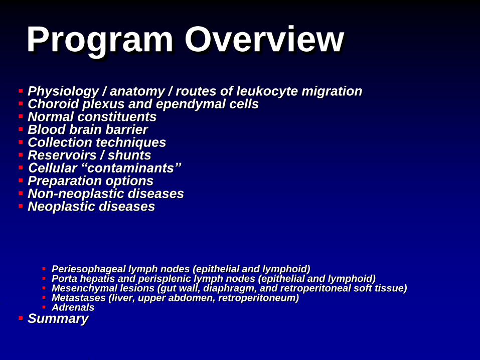
Program Overview
Physiology anatomy routes of leukocyte migration Choroid plexus and ependymal cells Normal constituents Blood brain barrier Collection techniques Reservoirs shunts Cellular ldquocontaminantsrdquo Preparation options Non-neoplastic diseases Neoplastic diseases
Periesophageal lymph nodes (epithelial and lymphoid) Porta hepatis and perisplenic lymph nodes (epithelial and lymphoid) Mesenchymal lesions (gut wall diaphragm and retroperitoneal soft tissue) Metastases (liver upper abdomen retroperitoneum) Adrenals
Summary
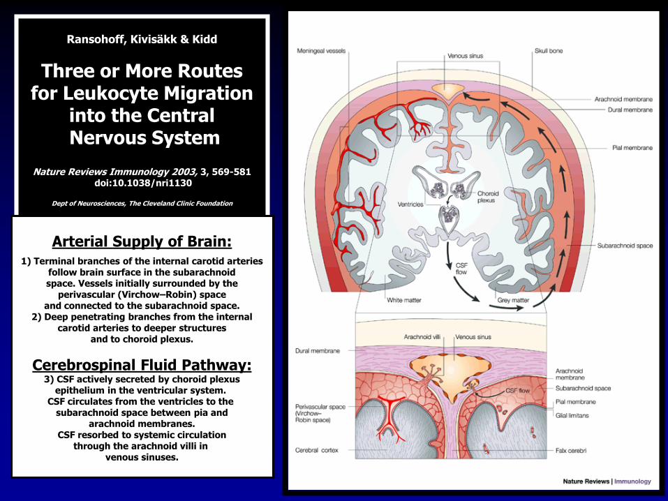
Ransohoff Kivisaumlkk amp Kidd
Three or More Routesfor Leukocyte Migration
into the CentralNervous System
Nature Reviews Immunology 2003 3 569-581doi101038nri1130
Dept of Neurosciences The Cleveland Clinic Foundation
Arterial Supply of Brain1) Terminal branches of the internal carotid arteries
follow brain surface in the subarachnoidspace Vessels initially surrounded by the
perivascular (VirchowndashRobin) spaceand connected to the subarachnoid space
2) Deep penetrating branches from the internalcarotid arteries to deeper structures
and to choroid plexus
Cerebrospinal Fluid Pathway3) CSF actively secreted by choroid plexus
epithelium in the ventricular system CSF circulates from the ventricles to the
subarachnoid space between pia andarachnoid membranes
CSF resorbed to systemic circulationthrough the arachnoid villi in
venous sinuses
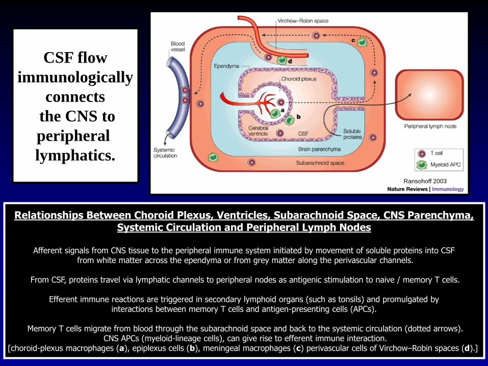
Relationships Between Choroid Plexus Ventricles Subarachnoid Space CNS ParenchymaSystemic Circulation and Peripheral Lymph Nodes
Afferent signals from CNS tissue to the peripheral immune system initiated by movement of soluble proteins into CSFfrom white matter across the ependyma or from grey matter along the perivascular channels
From CSF proteins travel via lymphatic channels to peripheral nodes as antigenic stimulation to naive memory T cells
Efferent immune reactions are triggered in secondary lymphoid organs (such as tonsils) and promulgated byinteractions between memory T cells and antigen-presenting cells (APCs)
Memory T cells migrate from blood through the subarachnoid space and back to the systemic circulation (dotted arrows)CNS APCs (myeloid-lineage cells) can give rise to efferent immune interaction
[choroid-plexus macrophages (a) epiplexus cells (b) meningeal macrophages (c) perivascular cells of VirchowndashRobin spaces (d)]
CSF flow
immunologically
connects
the CNS to
peripheral
lymphatics
Ransohoff 2003
CSF classically regarded as an ultrafiltrate of plasmaCSF more aptly described as a product of the
secretory epithelium of the CP_________________________________________
Persons aged gt= 5 years of age total CSF volume of 150 ml Human CSF volume turns over roughly FOUR times each day
CNS lacks lymphatic channelsIn some ways CSF may be thought of as lymph for the CNS
CSF can however drain directly into head and neck nodesOlfactory bulbs associated with extensions of SA space
Fluid from these locations drains across cribriform plate intolymphatics of the sinonasal submucosa
Lymphatics from other cranial nerves may also drain CSFto regional lymph nodes
Choroid plexus
Choroid plexus
31M VP shunt
Ependymal cells Ependymocytes
Cells that line the CSF-filled ventricles in the brain and the central canal of the spinal cordCiliated simple cuboidal epithelial-like cells
Apical surfaces covered in a layer of cilia which aid in circulation of CSFApical surfaces also covered with microvilli that can absorb CSF
Modified tight junctions between ependymal cells control fluid release across the liningHelp to control the exchange of substances between CSF and tissues of the brain and cord
The basal membrane of these cells is contacted by tentacle-like extensions of subjacent glia
httpbrainyinfocomstructuresthe-synapseependymal-cells
Ependymal cells Are they
ldquoepithelialrdquoor ldquoglialrdquo
Seems a bit of bothhellip
5 wk F CSF undergoing work up to rule out CNS infection
5 wk F CSF undergoing work up to rule out CNS infection
P Yeh amp D Westerman Nov 2015
Arachnoid granulations villi are small protrusions of arachnoid membrane through the duraVilli protrude into venous sinuses allowing CSF to exit the sub-arachnoid space into theblood Largest granulations lie in the superior saggittal sinus but are present in other
dural sinuses CSF pressures generally higher than venous pressures CSF flowsthrough the villi into the blood If pressures are reversed fluid will not
pass back into the subarachnoid space
ldquoNormalrdquo Cellular Constituents of CSF
Rare few small mature-appearing lymphocytes
Rare monocyte
55M headache and numbness in extremities
What the Heck
is the
Blood Brain Barrier
Separation of circulating blood from the extracellular CNS fluidOccurs along all capillaries
Consists of selective tight junctions between endothelial cellsAlso includes thick basement membrane and astrocytic endfeet or glia limitans
Restricts diffusion of organisism and large molecules into the substance of the CNS
Sounds like an alternative rock band to mehellipCould ldquoGreen Dayrdquo have beenldquoBBBrdquo
ALL marked propensity for CNS involvement especially as
leptomeningeal disease (50 of patients without prophylaxis)
Blood vessel basement membrane rich in laminin (known to
coordinate pathfinding in neuronal progenitor cells)
Most ALL cells express laminin receptor α6 integrin
Mice with ALL xenografts treated with inhibitors of α6 integrin
expression or α6 integrin neutralizing antibodies showed significant
reductions in ALL CNS transit
ALL cells use α6 integrin interactions with laminin in emissary blood
vessels passing between bone marrow and SAS to invade CNS and
do not conventionally traverse the BBB
How is CSF collected
Heinrich QuinckeGerman internist
Developed lumbarpuncture fortherapy of
hydrocephalusin 1891
Lumbar puncture ldquocontaminantsrdquohellipChondrocyte
Anucleate squamous cellOsteoblast
Marrow elementsMegakaryocyte
Ayub K Ommaya MD ScD (hc) FRCS FACS
1930-2008Pakistani neurosurgeon and
inventor of the Ommaya reservoir
Ommaya ReservoirUsed for repeated extractions of isovolumic aliquots of CSF
and concomitant intrathecal infusions of CNS therapies
Schematic representation ofan implanted Ommaya reservoir
26M Omaya Reservoir CSF Hx of Ewings sarcoma
Cerebral Shunts
Ventriculoperitoneal (VP) shunts most common
Most cerebral shunts connect ventricular system by tubing with a pumpvalve to a long catheter
CSF shunts can also drain to right atrium of heart and to pleural spaceLP connect the lumbar spine to the peritoneum
Shunts can be a conduit for infections into the CNS
61F VP Shunt
61 M HX of primaryCNS LBCL
reservoir tap
Diagnostic Accuracy of Cerebrospinal Fluid (CSF) Cytology in Metastatic Tumors An Analysis of Consecutive CSF Samples
Yoon Sung Bae June-Won Cheong Won Seok Chang Sewha Kim Eun Ji Oh Se Hoon Kim
Korean J Pathol Dec 2013 47(6) 563ndash568
385 CSF cytology samples from 42 patients were collected The samples were gathered using a ventricular catheter and reservoir CSF cytology of all patients was examined more than two
times with immunocytochemistry for cytokeratinPrimary neoplastic sites and histologic types of patients metastatic cancer were diverse The
overall sensitivity for detecting malignancy was 413 Even within short-term intervals diagnoses frequently changed
CSF cytology is currently the ldquogold
standardrdquo for the diagnosis of
malignant leptomeningeal disease
High specificity gt95
Low sensitivity 50
Loeb KR et al May 2015
Pressure 70 - 180 mm H20 (lateral recumbent)Appearance clear colorless Total protein 15 - 60 mg100 mL Gamma globulin 3 - 12 of the total proteinGlucose 50 - 80 mg100 mLCell count 0 - 5 white blood cells (all mononuclear)
No red blood cells and no PMNsChloride 110 - 125 mEqL
CSF Normal Values
Traumatic Tap Versus Pathologic Bleeding
Traumatic Tap Pathologic BleedBlood Most in 1st tube then less Same in all tubes
Post-centrifuge Clear supernatant Xanthochromic
Clot May clot Does not clot
Microscopic Well preserved RBCs Poorly preserved RBCs
Fresh blood Erythorphagocytosis
Siderophages
Based on Demay RM The Art and Science of Cytopathology 2nd Ed Exfoliative Cytology CSF Chapter 6 p 492
77M HX of GBM status post surgery 3 months remote
77M known CLLSLL presents with headaches
CSF Pressures
Increased CSF Pressure
Congestive heart failureMass lesions (tumor abscess etc)hellipInflammationCerebral edemaSuperior vena cava syndromeImpaired resorptionIntracranial veinous thrombosisIncreased CSF proteinAcute hypo-osmolalitySubarachnoid hemorrhageLead poisoning
Decreased CSF Pressure
Hypotension shockSevere dehydrationSpinal blockAcute hyperosmolality
Based on Demay RM The Art and Science of Cytopathology 2nd Ed Exfoliative Cytology CSF Chapter 6 p 491
CSF Protein amp Glucose
Increased ProteinTumorInflammation infectionSubarachnoid hemorrhageCerebral infarctionDegenerative diseases such as MSGuillain-Barre syndromeDiabetes with peripheral neuropathyDrugs (phenothiazenes)Traumatic tap
Decreased ProteinWater intoxicationSome leukemiasCSF leakageHyperthyroidismPostpneumoencephalogramNormal in some children
Increased GlucoseHyperglycemiaDiabetesIntravenous drugs
Decreased GlucoseBacterial meningitisMyobacterial meningitisFungal meningitisHypoglycemiaSubarachoid hemorrhageTumorNot viral meningitis
Based on Demay RM The Art and Science of Cytopathology 2nd Ed Exfoliative Cytology CSF Chapter 6 p 492
Cytology Preparation Options
Concentration Techniques RequiredCytocentrifugation preps - favoredProprietary liquid based preps
Membrane filter preps
Coated plus charged slides - favored
Two stains - favoredModified giemsa (Diff-Quik Quik-Dip)Papanicolaou
Cell blocks may be of great value inselected cases
BACKGROUND The present study was designed to determine whether the Thinprep plus Papanicolaou stain (Thinprep)
method is more sensitive than the Cytospin-coupled Wright-Giemsa (WG) stain (Cytospin) method in diagnosis of leptomeningeal metastasis (LM) from malignant solid tumors in cerebrospinal fluid (CSF)
METHODS The morphological features of tumor cells in fresh CSF samples were analyzed using both methods The tumor
cell detection rates were compared between the two methods
RESULTS Using the Thinprep method we found that each type of tumor cells in the CSF samples had specific identifiable
morphological features linked to their primary cancer origins such as adenocarcinomas originated from the lungs breast andstomach and lung squamous cell carcinomas small cell lung cancer large-cell neuroendocrine lung cancer hepatocellular carcinoma and malignant melanoma In a retrospective study with 88 LM patients cancer cells were detected in 80 out of the 88 CSF samples In the comparative study with 45 LM patients the initial detection rate of the Thinprep method was significantly higher than that of the Cytospin method (733 vs 578 Plt001) The cell morphology was better preserved and subcellular structures were clearer using the Thinprep method compared to the Cytospin method
CONCLUSIONS The Thinprep method is more sensitive and suitable for LM diagnosis in CSF in patients with malignant
solid tumors than the Cytospin method The Thinprep method may facilitate primary tumor detection and help design early treatment regimens for LM patients with tumors of unknown primary origin
April 7 2015
Tumor Cells Detected in CSF Specimens
ThinPrep plus Papanicolaou Stain Method Is More Sensitive than Cytospin-Coupled Wright Giemsa Stain Method in CSF Cytology for Diagnosis of Leptomeningeal Metastasis from Solid Tumors Pan et al Changchun China April 7 2015
a-n adenocarcinomas t-u hepatocellular carcinomaso-p squamous carcinomas v-x large cell carcinomasr-s small cell carcinomas y-z melanomas
Concordance with reference diagnosis was
slightly but significantly higher for modified
Giemsa-stained preparations compared with
Pap-stained slides
APLMJULY
2018
67F headaches reactive pleocytosis
67F headaches reactive pleocytosis
45F with shunt after head trauma from MVA
68M West Nile Virus Meningo-Encephalitis
Cerebrospinal Fluid Cytology in Seasonal Epidemic West Nile
Virus Meningo-Encephalitis
Ajay Rawal MD Patrick J Gavin MD Charles D Sturgis MD
Diagnostic CytopathologyVolume 34 Issue 2 pages 127ndash129 February 2006
DOI 101002dc20410 Copyright copy 2006 Wiley-Liss Inc
82F West Nile Virus Meningo-Encephalitis
Cerebrospinal fluid cytology an 11-year experience with 5951 specimensPrayson RA amp Fischler DF Department of Anatomic Pathology Cleveland Clinic Foundation OH 44195 USA
DESIGN AND SETTING A retrospective study of 5951 CSF specimens generated between 1985 and 1995 Specimens from pediatric patients (lt19 years of age) from the same time period were separately identified
RESULTS A total of 5561 adult and 390 pediatric CSF specimens were interpreted A diagnosis of negative for malignant cells was assigned in 5171 (93) of the adult cases and in 351 (90) of the pediatric cases Specific infectious organisms were identified in 26 adult specimens and one pediatric specimen Cryptococcus was the most common infectious agent observed (n = 23 adults) and Toxoplasma was the sole pediatric infectious agent Two hundred seventy-six (5) adult cases and 31 (8) pediatric cases were positive for malignant cells Diagnoses included metastatic tumors (adult 140 [51] pediatric 0) lymphomaleukemia (adult 112 [41] pediatric 4 [13]) malignant unclassified neoplasms (adult 9 [3] pediatric 0) and primary central nervous system neoplasms (adult 12 [4] pediatric 27 [87]) Medulloblastoma was the most common pediatric neoplasm (n = 21) There were 105 (2) adult cases and 8 (2) pediatric cases with atypical cells present Atypical lymphoid cells were the most common type in adult cases (53)
CONCLUSIONS In our experience infectious agents were rarely identified in pediatric CSF specimens In adult specimens the most commonly identified organisms was Cryptococcus Primary central nervous system neoplasms accounted for a higher percentage of CSF specimens in the pediatric population than in the adult population The most commonly identified malignancy in adults was metastatic neoplasms and in children medulloblastoma
21M HIVAIDS Cryptococcal Meningitis
76 M being treated systemically for Hodgkinrsquos
lymphoma developed cryptococcal
meningitis
ThinPrep Papanicolaou
76 M being treated systemically for
Hodgkinrsquos lymphoma developed
cryptococcal meningitis
Cell Block
HampE AlcBlue-PAS GMS
ldquoIndia Inkrdquo
Mucicarmine
21M HIVAIDS Cryptococcal Meningitis
VDRL (detects anti-cardiolipin antibodies that are present
in cases of syphilis but does not directly detect treponemal
antigens) has imperfect specificity and sensitivity
Newer treponemal EIAs and CIAs (enzyme linked and
chemiluminescent immunoassays) developed for
serum show enhanced sensitivity on CSF
May be of value with plasma cell rich pleocytoses
58 F
headache and photophobia
1 year after
small bowel transplant
PTLD DQ TP HampE EBER
CSFThinPrep Pap
42 Guatemalan MaleHeadache and Seizures
Neurocysticercosis(Taenia solium)
Racemose NeurocysticercosisType of neurocysticercosis with
involvement of the subarachnoid space
42M Guatemalan with neurocysticercosis (Taenia solium)
42M Guatemalan with neurocysticercosis (Taenia solium)
39M history of non-small cell lung cancer and bacteremia
20F Rheumatoid arthritis VZV meningitis
71M hx of metastatic prostate carcinoma now with headachesand single 28 cm irregularly shaped white matter lesion
Patients with known history of DLBCL
81F 53M HIV
32F B-ALL DQ amp MWG from Heme Lab
74F history of systemic mantle cell lymphoma
23M hx of medulloblastoma of cerebellum
14 M
HX of alveolar
rhabdomyosarcoma left hand
Now with widely metastatic
disease
ThinPrep Papanicolaou
74F smoker small cell carcinoma of lung
CSFEBUS FNA of level 7 lymph node
71F history of pT2 pN1 grade 2 ductal carcinoma of breast
76F history of pT2 pN0 invasive lobular carcinoma of breast
44 F presented with CNS
symptoms including headache
and confusion
later developed seizures
44 F Presented with CNS Symptoms
Subsequent mammography and
chest CT confirmed right
breast mass lesion
MRI confirmed enhancement of
lumbar spinal cord and
cauda equina
44 F Presented with CNS Symptoms
Found to have e-cadherin negative
invasive lobular breast carcinoma
Interestingly strongly ER amp Her-2 +
ER
HER-2
69M smoker with large lung lesionmediastinal adenopathy and
presentation of seizures
CSF
Wang needleof level 7 lymph node
57 M ConfusionRule out meningitis
MRI head with diffuseleptomeningealenhancement
Small enhancing softtissue mass in
left maxillary sinus
40F dural based mass lesion
CSF
Touch preparation ofresected tumor
Glioblastoma multiforme with epithelial differentiation A potential diagnostic pitfall in cerebrospinal fluid cytologySK Gill MD V Padmanabhan MD WF Hickey MD and JD Marotti MD 23 APR 2015 DOI 101002dc23274 copy 2015 Wiley Periodicals IncDartmouth Hitchcock Medical Center amp Geisel School of Medicine at Dartmouth
Cerebrospinal fluid lumbar (A) Hypercellular specimen with loosely cohesive sheets and groups of atypical cells (Cytospin Diff-Quik 60X) B Nuclear pleomorphism and prominent cytoplasmic vacuoles resembling metastatic carcinoma are evident (Cytospin Diff-Quik 300X inset 400X)
MRI of the brain showed a 64 cmring-enhancing lesion in the lefttemporal lobe
Surgical resection specimen (A) Scattered groups of small densely packed epithelioid cells are present in a background of conventional GBM (HampE 140X) B Cytoplasmic vacuoles are present (HampE 300X) C GFAP is negative in epithelioid cells but demonstrates diffuse positivity in the background (Immunostain 100X) D Epithelioid cells are focally positive for EMA (Immunostain 200X) E Focal positivity for cytokeratin AE1AE3 is also present (Immunostain 200X)
78M CHF
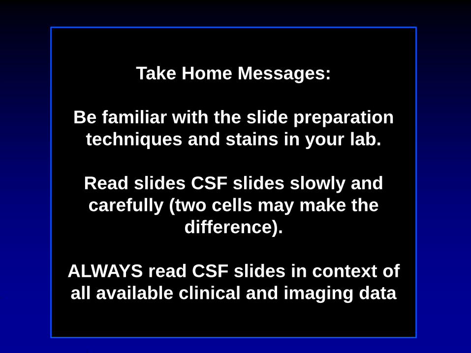
Take Home Messages
Be familiar with the slide preparation
techniques and stains in your lab
Read slides CSF slides slowly and
carefully (two cells may make the
difference)
ALWAYS read CSF slides in context of
all available clinical and imaging data
My pleasure to speak with you today
CommentsCritiquesInsights
Questions
Disclosures for Dr Sturgis
Consultant amp Trainer for Ventana (PDL-1 IHC) 2015Consultant for Philips (Pivotal Study ndash Digital Imaging) 2016Consultant for Preora Healthcare (Aerosol Cytology) 2018
(No conflicts of interest with this program)
Program Overview
Physiology anatomy routes of leukocyte migration Choroid plexus and ependymal cells Normal constituents Blood brain barrier Collection techniques Reservoirs shunts Cellular ldquocontaminantsrdquo Preparation options Non-neoplastic diseases Neoplastic diseases
Periesophageal lymph nodes (epithelial and lymphoid) Porta hepatis and perisplenic lymph nodes (epithelial and lymphoid) Mesenchymal lesions (gut wall diaphragm and retroperitoneal soft tissue) Metastases (liver upper abdomen retroperitoneum) Adrenals
Summary
Ransohoff Kivisaumlkk amp Kidd
Three or More Routesfor Leukocyte Migration
into the CentralNervous System
Nature Reviews Immunology 2003 3 569-581doi101038nri1130
Dept of Neurosciences The Cleveland Clinic Foundation
Arterial Supply of Brain1) Terminal branches of the internal carotid arteries
follow brain surface in the subarachnoidspace Vessels initially surrounded by the
perivascular (VirchowndashRobin) spaceand connected to the subarachnoid space
2) Deep penetrating branches from the internalcarotid arteries to deeper structures
and to choroid plexus
Cerebrospinal Fluid Pathway3) CSF actively secreted by choroid plexus
epithelium in the ventricular system CSF circulates from the ventricles to the
subarachnoid space between pia andarachnoid membranes
CSF resorbed to systemic circulationthrough the arachnoid villi in
venous sinuses
Relationships Between Choroid Plexus Ventricles Subarachnoid Space CNS ParenchymaSystemic Circulation and Peripheral Lymph Nodes
Afferent signals from CNS tissue to the peripheral immune system initiated by movement of soluble proteins into CSFfrom white matter across the ependyma or from grey matter along the perivascular channels
From CSF proteins travel via lymphatic channels to peripheral nodes as antigenic stimulation to naive memory T cells
Efferent immune reactions are triggered in secondary lymphoid organs (such as tonsils) and promulgated byinteractions between memory T cells and antigen-presenting cells (APCs)
Memory T cells migrate from blood through the subarachnoid space and back to the systemic circulation (dotted arrows)CNS APCs (myeloid-lineage cells) can give rise to efferent immune interaction
[choroid-plexus macrophages (a) epiplexus cells (b) meningeal macrophages (c) perivascular cells of VirchowndashRobin spaces (d)]
CSF flow
immunologically
connects
the CNS to
peripheral
lymphatics
Ransohoff 2003
CSF classically regarded as an ultrafiltrate of plasmaCSF more aptly described as a product of the
secretory epithelium of the CP_________________________________________
Persons aged gt= 5 years of age total CSF volume of 150 ml Human CSF volume turns over roughly FOUR times each day
CNS lacks lymphatic channelsIn some ways CSF may be thought of as lymph for the CNS
CSF can however drain directly into head and neck nodesOlfactory bulbs associated with extensions of SA space
Fluid from these locations drains across cribriform plate intolymphatics of the sinonasal submucosa
Lymphatics from other cranial nerves may also drain CSFto regional lymph nodes
Choroid plexus
Choroid plexus
31M VP shunt
Ependymal cells Ependymocytes
Cells that line the CSF-filled ventricles in the brain and the central canal of the spinal cordCiliated simple cuboidal epithelial-like cells
Apical surfaces covered in a layer of cilia which aid in circulation of CSFApical surfaces also covered with microvilli that can absorb CSF
Modified tight junctions between ependymal cells control fluid release across the liningHelp to control the exchange of substances between CSF and tissues of the brain and cord
The basal membrane of these cells is contacted by tentacle-like extensions of subjacent glia
httpbrainyinfocomstructuresthe-synapseependymal-cells
Ependymal cells Are they
ldquoepithelialrdquoor ldquoglialrdquo
Seems a bit of bothhellip
5 wk F CSF undergoing work up to rule out CNS infection
5 wk F CSF undergoing work up to rule out CNS infection
P Yeh amp D Westerman Nov 2015
Arachnoid granulations villi are small protrusions of arachnoid membrane through the duraVilli protrude into venous sinuses allowing CSF to exit the sub-arachnoid space into theblood Largest granulations lie in the superior saggittal sinus but are present in other
dural sinuses CSF pressures generally higher than venous pressures CSF flowsthrough the villi into the blood If pressures are reversed fluid will not
pass back into the subarachnoid space
ldquoNormalrdquo Cellular Constituents of CSF
Rare few small mature-appearing lymphocytes
Rare monocyte
55M headache and numbness in extremities
What the Heck
is the
Blood Brain Barrier
Separation of circulating blood from the extracellular CNS fluidOccurs along all capillaries
Consists of selective tight junctions between endothelial cellsAlso includes thick basement membrane and astrocytic endfeet or glia limitans
Restricts diffusion of organisism and large molecules into the substance of the CNS
Sounds like an alternative rock band to mehellipCould ldquoGreen Dayrdquo have beenldquoBBBrdquo
ALL marked propensity for CNS involvement especially as
leptomeningeal disease (50 of patients without prophylaxis)
Blood vessel basement membrane rich in laminin (known to
coordinate pathfinding in neuronal progenitor cells)
Most ALL cells express laminin receptor α6 integrin
Mice with ALL xenografts treated with inhibitors of α6 integrin
expression or α6 integrin neutralizing antibodies showed significant
reductions in ALL CNS transit
ALL cells use α6 integrin interactions with laminin in emissary blood
vessels passing between bone marrow and SAS to invade CNS and
do not conventionally traverse the BBB
How is CSF collected
Heinrich QuinckeGerman internist
Developed lumbarpuncture fortherapy of
hydrocephalusin 1891
Lumbar puncture ldquocontaminantsrdquohellipChondrocyte
Anucleate squamous cellOsteoblast
Marrow elementsMegakaryocyte
Ayub K Ommaya MD ScD (hc) FRCS FACS
1930-2008Pakistani neurosurgeon and
inventor of the Ommaya reservoir
Ommaya ReservoirUsed for repeated extractions of isovolumic aliquots of CSF
and concomitant intrathecal infusions of CNS therapies
Schematic representation ofan implanted Ommaya reservoir
26M Omaya Reservoir CSF Hx of Ewings sarcoma
Cerebral Shunts
Ventriculoperitoneal (VP) shunts most common
Most cerebral shunts connect ventricular system by tubing with a pumpvalve to a long catheter
CSF shunts can also drain to right atrium of heart and to pleural spaceLP connect the lumbar spine to the peritoneum
Shunts can be a conduit for infections into the CNS
61F VP Shunt
61 M HX of primaryCNS LBCL
reservoir tap
Diagnostic Accuracy of Cerebrospinal Fluid (CSF) Cytology in Metastatic Tumors An Analysis of Consecutive CSF Samples
Yoon Sung Bae June-Won Cheong Won Seok Chang Sewha Kim Eun Ji Oh Se Hoon Kim
Korean J Pathol Dec 2013 47(6) 563ndash568
385 CSF cytology samples from 42 patients were collected The samples were gathered using a ventricular catheter and reservoir CSF cytology of all patients was examined more than two
times with immunocytochemistry for cytokeratinPrimary neoplastic sites and histologic types of patients metastatic cancer were diverse The
overall sensitivity for detecting malignancy was 413 Even within short-term intervals diagnoses frequently changed
CSF cytology is currently the ldquogold
standardrdquo for the diagnosis of
malignant leptomeningeal disease
High specificity gt95
Low sensitivity 50
Loeb KR et al May 2015
Pressure 70 - 180 mm H20 (lateral recumbent)Appearance clear colorless Total protein 15 - 60 mg100 mL Gamma globulin 3 - 12 of the total proteinGlucose 50 - 80 mg100 mLCell count 0 - 5 white blood cells (all mononuclear)
No red blood cells and no PMNsChloride 110 - 125 mEqL
CSF Normal Values
Traumatic Tap Versus Pathologic Bleeding
Traumatic Tap Pathologic BleedBlood Most in 1st tube then less Same in all tubes
Post-centrifuge Clear supernatant Xanthochromic
Clot May clot Does not clot
Microscopic Well preserved RBCs Poorly preserved RBCs
Fresh blood Erythorphagocytosis
Siderophages
Based on Demay RM The Art and Science of Cytopathology 2nd Ed Exfoliative Cytology CSF Chapter 6 p 492
77M HX of GBM status post surgery 3 months remote
77M known CLLSLL presents with headaches
CSF Pressures
Increased CSF Pressure
Congestive heart failureMass lesions (tumor abscess etc)hellipInflammationCerebral edemaSuperior vena cava syndromeImpaired resorptionIntracranial veinous thrombosisIncreased CSF proteinAcute hypo-osmolalitySubarachnoid hemorrhageLead poisoning
Decreased CSF Pressure
Hypotension shockSevere dehydrationSpinal blockAcute hyperosmolality
Based on Demay RM The Art and Science of Cytopathology 2nd Ed Exfoliative Cytology CSF Chapter 6 p 491
CSF Protein amp Glucose
Increased ProteinTumorInflammation infectionSubarachnoid hemorrhageCerebral infarctionDegenerative diseases such as MSGuillain-Barre syndromeDiabetes with peripheral neuropathyDrugs (phenothiazenes)Traumatic tap
Decreased ProteinWater intoxicationSome leukemiasCSF leakageHyperthyroidismPostpneumoencephalogramNormal in some children
Increased GlucoseHyperglycemiaDiabetesIntravenous drugs
Decreased GlucoseBacterial meningitisMyobacterial meningitisFungal meningitisHypoglycemiaSubarachoid hemorrhageTumorNot viral meningitis
Based on Demay RM The Art and Science of Cytopathology 2nd Ed Exfoliative Cytology CSF Chapter 6 p 492
Cytology Preparation Options
Concentration Techniques RequiredCytocentrifugation preps - favoredProprietary liquid based preps
Membrane filter preps
Coated plus charged slides - favored
Two stains - favoredModified giemsa (Diff-Quik Quik-Dip)Papanicolaou
Cell blocks may be of great value inselected cases
BACKGROUND The present study was designed to determine whether the Thinprep plus Papanicolaou stain (Thinprep)
method is more sensitive than the Cytospin-coupled Wright-Giemsa (WG) stain (Cytospin) method in diagnosis of leptomeningeal metastasis (LM) from malignant solid tumors in cerebrospinal fluid (CSF)
METHODS The morphological features of tumor cells in fresh CSF samples were analyzed using both methods The tumor
cell detection rates were compared between the two methods
RESULTS Using the Thinprep method we found that each type of tumor cells in the CSF samples had specific identifiable
morphological features linked to their primary cancer origins such as adenocarcinomas originated from the lungs breast andstomach and lung squamous cell carcinomas small cell lung cancer large-cell neuroendocrine lung cancer hepatocellular carcinoma and malignant melanoma In a retrospective study with 88 LM patients cancer cells were detected in 80 out of the 88 CSF samples In the comparative study with 45 LM patients the initial detection rate of the Thinprep method was significantly higher than that of the Cytospin method (733 vs 578 Plt001) The cell morphology was better preserved and subcellular structures were clearer using the Thinprep method compared to the Cytospin method
CONCLUSIONS The Thinprep method is more sensitive and suitable for LM diagnosis in CSF in patients with malignant
solid tumors than the Cytospin method The Thinprep method may facilitate primary tumor detection and help design early treatment regimens for LM patients with tumors of unknown primary origin
April 7 2015
Tumor Cells Detected in CSF Specimens
ThinPrep plus Papanicolaou Stain Method Is More Sensitive than Cytospin-Coupled Wright Giemsa Stain Method in CSF Cytology for Diagnosis of Leptomeningeal Metastasis from Solid Tumors Pan et al Changchun China April 7 2015
a-n adenocarcinomas t-u hepatocellular carcinomaso-p squamous carcinomas v-x large cell carcinomasr-s small cell carcinomas y-z melanomas
Concordance with reference diagnosis was
slightly but significantly higher for modified
Giemsa-stained preparations compared with
Pap-stained slides
APLMJULY
2018
67F headaches reactive pleocytosis
67F headaches reactive pleocytosis
45F with shunt after head trauma from MVA
68M West Nile Virus Meningo-Encephalitis
Cerebrospinal Fluid Cytology in Seasonal Epidemic West Nile
Virus Meningo-Encephalitis
Ajay Rawal MD Patrick J Gavin MD Charles D Sturgis MD
Diagnostic CytopathologyVolume 34 Issue 2 pages 127ndash129 February 2006
DOI 101002dc20410 Copyright copy 2006 Wiley-Liss Inc
82F West Nile Virus Meningo-Encephalitis
Cerebrospinal fluid cytology an 11-year experience with 5951 specimensPrayson RA amp Fischler DF Department of Anatomic Pathology Cleveland Clinic Foundation OH 44195 USA
DESIGN AND SETTING A retrospective study of 5951 CSF specimens generated between 1985 and 1995 Specimens from pediatric patients (lt19 years of age) from the same time period were separately identified
RESULTS A total of 5561 adult and 390 pediatric CSF specimens were interpreted A diagnosis of negative for malignant cells was assigned in 5171 (93) of the adult cases and in 351 (90) of the pediatric cases Specific infectious organisms were identified in 26 adult specimens and one pediatric specimen Cryptococcus was the most common infectious agent observed (n = 23 adults) and Toxoplasma was the sole pediatric infectious agent Two hundred seventy-six (5) adult cases and 31 (8) pediatric cases were positive for malignant cells Diagnoses included metastatic tumors (adult 140 [51] pediatric 0) lymphomaleukemia (adult 112 [41] pediatric 4 [13]) malignant unclassified neoplasms (adult 9 [3] pediatric 0) and primary central nervous system neoplasms (adult 12 [4] pediatric 27 [87]) Medulloblastoma was the most common pediatric neoplasm (n = 21) There were 105 (2) adult cases and 8 (2) pediatric cases with atypical cells present Atypical lymphoid cells were the most common type in adult cases (53)
CONCLUSIONS In our experience infectious agents were rarely identified in pediatric CSF specimens In adult specimens the most commonly identified organisms was Cryptococcus Primary central nervous system neoplasms accounted for a higher percentage of CSF specimens in the pediatric population than in the adult population The most commonly identified malignancy in adults was metastatic neoplasms and in children medulloblastoma
21M HIVAIDS Cryptococcal Meningitis
76 M being treated systemically for Hodgkinrsquos
lymphoma developed cryptococcal
meningitis
ThinPrep Papanicolaou
76 M being treated systemically for
Hodgkinrsquos lymphoma developed
cryptococcal meningitis
Cell Block
HampE AlcBlue-PAS GMS
ldquoIndia Inkrdquo
Mucicarmine
21M HIVAIDS Cryptococcal Meningitis
VDRL (detects anti-cardiolipin antibodies that are present
in cases of syphilis but does not directly detect treponemal
antigens) has imperfect specificity and sensitivity
Newer treponemal EIAs and CIAs (enzyme linked and
chemiluminescent immunoassays) developed for
serum show enhanced sensitivity on CSF
May be of value with plasma cell rich pleocytoses
58 F
headache and photophobia
1 year after
small bowel transplant
PTLD DQ TP HampE EBER
CSFThinPrep Pap
42 Guatemalan MaleHeadache and Seizures
Neurocysticercosis(Taenia solium)
Racemose NeurocysticercosisType of neurocysticercosis with
involvement of the subarachnoid space
42M Guatemalan with neurocysticercosis (Taenia solium)
42M Guatemalan with neurocysticercosis (Taenia solium)
39M history of non-small cell lung cancer and bacteremia
20F Rheumatoid arthritis VZV meningitis
71M hx of metastatic prostate carcinoma now with headachesand single 28 cm irregularly shaped white matter lesion
Patients with known history of DLBCL
81F 53M HIV
32F B-ALL DQ amp MWG from Heme Lab
74F history of systemic mantle cell lymphoma
23M hx of medulloblastoma of cerebellum
14 M
HX of alveolar
rhabdomyosarcoma left hand
Now with widely metastatic
disease
ThinPrep Papanicolaou
74F smoker small cell carcinoma of lung
CSFEBUS FNA of level 7 lymph node
71F history of pT2 pN1 grade 2 ductal carcinoma of breast
76F history of pT2 pN0 invasive lobular carcinoma of breast
44 F presented with CNS
symptoms including headache
and confusion
later developed seizures
44 F Presented with CNS Symptoms
Subsequent mammography and
chest CT confirmed right
breast mass lesion
MRI confirmed enhancement of
lumbar spinal cord and
cauda equina
44 F Presented with CNS Symptoms
Found to have e-cadherin negative
invasive lobular breast carcinoma
Interestingly strongly ER amp Her-2 +
ER
HER-2
69M smoker with large lung lesionmediastinal adenopathy and
presentation of seizures
CSF
Wang needleof level 7 lymph node
57 M ConfusionRule out meningitis
MRI head with diffuseleptomeningealenhancement
Small enhancing softtissue mass in
left maxillary sinus
40F dural based mass lesion
CSF
Touch preparation ofresected tumor
Glioblastoma multiforme with epithelial differentiation A potential diagnostic pitfall in cerebrospinal fluid cytologySK Gill MD V Padmanabhan MD WF Hickey MD and JD Marotti MD 23 APR 2015 DOI 101002dc23274 copy 2015 Wiley Periodicals IncDartmouth Hitchcock Medical Center amp Geisel School of Medicine at Dartmouth
Cerebrospinal fluid lumbar (A) Hypercellular specimen with loosely cohesive sheets and groups of atypical cells (Cytospin Diff-Quik 60X) B Nuclear pleomorphism and prominent cytoplasmic vacuoles resembling metastatic carcinoma are evident (Cytospin Diff-Quik 300X inset 400X)
MRI of the brain showed a 64 cmring-enhancing lesion in the lefttemporal lobe
Surgical resection specimen (A) Scattered groups of small densely packed epithelioid cells are present in a background of conventional GBM (HampE 140X) B Cytoplasmic vacuoles are present (HampE 300X) C GFAP is negative in epithelioid cells but demonstrates diffuse positivity in the background (Immunostain 100X) D Epithelioid cells are focally positive for EMA (Immunostain 200X) E Focal positivity for cytokeratin AE1AE3 is also present (Immunostain 200X)
78M CHF
Take Home Messages
Be familiar with the slide preparation
techniques and stains in your lab
Read slides CSF slides slowly and
carefully (two cells may make the
difference)
ALWAYS read CSF slides in context of
all available clinical and imaging data
My pleasure to speak with you today
CommentsCritiquesInsights
Questions
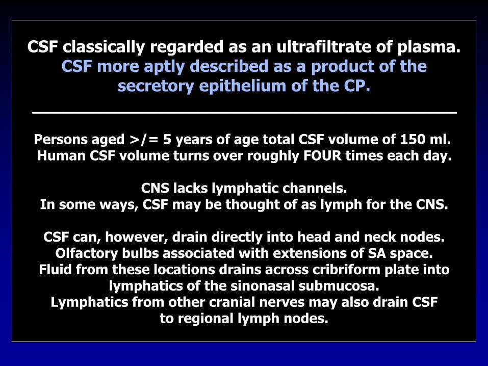
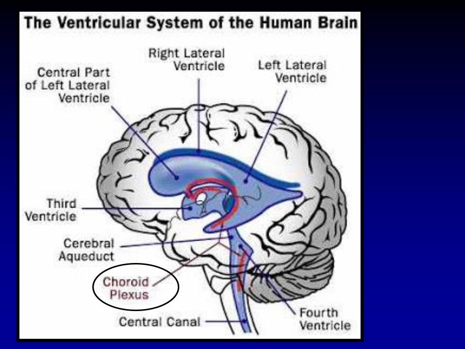
Choroid plexus
Choroid plexus
31M VP shunt
Ependymal cells Ependymocytes
Cells that line the CSF-filled ventricles in the brain and the central canal of the spinal cordCiliated simple cuboidal epithelial-like cells
Apical surfaces covered in a layer of cilia which aid in circulation of CSFApical surfaces also covered with microvilli that can absorb CSF
Modified tight junctions between ependymal cells control fluid release across the liningHelp to control the exchange of substances between CSF and tissues of the brain and cord
The basal membrane of these cells is contacted by tentacle-like extensions of subjacent glia
httpbrainyinfocomstructuresthe-synapseependymal-cells
Ependymal cells Are they
ldquoepithelialrdquoor ldquoglialrdquo
Seems a bit of bothhellip
5 wk F CSF undergoing work up to rule out CNS infection
5 wk F CSF undergoing work up to rule out CNS infection
P Yeh amp D Westerman Nov 2015
Arachnoid granulations villi are small protrusions of arachnoid membrane through the duraVilli protrude into venous sinuses allowing CSF to exit the sub-arachnoid space into theblood Largest granulations lie in the superior saggittal sinus but are present in other
dural sinuses CSF pressures generally higher than venous pressures CSF flowsthrough the villi into the blood If pressures are reversed fluid will not
pass back into the subarachnoid space
ldquoNormalrdquo Cellular Constituents of CSF
Rare few small mature-appearing lymphocytes
Rare monocyte
55M headache and numbness in extremities
What the Heck
is the
Blood Brain Barrier
Separation of circulating blood from the extracellular CNS fluidOccurs along all capillaries
Consists of selective tight junctions between endothelial cellsAlso includes thick basement membrane and astrocytic endfeet or glia limitans
Restricts diffusion of organisism and large molecules into the substance of the CNS
Sounds like an alternative rock band to mehellipCould ldquoGreen Dayrdquo have beenldquoBBBrdquo
ALL marked propensity for CNS involvement especially as
leptomeningeal disease (50 of patients without prophylaxis)
Blood vessel basement membrane rich in laminin (known to
coordinate pathfinding in neuronal progenitor cells)
Most ALL cells express laminin receptor α6 integrin
Mice with ALL xenografts treated with inhibitors of α6 integrin
expression or α6 integrin neutralizing antibodies showed significant
reductions in ALL CNS transit
ALL cells use α6 integrin interactions with laminin in emissary blood
vessels passing between bone marrow and SAS to invade CNS and
do not conventionally traverse the BBB
How is CSF collected
Heinrich QuinckeGerman internist
Developed lumbarpuncture fortherapy of
hydrocephalusin 1891
Lumbar puncture ldquocontaminantsrdquohellipChondrocyte
Anucleate squamous cellOsteoblast
Marrow elementsMegakaryocyte
Ayub K Ommaya MD ScD (hc) FRCS FACS
1930-2008Pakistani neurosurgeon and
inventor of the Ommaya reservoir
Ommaya ReservoirUsed for repeated extractions of isovolumic aliquots of CSF
and concomitant intrathecal infusions of CNS therapies
Schematic representation ofan implanted Ommaya reservoir
26M Omaya Reservoir CSF Hx of Ewings sarcoma
Cerebral Shunts
Ventriculoperitoneal (VP) shunts most common
Most cerebral shunts connect ventricular system by tubing with a pumpvalve to a long catheter
CSF shunts can also drain to right atrium of heart and to pleural spaceLP connect the lumbar spine to the peritoneum
Shunts can be a conduit for infections into the CNS
61F VP Shunt
61 M HX of primaryCNS LBCL
reservoir tap
Diagnostic Accuracy of Cerebrospinal Fluid (CSF) Cytology in Metastatic Tumors An Analysis of Consecutive CSF Samples
Yoon Sung Bae June-Won Cheong Won Seok Chang Sewha Kim Eun Ji Oh Se Hoon Kim
Korean J Pathol Dec 2013 47(6) 563ndash568
385 CSF cytology samples from 42 patients were collected The samples were gathered using a ventricular catheter and reservoir CSF cytology of all patients was examined more than two
times with immunocytochemistry for cytokeratinPrimary neoplastic sites and histologic types of patients metastatic cancer were diverse The
overall sensitivity for detecting malignancy was 413 Even within short-term intervals diagnoses frequently changed
CSF cytology is currently the ldquogold
standardrdquo for the diagnosis of
malignant leptomeningeal disease
High specificity gt95
Low sensitivity 50
Loeb KR et al May 2015
Pressure 70 - 180 mm H20 (lateral recumbent)Appearance clear colorless Total protein 15 - 60 mg100 mL Gamma globulin 3 - 12 of the total proteinGlucose 50 - 80 mg100 mLCell count 0 - 5 white blood cells (all mononuclear)
No red blood cells and no PMNsChloride 110 - 125 mEqL
CSF Normal Values
Traumatic Tap Versus Pathologic Bleeding
Traumatic Tap Pathologic BleedBlood Most in 1st tube then less Same in all tubes
Post-centrifuge Clear supernatant Xanthochromic
Clot May clot Does not clot
Microscopic Well preserved RBCs Poorly preserved RBCs
Fresh blood Erythorphagocytosis
Siderophages
Based on Demay RM The Art and Science of Cytopathology 2nd Ed Exfoliative Cytology CSF Chapter 6 p 492
77M HX of GBM status post surgery 3 months remote
77M known CLLSLL presents with headaches
CSF Pressures
Increased CSF Pressure
Congestive heart failureMass lesions (tumor abscess etc)hellipInflammationCerebral edemaSuperior vena cava syndromeImpaired resorptionIntracranial veinous thrombosisIncreased CSF proteinAcute hypo-osmolalitySubarachnoid hemorrhageLead poisoning
Decreased CSF Pressure
Hypotension shockSevere dehydrationSpinal blockAcute hyperosmolality
Based on Demay RM The Art and Science of Cytopathology 2nd Ed Exfoliative Cytology CSF Chapter 6 p 491
CSF Protein amp Glucose
Increased ProteinTumorInflammation infectionSubarachnoid hemorrhageCerebral infarctionDegenerative diseases such as MSGuillain-Barre syndromeDiabetes with peripheral neuropathyDrugs (phenothiazenes)Traumatic tap
Decreased ProteinWater intoxicationSome leukemiasCSF leakageHyperthyroidismPostpneumoencephalogramNormal in some children
Increased GlucoseHyperglycemiaDiabetesIntravenous drugs
Decreased GlucoseBacterial meningitisMyobacterial meningitisFungal meningitisHypoglycemiaSubarachoid hemorrhageTumorNot viral meningitis
Based on Demay RM The Art and Science of Cytopathology 2nd Ed Exfoliative Cytology CSF Chapter 6 p 492
Cytology Preparation Options
Concentration Techniques RequiredCytocentrifugation preps - favoredProprietary liquid based preps
Membrane filter preps
Coated plus charged slides - favored
Two stains - favoredModified giemsa (Diff-Quik Quik-Dip)Papanicolaou
Cell blocks may be of great value inselected cases
BACKGROUND The present study was designed to determine whether the Thinprep plus Papanicolaou stain (Thinprep)
method is more sensitive than the Cytospin-coupled Wright-Giemsa (WG) stain (Cytospin) method in diagnosis of leptomeningeal metastasis (LM) from malignant solid tumors in cerebrospinal fluid (CSF)
METHODS The morphological features of tumor cells in fresh CSF samples were analyzed using both methods The tumor
cell detection rates were compared between the two methods
RESULTS Using the Thinprep method we found that each type of tumor cells in the CSF samples had specific identifiable
morphological features linked to their primary cancer origins such as adenocarcinomas originated from the lungs breast andstomach and lung squamous cell carcinomas small cell lung cancer large-cell neuroendocrine lung cancer hepatocellular carcinoma and malignant melanoma In a retrospective study with 88 LM patients cancer cells were detected in 80 out of the 88 CSF samples In the comparative study with 45 LM patients the initial detection rate of the Thinprep method was significantly higher than that of the Cytospin method (733 vs 578 Plt001) The cell morphology was better preserved and subcellular structures were clearer using the Thinprep method compared to the Cytospin method
CONCLUSIONS The Thinprep method is more sensitive and suitable for LM diagnosis in CSF in patients with malignant
solid tumors than the Cytospin method The Thinprep method may facilitate primary tumor detection and help design early treatment regimens for LM patients with tumors of unknown primary origin
April 7 2015
Tumor Cells Detected in CSF Specimens
ThinPrep plus Papanicolaou Stain Method Is More Sensitive than Cytospin-Coupled Wright Giemsa Stain Method in CSF Cytology for Diagnosis of Leptomeningeal Metastasis from Solid Tumors Pan et al Changchun China April 7 2015
a-n adenocarcinomas t-u hepatocellular carcinomaso-p squamous carcinomas v-x large cell carcinomasr-s small cell carcinomas y-z melanomas
Concordance with reference diagnosis was
slightly but significantly higher for modified
Giemsa-stained preparations compared with
Pap-stained slides
APLMJULY
2018
67F headaches reactive pleocytosis
67F headaches reactive pleocytosis
45F with shunt after head trauma from MVA
68M West Nile Virus Meningo-Encephalitis
Cerebrospinal Fluid Cytology in Seasonal Epidemic West Nile
Virus Meningo-Encephalitis
Ajay Rawal MD Patrick J Gavin MD Charles D Sturgis MD
Diagnostic CytopathologyVolume 34 Issue 2 pages 127ndash129 February 2006
DOI 101002dc20410 Copyright copy 2006 Wiley-Liss Inc
82F West Nile Virus Meningo-Encephalitis
Cerebrospinal fluid cytology an 11-year experience with 5951 specimensPrayson RA amp Fischler DF Department of Anatomic Pathology Cleveland Clinic Foundation OH 44195 USA
DESIGN AND SETTING A retrospective study of 5951 CSF specimens generated between 1985 and 1995 Specimens from pediatric patients (lt19 years of age) from the same time period were separately identified
RESULTS A total of 5561 adult and 390 pediatric CSF specimens were interpreted A diagnosis of negative for malignant cells was assigned in 5171 (93) of the adult cases and in 351 (90) of the pediatric cases Specific infectious organisms were identified in 26 adult specimens and one pediatric specimen Cryptococcus was the most common infectious agent observed (n = 23 adults) and Toxoplasma was the sole pediatric infectious agent Two hundred seventy-six (5) adult cases and 31 (8) pediatric cases were positive for malignant cells Diagnoses included metastatic tumors (adult 140 [51] pediatric 0) lymphomaleukemia (adult 112 [41] pediatric 4 [13]) malignant unclassified neoplasms (adult 9 [3] pediatric 0) and primary central nervous system neoplasms (adult 12 [4] pediatric 27 [87]) Medulloblastoma was the most common pediatric neoplasm (n = 21) There were 105 (2) adult cases and 8 (2) pediatric cases with atypical cells present Atypical lymphoid cells were the most common type in adult cases (53)
CONCLUSIONS In our experience infectious agents were rarely identified in pediatric CSF specimens In adult specimens the most commonly identified organisms was Cryptococcus Primary central nervous system neoplasms accounted for a higher percentage of CSF specimens in the pediatric population than in the adult population The most commonly identified malignancy in adults was metastatic neoplasms and in children medulloblastoma
21M HIVAIDS Cryptococcal Meningitis
76 M being treated systemically for Hodgkinrsquos
lymphoma developed cryptococcal
meningitis
ThinPrep Papanicolaou
76 M being treated systemically for
Hodgkinrsquos lymphoma developed
cryptococcal meningitis
Cell Block
HampE AlcBlue-PAS GMS
ldquoIndia Inkrdquo
Mucicarmine
21M HIVAIDS Cryptococcal Meningitis
VDRL (detects anti-cardiolipin antibodies that are present
in cases of syphilis but does not directly detect treponemal
antigens) has imperfect specificity and sensitivity
Newer treponemal EIAs and CIAs (enzyme linked and
chemiluminescent immunoassays) developed for
serum show enhanced sensitivity on CSF
May be of value with plasma cell rich pleocytoses
58 F
headache and photophobia
1 year after
small bowel transplant
PTLD DQ TP HampE EBER
CSFThinPrep Pap
42 Guatemalan MaleHeadache and Seizures
Neurocysticercosis(Taenia solium)
Racemose NeurocysticercosisType of neurocysticercosis with
involvement of the subarachnoid space
42M Guatemalan with neurocysticercosis (Taenia solium)
42M Guatemalan with neurocysticercosis (Taenia solium)
39M history of non-small cell lung cancer and bacteremia
20F Rheumatoid arthritis VZV meningitis
71M hx of metastatic prostate carcinoma now with headachesand single 28 cm irregularly shaped white matter lesion
Patients with known history of DLBCL
81F 53M HIV
32F B-ALL DQ amp MWG from Heme Lab
74F history of systemic mantle cell lymphoma
23M hx of medulloblastoma of cerebellum
14 M
HX of alveolar
rhabdomyosarcoma left hand
Now with widely metastatic
disease
ThinPrep Papanicolaou
74F smoker small cell carcinoma of lung
CSFEBUS FNA of level 7 lymph node
71F history of pT2 pN1 grade 2 ductal carcinoma of breast
76F history of pT2 pN0 invasive lobular carcinoma of breast
44 F presented with CNS
symptoms including headache
and confusion
later developed seizures
44 F Presented with CNS Symptoms
Subsequent mammography and
chest CT confirmed right
breast mass lesion
MRI confirmed enhancement of
lumbar spinal cord and
cauda equina
44 F Presented with CNS Symptoms
Found to have e-cadherin negative
invasive lobular breast carcinoma
Interestingly strongly ER amp Her-2 +
ER
HER-2
69M smoker with large lung lesionmediastinal adenopathy and
presentation of seizures
CSF
Wang needleof level 7 lymph node
57 M ConfusionRule out meningitis
MRI head with diffuseleptomeningealenhancement
Small enhancing softtissue mass in
left maxillary sinus
40F dural based mass lesion
CSF
Touch preparation ofresected tumor
Glioblastoma multiforme with epithelial differentiation A potential diagnostic pitfall in cerebrospinal fluid cytologySK Gill MD V Padmanabhan MD WF Hickey MD and JD Marotti MD 23 APR 2015 DOI 101002dc23274 copy 2015 Wiley Periodicals IncDartmouth Hitchcock Medical Center amp Geisel School of Medicine at Dartmouth
Cerebrospinal fluid lumbar (A) Hypercellular specimen with loosely cohesive sheets and groups of atypical cells (Cytospin Diff-Quik 60X) B Nuclear pleomorphism and prominent cytoplasmic vacuoles resembling metastatic carcinoma are evident (Cytospin Diff-Quik 300X inset 400X)
MRI of the brain showed a 64 cmring-enhancing lesion in the lefttemporal lobe
Surgical resection specimen (A) Scattered groups of small densely packed epithelioid cells are present in a background of conventional GBM (HampE 140X) B Cytoplasmic vacuoles are present (HampE 300X) C GFAP is negative in epithelioid cells but demonstrates diffuse positivity in the background (Immunostain 100X) D Epithelioid cells are focally positive for EMA (Immunostain 200X) E Focal positivity for cytokeratin AE1AE3 is also present (Immunostain 200X)
78M CHF
Take Home Messages
Be familiar with the slide preparation
techniques and stains in your lab
Read slides CSF slides slowly and
carefully (two cells may make the
difference)
ALWAYS read CSF slides in context of
all available clinical and imaging data
My pleasure to speak with you today
CommentsCritiquesInsights
Questions
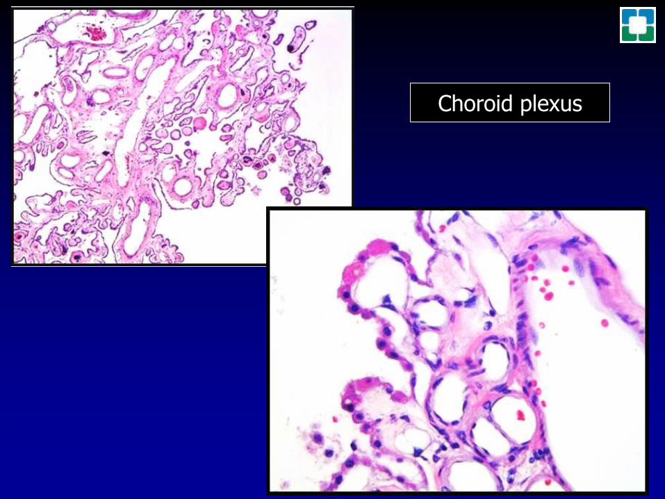
Choroid plexus
31M VP shunt
Ependymal cells Ependymocytes
Cells that line the CSF-filled ventricles in the brain and the central canal of the spinal cordCiliated simple cuboidal epithelial-like cells
Apical surfaces covered in a layer of cilia which aid in circulation of CSFApical surfaces also covered with microvilli that can absorb CSF
Modified tight junctions between ependymal cells control fluid release across the liningHelp to control the exchange of substances between CSF and tissues of the brain and cord
The basal membrane of these cells is contacted by tentacle-like extensions of subjacent glia
httpbrainyinfocomstructuresthe-synapseependymal-cells
Ependymal cells Are they
ldquoepithelialrdquoor ldquoglialrdquo
Seems a bit of bothhellip
5 wk F CSF undergoing work up to rule out CNS infection
5 wk F CSF undergoing work up to rule out CNS infection
P Yeh amp D Westerman Nov 2015
Arachnoid granulations villi are small protrusions of arachnoid membrane through the duraVilli protrude into venous sinuses allowing CSF to exit the sub-arachnoid space into theblood Largest granulations lie in the superior saggittal sinus but are present in other
dural sinuses CSF pressures generally higher than venous pressures CSF flowsthrough the villi into the blood If pressures are reversed fluid will not
pass back into the subarachnoid space
ldquoNormalrdquo Cellular Constituents of CSF
Rare few small mature-appearing lymphocytes
Rare monocyte
55M headache and numbness in extremities
What the Heck
is the
Blood Brain Barrier
Separation of circulating blood from the extracellular CNS fluidOccurs along all capillaries
Consists of selective tight junctions between endothelial cellsAlso includes thick basement membrane and astrocytic endfeet or glia limitans
Restricts diffusion of organisism and large molecules into the substance of the CNS
Sounds like an alternative rock band to mehellipCould ldquoGreen Dayrdquo have beenldquoBBBrdquo
ALL marked propensity for CNS involvement especially as
leptomeningeal disease (50 of patients without prophylaxis)
Blood vessel basement membrane rich in laminin (known to
coordinate pathfinding in neuronal progenitor cells)
Most ALL cells express laminin receptor α6 integrin
Mice with ALL xenografts treated with inhibitors of α6 integrin
expression or α6 integrin neutralizing antibodies showed significant
reductions in ALL CNS transit
ALL cells use α6 integrin interactions with laminin in emissary blood
vessels passing between bone marrow and SAS to invade CNS and
do not conventionally traverse the BBB
How is CSF collected
Heinrich QuinckeGerman internist
Developed lumbarpuncture fortherapy of
hydrocephalusin 1891
Lumbar puncture ldquocontaminantsrdquohellipChondrocyte
Anucleate squamous cellOsteoblast
Marrow elementsMegakaryocyte
Ayub K Ommaya MD ScD (hc) FRCS FACS
1930-2008Pakistani neurosurgeon and
inventor of the Ommaya reservoir
Ommaya ReservoirUsed for repeated extractions of isovolumic aliquots of CSF
and concomitant intrathecal infusions of CNS therapies
Schematic representation ofan implanted Ommaya reservoir
26M Omaya Reservoir CSF Hx of Ewings sarcoma
Cerebral Shunts
Ventriculoperitoneal (VP) shunts most common
Most cerebral shunts connect ventricular system by tubing with a pumpvalve to a long catheter
CSF shunts can also drain to right atrium of heart and to pleural spaceLP connect the lumbar spine to the peritoneum
Shunts can be a conduit for infections into the CNS
61F VP Shunt
61 M HX of primaryCNS LBCL
reservoir tap
Diagnostic Accuracy of Cerebrospinal Fluid (CSF) Cytology in Metastatic Tumors An Analysis of Consecutive CSF Samples
Yoon Sung Bae June-Won Cheong Won Seok Chang Sewha Kim Eun Ji Oh Se Hoon Kim
Korean J Pathol Dec 2013 47(6) 563ndash568
385 CSF cytology samples from 42 patients were collected The samples were gathered using a ventricular catheter and reservoir CSF cytology of all patients was examined more than two
times with immunocytochemistry for cytokeratinPrimary neoplastic sites and histologic types of patients metastatic cancer were diverse The
overall sensitivity for detecting malignancy was 413 Even within short-term intervals diagnoses frequently changed
CSF cytology is currently the ldquogold
standardrdquo for the diagnosis of
malignant leptomeningeal disease
High specificity gt95
Low sensitivity 50
Loeb KR et al May 2015
Pressure 70 - 180 mm H20 (lateral recumbent)Appearance clear colorless Total protein 15 - 60 mg100 mL Gamma globulin 3 - 12 of the total proteinGlucose 50 - 80 mg100 mLCell count 0 - 5 white blood cells (all mononuclear)
No red blood cells and no PMNsChloride 110 - 125 mEqL
CSF Normal Values
Traumatic Tap Versus Pathologic Bleeding
Traumatic Tap Pathologic BleedBlood Most in 1st tube then less Same in all tubes
Post-centrifuge Clear supernatant Xanthochromic
Clot May clot Does not clot
Microscopic Well preserved RBCs Poorly preserved RBCs
Fresh blood Erythorphagocytosis
Siderophages
Based on Demay RM The Art and Science of Cytopathology 2nd Ed Exfoliative Cytology CSF Chapter 6 p 492
77M HX of GBM status post surgery 3 months remote
77M known CLLSLL presents with headaches
CSF Pressures
Increased CSF Pressure
Congestive heart failureMass lesions (tumor abscess etc)hellipInflammationCerebral edemaSuperior vena cava syndromeImpaired resorptionIntracranial veinous thrombosisIncreased CSF proteinAcute hypo-osmolalitySubarachnoid hemorrhageLead poisoning
Decreased CSF Pressure
Hypotension shockSevere dehydrationSpinal blockAcute hyperosmolality
Based on Demay RM The Art and Science of Cytopathology 2nd Ed Exfoliative Cytology CSF Chapter 6 p 491
CSF Protein amp Glucose
Increased ProteinTumorInflammation infectionSubarachnoid hemorrhageCerebral infarctionDegenerative diseases such as MSGuillain-Barre syndromeDiabetes with peripheral neuropathyDrugs (phenothiazenes)Traumatic tap
Decreased ProteinWater intoxicationSome leukemiasCSF leakageHyperthyroidismPostpneumoencephalogramNormal in some children
Increased GlucoseHyperglycemiaDiabetesIntravenous drugs
Decreased GlucoseBacterial meningitisMyobacterial meningitisFungal meningitisHypoglycemiaSubarachoid hemorrhageTumorNot viral meningitis
Based on Demay RM The Art and Science of Cytopathology 2nd Ed Exfoliative Cytology CSF Chapter 6 p 492
Cytology Preparation Options
Concentration Techniques RequiredCytocentrifugation preps - favoredProprietary liquid based preps
Membrane filter preps
Coated plus charged slides - favored
Two stains - favoredModified giemsa (Diff-Quik Quik-Dip)Papanicolaou
Cell blocks may be of great value inselected cases
BACKGROUND The present study was designed to determine whether the Thinprep plus Papanicolaou stain (Thinprep)
method is more sensitive than the Cytospin-coupled Wright-Giemsa (WG) stain (Cytospin) method in diagnosis of leptomeningeal metastasis (LM) from malignant solid tumors in cerebrospinal fluid (CSF)
METHODS The morphological features of tumor cells in fresh CSF samples were analyzed using both methods The tumor
cell detection rates were compared between the two methods
RESULTS Using the Thinprep method we found that each type of tumor cells in the CSF samples had specific identifiable
morphological features linked to their primary cancer origins such as adenocarcinomas originated from the lungs breast andstomach and lung squamous cell carcinomas small cell lung cancer large-cell neuroendocrine lung cancer hepatocellular carcinoma and malignant melanoma In a retrospective study with 88 LM patients cancer cells were detected in 80 out of the 88 CSF samples In the comparative study with 45 LM patients the initial detection rate of the Thinprep method was significantly higher than that of the Cytospin method (733 vs 578 Plt001) The cell morphology was better preserved and subcellular structures were clearer using the Thinprep method compared to the Cytospin method
CONCLUSIONS The Thinprep method is more sensitive and suitable for LM diagnosis in CSF in patients with malignant
solid tumors than the Cytospin method The Thinprep method may facilitate primary tumor detection and help design early treatment regimens for LM patients with tumors of unknown primary origin
April 7 2015
Tumor Cells Detected in CSF Specimens
ThinPrep plus Papanicolaou Stain Method Is More Sensitive than Cytospin-Coupled Wright Giemsa Stain Method in CSF Cytology for Diagnosis of Leptomeningeal Metastasis from Solid Tumors Pan et al Changchun China April 7 2015
a-n adenocarcinomas t-u hepatocellular carcinomaso-p squamous carcinomas v-x large cell carcinomasr-s small cell carcinomas y-z melanomas
Concordance with reference diagnosis was
slightly but significantly higher for modified
Giemsa-stained preparations compared with
Pap-stained slides
APLMJULY
2018
67F headaches reactive pleocytosis
67F headaches reactive pleocytosis
45F with shunt after head trauma from MVA
68M West Nile Virus Meningo-Encephalitis
Cerebrospinal Fluid Cytology in Seasonal Epidemic West Nile
Virus Meningo-Encephalitis
Ajay Rawal MD Patrick J Gavin MD Charles D Sturgis MD
Diagnostic CytopathologyVolume 34 Issue 2 pages 127ndash129 February 2006
DOI 101002dc20410 Copyright copy 2006 Wiley-Liss Inc
82F West Nile Virus Meningo-Encephalitis
Cerebrospinal fluid cytology an 11-year experience with 5951 specimensPrayson RA amp Fischler DF Department of Anatomic Pathology Cleveland Clinic Foundation OH 44195 USA
DESIGN AND SETTING A retrospective study of 5951 CSF specimens generated between 1985 and 1995 Specimens from pediatric patients (lt19 years of age) from the same time period were separately identified
RESULTS A total of 5561 adult and 390 pediatric CSF specimens were interpreted A diagnosis of negative for malignant cells was assigned in 5171 (93) of the adult cases and in 351 (90) of the pediatric cases Specific infectious organisms were identified in 26 adult specimens and one pediatric specimen Cryptococcus was the most common infectious agent observed (n = 23 adults) and Toxoplasma was the sole pediatric infectious agent Two hundred seventy-six (5) adult cases and 31 (8) pediatric cases were positive for malignant cells Diagnoses included metastatic tumors (adult 140 [51] pediatric 0) lymphomaleukemia (adult 112 [41] pediatric 4 [13]) malignant unclassified neoplasms (adult 9 [3] pediatric 0) and primary central nervous system neoplasms (adult 12 [4] pediatric 27 [87]) Medulloblastoma was the most common pediatric neoplasm (n = 21) There were 105 (2) adult cases and 8 (2) pediatric cases with atypical cells present Atypical lymphoid cells were the most common type in adult cases (53)
CONCLUSIONS In our experience infectious agents were rarely identified in pediatric CSF specimens In adult specimens the most commonly identified organisms was Cryptococcus Primary central nervous system neoplasms accounted for a higher percentage of CSF specimens in the pediatric population than in the adult population The most commonly identified malignancy in adults was metastatic neoplasms and in children medulloblastoma
21M HIVAIDS Cryptococcal Meningitis
76 M being treated systemically for Hodgkinrsquos
lymphoma developed cryptococcal
meningitis
ThinPrep Papanicolaou
76 M being treated systemically for
Hodgkinrsquos lymphoma developed
cryptococcal meningitis
Cell Block
HampE AlcBlue-PAS GMS
ldquoIndia Inkrdquo
Mucicarmine
21M HIVAIDS Cryptococcal Meningitis
VDRL (detects anti-cardiolipin antibodies that are present
in cases of syphilis but does not directly detect treponemal
antigens) has imperfect specificity and sensitivity
Newer treponemal EIAs and CIAs (enzyme linked and
chemiluminescent immunoassays) developed for
serum show enhanced sensitivity on CSF
May be of value with plasma cell rich pleocytoses
58 F
headache and photophobia
1 year after
small bowel transplant
PTLD DQ TP HampE EBER
CSFThinPrep Pap
42 Guatemalan MaleHeadache and Seizures
Neurocysticercosis(Taenia solium)
Racemose NeurocysticercosisType of neurocysticercosis with
involvement of the subarachnoid space
42M Guatemalan with neurocysticercosis (Taenia solium)
42M Guatemalan with neurocysticercosis (Taenia solium)
39M history of non-small cell lung cancer and bacteremia
20F Rheumatoid arthritis VZV meningitis
71M hx of metastatic prostate carcinoma now with headachesand single 28 cm irregularly shaped white matter lesion
Patients with known history of DLBCL
81F 53M HIV
32F B-ALL DQ amp MWG from Heme Lab
74F history of systemic mantle cell lymphoma
23M hx of medulloblastoma of cerebellum
14 M
HX of alveolar
rhabdomyosarcoma left hand
Now with widely metastatic
disease
ThinPrep Papanicolaou
74F smoker small cell carcinoma of lung
CSFEBUS FNA of level 7 lymph node
71F history of pT2 pN1 grade 2 ductal carcinoma of breast
76F history of pT2 pN0 invasive lobular carcinoma of breast
44 F presented with CNS
symptoms including headache
and confusion
later developed seizures
44 F Presented with CNS Symptoms
Subsequent mammography and
chest CT confirmed right
breast mass lesion
MRI confirmed enhancement of
lumbar spinal cord and
cauda equina
44 F Presented with CNS Symptoms
Found to have e-cadherin negative
invasive lobular breast carcinoma
Interestingly strongly ER amp Her-2 +
ER
HER-2
69M smoker with large lung lesionmediastinal adenopathy and
presentation of seizures
CSF
Wang needleof level 7 lymph node
57 M ConfusionRule out meningitis
MRI head with diffuseleptomeningealenhancement
Small enhancing softtissue mass in
left maxillary sinus
40F dural based mass lesion
CSF
Touch preparation ofresected tumor
Glioblastoma multiforme with epithelial differentiation A potential diagnostic pitfall in cerebrospinal fluid cytologySK Gill MD V Padmanabhan MD WF Hickey MD and JD Marotti MD 23 APR 2015 DOI 101002dc23274 copy 2015 Wiley Periodicals IncDartmouth Hitchcock Medical Center amp Geisel School of Medicine at Dartmouth
Cerebrospinal fluid lumbar (A) Hypercellular specimen with loosely cohesive sheets and groups of atypical cells (Cytospin Diff-Quik 60X) B Nuclear pleomorphism and prominent cytoplasmic vacuoles resembling metastatic carcinoma are evident (Cytospin Diff-Quik 300X inset 400X)
MRI of the brain showed a 64 cmring-enhancing lesion in the lefttemporal lobe
Surgical resection specimen (A) Scattered groups of small densely packed epithelioid cells are present in a background of conventional GBM (HampE 140X) B Cytoplasmic vacuoles are present (HampE 300X) C GFAP is negative in epithelioid cells but demonstrates diffuse positivity in the background (Immunostain 100X) D Epithelioid cells are focally positive for EMA (Immunostain 200X) E Focal positivity for cytokeratin AE1AE3 is also present (Immunostain 200X)
78M CHF
Take Home Messages
Be familiar with the slide preparation
techniques and stains in your lab
Read slides CSF slides slowly and
carefully (two cells may make the
difference)
ALWAYS read CSF slides in context of
all available clinical and imaging data
My pleasure to speak with you today
CommentsCritiquesInsights
Questions
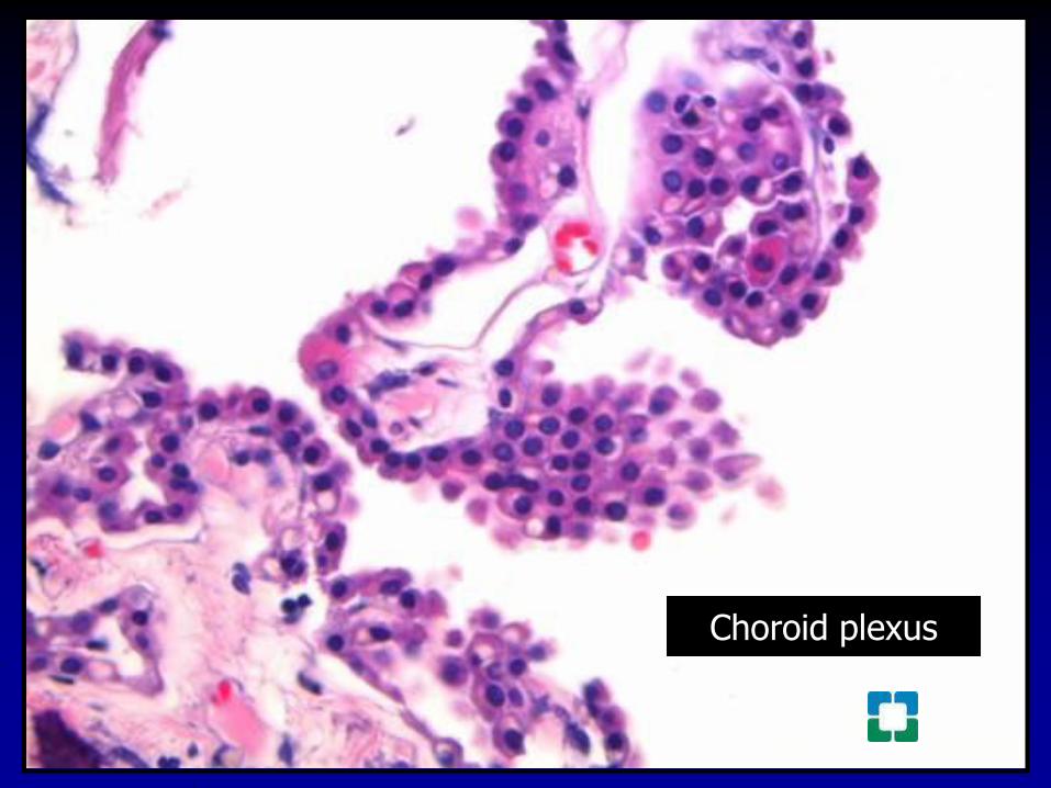
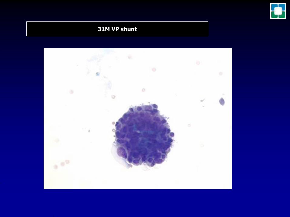
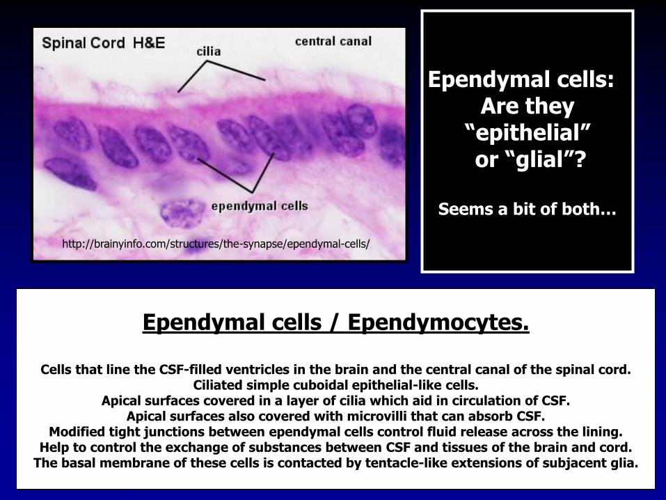
5 wk F CSF undergoing work up to rule out CNS infection
5 wk F CSF undergoing work up to rule out CNS infection
P Yeh amp D Westerman Nov 2015
Arachnoid granulations villi are small protrusions of arachnoid membrane through the duraVilli protrude into venous sinuses allowing CSF to exit the sub-arachnoid space into theblood Largest granulations lie in the superior saggittal sinus but are present in other
dural sinuses CSF pressures generally higher than venous pressures CSF flowsthrough the villi into the blood If pressures are reversed fluid will not
pass back into the subarachnoid space
ldquoNormalrdquo Cellular Constituents of CSF
Rare few small mature-appearing lymphocytes
Rare monocyte
55M headache and numbness in extremities
What the Heck
is the
Blood Brain Barrier
Separation of circulating blood from the extracellular CNS fluidOccurs along all capillaries
Consists of selective tight junctions between endothelial cellsAlso includes thick basement membrane and astrocytic endfeet or glia limitans
Restricts diffusion of organisism and large molecules into the substance of the CNS
Sounds like an alternative rock band to mehellipCould ldquoGreen Dayrdquo have beenldquoBBBrdquo
ALL marked propensity for CNS involvement especially as
leptomeningeal disease (50 of patients without prophylaxis)
Blood vessel basement membrane rich in laminin (known to
coordinate pathfinding in neuronal progenitor cells)
Most ALL cells express laminin receptor α6 integrin
Mice with ALL xenografts treated with inhibitors of α6 integrin
expression or α6 integrin neutralizing antibodies showed significant
reductions in ALL CNS transit
ALL cells use α6 integrin interactions with laminin in emissary blood
vessels passing between bone marrow and SAS to invade CNS and
do not conventionally traverse the BBB
How is CSF collected
Heinrich QuinckeGerman internist
Developed lumbarpuncture fortherapy of
hydrocephalusin 1891
Lumbar puncture ldquocontaminantsrdquohellipChondrocyte
Anucleate squamous cellOsteoblast
Marrow elementsMegakaryocyte
Ayub K Ommaya MD ScD (hc) FRCS FACS
1930-2008Pakistani neurosurgeon and
inventor of the Ommaya reservoir
Ommaya ReservoirUsed for repeated extractions of isovolumic aliquots of CSF
and concomitant intrathecal infusions of CNS therapies
Schematic representation ofan implanted Ommaya reservoir
26M Omaya Reservoir CSF Hx of Ewings sarcoma
Cerebral Shunts
Ventriculoperitoneal (VP) shunts most common
Most cerebral shunts connect ventricular system by tubing with a pumpvalve to a long catheter
CSF shunts can also drain to right atrium of heart and to pleural spaceLP connect the lumbar spine to the peritoneum
Shunts can be a conduit for infections into the CNS
61F VP Shunt
61 M HX of primaryCNS LBCL
reservoir tap
Diagnostic Accuracy of Cerebrospinal Fluid (CSF) Cytology in Metastatic Tumors An Analysis of Consecutive CSF Samples
Yoon Sung Bae June-Won Cheong Won Seok Chang Sewha Kim Eun Ji Oh Se Hoon Kim
Korean J Pathol Dec 2013 47(6) 563ndash568
385 CSF cytology samples from 42 patients were collected The samples were gathered using a ventricular catheter and reservoir CSF cytology of all patients was examined more than two
times with immunocytochemistry for cytokeratinPrimary neoplastic sites and histologic types of patients metastatic cancer were diverse The
overall sensitivity for detecting malignancy was 413 Even within short-term intervals diagnoses frequently changed
CSF cytology is currently the ldquogold
standardrdquo for the diagnosis of
malignant leptomeningeal disease
High specificity gt95
Low sensitivity 50
Loeb KR et al May 2015
Pressure 70 - 180 mm H20 (lateral recumbent)Appearance clear colorless Total protein 15 - 60 mg100 mL Gamma globulin 3 - 12 of the total proteinGlucose 50 - 80 mg100 mLCell count 0 - 5 white blood cells (all mononuclear)
No red blood cells and no PMNsChloride 110 - 125 mEqL
CSF Normal Values
Traumatic Tap Versus Pathologic Bleeding
Traumatic Tap Pathologic BleedBlood Most in 1st tube then less Same in all tubes
Post-centrifuge Clear supernatant Xanthochromic
Clot May clot Does not clot
Microscopic Well preserved RBCs Poorly preserved RBCs
Fresh blood Erythorphagocytosis
Siderophages
Based on Demay RM The Art and Science of Cytopathology 2nd Ed Exfoliative Cytology CSF Chapter 6 p 492
77M HX of GBM status post surgery 3 months remote
77M known CLLSLL presents with headaches
CSF Pressures
Increased CSF Pressure
Congestive heart failureMass lesions (tumor abscess etc)hellipInflammationCerebral edemaSuperior vena cava syndromeImpaired resorptionIntracranial veinous thrombosisIncreased CSF proteinAcute hypo-osmolalitySubarachnoid hemorrhageLead poisoning
Decreased CSF Pressure
Hypotension shockSevere dehydrationSpinal blockAcute hyperosmolality
Based on Demay RM The Art and Science of Cytopathology 2nd Ed Exfoliative Cytology CSF Chapter 6 p 491
CSF Protein amp Glucose
Increased ProteinTumorInflammation infectionSubarachnoid hemorrhageCerebral infarctionDegenerative diseases such as MSGuillain-Barre syndromeDiabetes with peripheral neuropathyDrugs (phenothiazenes)Traumatic tap
Decreased ProteinWater intoxicationSome leukemiasCSF leakageHyperthyroidismPostpneumoencephalogramNormal in some children
Increased GlucoseHyperglycemiaDiabetesIntravenous drugs
Decreased GlucoseBacterial meningitisMyobacterial meningitisFungal meningitisHypoglycemiaSubarachoid hemorrhageTumorNot viral meningitis
Based on Demay RM The Art and Science of Cytopathology 2nd Ed Exfoliative Cytology CSF Chapter 6 p 492
Cytology Preparation Options
Concentration Techniques RequiredCytocentrifugation preps - favoredProprietary liquid based preps
Membrane filter preps
Coated plus charged slides - favored
Two stains - favoredModified giemsa (Diff-Quik Quik-Dip)Papanicolaou
Cell blocks may be of great value inselected cases
BACKGROUND The present study was designed to determine whether the Thinprep plus Papanicolaou stain (Thinprep)
method is more sensitive than the Cytospin-coupled Wright-Giemsa (WG) stain (Cytospin) method in diagnosis of leptomeningeal metastasis (LM) from malignant solid tumors in cerebrospinal fluid (CSF)
METHODS The morphological features of tumor cells in fresh CSF samples were analyzed using both methods The tumor
cell detection rates were compared between the two methods
RESULTS Using the Thinprep method we found that each type of tumor cells in the CSF samples had specific identifiable
morphological features linked to their primary cancer origins such as adenocarcinomas originated from the lungs breast andstomach and lung squamous cell carcinomas small cell lung cancer large-cell neuroendocrine lung cancer hepatocellular carcinoma and malignant melanoma In a retrospective study with 88 LM patients cancer cells were detected in 80 out of the 88 CSF samples In the comparative study with 45 LM patients the initial detection rate of the Thinprep method was significantly higher than that of the Cytospin method (733 vs 578 Plt001) The cell morphology was better preserved and subcellular structures were clearer using the Thinprep method compared to the Cytospin method
CONCLUSIONS The Thinprep method is more sensitive and suitable for LM diagnosis in CSF in patients with malignant
solid tumors than the Cytospin method The Thinprep method may facilitate primary tumor detection and help design early treatment regimens for LM patients with tumors of unknown primary origin
April 7 2015
Tumor Cells Detected in CSF Specimens
ThinPrep plus Papanicolaou Stain Method Is More Sensitive than Cytospin-Coupled Wright Giemsa Stain Method in CSF Cytology for Diagnosis of Leptomeningeal Metastasis from Solid Tumors Pan et al Changchun China April 7 2015
a-n adenocarcinomas t-u hepatocellular carcinomaso-p squamous carcinomas v-x large cell carcinomasr-s small cell carcinomas y-z melanomas
Concordance with reference diagnosis was
slightly but significantly higher for modified
Giemsa-stained preparations compared with
Pap-stained slides
APLMJULY
2018
67F headaches reactive pleocytosis
67F headaches reactive pleocytosis
45F with shunt after head trauma from MVA
68M West Nile Virus Meningo-Encephalitis
Cerebrospinal Fluid Cytology in Seasonal Epidemic West Nile
Virus Meningo-Encephalitis
Ajay Rawal MD Patrick J Gavin MD Charles D Sturgis MD
Diagnostic CytopathologyVolume 34 Issue 2 pages 127ndash129 February 2006
DOI 101002dc20410 Copyright copy 2006 Wiley-Liss Inc
82F West Nile Virus Meningo-Encephalitis
Cerebrospinal fluid cytology an 11-year experience with 5951 specimensPrayson RA amp Fischler DF Department of Anatomic Pathology Cleveland Clinic Foundation OH 44195 USA
DESIGN AND SETTING A retrospective study of 5951 CSF specimens generated between 1985 and 1995 Specimens from pediatric patients (lt19 years of age) from the same time period were separately identified
RESULTS A total of 5561 adult and 390 pediatric CSF specimens were interpreted A diagnosis of negative for malignant cells was assigned in 5171 (93) of the adult cases and in 351 (90) of the pediatric cases Specific infectious organisms were identified in 26 adult specimens and one pediatric specimen Cryptococcus was the most common infectious agent observed (n = 23 adults) and Toxoplasma was the sole pediatric infectious agent Two hundred seventy-six (5) adult cases and 31 (8) pediatric cases were positive for malignant cells Diagnoses included metastatic tumors (adult 140 [51] pediatric 0) lymphomaleukemia (adult 112 [41] pediatric 4 [13]) malignant unclassified neoplasms (adult 9 [3] pediatric 0) and primary central nervous system neoplasms (adult 12 [4] pediatric 27 [87]) Medulloblastoma was the most common pediatric neoplasm (n = 21) There were 105 (2) adult cases and 8 (2) pediatric cases with atypical cells present Atypical lymphoid cells were the most common type in adult cases (53)
CONCLUSIONS In our experience infectious agents were rarely identified in pediatric CSF specimens In adult specimens the most commonly identified organisms was Cryptococcus Primary central nervous system neoplasms accounted for a higher percentage of CSF specimens in the pediatric population than in the adult population The most commonly identified malignancy in adults was metastatic neoplasms and in children medulloblastoma
21M HIVAIDS Cryptococcal Meningitis
76 M being treated systemically for Hodgkinrsquos
lymphoma developed cryptococcal
meningitis
ThinPrep Papanicolaou
76 M being treated systemically for
Hodgkinrsquos lymphoma developed
cryptococcal meningitis
Cell Block
HampE AlcBlue-PAS GMS
ldquoIndia Inkrdquo
Mucicarmine
21M HIVAIDS Cryptococcal Meningitis
VDRL (detects anti-cardiolipin antibodies that are present
in cases of syphilis but does not directly detect treponemal
antigens) has imperfect specificity and sensitivity
Newer treponemal EIAs and CIAs (enzyme linked and
chemiluminescent immunoassays) developed for
serum show enhanced sensitivity on CSF
May be of value with plasma cell rich pleocytoses
58 F
headache and photophobia
1 year after
small bowel transplant
PTLD DQ TP HampE EBER
CSFThinPrep Pap
42 Guatemalan MaleHeadache and Seizures
Neurocysticercosis(Taenia solium)
Racemose NeurocysticercosisType of neurocysticercosis with
involvement of the subarachnoid space
42M Guatemalan with neurocysticercosis (Taenia solium)
42M Guatemalan with neurocysticercosis (Taenia solium)
39M history of non-small cell lung cancer and bacteremia
20F Rheumatoid arthritis VZV meningitis
71M hx of metastatic prostate carcinoma now with headachesand single 28 cm irregularly shaped white matter lesion
Patients with known history of DLBCL
81F 53M HIV
32F B-ALL DQ amp MWG from Heme Lab
74F history of systemic mantle cell lymphoma
23M hx of medulloblastoma of cerebellum
14 M
HX of alveolar
rhabdomyosarcoma left hand
Now with widely metastatic
disease
ThinPrep Papanicolaou
74F smoker small cell carcinoma of lung
CSFEBUS FNA of level 7 lymph node
71F history of pT2 pN1 grade 2 ductal carcinoma of breast
76F history of pT2 pN0 invasive lobular carcinoma of breast
44 F presented with CNS
symptoms including headache
and confusion
later developed seizures
44 F Presented with CNS Symptoms
Subsequent mammography and
chest CT confirmed right
breast mass lesion
MRI confirmed enhancement of
lumbar spinal cord and
cauda equina
44 F Presented with CNS Symptoms
Found to have e-cadherin negative
invasive lobular breast carcinoma
Interestingly strongly ER amp Her-2 +
ER
HER-2
69M smoker with large lung lesionmediastinal adenopathy and
presentation of seizures
CSF
Wang needleof level 7 lymph node
57 M ConfusionRule out meningitis
MRI head with diffuseleptomeningealenhancement
Small enhancing softtissue mass in
left maxillary sinus
40F dural based mass lesion
CSF
Touch preparation ofresected tumor
Glioblastoma multiforme with epithelial differentiation A potential diagnostic pitfall in cerebrospinal fluid cytologySK Gill MD V Padmanabhan MD WF Hickey MD and JD Marotti MD 23 APR 2015 DOI 101002dc23274 copy 2015 Wiley Periodicals IncDartmouth Hitchcock Medical Center amp Geisel School of Medicine at Dartmouth
Cerebrospinal fluid lumbar (A) Hypercellular specimen with loosely cohesive sheets and groups of atypical cells (Cytospin Diff-Quik 60X) B Nuclear pleomorphism and prominent cytoplasmic vacuoles resembling metastatic carcinoma are evident (Cytospin Diff-Quik 300X inset 400X)
MRI of the brain showed a 64 cmring-enhancing lesion in the lefttemporal lobe
Surgical resection specimen (A) Scattered groups of small densely packed epithelioid cells are present in a background of conventional GBM (HampE 140X) B Cytoplasmic vacuoles are present (HampE 300X) C GFAP is negative in epithelioid cells but demonstrates diffuse positivity in the background (Immunostain 100X) D Epithelioid cells are focally positive for EMA (Immunostain 200X) E Focal positivity for cytokeratin AE1AE3 is also present (Immunostain 200X)
78M CHF
Take Home Messages
Be familiar with the slide preparation
techniques and stains in your lab
Read slides CSF slides slowly and
carefully (two cells may make the
difference)
ALWAYS read CSF slides in context of
all available clinical and imaging data
My pleasure to speak with you today
CommentsCritiquesInsights
Questions
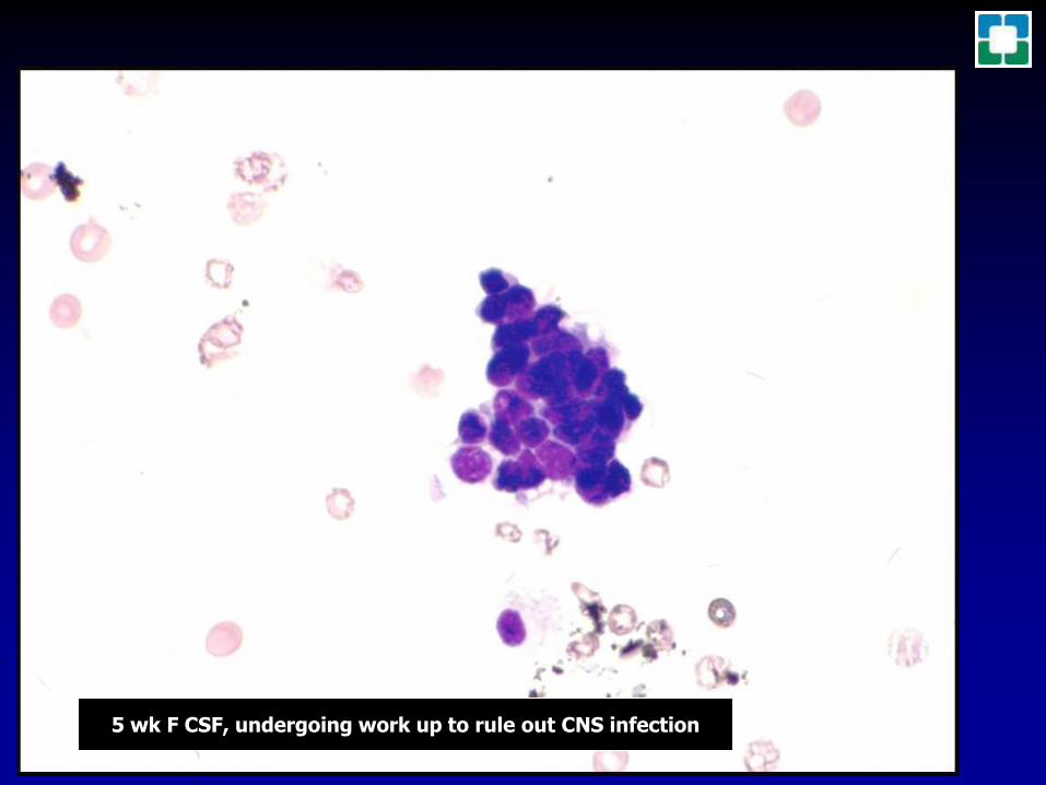
5 wk F CSF undergoing work up to rule out CNS infection
P Yeh amp D Westerman Nov 2015
Arachnoid granulations villi are small protrusions of arachnoid membrane through the duraVilli protrude into venous sinuses allowing CSF to exit the sub-arachnoid space into theblood Largest granulations lie in the superior saggittal sinus but are present in other
dural sinuses CSF pressures generally higher than venous pressures CSF flowsthrough the villi into the blood If pressures are reversed fluid will not
pass back into the subarachnoid space
ldquoNormalrdquo Cellular Constituents of CSF
Rare few small mature-appearing lymphocytes
Rare monocyte
55M headache and numbness in extremities
What the Heck
is the
Blood Brain Barrier
Separation of circulating blood from the extracellular CNS fluidOccurs along all capillaries
Consists of selective tight junctions between endothelial cellsAlso includes thick basement membrane and astrocytic endfeet or glia limitans
Restricts diffusion of organisism and large molecules into the substance of the CNS
Sounds like an alternative rock band to mehellipCould ldquoGreen Dayrdquo have beenldquoBBBrdquo
ALL marked propensity for CNS involvement especially as
leptomeningeal disease (50 of patients without prophylaxis)
Blood vessel basement membrane rich in laminin (known to
coordinate pathfinding in neuronal progenitor cells)
Most ALL cells express laminin receptor α6 integrin
Mice with ALL xenografts treated with inhibitors of α6 integrin
expression or α6 integrin neutralizing antibodies showed significant
reductions in ALL CNS transit
ALL cells use α6 integrin interactions with laminin in emissary blood
vessels passing between bone marrow and SAS to invade CNS and
do not conventionally traverse the BBB
How is CSF collected
Heinrich QuinckeGerman internist
Developed lumbarpuncture fortherapy of
hydrocephalusin 1891
Lumbar puncture ldquocontaminantsrdquohellipChondrocyte
Anucleate squamous cellOsteoblast
Marrow elementsMegakaryocyte
Ayub K Ommaya MD ScD (hc) FRCS FACS
1930-2008Pakistani neurosurgeon and
inventor of the Ommaya reservoir
Ommaya ReservoirUsed for repeated extractions of isovolumic aliquots of CSF
and concomitant intrathecal infusions of CNS therapies
Schematic representation ofan implanted Ommaya reservoir
26M Omaya Reservoir CSF Hx of Ewings sarcoma
Cerebral Shunts
Ventriculoperitoneal (VP) shunts most common
Most cerebral shunts connect ventricular system by tubing with a pumpvalve to a long catheter
CSF shunts can also drain to right atrium of heart and to pleural spaceLP connect the lumbar spine to the peritoneum
Shunts can be a conduit for infections into the CNS
61F VP Shunt
61 M HX of primaryCNS LBCL
reservoir tap
Diagnostic Accuracy of Cerebrospinal Fluid (CSF) Cytology in Metastatic Tumors An Analysis of Consecutive CSF Samples
Yoon Sung Bae June-Won Cheong Won Seok Chang Sewha Kim Eun Ji Oh Se Hoon Kim
Korean J Pathol Dec 2013 47(6) 563ndash568
385 CSF cytology samples from 42 patients were collected The samples were gathered using a ventricular catheter and reservoir CSF cytology of all patients was examined more than two
times with immunocytochemistry for cytokeratinPrimary neoplastic sites and histologic types of patients metastatic cancer were diverse The
overall sensitivity for detecting malignancy was 413 Even within short-term intervals diagnoses frequently changed
CSF cytology is currently the ldquogold
standardrdquo for the diagnosis of
malignant leptomeningeal disease
High specificity gt95
Low sensitivity 50
Loeb KR et al May 2015
Pressure 70 - 180 mm H20 (lateral recumbent)Appearance clear colorless Total protein 15 - 60 mg100 mL Gamma globulin 3 - 12 of the total proteinGlucose 50 - 80 mg100 mLCell count 0 - 5 white blood cells (all mononuclear)
No red blood cells and no PMNsChloride 110 - 125 mEqL
CSF Normal Values
Traumatic Tap Versus Pathologic Bleeding
Traumatic Tap Pathologic BleedBlood Most in 1st tube then less Same in all tubes
Post-centrifuge Clear supernatant Xanthochromic
Clot May clot Does not clot
Microscopic Well preserved RBCs Poorly preserved RBCs
Fresh blood Erythorphagocytosis
Siderophages
Based on Demay RM The Art and Science of Cytopathology 2nd Ed Exfoliative Cytology CSF Chapter 6 p 492
77M HX of GBM status post surgery 3 months remote
77M known CLLSLL presents with headaches
CSF Pressures
Increased CSF Pressure
Congestive heart failureMass lesions (tumor abscess etc)hellipInflammationCerebral edemaSuperior vena cava syndromeImpaired resorptionIntracranial veinous thrombosisIncreased CSF proteinAcute hypo-osmolalitySubarachnoid hemorrhageLead poisoning
Decreased CSF Pressure
Hypotension shockSevere dehydrationSpinal blockAcute hyperosmolality
Based on Demay RM The Art and Science of Cytopathology 2nd Ed Exfoliative Cytology CSF Chapter 6 p 491
CSF Protein amp Glucose
Increased ProteinTumorInflammation infectionSubarachnoid hemorrhageCerebral infarctionDegenerative diseases such as MSGuillain-Barre syndromeDiabetes with peripheral neuropathyDrugs (phenothiazenes)Traumatic tap
Decreased ProteinWater intoxicationSome leukemiasCSF leakageHyperthyroidismPostpneumoencephalogramNormal in some children
Increased GlucoseHyperglycemiaDiabetesIntravenous drugs
Decreased GlucoseBacterial meningitisMyobacterial meningitisFungal meningitisHypoglycemiaSubarachoid hemorrhageTumorNot viral meningitis
Based on Demay RM The Art and Science of Cytopathology 2nd Ed Exfoliative Cytology CSF Chapter 6 p 492
Cytology Preparation Options
Concentration Techniques RequiredCytocentrifugation preps - favoredProprietary liquid based preps
Membrane filter preps
Coated plus charged slides - favored
Two stains - favoredModified giemsa (Diff-Quik Quik-Dip)Papanicolaou
Cell blocks may be of great value inselected cases
BACKGROUND The present study was designed to determine whether the Thinprep plus Papanicolaou stain (Thinprep)
method is more sensitive than the Cytospin-coupled Wright-Giemsa (WG) stain (Cytospin) method in diagnosis of leptomeningeal metastasis (LM) from malignant solid tumors in cerebrospinal fluid (CSF)
METHODS The morphological features of tumor cells in fresh CSF samples were analyzed using both methods The tumor
cell detection rates were compared between the two methods
RESULTS Using the Thinprep method we found that each type of tumor cells in the CSF samples had specific identifiable
morphological features linked to their primary cancer origins such as adenocarcinomas originated from the lungs breast andstomach and lung squamous cell carcinomas small cell lung cancer large-cell neuroendocrine lung cancer hepatocellular carcinoma and malignant melanoma In a retrospective study with 88 LM patients cancer cells were detected in 80 out of the 88 CSF samples In the comparative study with 45 LM patients the initial detection rate of the Thinprep method was significantly higher than that of the Cytospin method (733 vs 578 Plt001) The cell morphology was better preserved and subcellular structures were clearer using the Thinprep method compared to the Cytospin method
CONCLUSIONS The Thinprep method is more sensitive and suitable for LM diagnosis in CSF in patients with malignant
solid tumors than the Cytospin method The Thinprep method may facilitate primary tumor detection and help design early treatment regimens for LM patients with tumors of unknown primary origin
April 7 2015
Tumor Cells Detected in CSF Specimens
ThinPrep plus Papanicolaou Stain Method Is More Sensitive than Cytospin-Coupled Wright Giemsa Stain Method in CSF Cytology for Diagnosis of Leptomeningeal Metastasis from Solid Tumors Pan et al Changchun China April 7 2015
a-n adenocarcinomas t-u hepatocellular carcinomaso-p squamous carcinomas v-x large cell carcinomasr-s small cell carcinomas y-z melanomas
Concordance with reference diagnosis was
slightly but significantly higher for modified
Giemsa-stained preparations compared with
Pap-stained slides
APLMJULY
2018
67F headaches reactive pleocytosis
67F headaches reactive pleocytosis
45F with shunt after head trauma from MVA
68M West Nile Virus Meningo-Encephalitis
Cerebrospinal Fluid Cytology in Seasonal Epidemic West Nile
Virus Meningo-Encephalitis
Ajay Rawal MD Patrick J Gavin MD Charles D Sturgis MD
Diagnostic CytopathologyVolume 34 Issue 2 pages 127ndash129 February 2006
DOI 101002dc20410 Copyright copy 2006 Wiley-Liss Inc
82F West Nile Virus Meningo-Encephalitis
Cerebrospinal fluid cytology an 11-year experience with 5951 specimensPrayson RA amp Fischler DF Department of Anatomic Pathology Cleveland Clinic Foundation OH 44195 USA
DESIGN AND SETTING A retrospective study of 5951 CSF specimens generated between 1985 and 1995 Specimens from pediatric patients (lt19 years of age) from the same time period were separately identified
RESULTS A total of 5561 adult and 390 pediatric CSF specimens were interpreted A diagnosis of negative for malignant cells was assigned in 5171 (93) of the adult cases and in 351 (90) of the pediatric cases Specific infectious organisms were identified in 26 adult specimens and one pediatric specimen Cryptococcus was the most common infectious agent observed (n = 23 adults) and Toxoplasma was the sole pediatric infectious agent Two hundred seventy-six (5) adult cases and 31 (8) pediatric cases were positive for malignant cells Diagnoses included metastatic tumors (adult 140 [51] pediatric 0) lymphomaleukemia (adult 112 [41] pediatric 4 [13]) malignant unclassified neoplasms (adult 9 [3] pediatric 0) and primary central nervous system neoplasms (adult 12 [4] pediatric 27 [87]) Medulloblastoma was the most common pediatric neoplasm (n = 21) There were 105 (2) adult cases and 8 (2) pediatric cases with atypical cells present Atypical lymphoid cells were the most common type in adult cases (53)
CONCLUSIONS In our experience infectious agents were rarely identified in pediatric CSF specimens In adult specimens the most commonly identified organisms was Cryptococcus Primary central nervous system neoplasms accounted for a higher percentage of CSF specimens in the pediatric population than in the adult population The most commonly identified malignancy in adults was metastatic neoplasms and in children medulloblastoma
21M HIVAIDS Cryptococcal Meningitis
76 M being treated systemically for Hodgkinrsquos
lymphoma developed cryptococcal
meningitis
ThinPrep Papanicolaou
76 M being treated systemically for
Hodgkinrsquos lymphoma developed
cryptococcal meningitis
Cell Block
HampE AlcBlue-PAS GMS
ldquoIndia Inkrdquo
Mucicarmine
21M HIVAIDS Cryptococcal Meningitis
VDRL (detects anti-cardiolipin antibodies that are present
in cases of syphilis but does not directly detect treponemal
antigens) has imperfect specificity and sensitivity
Newer treponemal EIAs and CIAs (enzyme linked and
chemiluminescent immunoassays) developed for
serum show enhanced sensitivity on CSF
May be of value with plasma cell rich pleocytoses
58 F
headache and photophobia
1 year after
small bowel transplant
PTLD DQ TP HampE EBER
CSFThinPrep Pap
42 Guatemalan MaleHeadache and Seizures
Neurocysticercosis(Taenia solium)
Racemose NeurocysticercosisType of neurocysticercosis with
involvement of the subarachnoid space
42M Guatemalan with neurocysticercosis (Taenia solium)
42M Guatemalan with neurocysticercosis (Taenia solium)
39M history of non-small cell lung cancer and bacteremia
20F Rheumatoid arthritis VZV meningitis
71M hx of metastatic prostate carcinoma now with headachesand single 28 cm irregularly shaped white matter lesion
Patients with known history of DLBCL
81F 53M HIV
32F B-ALL DQ amp MWG from Heme Lab
74F history of systemic mantle cell lymphoma
23M hx of medulloblastoma of cerebellum
14 M
HX of alveolar
rhabdomyosarcoma left hand
Now with widely metastatic
disease
ThinPrep Papanicolaou
74F smoker small cell carcinoma of lung
CSFEBUS FNA of level 7 lymph node
71F history of pT2 pN1 grade 2 ductal carcinoma of breast
76F history of pT2 pN0 invasive lobular carcinoma of breast
44 F presented with CNS
symptoms including headache
and confusion
later developed seizures
44 F Presented with CNS Symptoms
Subsequent mammography and
chest CT confirmed right
breast mass lesion
MRI confirmed enhancement of
lumbar spinal cord and
cauda equina
44 F Presented with CNS Symptoms
Found to have e-cadherin negative
invasive lobular breast carcinoma
Interestingly strongly ER amp Her-2 +
ER
HER-2
69M smoker with large lung lesionmediastinal adenopathy and
presentation of seizures
CSF
Wang needleof level 7 lymph node
57 M ConfusionRule out meningitis
MRI head with diffuseleptomeningealenhancement
Small enhancing softtissue mass in
left maxillary sinus
40F dural based mass lesion
CSF
Touch preparation ofresected tumor
Glioblastoma multiforme with epithelial differentiation A potential diagnostic pitfall in cerebrospinal fluid cytologySK Gill MD V Padmanabhan MD WF Hickey MD and JD Marotti MD 23 APR 2015 DOI 101002dc23274 copy 2015 Wiley Periodicals IncDartmouth Hitchcock Medical Center amp Geisel School of Medicine at Dartmouth
Cerebrospinal fluid lumbar (A) Hypercellular specimen with loosely cohesive sheets and groups of atypical cells (Cytospin Diff-Quik 60X) B Nuclear pleomorphism and prominent cytoplasmic vacuoles resembling metastatic carcinoma are evident (Cytospin Diff-Quik 300X inset 400X)
MRI of the brain showed a 64 cmring-enhancing lesion in the lefttemporal lobe
Surgical resection specimen (A) Scattered groups of small densely packed epithelioid cells are present in a background of conventional GBM (HampE 140X) B Cytoplasmic vacuoles are present (HampE 300X) C GFAP is negative in epithelioid cells but demonstrates diffuse positivity in the background (Immunostain 100X) D Epithelioid cells are focally positive for EMA (Immunostain 200X) E Focal positivity for cytokeratin AE1AE3 is also present (Immunostain 200X)
78M CHF
Take Home Messages
Be familiar with the slide preparation
techniques and stains in your lab
Read slides CSF slides slowly and
carefully (two cells may make the
difference)
ALWAYS read CSF slides in context of
all available clinical and imaging data
My pleasure to speak with you today
CommentsCritiquesInsights
Questions
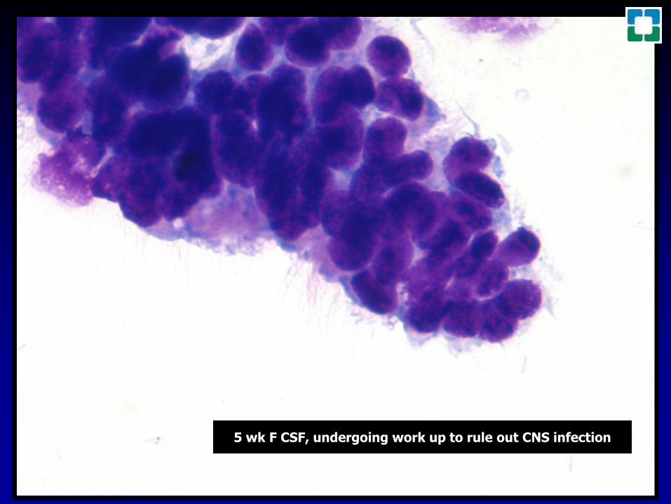
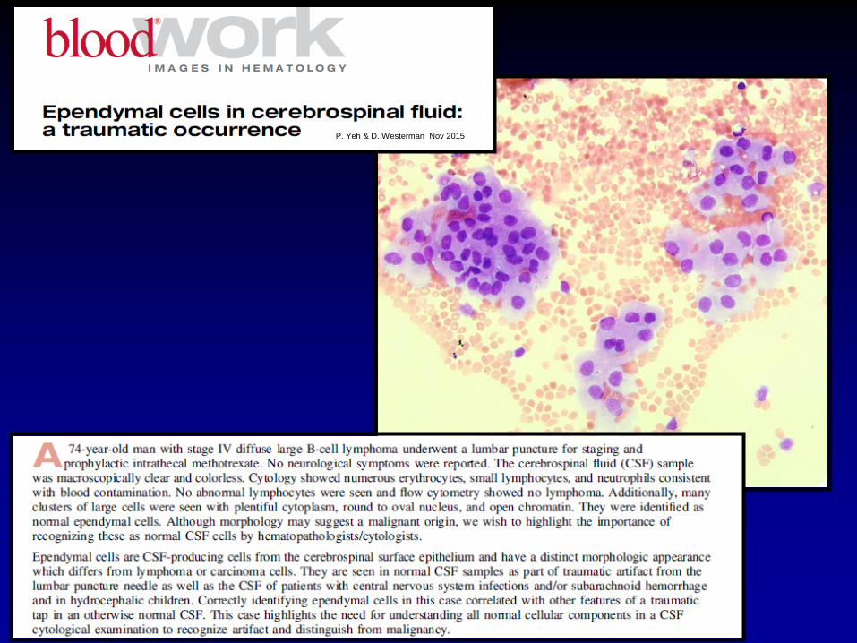
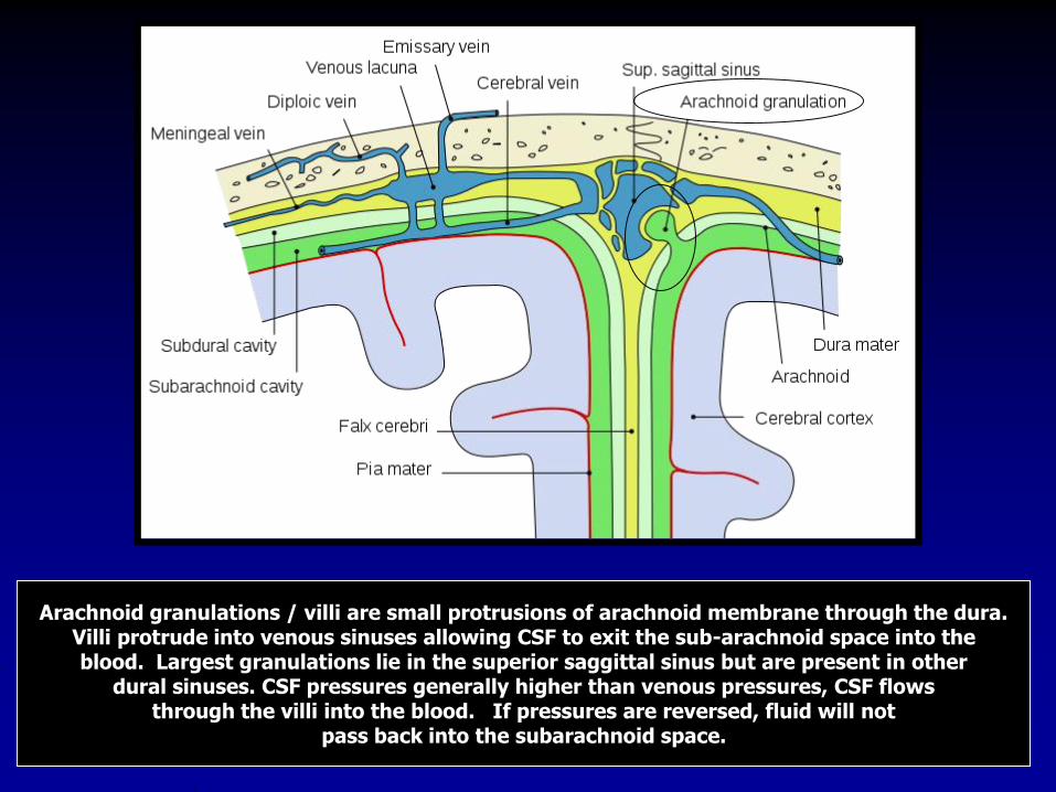
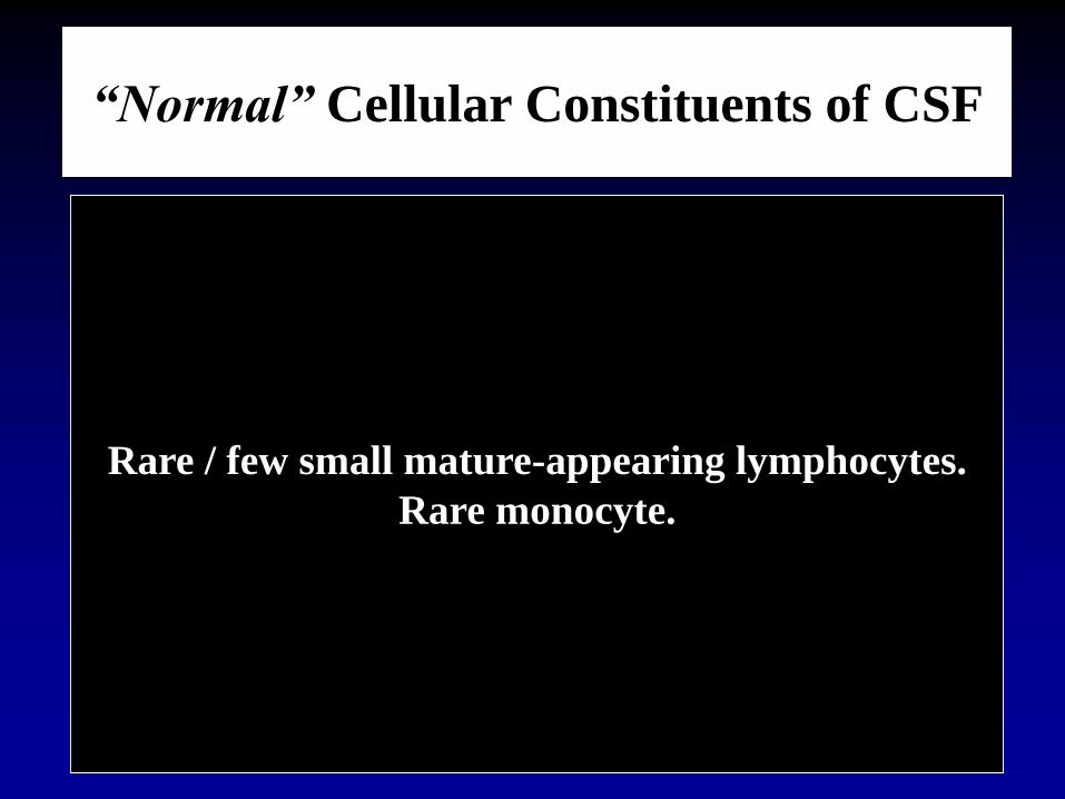
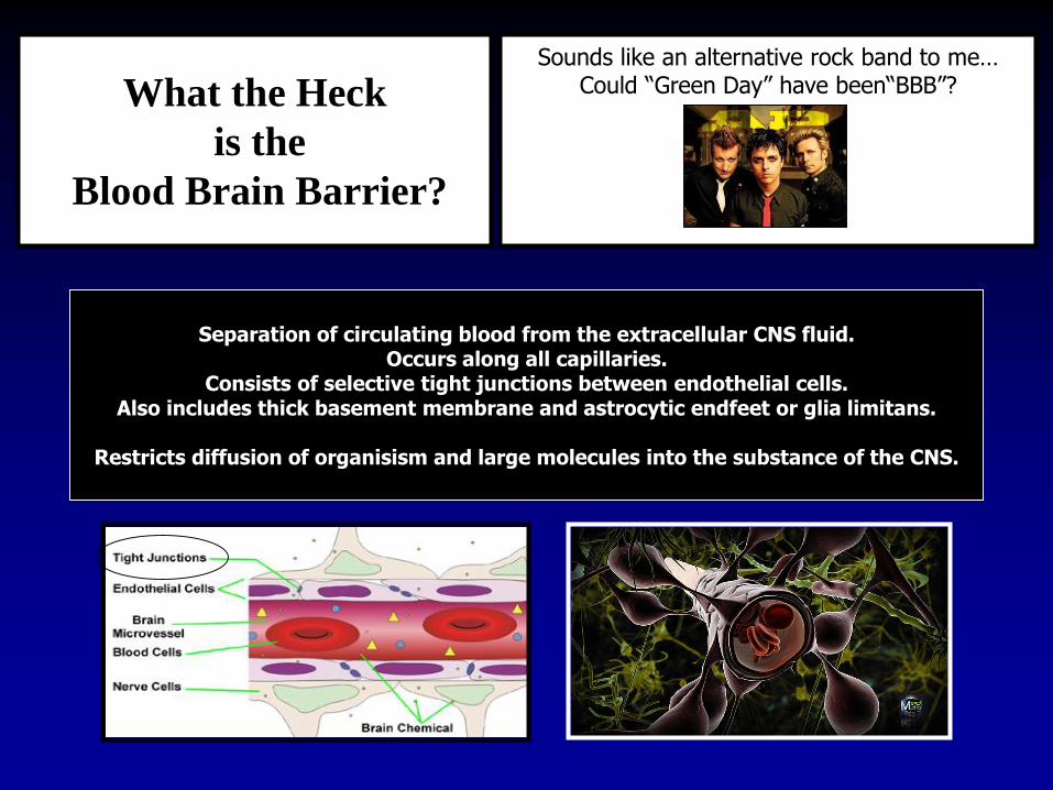
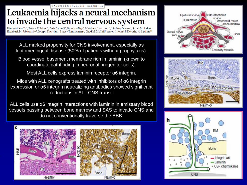
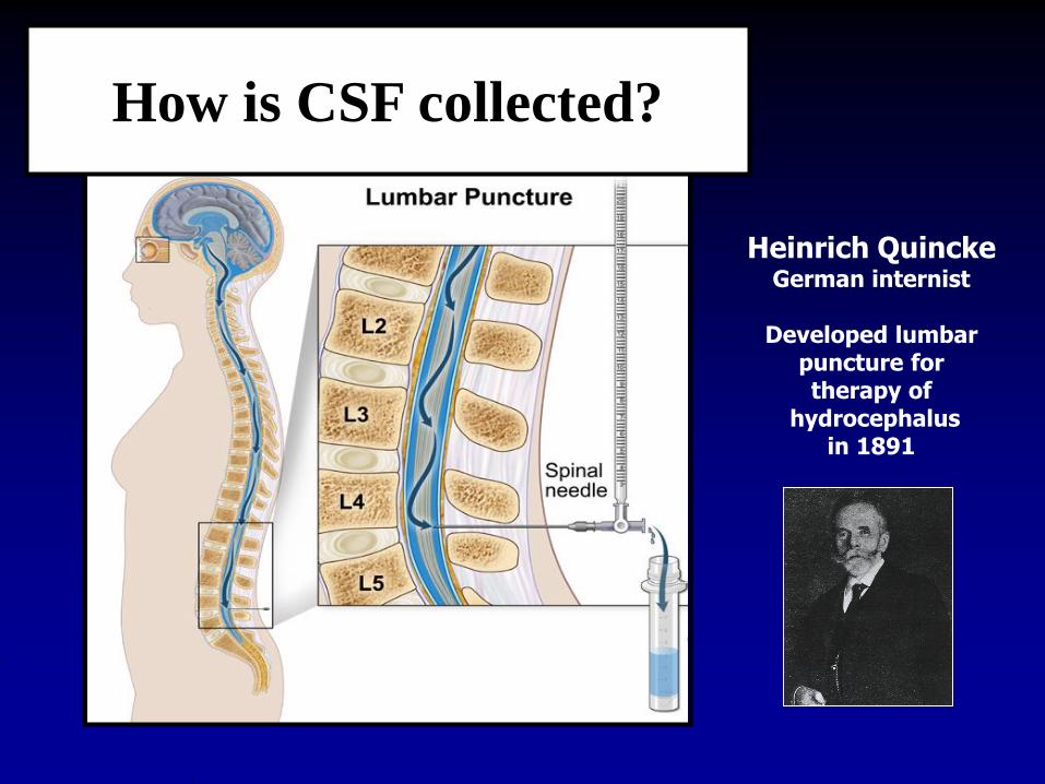
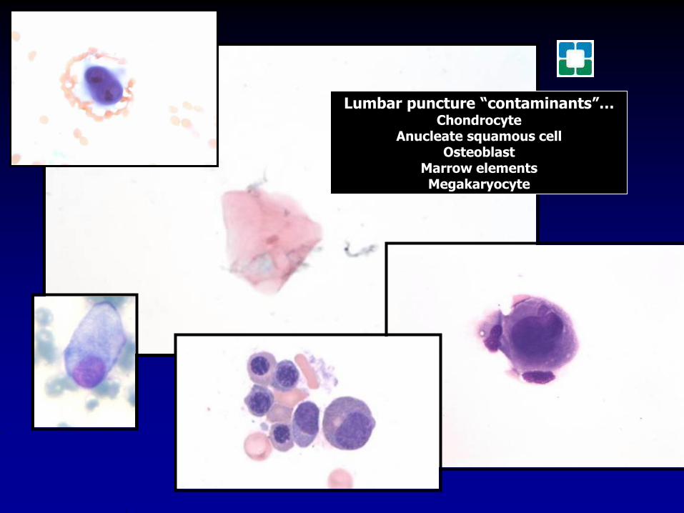
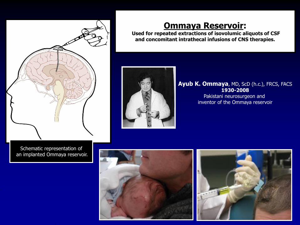
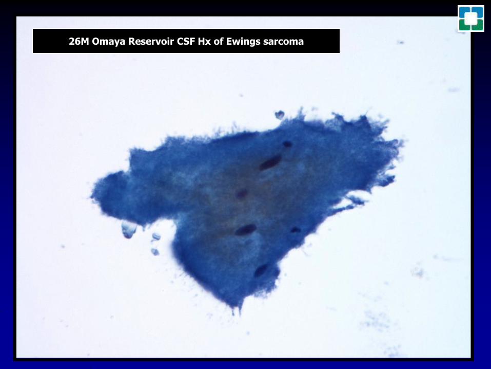
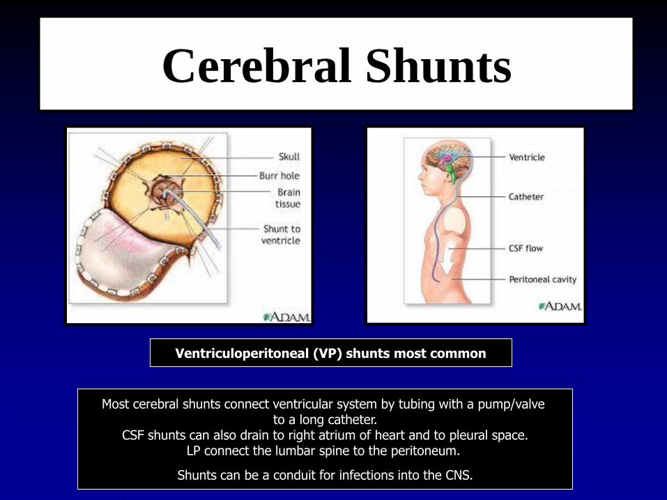
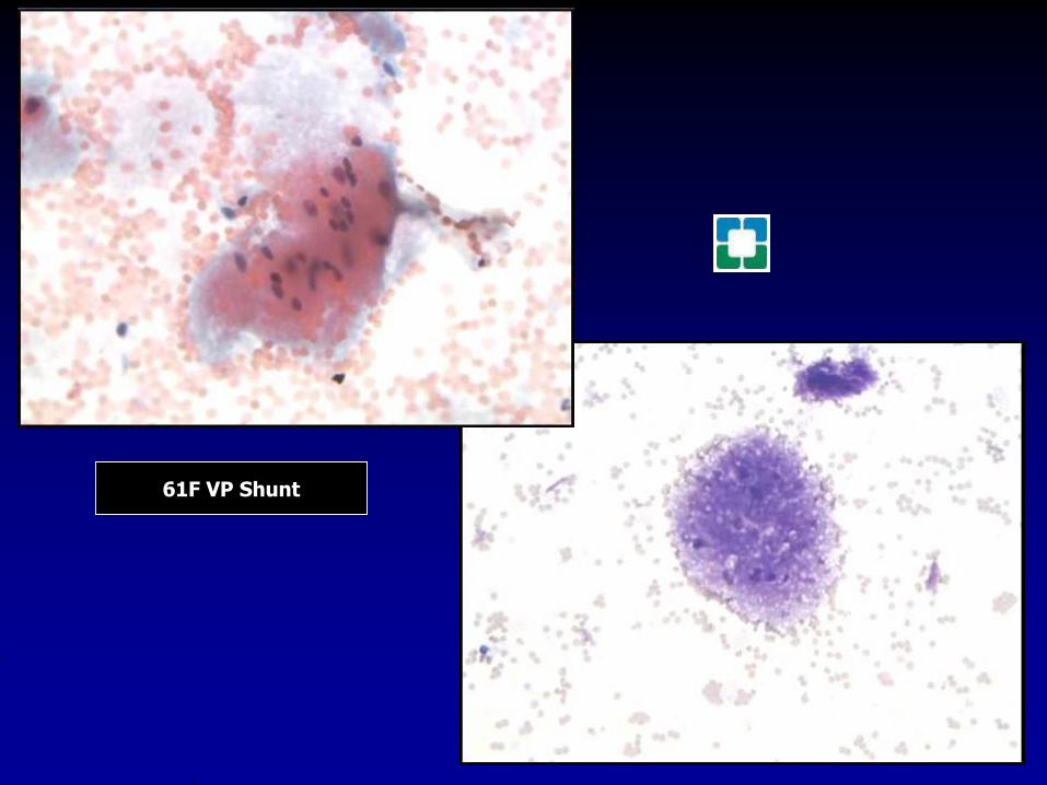
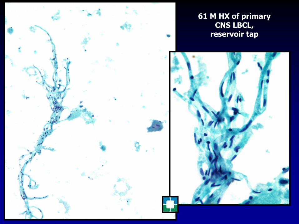
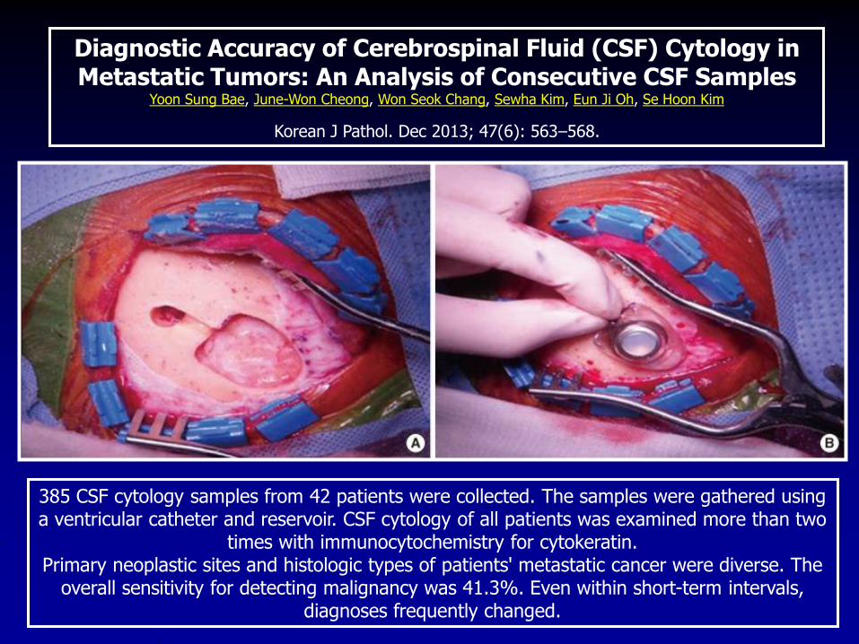
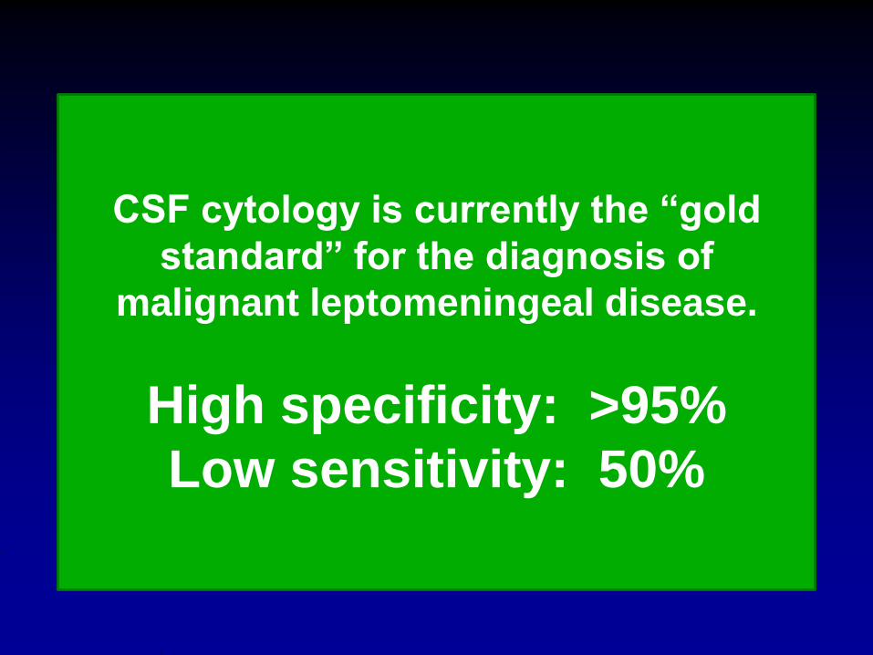
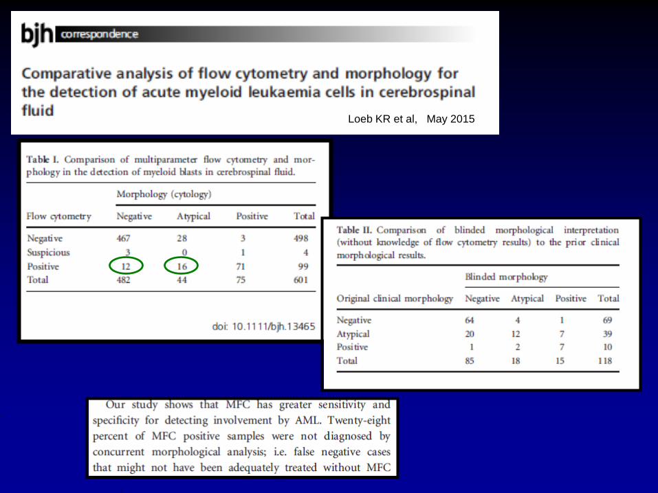
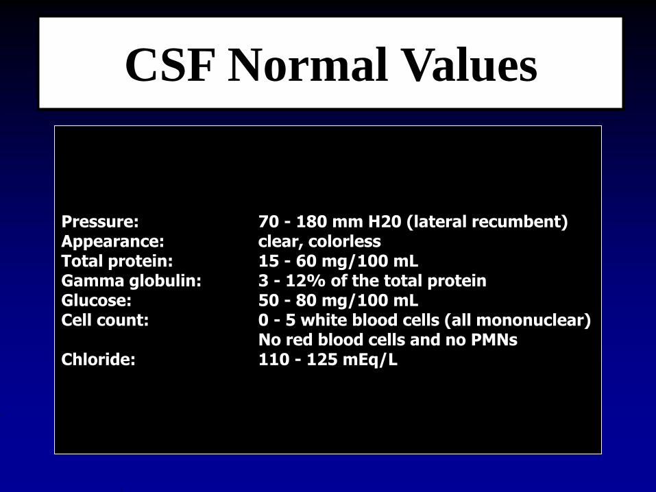
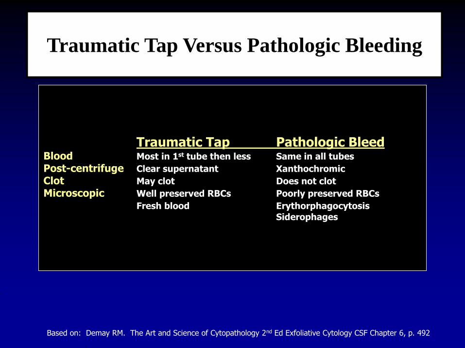
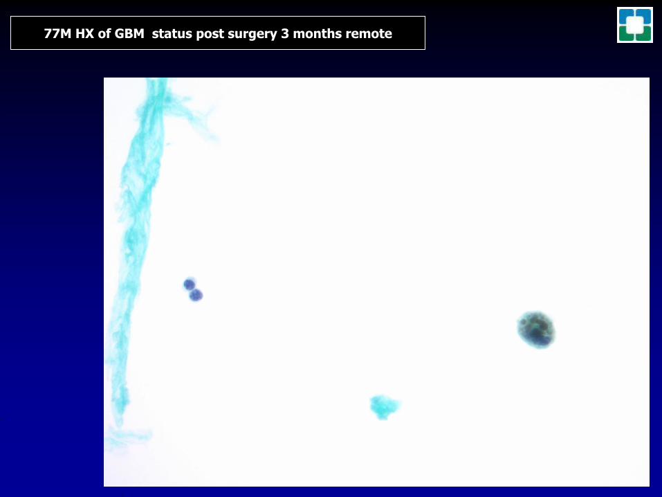
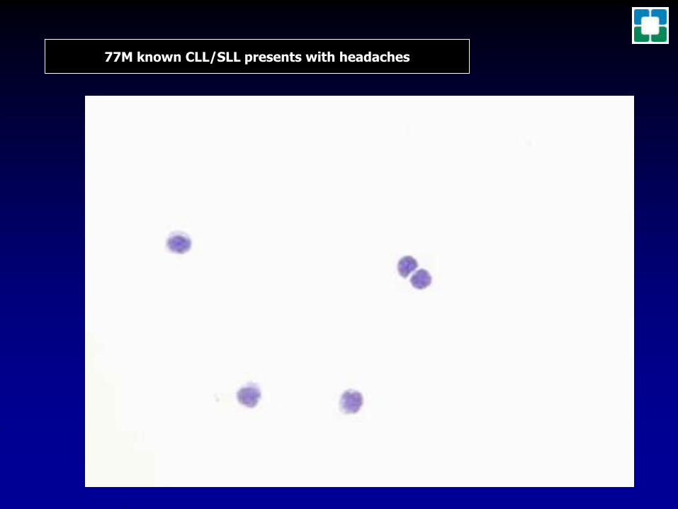
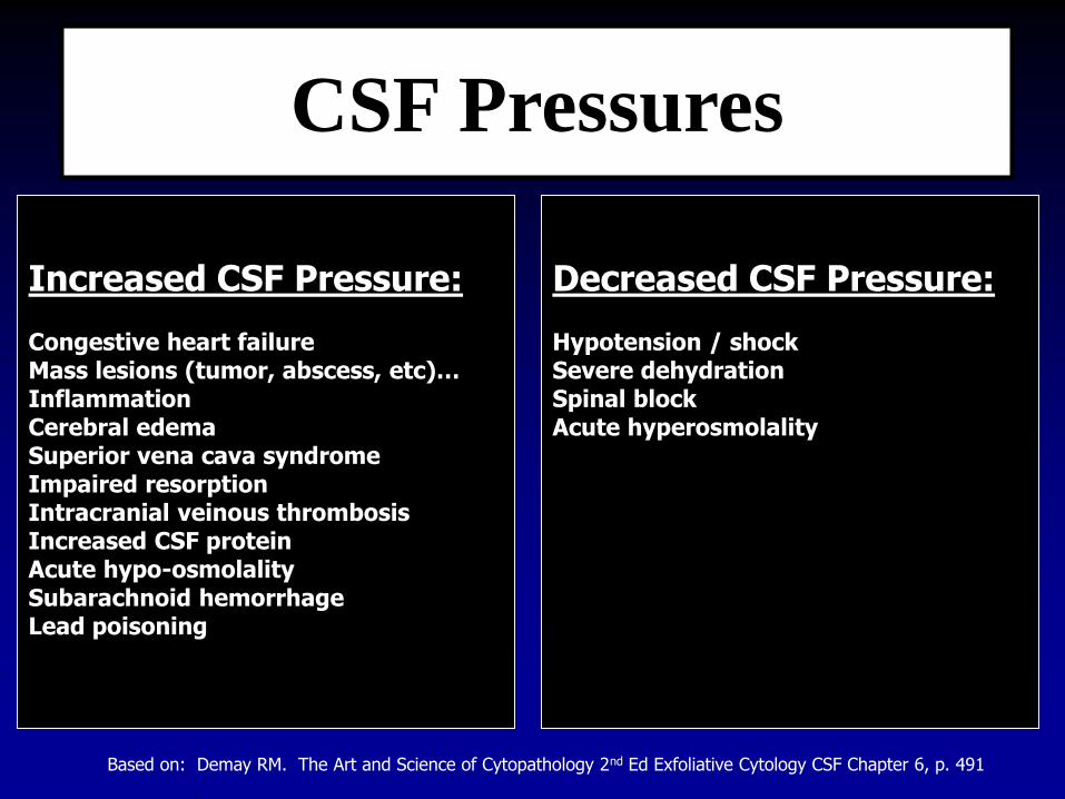
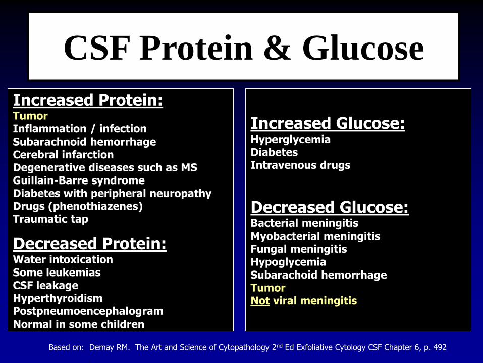
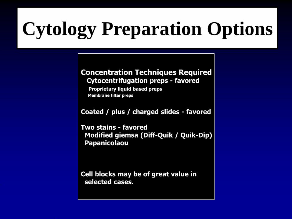
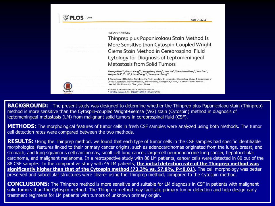
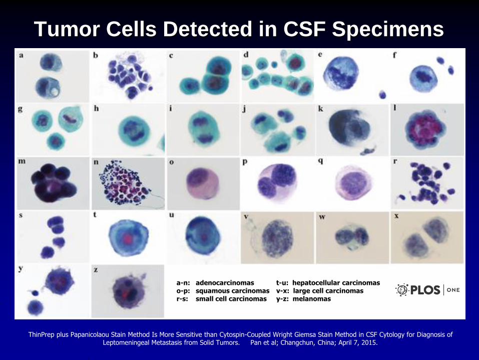
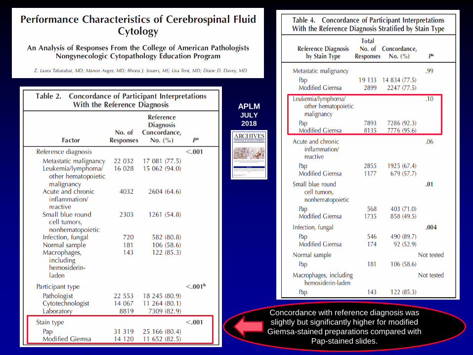
67F headaches reactive pleocytosis
67F headaches reactive pleocytosis
45F with shunt after head trauma from MVA
68M West Nile Virus Meningo-Encephalitis
Cerebrospinal Fluid Cytology in Seasonal Epidemic West Nile
Virus Meningo-Encephalitis
Ajay Rawal MD Patrick J Gavin MD Charles D Sturgis MD
Diagnostic CytopathologyVolume 34 Issue 2 pages 127ndash129 February 2006
DOI 101002dc20410 Copyright copy 2006 Wiley-Liss Inc
82F West Nile Virus Meningo-Encephalitis
Cerebrospinal fluid cytology an 11-year experience with 5951 specimensPrayson RA amp Fischler DF Department of Anatomic Pathology Cleveland Clinic Foundation OH 44195 USA
DESIGN AND SETTING A retrospective study of 5951 CSF specimens generated between 1985 and 1995 Specimens from pediatric patients (lt19 years of age) from the same time period were separately identified
RESULTS A total of 5561 adult and 390 pediatric CSF specimens were interpreted A diagnosis of negative for malignant cells was assigned in 5171 (93) of the adult cases and in 351 (90) of the pediatric cases Specific infectious organisms were identified in 26 adult specimens and one pediatric specimen Cryptococcus was the most common infectious agent observed (n = 23 adults) and Toxoplasma was the sole pediatric infectious agent Two hundred seventy-six (5) adult cases and 31 (8) pediatric cases were positive for malignant cells Diagnoses included metastatic tumors (adult 140 [51] pediatric 0) lymphomaleukemia (adult 112 [41] pediatric 4 [13]) malignant unclassified neoplasms (adult 9 [3] pediatric 0) and primary central nervous system neoplasms (adult 12 [4] pediatric 27 [87]) Medulloblastoma was the most common pediatric neoplasm (n = 21) There were 105 (2) adult cases and 8 (2) pediatric cases with atypical cells present Atypical lymphoid cells were the most common type in adult cases (53)
CONCLUSIONS In our experience infectious agents were rarely identified in pediatric CSF specimens In adult specimens the most commonly identified organisms was Cryptococcus Primary central nervous system neoplasms accounted for a higher percentage of CSF specimens in the pediatric population than in the adult population The most commonly identified malignancy in adults was metastatic neoplasms and in children medulloblastoma
21M HIVAIDS Cryptococcal Meningitis
76 M being treated systemically for Hodgkinrsquos
lymphoma developed cryptococcal
meningitis
ThinPrep Papanicolaou
76 M being treated systemically for
Hodgkinrsquos lymphoma developed
cryptococcal meningitis
Cell Block
HampE AlcBlue-PAS GMS
ldquoIndia Inkrdquo
Mucicarmine
21M HIVAIDS Cryptococcal Meningitis
VDRL (detects anti-cardiolipin antibodies that are present
in cases of syphilis but does not directly detect treponemal
antigens) has imperfect specificity and sensitivity
Newer treponemal EIAs and CIAs (enzyme linked and
chemiluminescent immunoassays) developed for
serum show enhanced sensitivity on CSF
May be of value with plasma cell rich pleocytoses
58 F
headache and photophobia
1 year after
small bowel transplant
PTLD DQ TP HampE EBER
CSFThinPrep Pap
42 Guatemalan MaleHeadache and Seizures
Neurocysticercosis(Taenia solium)
Racemose NeurocysticercosisType of neurocysticercosis with
involvement of the subarachnoid space
42M Guatemalan with neurocysticercosis (Taenia solium)
42M Guatemalan with neurocysticercosis (Taenia solium)
39M history of non-small cell lung cancer and bacteremia
20F Rheumatoid arthritis VZV meningitis
71M hx of metastatic prostate carcinoma now with headachesand single 28 cm irregularly shaped white matter lesion
Patients with known history of DLBCL
81F 53M HIV
32F B-ALL DQ amp MWG from Heme Lab
74F history of systemic mantle cell lymphoma
23M hx of medulloblastoma of cerebellum
14 M
HX of alveolar
rhabdomyosarcoma left hand
Now with widely metastatic
disease
ThinPrep Papanicolaou
74F smoker small cell carcinoma of lung
CSFEBUS FNA of level 7 lymph node
71F history of pT2 pN1 grade 2 ductal carcinoma of breast
76F history of pT2 pN0 invasive lobular carcinoma of breast
44 F presented with CNS
symptoms including headache
and confusion
later developed seizures
44 F Presented with CNS Symptoms
Subsequent mammography and
chest CT confirmed right
breast mass lesion
MRI confirmed enhancement of
lumbar spinal cord and
cauda equina
44 F Presented with CNS Symptoms
Found to have e-cadherin negative
invasive lobular breast carcinoma
Interestingly strongly ER amp Her-2 +
ER
HER-2
69M smoker with large lung lesionmediastinal adenopathy and
presentation of seizures
CSF
Wang needleof level 7 lymph node
57 M ConfusionRule out meningitis
MRI head with diffuseleptomeningealenhancement
Small enhancing softtissue mass in
left maxillary sinus
40F dural based mass lesion
CSF
Touch preparation ofresected tumor
Glioblastoma multiforme with epithelial differentiation A potential diagnostic pitfall in cerebrospinal fluid cytologySK Gill MD V Padmanabhan MD WF Hickey MD and JD Marotti MD 23 APR 2015 DOI 101002dc23274 copy 2015 Wiley Periodicals IncDartmouth Hitchcock Medical Center amp Geisel School of Medicine at Dartmouth
Cerebrospinal fluid lumbar (A) Hypercellular specimen with loosely cohesive sheets and groups of atypical cells (Cytospin Diff-Quik 60X) B Nuclear pleomorphism and prominent cytoplasmic vacuoles resembling metastatic carcinoma are evident (Cytospin Diff-Quik 300X inset 400X)
MRI of the brain showed a 64 cmring-enhancing lesion in the lefttemporal lobe
Surgical resection specimen (A) Scattered groups of small densely packed epithelioid cells are present in a background of conventional GBM (HampE 140X) B Cytoplasmic vacuoles are present (HampE 300X) C GFAP is negative in epithelioid cells but demonstrates diffuse positivity in the background (Immunostain 100X) D Epithelioid cells are focally positive for EMA (Immunostain 200X) E Focal positivity for cytokeratin AE1AE3 is also present (Immunostain 200X)
78M CHF
Take Home Messages
Be familiar with the slide preparation
techniques and stains in your lab
Read slides CSF slides slowly and
carefully (two cells may make the
difference)
ALWAYS read CSF slides in context of
all available clinical and imaging data
My pleasure to speak with you today
CommentsCritiquesInsights
Questions
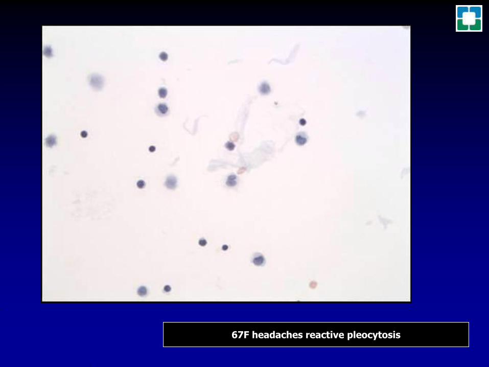
67F headaches reactive pleocytosis
45F with shunt after head trauma from MVA
68M West Nile Virus Meningo-Encephalitis
Cerebrospinal Fluid Cytology in Seasonal Epidemic West Nile
Virus Meningo-Encephalitis
Ajay Rawal MD Patrick J Gavin MD Charles D Sturgis MD
Diagnostic CytopathologyVolume 34 Issue 2 pages 127ndash129 February 2006
DOI 101002dc20410 Copyright copy 2006 Wiley-Liss Inc
82F West Nile Virus Meningo-Encephalitis
Cerebrospinal fluid cytology an 11-year experience with 5951 specimensPrayson RA amp Fischler DF Department of Anatomic Pathology Cleveland Clinic Foundation OH 44195 USA
DESIGN AND SETTING A retrospective study of 5951 CSF specimens generated between 1985 and 1995 Specimens from pediatric patients (lt19 years of age) from the same time period were separately identified
RESULTS A total of 5561 adult and 390 pediatric CSF specimens were interpreted A diagnosis of negative for malignant cells was assigned in 5171 (93) of the adult cases and in 351 (90) of the pediatric cases Specific infectious organisms were identified in 26 adult specimens and one pediatric specimen Cryptococcus was the most common infectious agent observed (n = 23 adults) and Toxoplasma was the sole pediatric infectious agent Two hundred seventy-six (5) adult cases and 31 (8) pediatric cases were positive for malignant cells Diagnoses included metastatic tumors (adult 140 [51] pediatric 0) lymphomaleukemia (adult 112 [41] pediatric 4 [13]) malignant unclassified neoplasms (adult 9 [3] pediatric 0) and primary central nervous system neoplasms (adult 12 [4] pediatric 27 [87]) Medulloblastoma was the most common pediatric neoplasm (n = 21) There were 105 (2) adult cases and 8 (2) pediatric cases with atypical cells present Atypical lymphoid cells were the most common type in adult cases (53)
CONCLUSIONS In our experience infectious agents were rarely identified in pediatric CSF specimens In adult specimens the most commonly identified organisms was Cryptococcus Primary central nervous system neoplasms accounted for a higher percentage of CSF specimens in the pediatric population than in the adult population The most commonly identified malignancy in adults was metastatic neoplasms and in children medulloblastoma
21M HIVAIDS Cryptococcal Meningitis
76 M being treated systemically for Hodgkinrsquos
lymphoma developed cryptococcal
meningitis
ThinPrep Papanicolaou
76 M being treated systemically for
Hodgkinrsquos lymphoma developed
cryptococcal meningitis
Cell Block
HampE AlcBlue-PAS GMS
ldquoIndia Inkrdquo
Mucicarmine
21M HIVAIDS Cryptococcal Meningitis
VDRL (detects anti-cardiolipin antibodies that are present
in cases of syphilis but does not directly detect treponemal
antigens) has imperfect specificity and sensitivity
Newer treponemal EIAs and CIAs (enzyme linked and
chemiluminescent immunoassays) developed for
serum show enhanced sensitivity on CSF
May be of value with plasma cell rich pleocytoses
58 F
headache and photophobia
1 year after
small bowel transplant
PTLD DQ TP HampE EBER
CSFThinPrep Pap
42 Guatemalan MaleHeadache and Seizures
Neurocysticercosis(Taenia solium)
Racemose NeurocysticercosisType of neurocysticercosis with
involvement of the subarachnoid space
42M Guatemalan with neurocysticercosis (Taenia solium)
42M Guatemalan with neurocysticercosis (Taenia solium)
39M history of non-small cell lung cancer and bacteremia
20F Rheumatoid arthritis VZV meningitis
71M hx of metastatic prostate carcinoma now with headachesand single 28 cm irregularly shaped white matter lesion
Patients with known history of DLBCL
81F 53M HIV
32F B-ALL DQ amp MWG from Heme Lab
74F history of systemic mantle cell lymphoma
23M hx of medulloblastoma of cerebellum
14 M
HX of alveolar
rhabdomyosarcoma left hand
Now with widely metastatic
disease
ThinPrep Papanicolaou
74F smoker small cell carcinoma of lung
CSFEBUS FNA of level 7 lymph node
71F history of pT2 pN1 grade 2 ductal carcinoma of breast
76F history of pT2 pN0 invasive lobular carcinoma of breast
44 F presented with CNS
symptoms including headache
and confusion
later developed seizures
44 F Presented with CNS Symptoms
Subsequent mammography and
chest CT confirmed right
breast mass lesion
MRI confirmed enhancement of
lumbar spinal cord and
cauda equina
44 F Presented with CNS Symptoms
Found to have e-cadherin negative
invasive lobular breast carcinoma
Interestingly strongly ER amp Her-2 +
ER
HER-2
69M smoker with large lung lesionmediastinal adenopathy and
presentation of seizures
CSF
Wang needleof level 7 lymph node
57 M ConfusionRule out meningitis
MRI head with diffuseleptomeningealenhancement
Small enhancing softtissue mass in
left maxillary sinus
40F dural based mass lesion
CSF
Touch preparation ofresected tumor
Glioblastoma multiforme with epithelial differentiation A potential diagnostic pitfall in cerebrospinal fluid cytologySK Gill MD V Padmanabhan MD WF Hickey MD and JD Marotti MD 23 APR 2015 DOI 101002dc23274 copy 2015 Wiley Periodicals IncDartmouth Hitchcock Medical Center amp Geisel School of Medicine at Dartmouth
Cerebrospinal fluid lumbar (A) Hypercellular specimen with loosely cohesive sheets and groups of atypical cells (Cytospin Diff-Quik 60X) B Nuclear pleomorphism and prominent cytoplasmic vacuoles resembling metastatic carcinoma are evident (Cytospin Diff-Quik 300X inset 400X)
MRI of the brain showed a 64 cmring-enhancing lesion in the lefttemporal lobe
Surgical resection specimen (A) Scattered groups of small densely packed epithelioid cells are present in a background of conventional GBM (HampE 140X) B Cytoplasmic vacuoles are present (HampE 300X) C GFAP is negative in epithelioid cells but demonstrates diffuse positivity in the background (Immunostain 100X) D Epithelioid cells are focally positive for EMA (Immunostain 200X) E Focal positivity for cytokeratin AE1AE3 is also present (Immunostain 200X)
78M CHF
Take Home Messages
Be familiar with the slide preparation
techniques and stains in your lab
Read slides CSF slides slowly and
carefully (two cells may make the
difference)
ALWAYS read CSF slides in context of
all available clinical and imaging data
My pleasure to speak with you today
CommentsCritiquesInsights
Questions
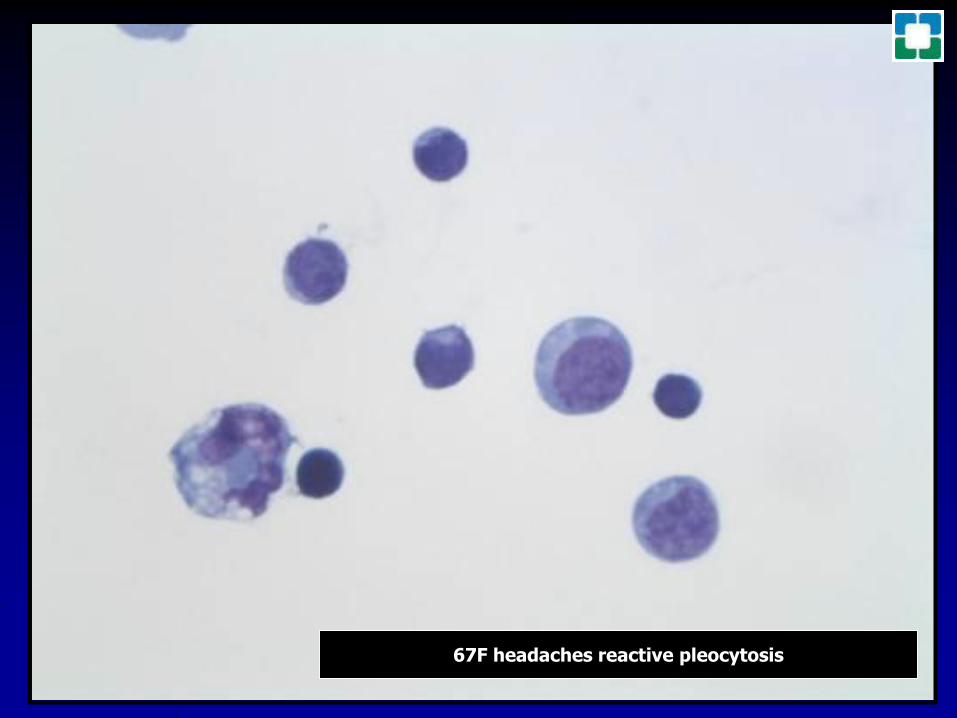
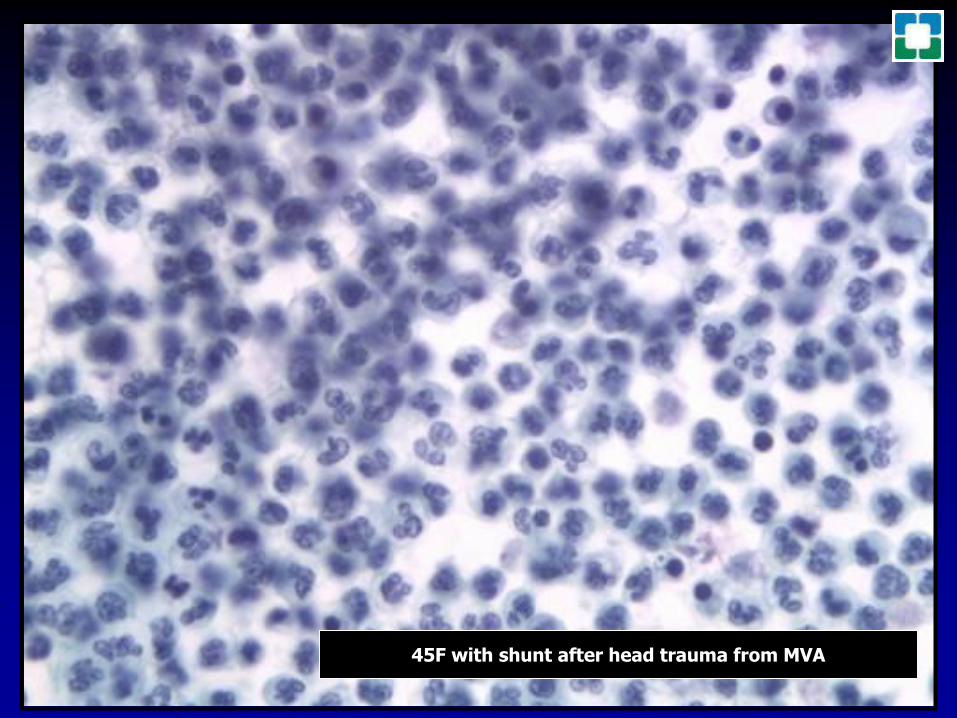
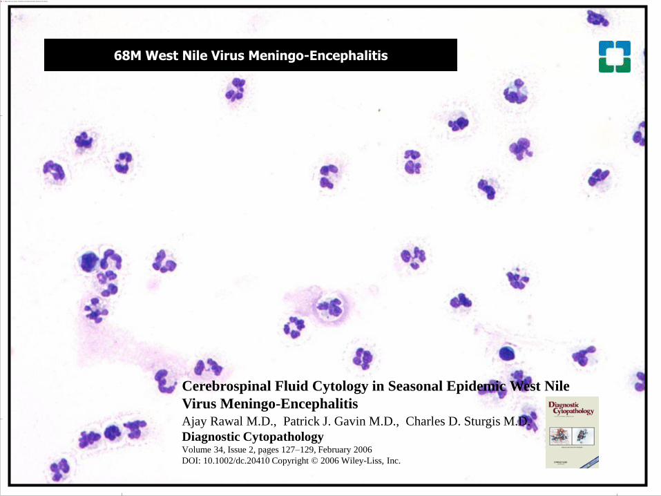
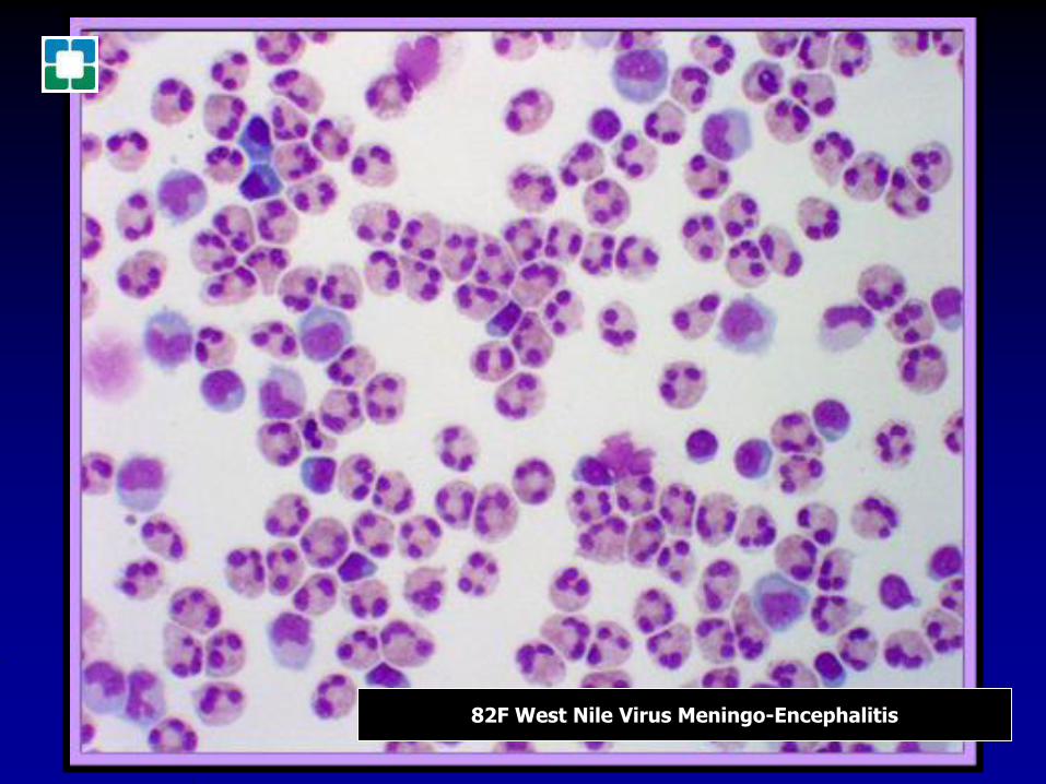
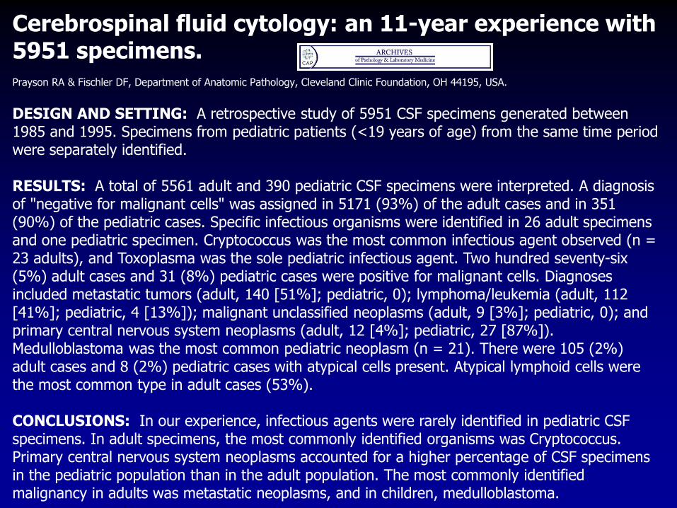
21M HIVAIDS Cryptococcal Meningitis
76 M being treated systemically for Hodgkinrsquos
lymphoma developed cryptococcal
meningitis
ThinPrep Papanicolaou
76 M being treated systemically for
Hodgkinrsquos lymphoma developed
cryptococcal meningitis
Cell Block
HampE AlcBlue-PAS GMS
ldquoIndia Inkrdquo
Mucicarmine
21M HIVAIDS Cryptococcal Meningitis
VDRL (detects anti-cardiolipin antibodies that are present
in cases of syphilis but does not directly detect treponemal
antigens) has imperfect specificity and sensitivity
Newer treponemal EIAs and CIAs (enzyme linked and
chemiluminescent immunoassays) developed for
serum show enhanced sensitivity on CSF
May be of value with plasma cell rich pleocytoses
58 F
headache and photophobia
1 year after
small bowel transplant
PTLD DQ TP HampE EBER
CSFThinPrep Pap
42 Guatemalan MaleHeadache and Seizures
Neurocysticercosis(Taenia solium)
Racemose NeurocysticercosisType of neurocysticercosis with
involvement of the subarachnoid space
42M Guatemalan with neurocysticercosis (Taenia solium)
42M Guatemalan with neurocysticercosis (Taenia solium)
39M history of non-small cell lung cancer and bacteremia
20F Rheumatoid arthritis VZV meningitis
71M hx of metastatic prostate carcinoma now with headachesand single 28 cm irregularly shaped white matter lesion
Patients with known history of DLBCL
81F 53M HIV
32F B-ALL DQ amp MWG from Heme Lab
74F history of systemic mantle cell lymphoma
23M hx of medulloblastoma of cerebellum
14 M
HX of alveolar
rhabdomyosarcoma left hand
Now with widely metastatic
disease
ThinPrep Papanicolaou
74F smoker small cell carcinoma of lung
CSFEBUS FNA of level 7 lymph node
71F history of pT2 pN1 grade 2 ductal carcinoma of breast
76F history of pT2 pN0 invasive lobular carcinoma of breast
44 F presented with CNS
symptoms including headache
and confusion
later developed seizures
44 F Presented with CNS Symptoms
Subsequent mammography and
chest CT confirmed right
breast mass lesion
MRI confirmed enhancement of
lumbar spinal cord and
cauda equina
44 F Presented with CNS Symptoms
Found to have e-cadherin negative
invasive lobular breast carcinoma
Interestingly strongly ER amp Her-2 +
ER
HER-2
69M smoker with large lung lesionmediastinal adenopathy and
presentation of seizures
CSF
Wang needleof level 7 lymph node
57 M ConfusionRule out meningitis
MRI head with diffuseleptomeningealenhancement
Small enhancing softtissue mass in
left maxillary sinus
40F dural based mass lesion
CSF
Touch preparation ofresected tumor
Glioblastoma multiforme with epithelial differentiation A potential diagnostic pitfall in cerebrospinal fluid cytologySK Gill MD V Padmanabhan MD WF Hickey MD and JD Marotti MD 23 APR 2015 DOI 101002dc23274 copy 2015 Wiley Periodicals IncDartmouth Hitchcock Medical Center amp Geisel School of Medicine at Dartmouth
Cerebrospinal fluid lumbar (A) Hypercellular specimen with loosely cohesive sheets and groups of atypical cells (Cytospin Diff-Quik 60X) B Nuclear pleomorphism and prominent cytoplasmic vacuoles resembling metastatic carcinoma are evident (Cytospin Diff-Quik 300X inset 400X)
MRI of the brain showed a 64 cmring-enhancing lesion in the lefttemporal lobe
Surgical resection specimen (A) Scattered groups of small densely packed epithelioid cells are present in a background of conventional GBM (HampE 140X) B Cytoplasmic vacuoles are present (HampE 300X) C GFAP is negative in epithelioid cells but demonstrates diffuse positivity in the background (Immunostain 100X) D Epithelioid cells are focally positive for EMA (Immunostain 200X) E Focal positivity for cytokeratin AE1AE3 is also present (Immunostain 200X)
78M CHF
Take Home Messages
Be familiar with the slide preparation
techniques and stains in your lab
Read slides CSF slides slowly and
carefully (two cells may make the
difference)
ALWAYS read CSF slides in context of
all available clinical and imaging data
My pleasure to speak with you today
CommentsCritiquesInsights
Questions
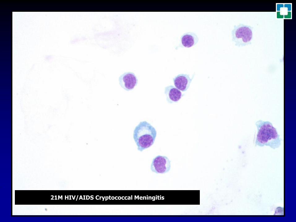
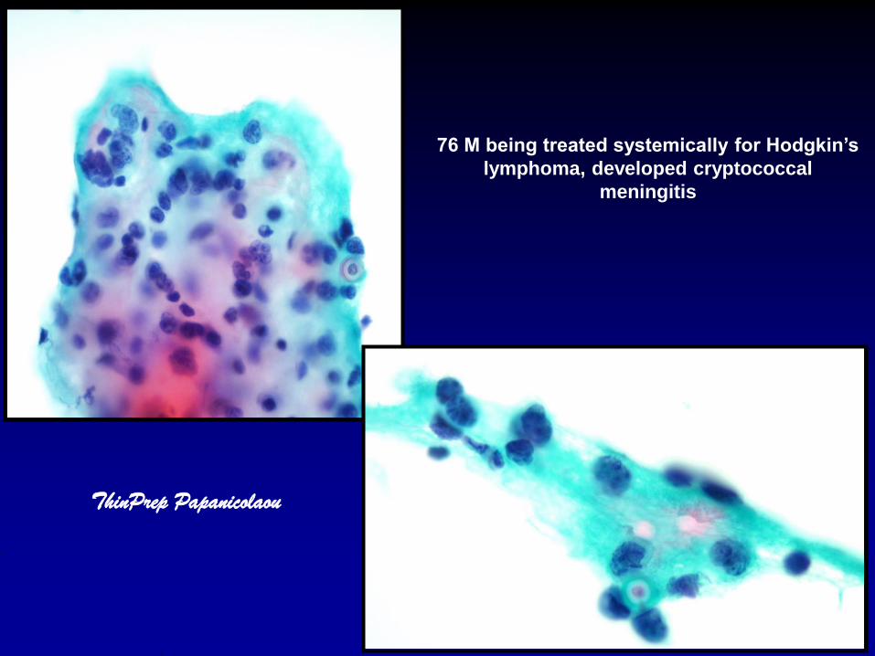
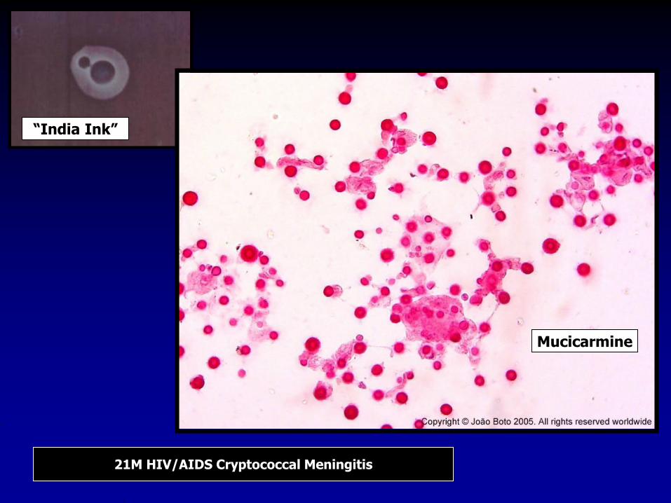
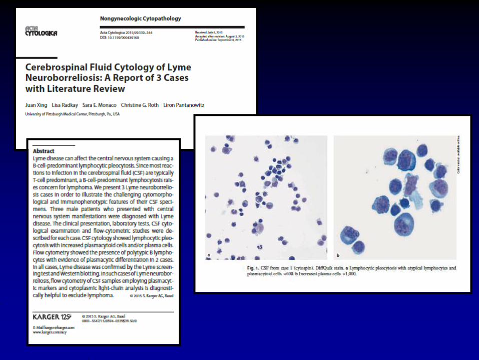
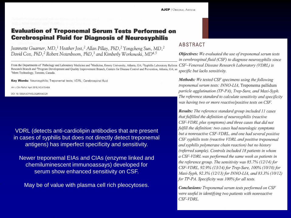
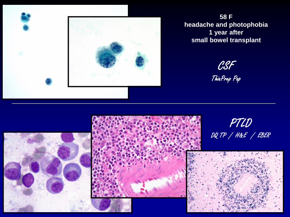
42M Guatemalan with neurocysticercosis (Taenia solium)
42M Guatemalan with neurocysticercosis (Taenia solium)
39M history of non-small cell lung cancer and bacteremia
20F Rheumatoid arthritis VZV meningitis
71M hx of metastatic prostate carcinoma now with headachesand single 28 cm irregularly shaped white matter lesion
Patients with known history of DLBCL
81F 53M HIV
32F B-ALL DQ amp MWG from Heme Lab
74F history of systemic mantle cell lymphoma
23M hx of medulloblastoma of cerebellum
14 M
HX of alveolar
rhabdomyosarcoma left hand
Now with widely metastatic
disease
ThinPrep Papanicolaou
74F smoker small cell carcinoma of lung
CSFEBUS FNA of level 7 lymph node
71F history of pT2 pN1 grade 2 ductal carcinoma of breast
76F history of pT2 pN0 invasive lobular carcinoma of breast
44 F presented with CNS
symptoms including headache
and confusion
later developed seizures
44 F Presented with CNS Symptoms
Subsequent mammography and
chest CT confirmed right
breast mass lesion
MRI confirmed enhancement of
lumbar spinal cord and
cauda equina
44 F Presented with CNS Symptoms
Found to have e-cadherin negative
invasive lobular breast carcinoma
Interestingly strongly ER amp Her-2 +
ER
HER-2
69M smoker with large lung lesionmediastinal adenopathy and
presentation of seizures
CSF
Wang needleof level 7 lymph node
57 M ConfusionRule out meningitis
MRI head with diffuseleptomeningealenhancement
Small enhancing softtissue mass in
left maxillary sinus
40F dural based mass lesion
CSF
Touch preparation ofresected tumor
Glioblastoma multiforme with epithelial differentiation A potential diagnostic pitfall in cerebrospinal fluid cytologySK Gill MD V Padmanabhan MD WF Hickey MD and JD Marotti MD 23 APR 2015 DOI 101002dc23274 copy 2015 Wiley Periodicals IncDartmouth Hitchcock Medical Center amp Geisel School of Medicine at Dartmouth
Cerebrospinal fluid lumbar (A) Hypercellular specimen with loosely cohesive sheets and groups of atypical cells (Cytospin Diff-Quik 60X) B Nuclear pleomorphism and prominent cytoplasmic vacuoles resembling metastatic carcinoma are evident (Cytospin Diff-Quik 300X inset 400X)
MRI of the brain showed a 64 cmring-enhancing lesion in the lefttemporal lobe
Surgical resection specimen (A) Scattered groups of small densely packed epithelioid cells are present in a background of conventional GBM (HampE 140X) B Cytoplasmic vacuoles are present (HampE 300X) C GFAP is negative in epithelioid cells but demonstrates diffuse positivity in the background (Immunostain 100X) D Epithelioid cells are focally positive for EMA (Immunostain 200X) E Focal positivity for cytokeratin AE1AE3 is also present (Immunostain 200X)
78M CHF
Take Home Messages
Be familiar with the slide preparation
techniques and stains in your lab
Read slides CSF slides slowly and
carefully (two cells may make the
difference)
ALWAYS read CSF slides in context of
all available clinical and imaging data
My pleasure to speak with you today
CommentsCritiquesInsights
Questions
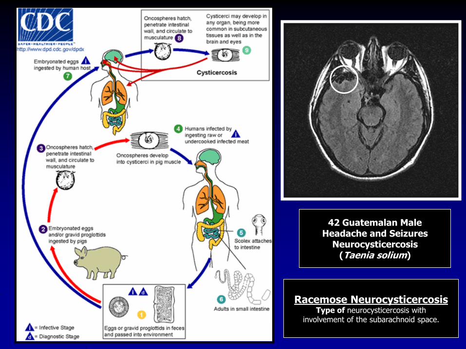
42M Guatemalan with neurocysticercosis (Taenia solium)
39M history of non-small cell lung cancer and bacteremia
20F Rheumatoid arthritis VZV meningitis
71M hx of metastatic prostate carcinoma now with headachesand single 28 cm irregularly shaped white matter lesion
Patients with known history of DLBCL
81F 53M HIV
32F B-ALL DQ amp MWG from Heme Lab
74F history of systemic mantle cell lymphoma
23M hx of medulloblastoma of cerebellum
14 M
HX of alveolar
rhabdomyosarcoma left hand
Now with widely metastatic
disease
ThinPrep Papanicolaou
74F smoker small cell carcinoma of lung
CSFEBUS FNA of level 7 lymph node
71F history of pT2 pN1 grade 2 ductal carcinoma of breast
76F history of pT2 pN0 invasive lobular carcinoma of breast
44 F presented with CNS
symptoms including headache
and confusion
later developed seizures
44 F Presented with CNS Symptoms
Subsequent mammography and
chest CT confirmed right
breast mass lesion
MRI confirmed enhancement of
lumbar spinal cord and
cauda equina
44 F Presented with CNS Symptoms
Found to have e-cadherin negative
invasive lobular breast carcinoma
Interestingly strongly ER amp Her-2 +
ER
HER-2
69M smoker with large lung lesionmediastinal adenopathy and
presentation of seizures
CSF
Wang needleof level 7 lymph node
57 M ConfusionRule out meningitis
MRI head with diffuseleptomeningealenhancement
Small enhancing softtissue mass in
left maxillary sinus
40F dural based mass lesion
CSF
Touch preparation ofresected tumor
Glioblastoma multiforme with epithelial differentiation A potential diagnostic pitfall in cerebrospinal fluid cytologySK Gill MD V Padmanabhan MD WF Hickey MD and JD Marotti MD 23 APR 2015 DOI 101002dc23274 copy 2015 Wiley Periodicals IncDartmouth Hitchcock Medical Center amp Geisel School of Medicine at Dartmouth
Cerebrospinal fluid lumbar (A) Hypercellular specimen with loosely cohesive sheets and groups of atypical cells (Cytospin Diff-Quik 60X) B Nuclear pleomorphism and prominent cytoplasmic vacuoles resembling metastatic carcinoma are evident (Cytospin Diff-Quik 300X inset 400X)
MRI of the brain showed a 64 cmring-enhancing lesion in the lefttemporal lobe
Surgical resection specimen (A) Scattered groups of small densely packed epithelioid cells are present in a background of conventional GBM (HampE 140X) B Cytoplasmic vacuoles are present (HampE 300X) C GFAP is negative in epithelioid cells but demonstrates diffuse positivity in the background (Immunostain 100X) D Epithelioid cells are focally positive for EMA (Immunostain 200X) E Focal positivity for cytokeratin AE1AE3 is also present (Immunostain 200X)
78M CHF
Take Home Messages
Be familiar with the slide preparation
techniques and stains in your lab
Read slides CSF slides slowly and
carefully (two cells may make the
difference)
ALWAYS read CSF slides in context of
all available clinical and imaging data
My pleasure to speak with you today
CommentsCritiquesInsights
Questions
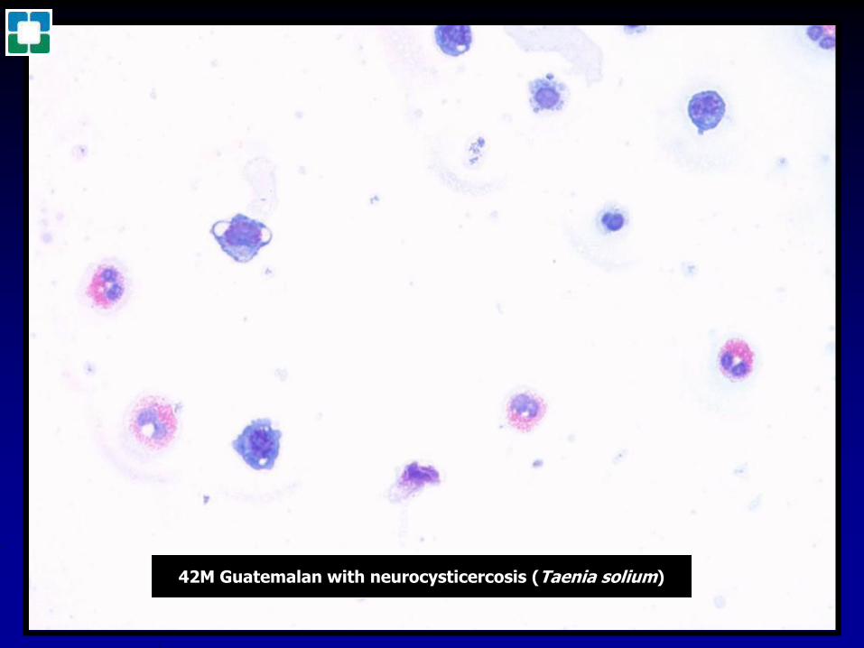
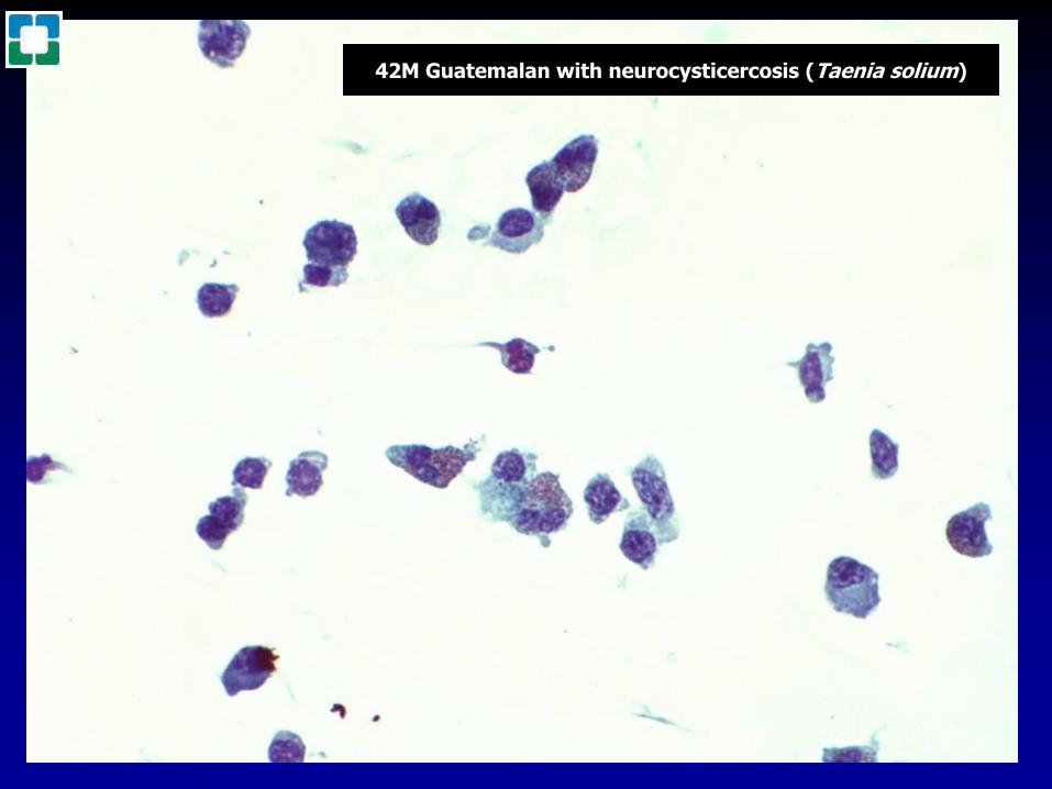
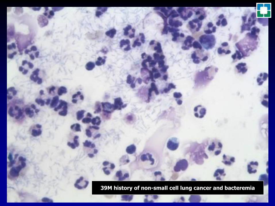
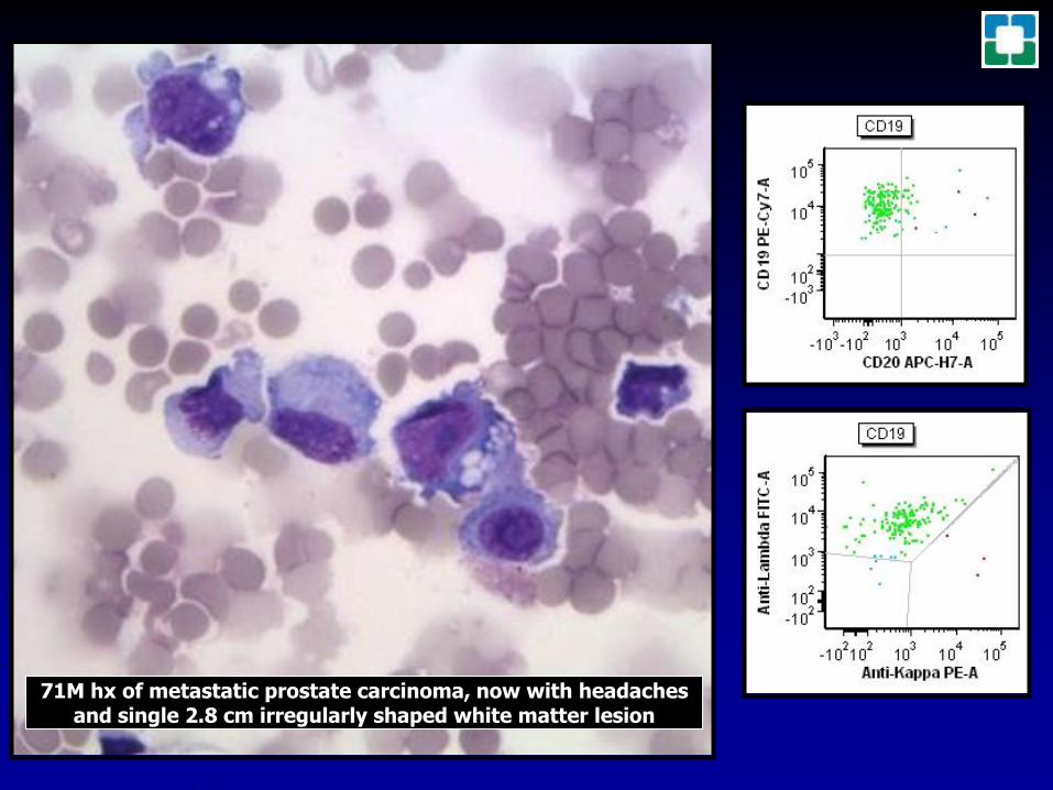
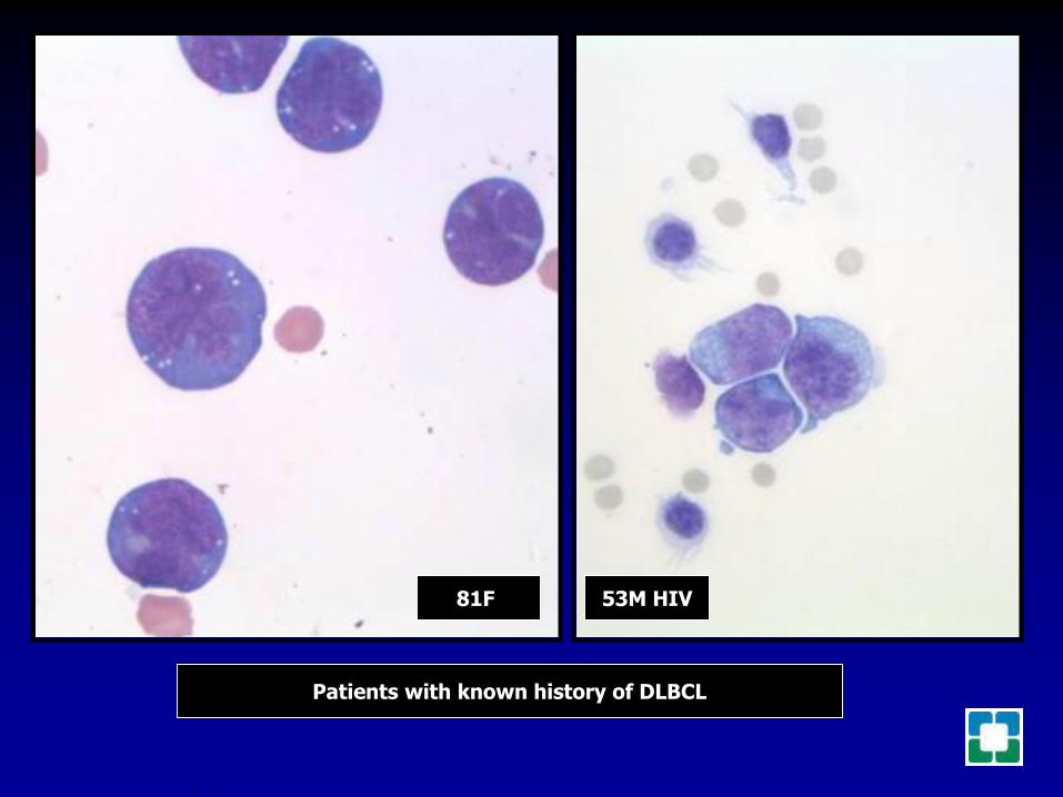
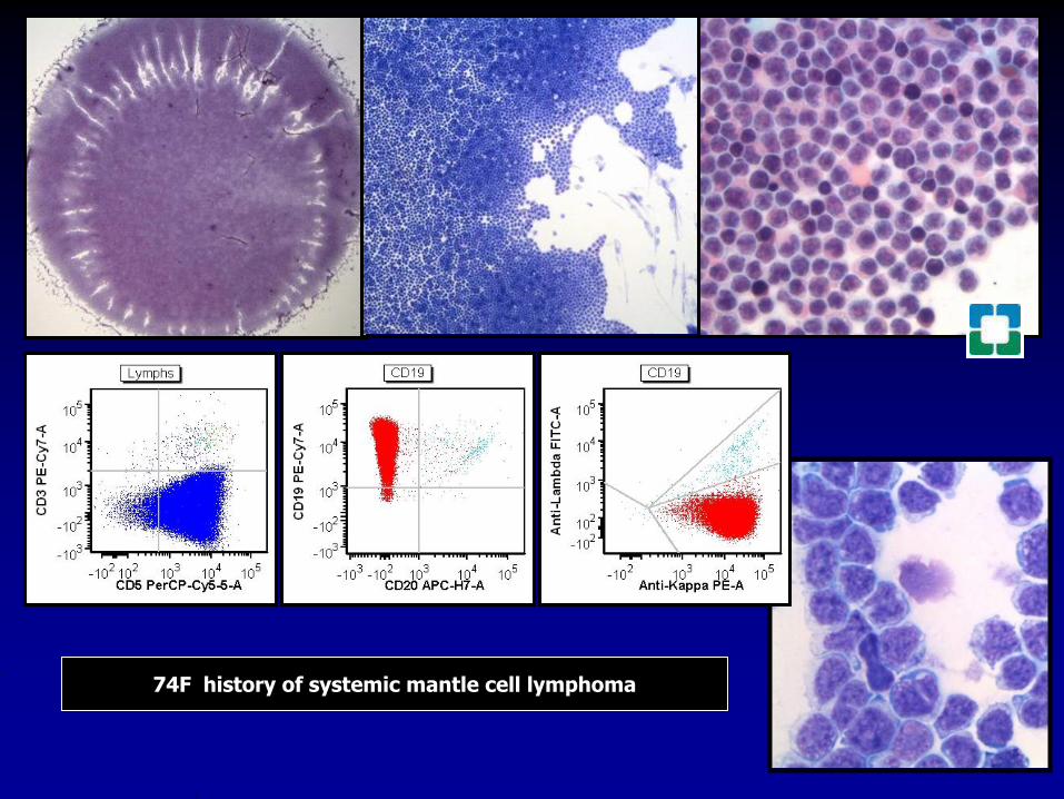
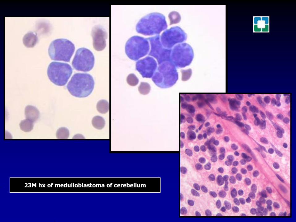
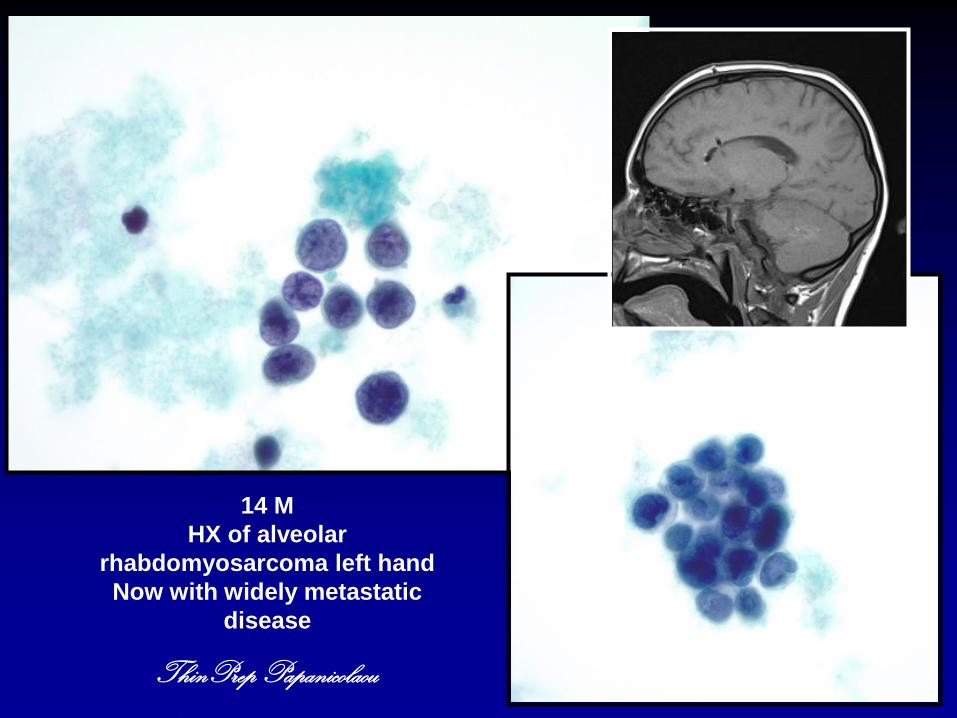
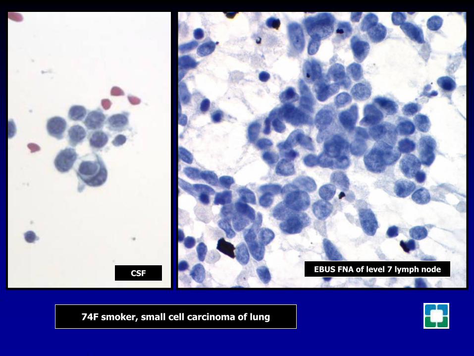
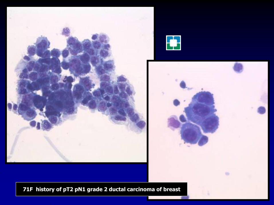
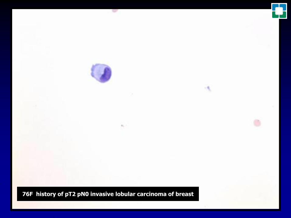
44 F Presented with CNS Symptoms
Subsequent mammography and
chest CT confirmed right
breast mass lesion
MRI confirmed enhancement of
lumbar spinal cord and
cauda equina
44 F Presented with CNS Symptoms
Found to have e-cadherin negative
invasive lobular breast carcinoma
Interestingly strongly ER amp Her-2 +
ER
HER-2
69M smoker with large lung lesionmediastinal adenopathy and
presentation of seizures
CSF
Wang needleof level 7 lymph node
57 M ConfusionRule out meningitis
MRI head with diffuseleptomeningealenhancement
Small enhancing softtissue mass in
left maxillary sinus
40F dural based mass lesion
CSF
Touch preparation ofresected tumor
Glioblastoma multiforme with epithelial differentiation A potential diagnostic pitfall in cerebrospinal fluid cytologySK Gill MD V Padmanabhan MD WF Hickey MD and JD Marotti MD 23 APR 2015 DOI 101002dc23274 copy 2015 Wiley Periodicals IncDartmouth Hitchcock Medical Center amp Geisel School of Medicine at Dartmouth
Cerebrospinal fluid lumbar (A) Hypercellular specimen with loosely cohesive sheets and groups of atypical cells (Cytospin Diff-Quik 60X) B Nuclear pleomorphism and prominent cytoplasmic vacuoles resembling metastatic carcinoma are evident (Cytospin Diff-Quik 300X inset 400X)
MRI of the brain showed a 64 cmring-enhancing lesion in the lefttemporal lobe
Surgical resection specimen (A) Scattered groups of small densely packed epithelioid cells are present in a background of conventional GBM (HampE 140X) B Cytoplasmic vacuoles are present (HampE 300X) C GFAP is negative in epithelioid cells but demonstrates diffuse positivity in the background (Immunostain 100X) D Epithelioid cells are focally positive for EMA (Immunostain 200X) E Focal positivity for cytokeratin AE1AE3 is also present (Immunostain 200X)
78M CHF
Take Home Messages
Be familiar with the slide preparation
techniques and stains in your lab
Read slides CSF slides slowly and
carefully (two cells may make the
difference)
ALWAYS read CSF slides in context of
all available clinical and imaging data
My pleasure to speak with you today
CommentsCritiquesInsights
Questions
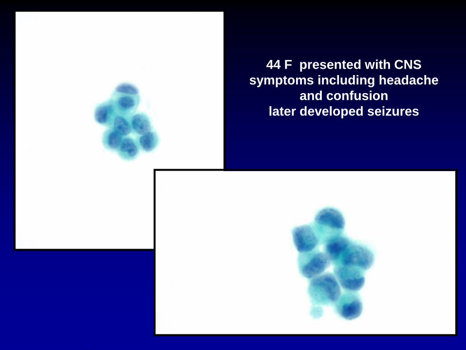
44 F Presented with CNS Symptoms
Found to have e-cadherin negative
invasive lobular breast carcinoma
Interestingly strongly ER amp Her-2 +
ER
HER-2
69M smoker with large lung lesionmediastinal adenopathy and
presentation of seizures
CSF
Wang needleof level 7 lymph node
57 M ConfusionRule out meningitis
MRI head with diffuseleptomeningealenhancement
Small enhancing softtissue mass in
left maxillary sinus
40F dural based mass lesion
CSF
Touch preparation ofresected tumor
Glioblastoma multiforme with epithelial differentiation A potential diagnostic pitfall in cerebrospinal fluid cytologySK Gill MD V Padmanabhan MD WF Hickey MD and JD Marotti MD 23 APR 2015 DOI 101002dc23274 copy 2015 Wiley Periodicals IncDartmouth Hitchcock Medical Center amp Geisel School of Medicine at Dartmouth
Cerebrospinal fluid lumbar (A) Hypercellular specimen with loosely cohesive sheets and groups of atypical cells (Cytospin Diff-Quik 60X) B Nuclear pleomorphism and prominent cytoplasmic vacuoles resembling metastatic carcinoma are evident (Cytospin Diff-Quik 300X inset 400X)
MRI of the brain showed a 64 cmring-enhancing lesion in the lefttemporal lobe
Surgical resection specimen (A) Scattered groups of small densely packed epithelioid cells are present in a background of conventional GBM (HampE 140X) B Cytoplasmic vacuoles are present (HampE 300X) C GFAP is negative in epithelioid cells but demonstrates diffuse positivity in the background (Immunostain 100X) D Epithelioid cells are focally positive for EMA (Immunostain 200X) E Focal positivity for cytokeratin AE1AE3 is also present (Immunostain 200X)
78M CHF
Take Home Messages
Be familiar with the slide preparation
techniques and stains in your lab
Read slides CSF slides slowly and
carefully (two cells may make the
difference)
ALWAYS read CSF slides in context of
all available clinical and imaging data
My pleasure to speak with you today
CommentsCritiquesInsights
Questions
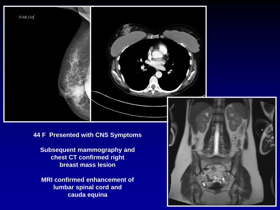
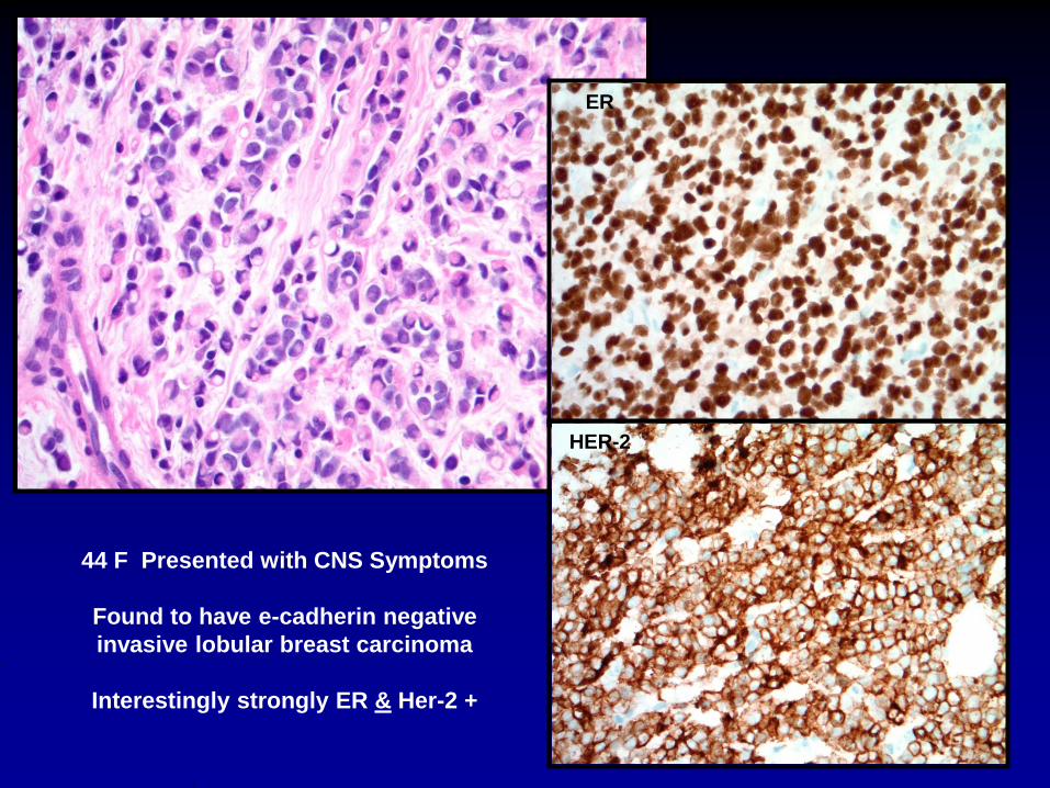
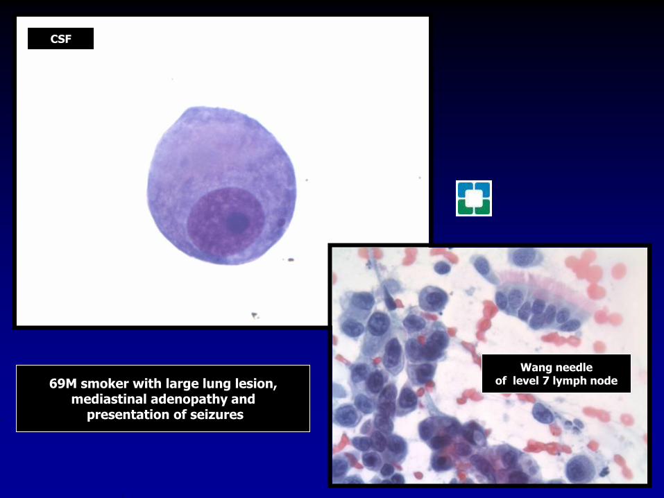
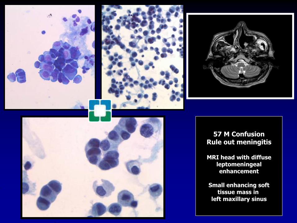
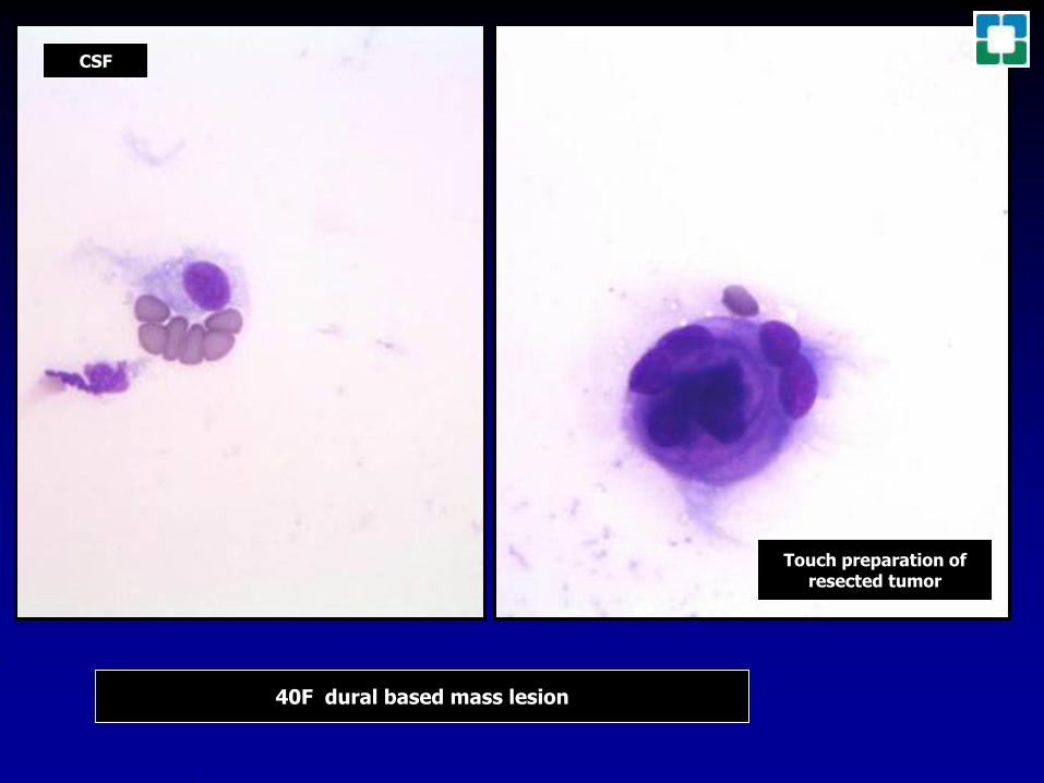
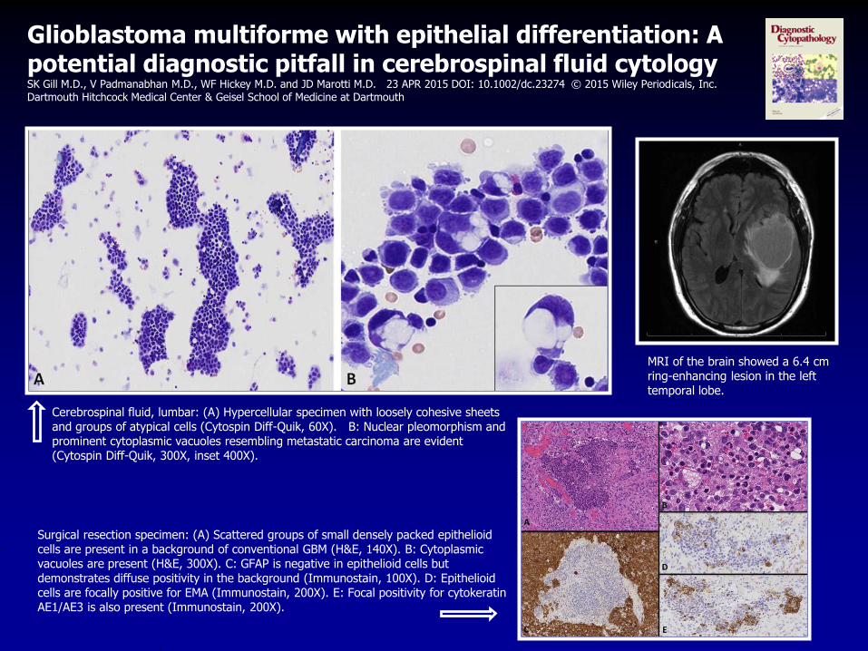
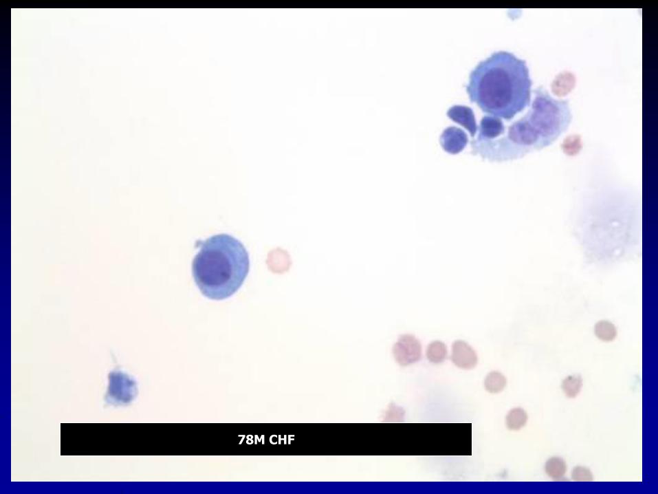
My pleasure to speak with you today
CommentsCritiquesInsights
Questions