Embed Size (px)
Citation preview

NO counterbalances HO-1 overexpression-inducedacceleration of hepatocyte proliferation in miceHarald Schuett1, Christian Eipel1, Claudia Maletzki1, Michael D Menger2 and Brigitte Vollmar1
The trigger for liver regeneration, including shear stress, has been the subject of ongoing debate. Blood vessel-derivedgaseous molecules carbon monoxide (CO) and nitric oxide (NO) regulate vascular tone and play an important role in liverregeneration. In heme oxygenase-1 (HO-1) transgenic mice, it has been shown that CO-mediated impairment ofvasorelaxation is an NO-dependent event. We therefore studied liver regeneration in HO-1 overexpressing animals independency of NO availability. Mice were subjected to 2/3 hepatectomy and were treated with either cobaltprotoporphyrin-IX for induction of CO-liberating HO-1, No-nitro-L-arginine methyl ester (L-NAME) for blockade of NOsynthase (NOS) or both. Application of molsidomine in L-NAME treated animals served for resubstitution of NO. Vehicle-treated animals served as respective control animals. We examined 5-bromo-20-deoxyuridine incorporation andproliferating cell nuclear antigen expression as well as HO-1 and NOS-2 protein levels. Intrahepatic red blood cell velocityand volumetric blood flow were evaluated by in vivo fluorescence microscopy as indicators for microvascular shear stress.Hepatic regeneration remained unaffected by L-NAME application for NOS blockade. However, NOS blockade in HO-1induced animals caused increased 5-bromo-20-deoxyuridine and proliferating cell nuclear antigen measures of liverregeneration. In parallel, these animals revealed increased velocities and volumetric blood flow in the terminal afferentvessels and postsinusoidal venules. These local hemodynamic changes including enhanced hepatocyte proliferationcould be reversed by NO liberation via molsidomine. The present findings stress the role of NO to counterbalancevascular tone in HO-1 overexpressing animals for maintenance of adequate perfusion and salutary shear force within thehepatic microvasculature upon liver resection.Laboratory Investigation (2007) 87, 602–612; doi:10.1038/labinvest.3700548; published online 2 April 2007
KEYWORDS: carbon monoxide; hepatectomy; intravital fluorescence microscopy; proliferation; shear stress
In contrast to most other organs, the liver comprises a uniqueand remarkable property, ie to regenerate, resulting in aprecise reconstitution of the lost tissue mass.1–3 Following atwo-third hepatectomy, liver cells which normally remain inproliferative quiescence are primed and achieve competencefor proliferation. During the subsequent proliferation, thehepatocyte population is expanded. Once the original massof cells is achieved, regeneration is terminated.1–3
The tightly regulated process of liver regeneration is so farnot completely understood. Beside numerous cytokine- andgrowth factor-mediated pathways,1,3 hemodynamic altera-tions, in particular enhanced shear stress upon liver massresection is one of the main factors contributing to cellproliferation.4–7 Within this context, nitric oxide (NO) andcarbon monoxide (CO)-liberating enzyme systems may play
a particular role as both gaseous mediators are released uponenhanced shear stress.7–11
It has been reported that these two gaseous transmittersystems are closely linked in that both systems NO producingnitric oxide synthase (NOS) as well as CO releasing hemeoxygenase (HO) are capable of modulating each othersactivity12–14 and to some extent even in a reciprocal organspecific manner.15 This close association seems to be ofparticular importance since only the synergistic interaction ofCO, NO and HO-1 could demonstrate beneficial effectson the cellular level in a model of tumor necrosis factora-induced hepatocyte cell death in mice.16
In the context of vasoregulation the interaction is probablyeven more complex, as there is evidence for a partial agonismof CO for the soluble guanylate cyclase (sGC), which could
Received 19 October 2006; revised 22 December 2006; accepted 26 December 2006
1Institute for Experimental Surgery, University of Rostock, Rostock, Germany and 2Institute of Clinical and Experimental Surgery, University of Saarland, Homburg-Saar,GermanyCorrespondence: Professor B Vollmar, MD, Institute for Experimental Surgery, University of Rostock, 18055 Rostock, Schillingallee 70, Germany.E-mail: [email protected]
Laboratory Investigation (2007) 87, 602–612
& 2007 USCAP, Inc All rights reserved 0023-6837/07 $30.00
602 Laboratory Investigation | Volume 87 June 2007 | www.laboratoryinvestigation.org

cause a suppression of the vasodilatory response to NO.17
Moreover, the dual blood supply of the liver is subject ofdifferential regulation by NO and CO.18
So far there are no studies, addressing this intriguing in-terplay of NO and CO in the hepatic microvasculature uponregeneration following partial hepatectomy. We herein de-monstrate that cobalt protoporphyrin-IX (CoPP-IX)-inducedHO-1 overexpression combined with inhibition of NOS byNo-nitro-L-arginine methyl ester (L-NAME) accelerates liverregeneration. This observation is most probably due to thedifferential modulation of the pre- and intrahepatic vascularsystem that causes in turn elevated volumetric blood flow andshear stress as trigger for hepatic proliferation. Attenuation ofhyperperfusion by additional NO substitution reduced thedegree of hepatocyte proliferation to levels of unaffectedregenerating livers and thus emphasizing the relevance ofbasal NOS activity for physiological liver regeneration.
MATERIALS AND METHODSLiver Regeneration ModelUpon approval by the local government, all experiments wereperformed in accordance with the German legislation onprotection of animals and the National Institute of Health‘Guide for the Care and Use of Laboratory Animals’ (In-stitute of Laboratory Animal Resources, National ResearchCouncil; NIH publication 86-23 revised 1985). Male C57BL/6J mice (8–10 week old, Charles River Laboratories, Sulzfeld,Germany) were anesthetized by breathing isoflurane (1.5vol%) and subjected to a 2/3 hepatectomy.19 Animals wereplaced in supine position and an upper midline incision wasfollowed by retraction of the xyphoid cartilage for adequateexposure of the liver and division of hepatic ligaments. Theright upper, the left upper and the left lower liver lobes wereresected by placing 4–0 silk suture ties most proximally to theorigin of the lobes. After removal of the tied lobes and irri-gation of the abdomen with warm saline, the peritoneum andthe skin were closed with running 6–0 and 5–0 sutures, re-spectively. Subcutaneous 5 ml saline depots served for volumereplacement. Animals were allowed to recover from an-esthesia and surgery under red warming lamp and held insingle cages until the subsequent experiments.
Experimental Groups and ProtocolFor kinetics of HO-1 and NOS-2 expression upon 2/3 he-patectomy, animals without any pretreatment were subjectedto 2/3 hepatectomy and killed after 2, 4, 8, 12, 18, 24, 48 and96 h for sampling of liver tissue and subsequent protein ex-traction (n¼ 3 animals per point in time). In a second set ofexperiments, animals were subjected to the following treat-ment in an investigator-blinded fashion: 24 h before hepa-tectomy, mice (n¼ 6) were applied CoPP-IX (15 mmol/kgbody weight (bw) intraperitoneal (i.p.)) for HO-1 induction.Control animals received equivalent volumes of the vehicle8.4% NaHCO3/phosphate-buffered saline (PBS) (n¼ 7).To address the role of NOS, both CoPP-IX- (n¼ 5) and
vehicle-treated control animals (n¼ 7) additionally receivedthe NOS inhibitor L-NAME (100 mg/kg bw, i.p.) 24 h beforeand immediately after liver resection. To verify the role ofNOS blockade together with concomitant HO-1 over-expression on liver regeneration and hepatic microcirculationanimals were treated with molsidomine (10 mg/kg bw, i.p.)24 h before and immediately after liver resection inaddition to CoPP-IX and L-NAME (n¼ 6). Molsidomine isenzymatically converted in the liver to yield the activemetabolite SIN-1 which consecutively releases NO.20,21 For acondensed survey of the experimental schedule and thenumber of animals used in each subset as well as thedescription of the respective groups used in this study seeFigure 1 and Table 1.
The HO-1 inductor CoPP-IX was dissolved in NaHCO3/PBS to achieve a final concentration of 1.5 mmol/ml. L-NAME(Axxora Life Sciences, Grunberg, Germany) and molsido-mine (Sigma-Aldrich Chemie GmbH, Munich, Germany)were dissolved in sterile saline. All solutions were freshlyprepared at the day of the experiment. Dose and applicationmode of drugs were chosen in accordance to previouslypublished work of our and other groups.22–25
At 48 h after liver resection, during maximum of DNAsynthesis,26 animals were anesthetized with ketamine/xyla-zine (90/25 mg/kg bw, ip) for retrobulbar sampling of blood.In addition, liver tissue was excised, weighed and harvestedfor subsequent analysis.
Being aware that liver weight is influenced by various ex-trinsic and intrinsic factors that are often unrelated to hepaticregeneration, we assessed expression of proliferating cellnuclear antigen (PCNA) by immunohistochemistry, whichhas been described to serve as an accurate and reliable markerto quantitatively assess hepatic regeneration.27 As PCNA mayonly reflect regeneration on a cell-to-cell basis, we ad-ditionally used 5-bromo-20-deoxyuridine (BrdU, Sigma-Al-drich) incorporation to study DNA synthesis upon liverresection.28 For this purpose, BrdU (50 mg/kg bw, i.p.) wasdissolved in PBS to achieve a final concentration of 5 mg/mland applied intraperitoneally 1 h before sacrifice.29
Western Blot Analysis of Liver TissueComplementary to the conventional measurement of plasmanitrate/nitrite as metabolites of NO, we determined the directeffect of NO in liver tissue. Formation of free and protein-bound 3-nitrotyrosine (3-NT) adducts was used as a probefor NO and its reactive nitrogen species mediated tissue in-terference.30 Thus, detection of these adducts could be uti-lized to determine the amount of NO applied to the liver inthe experimental groups.
For whole protein extracts and Western blot analysis ofHO-1, NOS-2, and 3-NT, liver tissue was homogenized inlysis buffer (10 mM Tris, pH 7.5, 10 mM NaCl, 0.1 mMEDTA, 0.5% Triton-X 100, 0.02% NaN3, and 0.2 mM phe-nylmethylsulfonyl fluoride), incubated for 30 min on ice,and centrifuged for 15 min at 10 000 g. Before use, all buffers
Vasoactive systems and liver regeneration
H Schuett et al
www.laboratoryinvestigation.org | Laboratory Investigation | Volume 87 June 2007 603

received a protease inhibitor cocktail (1:100, v/v; Sigma-Aldrich). Protein concentrations were determined using thebicinchoninic acid protein assay (Pierce Biotechnology,Bonn, Germany) with bovine serum albumin as standard.
Equal amounts of protein (HO-1: 20 mg, NOS-2: 20 mg,3-NT: 40 mg) were separated discontinuously on sodiumdodecyl sulfate polyacrylamide gels (12%) and transferred toa polyvinyldifluoride membrane (Immobilon-P, Millipore,Eschborn, Germany). After blockade of non-specific bindingsites, membranes were incubated for 2 h at room temperaturewith rabbit-polyclonal anti-HO-1 (1:5000, Stressgen Biotech,San Diego, CA, USA), rabbit-polyclonal anti-NOS-2 (1:2000,Calbiochem, San Diego, CA, USA) and mouse-monoclonal
anti-3-NT (1:1000, Santa Cruz Biotechnology, Heidelberg,Germany), followed by a peroxidase-conjugated goat anti-rabbit immunoglobulin G (IgG) (HO-1 1:60 000, NOS-21:20 000; Cell Signalling Technology, Frankfurt, Germany)and a peroxidase-conjugated rabbit anti-mouse IgG respec-tively (3-NT 1:5 000; Sigma-Aldrich) as secondary antibodies.Protein expression was visualized by means of luminol en-hanced chemiluminescence (ECL plus; Amersham PharmaciaBiotech, Freiburg, Germany) and exposure of the membraneto a blue light sensitive autoradiography film (Kodak BioMaxLight Film, Kodak-Industrie, Chalon-sur-Saone, France).Signals were densitometrically assessed (Gel-Dokumenta-tionssystem TotalLab, Nonlinear Dynamics, New Castle upon
Figure 1 Experiments were performed in three seperate sets of animals: In set 1 animals without any pretreatment were subjected to 2/3 hepatectomy (PH)
and sacrificed (z) at the indicated points in time for sampling of liver tissue and subsequent analysis. In set 2 intravital fluorescence microscopy (IVM)
was performed to assess hepatic microhemodynamics after respective pretreatment at the point in time which equals to the point in time of resection in set
3. In set 3 animals were subjected to PH after the respective pre-treatment and sacrificed at 48 h post-PH for sampling of liver tissue. For further information,
see Materials and methods as well as Table 1.
Vasoactive systems and liver regeneration
H Schuett et al
604 Laboratory Investigation | Volume 87 June 2007 | www.laboratoryinvestigation.org
Tyne, UK) and normalized to the b-actin signals as loadingcontrols (mouse monoclonal anti-b-actin antibody, 1:20 000;Sigma-Aldrich).
Histology and Immunohistochemistry of Liver TissueLiver tissue was fixed in 4% phosphate buffered formalin for2–3 days and embedded in paraffin. From the paraffin-embedded tissue blocks, 4 mm sections were cut and stainedwith hematoxylin-eosin for histological analysis. For im-munohistochemical demonstration of HO-1, PCNA andBrdU, sections collected on poly-L-lysine-coated glass slideswere treated by microwave for antigen unmasking. Rabbitpolyclonal anti-HO-1 (1:2000; Stressgen Biotech), rabbitpolyclonal anti-PCNA (1:50; Santa Cruz Biotechnology) andmouse monoclonal anti-BrdU (1:50; Dako Cytomation,Hamburg, Germany) were used as primary antibodies andincubated for 18 h at 41C. After equilibrating to room tem-perature, sections were incubated with horseradish perox-idase-conjugated goat anti-rabbit IgG (HO-1 1:100), alkalinephosphatase-conjugated goat anti-rabbit IgG (PCNA; 1:20)or horseradish peroxidase-conjugated goat anti-mouse IgG(BrdU, 1:100) for 30 min (all from Dako Cytomation). 3,30-diaminobenzidine (HO-1 and BrdU) or fuchsin (PCNA)were used as chromogens. Then the sections were counter-stained with hemalaun and examined by light microscopy(Axioskop 40, Zeiss, Gottingen, Germany). PCNA- andBrdU-positive nuclei as well as HO-1-positive hepatocyteswere counted within 40 consecutive high power fields(� 40 objective, numerical aperture 0.65) and are given ascells/mm2.
Intravital Fluorescence MicroscopyTo analyze the effect of concomitant HO-1 induction (CoPP-IX) and NOS blockade (L-NAME) as well as the effect ofrestoration of the NO balance (molsidomine) in comparisonto control animals on hepatic microcirculation, in a thirdstudy set pretreated animals were studied at the momentof resection (n¼ 5–7 per group; for a condensed survey ofthe number of animals used in each subset as well as the
description of the respective groups used in this set see Figure1 and Table 1). For this purpose, spontaneously breathingketamine/xylazine (90/25 mg/kg bw, i.p.) anesthetized ani-mals were placed on their left side on a heating pad formaintenance of body temperature at 36–371C. The left liverlobe was exteriorized and covered with a glass slide formicroscopy and assessment of microhemodynamics withinthe afferent terminal vessels, the sinusoids and outflowingpostsinusoidal venules.
Using a fluorescence microscope equipped with a 100 Wmercury lamp (Axiotech vario, Zeiss, Jena, Germany) and afilter for blue light epi-illumination (excitation/emissionwavelength: 450–490 nm/4520 nm), microscopic imageswere taken by a water immersion objective (� 20/numericalaperture 0.50 W, Zeiss), recorded by a CCD video camera (FK6990A-IQ, Pieper, Berlin, Germany) and transferred to avideo system (S-VHS Panasonic AG 7350-E, Matsushita,Tokyo, Japan).
Quantitative Video AnalysisFor contrast enhancement, the plasma marker fluoresceinisothiocyanate-dextran (5%, 0.1 ml/100 g bw; Sigma-Aldrich)was used to assess red blood cell velocity (vRBC) within theindividual microvessels, that is, afferent terminal vessels(terminal hepatic arterioles and terminal portal venules),hepatic sinusoids, and postsinusoidal venules, as describedpreviously.31 Volumetric blood flow (VQ) in the individualmicrovessels was estimated from vRBC and microvascularcross-sectional area (pr2) according to the equation of Grossand Aroesty,32 that is, VQ¼ vRBC p r2. Though the equation isvery simplistic inasmuch as a cylindrical shape of the re-spective microvessel is assumed and values may not accu-rately reflect the actual flow, they allow assessment of relativedifferences between groups. Shear stress (t) applied to thehepatic microvasculature was calculated according to thefollowing formula: t¼ 4ZVQ/pr3, where r is the radius andZ the blood viscosity.33,34
Quantification of microhemodynamic parameterswas performed offline by frame-to-frame analysis of the
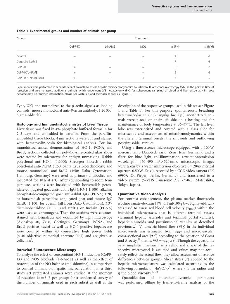
Table 1 Experimental groups and number of animals per group
Groups Treatment
CoPP-IX L-NAME MOL n (PH) n (IVM)
Control � � � 7 6
Control/L-NAME � + � 7 6
CoPP-IX + � � 6 7
CoPP-IX/L-NAME + + � 5 6
CoPP-IX/L-NAME/MOL + + + 6 5
Experiments were performed in separate sets of animals, to assess hepatic microhemodynamics by intravital fluorescence microscopy (IVM) at the point in time ofresection and also to assess additional animals which underwent 2/3 hepatectomy (PH) for subsequent sampling of blood and liver tissue at 48 h posthepatectomy. For further information, please see Materials and methods as well as Figure 1.
Vasoactive systems and liver regeneration
H Schuett et al
www.laboratoryinvestigation.org | Laboratory Investigation | Volume 87 June 2007 605

videotaped images using a computer-assisted image analysissystem (Cap-Image; Zeintl, Heidelberg, Germany). In eachanimal five sinusoidal observation fields were recorded. Perobservation field, red blood cell velocity and diameter inmidzonal regions of the sinusoidal pathway (classificationaccording to Rappaport35) were assessed in a total of 5–10individual sinusoids. A total of 5–10 terminal hepatic arterialand portal venous vessels and 5–10 postsinusoidal venulesper animal each were recorded and analyzed for red bloodcell velocity and diameter. Mean values for red blood cellvelocity and diameter (3–5 measurements per individualmicrovessel with subsequent calculation of mean values) wereused for calculation of volumetric blood flow and shear stressfor each individual microvascular segment (5–10 terminalhepatic arteriolar, terminal portal venular, sinusoidal andpostsinuoidal vessels per animal). Subsequently, these datawere averaged for each individual animal and there uponthe data of all animals per group were summarized asmean7s.e.m.
Statistical AnalysisAll experiments were performed in an investigator-blindedfashion. Data are expressed as means7s.e.m. After provingthe assumption of normality and equal variance acrossgroups, differences between groups were assessed usinganalysis of variance followed by the appropriate post hoccomparison test. Statistical significance was set at Po0.05.Statistics were performed using the software packageSigmaStat (Jandel Corporation, San Rafael, CA, USA).
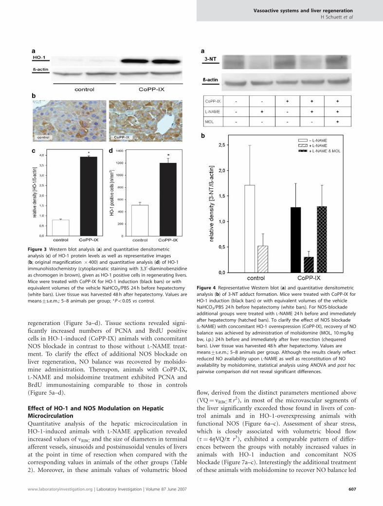
RESULTSEffect of Liver Resection on HO-1 Expression and ItsModulation by CoPP-IXAs revealed by Western blot analysis, regeneration of livertissue upon 2/3 resection caused a marked increase of HO-1expression over time with an apparent maximum at 48 h afterhepatectomy when compared with that of normal quiescentliver tissue (Figure 2a and b). In contrast, there was no rise inNOS-2 protein expression over the time course of 96 h afterhepatectomy (Figure 2c and d). CoPP-IX pretreated animalswhich were subjected to hepatectomy showed a B5-foldincrease of HO-1 protein levels in liver tissue (Figure 3a andc). In line with these densitometric data, histochemistry forHO-1 expression revealed marked immunoreactivity ofhepatocytes upon liver regeneration. The number of HO-1positive cells was found to be even 2.5-fold higher byadditional HO-1 induction with CoPP-IX in comparison tovehicle-treated controls (Figure 3b and d).
Effect of L-NAME and Molsidomine on Formationof 3-NT AdductsThough not statistically significant, there was at least a dis-tinct attenuation of 3-NT adducts in animals treated withL-NAME, while administration of the NO-donor molsido-mine in addition to L-NAME resulted in a normalized 3-NT levelindicating a recovery of the NO balance (Figure 4a and b).
Effect of HO-1 and NOS Modulation on HepaticRegenerationImmunohistochemical analysis of PCNA protein and BrdUincorporation were performed for reliable assessment of liver
Figure 2 Representative Western blots and quantitative densitometric analysis of HO-1 (a and b) and NOS-2 (c and d) in liver tissue of animals over 2 h to
96 h after 2/3 hepatectomy without any pretreatment (n¼ 3 per point in time). Values are means7s.e.m.
Vasoactive systems and liver regeneration
H Schuett et al
606 Laboratory Investigation | Volume 87 June 2007 | www.laboratoryinvestigation.org
regeneration (Figure 5a–d). Tissue sections revealed signi-ficantly increased numbers of PCNA and BrdU positivecells in HO-1-induced (CoPP-IX) animals with concomitantNOS blockade in contrast to those without L-NAME treat-ment. To clarify the effect of additional NOS blockade onliver regeneration, NO balance was recovered by molsido-mine administration. Thereupon, animals with CoPP-IX,L-NAME and molsidomine treatment exhibited PCNA andBrdU immunostaining comparable to those in controls(Figure 5a–d).
Effect of HO-1 and NOS Modulation on HepaticMicrocirculationQuantitative analysis of the hepatic microcirculation inHO-1-induced animals with L-NAME application revealedincreased values of vRBC and the size of diameters in terminalafferent vessels, sinusoids and postsinusoidal venules of liversat the point in time of resection when compared with thecorresponding values in animals of the other groups (Table2). Moreover, in these animals values of volumetric blood
flow, derived from the distinct parameters mentioned above(VQ¼ vRBC p r2), in most of the microvascular segments ofthe liver significantly exceeded those found in livers of con-trol animals and in HO-1-overexpressing animals withfunctional NOS (Figure 6a–c). Assessment of shear stress,which is closely associated with volumetric blood flow(t¼ 4ZVQ/p r3), exhibited a comparable pattern of differ-ences between the groups with notably increased values inanimals with HO-1 induction and concomitant NOSblockade (Figure 7a–c). Interestingly the additional treatmentof these animals with molsidomine to recover NO balance led
Figure 3 Western blot analysis (a) and quantitative densitometric
analysis (c) of HO-1 protein levels as well as representative images
(b; original magnification � 400) and quantitative analysis (d) of HO-1
immunohistochemistry (cytoplasmatic staining with 3,30-diaminobenzidine
as chromogen in brown), given as HO-1 positive cells in regenerating livers.
Mice were treated with CoPP-IX for HO-1 induction (black bars) or with
equivalent volumes of the vehicle NaHCO3/PBS 24 h before hepatectomy
(white bars). Liver tissue was harvested 48 h after hepatectomy. Values are
means7s.e.m.; 5–8 animals per group; *Po0.05 vs control.
Figure 4 Representative Western blot (a) and quantitative densitometric
analysis (b) of 3-NT adduct formation. Mice were treated with CoPP-IX for
HO-1 induction (black bars) or with equivalent volumes of the vehicle
NaHCO3/PBS 24 h before hepatectomy (white bars). For NOS-blockade
additional groups were treated with L-NAME 24 h before and immediately
after hepatectomy (hatched bars). To clarify the effect of NOS blockade
(L-NAME) with concomitant HO-1 overexpression (CoPP-IX), recovery of NO
balance was achieved by administration of molsidomine (MOL, 10 mg/kg
bw, i.p.) 24 h before and immediately after liver resection (chequered
bars). Liver tissue was harvested 48 h after hepatectomy. Values are
means7s.e.m.; 5–8 animals per group. Although the results clearly reflect
reduced NO availability upon L-NAME as well as reconstitution of NO
availability by molsidomine, statistical analysis using ANOVA and post hoc
pairwise comparison did not reveal significant differences.
Vasoactive systems and liver regeneration
H Schuett et al
www.laboratoryinvestigation.org | Laboratory Investigation | Volume 87 June 2007 607

to a decline of all microhemodynamic parameters towardscontrol values, comparable to the changes observed forhepatocyte proliferation (Figures 5–7).
DISCUSSIONHerein, we communicate the following major findings: (i)hepatocyte regeneration upon liver resection was associatedwith increased levels of HO-1, but not NOS-2 protein; and(ii) inhibition of NO synthesis by L-NAME did not influenceregeneration parameters, as assessed by PCNA and BrdUimmunostaining at 48 h after resection (point in time ofmaximal DNA synthesis). However, if HO-1 is overexpressed,NO seems to be mandatory to keep up the appropriatevascular tone, as L-NAME treatment in CoPP-IX-exposedanimals caused intrahepatic hyperperfusion which in turncaused elevation in shear stress consecutively triggeringhepatic proliferation. This view is supported by the fact thatattenuation of this hyperperfusion-induced acceleration ofhepatocyte proliferation to levels found in untreated re-generating livers could be achieved by resubstitution of NOusing molsidomine.
CO as a product of the HO-1 pathway is known to up-regulate the sGC activity with release of cyclic guanosinemonophosphate and therefore exhibits similar physiologicalproperties to NO, such as smooth muscle relaxation andinhibition of platelet aggregation.36–38 As the liver representsone of the most abundant sources of HO, constitutive levelsof CO are considered to be necessary for maintenance of itslow vascular resistance.39,40 Concomitantly, the absence ofboth constitutive and inducible NOS in liver tissue impliesthe negligible role of NO in tonus control of the hepaticmicrovasculature, at least under physiological conditions,39
though CO seems to be weaker in stimulation of sGCthan NO.36,41
Both gaseous mediators have been shown to be released ina shear-stress dependent manner.11,33 Shear stress, beingproportional to the blood flow and the inverse of the cube ofthe vessel radius,33,42 characteristically appears upon the in-crease in the blood flow-to-liver mass ratio during liverresection and has been noted as a possible trigger in the earlystages of liver regeneration.43 In perfused livers isolated fromnormal rats, it has been shown that NO was released bynorepinephrine-induced vasoconstriction at constant flow
Figure 5 Representative images (a and c; original magnification � 400) and quantitative analysis (b and d) of PCNA (nuclear staining with fuchsin as
chromogen in red) and BrdU immunohistochemistry (nuclear staining with 3,30-diaminobenzidine as chromogen in brown), given as number of PCNA or
BrdU-positive cells/mm2 in regenerating livers. Mice were treated with CoPP-IX for HO-1 induction (black bars) or with equivalent volumes of the vehicle
NaHCO3/PBS 24 h before hepatectomy (white bars). For NOS blockade additional groups were treated with L-NAME 24 h before and immediately after
hepatectomy (hatched bars). To clarify the effect of NOS blockade (L-NAME) with concomitant HO-1 overexpression (CoPP-IX) on the liver regeneration,
recovery of NO balance was achieved by administration of molsidomine (MOL, 10 mg/kg bw, i.p.) 24 h before and immediately after liver resection
(chequered bars). Liver tissue was harvested 48 h after hepatectomy. Values are means7s.e.m.; 5–8 animals per group; *Po0.05 vs control.
Vasoactive systems and liver regeneration
H Schuett et al
608 Laboratory Investigation | Volume 87 June 2007 | www.laboratoryinvestigation.org
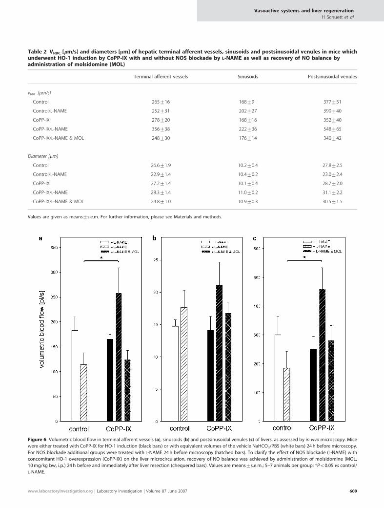
Table 2 VRBC [lm/s] and diameters [lm] of hepatic terminal afferent vessels, sinusoids and postsinusoidal venules in mice whichunderwent HO-1 induction by CoPP-IX with and without NOS blockade by L-NAME as well as recovery of NO balance byadministration of molsidomine (MOL)
Terminal afferent vessels Sinusoids Postsinusoidal venules
vRBC [mm/s]
Control 265716 16879 377751
Control/L-NAME 252731 202727 390740
CoPP-IX 278720 168716 352740
CoPP-IX/L-NAME 356738 222736 548765
CoPP-IX/L-NAME & MOL 248730 176714 340742
Diameter [mm]
Control 26.671.9 10.270.4 27.872.5
Control/L-NAME 22.971.4 10.470.2 23.072.4
CoPP-IX 27.271.4 10.170.4 28.772.0
CoPP-IX/L-NAME 28.371.4 11.070.2 31.172.2
CoPP-IX/L-NAME & MOL 24.871.0 10.970.3 30.571.5
Values are given as means7s.e.m. For further information, please see Materials and methods.
Figure 6 Volumetric blood flow in terminal afferent vessels (a), sinusoids (b) and postsinusoidal venules (c) of livers, as assessed by in vivo microscopy. Mice
were either treated with CoPP-IX for HO-1 induction (black bars) or with equivalent volumes of the vehicle NaHCO3/PBS (white bars) 24 h before microscopy.
For NOS blockade additional groups were treated with L-NAME 24 h before microscopy (hatched bars). To clarify the effect of NOS blockade (L-NAME) with
concomitant HO-1 overexpression (CoPP-IX) on the liver microcirculation, recovery of NO balance was achieved by administration of molsidomine (MOL,
10 mg/kg bw, i.p.) 24 h before and immediately after liver resection (chequered bars). Values are means7s.e.m.; 5–7 animals per group; *Po0.05 vs control/
L-NAME.
Vasoactive systems and liver regeneration
H Schuett et al
www.laboratoryinvestigation.org | Laboratory Investigation | Volume 87 June 2007 609

rate, while increasing flow rate did not release NO, implyingthe contribution of other vasodilators in maintenance ofhepatic vascular resistance under high flow conditions.44 Insupport of this, and as observed in the present study, shearstress did not stimulate NOS-2 protein expression however itwas capable of inducing HO-1 protein expression.11 Thus, itis reasonable to speculate that at least under normal andincreased flow conditions in liver microcirculation, CO ra-ther than NO serves as effective mediator of vasoregulationeither via agonism for the sGC or cGMP-independent viaactivation of vascular smooth muscle Ca2þ -activated Kþ
channels.37,45 At the same time, however, there is evidencethat NO availability is mandatory to control vasotonus incase of HO-1 associated hyperperfusion of the liver.
NO is reported to be released after partial hepatectomy46
and seems to be required for adequate liver regenerationinasmuch as NOS-2 deficient mice exhibited impaired re-generation.47 In line with this, the increase of proliferativefactor activity and c-fos mRNA expression in hepatectomizedrats, serving as two indices of the initiation of the liver re-generation cascade, could be inhibited by the NOS antagonistL-NAME, while a NO donor reversed inhibition.10 On the
contrary, it has been shown that both endogenous NOsynthesis and exogenous NO delivery resulted in delayed liverrecovery in hepatectomized mice.48 The present data pro-vides an explanation to this contradiction in that NO seemsnot to be mandatory for the liver to sufficiently regenerateupon resection, as far as vasotonic control is adequatelymaintained by a proportionate HO-1 response. However, incase of HO-1 overexpression, NO is essential to counter-balance hyperperfusion-induced surpassing shear stress andsubsequently, shear stress-induced enhancement of pro-liferation. This has conclusively been shown by the fact thatsubstitution of NO by molsidomine in the CoPP-IX/L-NAMEtreated animals could reverse the hepatic hyperdynamiccondition with super-physiologically accelerated prolifera-tion.
In HO-1 transgenic mice, CO overproduced through theHO reaction has recently been shown to interfere with NO-mediated vasorelaxing mechanisms.17 We now show that theCoPP-IX/L-NAME-treated animals present higher bloodflow velocities as well as volumetric blood flow and thusincreased shear stress. Moreover, restoration of NO balanceby application of molsidomine returned microhemodynamic
Figure 7 Shear stress (t) in terminal afferent vessels (a), sinusoids (b) and postsinusoidal venules (c) of livers, as assessed by in vivo microscopy and
calculated according to the following formula: t¼ 4ZVQ/pr3. Mice were either treated with CoPP-IX for HO-1 induction (black bars) or with equivalent
volumes of the vehicle NaHCO3/PBS (white bars) 24 h before microscopy. For NOS-blockade additional groups were treated with L-NAME 24 h before
microscopy (hatched bars). To clarify the effect of NOS blockade (L-NAME) with concomitant HO-1 overexpression (CoPP-IX) on the liver microcirculation,
recovery of NO balance was achieved by administration of molsidomine (MOL, 10 mg/kg bw, i.p.) 24 h before and immediately after liver resection
(chequered bars). Values are means7s.e.m.; 5–7 animals per group.
Vasoactive systems and liver regeneration
H Schuett et al
610 Laboratory Investigation | Volume 87 June 2007 | www.laboratoryinvestigation.org
parameters and proliferation capacity of the liver to values ofregenerating livers from animals without any pretreatment.Thus, the observed increase of red blood cell velocity andvolumetric blood flow in CoPP-IX/L-NAME-treated animalsis most likely attributable to the differential modulation ofthe pre- and intrahepatic vascular system by overabundantCO and to loss of NO action owing to L-NAME application.The increase of blood volume causes in turn an elevation ofshear stress, which consecutively triggers hepatic prolifera-tion.4,5,7 At first sight, one might wonder why NO-blockadeby L-NAME or HO-1 induction by CoPP-IX alone waswithout effect and did increase microhemodynamics only incase of NOS-blocked and concomitant HO-1-induced ani-mals. This might be explained by the desensitization of sGCfor NO through CO by overexpression of HO-1, as this hasbeen described for HO-1 transgenic mice17 as well as theabolishment of NO-mediated attenuation of CO-mediatedstimulatory effects on vascular smooth muscle Ca2þ -acti-vated Kþ channels.49 In conclusion, our model providesevidence that NO plays a major role in adjusting vasculartone in HO-1 overexpressing animals. This is important inorder to maintain adequate perfusion and salutary effect onshear force within the hepatic microvasculature upon liverresection to assure a physiological regeneration process.
ACKNOWLEDGEMENT
We thank Berit Blendow, Doris Butzlaff, Dorothea Frenz, Maren Nerowski,
and Kathrin Sievert (Institute of Experimental Surgery, University of Rostock)
for excellent technical assistance and Evelyn Kidess (Institute of
Experimental Surgery, University of Rostock) for her expert assistance in
editing the manuscript.
Grants: This study is supported in part by a grant of the Deutsche
Forschungsgemeinschaft, Bonn-Bad Godesberg, Germany (Vo 450/7-3).
1. Fausto N. Liver regeneration. J Hepatol 2000;32:19–31.2. Michalopoulos GK, deFrances MC. Liver regeneration. Science
1997;276:60–66.3. Taub R. Liver regeneration: from myth to mechanism. Nat Rev Mol Cell
Biol 2004;5:836–847.4. Niiya T, Murakami M, Aoki T, et al. Immediate increase of portal
pressure, reflecting sinusoidal shear stress, induced liver regenerationafter partial hepatectomy. J Hepatobiliary Pancreat Surg 1999;6:275–280.
5. Sato Y, Tsukada K, Hatakeyama K. Role of shear stress and immuneresponses in liver regeneration after a partial hepatectomy. Surg Today1999;29:1–9.
6. Thomson RY, Clarke AM. Role of portal blood supply in liverregeneration. Nature 1965;208:392–393.
7. Wang HH, Lautt WW. Evidence of nitric oxide, a flow-dependent factor,being a trigger of liver regeneration in rats. Can J Physiol Pharmacol1998;76:1072–1079.
8. Busse R, Fleming I. Pulsatile stretch and shear stress: physical stimulidetermining the production of endothelium-derived relaxing factors. JVasc Res 1998;35:73–84.
9. Hirano K, Sato Y, Kobayashi T, et al. Carbon monoxide hemoglobin andbilirubin metabolism in adult living-related liver transplantation.Hepatogastroenterology 2003;50:1745–1748.
10. Schoen JM, Wang HH, Minuk GY, et al. Shear stress-induced nitric oxiderelease triggers the liver regeneration cascade. Nitric Oxide2001;5:453–464.
11. Wagner CT, Durante W, Christodoulides N, et al. Hemodynamic forcesinduce the expression of heme oxygenase in cultured vascular smoothmuscle cells. J Clin Invest 1997;100:589–596.
12. Naughton P, Foresti R, Bains SK, et al. Induction of heme oxygenase 1by nitrosative stress. A role for nitroxyl anion. J Biol Chem2002;277:40666–40674.
13. Kim YM, Bergonia HA, Muller C, et al. Loss and degradation of enzyme-bound heme induced by cellular nitric oxide synthesis. J Biol Chem1995;270:5710–5713.
14. Maulik N, Engelman DT, Watanabe M, et al. Nitric oxide-a retrogrademessenger for carbon monoxide signaling in ischemic heart. Mol CellBiochem 1996;157:75–86.
15. Sarady JK, Zuckerbraun BS, Bilban M, et al. Carbon monoxideprotection against endotoxic shock involves reciprocal effects on iNOSin the lung and liver. FASEB J 2004;18:854–866.
16. Zuckerbraun BS, Billiar TR, Otterbein SL, et al. Carbon monoxideprotects against liver failure through nitric oxide-induced hemeoxygenase 1. J Exp Med 2003;198:1707–1716.
17. Imai T, Morita T, Shindo T, et al. Vascular smooth muscle cell-directedoverexpression of heme oxygenase-1 elevates blood pressure throughattenuation of nitric oxide-induced vasodilation in mice. Circ Res2001;89:55–62.
18. Pannen BH, Bauer M. Differential regulation of hepatic arterial andportal venous vascular resistance by nitric oxide and carbon monoxidein rats. Life Sci 1998;62:2025–2033.
19. Greene AK, Puder M. Partial hepatectomy in the mouse: technique andperioperative management. J Invest Surg 2003;16:99–102.
20. Feelisch M, Ostrowski J, Noak E. On the mechanism of NO release fromsydnonimines. J Cardiovasc Pharmacol 1989;14(Suppl 11):13–22.
21. Yamamoto T, Bing RJ. Nitric oxide donors. Proc Soc Exp Biol Med2000;225:200–206.
22. Amon M, Menger MD, Vollmar B. Heme oxygenase and nitric oxidesynthase mediate cooling-associated protection against TNF-a-induced microcirculatory dysfunction and apoptotic cell death. FASEBJ 2003;17:175–185.
23. Rahman TM, Hodgson HJ. The effects of early and late administrationof inhibitors of inducible nitric oxide synthase in a thioacetamide-induced model of acute hepatic failure in the rat. J Hepatol2003;38:583–590.
24. Osna NA, Haorah J, Krutik VM, et al. Peroxynitrite alters the catalyticactivity of rodent liver proteasome in vitro and in vivo. Hepatology2004;40:574–582.
25. Sass G, Soares MC, Yamashita K, et al. Heme oxygenase-1 and itsreaction product, carbon monoxide, prevent inflammation-relatedapoptotic liver damage in mice. Hepatology 2003;38:909–918.
26. Kountouras J, Boura P, Lygidakis NJ. Liver regeneration afterhepatectomy. Hepatogastroenterology 2001;48:556–562.
27. Assy N, Gong Y, Zhang M, et al. Use of proliferating cell nuclear antigenas a marker of liver regeneration after partial hepatectomy in rats. JLab Clin Med 1998;131:251–256.
28. Assy N, Minuk GY. Liver regeneration: methods for monitoring andtheir applications. J Hepatol 1997;26:945–952.
29. Furnus CC, Inda AM, Andrini LB, et al. Chronobiology of theproliferative events related to angiogenesis in mice liver regenerationafter partial hepatectomy. Cell Biol Int 2003;27:383–386.
30. Tarpey MM, Fridovich I. Methods of detection of vascular reactivespecies: nitric oxide, superoxide, hydrogen peroxide, and peroxynitrite.Circ Res 2001;89:224–236.
31. Richter S, Vollmar B, Mucke I, et al. Hepatic arteriolo-portal venularshunting guarantees maintenance of nutritional microvascular supplyin hepatic arterial buffer response of rat livers. J Physiol 2001;531:193–201.
32. Gross JF, Aroesty J. Mathematical models of capillary flow. A criticalreview. Biorheology 1972;9:225–264.
33. Hori N, Wiest R, Groszmann RJ. Enhanced release of nitric oxide inresponse to changes in flow and shear stress in the superiormesenteric arteries of portal hypertensive rats. Hepatology1998;28:1467–1473.
34. Windberger U, Bartholovitsch A, Plasenzotti R, et al. Whole bloodviscosity, plasma viscosity and erythrocyte aggregation in ninemammalian species: reference values and comparison of data. ExpPhysiol 2003;88:431–440.
35. Rappaport AM. The microcirculatory hepatic unit. Microvasc Res1973;6:212–228.
36. Burstyn JN, Yu AE, Dierks EA, et al. Studies of the heme coordinationand ligand binding properties of soluble guanylyl cyclase (sGC):
Vasoactive systems and liver regeneration
H Schuett et al
www.laboratoryinvestigation.org | Laboratory Investigation | Volume 87 June 2007 611
characterization of Fe(II)sGC and Fe(II)sGC(CO) by electronicabsorption and magnetic circular dichroism spectroscopies andfailure of CO to activate the enzyme. Biochemistry 1995;34:5896–5903.
37. Wang R. Resurgence of carbon monoxide: an endogenous gaseousvasorelaxing factor. Can J Physiol Pharmacol 1998;76:1–15.
38. Brune B, Ullrich V. Inhibition of platelet aggregation by carbonmonoxide is mediated by activation of guanylate cyclase. MolPharmacol 1987;32:497–504.
39. Suematsu M, Goda N, Sano T, et al. Carbon monoxide: an endogenousmodulator of sinusoidal tone in the perfused rat liver. J Clin Invest1995;96:2431–2437.
40. Suematsu M, Kashiwagi S, Sano T, et al. Carbon monoxide as anendogenous modulator of hepatic vascular perfusion. BiochemBiophys Res Commun 1994;205:1333–1337.
41. Sharma VS, Magde D. Activation of soluble guanylate cyclase bycarbon monoxide and nitric oxide: a mechanistic model. Methods1999;19:494–505.
42. Kamiya A, Togawa T. Adaptive regulation of wall shear stress to flowchange in the canine carotid artery. Am J Physiol 1980;239:H14–H21.
43. Braet F, Shleper M, Paizi M, et al. Liver sinusoidal endothelial cellmodulation upon resection and shear stress in vitro. Comp Hepatol2004;3:7.
44. Pastor CM, Hadengue A. Shear stress modulates the vascular tone inperfused livers isolated from normal rats. Hepatology 2000;32:786–791.
45. Naik JS, Walker BR. Heme oxygenase-mediated vasodilation involvesvascular smooth muscle cell hyperpolarization. Am J Physiol Heart CircPhysiol 2003;285:H220–H228.
46. Hortelano S, Dewez B, Genaro AM, et al. Nitric oxide is released in rege-nerating liver after partial hepatectomy. Hepatology 1995;21:776–786.
47. Rai RM, Lee FY, Rosen A, et al. Impaired liver regeneration in induciblenitric oxide synthase deficient mice. Proc Natl Acad Sci USA1998;95:13829–13834.
48. Zeini M, Hortelano S, Traves PG, et al. Assessment of a dual regulatoryrole for NO in liver regeneration after partial hepatectomy: protectionagainst apoptosis and retardation of hepatocyte proliferation. FASEB J2005;19:995–1007.
49. Wu L, Cao K, Lu Y, et al. Different mechanisms underlying thestimulation of K(Ca) channels by nitric oxide and carbon monoxide. JClin Invest 2002;110:691–700.
Vasoactive systems and liver regeneration
H Schuett et al
612 Laboratory Investigation | Volume 87 June 2007 | www.laboratoryinvestigation.org