Embed Size (px)
Citation preview

1
No Association Between Types of Unilateral Mandibular
Condylar Abnormalities and Facial Asymmetry in orthopedic
treated Patients with Juvenile Idiopathic Arthritis
Highlights
Dentofacial asymmetries often occurred in patients with juvenile idiopathic arthritis
(JIA).
Dentofacial development was affected more severely in patients with condylar
abnormalities.
Dysmorphic dentofacial development was not associated with a specific type of
condylar abnormality.
Combined condylar deformations and erosions were observed in one third of the TMJs
in JIA.

2
Abstract
Introduction: Dentofacial asymmetries are often observed in patients with juvenile idiopathic
arthritis (JIA) and temporomandibular joint (TMJ) involvements. The aim of this split-face
study was to associate types of radiological TMJ abnormalities with the degree of dentofacial
asymmetry in patients with unilateral TMJ involvements assessed with cone beam
computerized tomography (CBCT).
Methods: Forty-seven JIA patients and 19 non-arthritic control subjects were included. Normal
condylar radiological CBCT appearance in a minimum of one TMJ was set as the inclusion
criterion for all patients with JIA. The contralateral TMJ was thereafter scored as either
“normal”, “deformed” or “erosive” consistent with predefined criteria. Based on the bilateral
radiological TMJ appearances, three JIA groups were assigned: 1) normal/normal, 2)
normal/deformed, and 3) normal/erosive. The severity of dentofacial asymmetry was compared
between the JIA groups and control subjects. Dentofacial asymmetry was expressed as inter-
side ratios and angular measurements.
Results: Eighty-seven percent of the JIA patients were currently being treated or had previously
received treatment with functional orthopedic appliance at the time of the CBCT. Significantly
greater dentofacial asymmetries were observed in the two groups of JIA patients with unilateral
condylar abnormalities (deformation or erosion) than in the other groups. A similar degree of
dentofacial asymmetry was observed in JIA patients with bilateral normal TMJs and in the non-
arthritic control group.
Conclusion: JIA patients with unilateral condylar abnormalities (deformation or erosion)
exhibited significantly more severe dentofacial asymmetries than JIA patients without condylar
abnormalities and control subjects. We found the same degree of dentofacial asymmetry when
dividing condylar abnormalities into deformation or erosion groups.
3
Introduction
The involvement of the temporomandibular joint (TMJ) is a common finding in patients with
juvenile idiopathic arthritis (JIA) and has a varying prevalence of up to 96%. 1 A clinical
consequence of TMJ involvement can be dysmorphic mandibular development resulting in
dentofacial asymmetry, occlusal instability, alterations in muscular activity, and suboptimal
TMJ function, which, in turn, may lead to the development of orofacial symptoms and a
compromised aesthetic appearance. 2-9 One of the primary treatment goals in JIA is to avoid
this unwanted dysmorphic dentofacial development. However, the underlying processes are
not yet fully understood.
Historically, dysmorphic dentofacial alterations in JIA patients have been regarded as a
consequence of arthritis-induced TMJ degeneration resulting in the loss of condylar vertical
height, which is an arthritis-induced degradation similar to what occurs in adult patients with
rheumatoid arthritis with TMJ involvement. 10 For decades, extensive research has fueled
debates on the implications of TMJ lesions/degeneration on mandibular development and
growth. 11-14 However, the contemporary dominant theory explains the dysmorphic dentofacial
development in JIA patients as a consequence of condylar growth disturbances rather than
arthritis-induced condylar damage alone. 9,15,16-18 In a recent review, Peltomäki et al. supported
this perception and emphasized the need for documentation in future prospective longitudinal
studies. 19 They further noted that altered dentofacial development may be caused by a
combination of inflammatory effects on the intra-articular growth site of the condylar cartilage
and impaired masticatory function. 19
There is currently a need to elucidate the intra-articular processes that occur during dysmorphic
dentofacial development in JIA. This is important from a pathogenic perspective to advance
our diagnostic and therapeutic understanding of this challenging condition. Combined
imaging/radiological TMJ scoring systems have recently been published exclusively for JIA.
20,21 Components of these scoring systems aim to score the severity of arthritis-induced osseous
changes. The scoring systems imply the progression of the arthritis-induced condylar osseous
changes from minor condylar head deformation (flattening) to condylar destructions that
progressively reduce normal mandibular growth. 20,21 Both scoring systems grade erosive
osseous changes higher than condylar deformation (flattening). With an isolated focus on
condylar conditions, these scoring systems may be useful for radiological TMJ assessment.
However, no human studies have determined if particular types of abnormal TMJ condylar
4
osseous changes (e.g., erosive changes) are associated with a greater extent of mandibular
dysmorphic development than others (e.g., condylar deformations). Notably, previous
experimental studies found no relationship between the severity of condylar lesions and a
reduction in mandibular growth and development in young growing rabbits with
experimentally induced and histologically confirmed TMJ arthritis. 17,22
Previous research on TMJ abnormality and dentofacial development in JIA was primarily
based on conventional radiological techniques, 11,14,23,24 and only few studies with more
advanced techniques exist. 25-29 The introduction of cone beam computed tomography (CBCT),
a cost- and dose-effective three-dimensional imaging modality, has enabled the radiological
examination of TMJ hard tissue pathologies, which is superior to conventional radiological
methods. 30 The aim of this cross-sectional CBCT-based study was to associate radiological
TMJ abnormalities with the degree of dentofacial asymmetry in patients with unilateral TMJ
involvements. A control group was used to compare asymmetries to the normal population.
Materials and Methods
JIA patients
Eighty-six consecutive JIA patients at the Section of Orthodontics; Aarhus University,
Denmark were eligible for inclusion. All candidates had received a craniofacial CBCT scan
between February 2011 and April 2014. Patients were included when complying with the
following inclusion criteria: 1) a diagnosis of JIA according to the ILAR criteria 31 and 2) at
least one TMJ with radiological normal/healthy osseous TMJ appearance. The exclusion
criteria were 1) patients with previous craniofacial traumas or patients with an underlying
diagnosis of a syndrome or congenital birth defect involving the craniofacial area or 2) CBCT
images of poor quality. The data on patient characteristics were collected from medical hospital
records. The present study was approved by the Danish Health and Medicines Authorities
(DOK2129859) and the Danish Data Protection Agency (2007-58-0010) and was conducted in
accordance with the ethics of the Helsinki Declaration.
Control group
Nineteen controls without JIA previously treated at the Section of Orthodontics, Aarhus
University, Denmark, for other reasons were identified. All had received a full-face CBCT scan
in relation to an orthodontic treatment. Control subject criteria were: 1) no previous or current
diagnosis of temporomandibular dysfunction; 2) a high-quality CBCT allowing optimal

5
radiological assessment; and 3) an age comparable to the JIA patients. Although control
subjects with a neutral dental occlusion (Class I) were preferred, full-face CBCTs were rarely
taken in this group; we therefore accepted subjects with dental class II subdivision
malocclusions and impacted canines.
CBCT scan
CBCT scans (NewTom 5G, QR s.r.l., Italy) were acquired in accordance with the
manufacturer’s instructions. The image acquisition parameters included a FOV of 18 ×16 cm,
a scanning time of approximately 18 sec, and 3.6 sec of active radiation (pulsed mode) with
settings of 110 kV and 3-7 mA. All CBCT scans were reconstructed with a 0.30-mm isotropic
voxel dimension.
Radiological TMJ evaluation
The radiological evaluation was carried out on the joint level, giving each individual two
independently assessed TMJ scores (one score for the right joint and one score for the left
joint). The interpretation of the CBCT images was conducted using NewTom NNT Viewer
software (version 4.6). The type of mandibular condylar abnormality was independently scored
by a specially trained maxillofacial radiologist (LZA) who was blinded to the patient diagnosis
and the order in which the TMJs were presented (control and JIA subjects were mixed).
Cropped CBCT images only including the TMJs were viewed in axial, oblique coronal and
oblique sagittal planes. The radiologist was therefore also blinded to the presence of any
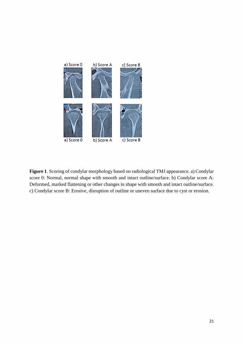
dentofacial asymmetry. Prior to patient inclusion, three definitions of condylar scores were
decided based on a categorization of the condylar radiological appearance (Fig. 1):
1. Normal: normal shape with smooth and intact outline/surface (score 0)
2. Deformed: marked flattening or other changes in shape with smooth and intact
outline/surface (score A)
3. Erosive: disruption of outline or uneven surface due to cysts or erosion (score B)
The maxillofacial radiologist was asked to subgroup each mandibular condyle based on the
radiological appearances into one of three groups: normal condylar outline (score 0) or
abnormal condylar outline (score A or B). When both deformations (score A) and erosive
changes (score B) were found in the same TMJ, the score was based on the most prominent
6
radiological feature present. Each individual was thereafter categorized by the bilateral joint
scores and assigned to one of six groups: JIA 0-0, JIA 0-A, JIA 0-B, JIA A-A, JIA A-B or JIA
B-B. In compliance with the inclusion criteria of this split-face designed study, only patients
from the JIA 0-0, JIA 0-A, and JIA 0-B sub-groups were included in our data analyses because
a minimum of one joint in each patient was required to have a normal appearance. The control
subjects were also sub-grouped based on their bilateral joint scores. However, to represent the
population-based variation in radiological condylar appearance, all control subjects were
accepted without considering their bilateral joint scores. Therefore, control subjects with
radiological abnormalities A-A or B-B were also included. Double assessments of 20
consecutive TMJs were conducted with a two-week interval to assess the intra-observer
agreement.
3D analysis
To assess the degree of dentofacial asymmetry, a three-dimensional dentofacial analysis was
conducted using Mimics software 16.0 (Materialise Interactive Medical Image Control System,
Leuven, Belgium). The aim was to assess the intra-patient mandibular asymmetries based on
outcome variables representing facial asymmetries between the left and right side of the face.
Information on sagittal, vertical, and transversal mandibular asymmetries was collected using
eight anatomical landmarks. The landmarks are defined and presented in Table 1 and Fig. 2.
Reference planes
For the analysis, two main reference planes were constructed:
1. Axial plane: through S and LOr and LOl.
2. Midsagittal plane: through N, S and perpendicular to the axial plane.
The additional planes necessary for the analysis were constructed with reference to these two
planes. The points and additional planes are defined in Table 1 and shown in Fig. 2ab.
Outcome variables of craniofacial asymmetry
A total of nine predefined outcome variables were assessed (Table 1, Fig. 2cd). In the vertical
direction, five bilateral length variables of interest were defined: 1) total mandibular height
(from Co to Go), 2) condylar height (from Co to Inc plane), 3) ramus height (from Go to Inc
plane), 4) the distance from the axial plane to the gonion, and 5) the distance from the axial

7
plane to Minf. One variable was defined in the sagittal direction; the length of the mandible
left and right side (from Go to Gn). In the transversal direction, one variable was defined:
transversally, the distance from the midsagittal plane to the left and right gonion. Additionally,
two angular measurements were defined: 1) the jaw angle left and right side (the angle between
the line connecting Co and Go and the line connecting Go and Gn) and 2) the gonial plane
angle (the angle formed between a plane passing through the right gonion and the left gonion
(perpendicular to the coronal plane) and the midsagittal plane (degrees) measured to the
affected side). In group 0-0, the angle was measured to the shortest side. For all bilateral
outcome variables, a ratio of asymmetry was calculated to obtain a measure of intra-patient
dentofacial asymmetry. The ratio was calculated by dividing the value of the affected side (TMJ
score A or B) with the value of the non-affected side (TMJ score 0). In group 0-0, the ratio was
calculated using the smallest value divided by the largest value.
Statistics
The data concerning the nine outcome variables were tested for a normal distribution by visual
inspections of Q–Q plots. Inter-group differences in dentofacial asymmetry were assessed
using analysis of variance (ANOVA) tests with independent Student’s t-tests serving as post-
ANOVA tests. Post-ANOVA testing was only performed in outcome variables where a
statistically significant difference was observed in the primary ANOVA test. The significance
level was adjusted in accordance with Bonferroni to avoid a type 1 error due to multiple testing.
In the primary ANOVA tests, the significance level was lowered with the number of tests
conducted (0.05/9 → p<0.006). In the post-ANOVA tests, the significance level was adjusted
based on the number of t-tests conducted within each individual outcome variable (0.05/6 →
p<0.008).
Intra-rater agreement of the radiological condylar TMJ scores was assessed by kappa statistics
based on duplicate assessments two weeks apart. The error of the method of the nine outcome
variables describing craniofacial asymmetry was also evaluated based on duplicate assessments
two weeks apart using scatterplots and correlation coefficients. 32 For each of the nine outcome
variables, the error of measurement was assessed based on Bland–Altman plots. 32 The limits
of agreements derived from the Bland–Altman plots were used to define the smallest detectable
difference for each specific outcome variable: The smallest detectable difference was defined
as the minimal amount of change in the inter-side ratios and angles needed to overcome the
measurement error within each of the specific outcome variables defined.

8
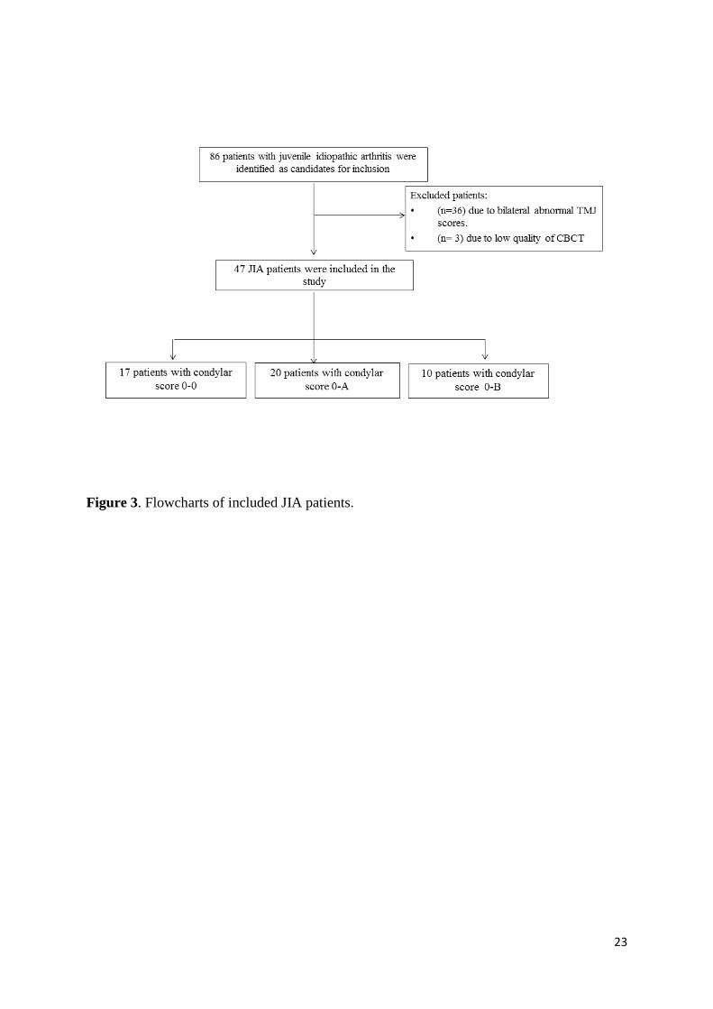
Results
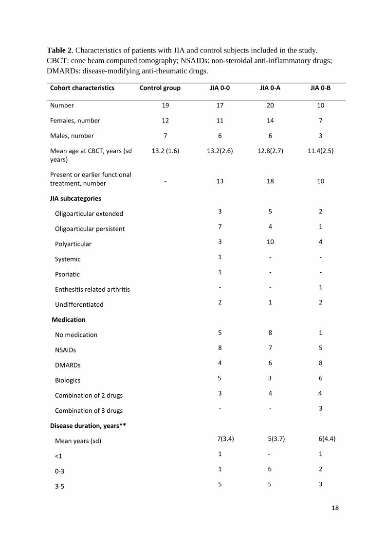
JIA groups: Forty-seven of the examined 86 patients met the inclusion criteria. All patients
presented inflammatory involvements in other joints besides the TMJs. Due to similar
bilaterally abnormal radiological TMJ assessment scores (JIA A-A, JIA B-B and JIA A-B), 36
patients (41%) were excluded from the study. Three JIA patients were excluded due to low
CBCT quality (Fig. 3). The remaining 47 included JIA patients who were grouped into three
sub-groups based on the radiological findings: JIA 0-0 (n=17), JIA 0-A (n=20), and JIA 0-B
(n=10). The mean ages of the patients in the sub-groups were 13.2 years (std. 2.6) in the JIA
0-0 group, 12.8 years (std. 2.7) in the JIA 0-A group and 11.4 years (std. 2.5) in the JIA 0-B
group (Table 2). Comparable distribution of disease duration of TMJ arthritis was observed
among the JIA groups (Table 2). Nine of the 47 patients (19%) had radiological findings of
both score A and B within the same joint. Forty-one of the included 47 JIA patients were
currently being treated or had previously been treated with a functional orthopedic appliance.
(33) The distribution of patients receiving treatment with functional orthopedic appliance were:
JIA 0-0 (n=13/17, 77%), JIA 0-A (n=18/20, 90%), and JIA 0-B (n=10/10, 100%). No
significant intergroup difference was observed in the duration of treatment with functional
orthopedic appliance at the time of CBCT.
Control subjects: Nineteen subjects were included. Based on their TMJ abnormality scores, the
control group comprised the following subgroups: 0-0 (n=14), 0-B (n=3), A-A (n=1), and B-B
(n=1), which indicates that abnormal joint appearances were also present among the control
subjects as a random finding. The mean age at CBCT for the control subjects was 13.2 years
(std. 1.6) (Table 2).
The intra-rater reliability of the radiological TMJ abnormality scorings revealed an agreement
of 83.3 percent (κ=0.67) in the assessment of normal versus abnormal radiological TMJ
appearance. In the TMJ assessment of joints that were scored with “abnormal findings,” an
intra-rater agreement of 76.7% (κ=0.63) was found in the subgroups (score A versus score B).
According to Fleiss et al., this represents a fair agreement (κ=0.4< fair agreement ≤0.75). 34
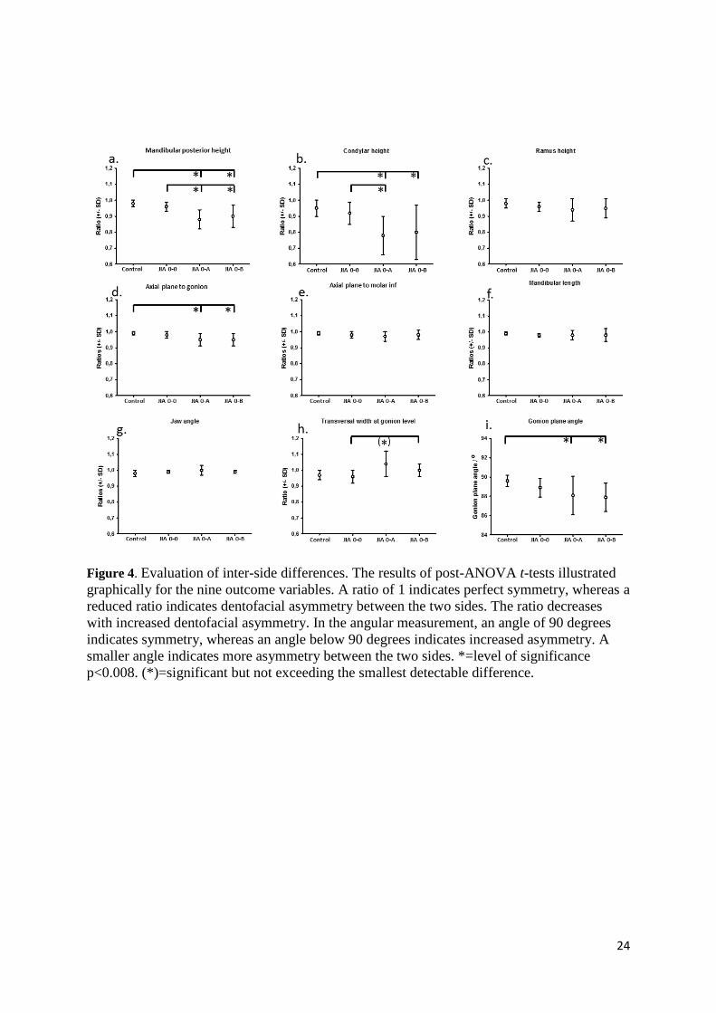
Dentofacial asymmetry
Inter-group differences in dentofacial asymmetry are listed in Table 3. The primary ANOVA
tests revealed significant inter-group differences in the following outcome variables (p<0.006):
1) mandibular posterior height, 2) condylar height, 3) axial plane to gonion, 4) mandibular

9
transversal width at the gonion level, and 5) gonion plane angle (Table 3). The results of the
secondary post-ANOVA t-tests (level of significance p<0.008) are shown in Fig. 4. The
significant results are presented below.
Condylar height: A significantly larger degree of asymmetry was observed in the JIA 0-A and
0-B groups than in the control group. Additionally, the condylar height asymmetry was
significantly more pronounced in JIA 0-A patients than in the JIA 0-0 group. No significant
differences were observed between the control and JIA 0-0 groups or between the JIA 0-A and
JIA 0-B groups (Table 3 and Fig. 4a).
Mandibular posterior height: Significantly larger asymmetries were found in the JIA 0-A and
JIA 0-B groups than in the control and JIA 0-0 groups. No significant differences were
observed between the control group and the JIA 0-0 group or between the JIA 0-A and JIA 0-
B groups (Table 3 and Fig. 4b).
Axial plane to gonion: Asymmetry was significantly larger in the JIA 0-A and 0-B groups than
in the control group. Significant differences were not found between the three JIA groups or
between the JIA 0-0 and control groups (Table 3 and Fig. 4d).
Transversal width at gonion level: A significant difference was observed between the JIA 0-0
group and JIA 0-B group. However, although significant, this inter-group difference in ratio
did not exceed the smallest detectable difference of this specific outcome variable (Table 3 and
Fig. 4h).
Gonion plane angle: A smaller angle was observed in the JIA 0-A and 0-B groups than in the
control group, indicating a larger difference in the vertical position of the gonion point right
and left. JIA 0-0 was not significantly different from the two other groups, JIA 0-A and JIA 0-
B (Table 3 and Fig. 4i). The outcome variables with no significant differences were ramus
height, axial plane to inferior molar, mandibular length and jaw angle (Table 3 and Fig. 4c, e,
f, g).
The intra-rater reliability of the nine outcome variables showed acceptable correlation levels
between the duplicate assessments (r>0.90) with the exception of the outcome measure
reflecting the transversal width at the gonion level (r=0.69). The ranges of the smallest
detectable differences for the inter-side ratios were calculated for each of the nine outcome
variables using Bland–Altman plots (range=0.02-0.09). The smallest detectable differences for
the two angular outcome variables were 0.02 degrees for the “jaw angle” and 1.32 degrees for
the “gonion plane angle” outcome measure.
10
Discussion
Dentofacial asymmetry is a common feature in patients with JIA and unilateral TMJ arthritis
involvement. 24,26,29 Minor dentofacial and mandibular asymmetries are also well known
findings in a normal population; however, a precisely defined normal range has not been
established. 35 In the present study, the JIA patients with unilateral TMJ abnormalities exhibited
significantly more severe dentofacial inter-side asymmetries than the JIA patients without TMJ
abnormalities and control subjects. This is in accordance with other studies reporting
pronounced asymmetries and smaller mandibular dimensions in JIA patients with condylar
abnormalities. 14,18,24 Koos et al. investigated mandibular asymmetries in 23 JIA patients using
CBCT and compared them with 23 age- and gender-matched controls. 29 Significantly more
pronounced asymmetries were found in the patients with JIA than in the controls. However,
Koos et al. made no attempt to correlate the intra-individual asymmetries with TMJ
abnormalities, as was done in this study. Huntjens et al. examined the degree of condylar head
asymmetry in children affected with JIA based on CBCT. 26 Their results showed that
asymmetry of the condyles was frequently present; however, condylar asymmetry did not
correlate with facial 3D appearance.
To our knowledge, this is the first CBCT-based human study to investigate the association
between types of radiological TMJ abnormalities and dentofacial development in JIA patients
with TMJ involvement. In the present study, mandibular dysmorphic development was affected
to the same extent in patients with either unilateral condylar deformations (score A) or
unilateral erosions (score B). The cross-sectional nature of our study restricts our ability to
draw conclusions regarding the causal relationship between TMJ abnormalities and
dysmorphic dentofacial development; however, our findings call for a larger debate. It is not
our impression that the two radiological features (deformation and erosion) necessarily
represent a disease progression going from A towards B when attention is paid to general
dentofacial development. Instead, A and B could be regarded as two expressions of the same process
that vary over time based on the severity of the TMJ inflammation. This hypothesis is supported by
a 5-year cohort follow-up study providing evidence for a condylar regenerative capacity in
patients with low disease activity. 9 We allow ourselves to hypothesize that TMJs with scores
A and B are both expressions of a reaction to inflammation in the TMJs where category A is
likely to represent a stable phase and category B represent a reaction to a more active high
inflammatory phase. Future studies based on contrast-enhanced MRI techniques are needed to
confirm this hypothesis. Notably, in light of mandibular development, it remains an open

11
question whether the presence of erosion (score B) can be considered worse than the presence
of deformation (score A) or vice versa. The overlap of deformation (Score A) and erosions
(Score B) in 30% (9/30 joints) of the joints assigned to an abnormal radiological appearance
leads to the question of whether it is useful to distinguish between TMJ deformations and
erosions in future scoring system when the results provide little information on general
dentofacial development. Future longitudinal studies are warranted to assess the association
between the intra-articular conditions and dysmorphic dentofacial development. Additionally,
there is a need to develop 3D CBCT-based cephalometric standards for the assessment of JIA-
specific dentofacial morphological changes.
In a recent publication from Peck et al., condylar deformation (flattening) was considered an
indeterminate finding and was not included in the diagnostic algorithm for TMJ arthritis caused
by “systemic arthritides.” 36 In support of this viewpoint, 13% (5/38) of the TMJs in the non-
JIA control group in our study were assigned an “abnormal” condylar score. However, our
findings do not support this viewpoint in terms of patients with JIA, and we suggest an
alternative approach to JIA radiological evaluation: the presence of an abnormal radiological
joint appearance should always call for further assessment of dentofacial morphology despite
the type of condylar deformity revealed by radiological evaluation (deformation or erosion).
We found the site of the asymmetry to be primarily related to a short condyle on the affected
side. Significant differences were found for mandibular posterior height in JIA 0-A and JIA 0-
B when compared to control and JIA 0-0 groups; however, there were no significant differences
for ramus height, indicating that a shorter condyle was responsible for the decreased posterior
face height in our patient group. No further morphological differences were found. The reason
for a lack of other deformities, such as inter-side difference in Ramus height and occlusal
canting (inter-side difference in axial plane to Minf), could be ascribed to the orthopedic
treatment that 41 of the patients received after a diagnosis of TMJ arthritis had been confirmed.
To our knowledge, this is the largest CBCT-based case-control study of its kind on JIA.
However, there are limitations to the present study. First, only a fair agreement (κ=0.67) was
obtained in the radiological assessment of intra-rater reliability between normal and abnormal
TMJ appearance. This may be explained by the fact that the subchondral cortical bone is under
development in children and adolescents, which may hamper radiological assessments in the
age group included in this study. 37 The kappa value for differentiating the abnormal
morphology into group A or B was slightly lower (κ=0.63), which may be explained by the

12
fact that some joints exhibited features from both groups that also may have interfered with
interpretation. Second, a correlation between the duration of TMJ arthritis and the degree of
facial asymmetry was not considered because initial MRI verification of TMJ arthritis was not
routinely conducted in all patients. Third, the included JIA patients represented a group of well
treated patients with orthopedic splints; this may have moderated the intra-patient asymmetries.
To avoid this bias, the study should have been conducted with untreated JIA patients; however,
this would have been unethical in our opinion. Forty-one of the 47 included JIA patients were
undergoing or had previously received functional appliance treatment with an orthopedic
distraction splint. In five of the remaining six patients, an orthopedic distraction splint was
fabricated immediately after the CBCT scan date and functional treatment was commenced,
whereas the final patient was followed to observe further development. There were no
differences in treatment protocols offered to the three JIA groups. We previously showed that
treatment with an orthopedic distraction splint facilitates comparable inter-side growth rates in
patients with unilateral TMJ arthritis and prevents mandibular asymmetry from being
exacerbated. 33 All patients received routine orofacial examination, and distraction splint
treatment was initiated as soon as facial asymmetry was noted; thus, we hypothesize that only
a minor aggravation of asymmetries occurred after the initiation of the distraction splint
treatment.
Conclusions
JIA patients with unilateral condylar abnormalities (deformation or erosion) exhibited
significantly more severe dentofacial asymmetries than JIA patients without condylar
abnormalities and control subjects.
A similar degree of dentofacial asymmetry was observed in the two groups of patients
with unilateral condylar abnormalities (deformation or erosion).
Mandibular asymmetry was exclusively related to a short condyle on the affected side.
In patients with JIA, both deformation and erosion were present in 30% of the TMJs
examined.
Radiological signs of condylar abnormalities (deformation and/or erosion) were
observed in 13% of the TMJs in the non-JIA control group.
Generally, in JIA, the presence of abnormal radiological TMJ appearance should
always call for the further assessment of dentofacial morphology.

13
Acknowledgement
We thank Professor Tore A. Larheim for his help and guidance during the radiological
assessments.
Funding
No funding was received for this project.
References
1. Larheim TA, Doria AS, Kirkhus E, Parra DA, Kellenberger CJ, Arvidsson LZ. TMJ imaging in JIA patients; An overview. Semin Orthod.2015; 21:102-10. 2. Arvidsson LZ, Fjeld MG, Smith HJ, Flato B, Ogaard B, Larheim TA. Craniofacial growth disturbance is related to temporomandibular joint abnormality in patients with juvenile idiopathic arthritis, but normal facial profile was also found at the 27-year follow-up. Scand J Rheumatol. 2010;39:373-9. 3. Arvidsson LZ, Flato B, Larheim TA. Radiographic TMJ abnormalities in patients with juvenile idiopathic arthritis followed for 27 years. Oral Surg Oral Med Oral Pathol Oral Radiol Endod. 2009;108:114-23. 4. Bakke M, Zak M, Jensen BL, Pedersen FK, Kreiborg S. Orofacial pain, jaw function, and temporomandibular disorders in women with a history of juvenile chronic arthritis or persistent juvenile chronic arthritis. Oral Surg Oral Med Oral Pathol Oral Radiol Endod. 2001;92:406-14. 5. Engstrom AL, Wanman A, Johansson A, Keshishian P, Forsberg M. Juvenile arthritis and development of symptoms of temporomandibular disorders: a 15-year prospective cohort study. J Orofac Pain. 2007;21:120-6. 6. Fjeld MG, Arvidsson LZ, Stabrun AE, Birkeland K, Larheim TA, Ogaard B. Average craniofacial development from 6 to 35 years of age in a mixed group of patients with juvenile idiopathic arthritis. Acta Odontol Scand. 2009;67:153-60. 7. Kuseler A, Pedersen TK, Gelineck J, Herlin T. A 2 year followup study of enhanced magnetic resonance imaging and clinical examination of the temporomandibular joint in children with juvenile idiopathic arthritis. J Rheumatol. 2005;32:162-9. 8. Stoustrup P, Kristensen KD, Verna C, Kuseler A, Herlin T, Pedersen TK. Orofacial symptoms related to temporomandibular joint arthritis in juvenile idiopathic arthritis: smallest detectable difference in self-reported pain intensity. J Rheumatol. 2012;39:2352-8. 9. Twilt M, Schulten AJ, Verschure F, Wisse L, Prahl-Andersen B, Suijlekom-Smit LW. Long-term followup of temporomandibular joint involvement in juvenile idiopathic arthritis. Arthritis Rheum. 2008;59:546-52. 10. Ahmed N, Petersson A, Catrina AI, Mustafa H, Alstergren P. Tumor necrosis factor mediates temporomandibular joint bone tissue resorption in rheumatoid arthritis. Acta Odontol Scand. 2015;73:232-40. 11. Billiau AD, Hu Y, Verdonck A, Carels C, Wouters C. Temporomandibular Joint Arthritis in Juvenile Idiopathic Arthritis: Prevalence, Clinical and Radiological Signs, and Relation to Dentofacial Morphology. J Rheumatol. 2007;34:1925-33. 12. Hu YS, Schneiderman ED, Harper RP. The temporomandibular joint in juvenile rheumatoid arthritis: Part II. Relationship between computed tomographic and clinical findings. Pediatr Dent. 1996;18:312-9. 13. Kjellberg H. Juvenile chronic arthritis. Dentofacial morphology, growth, mandibular function and orthodontic treatment. Swed Dent J Suppl. 1995;109:1-56.

14
14. Kjellberg H. Craniofacial growth in juvenile chronic arthritis. Acta Odontol Scand. 1998;56:360-5. 15. Kirkhus E, Arvidsson LZ, Smith HJ, Flato B, Hetlevik SO, Larheim TA. Disk abnormality coexists with any degree of synovial and osseous abnormality in the temporomandibular joints of children with juvenile idiopathic arthritis. Pediatr Radiol 2016;46:331-41. 16. Kristensen KD, Hauge EM, Dalstra M, Stoustrup P, Kuseler A, Pedersen TK, et al. Association between condylar morphology and changes in bony microstructure and sub-synovial inflammation in experimental temporomandibular joint arthritis. J Oral Pathol Med. 2011;40:111-20. 17. Stoustrup P, Kristensen KD, Kuseler A, Gelineck J, Cattaneo PM, Pedersen TK, et al. Condylar lesions in relation to mandibular growth in untreated and intra-articular corticosteroid-treated experimental temporomandibular joint arthritis. Clin Exp Rheumatol. 2010;28:576-83. 18. Twilt M, Schulten AJ, Nicolaas P, Dulger A, Suijlekom-Smit LW. Facioskeletal changes in children with juvenile idiopathic arthritis. Ann Rheum Dis. 2006;65:823-5. 19. Peltomäki T, Kreiborg S, Pedersen, TK; Ogaard, B. Craniofacial growth and dentoalveolar development in juvenile idiopathic arthritis patients. Semin Orthod. 2015;21:84-93. 20. Koos B, Tzaribachev N, Bott S, Ciesielski R, Godt A. Classification of temporomandibular joint erosion, arthritis, and inflammation in patients with juvenile idiopathic arthritis. J Orofac Orthop. 2013;74:506-19. 21. Vaid YN, Dunnavant FD, Royal SA, Beukelman T, Stoll ML, Cron RQ. Imaging of the temporomandibular joint in juvenile idiopathic arthritis. Arthritis Care Res (Hoboken). 2014;66:47-54. 22. Kristensen KD, Stoustrup P, Kuseler A, Pedersen TK, Nyengaard JR, Hauge EM, et al. Quantitative histological changes of repeated antigen-induced arthritis in the temporomandibular joints of rabbits treated with intra-articular corticosteroid. J Oral Pathol Med. 2008;37:437-44. 23. Ronchezel MV, Hilario MO, Goldenberg J, Lederman HM, Faltin K, Jr., de Azevedo MF, et al. Temporomandibular joint and mandibular growth alterations in patients with juvenile rheumatoid arthritis. J Rheumatol. 1995;22:1956-61. 24. Stabrun AE, Larheim TA, Hoyeraal HM, Rosler M. Reduced mandibular dimensions and asymmetry in juvenile rheumatoid arthritis. Pathogenetic factors. Arthritis Rheum. 1988;31:602-11. 25. Arvidsson LZ, Smith HJ, Flato B, Larheim TA. Temporomandibular joint findings in adults with long-standing juvenile idiopathic arthritis: CT and MR imaging assessment. Radiology. 2010;256:191-200. 26. Huntjens E, Kiss G, Wouters C, Carels C. Condylar asymmetry in children with juvenile idiopathic arthritis assessed by cone-beam computed tomography. Eur J Orthod. 2008;30:545-51. 27. Farronato G, Garagiola U, Carletti V, Cressoni P, Mercatali L, Farronato D. Change in condylar and mandibular morphology in juvenile idiopathic arthritis: Cone Beam volumetric imaging. Minerva Stomatol. 2010;59:519-34. 28. Lima Ferraz Junior AM, Devito KL, Guimaraes JP. Temporomandibular disorder in patients with juvenile idiopathic arthritis: clinical evaluation and correlation with the findings of cone beam computed tomography. Oral Surg Oral Med Oral Pathol Oral Radiol Endod.. 2012;114:E51-57. 29. Koos B, Gassling V, Bott S, Tzaribachev N, Godt A. Pathological changes in the TMJ and the length of the ramus in patients with confirmed juvenile idiopathic arthritis. J Craniomaxillofac Surg 2014;42:1802-7. 30. Larheim TA, Abrahamsson AK, Kristensen M, Arvidsson LZ. Temporomandibular joint diagnostics using cone beam computed tomography. Dentomaxillofac Radiol. 2014:20140235. 31. Petty RE, Southwood TR, Baum J, Bhettay E, Glass DN, Manners P, et al. Revision of the proposed classification criteria for juvenile idiopathic arthritis: Durban, 1997. J Rheumatol. 1998;25:1991-4. 32. Bland JM, Altman DG. Statistical methods for assessing agreement between two methods of clinical measurement. Lancet. 1986;1:307-10.

15
33. Stoustrup P, Kuseler A, Kristensen KD, Herlin T, Pedersen TK. Orthopaedic splint treatment can reduce mandibular asymmetry caused by unilateral temporomandibular involvement in juvenile idiopathic arthritis. Eur J Orthod. 2013;35:191-8
34. Fleiss J LLB, A; Paik M,C. Statistical methods for rates and proportions. J.Wiley, Hoboken, N.J); 2003. 35. Liukkonen M, Sillanmaki L, Peltomaki T. Mandibular asymmetry in healthy children. Acta Odontol Scand. 2005;63:168-72. 36. Peck CC, Goulet JP, Lobbezoo F, Schiffman EL, Alstergren P, Anderson GC, et al. Expanding the taxonomy of the diagnostic criteria for temporomandibular disorders. J Oral Rehabil 2014;41:2-23. 37. Lei J, Liu MQ, Yap AU, Fu KY. Condylar subchondral formation of cortical bone in adolescents and young adults. Br J Oral Maxillofac Surg. 2013;51:63-8.
16
Table 1. Definition and description of the landmarks, planes and variables used in the study.
Colors refer to the planes illustrated in Fig. 2.
Landmarks Definition Abbreviation
Condyle point The midpoint on the superior surface of the condyle Cor/Col
Gnathion The lowest point on the lower border of the chin Gn
Gonion (right/left) Constructed by the bisection by the angle formed by the tangents
to the lower and posterior borders of the mandible
Gor/Gol
Incisura point
(right/left)
Lowest point in the concavity between processus coronoideus
and processus condylaris in relation to the Axial Plane
Incr/Incl
Latero-orbital point
(right/left)
At the zygomaticofrontal suture at the lateral aspect of the orbit
wall
LOr/LOl
Lower first molar point
(right/left)
The mesio-buccal cusp of the lower first molar Minfr/Minfl
Nasion Midpoint between maxillary-nasal-frontal right and left junction N
Sella turcica The centre of the hypophyseal fossa S
Planes Definition Color
Axial plane Through S, LOr and LOl Blue
Mid-sagittal plane Through N, S and perpendicular to the axial plane Dark blue
Coronal plane Through S and perpendicular to axial plane and mid-sagittal
plane Pink
Inc plane (right/left) Through Inc (left / right) and perpendicular to the mid-sagittal
plane and the coronal plane. One plane for each side was created
Purple
Gonion plane Plane through Gonion (right/left) and Gnathion
Variables Definition
Mandibular posterior
height (right/left) From Go to Co (mm)
Condylar height
(right/left) From Co to Inc plane
Ramus height
(right/left) Go to Inc plane (mm)

17
Axial plane-Go
(right/left) Axial plane to gonion (mm)
Axial plane-Minf
(right/left)
Axial plane to lower molar mesio-buccal cusp tip (mm)
Mandibular length
right/left)
Gn to Go (mm)
Jaw angle (right/left ) Angle between the line connecting Co and Go and the line
connecting Go and Gn (degrees)
Transversal width at
gonion level (right/left)
Distance from mid-sagittal plane to gonion (mm)
Gonion plane angle Angle between gonial plane and the mid-sagittal plane (degrees)
measured to the affected side / group 0-0 to the smallest side
18
Table 2. Characteristics of patients with JIA and control subjects included in the study.
CBCT: cone beam computed tomography; NSAIDs: non-steroidal anti-inflammatory drugs;
DMARDs: disease-modifying anti-rheumatic drugs.
Cohort characteristics Control group JIA 0-0 JIA 0-A JIA 0-B
Number
Females, number
Males, number
Mean age at CBCT, years (sd years)
Present or earlier functional treatment, number
JIA subcategories
Oligoarticular extended
Oligoarticular persistent
Polyarticular
Systemic
Psoriatic
Enthesitis related arthritis
Undifferentiated
Medication
No medication
NSAIDs
DMARDs
Biologics
Combination of 2 drugs
Combination of 3 drugs
Disease duration, years**
Mean years (sd)
<1
0-3
3-5
19
12
7
13.2 (1.6)
-
17
11
6
13.2(2.6)
13
3
7
3
1
1
-
2
5
8
4
5
3
-
7(3.4)
1
1
5
20
14
6
12.8(2.7)
18
5
4
10
-
-
-
1
8
7
6
3
4
-
5(3.7)
-
6
5
10
7
3
11.4(2.5)
10
2
1
4
-
-
1
2
1
5
8
6
4
3
6(4.4)
1
2
3

19
>5
TMJ involvement, years***
<1
0-2
3-5
>5
10
4
5
3
5
8
3
7
2
8
4
4
1
2
3

20
Table 3. ANOVA tests analyzing ratios and angels of inter-side dentofacial asymmetries in
patients and control subjects. Numbers indicate inter-side ratios, standard deviations in
brackets. /*=linear (ratio) measurement, /◦=degrees, **=level of significance p<0.006. Post-
ANOVA tests were only performed for outcome variables in which a statistically significant
difference was observed in the primary ANOVA test. N.S.=non-significant.
Outcome
measures
Control group JIA
0-0
JIA
0-A
JIA
0-B
ANOVA
p-value**
Condylar height
/*
0.95 (0.05)
0.92 (0.07)
0.78 (0.12)
0.80 (0.17)
<0.001
Mandibular
posterior
height/*
0.98 (0.02) 0.96 (0.03) 0.88 (0.06) 0.90 (0.07) <0.001
Ramus height/* 0.98 (0.03) 0.96 (0.03) 0.94 (0.07) 0.95 (0.06) N.S.
Axial plane to
gonion /*
0.99 (0.01) 0.98 (0.02) 0.95 (0.04) 0.95 ( 0.04) <0.001
Axial plane to
molar inf. /*
0.99 (0.01) 0.98 (0.02) 0.97 (0.03)
0.98 (0.03) N.S.
Mandibular
length /*
0.99 (0.01) 0.98 (0.01) 0.98 (0.03) 0.98 (0.04) N.S
Jaw angle /◦ 0.98 (0.02) 0.99 (0.01) 1.00 (0.03) 0.99 (0.01) N.S.
Transversal
width at gonion
level/*
0.97 (0.03) 0.96 (0.04) 1.04 (0.08) 1.0 (0.04) <0.001
Gonion plane
angle /◦
89.6 (0.60) 88.9 (0.98) 88.1 (1.98) 87.9 (1.48) 0.002
21
Figure 1. Scoring of condylar morphology based on radiological TMJ appearance. a) Condylar
score 0: Normal, normal shape with smooth and intact outline/surface. b) Condylar score A:
Deformed, marked flattening or other changes in shape with smooth and intact outline/surface.
c) Condylar score B: Erosive, disruption of outline or uneven surface due to cyst or erosion.

22
Figure 2. Illustration of the landmarks, planes and variables from the 3D analysis. a & b:
Frontal and sagittal view with landmarks. c & d: Frontal and sagittal view with planes and
variables. Color code of planes: axial plane, blue; midsagittal plane, dark blue; coronal plane,
pink; incisura plane (left / right), purple. Further descriptions of landmarks and variables are
presented in Table 1.
23
Figure 3. Flowcharts of included JIA patients.
24
Figure 4. Evaluation of inter-side differences. The results of post-ANOVA t-tests illustrated
graphically for the nine outcome variables. A ratio of 1 indicates perfect symmetry, whereas a
reduced ratio indicates dentofacial asymmetry between the two sides. The ratio decreases
with increased dentofacial asymmetry. In the angular measurement, an angle of 90 degrees
indicates symmetry, whereas an angle below 90 degrees indicates increased asymmetry. A
smaller angle indicates more asymmetry between the two sides. *=level of significance
p<0.008. (*)=significant but not exceeding the smallest detectable difference.


![CaseReport …[8] E.M.Ongkosuwito,J.vanVooren,J.W.vanNecketal., “Changes of mandibular ramal height, during growth in unilateral hemifacial microsomia patients and unaected](https://img.pdfslide.us/doc/110x75/60c4829f23e96b545e31e549/casereport-8-emongkosuwitojvanvoorenjwvannecketal-aoechanges-of-mandibular.jpg)

![Orthodontic Treatment of Bilateral Impacted Mandibular ...downloads.hindawi.com/journals/crid/2019/7638959.pdf · than unilateral impaction [7–10]. Even though lower canine impaction](https://img.pdfslide.us/doc/110x75/5f0a053a7e708231d429a0ad/orthodontic-treatment-of-bilateral-impacted-mandibular-than-unilateral-impaction.jpg)