Embed Size (px)
Citation preview
Biochemistry 1990, 29. 3509-35 14 3509
1990) studies is not an artifact of the high salt concentration used. The various physicochemical differences seen between oxidized and reduced cytochrome c in solution at moderate salt appear to reflect differences in stability, and the conjugate parameter, structural dynamics, which occur without sub- stantial changes in the three-dimensional structure of the average native state. The selective salt binding observed here and in prior work suggests that electrostatic rather than structural difference should be considered.
Registry No. Cytochrome c, 9007-43-6; phosphate, 14265-44-2.
REFERENCES Anderson, T., Thulin, E., & Forsen, S. (1979) Biochemistry
Arean, C. O., Moore, G. R., Williams, G., & Williams, R.
Barlow, G. H., & Margoliash, E. (1966) J . Biol. Chem. 241,
Brautigan, D. L., Ferguson-Miller, S., Tarr, G. E., & Mar-
Clayden, N. J., & Williams, R. J. P. (1982) J . Magn. Reson.
Eden, D., Matthew, J . B., Rosa, J. J . , & Richards, F. M.
Englander, S. W., & Crowe, D. (1965) Anal. Biochem. 12,
Englander, S. W., & Kallenbach, N. R. (1984) Q. Rev. Bio-
Feng, Y. , Roder, H., Englander, S. W., Wand, A. J . , &
Feng, Y., Roder, H., & Englander, S. W. (1990) Biochemistry
Kharakoz, D. P., & Mkhitaryan, A. G. (1986) Mol. Biol. 20,
18, 2487-2493.
J. P. (1988) Eur. J . Biochem. 173, 607-615.
1437-1 440.
goliash, E. (1978) J . Biol. Chem. 253, 140-148.
49, 383-396.
(1982) Proc. Natl. Acad. Sci. U.S.A. 79, 815-819.
579-584.
phys. 16, 521-655.
DiStefano, D. L. (1989) Biochemistry 28, 195-203.
(preceding paper in this issue).
3 96-406.
Lattman, E. E. (1989) Proteins: Struct., Funct., Genet. 5,
Liu, G., Grygon, A., & Spiro, T. G. (1989) Biochemistry 28,
Margalit, R., & Schejter, A. (1973) Eur. J . Biochem. 32,
Margoliash, E., & Schejter, A. (1966) Adv. Protein Chem.
Osheroff, N., Brautigan, D. L., & Margoliash, E. (1980) Proc.
Patel, D. J. , & Canuel, L. L. (1976) Proc. Natl. Acad. Sci.
Rush, J. D., Koppenol, W. H., Garber, E. A. E., & Margo-
Stellwagen, E., & Shulman, R. G. (1973) J . Mol. Biol. 80,
Taborsky, G., & McCollum, K. (1979) J . Biol. Chem. 254,
Takano, T., & Dickerson, R. E. (1981a) J . Mol. Biol. 153,
Takano, T., & Dickerson, R. E. (1981 b) J . Mol. Biol. 153,
Trewhella, J., Carlson, V., Curtis, E. H., & Heidorn, D. B.
Ulmer, D. D., & Kagi, J. H. (1968) Biochemistry 7,
Wand, A. J., Roder, H., & Englander, S. W. (1986) Bio-
Wand, A. J., DiStefano, D. L., Feng, Y., Roder, H., & En-
Williams, G., Clayden, N. J., Moore, G. R., & Williams, R.
149-1 55.
5046-5050.
49 2-49 9.
21, 113.
Natl. Acad. Sci. U.S.A. 77, 4439-4443.
U.S.A. 73, 1998.
liash, E. (1988) J . Biol. Chem. 263, 7514-7520.
5 59-57 3 *
7069-7075.
79-94.
95-1 15.
(1988) Biochemistry 27, 1121-1125.
27 10-27 17.
chemistry 25, 1107-1 114.
glander, s. W. (1989) Biochemistry 28, 186-194.
J. P. (1985) J . Mol. Biol. 183, 447-460.
N M R Study of the Phosphoryl Binding Loop in Purine Nucleotide Proteins: Evidence for Strong Hydrogen Bonding in Human N-ras p21t
Alfred G. Redfield* and Mary Z. Papastavros Department of Biochemistry, Brandeis University, Waltham, Massachusetts 02254
Received October 10, 1989; Revised Manuscript Received December I, I989
ABSTRACT: The structure of the phosphoryl binding region of human N-ras p21 was probed by using heteronuclear proton-observed N M R methods. Normal protein and a Gly-12 - Asp- 12 mutant protein were prepared with two amino acids labeled with 15N a t their amide positions: valine and glycine, aspartic acid and glycine, and lysine and glycine. W e completed the identification of amide 15NH resonances from Gly-12 and Asp-I2 to the end of the phosphoryl binding domain consensus sequence (Lys-16) in protein complexed with G D P and have made tentative amide identifications from Val-9 to Ser-17. The methods used, together with initial identifications of the Gly- 12 and - 13 amide resonances, were described previously [Campbell-Burk, S. (1989) Biochemistry 28,9478-94841. The amide resonances of both Gly-13 and Lys-16 are shifted downfield below 10.4 ppm in both the normal and mutant proteins. These downfield shifts are presumed to be due to strong hydrogen bonds with the P-phosphate oxygens of GDP.
T e r e is a large class of purine nucleotide binding proteins having a consensus sequence Gly-X-X-X-X-Gly-Lys at the phosphoryl binding site (Walker et al., 1982; Higgins et al., 1986). We have been studying members of an important
'Partially supported by USPHS Grant GM20168 and by the Cetus and DuPont Corporations.
0006-2960/90/0429-3509$02.50/0
subclass of these proteins, the 21-kilodalton products of the human N-ras gene, human N-ras p21 (henceforth p21) and an oncogenic mutant thereof, using NMR (Campbell-Burk et al., 1989; Campbell-Burk, 1989). For N-ras p21 protein, the version of the consensus sequence starts at Gly-10 and is Gly-Ala-Gly-Gly-Val-Gly-Lys, while the mutant has aspartic acid substituted for glycine at position 12 (Barbacid, 1987).
0 1990 American Chemical Society

3510
The recent discovery of a p21 GTPase activator and probable downstream effector protein makes the study of these proteins particularly interesting (Trahey & McCormick, 1987; McCormick, 1989).
One of the strengths of N M R is its ability to provide chemical as well as structural (distance) information, in the present case concerning unusual hydrogen bonding interactions. Such information may be difficult to obtain by X-ray dif- fraction or by two-dimensional (2D)' N M R distance mea- surements because of the inaccuracy of distances often ob- tained by these methods. In the present paper, we discuss our evidence that the Gly- 13 and Lys- 16 amide protons interact unusually strongly with phosphate oxygens of the bound GDP. The. interaction of these amide groups in a loop with GDP phosphate oxygens was also indicated by X-ray diffraction results in p21 and related proteins. However, the X-ray evidence for especially strong amide-phosphate interaction in members of this class of proteins either seems ambiguous (Dreusike & Schulz, 1988; Jurnak, 1985) or appears to differ in some ways from the simplest interpretation of our data (Tong et al., 1989a).
Previously we found that the amide proton resonance of Gly-13 was shifted unusually far downfield in the normal protein, and was shifted even further downfield in the Asp- 12 mutant (Campbell-Burk et al., 1989; Campbell-Burk, 1989). It was suggested that these unusual shifts were due to a strong interaction, such as a hydrogen bond, of the Gly-13 amide group with a phosphate oxygen of bound GDP. Here we extend these observations to the end of the consensus sequence, Lys-16. Unexpectedly, we find that the amide resonance of Lys-16 is also shifted far downfield in both normal and mutant p21. This observation suggests that the amide proton of Lys-16, as well as that of Gly-13, is involved in a strong in- teraction with a GDP phosphate oxygen, to a greater extent than other amide protons in this loop.
We find by nuclear Overhauser effect (NOE) that the ad- jacent amide protons of the consensus sequence are close to each other, and we also conclude that there are not major changes in the structure of most of the protein as a result of mutation of Gly-12. Both of these latter conclusions are consistent with X-ray results (deVos et a]., 1988; Tong et al., 1989a,b; Pai et al., 1989).
The present study continues our use of isotope labels with proton N M R to target an interesting region of the molecule. Other recent N M R studies of p21 have used purely proton NMR, with deuteration or mutations, to learn about regions of the molecule somewhat remote from the phosphoryl binding loop (Schlichting et al., 1988, 1990; Ha et a]., 1989; Yamasaki et al., 1989).
MATERIALS AND METHODS Doubly labeled p21 was produced from an expression vector
transformed into a polyauxotrophic transaminase-deficient Escherichia coli strain as described (Campbell-Burk et al., 1989). Both mutant and normal p21 were purified from cells grown in the presence of I5N-labeled glycine and valine. Mutant p21 was likewise purified from cells grown in [a- '5N]lysine and [I5N]glycine, normal p21 was labeled with [ 15N]lysine, and mutant protein was labeled with [15N]aspartic acid and [15N]glycine. Generally, we used 1 g of each labeled amino acid for 8 L of fermentation medium, to obtain 10 mg of p21. Even though the cell line used is not a lysine auxo-
' Abbreviations: HMQC, heteronuclear multiple quantum coherence; p21, human N-ras p21 gene product; 2D, two dimensional; NOE, nuclear Overhauser effect: GAP, GTPase activating protein; N-ras, human neuroblastoma ras; H-ras. Harvey ras.
Biochemistry, Vol. 29, No. 14, 1990 Redfield and Papastavros
0 /
D,' 10 or 115 / By 5
I I 7 LYSINE ,
128 120 112 104 NITROGEN SHIFT PPM
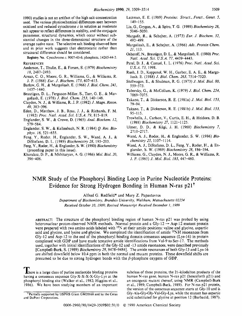
FIGURE 1: HMQC spectrum of mutant (Asp-12) p21 protein labeled with ISN at the peptide amides of glycine and lysine. The dashed line separates the glycine peaks from the lysine peaks, based on our previous spectra obtained from protein labeled at glycine alone. Resonances with extreme proton shifts are weak because the selective pulse used was optimized for the center of the spectrum, but their existence has been confirmed repeatedly in other runs optimized for them. The peaks on this spectrum which have been identified at this writing are indicated by their position in the amino acid sequence. The sample contains approximately 10 mg of protein in 0.3 mL, and the spectrum was acquired overnight at 18 OC. Usable spectra are obtainable in less than 1 h, however. The buffer consists of 20 mM Tris, 100 m M NaC1, 5 mM MgC12, and 5 mM dithiothreitol, pH 7.5, in 10% D 2 0 .
trophe, nearly 100% incorporation of lysine was achieved, judging from the intensities of the lysine N M R lines.
Standard two dimensional (2D) proton-I5N heteronuclear multiple quantum correlation (HMQC) spectra were obtained for all samples, and edited one-dimensional NOE spectra were used to establish nearest-neighbor connections. These methods have been described previously (Bax et al., 1983; McIntosh et al., 1987; Griffey & Redfield, 1987). Spectra were obtained on a 500-MHz instrument built a t Brandeis University.
RESULTS Figure 1 shows a two-dimensional 500-MHz I5N-proton
HMQC N M R spectrum for a sample of mutant (Asp-12) N-ras p21 that has been labeled with the rare isotope I5N a t the amide positions of the glycine and lysine residues in the molecule. The HMQC method used here exploits the large 90-Hz spin-spin coupling between a I5N nucleus and the proton attached to it to help form the resonance peaks. Each peak in such a spectrum arises from a single I5NH group. Protons not covalently attached to I5N nuclei, Le., carbon protons or protons attached to I4N, do not give signals. The chemical shifts of the proton and the I5N for each peak can be read off the spectrum from the projections of the peak position onto the abscissa and ordinate. Figure 1 shows only peaks due to the glycine and lysine amide groups in p21, because these were the only sites that were I5N labeled, and there is one peak for each such amide. Spectra of this type have been obtained for several proteins which have been fully I5N labeled, and virtually all the amide resonances in such spectra have been identified for proteins as large as 19 kilo- daltons (Torchia et al., 1989; L. McIntosh et al., unpublished results).
Such a spectrum is often immediately useful as a fingerprint. For example, we have obtained a spectrum like that of Figure 1 for both normal and mutant (Asp-12) p21 with I5N labels at the amide groups of glycine, lysine, and valine. We can
N M R Evidence for Hydrogen Bonding in N-ras p21
I ~ R R . n I
1 I I t I
i 0.0 8.0 PROTON SHIFT PPM
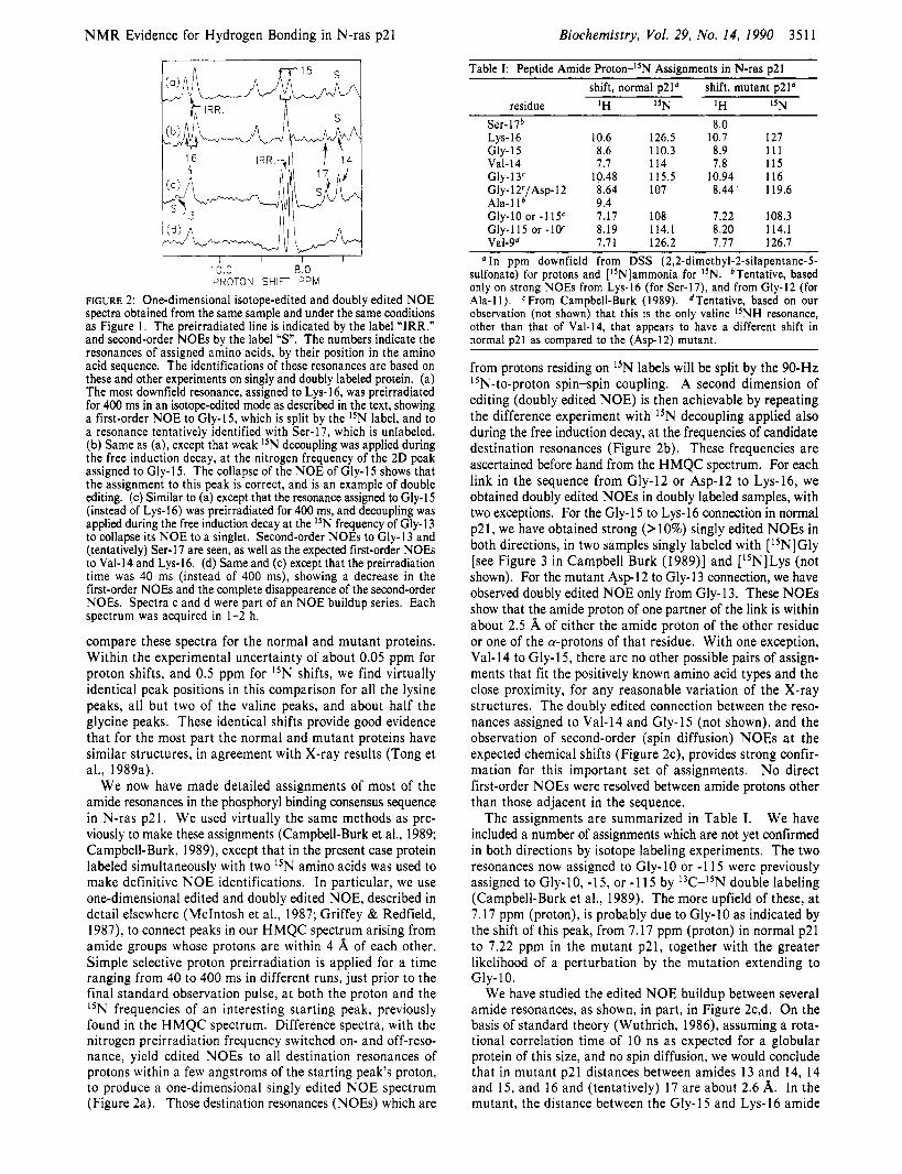
FIGURE 2: One-dimensional isotope-edited and doubly edited NOE spectra obtained from the same sample and under the same conditions as Figure 1 . The preirradiated line is indicated by the label "IRR." and second-order NOEs by the label "S". The numbers indicate the resonances of assigned amino acids, by their position in the amino acid sequence. The identifications of these resonances are based on these and other experiments on singly and doubly labeled protein. (a) The most downfield resonance, assigned to Lys- 16, was preirradiated for 400 ms in an isotope-edited mode as described in the text, showing a first-order NOE to Gly-15, which is split by the I5N label, and to a resonance tentatively identified with Ser-17, which is unlabeled. (b) Same as (a), except that weak I5N decoupling was applied during the free induction decay, at the nitrogen frequency of the 2D peak assigned to Gly- 15. The collapse of the NOE of Gly-15 shows that the assignment to this peak is correct, and is an example of double editing. (c) Similar to (a) except that the resonance assigned to Gly-15 (instead of Lys-16) was preirradiated for 400 ms, and decoupling was applied during the free induction decay at the I5N frequency of Gly-13 to collapse its NOE to a singlet. Second-order NOEs to Gly-13 and (tentatively) Ser-17 are seen, as well as the expected first-order NOEs to Val-14 and Lys-16. (d) Same and (c) except that the preirradiation time was 40 ms (instead of 400 ms), showing a decrease in the first-order NOEs and the complete disappearence of the second-order NOEs. Spectra c and d were part of an NOE buildup series. Each spectrum was acquired in 1-2 h.
compare these spectra for the normal and mutant proteins. Within the experimental uncertainty of about 0.05 ppm for proton shifts, and 0.5 ppm for I5N shifts, we find virtually identical peak positions in this comparison for all the lysine peaks, all but two of the valine peaks, and about half the glycine peaks. These identical shifts provide good evidence that for the most part the normal and mutant proteins have similar structures, in agreement with X-ray results (Tong et al., 1989a).
We now have made detailed assignments of most of the amide resonances in the phosphoryl binding consensus sequence in N-ras p21. We used virtually the same methods as pre- viously to make these assignments (Campbell-Burk et al., 1989; Campbell-Burk, 1989), except that in the present case protein labeled simultaneously with two 15N amino acids was used to make definitive NOE identifications. In particular, we use one-dimensional edited and doubly edited NOE, described in detail elsewhere (McIntosh et al., 1987; Griffey & Redfield, 1987), to connect peaks in our HMQC spectrum arising from amide groups whose protons are within 4 A of each other. Simple selective proton preirradiation is applied for a time ranging from 40 to 400 ms in different runs, just prior to the final standard observation pulse, a t both the proton and the I5N frequencies of an interesting starting peak, previously found in the HMQC spectrum. Difference spectra, with the nitrogen preirradiation frequency switched on- and off-reso- nance, yield edited NOES to all destination resonances of protons within a few angstroms of the starting peak's proton, to produce a one-dimensional singly edited N O E spectrum (Figure 2a). Those destination resonances (NOEs) which are
Biochemistry, Vol. 29, No. 14, 1990 351 1
Table I: Peptide Amide P ro t~n-~~N Assignments in N-ras p21 shift, normal p21' shift, mutant p21'
residue 1H 15N 1H "N Ser-17b 8.0 LYS- 16 10.6 126.5 10.7 127 Gly-15 8.6 110.3 8.9 111 Val-I4 7.7 1 I4 7.8 115 Gly-13' 10.48 115.5 10.94 116 Gly-12C/A~p-12 8.64 107 8.44 119.6 Ala- 1 1 9.4 Gly-10 or -115c 7.17 108 1.22 108.3 Gly-115 or -10' 8.19 114.1 8.20 114.1 Vai-gd 7.71 126.2 7.77 126.1 In ppm downfield from DSS (2,2-dimethyl-2-silapentane-5-
sulfonate) for protons and [15N]ammonia for I5N. bTentative, based only on strong NOEs from Lys-16 (for Ser-17), and from Gly-12 (for Ala-I 1). CFrom Campbell-Burk (1989). dTentative, based on our observation (not shown) that this is the only valine I5NH resonance, other than that of Val-14, that appears to have a different shift in normal ~ 2 1 as comDared to the ( A s D - ~ ~ ) mutant.
from protons residing on 15N labels will be split by the 90-Hz 15N-to-proton spin-spin coupling. A second dimension of editing (doubly edited NOE) is then achievable by repeating the difference experiment with 15N decoupling applied also during the free induction decay, at the frequencies of candidate destination resonances (Figure 2b). These frequencies are ascertained before hand from the HMQC spectrum. For each link in the sequence from Gly- 12 or Asp- 12 to Lys- 16, we obtained doubly edited NOES in doubly labeled samples, with two exceptions. For the Gly- 15 to Lys- 16 connection in normal p21, we have obtained strong (>lo%) singly edited NOEs in both directions, in two samples singly labeled with [15N]Gly [see Figure 3 in Campbell Burk (1989)l and ["NILys (not shown). For the mutant Asp-12 to Gly-13 connection, we have observed doubly edited NOE only from Gly-13. These NOEs show that the amide proton of one partner of the link is within about 2.5 A of either the amide proton of the other residue or one of the a-protons of that residue. With one exception, Val-14 to Gly-15, there are no other possible pairs of assign- ments that fit the positively known amino acid types and the close proximity, for any reasonable variation of the X-ray structures. The doubly edited connection between the reso- nances assigned to Val-14 and Gly-15 (not shown), and the observation of second-order (spin diffusion) NOEs a t the expected chemical shifts (Figure 2c), provides strong confir- mation for this important set of assignments. No direct first-order NOES were resolved between amide protons other than those adjacent in the sequence.
We have included a number of assignments which are not yet confirmed in both directions by isotope labeling experiments. The two resonances now assigned to Gly-10 or -1 15 were previously assigned to Gly-1 0, - 15, or - 1 15 by l3C-I5N double labeling (Campbell-Burk et al., 1989). The more upfield of these, a t 7.17 ppm (proton), is probably due to Gly-10 as indicated by the shift of this peak, from 7.17 ppm (proton) in normal p21 to 7.22 ppm in the mutant p21, together with the greater likelihood of a perturbation by the mutation extending to
We have studied the edited NOE buildup between several amide resonances, as shown, in part, in Figure 2c,d. On the basis of standard theory (Wuthrich, 1986), assuming a rota- tional correlation time of 10 ns as expected for a globular protein of this size, and no spin diffusion, we would conclude that in mutant p21 distances between amides 13 and 14, 14 and 15, and 16 and (tentatively) 17 are about 2.6 A. In the mutant, the distance between the Gly-15 and Lys-16 amide
The assignments are summarized in Table I.
Gly-10.

3512 Biochemistry, Vol. 29, No. 14, 1990 Redfield and Papastavros
structural changes than are the less shifted amides. As already mentioned, the Gly- 13 amide resonances are
shifted far downfield, being at 10.48 ppm (proton) in normal p21 and at 10.94 ppm in the mutant (Campbell-Burk, 1989). As will be discussed later, the simplest explanation for these unusual downfield shifts is the existence of a hydrogen bond between the Gly- 13 amide and a phosphate oxygen, in both proteins. The greater downfield shift in the mutant argues strongly for the maintenance, and possible strengthening, of this bond in the mutant.
Published X-ray structures of normal H-ras p21, and of a Val-12 mutant, are consistent with our conclusions that there is no global change in the structure far from the mutation and that there is a hydrogen bond between the Gly-13 amide group and a phosphate oxygen in the normal protein (Tong et al., 1989a). The X-ray structure indicates that the amide is hy- drogen bonded to an oxygen of the @-phosphate in normal H-ras p21.
In the case of the mutant-to-normal protein comparison at the phosphoryl binding loop, there are important differences between our conclusions and these X-ray results. The latter indicate that the hydrogen bond at Gly-13 is broken, and also that there are major changes at Gly- 10 and - 15, in a Val- 12 H-ras p21 mutant, compared to normal H-ras p21.
Since a Val- 12 mutant was used for X-ray studies and an Asp- 12 mutant for NMR studies, these apparent discrepancies may not be real. Perturbations resulting from substitution at position 12 might depend on the amino acid change at this position, so that a common conformational change between various position 12 transforming mutants does not exist. Another possibility is that the original X-ray comparison of wild-type and mutant proteins is no longer valid, since the topology of the protein has recently been revised (Pai et a]., 1989; Tong et al., 1989b; Yamasaki et al., 1989).
Downfield Proton Shifts as Indicators of Hydrogen Bond- ing. Since information in this area is developing rapidly, we present a brief review of this subject. Amide proton resonances are found in a region centered at about 8.3 ppm. In typical proteins, about 15% of the amide protons will be found more downfield than 9 ppm, and about 2% will be between 9.5 and 9.85 ppm. These shifts have previously been correlated with close amide-to-carbonyl distances (Wagner et al., 1983). We have found no previous literature reference to proton NMR of a proton involved in an amide-to-phosphate interaction in any protein or model compound. A number of downfield- shifted amide resonances have been attributed to strong hy- drogen bonds (Llinas et al., 1972; Wagner et al., 1983; Ecker et al., 1897) with carbonyl, hydroxyl, and carboxyl oxygens. A survey of NMR results on proteins containing more than 45 amino acids, published through the end of 1987, indicates that out of 1486 peptide amide resonances identified in these proteins, 19 were shifted more downfield than 9.85 ppm and, of these, 5 were shifted below 10.4 ppm [E. Ulrich, unpublished results; see also Grob and Kalbitzer (1 988)]. In an informal survey of more recently published results containing about 500 additional amide resonance identifications, we find 7 additional resonances more downfield than 9.85 ppm, not counting 2 such resonances in the interesting special case of the site of Ca2+ binding in several similar proteins (Ikura et al., 1987; Kordel et al., 1989). In a few cases, these downfield shifts can be attributed to strong hydrogen bonds, but in most cases, there is not a good enough X-ray or N M R structure to make this correlation for all these resonances.
Only a few studies of proteins have been published using HMQC with I5N-labeled amide groups, and of these, the only
protons appears to be even shorter, as might be inferred from the rapid NOE buildup shown in Figure 2c,d. However, preliminary experiments, on samples incorporating deuterated ['SN]glycine, show much weaker amide-amide NOES than in the samples described in this paper, strongly indicating involvement of the a-protons of the glycines in multistep (spin diffusion) transfer. Such multistep transfer might not oth- erwise be obvious even though irradiation times as low as 50 ms are used. The distances inferred solely from NOE buildup are likely to be those associated with a rate-determining step, which could be an amide proton to a-proton transfer. Further characterization of these processes using other methods (Massefski & Redfield, 1988) is needed before detailed structural conclusions can be made.
DISCUSSION Comparison between Normal and Mutant p21. By com-
paring the chemical shifts of the amide peaks in normal and mutant p21, we can make some inferences about the extent of structural differences between them. We will discuss the validity of these inferences later, and we will restrict ourselves to inferences based either on very large shifts or shift differ- ences or on the lack of any shift difference of more than 0.1 ppm (proton).
As already mentioned, we conclude that the structure of much of the protein is unperturbed by the mutation at position 12, from the identical shifts of many of the labeled amide groups in the normal and mutant protein. However, there is evidence for some structural change outside of the Gly-10 to Gly-15 loop, due to the mutation at position 12, from the previously reported comparison of glycine shift data by Campbell-Burk (1989). Furthermore, the three amino acids which we labeled in both normal and mutant p21 are not found in every region of the molecule, so that there are regions of the protein where structural changes would not be reported by them.
The inference of no important structural change, from no chemical shift change, is generally reliable but could be in- correct in specific cases. There are few published results on the effect of mutations on HMQC spectra (McIntosh et al., 1987; Campbell-Burk, 1989), but we have participated in such comparisons for T4 lysozyme (L. McIntosh et al., unpublished results) for which changes of the size indicated here are always observed near points of mutation, in support of this assumption. Shifts produced by opposing mechanisms might cancel in specific cases, although such cancelation in both "N and proton dimensions seems highly unlikely.
Turning to the phosphoryl binding domain, Table I indicates that the shifts of the amide groups of Gly-10 and Lys-16 are changed by less than 0.1 ppm between normal and mutant protein. It is therefore most unlikely that the mutation pro- duces a major structural change such as a bond rotation, or a relative displacement as large as 0.5 A, at these amide protons. As might be expected, there does appear to be a change in the structure near the mutation, at residues 14 and 15, indicated by changes of 0.1 and 0.3 ppm in their amide proton shifts. These changes will not be interpreted in detail except to note that the perturbation at Gly-15 (0.3 ppm) appears greater than that at Gly-10 (0.1 ppm, or less, de- pending on the correct assignment of the Gly-10 peak), which is closer in the sequence. It may appear that we are incon- sistent in inferring that there is no important change in the immediate vicinity of Lys-16, whose shift changes by 0.1 ppm, when a similar change is seen, for example, at Val-14. However, Lys-16 is unusually downfield-shifted in both normal and mutant protein, so that it would be more sensitive to small

N M R Evidence for Hydrogen Bonding in N-ras p21
protein known to us, having amide resonances below 9.85 ppm, is staphylococcol nuclease, for which there are four such resonances (Torchia et al., 1989). Two of these are from residues (Lys-70 and Thr-120) whose amide protons appear to be hydrogen bonded to carboxyl groups, according to the X-ray structure (D. Torchia, private communication). Another ISNH, assigned to Val-99, has an anomalous downfield 15N shift. The fourth resonance at 10.2 ppm is assigned to Lys-78 and does not appear to be anomalous in the X-ray structure. The only other far-downfield resonance studied by ISN HMQC, that is known to us, is that of Tyr-23 in bovine pancreatic trypsin inhibitor (Glushka & Cowburn, 1987), at 10.46 ppm (proton). This ISNH is in a hydrogen bond with a glutamine side-chain carbonyl oxygen (Richarz et al., 1979) and has a normal ISN shift.
In summary, there is a reasonable correlation between downfield shifts and strong hydrogen bonds (Wagner et al., 1983), but the nature of the acceptor (carboxyl, carbonyl, or hydroxyl) apparently cannot be inferred from N M R shifts at this time. The ISN shifts of hydrogen-bonded amide groups are not unusual. Further comparisons, such as those made in the present study, between N M R shifts and three-dimen- sional structure determinations are needed to evaluate the reliability of inferences that connect N M R shifts with hy- drogen bonding.
Other Sources of Downfield Shifts. In the case of p21, it is possible that the downfield shifts of residues 13 and 16 could be due to interaction with a water molecule or hydroxyl ion complexed to the nearby Mg2+ ion or that the shifts are produced by an unusual peptide conformation in this loop, but neither possibility is indicated by any other information on p21. Such mechanisms have not been invoked to explain downfield shifts in other proteins, to our knowledge. Resonances can also be shifted downfield if they come from protons close to aromatic or paramagnetic groups, but no X-ray or other structure has placed any aromatic groups near to Gly- 13 or Lys- 16 in p2 1 , and there are no paramagnetic groups in the protein.
Hydrogen Bonds between Amide Groups and GDP Phos- phate Oxygens. The most interesting result of this work is the finding that two amide protons in the phosphoryl binding domain are shifted far downfield. For reasons summarized above, and as suggested by some of the X-ray structures, we attribute these shifts to especially strong hydrogen bonds with phosphate oxygens. It remains to be seen whether these shifts generally occur in the phosphoryl binding loop in other proteins of this large class; it seems likely that the downfield shift of lysine-16, at least, will be a fingerprint for such a binding domain, since this amino acid is invariant in the consensus sequence. Interestingly, in a new structure of triphosphate- ligated H-ras p21 (Pai et al., 1989), the amide nitrogen of Lys- 16 is 3 A from one of the oxygens of the @-phosphate of the GTP analogue, and is reported to be the closest of any amide nitrogen to a phosphate oxygen, consistent with our interpretation.
We can speculate as to why amide proton coordination is used by these proteins instead of amino group coordination, at the @-phosphate. The protein appears to be designed to bind GTP without hydrolyzing it. The slow GTPase activity of the isolated protein is probably irrelevant to its function; only when activated by the GTPase activating protein, or GAP, does significant GTP hydrolysis occur (McCormick, 1989). Amide coordination may help prevent rapid GTPase by providing a poor environment for the transition state. The transition state for hydrolysis presumably requires electron withdrawal at the
Biochemistry, Vo1. 29, No. 14, 1990 3513
phosphate oxygens by a proton provided by the protein. The relatively strongly bound (weakly basic) amide protons are less favorable for this purpose than are protons on amino groups. A favorable catalytic group could replace one of the amide coordinating groups transiently, during normal catalysis pro- moted by GAP. It has been suggested that such a catalytic group might be provided by GAP rather than by p21 (Pai et al., 1989).
Tucker et al. (1986) found that the affinity of GDP@S to p21 was nearly as high as that for GDP, and that the P- phosphate of GDP interacts with the bound Mg2+ ion. From this observation, they concluded that there is probably only one energetically important point of interaction of the 0- phosphate oxygens with the protein backbone or side chains. This conclusion is not necessarily in conflict with our inference of two strongly hydrogen-bonding interactions of amides with the @-phosphate, because our observation of these protons does not give detailed information about the stability of the hy- drogen bonds. The formation of a hydrogen bond could be linked to an unfavorable conformational change. In fact, it was previously reported that the proton solvent exchange rate of the Gly-13 amide was moderately fast (faster than one day-' at room temperature), while the solvent exchange rate of the Gly-15 amide proton was found to be slow [peak K in Campbell-Burk (1 989)]. This observation suggests that, as might be expected from the invariance of Lys-16 in the con- sensus sequence, the Gly- 13 hydrogen bond is relatively labile while the region around the Gly- 1 5 and Lys- 16 bond is stable and structural.
The previously reported slow solvent exchange rate of the Gly-15 amide proton suggests that this proton may also be involved in a moderately strong hydrogen bond. We do not mean to imply that the downfield shifts of the Gly-13 and Lys-16 resonances show that the amide protons of these res- idues are the only ones involved in hydrogen bonds with phosphate or other oxygens. Given the complicated nature of hydrogen exchange kinetics, the limited structural infor- mation, and the small amount of proton exchange data, it seems premature to try to interpret these exchange data in detail a t this time.
Studies of amino acid mutations at positions 13 and 16 do not shed much light on the role of the amide hydrogen bonds discussed here. Mutation of Lys-16 to aspartic acid dra- matically decreases the affinity of GDP and GTP (Sigal et al., 1986), but this change of affinity might be associated with the disruption of the probable interaction of the side chain of this lysine with the carbonyl groups of residues 10 and 11 (Pai et al., 1989; Dreusike et al., 1988). Oncogenic and trans- forming forms of p21 have been found with mutations at Gly- 13, but have not been characterized biochemically (Fasano et al., 1984; Bos et al., 1985).
In conclusion, we have reported the first assignments of the amide resonances of the phosphoryl binding motif of a member of a large class of purine nucleotide binding proteins. These results provide a basis for further experiments such as a de- termination of variations of the three-dimensional structure in this binding motif, or further N M R assignments and structure determination elsewhere in the protein. The present study is intended to some extent as a prototype of methodology applicable to even larger proteins where complete proton as- signments either may be out of the question or may be too time consuming. The specific results and methods we used should also be readily applicable to other members of this class of proteins for comparison, without performing the time-con- suming task of assigning the entire protein. In fact, D. Lowry
3514
of the Brandeis laboratory has recently labeled a GDP binding domain of elongation factor Tu (Parmeggiani et al., 1987) with [a-i5N]lysine and [15N]glycine, and he finds an HMQC I5NH resonance at almost the same position in both proton and 15N dimensions ( 1 1.1 ppm proton, 126.5 ppm lSN) as that of Lys-16 in p21 (unpublished results). Edited NOE shows that this is the amide resonance of Lys-24, which corresponds to Lys-16 in p21. This observation lends support to the proposal that the strong hydrogen bond at Lys-16 is a universal structural feature of purine binding proteins (Pai et al., 1989; Dreusike & Schulz, 1988).
ACKNOWLEDGMENTS We are indebted to S. Campbell-Burk for contributions to
all aspects of this research. We thank F. McCormick for continued encouragement, E. Pai and A. Wittinghofer and their co-workers for information on their X-ray structure of p21, and K. Hall for useful comments on the manuscript. We also thank P. Weber, J . Wand, G. Wagner, W. Chazin, and D. Torchia for information about chemical shifts, and espe- cially E. Ulrich for information based on his NMR-shift data set.
REFERENCES Barbacid, M. (1987) Annu. Reo. Biochem. 56, 799-827. Bax, A., Griffey, R. H., & Hawkins, B. L. (1983) J . Magn.
Reson. 55, 301-315. Bos, J. L., Tokosz, D., Marshall, C. J., Verlaan-de Vries, M.,
Veeneman, G. H., van der Eb, A. J., van Boom, J. H., Janssen, J. W. G., & Steenvoorden, A. C. M. (1985) Nature
Biochemistry, Vol. 29, No. 14, 1990
315, 726-730. Campbell-Burk, S . (1 989) Biochemistry 28, 9478-9484. Campbell-Burk, S., Papastavros, M. Z., McCormick, F., &
Redfield, A. G. (1988) Proc. Natl. Acad. Sci. U.S.A. 86,
deVos, A. M., Tong, L., Milburn, M. V., Mattas, P. M., Jancarik, J., Noguchi, S., Nishimura, S., Miura, K., Oht- suka, E., & Kim, S.-H. (1988) Science 239, 888-893.
Dreusike, D., Karplus, P. A., & Schulz, G. E. (1988) J . Mol. Biol. 199, 359-371.
Ecker, D. J., Butt, T. R., Marsh, J., Sternberg, E. J., Margolis, N., Monia, B. P., Jonnalagadda, S., Kahn, M. I., Weber, P. L., Mueller, L., & Crooke, S. T. (1987) J . Biol. Chem.
Fasano, O., Aldrich, T., Tamanoi, F., Taparowski, E., Furth, M., & Wigler, M. (1984) Proc. Natl. Acad. Sci. U.S.A. 81,
Glushka, J., & Cowburn, D. (1987) J . Am. Chem. SOC. 109,
Griffey, R. H., & Redfield, A. G. (1987) Q. Reo. Biophys.
Grob, K.-H., & Kalbitzer, H. R. (1988) J . Magn. Reson. 76,
Ha, J.-M., Ito, T., Kawai, G., Miyazawa, T., Miura, K., Ohtsuka, E., Noguchi, S., Nishimura, S., & Yokayama, S. (1989) Biochemistry 28, 841 1-8416.
8 17-820.
262, 142 13-14221.
4008-40 1 3.
7879-789 I .
19, 51-82.
87-99.
Redfield and Papastavros
Haruyama, H., Qian, Y.-Q., & Wuthrich, K. (1989) Bio- chemistry 28, 43 12-43 18.
Higgins, C. F., Hiles, I. D., Salmond, G. P., Gill, D. R., Downie, J . A,, Evans, I. J., Holland, I. B., Gray, L., Bull, A. W., & Hermondson (1986) Nature (London) 323,
Ikura, M., Minowa, O., Yazawa, M., Yagi, M., & Hikichi, K. (1987) FEBS Lett. 219, 17-21.
Jurnak, F. (1985) Science 230, 32-36. Kordel, J., Forsen, S., & Chazin, W. J. (1988) Biochemistry
Llinas, M., Klein, M. P., & Nielands, J. B. (1972) J . Mol.
Massefski, W., & Redfield, A. G. (1 988) J . Magn. Reson. 78,
McCormick, F. (1989) Cell 56, 5-8. McIntosh, L. P., Griffey, R. H., Muchmore, D. C., Nelson,
C. P., Redfield, A. G., & Dahlquist, F. W. (1987) Proc. Natl. Acad. Sci. U.S.A. 84, 1244-1248.
Pai, E. F., Kabsch, W., Krengel, U., Holmes, K. E., John, J., & Wittinghofer, A. (1989) Nature 341, 209-214.
Parmeggiani, A., Swart, G. W. M., Mortensen, K. K., Jensen, M., Clark, B. F. C., Dente, L., & Cortese, R. (1987) Proc. Natl. Acad. Sci. U.S.A. 84, 3141-3145.
Richarz, R., Sehr, P., Wagner, G., & Wuthrich, K. (1979) J . Mol. Biol. 130, 19-30.
Rosch, P., Wittinghofer, A,, Tucker, J., Sczakiel, G., Leber- man, R., & Schlichting, I. (1981) Biochem. Biophys. Res. Commun. 135, 549-555.
Schlichting, I., Wittinghofer, A., & Rosch, P. (1988) Biochem. Biophys. Res. Commun. 150, 444-448.
Schlichting, I., John, J., Frech, M., Chardin, P., Wittinghofer, A., Zimmerman, H., & Rosch, P. (1 990) Biochemistry 29,
Sigal, I. S., Gibbs, J. B., D’Alonzo, J. S., Temeles, G. L., Wolanski, B. S., Socher, S. H., & Scolnick, E. M. (1986) Proc. Natl. Acad. Sci. U.S.A. 83, 952-956.
Tong, L., deVos, A. M., Milbum, M. V., Jancarik, J., Noguchi, S., Nishimura, S., Miura, K., Ohtsuka, E., & Kim, S.-H. (1989a) Nature 337, 90-93.
Tong, L., Milburn, M. V., deVos, A., & Kim, S.-H. (1989b) Science 242, 244.
Torchia, D. A., Sparks, S. W., & Bax, A. (1 989) Biochemistry
Trahey, M., & McCormick, F. (1987) Science 238, 542-545. Tucker, J., Sczakiel, G., Feuerstein, J., John, J., Goody, R.
S., & Wittinghofer, A. (1986) EMBO J . 5, 1351-1358. Wagner, G., Pardi, A,, & Wuthrich, K. (1983) J . Am. Chem.
Walker, J. E., Saraste, M., Runswick, M. J., & Gay, N. J.
Wuthrich, K. (1986) N M R of Proteins and Nucleic Acids, Wiley, New York.
Yamasaki, K., Kawai, G., Yukata, I., Yukata, M., Fujita, J., Miyazawa, T., Nishimura, S., & Yokayama, S. (1989) Biochem. Biophys. Res. Commun. 162, 1054-1062.
448-450.
28, 7065-7074.
Biol. 68, 265-284.
150-1 55.
504-5 1 1.
28, 5509-5524.
SOC. 105, 5948-5949.
(1982) EMBO J . 1, 945-982.