Embed Size (px)
Citation preview

© Endeavour College of Natural Health endeavour.edu.au 1
NMDF211
Nutritional Biochemistry
Hormonal regulation of the digestive system
Macronutrient Pharmacokinetics
Session: 1

© Endeavour College of Natural Health endeavour.edu.au 2
Session
Objectives
❖ Understand the
biochemical processes
underpinning the
digestion, absorption,
transportation and
metabolism of:
• Carbohydrates
• Lipids
• Proteins
Source: Biology forums.com

© Endeavour College of Natural Health endeavour.edu.au 3
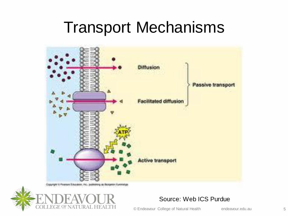
Transport Mechanisms
Active transport - utilises energy (ATP).
❖ The energy molecule is needed because the nutrient travels
against the concentration gradient.
❖ Types of molecules transported are: proteins, ions, large
cells, complex sugars. Such as iodine uptake by thyroid
gland.
❖ A good example is the sodium-potassium, pump (NA/K
pump). This allows sodium and potassium to move against
the concentration gradient. (Gropper & Smith, 2018)
Watch video on Sodium-potassium pump
By O’ Loughlin, M 2019 –for McGrawHills animations at
http://highered.mheducation.com/sites/0072495855/student_view0/chapter2/animation__how_the_sodium_potassium_pump_works.html or at https://www.youtube.com/watch?v=M6_NCdV7YO8&t=5s

© Endeavour College of Natural Health endeavour.edu.au 4
Transport Mechanisms
Passive transport – movement of nutrients via a
concentration gradient (movement from an area of high
concentration to an area of low concentration).
This transport mechanism includes:
❖Simple diffusion (without carrier)
❖Facilitated diffusion (with help of carrier)
(Gropper & Smith, 2018)
© Endeavour College of Natural Health endeavour.edu.au 5
Transport Mechanisms
Source: Web ICS Purdue

© Endeavour College of Natural Health endeavour.edu.au 6
Review of Digestion
Source: Health Medicine and Anatomy
Reference Pictures (2013).
© Endeavour College of Natural Health endeavour.edu.au 7
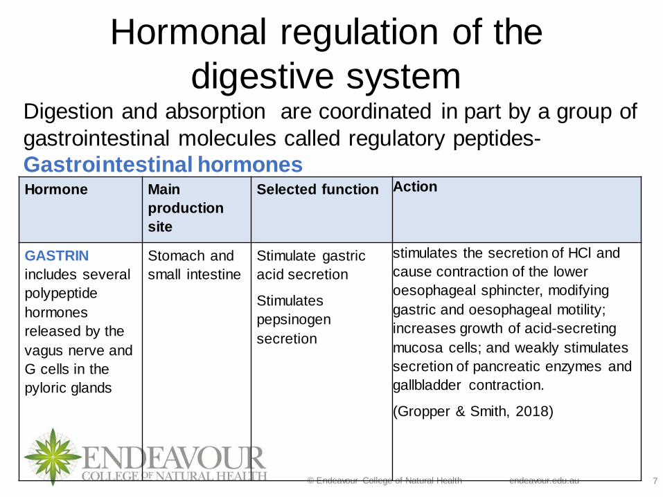
Hormonal regulation of the
digestive system Digestion and absorption are coordinated in part by a group of
gastrointestinal molecules called regulatory peptides-
Gastrointestinal hormonesHormone Main
production
site
Selected function Action
GASTRIN
includes several
polypeptide
hormones
released by the
vagus nerve and
G cells in the
pyloric glands
Stomach and
small intestine
Stimulate gastric
acid secretion
Stimulates
pepsinogen
secretion
stimulates the secretion of HCl and
cause contraction of the lower
oesophageal sphincter, modifying
gastric and oesophageal motility;
increases growth of acid-secreting
mucosa cells; and weakly stimulates
secretion of pancreatic enzymes and
gallbladder contraction.
(Gropper & Smith, 2018)

© Endeavour College of Natural Health endeavour.edu.au 8
Hormonal regulation of the
digestive system Hormone Main
productio
n site
Selected
function
Action
SECRETIN
A strongly basic
polypeptide
hormone
secreted by the
mucosa of the
duodenum and
upper jejunum
when acid chyme
enters the
intestine
Small
intestine
Stimulates
pancreatic juice
secretion
Diminishes
gastric
emptying
Diminishes
gastric acid
secretion
It stimulates the release of
pancreatic juice by the
pancreas and to a lesser extent
bile by the liver, both of which
contain bicarbonate and
change the pH of the
duodenum from acid to
alkaline, thereby facilitating the
action of intestinal digestive
enzymes.
(Gropper & Smith, 2018)
© Endeavour College of Natural Health endeavour.edu.au 9
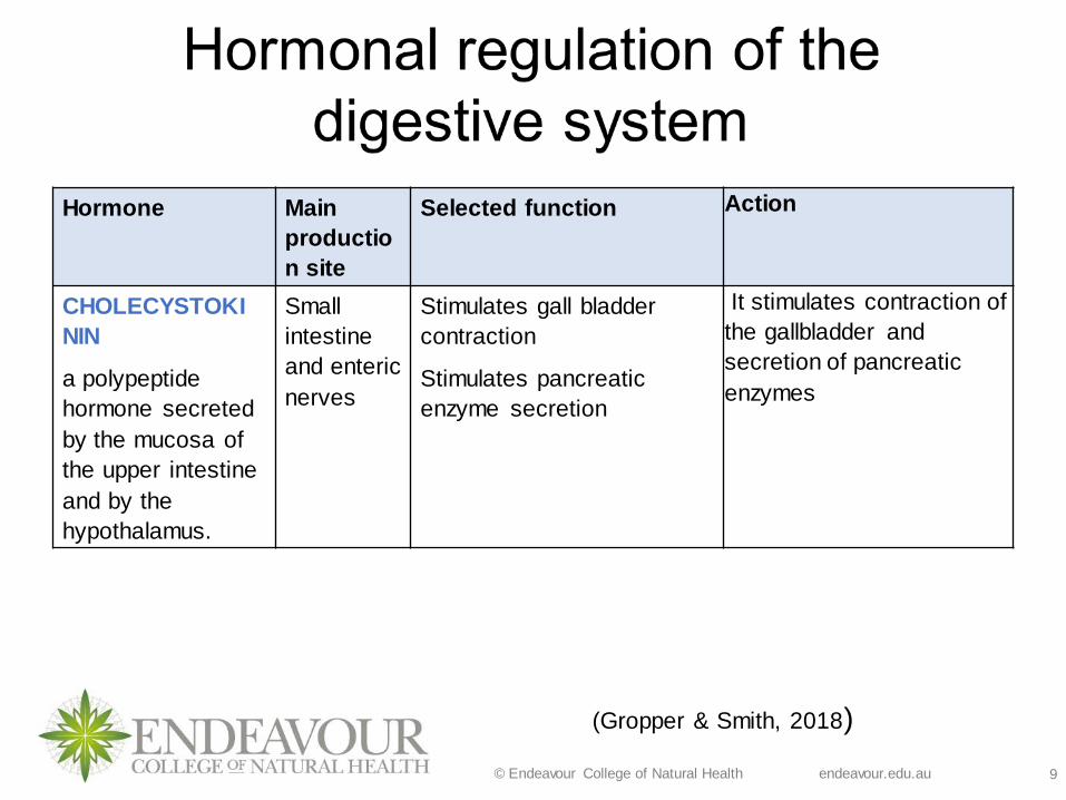
Hormone Main
productio
n site
Selected function Action
CHOLECYSTOKI
NIN
a polypeptide
hormone secreted
by the mucosa of
the upper intestine
and by the
hypothalamus.
Small
intestine
and enteric
nerves
Stimulates gall bladder
contraction
Stimulates pancreatic
enzyme secretion
It stimulates contraction of
the gallbladder and
secretion of pancreatic
enzymes
(Gropper & Smith, 2018)

© Endeavour College of Natural Health endeavour.edu.au 10
Discuss nutritional
impact of Gastric
bypass surgery!!!!
© Endeavour College of Natural Health endeavour.edu.au 11
Macronutrient Pharmacokinetics ❖Macronutrient Pharmacokinetics - absorption,
distribution, metabolism and excretion (ADME) of all the
macronutrients that influences a healthy or disease
status.
❖ These processes are impacted by:
• Poor digestive function
• Mucosal integrity
• Intestinal microbiota
• Hepatic and kidney function
• The presence or lack of dietary fibre
• Agonists and antagonists – nutrient and non-nutrient substances
• Stress – Sympathetic (SNS) or parasympathetic nervous system
(PSNS)
(Gropper & Smith, 2018)
© Endeavour College of Natural Health endeavour.edu.au 12
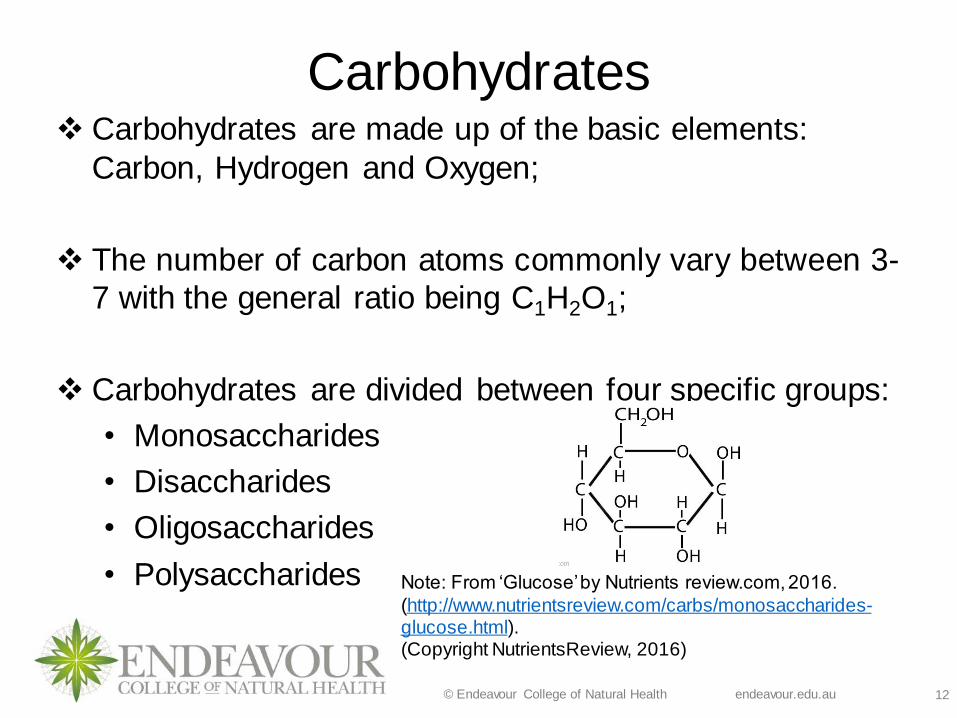
Carbohydrates❖ Carbohydrates are made up of the basic elements:
Carbon, Hydrogen and Oxygen;
❖ The number of carbon atoms commonly vary between 3-
7 with the general ratio being C1H2O1;
❖ Carbohydrates are divided between four specific groups:
• Monosaccharides
• Disaccharides
• Oligosaccharides
• Polysaccharides Note: From ‘Glucose’ by Nutrients review.com, 2016.
(http://www.nutrientsreview.com/carbs/monosaccharides-glucose.html). (Copyright NutrientsReview, 2016)

© Endeavour College of Natural Health endeavour.edu.au 13
Types of Carbohydrates
Monosaccharides Structurally the simplest carbohydrate and
cannot be reduced into smaller units by hydrolysis;
The most abundant and nutritionally relevant are the 6-carbon sugars
Fructose
Galactose
Glucose
Disaccharides Have just two monosaccharide units joined by covalent bonds.
Sucrose if the most nutritionally significant furnishing approximately 1/3 of total dietary carbohydrate in the average diet.
Lactose
Maltose
Sucrose
Oligosaccharides Consists of a short-chain monosaccharide (3 to 6) units also
joined by covalent bonds.
The number of units is designated by the prefix tri-, tetra-,
penta, and so on, followed by the word saccharide. Trisaccharides occur most frequently in nature
Human digestive enzymes cannot digest these and they are
broken down by intestinal bacteria.
Raffinose (tri)
Stachyose (tetra)Verbascose (penta)
Polysaccharides Consists of long chains of monosaccharide units that may
number from several into the hundreds or even thousands.
Glycogen
StarchCellulose
© Endeavour College of Natural Health endeavour.edu.au 14
Digestion of Carbohydrates
❖ The cellular use of carbohydrates depends on their absorption
from the gastrointestinal tract into the blood stream.
❖ The enterocytes can only absorb monosaccharides form of
carbohydrates (across brush border), however, a small
concentration of disaccharides may also be absorbed.
❖ Large structures (polysaccharides and oligosaccharides
chains) must first be hydrolyzed to release their
monosaccharide and disaccharide content.
❖ This process requires the use of hydrolytic enzymes
collectively known as either glycosidases or carbohydrases.(Gropper & Smith, 2018)

© Endeavour College of Natural Health endeavour.edu.au 15
Digestion of Disaccharides❖ Digestion of disaccharides occurs entirely within the
microvilli of upper small intestine.
❖ Enzymes responsible for their digestion are collectively
known as the disaccharidases.
❖ Lactase, sucrase, maltase and isomaltase are all forms
of disaccharidases.
Picture source: Groschwitz & Hogan
(2009)
© Endeavour College of Natural Health endeavour.edu.au 16
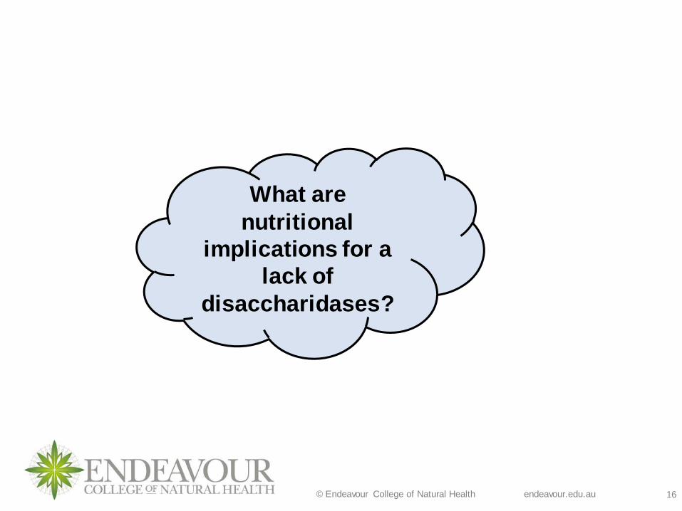
What are
nutritional
implications for a
lack of
disaccharidases?

© Endeavour College of Natural Health endeavour.edu.au 17
Digestion of Polysaccharides❖ This process of digestion of polysaccharides begins in the
mouth. Salivary α - amylase enzyme hydrolysis α 1, 4-glycosidic linkages.
❖ The enzyme is rendered inactive in the stomach by gastric acid.
❖ The pancreas also produces pancreatic α –amylase in the upper part of the duodenum.
❖ Pancreatic bicarbonate ions neutralize gastric acid in the upper part of the duodenum which favor the digestive
activity of pancreatic amylase.
❖ Amylose and amylopectin are thus broken down to
oligosaccharides/dextrins and then further to glucose by α –
dextrinase.(Gropper & Smith, 2018)

© Endeavour College of Natural Health endeavour.edu.au 18
Carbohydrate DigestionEnzyme Action Site
a-amylase a1 – 4 bonds in starch and dextrins.
Mo
uth
a-amylase a1 – 4 bonds in starch, maltotriose.
Pa
nc
reas
Lactase Lactose
Sm
all In
tes
tine
s
Maltase Maltose
Sucrase Sucrose
a-Dextrinase a1 – 6 bonds in dextrins,
oligosaccharides.
Isomaltase a1 – 6 bonds in dextrins,
oligosaccharides.
Glucoamylase a1 – 4 bonds in maltose,
maltotriose.
Glucosidase a1 – 4 bonds in maltose,
maltotriose.

© Endeavour College of Natural Health endeavour.edu.au 19
Glucose Transport❖Most cells in the body are dependent on glucose for the
supply of ATP. The cellular uptake of glucose requires that
it crosses the cell membrane. Its concentration in the blood
must be precisely controlled.
❖ But, the highly polar glucose molecule cannot move across
the cell membrane by simple diffusion because it cannot
pass through the nonpolar matrix of the lipid layer.
❖ Hence an efficient transport system is required.
❖ The classical pathway of glucose absorption across the
intestinal brush-border membrane, predominantly is
mediated by SGLT1, a membrane protein that couples two
molecules of Na+ together with one molecule of glucose:-
Energy dependent (Na/K-ATPase symport system
(SGLT1).
❖ (Shils et al. 2008)
© Endeavour College of Natural Health endeavour.edu.au 20
Glucose Transport❖ Most other human cells use non energy dependent
transporters for glucose uptake.
❖ A family of protein carriers function in the facilitated
transport of glucose (and other monosaccharides) and
are called glucose transporters, abbreviated as GLUT.
❖ 14 glucose transport proteins have been identified.
❖ For example, the passive move out of the basolateral
surface of enterocytes contains a facilitated-diffusion
glucose transporter (GLUT2) which allows glucose to
move to the blood capillaries. (Chen, To & Dong, 2016)

© Endeavour College of Natural Health endeavour.edu.au 21
Glucose Transporters
A transporter protein:
❖Has a specific combining site for the molecule being
transported.
❖Undergoes a conformational change upon binding the
molecule allowing the molecule to be translocated to
the other side of the membrane and released.
❖Has the ability to reverse the conformational changes
without the molecule’s being bound to the transporter
therefore the process can be repeated.
(Gropper & Smith, 2018)

© Endeavour College of Natural Health endeavour.edu.au 22
Source:Slideshare.net, 2014-wwwmedicinemcq.com
(Refer to page 74 for more details in Gropper & Smith, 2018)

© Endeavour College of Natural Health endeavour.edu.au 23
Glucose Transporters and Insulin Regulation
Glucose
Transporter
Insulin Regulatable Cellular
Location
GLUT1 No o Erythrocyte
o Blood Brain Barrier
o Placenta
o Fetal Tissue
GLUT2 No o Liver
o Pancreatic b-cell
o Kidney
o Small Intestines
GLUT3 No o Brain (Neurons)
GLUT4 Yes o Muscle (Skeletal and Smooth)
oAdipose
o Heart
GLUT5 No o Small Intestines
GLUT7 No o Endoplasmic Reticulum (Hepatocytes)
SerumGlycolysis
G
L
U
T

© Endeavour College of Natural Health endeavour.edu.au 24
Monosaccharide Absorption❖ Glucose and Galactose are absorbed from the small
intestines to the mucosal cells by sodium/glucose
symporter 1 (SGLT 1) utilizing ATP
❖ At high concentrations (after large carbohydrate meal),
they are absorbed by facilitated transport via specific
glucose transporter type 2 (GLUT 2)
❖ Glucose, galactose and fructose all exit the enterocyte via
GLUT 2
(Gropper & Smith, 2018)

© Endeavour College of Natural Health endeavour.edu.au 25
Monosaccharide Absorption
❖ GLUT 2 transporters are shown to be regulated by glucose
concentration in the intestinal lumen in humans
❖ Due to its low affinity and high capacity, GLUT2 transports
dietary sugars, glucose, fructose and galactose in a large
range of physiological concentrations, displaying large
bidirectional fluxes in and out the cells.
❖ GLUT2 triggers a metabolic-signalling cascade that
participates in the detection of sugar abundance.
❖Other factors involved in GLUT 2 regulation include
sweetness receptors, high-fructose diets, high-saturated fat
diets and artificial sweeteners.
(Shils et al, 2006, Leturque et al, 2005)

© Endeavour College of Natural Health endeavour.edu.au 26
Monosaccharide Absorption
❖ Fructose is absorbed over the brush border intoenterocytes via facilitated transport with a specific GLUT5 transporter
(Rate of uptake is slower than that of glucose and galactose
however is increased when GLUT 2 is present)
❖ Exits enterocytes via GLUT 2 transporter (only down a
concentration gradient), travels to liver via portal
circulation for metabolism.
❖ It is then rapidly phosphorylated by the liver, leaving no
circulating fructose in the bloodstream which creates a
downhill concentration gradient to allow absorption in the
small intestine.

© Endeavour College of Natural Health endeavour.edu.au 27
Carbohydrate Absorption
Source: Biology forums.com

© Endeavour College of Natural Health endeavour.edu.au 28
Monosaccharide Transport
❖ The monosaccharides (glucose, galactose and fructose)
enter the portal circulation once absorbed across the
basolateral border of the small intestines
❖ The liver is the major site of metabolism
❖Monosaccharides enter hepatocytes by facilitated
transport.
❖ Both galactose and fructose are converted to glucose
derivatives or catabolised for energy depending on
liver’s requirements.(Gropper & Smith, 2018)

© Endeavour College of Natural Health endeavour.edu.au 29
Absorption Across Basolateral Border
Glucose, galactose
and fructose are all
absorbed across the
basolateral border by
a process of facilitated
transport (GLUT 2)

© Endeavour College of Natural Health endeavour.edu.au 30
Monosaccharide Transport❖Glucose is the most important monosaccharide and is
also extensively metabolised by the liver but its removal
is not complete as is fructose and galactose.
❖ The remainder of the glucose passes into the systemic
blood stream and is distributed among muscles, kidney
and adipose tissue, only glucose is found in circulation.
❖ For uptake in skeletal muscles, heart and adipose tissue,
the process is insulin dependent (GLUT 4).
❖ Chromium, glutamine and Vitamin B3 are required for
insulin receptors to function.(Gropper & Smith, 2018)
© Endeavour College of Natural Health endeavour.edu.au 31
Lipids
© Endeavour College of Natural Health endeavour.edu.au 32
Lipids❖ Lipids are a diverse group of organic compounds
including fats, oils, waxes (sterols and non-sterol esters), steroid hormones, fat soluble vitamins A, D, E, K.
❖ Triacylglycerols (TAGs) previously termed triglycerides (TG’s), account for 95% of dietary fat and are the predominant storage form in adipose tissue.
❖ Structurally they comprise of a tri-hydroxyalcohol, glycerol, to which three fatty acids are attached by ester bonds.
❖ The fatty acids tails in the TAG determines the type of lipid, based on chain length and degree of saturation.
(Gropper & Smith, 2018)
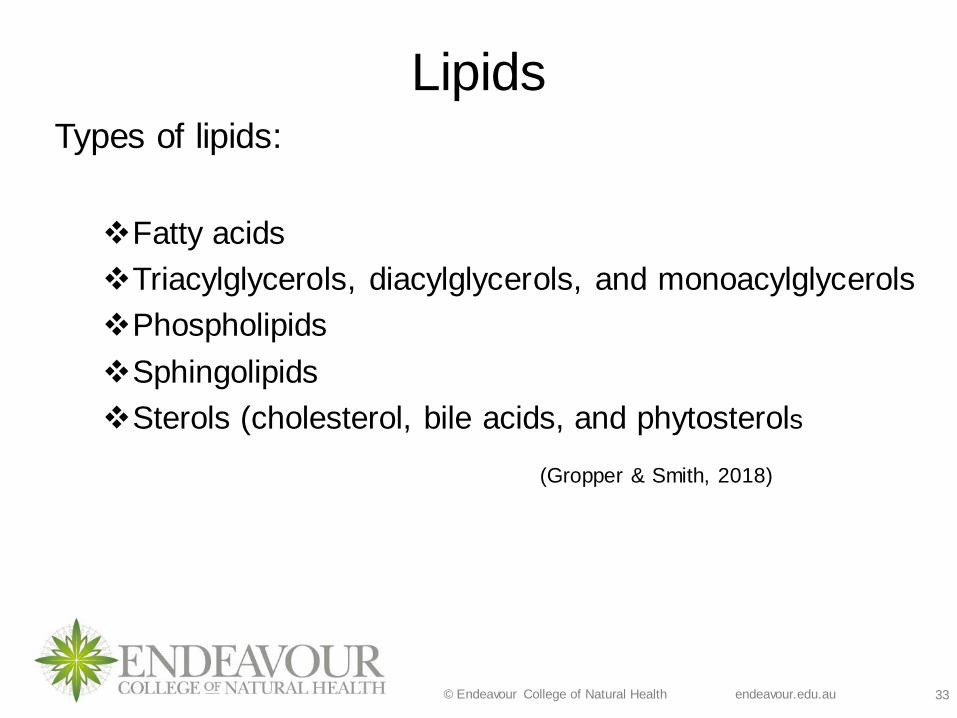
© Endeavour College of Natural Health endeavour.edu.au 33
LipidsTypes of lipids:
❖Fatty acids
❖Triacylglycerols, diacylglycerols, and monoacylglycerols
❖Phospholipids
❖Sphingolipids
❖Sterols (cholesterol, bile acids, and phytosterols
(Gropper & Smith, 2018)

© Endeavour College of Natural Health endeavour.edu.au 34
Classification of Lipids1. Simple lipids
❖ Fatty acids
❖ Triacylglycerols (TAGs), diacylglycerols (DAGs), and
monoacylglycerols (MAGs)
❖ Waxes (esters of fatty acids with higher alcohols (e.g. cholesterol)
and non-sterol esters (e.g. vitamin A)
2. Compound lipids
❖ Phospholipids
❖ Phosphatidic acids (e.g. lecithin)
❖ Plasmalogens
❖ Sphingomyelins
❖ Glycolipids
❖ Lipoproteins (LDL’s, HDL’s etc.)
3. Derived lipids: hydrolysis of lipids in group one or two that still
possess lipid properties
4. Ethyl alcohol (Gropper & Smith, 2018)
© Endeavour College of Natural Health endeavour.edu.au 35
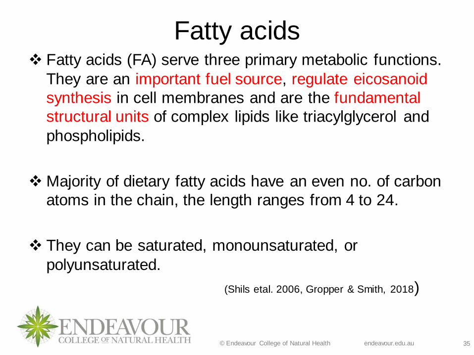
Fatty acids ❖ Fatty acids (FA) serve three primary metabolic functions.
They are an important fuel source, regulate eicosanoid
synthesis in cell membranes and are the fundamental
structural units of complex lipids like triacylglycerol and
phospholipids.
❖Majority of dietary fatty acids have an even no. of carbon
atoms in the chain, the length ranges from 4 to 24.
❖ They can be saturated, monounsaturated, or
polyunsaturated.
(Shils etal. 2006, Gropper & Smith, 2018)

© Endeavour College of Natural Health endeavour.edu.au 36
Fatty acids
Picture source: From Biomolecules-The Lipids retrieved from
https://dlc.dcccd.edu/biology1-3/lipids

© Endeavour College of Natural Health endeavour.edu.au 37
Fatty acids ❖ These are further categorised according to the length of
the carbon chain, short chain fatty acids (< 6), medium
chain fatty acids (6 to 10), and long chain fatty acids
(>12).
❖ Essential fatty acids (EFA’s) must be consumed in the
diet and can be obtained from the plant foods in the form
of: Linoleic acid and a-linolenic acid
❖W-6 versus W-3 fatty acids (are structurally and
functionally different) (Gropper & Smith, 2018)

© Endeavour College of Natural Health endeavour.edu.au 38
Nomenclature of fatty acidsIn the ‘omega classification system’, FAs are identified by
• their chain length,
• the number of double bonds present,
• and the position of the first double bond from the methyl
(CH3) end of the molecule. For Example:
❖ A PUFA with its first double bond three carbons away from methyl
end is omega-3 fatty acids
❖ A PUFA with its first double bond six carbons away from methyl end
is omega 6 fatty acids.
(Encyclopaedia of Human Nutrition. 2013)
Picture source: https://dlc.dcccd.edu/biology1-3/lipids

© Endeavour College of Natural Health endeavour.edu.au 39
Activity
(Valenzuela & Valenzuela, 2013)
© Endeavour College of Natural Health endeavour.edu.au 40
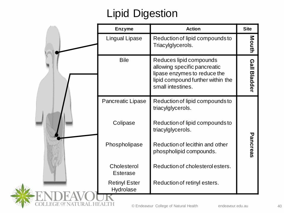
Lipid Digestion
Enzyme Action Site
Lingual Lipase Reduction of lipid compounds to
Triacylglycerols.
Mo
uth
Bile Reduces lipid compounds
allowing specific pancreatic lipase enzymes to reduce the lipid compound further within the
small intestines.
Ga
ll Bla
dd
er
Pancreatic Lipase Reduction of lipid compounds to
triacylglycerols.
Pa
nc
reas
Colipase Reduction of lipid compounds to
triacylglycerols.
Phospholipase Reduction of lecithin and other
phospholipid compounds.
Cholesterol
Esterase
Reduction of cholesterol esters.
Retinyl Ester
Hydrolase
Reduction of retinyl esters.

© Endeavour College of Natural Health endeavour.edu.au 41
Lipid Digestion
❖ As fats are hydrophobic, digestion requires a special
process because the necessary enzymes are
hydrophilic.
❖ Hence an efficient emulsification process mediated
mainly by bile salts is required for the digestion of lipids.
❖ Stimulation of gall bladder to release bile occurs in
presence of 7 – 9 gms of fat in a meal.

© Endeavour College of Natural Health endeavour.edu.au 42
Absorption of Lipids
Khan academy-/digestion-mobilization-and-transport-of-fats
Source: Khan Academy.org
Watch the following video to understand the digestion
and absorption of fats.

© Endeavour College of Natural Health endeavour.edu.au 43
Digestion and Absorption of Lipids
The bulk of dietary lipid is triglyceride. Foods also
contain phospholipids, cholesterol and fat-soluble
vitamins.
As TAGs are water insoluble, its absorption requires:
❖ Break down of the large aggregates that are held in
suspension - a process called emulsification.
❖ Triglyceride molecules must be enzymatically
digested to yield monoglyceride and fatty acids, both
of which can efficiently diffuse or be transported into
the enterocyte.

© Endeavour College of Natural Health endeavour.edu.au 44
Digestion and Absorption of Lipids Emulsification by bile salts make lipids more soluble.
• Being derivatives of cholesterol, they have both
hydrophilic and hydrophobic domains.
• On exposure to a large aggregate of TAG’s, the
hydrophobic portions of bile acids intercalate into the lipid,
with the hydrophilic domains remaining at the surface.
• Such coating with bile acids aids in breakdown of large
aggregates or droplets into smaller and smaller droplets (Bowen, 2019)
Pic source: Colorado State University (n.d)
http://www.vivo.colostate.edu/hbooks/pathp
hys/digestion/smallgut/absorb_lipids.html

© Endeavour College of Natural Health endeavour.edu.au 45
Absorption of Lipids
• Hydrolysis of TAG into
monoglyceride and free fatty
acids is accomplished by
the pancreatic lipase (a
subclass of esterases).
• The free fatty acids remain
associated with bile acids
and complex with other
lipids to form structures
called micelles (Bowen, 2019)
Pic source: Colorado State University (n.d)
http://www.vivo.colostate.edu/hbooks/pathphys/digestio
n/smallgut/absorb_lipids.html

© Endeavour College of Natural Health endeavour.edu.au 46
Absorption of Lipids
Source: Biology forums.com
© Endeavour College of Natural Health endeavour.edu.au 47
Lipid Absorption❖ Stabilised by the polar bile salts, the micellar particles are
sufficiently water soluble to penetrate the enterocytes in the
small intestine.
❖ The lipid part of the micelle diffuse out of the micelles and
into the enterocytes on a concentration gradient .
❖ Then re-formulation or re-esterification of TAGs,
phosphatidyl-choline and cholesteryl esters takes place in
the intestinal cells. They get attached to chylomicrons
(along with fat soluble vitamins) and leave the basolateral
layer through lacteals (and travel via lymph system) (refer to pic
on the next slide).
(Gropper and Smith, 2018)

© Endeavour College of Natural Health endeavour.edu.au 48
Lipid Absorption❖ Bile salts that are not absorbed return to the liver via the
portal vein unless fibre in the diet binds the bile for
excretion.
❖ After absorption, re-formulation or re-esterification of
triacylgycerols, phosphatidyl-choline and cholesteryl
esters takes place.
❖ In the blood, short-chain fatty acids attach to albumin for
transport to other tissues and don’t require solubilisation.

© Endeavour College of Natural Health endeavour.edu.au 49
Lipid Transport and Storage
Chylomicrons
❖ The primary form of lipoproteins formed from exogenous lipids
❖ Lipoproteins other than chylomicrons transport endogenous
lipids from tissue to tissue to supply different cells needs
❖ Lipoproteins differ according to ratio of lipid to protein e.g
triacylglycerols, cholesterol, phospholipids
❖VLDL’s contain higher levels of triacylglycerol (TAG)
and less protein and cholesterol
❖LDL’s contain less TAG, higher protein and highest
cholesterol
❖HDL’s contain lowest TAG, highest protein and
intermediate cholesterol which it delivers back to the
liver
(Gropper & Smith, 2018)

© Endeavour College of Natural Health endeavour.edu.au 50
Protein
© Endeavour College of Natural Health endeavour.edu.au 51
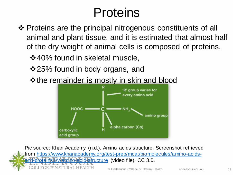
Proteins ❖ Proteins are the principal nitrogenous constituents of all
animal and plant tissue, and it is estimated that almost half
of the dry weight of animal cells is composed of proteins.
❖40% found in skeletal muscle,
❖25% found in body organs, and
❖the remainder is mostly in skin and blood
Pic source: Khan Academy (n.d.). Amino acids structure. Screenshot retrieved
from https://www.khanacademy.org/test-prep/mcat/biomolecules/amino-acids-
and-proteins1/v/amino-acid-structure (video file). CC 3.0.

© Endeavour College of Natural Health endeavour.edu.au 52
Classification of Proteins
Amino acids may be classified in a variety of ways including
by structure, net charge, polarity and essentiality.
Structure
❖ The basic structural units of proteins are the amino acids
have a central carbon (C) at least one amino group (NH2),
a carboxyl acid group (-COOH) and a side chain (R-group).
❖ The distinctive characteristics of amino acid side chains
that make up polypeptides bestow on a protein its structure
and influence its functional role in the body
❖ These distinctive features determine whether AA will be
synthesised in the body and their programming for specific
metabolic pathway (Gropper & Smith, 2018)
© Endeavour College of Natural Health endeavour.edu.au 53
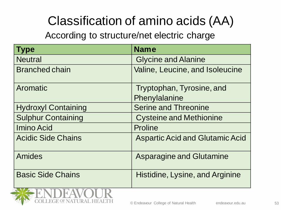
Classification of amino acids (AA)
Type Name
Neutral Glycine and Alanine
Branched chain Valine, Leucine, and Isoleucine
Aromatic Tryptophan, Tyrosine, and
Phenylalanine
Hydroxyl Containing Serine and Threonine
Sulphur Containing Cysteine and Methionine
Imino Acid Proline
Acidic Side Chains Aspartic Acid and Glutamic Acid
Amides Asparagine and Glutamine
Basic Side Chains Histidine, Lysine, and Arginine
According to structure/net electric charge

© Endeavour College of Natural Health endeavour.edu.au 54
Classification of amino acids (AA) According to essentiality
Essential/Indispensable Non-esential/Dispensable
Histidine
Isoleucine
Leucine
Lysine
Methionine Phenylalanine
Threonine
Tryptophan
Valine
Alanine
Arginine
Asparagine
Aspartic acid
Cysteine Glutamic acid
Glutamine
Glycine
Proline
Serine Tyrosine
In 1957, Rose categorized the amino acids as nutritionally essential or non
essential based on body’s requirement.(Gropper and Smith, 2018, Whitney etal. 2017)
Conditional
essential
AA????
© Endeavour College of Natural Health endeavour.edu.au 55
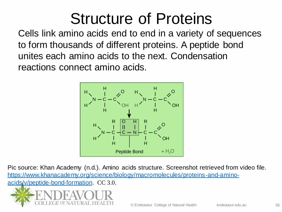
Structure of ProteinsCells link amino acids end to end in a variety of sequences
to form thousands of different proteins. A peptide bond
unites each amino acids to the next. Condensation
reactions connect amino acids.
Pic source: Khan Academy (n.d.). Amino acids structure. Screenshot retrieved from video file.
https://www.khanacademy.org/science/biology/macromolecules/proteins-and-amino-
acids/v/peptide-bond-formation. CC 3.0.
© Endeavour College of Natural Health endeavour.edu.au 56
❖ Peptide: The family of molecules formed from the linking of
various amino acids in a defined order. The link between one
amino acid and the next is an amide bond (or peptide bond).
Peptides differ to proteins only by their size.
❖ Polypeptide: A peptide, such as a small protein, containing
many molecules of amino acids, typically between 10 and
100.
❖ Oligopeptide: An oligopeptide (oligo=few) consists of
between two and 20 amino acids (includes dipeptides,
tripeptides, tetrapeptides, pentapeptides, etc.).
❖ Tripeptide: A tripeptide is a molecule consisting of three
amino acids joined by peptide bonds.
❖ Dipeptide: A dipeptide is a molecule consisting of two amino
acids joined by a single peptide bond.

© Endeavour College of Natural Health endeavour.edu.au 57
Example of protein structure
(Zewaila, Rahmanb & Naglah, 2014)

© Endeavour College of Natural Health endeavour.edu.au 58
Protein Digestion
(Gropper and Smith, 2018)

© Endeavour College of Natural Health endeavour.edu.au 59
Protein - Digestion❖ Digestion begins in stomach.
❖ Pepsinogen secreted from stomach requires adequate
HCl in order to activate pepsin.
❖ Pepsin breaks down peptide bonds in protein chains.
❖ Digestion continues in the small intestine by various
proenzymes secreted by pancreas.
❖ Oligo, tri- and di-peptidases are secreted by small
intestine to assist in liberation of the constituent amino
acids.(Gropper and Smith, 2018)
© Endeavour College of Natural Health endeavour.edu.au 60
Absorption - Amino Acid Transport
❖Occurs along the entire small intestine, however, most
amino acids are absorbed in the proximal (upper) small
intestine.
❖Multiple energy-dependent transport systems for amino
acids are located in the intestinal brush border
❖Most are transported across the brush border via Na+
dependent systems
(Gropper and Smith, 2018)

© Endeavour College of Natural Health endeavour.edu.au 61
Absorption of peptides and single amino acids by
the enterocyte
Absorption of amino acids
Pic source: Openclass/pearson
Sourced from:
https://content.openclass.com/eps/pear
son-reader/
© Endeavour College of Natural Health endeavour.edu.au 62
Absorption-Amino Acid Transport
❖ Affinity of a carrier for an amino acid is influenced by the
hydrocarbon mass of its side chain and by the net
electrical charge
❖ If hydrocarbon mass increases so does the affinity,
therefore:
a) Branched chain amino acids are absorbed faster than
smaller amino acids
b) Neutral amino acids tend to be absorbed faster than
basic or acidic amino acids
c) Essential amino acids are absorbed faster than non-
essential amino acids
d) Slowest to be absorbed are the dicarboxylic (acidic)
amino acids – glutamate and aspartate
© Endeavour College of Natural Health endeavour.edu.au 63
Amino Acid Transport
❖ Ingesting one amino acid or a particular group of amino
acids that use the same carrier system may create a
competition for absorption. Usually the amino acid in
highest concentration will be absorbed by inhibiting the
absorption of other amino acids.
❖ Nitrogen assimilation following ingestion of protein
containing foods is better than ingestion of free amino
acids.
❖ Specific amino acid transporters have been identified
for relevant amino acids absorption
❖ Peptides are transported via PEPT1 transporter,
associated with the co-movement of protons (H+)
Peptides once within the enterocytes are hydrolysed by
cytoplasmic peptidases to form free intracellular amino acids.
(Gropper & Smith ,2018)

© Endeavour College of Natural Health endeavour.edu.au 64
Basolateral Membrane Transport of
Amino Acids
❖ Transport of amino acids across the basolateral
membrane is mainly via sodium-independent transport
❖ Sodium dependent pathways are used when the
amino acid concentrations in the gut are low
❖ Active transport of amino acids into the enterocytes is
necessary to provide the cells own needs
(Gropper & Smith, 2018)
© Endeavour College of Natural Health endeavour.edu.au 65
Intestinal Cell Amino Acid Use
❖Many amino acids absorbed are used by the villi for
protein synthesis
❖Within intestinal cells, amino acids maybe used for:
• Energy
• Synthesis of:
• Apoproteins for lipoprotein formation
• New digestive enzymes
• Hormones
• Nitrogen-containing compounds
❖Metabolised into other amino acids or compounds
(Gropper & Smith, 2018)
© Endeavour College of Natural Health endeavour.edu.au 66
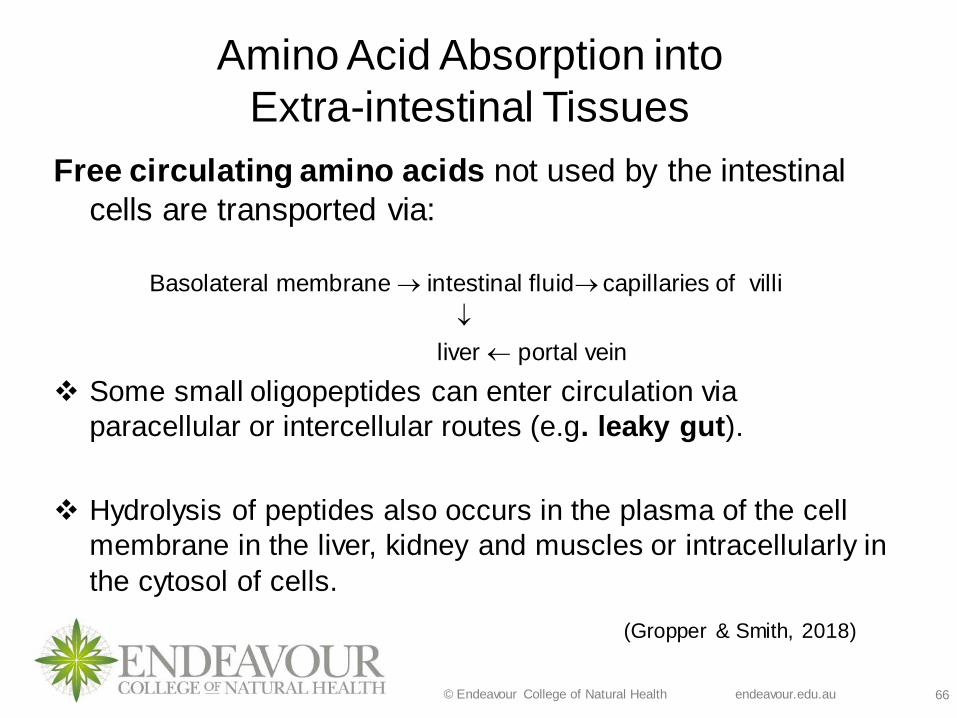
Amino Acid Absorption into
Extra-intestinal Tissues
Free circulating amino acids not used by the intestinal
cells are transported via:
Basolateral membrane → intestinal fluid→ capillaries of villi
liver portal vein
❖ Some small oligopeptides can enter circulation via
paracellular or intercellular routes (e.g. leaky gut).
❖ Hydrolysis of peptides also occurs in the plasma of the cell
membrane in the liver, kidney and muscles or intracellularly in
the cytosol of cells.
(Gropper & Smith, 2018)

© Endeavour College of Natural Health endeavour.edu.au 67
Amino Acid Absorption into
Extra-intestinal Tissues
Amino acids transported into the liver use the following
carrier systems:
1. Diffusion
2. Sodium dependent N system Glutamine, Histidine
3. Hormones and cytokines such as interleukin-1 and TNFa
4. System A induces glucagon and provides amino acid
substrates for gluconeogenesis
5. System Gly is sodium dependent for glycine
Amino acids transported into the kidneys use the following
carrier systems
1. Diffusion
2. -glutamyl cycle (glutathione is the carrier)

© Endeavour College of Natural Health endeavour.edu.au 68
Amino Acid Metabolism❖ The liver is the primary site of amino acid metabolism.
❖ Monitors rate of amino acid metabolism according to the
needs of the body
❖ 20% are used for the synthesis of proteins and nitrogen-
containing compounds
❖ Most of those synthesised will stay in the liver and the rest will
be released into the plasma.
❖ The concentration of total protein in human plasma is typically
7.5 g/dL and are mostly glycoproteins plus simple proteins
and lipoproteins (Gropper & Smith, 2018)

© Endeavour College of Natural Health endeavour.edu.au 69
Plasma ProteinsPlasma proteins perform a variety of functions:
1. Albumin – maintains oncotic pressure, transports nutrients.
2. Transthyretin - prealbumin
3. Retinol-binding protein – Vitamin A, thyroid hormone transport
4. Blood-clotting proteins
5. Globulins:
a) a1-globulins
b) a2-globulins
c) b-globulins
d) -globulins

© Endeavour College of Natural Health endeavour.edu.au 70
Non-Protein Nitrogen (NPNs) Compounds
Amino acids are also used to synthesise NPN that play an
important role in the body. Out of 15 NPNs six are clinically
significant.
Nitrogen-
Containing
Non-Protein
Compounds
Constituent Amino
Acids
Glutathione Cysteine, glycine,
glutamate
Carnitine Lysine, methionine
Creatine Arginine, glycine,
methionine
Carnosine Histidine, b-alanine
Choline Serine

© Endeavour College of Natural Health endeavour.edu.au 71
Storage of Proteins❖ Every cell contains protein especially:
• Muscles,
• Connective tissue,
• Mucus,
• Blood-clotting factors,
• Transport proteins in the bloodstream,
• Lipoproteins,
• Enzymes,
• Immune bodies,
• Hormones,
• Visual pigments,
• Support structure inside bones
❖ Excess protein in the diet doesn’t enhance the synthesis of these body components but eating too little can impede it
❖ Only the brain resists protein breakdown but all other structures continually undergo protein breakdown and repair

© Endeavour College of Natural Health endeavour.edu.au 72
References Allen, P. (Eds.). (2013). Encyclopedia of human nutrition. Retrieved from
http://www.credoreference.com
Bowen, R. (2019). Absorption of lipids. http://www.vivo.colostate.edu/hbooks/pathphys/digestion/smallgut/absorb
lipids.html
Chen,L., Tuo, B., and Dong, H. (2016). Regulation of Intestinal Glucose Absorption by Ion
Channels and Transporters. Nutrients. 8(1): 43.doi: 10.3390/nu8010043
Gropper, S. S., & Smith, J. L. (2018). Advanced nutrition and human
metabolism (7th ed.). Belmont, CA: Wadsworth Cengage Learning.
Hoey, L., Strain, J. J., & McNulty, H. (2009). Studies of biomarker responses to
intervention with vitamin B-12: a systematic review of randomized
controlled trials. The American Journal of Clinical Nutrition, 89(6),
1981S-1996S. doi:10.3945/ajcn.2009.27230C
Leturque, A., Brot-Laroche, E., Le Gall, M.,Stolarczyk, E, and Tobin, V. (2005). The role of GLUT2 in dietary sugar handling. J. Physiol. Biochem, 61
(4), 529-538.

© Endeavour College of Natural Health endeavour.edu.au 73
References
McMurry, J. (1992). Organic chemistry (3rd ed.). Pacific Grove, CA: Brooks/Cole
Publishing Company.
Marks, D. B., Marks, A. D., & Smith, C. M. (1996). Basic medical biochemistry – a
clinical approach. Baltimore, MD: Lippincott Williams & Wilkins.
Muir, J. G., Yeow, E. W., Keogh, J., Pizzey, C., Bird, A. R., Sharpe, K., ...
Macrae, F. A. (2004). Combining wheat bran with resistant
starch has more beneficial effects on fecal indexes than does wheat bran
alone. The American Journal of Clinical Nutrition, 79(6), 1020-1028.
O’ Loughlin, M. (2019). Human Anatomy. McGraw-Hill Global Education
Holdings, LLC. Access on 27th June 2019 from
http://highered.mheducation.com/sites/0072495855/student_view0/chapter2/animat
ion__how_the_sodium_potassium_pump_works.html

© Endeavour College of Natural Health endeavour.edu.au 74
References
Osiecki, H. (2002). Cancer: A nutritional/biochemical approach. Eagle Farm, QLD:
Bioconcepts Publishing.
Roach, J. (2003). Metabolism and Nutrition (2nd ed.). London, England: Mosby.
Shils M. E., Olson, J. A., Shike, M., & Ross, A. C. (2006). Modern nutrition in health
and disease (9th ed.). Baltimore, MD: Lippincott Williams & Wilkins.
Valenzuela, B.R., Valenzuela B. A. (2013). Overview About Lipid Structure. Lipid
Metabolism, Rodrigo Valenzuela Baez, IntechOpen.
Available from: https://www.intechopen.com/books/lipid-metabolism/overview-
about-lipid-structure. DOI: 10.5772/52306
Zubieta-Calleja, G., & Paulev, P. (2004). New Human Physiology. Retrieved from http://www.zuniv.net/physiology/book/chapter22.html

© Endeavour College of Natural Health endeavour.edu.au 75
References (For images-in order of appearance in the slides)
Nutrientsreview.com. (2016). Glucose. Retrieved January 11, 2019 from
http://www.nutrientsreview.com/carbs/monosaccharides-glucose.html
Groschwitz, K.R and Hogan, S.P. (2009). Intestinal Barrier Function: Molecular
Regulation and Disease Pathogenesis. J Allergy Clin Immunol, 124(1): 3–22.
doi:10.1016/j.jaci.2009.05.038.
Slideshare.net (2014). Insulin actions and receptor. Retrieved January 11, 201
9from https://www.slideshare.net/rajendransurendran/insulin-actions-and-receptors-
19-06-13-original, (wwwmedicinemcq.com)
Dlc.dcccd.edu. (n.d.). Biomolecules-The Lipids. Retrieved January 2019 from https://dlc.dcccd.edu/biology1-3/lipids
Valenzuela, R. B. and Valenzuela, A. B. (2013). Overview About Lipid Structure. Retrived from https://www.intechopen.com/books/lipid-metabolism/overview-about-
lipid-structure, http://dx.doi.org/10.5772/52306

© Endeavour College of Natural Health endeavour.edu.au 76
References (For images)
Colorado State University (n.d). Absorption of lipids . Retrieved from
http://www.vivo.colostate.edu/hbooks/pathphys/digestion/smallgut/absorb_lipids.html
Khan Academy (n.d.). Amino acids structure (video file-created by Tracy Kim Kovach).
Retrieved from https://www.khanacademy.org/test-prep/mcat/biomolecules/amino-acids-and-proteins1/v/amino-acid-structure
Khan Academy (n.d.). Peptide bond formation (video file-attributed to
https://cnx.org/contents/[email protected]:2zzm1QG9@7/Proteins).
Retrieved from https://www.khanacademy.org/science/biology/macromolecules/proteins-
and-amino-acids/v/peptide-bond-formation

© Endeavour College of Natural Health endeavour.edu.au 77
References (For images)
Digestion of peptides and amino acids. Retrieved from
https://content.openclass.com/eps/pearson-reader/api/item/ab914c98-1923-486b-bdb4-
b9187be18b9e/1/file/silverthornHP7-071415-MJ-
BO/OPS/s9ml/chapter21/filep7000495934000000000000000007215.xhtml#P70004959340
000000000000000072B7
Zewaila, M. A., Rahmanb,S. A., Naglaha, A. M. (2014). Synthesis of nonapeptide (
B22–B30) of insulin B-chain using modified solid-phase methods with and without
microwave energy. Egyptian Pharmaceutical Journal 13:21–26 DOI: 10.4103/1687-
4315.135594

© Endeavour College of Natural Health endeavour.edu.au 78
COMMONWEALTH OF AUSTRALIA
Copyright Regulations 1969
WARNING
This material has been reproduced and communicated to you by or on behalf of the Australian College of Natural Medicine Pty Ltd (ACNM) trading as Endeavour College of Natural Health, FIAFitnation, College of Natural Beauty, Wellnation - Pursuant Part VB of the Copyright Act 1968 (the Act).
The material in this communication may be subject to copyright under the Act. Any further reproduction or
communication of this material by you may be the subject of copyright protection under the Act.
Do not remove this notice.