Embed Size (px)
Citation preview

NK-CELLSVirus-infected
cell
NK-cell Lysis of infected cell
INFLAMMATION
BacteriumLPS
Cytokines
PhagocytosisIntracellular killing
PHAGOCYTOSIS Phagocyte
Bacterium
Neutrophil
NK-cell
Macrophage
TNF
IL-12
Bacterium
Complement proteins Lysis of bacteria
Inflammation
Complement-dependent phagocytosis
IFN
COMPLEMENT
CELLULAR AND HUMORAL MECHANISMS OF INNATE IMMUNITY
Vessel
Bone marrow
Stem cell
PU-1
Szerv/szövet Makrofág populáció
Bone Osteoclast
Central nervous system
Microglia
Connective tissue
Histiocytes
Placenta Hofbauer cells
Kidney Mesengial cells
Liver Kupffer cells
Peritoneum Peritoneal macrophages
Lung Alveolar macrophages
Skin Epidermal and dermal macrophages
Macrophages can act as stromal cells to help the differentiation of other cells.
Monocyte
Macrophage
Tissuesorgans
DEVELOPMENT OF MACROPHAGES

RECEPTORS AND OTHER MOLEKULES OF MACROPHAGES
LPS receptor (CD14) + TLR4
MHCI
MHCII
TLR – pathogen pattern
CR1 (CD35)
CR3 (CD11b/CD18)
LFA1 (CD11a/CD18)
FcRIII (CD16)
FcRII (CD32)
FcRI (CD64)
Ag + IgG complex
Mannose receptor
Scavanger receptor
peroxidázhidroláz

Copyright ©2008 American Society of Hematology. Copyright restrictions may apply.
Dale, D. C. et al. Blood 2008;112:935-945
Activation of macrophages

RECEPTOR LIGAND FUNCTION
FcR IgG, IgE Opsonized phagocytosis, ADCC, release of inflammatory mediators
CR3 iC3B, ICAM-1 Opsonized phagocytosis
Macrophage Mannose Receptor
Lectin, Endocytosis, phagocytosis, antigen capture and transport
SR-A LPS, polianions, lipoteikolic acid
Endocytosis, phagocytosis, adhesion
CD14 LPS Transduces LPS aktivation , TNFa release
CCR1 MIP1a, MCP-3 Recruitment, migration of monocytes
CCR3 Eotaxin Haematopoiesis, HIV-1 coreceptor
CCR5 MIP1 Haematopoiesis, HIV-1 coreceptor
CXCR4 SDF-1a Haematopoiesis, HIV-1 coreceptor
Receptors and molecules of macrophages

Activation of macrophages

IFN
IL-12IL-18
Th 1 cellNK cell
Inflammatory cytokines Antimicrobial substances
Alternative activation: Mannose receptor – endocytosis Th2 chemokines NOS inhibition Tissue regeneration
IL-4IL-13 Th 2 cell
MicroorganismTNF
IL-6 IL-12
IL-10
T cellAPC
Inactivation
Activation of macrophages
Inflammatory cytokines

Functions of activated macrophagesin anti-bacterial immunity
Macrophage Response** Role in Cell-mediated Immunity
Production of reactive oxygen intermediates, Killing of microbes in phagolysomesnitric oxide; increased lysosomal enzymes (effector function of macrophages)
Secretion of Cytokines TNF-, IL-1: leukocyte recruitment(TNF-, IL-1, IL-12) (inflammation)
IL-12: TH-1 differentiation, IFN- production(induction of response)
Increased expression of: Increased T cell activationCD80, CD86 (amplification)
Class I, Class II MHC
** These macrophage responses are induced by CD40 ligation to CD154 (CD40L) and T cell-derived IFN- in cell-mediated immunity; similar responses are induced by microbial products, particularly LPS, and NK cell-derived IFN- in innate immunity.

Phagocytosis

Intracellular Bacterial Killing

Intracellular bacterial killing
Reactive oxygen species
Intracellular bacteria in phagolysosomes are susceptible to reactive oxygen species, which damage cell wall components and fragment genomic DNA.
O2 + NADPHNADPH Oxidase
NADP + O2- + H+
O2- + H+
SODO2 + H2O2
Reactive Oxygen Intermediate (ROI) production is initiated by membrane-bound NADPH oxidase, which is activated by IFN-.
O2- is further metabolized by superoxide dismutase (SOD).

Intracellular bacterial killing
Reactive oxygen speciesIn the presence of appropriate iron catalysts, the Haber-Weiss reaction takes place:
O2- + Fe3+ O2 + Fe2+
H2O2 + Fe2+ OH + OH- + Fe3+
O2- + H2O2 OH +OH- + O2
O2- is transformed into 1O2. 1O2 and OH are short-lived, powerful
oxidants with high antibacterial activity, causing damage to DNA, membrane lipids, and proteins.
Nomenclature
O2- - superoxide anion
OH – hydroxyl radical containing a free electron 1O2 – singlet oxygen, a highly reactive form of O2

Intracellular bacterial killing
Reactive oxygen species
myeloperoxidase-dependent killing
The lysosomes of granulocytes and monocytes/macrophages contain the enzyme myeloperoxidase (MPO). This enzyme catalyzes the following reaction:
H2O2 + Cl- OCl- + H2OMPO
Hypochlorous acid and chloramines are formed – both agents further increase the bactericidal power of the ROI system by destroying biologically important proteins through chlorination (Halogenation).

Copyright ©2008 American Society of Hematology. Copyright restrictions may apply.
Dale, D. C. et al. Blood 2008;112:935-945

Intracellular bacterial killingReactive nitrogen species
Phagocytes possess an additional pathway for generating reactive species that possess bactericidal activity. These species are the reactive nitrogen intermediates (RNI).
The principal RNI is nitric oxide (NO), which is derived from the terminal guanidino-nitrogen atom of L-arginine. The reaction is catalyzed by the inducible form of nitric oxide synthase (iNOS; NOS2), leading to the formation of L-citrulline and NO.

Intracellular bacterial killing
Reactive nitrogen speciesNO can act as an oxidizing agent alone, or it interacts with O2
- to form unstable peroxynitrite (ONOO-). This may be transformed to the more stable anions, NO2
- and NO3-, or decomposed to NO.
O2- + NO ONOO-
ONOO- + H+ NO2- + .OH
NO2- + .OH NO3
- + H+
ONOO- + H+ .OH + NO.
NO· and ONOO- are highly reactive antimicrobial agents. NO· may be transformed to nitrosothiols expressing the most potent antimicrobial activity. In contrast, NO2
- and NO3- are without
notable effects on microorganisms.
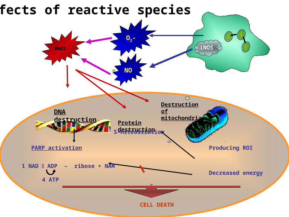
NO
O2-
0NO2-
Destruction of mitochondriaDNA destruction
PARP activation
CELL DEATH
Producing ROI
Decreased energy1 NAD ADP – ribose + NAM
4 ATP
Protein destruction
iNOS
S-nitrosilation
Effects of reactive species
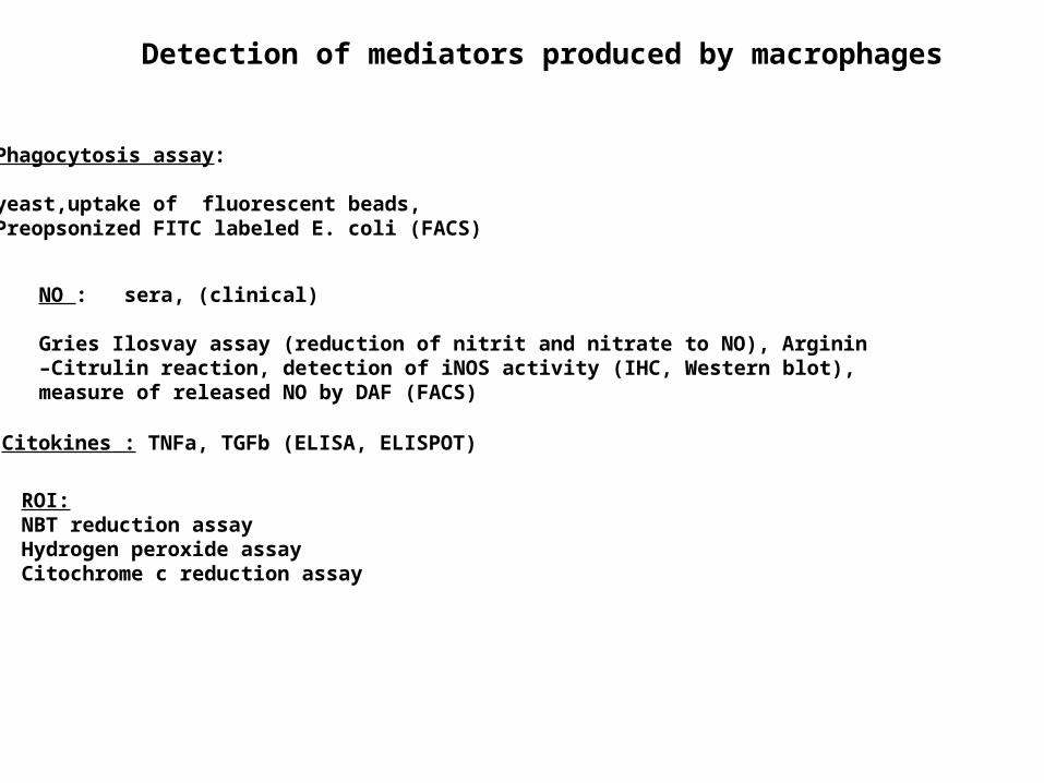
NO : sera, (clinical)
Gries Ilosvay assay (reduction of nitrit and nitrate to NO), Arginin –Citrulin reaction, detection of iNOS activity (IHC, Western blot), measure of released NO by DAF (FACS)
Detection of mediators produced by macrophages
Phagocytosis assay:
yeast,uptake of fluorescent beads,Preopsonized FITC labeled E. coli (FACS)
Citokines : TNFa, TGFb (ELISA, ELISPOT)
ROI:NBT reduction assayHydrogen peroxide assayCitochrome c reduction assay

Intracellular bacterial evasion ofkilling in phagocytes
Intracellular bacteria have evolved strategies to evade killing by the mechanisms available to the phagocyte.
Defensins Unknown
Phagosome acidification Phagosome neutralization
Phagosome–lysosome fusion Inhibition of phagosome–lysosome fusionLysosomal enzymes Resistance against enzymes
Intraphagolysosomal killing Evasion into cytosol Robust cell wall
C3b receptor-mediated uptake,
ROI ROI detoxifiers, ROI scavengers
RNI Unknown (ROI detoxifiers probably interfere with RNI)
Iron starvation Microbial iron scavengers (e.g., siderophores)
Tryptophan starvation Unknown
Macrophage effector capacity Microbial evasion mechanism
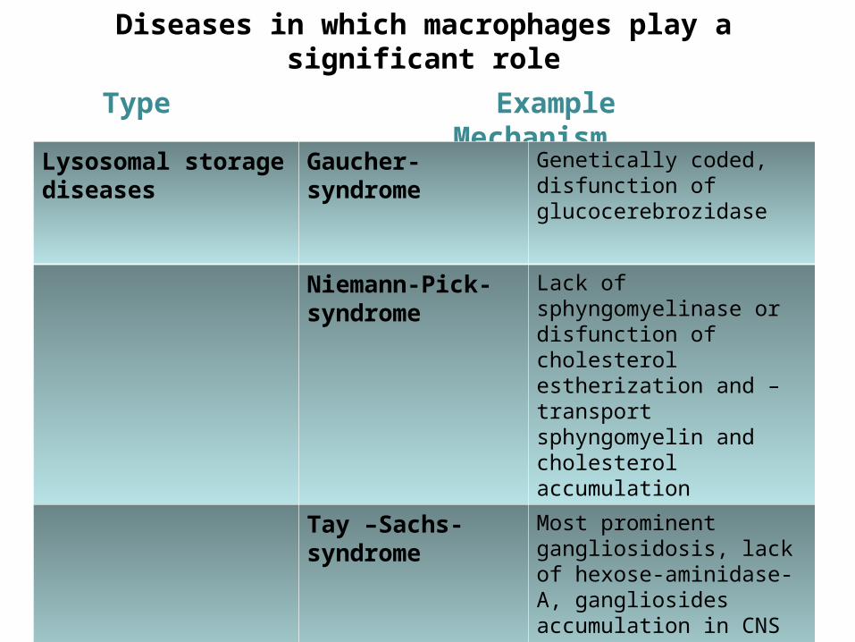
Diseases in which macrophages play a significant role
Type Example Mechanism
Lysosomal storage diseases
Gaucher-syndrome
Genetically coded, disfunction of glucocerebrozidase
Niemann-Pick-syndrome
Lack of sphyngomyelinase or disfunction of cholesterol estherization and –transport sphyngomyelin and cholesterol accumulation
Tay –Sachs-syndrome
Most prominent gangliosidosis, lack of hexose-aminidase-A, gangliosides accumulation in CNS

Diseases in which macrophages play a significant role
Type Example Mechanism
Infections AIDS Cellular immunodeficiency, lack of CD4+ T cells and macrophages
Malaria Host mononuclear phagocyte system hyperplasia, massive splenomegaly
Cerebrospinal diseases
Alzheimer-dysease
Senilis cerebralis amiloidosis, caused by improper elimination of amyloid-associated protein because of defects in macrophage enzymes

Diseases in which macrophages play a significant role
Type Example Mechanism
Chronic inflammation
Silicosis Chrystal quartz powder phagocytosed by alveolar macrophages - progrediated nodular fibrotizing pneumoconiosis
Asbestosis Asbestos filaments phagocytosed by alveolar macrophages - chromic desquamative alveolitis and interstitial inflammation become fibrosis
Atherosclerosis
Monocytes exit to the intima from the blood, become macrophages and store fat cytoplasmatically: foamy cells - inflammation

Granulomatosus inflammation
- chronic inflammation- epitheloid cells in the infiltrate, these are modified macrophages whit pale cytoplasm and nucleus - cells with no intercellular substances (epithelial cell-like tight connections)
- cells become multinucleated Langhans type giant cells
↓Granulomatosus inflammation:
granuloma formation with cell death

lymphocytes
syncytium (multinucleated giant cells)
Granulomas

Periapical granuloma = dental granuloma
Modified granulation tissue containing elements of chronic inflammation, located adjacent to the root apex of a tooth with infected, necrotic pulp.

Phatogen:Mycobacterium tuberculosisMycobacterium bovis, Mycobacterium africanum, Mycobacterium canetti, and Mycobacterium microti can also cause tuberculosis, but these species do not usually infect healthy adults
Tuberculosis most commonly attacks:• the lungs (as pulmonary TB)•central nervous system (meningitidis) • lymphatic and circulatory system (miliary TB) •genitourinary system, •bones, joints• skin
From 2000 to 2004, 20% of TB cases being resistant to standard treatments and 2% resistant to second-line drugs.
Tuberculosis (TBC)
2.000.000.000 infected worldwide

Mycobacterium infection
DS
1.Infection of macrophages
CD8+ T cell
CD4+ T cell
IL-12
2. Antigen presentation
Macrophage
perforingranulysin
IFN-TNF
3. T cell and macrophage activation
Macrophage
Macrophage
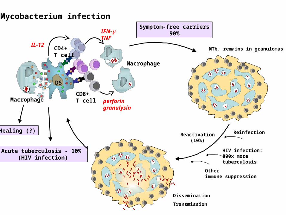
Mycobacterium infection
Healing (?)
Acute tuberculosis - 10% (HIV infection)
Symptom-free carriers90%
MTb. remains in granulomas
Reinfection
Dissemination
Transmission
Reactivation(10%)
HIV infection:800x more tuberculosis
Other immune suppression
DS
CD8+ T cell
CD4+ T cell
IL-12
Macrophage perforingranulysin
IFN-TNF
Macrophage
Morbus hungaricusMorbidity of TBC in Hungary:
• first decades of the later century: 340-380/100000 citizens
• 1955: 30/100000 citizens (lower than the European average!)
reason: regular screening, vaccination, up-to-date therapy
• In last years: increasing numbers of TBC
reason: optimistic attitude, ease of strict control
The 90% of people infected with bacteria are symptome-free, living with latent TBC (LTBI), their opportunity is 10% to develop disease.Without treatment, 50% of TBC diseases are lethal.
TBC is one of the three most dangerous infectious diseases worldwide, mortality is two times higher than to malaria.
2.000.000.000 infected persons
Appearance and frequency of TBC

Skin and bone - tuberculosis


Primary infection

Miliaris tuberculosis

CNS tuberculosis
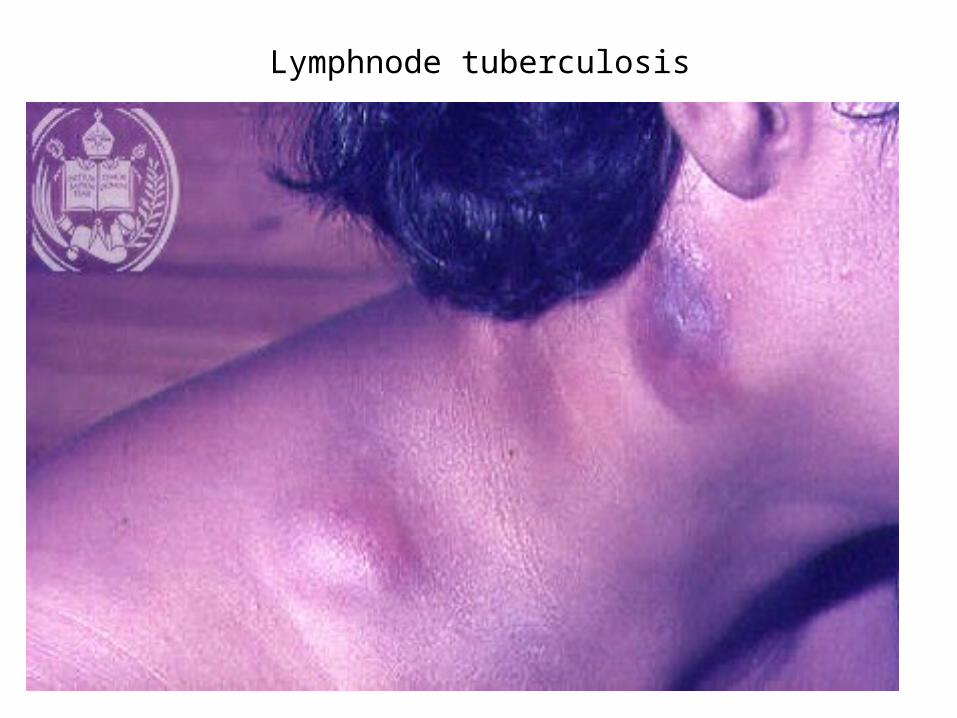
Lymphnode tuberculosis

Diagnostic testing of tuberculosis
Tuberculin skin test(TST) Most often applied tuberculin test:
Mantoux’s test PPD (purified protein derivative) Size of induratio (after 48h)
Disadvantages:
• Not specific for M. tuberculosis
• Positiv reaction: in case of atypical Mycobacterial diseases and BCG vaccination also

IFNγ release assay (IGRA) - ELISPOT ESAT-6 (early secrete antigen target 6) and CFP-10 (culture filtrate
protein) stimulatory antigens Measuring: release of IFNγ by T cells Results: SFU (Spot Forming Unit)
Advantages:
• More specific than TST
• Can be repeated
• The testing protocol requires only one visit
Disadvantages:Reversion: a previously positive IGRA results becomes negative upon revers testing, due to• clearing of TB infection (spontaneous or due to treatment) • biological variations among IGRA+ individuals• the life cycle of M. tuberculosis, where the Mycobacterium enters a dormant state in which it may not be secrete ESAT-6 and CFP-10 antigens (but instead secrete other antigens which are not used in currently available IGRAs)
Diagnostic testing of tuberculosis

Treatment
First line tuberculosis drugs
3-letter 1-letter Drug
EMB E Ethambutol
INH H Isoniazid
PZA Z Pyrazinamide
RMP R Rifampicin
STM S streptomycin
Second line tuberculosis drugs
CIP (none) Ciprofloxacin
MXF (none) Moxifloxacin
PAS P p-aminosalicylic acid
All first-line anti-tuberculous drug names have a standard three-letter and a single-letter abbreviation:•Streptomycin is STM or S, •isoniazid is INH or H, •rifampicin is RMP or R, •ethambutol is EMB or E, •pyrazinamide is PZA or Z. The US commonly uses abbreviations and names that are not internationally recognised: rifampicin is called rifampin and abbreviated RIF; streptomycin is commonly abbreviated SM.

The standard "short" course treatment for tuberculosis (TB):
2 months: isoniazid, rifampicin, pyrazinamide, and ethambutol+4 months: isoniazid and rifampicin alone
For latent tuberculosis, the standard treatment is six to nine months of isoniazid alone