Embed Size (px)
Citation preview

NK Cell Receptor/H2-Dk–Dependent Host Resistance toViral Infection Is Quantitatively Modulated by H2 q
Inhibitory SignalsNassima Fodil-Cornu1,2, J. Concepcion Loredo-Osti3, Silvia M. Vidal1,2*
1 Department of Human Genetics and Department of Microbiology and Immunology, McGill University, Life Sciences Complex, Montreal, Canada, 2 McGill Centre for the
Study of Host Resistance, McGill University, Montreal, Canada, 3 Department of Mathematics and Statistics, Memorial University of Newfoundland, St. Johns, Canada
Abstract
The cytomegalovirus resistance locus Cmv3 has been linked to an epistatic interaction between two loci: a Natural Killer (NK)cell receptor gene and the major histocompatibility complex class I (MHC-I) locus. To demonstrate the interaction betweenCmv3 and H2k, we generated double congenic mice between MA/My and BALB.K mice and an F2 cross between FVB/N (H-2q)and BALB.K (H2k) mice, two strains susceptible to mouse cytomegalovirus (MCMV). Only mice expressing H2k inconjunction with Cmv3MA/My or Cmv3FVB were resistant to MCMV infection. Subsequently, an F3 cross was carried outbetween transgenic FVB/H2-Dk and MHC-I deficient mice in which only the progeny expressing Cmv3FVB and a single H2-Dk
class-I molecule completely controlled MCMV viral loads. This phenotype was shown to be NK cell–dependent andassociated with subsequent NK cell proliferation. Finally, we demonstrated that a number of H2q alleles influence theexpression level of H2q molecules, but not intrinsic functional properties of NK cells; viral loads, however, werequantitatively proportional to the number of H2q alleles. Our results support a model in which H-2q molecules conveyLy49-dependent inhibitory signals that interfere with the action of H2-Dk on NK cell activation against MCMV infection.Thus, the integration of activating and inhibitory signals emanating from various MHC-I/NK cell receptor interactionsregulates NK cell–mediated control of viral load.
Citation: Fodil-Cornu N, Loredo-Osti JC, Vidal SM (2011) NK Cell Receptor/H2-Dk–Dependent Host Resistance to Viral Infection Is Quantitatively Modulated byH2
qInhibitory Signals. PLoS Genet 7(4): e1001368. doi:10.1371/journal.pgen.1001368
Editor: Derry C. Roopenian, The Jackson Laboratory, United States of America
Received September 24, 2010; Accepted March 8, 2011; Published April 21, 2011
Copyright: � 2011 Fodil-Cornu et al. This is an open-access article distributed under the terms of the Creative Commons Attribution License, which permitsunrestricted use, distribution, and reproduction in any medium, provided the original author and source are credited.
Funding: This work was supported by Canadian Institutes of Health Research MOP-7781 and the Canada Research Chairs Program (SMV). The funders had no rolein study design, data collection and analysis, decision to publish, or preparation of the manuscript.
Competing Interests: The authors have declared that no competing interests exist.
* E-mail: [email protected]
Introduction
Natural killer (NK) cells play an important role in the innate
immune response against tumors, MHC-mismatched bone-
marrow grafts, and pathogens [1–2]. These cells also contribute
to defense against parasites and intracellular bacteria, and they are
critical for the control of a variety of viral infections [3–6]. NK cell
actions are immediate and appear to be particularly important
during the first few days of infection; they involve direct lysis of
infected cells and production of proinflammatory cytokines [7].
NK cell activation is tightly regulated by output signals derived
from the engagement of inhibitory and activating receptors by
their respective ligands on potential targets [8]. Inhibitory human
killer immunoglobulin–like receptors (KIR), mouse killer C-type
lectin-like receptors family A (KLRA or Ly49), and NKG2A/
CD94 receptors recognize major histocompatibility (MHC) class I
molecules (H2 in mice), thus controlling NK cell reactivity against
‘‘self.’’ As virally infected cells downregulate the expression of
MHC class I molecules, the lack of inhibitory signals stimulates
NK cells. This mechanism is described as the ‘‘missing self’’
hypothesis, whereby NK cells eliminate targets that lack normal
levels of self-MHC class I molecules [9]. In addition, the
interaction between inhibitory receptors and self MHC-I mole-
cules is the basis of NK cell education (also termed licensing),
leading to the maturation of functional NK cells in homeostatic
conditions [10–17]. By contrast, several families of activating
receptors, such as activating KLRA (also known as Ly49)
receptors, KLRK1 (NKG2D) and the natural cytotoxicity receptor
(NCR) NKP46 (NCR1) can induce NK cell activation through the
recognition of viral ligands or stress-induced molecules [18–22].
Although it is clear that NK cell responses are modulated by a
balance of opposing signals received from self- or nonself-specific
ligands, the precise contribution of specific inhibitory and
activating pathways to the resolution of infection remains to be
fully understood.
The genetic dissection of host resistance or susceptibility to
mouse cytomegalovirus (MCMV) has provided a fresh view of the
precise role of activating NK cell receptors in the recognition of
infected cells and host protection against the infection. Using
informative crosses between various mouse strain combinations,
several MCMV-resistance loci have been mapped to the NK cell
gene complex (NKC) on mouse chromosome 6. The best
characterized, Cmv1 (also known as Klra8) and Cmv3, are defined
by two different modes of inheritance, which seem to correlate
with two different mechanisms of recognition. Cmv1 is a single
dominant locus whose resistance allele, described in C57BL/6 (B6)
mice, encodes the Ly49H activating receptor. Ly49H recognizes
MCMV-infected cells through a direct interaction with the viral
product m157 [21–22]. Engagement of Ly49H by m157 elicits
NK cell–mediated cytotoxicity, cytokine secretion, NK cell
PLoS Genetics | www.plosgenetics.org 1 April 2011 | Volume 7 | Issue 4 | e1001368
proliferation, and viral clearance [18,23–24]. The Cmv3 locus was
detected in a cross between resistant MA/My and susceptible
BALB/c mice. Expression of Cmv3-determined resistance account-
ed for a 100-fold decrease in splenic viral load, but it was only
observed in mice carrying a specific combination of MA/My alleles
at the NKC and MHC (H2k) loci. Functional candidate gene testing
of Ly49 receptors isolated from MA/My mice showed that another
DAP12-associated receptor, Ly49P, responded to MCMV-infected
cells [25]. In this case, Ly49P functional recognition of target cells
required surface expression of both the host H2-Dk molecule and
the viral component m04/gp34 [26]. The role of the H2k haplotype
in MCMV resistance was previously associated with greater survival
following infection with lethal inoculum doses of MCMV compared
to other H2 haplotypes [27]. In addition, linkage analyses in a cross
between resistant MA/My and susceptible C57L strains, as well as
the generation of congenic C57L.M-H2k mice carrying the H2k
allele from MA/My, confirmed a role for H2-Dk-linked resistance to
MCMV [28–29]. Nevertheless, the mechanism of resistance
regulated by the interaction between NK receptors and MHC class
I molecule is still unclear.
In MA/My mice, Cmv3-determined MCMV resistance served
as a model for researchers and allowed them to propose the
existence of a functional interaction between the activating Ly49P
receptor on NK cells and MHC class I H2-Dk molecules.
However, the role of a Ly49P-m04-H2-Dk stimulatory axis
remained to be clarified. In the present study, we sought to
replicate experimentally the statistical association between the
NKC and the MHC-I locus in MCMV resistance, to determine
the precise molecules involved in MCMV resistance in vivo, and to
evaluate the impact of MHC-I inhibitory signals on the NK cell
antiviral response.
Results
Interaction between NKC alleles and H2k locus isassociated with resistance to MCMV
To validate the epistatic interaction between the NKC and H2
detected by linkage analysis [25], we used a marker-assisted strategy
to construct congenic mouse lines in which a chromosome 6
segment (Cmv3) from MA/My MCMV-resistant mice was inde-
pendently introgressed into BALB/c (H2d) and BALB.K (H2k)
susceptible backgrounds. Congenic BALB.K mice have been
described previously [30]. The correlations between the current
physical maps and the genomic region introgressed in the respective
single and double congenic strains BALB-Cmv3MA/MyH2d and
BALB-Cmv3MA/MyH2k are shown in Figure 1 and Table 1.
To examine the effect of the genetic background on the
expression of NKC-encoded receptors in the Cmv3MA/My region,
we used a panel of antibodies with known antigen specificities
(Figure S1) [31]. Though several anti-Ly49 antibodies are cross-
reactive [31], we observed variations among the mouse strains in
terms of frequency of Ly49 subpopulations. Compared to BALB-
Cmv3MA/MyH2d mice, BALB-Cmv3MA/MyH2k animals had a
significantly increased frequency of NK cells stained with the
monoclonal antibodies 12A8 (against Ly49R; P = 0.02) and 14B11
(against Ly49I/U; P = 0.007) (Figure 1B and Figure S2). Notably,
14B11-stained NK cells were also significantly increased in BALB-
Cmv3MA/MyH2k mice compared to MA/My mice (P = 0.005).
These results demonstrated the influence of H2 alleles [32], as well
as the influence of an additional non-H2 mechanism, in the
formation of the Ly49 repertoire. We also observed a highly
significant increase in the frequency of NK cells labeled with 2F1
antibody (P = 0.004), which recognizes the maturation marker
KLRG1, in BALB-Cmv3MA/MyH2d mice compared to their H2k
counterparts. Finally, we observed that NK cells from the three
mouse strains that share the Cmv3MA/My allele lacked expression of
NKG2A/C/E and CD94-associated receptors at the protein but
not mRNA level (Figure 1B and Figure S3B). By contrast, there
was a normal expression of these receptors in the FVB/N mouse
strain, which carries an NKC haplotype similar to that of MA/My
mice (Figure S3A and S3B).
To evaluate the effect of the transferred MA/My chromosome 6
(Cmv3) segment on the response of MCMV-susceptible BALB/c
and BALB.K mice, we infected congenic and parental control
mice by intraperitoneal (i.p.) inoculation of MCMV sublethal
doses (Figure 1C). By day 3 post-infection (p.i), uncontrolled
MCMV replication was observed in the spleen of susceptible
BALB/c mice (log10 plaque-forming units [PFU] = 4.3960.16),
while MCMV-resistant MA/My mice had restricted viral
replication, as shown by a .100-fold lower viral titer (log10
PFU = 1.8160.05) than that seen in BALB/c mice. Single
congenic BALB.K and BALB-Cmv3MA/MyH2d mice had viral titers
that were indistinguishable from those observed in BALB/c mice.
More importantly, congenic mice with the BALB.K background,
which carry only one copy of the MA/My NKC (BALB-
NKCBALBCmv3MA/My.H2k), were as susceptible to MCMV as
BALB/c, BALB.K, and BALB-Cmv3MA/MyH2d mice. By contrast,
double congenic BALB-Cmv3MA/MyH2k mice had restricted virus
growth to the same extent as resistant MA/My mice. Viral titers in
the liver correlated with those observed in the spleens. Further-
more, by day 7 p.i., the virus was cleared from the spleen
(unpublished data), which had undergone a massive increase in
weight and cell number in MA/My and BALB-Cmv3MA/MyH2k
mice (Figure 1D). Collectively, these data demonstrate that the
interaction between Cmv3MA/My and H2k confers MCMV
resistance and is sufficient to explain the control of viral load
observed in MA/My mice.
Because we did not have antibodies that specifically recognize
Ly49P receptors and to examine a possible role of CD94
heterodimers, we attempted to confirm the results obtained in
the congenic mice in a new cross between two strains that
independently carried the H2k loci and the Ly49P gene at the
NKC. We examined the segregation of MCMV viral load in the
spleens of progeny mice from an F2 cross between the MCMV-
susceptible strains FVB/N and BALB.K. Although both parental
strains sustained a relatively high viral titer (5.2 log10 PFU), the
137 F2 progeny showed a continuous distribution ranging from 2
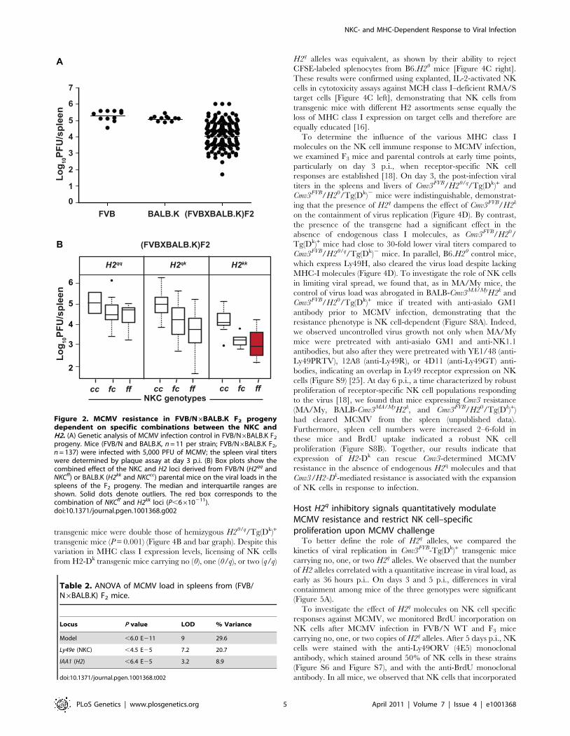
to 6 log10 PFU (Figure 2A). To evaluate the contribution of H2
Author Summary
Effective natural killer (NK) cell responses against virallyinfected cells are regulated by NK cell receptors thatspecifically recognize target cells. In the current study, wevalidated the specific interaction taking place between NKcell receptors and MHC class I molecules on the surface ofinfected cells, resulting in resistance to cytomegalovirus.Genetic dissection of this mechanism of interactionrevealed that the NK cell response occurs exclusivelythrough the triggering of the activating Ly49P receptor bythe MHC class I H2-Dk molecule. We observed, in thiscontext, that NK cells were incapable of clearing the viruswhen target cells also expressed MHC class I H2q
molecules, which strongly and quantitatively inhibit NKcells. Our findings reveal that the interplay betweeninhibitory and activating NK cell receptors and their MHCclass I ligands generate signals that shape the outcome ofinfection.
NKC- and MHC-Dependent Response to Viral Infection
PLoS Genetics | www.plosgenetics.org 2 April 2011 | Volume 7 | Issue 4 | e1001368

and NKC genes to MCMV resistance in this cross, F2 mice were
genotyped and distributed according to their NKC (Ly49e) and H2
(IAA1) genotypes. Mice homozygous for H2kk alleles from BALB.K
and NKCff alleles from FVB/N had the lowest viral load
(Figure 2B). The model that best fitted this phenotype/genotype
distribution in the analysis of variance had a joint logarithm of
odds (LOD) score of 9 (P,10211) and accounted for 29.6% of the
phenotypic variation (Table 2). Thus, there was a highly significant
association between NKC/H2 interaction and control of MCMV
infection in this second cross, indicating that Cmv3 was also present
in the FVB/N mouse strain and that its expression in the presence
of H2k was necessary and sufficient for viral control. Furthermore,
these data suggest that the same gene encodes Cmv3 in both the
MA/My and FVB/N NKC regions.
Transgenic expression of H2-Dk has a modest effect onMCMV control in the presence of H2q
Previously, we showed that activation of Ly49P-bearing
reporter cells requires the H2-Dk host molecule on MCMV-
infected cells [25–26]. Therefore, to better delineate the role of H2
in the host response against MCMV, we attempted in vivo rescue of
the FVB/N susceptible phenotype by genetic transfer of an 11 kb
H2-Dk genomic fragment cloned from AKR mice (Figure 3A). We
monitored for the presence of a diagnostic 300 bp fragment
corresponding to exon 3 of the H2-Dk gene to identify transgenic
FVB-Tg(Dk)+ mice among the founder population (unpublished
data). By surface staining of mouse embryo fibroblasts (MEF) from
FVB-Tg(Dk)+ mice, we observed the normal low levels of H2-Dk
expression under regular conditions (Figure 3B). However, IFN-btreatment up-regulated expression of H2-Dk on MEF cells from
either FVB-Tg(Dk)+ mice or AKR mice (H2-Dk transgene donor
mouse strain) to the same extent, indicating that the transgene
promoter regulatory sequences were intact (Figure 3B). We also
found that the level of expression of H2-Dk in splenocytes from the
FVB/N transgenic mice was similar to the natural H2-Dk
expression in splenocytes from MA/My or BALB.K strains
(Figure 3C). Finally, we investigated the expression of transgenic
H2-Dk and endogenous H2-Dq molecules in T and B cells isolated
from the spleen and observed that the two MHC-I molecules were
expressed in FVB-Tg(Dk)+ mice at levels similar to those found in
Figure 1. Generation, phenotype, and MCMV infection outcome of BALB mice congenic for the natural killer gene complex inheritedfrom MA/My mice. (A) Left, physical map of chromosome 6 markers used to determine the size of the MA/My fragment introgressed into the BALBbackground. The four versions of chromosome 6 indicate the genotypes of sub-congenic strains produced during the generation of BALB.Cmv3MA/My
congenic mice carrying a 10 Mb segment (between SNPs rs13479016 and rs13479061) spanning the NKC region from parental MA/My mice. Right,physical map of chromosome 17 markers used to characterize the 9.4 Mb segment (between D17Mit28 and D17Mit51) comprising the H2 region ofBALB.K mice (H2k). (B) NK cell receptor expression in MA/My parental mice and derived BALB-Cmv3MA/MyH2d and BALB-Cmv3MA/MyH2k congenic mice. Thereceptors (indicated on the bottom of the panel) were gated by FACS on NKp46+ splenic NK cells; the proportion of expression is indicated in eachhistogram. (C) Viral load in spleens (left) and livers (right) of mice of the indicated genotypes, as determined by plaque-forming assays 3 days p.i. (D)Spleen size (top) and weight and total cellularity (bottom) determined in MA/My and BALB-Cmv3MA/MyH2k mice at 7 days p.i. White bar, uninfected mice;black bar, MCMV-infected mice. Data were analyzed using two-way ANOVA analysis and the two-tailed Student’s test. Data are presented as mean 6
SEM and P values of significant differences between groups are indicated. Results shown in panel B are representative of three experiments using 2–3mice per group; results shown in panels C and D are representative of five independent experiments using 3–8 mice per group.doi:10.1371/journal.pgen.1001368.g001
NKC- and MHC-Dependent Response to Viral Infection
PLoS Genetics | www.plosgenetics.org 3 April 2011 | Volume 7 | Issue 4 | e1001368

H2q- and H2k-bearing inbred mice (Figure S4). To test whether the
H2-Dk transgene is capable of stimulating the Ly49P receptor, we
assessed the activation of Ly49P-bearing reporter cells and found
that these cells were equally activated by MCMV-infected MEF
cells from BALB.K or from FVB-Tg(Dk)+ (Figure S5A). Stimula-
tion of Ly49P reporters was also observed upon challenge by
MEFs infected with a mutant virus lacking m157 (the Ly49H
ligand). However, Ly49P reporter cell stimulation was lost upon
infection of MEF cells with a mutant virus lacking the m04 gene,
indicating that the transgenic H2-Dk molecule also requires m04/
gp34 to stimulate Ly49P, as reported (Figure S5B) [26].
To establish the contribution of H2-Dk to the MCMV response,
we first investigated a possible modulation of the Ly49 receptor
repertoire. No significant differences were found between
transgenic and non-transgenic mice in terms of the frequency of
the various NK cell populations tested (Figure S6). Since H2-Dk
has the potential to influence licensing through its interactions with
cognate Ly49 inhibitory receptors [16], we addressed the licensing
status of wild-type and transgenic NK cells. To do this, we
determined the ability of NK cells to mediate in vivo rejection of
MHC class I deficient splenocytes isolated from B6.129P-H2-
D1tm1Bpe H2-K1tm1Bpe (herein B6.H20) mice using a quantitative
CFSE-based method [33]. As previously described, B6.H20 mice
tolerated grafted syngeneic splenocytes (Figure 3D). By contrast,
both FVB-Tg(Dk)+ and FVB-Tg(Dk)2 mice rejected B6.H20
splenocytes with the same efficiency, suggesting that the presence
of the H2-Dk transgene does not alter the licensing status of NK
cells (Figure 3D). Finally, we monitored early viral replication
following MCMV infection in FVB-Tg(Dk)+ mice, along with
single and double BALB-Cmv3MA/MyH2d and BALB-Cmv3MA/
MyH2k congenic mice and parental MA/My mice. We observed
that FVB-Tg(Dk)+ mice had a statistically significant, albeit
modest, reduction in MCMV replication in the spleen (but not
in the liver) compared to nontrangenic littermates; this reduction
represented only a small fraction (1:7) of the reduction observed in
BALB-Cmv3MA/MyH2k congenic mice (Figure 3E). Collectively,
these data demonstrated that the H2-Dk transgene was fully
expressed and able to recognize and activate the Ly49P receptor in
vitro; however, it only provided partial control of MCMV infection.
Transgenic expression of H2-Dk has a major effect onMCMV control in the absence of H2q
Classical MHC class I molecules are the prototype ligands for
Ly49 receptors. Reporter cell assays and tetramer binding assays
suggest that H2-Dk molecules elicit both activating signals, through
Ly49P, and inhibitory signals, through Ly49I and Ly49V [25,31].
By contrast, H2q-encoded molecules can elicit strong inhibitory
signals through Ly49I or Ly49C, but are inert to Ly49G2 [34], as
well as to Cmv3-encoded activating receptors (Ly49P, Ly49R, and
Ly49U) [25]. Therefore, we hypothesized that the poor MCMV
infection control observed in FVB-Tg(Dk)+ mice resulted from
competition between the inhibitory and activating signals
emanating from H2q- and H2-Dk-encoded ligands, respectively.
To test this hypothesis, we crossed FVB-Tg(Dk)+ transgenic mice
with B6.H20 mice, which possess targeted deletions at the H2-D
and H2-K genes. This cross produced F3 progeny mice
homozygous for the FVB/N NKC (Cmv3FVB), but with a different
assortment of MHC class I alleles. F3 mice were either (1) deficient
in endogenous MHC class I alleles in the presence or absence of
the H2-Dk transgene (H20/Tg(Dk)+ or H20/Tg(Dk)2), (2) hemi-
zygous for H2q in the presence or absence of the H2-Dk transgene
(H20/q/Tg(Dk)2 or H20/q/Tg(Dk)+), or (3) homozygous for H2q in
the presence of the H2-Dk transgene (H2q/Tg(Dk)+) (Figure 4A and
Table 1). Again, we monitored whether the frequencies of various
NK cell populations were affected by the genetic makeup of F3
mice and detected no major variations in the NK cell populations,
the only exception being Ly49G+ NK cells, which were barely
detectable in Cmv3FVB/H20/Tg(Dk)2 mice (Figure S7). Similarly,
the level of H2-Dk and H2-Kk expression on splenocytes was
equivalent in transgenic F3 mice with different H2 genotypes
(Figure S7, right panel). By contrast, we noted that levels of H2-Dq
expression on lymphocytes from homozygous H2q/Tg(Dk)+
Table 1. Nomenclature, NKC/H2 genotype, and MCMV titer of mouse strains used in this paper.
Standard nomenclature Abbreviation used in this paper NKC genotype H2 genotype H2-Dk transgene MCMV titer
BALB/c BALB H2d high
MA/My MA/My H2k low
C.C3-H2k BALB.K BALB H2k high
C.MA-Cmv3r.C3- H2k BALB-Cmv3MA/MyH2k MA/My H2k 2 low
C.MA-Cmv3r.C-H2d BALB-Cmv3MA/MyH2d MA/My H2d 2 high
FVB/N FVB/N H2q high
FVB-Tg(H2-Dk) (funder#65864)
FVB-Tg(Dk)+ FVB/N H2q + high
FVB-Tg(Dk)2 FVB/N H2q 2 high
B6.129P-H2-D1tm1Bpe
H2-K1tm1BpeB6.H20 B6 H20 (MHC-I knock
out)2 low
(FVB-Tg(H2-Dk)1Sv xB6.129P-H2-D1tm1Bpe H2-K1tm1Bpe)F3
Cmv3FVB/H20/Tg(Dk)+ (F3) FVB/N H20 + low
Cmv3FVB/H20/Tg(Dk)2 (F3) FVB/N H20 2 high
Cmv3FVB/H20/q/Tg(Dk)+ (F3) FVB/N H20/q + high
Cmv3FVB/H20/q/Tg(Dk)2 (F3) FVB/N H20/q 2 high
Cmv3FVB/H2q/Tg(Dk)+ (F3) FVB/N H2q + high
Text in Bold indicates mouse strains whose genotypes correlate with low viral titers.doi:10.1371/journal.pgen.1001368.t001
NKC- and MHC-Dependent Response to Viral Infection
PLoS Genetics | www.plosgenetics.org 4 April 2011 | Volume 7 | Issue 4 | e1001368
transgenic mice were double those of hemizygous H20/q/Tg(Dk)+
transgenic mice (P = 0.001) (Figure 4B and bar graph). Despite this
variation in MHC class I expression levels, licensing of NK cells
from H2-Dk transgenic mice carrying no (0), one (0/q), or two (q/q)
H2q alleles was equivalent, as shown by their ability to reject
CFSE-labeled splenocytes from B6.H20 mice [Figure 4C right].
These results were confirmed using explanted, IL-2-activated NK
cells in cytotoxicity assays against MCH class I–deficient RMA/S
target cells [Figure 4C left], demonstrating that NK cells from
transgenic mice with different H2 assortments sense equally the
loss of MHC class I expression on target cells and therefore are
equally educated [16].
To determine the influence of the various MHC class I
molecules on the NK cell immune response to MCMV infection,
we examined F3 mice and parental controls at early time points,
particularly on day 3 p.i., when receptor-specific NK cell
responses are established [18]. On day 3, the post-infection viral
titers in the spleens and livers of Cmv3FVB/H20/q/Tg(Dk)+ and
Cmv3FVB/H20/Tg(Dk)2 mice were indistinguishable, demonstrat-
ing that the presence of H2q dampens the effect of Cmv3FVB/H2k
on the containment of virus replication (Figure 4D). By contrast,
the presence of the transgene had a significant effect in the
absence of endogenous class I molecules, as Cmv3FVB/H20/
Tg(Dk)+ mice had close to 30-fold lower viral titers compared to
Cmv3FVB/H20/q/Tg(Dk)2 mice. In parallel, B6.H20 control mice,
which express Ly49H, also cleared the virus load despite lacking
MHC-I molecules (Figure 4D). To investigate the role of NK cells
in limiting viral spread, we found that, as in MA/My mice, the
control of virus load was abrogated in BALB-Cmv3MA/MyH2k and
Cmv3FVB/H20/Tg(Dk)+ mice if treated with anti-asialo GM1
antibody prior to MCMV infection, demonstrating that the
resistance phenotype is NK cell-dependent (Figure S8A). Indeed,
we observed uncontrolled virus growth not only when MA/My
mice were pretreated with anti-asialo GM1 and anti-NK1.1
antibodies, but also after they were pretreated with YE1/48 (anti-
Ly49PRTV), 12A8 (anti-Ly49R), or 4D11 (anti-Ly49GT) anti-
bodies, indicating an overlap in Ly49 receptor expression on NK
cells (Figure S9) [25]. At day 6 p.i., a time characterized by robust
proliferation of receptor-specific NK cell populations responding
to the virus [18], we found that mice expressing Cmv3 resistance
(MA/My, BALB-Cmv3MA/MyH2k, and Cmv3FVB/H20/Tg(Dk)+)
had cleared MCMV from the spleen (unpublished data).
Furthermore, spleen cell numbers were increased 2–6-fold in
these mice and BrdU uptake indicated a robust NK cell
proliferation (Figure S8B). Together, our results indicate that
expression of H2-Dk can rescue Cmv3-determined MCMV
resistance in the absence of endogenous H2q molecules and that
Cmv3/H2-Dk-mediated resistance is associated with the expansion
of NK cells in response to infection.
Host H2q inhibitory signals quantitatively modulateMCMV resistance and restrict NK cell–specificproliferation upon MCMV challenge
To better define the role of H2q alleles, we compared the
kinetics of viral replication in Cmv3FVB-Tg(Dk)+ transgenic mice
carrying no, one, or two H2q alleles. We observed that the number
of H2 alleles correlated with a quantitative increase in viral load, as
early as 36 hours p.i.. On days 3 and 5 p.i., differences in viral
containment among mice of the three genotypes were significant
(Figure 5A).
To investigate the effect of H2q molecules on NK cell specific
responses against MCMV, we monitored BrdU incorporation on
NK cells after MCMV infection in FVB/N WT and F3 mice
carrying no, one, or two copies of H2q alleles. After 5 days p.i., NK
cells were stained with the anti-Ly49ORV (4E5) monoclonal
antibody, which stained around 50% of NK cells in these strains
(Figure S6 and Figure S7), and with the anti-BrdU monoclonal
antibody. In all mice, we observed that NK cells that incorporated
Figure 2. MCMV resistance in FVB/N6BALB.K F2 progenydependent on specific combinations between the NKC andH2. (A) Genetic analysis of MCMV infection control in FVB/N6BALB.K F2
progeny. Mice (FVB/N and BALB.K, n = 11 per strain; FVB/N6BALB.K F2,n = 137) were infected with 5,000 PFU of MCMV; the spleen viral titerswere determined by plaque assay at day 3 p.i. (B) Box plots show thecombined effect of the NKC and H2 loci derived from FVB/N (H2qq andNKCff) or BALB.K (H2kk and NKCcc) parental mice on the viral loads in thespleens of the F2 progeny. The median and interquartile ranges areshown. Solid dots denote outliers. The red box corresponds to thecombination of NKCff and H2kk loci (P,6610211).doi:10.1371/journal.pgen.1001368.g002
Table 2. ANOVA of MCMV load in spleens from (FVB/N6BALB.K) F2 mice.
Locus P value LOD % Variance
Model ,6.0 E211 9 29.6
Ly49e (NKC) ,4.5 E25 7.2 20.7
IAA1 (H2) ,6.4 E25 3.2 8.9
doi:10.1371/journal.pgen.1001368.t002
NKC- and MHC-Dependent Response to Viral Infection
PLoS Genetics | www.plosgenetics.org 5 April 2011 | Volume 7 | Issue 4 | e1001368

BrdU were negative for the Ly49ORV antibody staining.
Furthermore, the increase in BrdU incorporation was inversely
proportional to the number of H2q alleles (Figure 5B). This result
suggests that there is a dose-dependent inhibition of NK cell
proliferation by H2q alleles in response to MCMV infection.
Finally, to investigate whether host MHC-I molecules affect NK
cell activity upon MCMV infection, we adoptively transferred
CFSE-labeled NK cells enriched from Cmv3FVB/H20/Tg(Dk)+
donor mice into Cmv3FVB/H20/Tg(Dk)+ or Cmv3FVB/H20/q/
Tg(Dk)+ recipients (Figure 5C). After 5 days p.i., donor NK cells
had undergone more rounds of division in the Cmv3FVB/H20/
Tg(Dk)+ recipients than the NK cells that were transferred into
Cmv3FVB/H20/q/Tg(Dk)+ mice; this indicated that H2q alleles
limited NK cell proliferation induced by MCMV infection
(Figure 6C bar graph). Thus, NK cells carrying H2-Dk as the
sole MHC class I molecule were impaired in their ability to
proliferate if the recipient mice carried H2q alleles. Collectively,
these results suggest that expression of host H2q molecules
dampens the capacity of NK cells to protect against MCMV.
Discussion
In this study, we examined the combined contribution of the
NKC and H2 loci to the NK cell response against MCMV infection.
We report that MCMV resistance was recapitulated in double-
congenic mice and in an independent F2 cross that reconstituted the
combination of Cmv3 resistance alleles and H2k. Furthermore, we
established that the H2-Dk molecule is essential to the resistance
phenotype, because genetically susceptible mice bearing Cmv3 were
rendered resistant by acquisition of an H2-Dk transgene. However,
efficient virus control was observed only in the absence of
endogenous H2q molecules, whose inhibitory input quantitatively
modulated virus control. Thus, MHC class I molecules play
antagonistic roles in the NK response against viral infection.
Combined effect of the NKC and MHC loci on NK cellantiviral responses
The role of the MHC has been studied using panels of congenic
[27], sub-congenic, and transgenic mice or F2 crosses with the
Figure 3. Functional characterization of H2-Dk transgenic mice. (A) Schematic representation of the H2-Dk gene within the 11.5 kb EcoRIgenomic DNA fragment from AKR mice used to generate transgenic mice. (B) Expression of H2-Dk on MEFs from FVB/N nontransgenic (FVB-Tg(Dk)2,transgenic FVB-Tg(Dk)+, and AKR (H2k) mice. MEFs from FVB-Tg(Dk)+ and wild-type littermates were prepared from PCR-typed embryos, untreated orincubated overnight with 100 U/ml IFN-b prior to analysis of H2-Dk expression by FACS. (C) H2-Dk staining on lymphocytes from FVB-Tg(Dk)2 (redpeak), BALB.K (dashed peak), MA/My (black peak), and FVB-Tg(Dk)+ (dotted peak) mice. Bar graph shows quantification of the percentage of H2-Dk
expression from 2–3 mice per group. (D) Splenocytes from B6.H20 mice or NKC/H2 histocompatible mice were inoculated into either untreated orasialo-GM1–treated NK cell–depleted hosts. Ratio values indicate the relative survival in the test population (CFSEhigh) compared to thehistocompatible control population (CFSElow) at 18 hours after injection. Three mice per group were analyzed. Statistical significance betweenuntreated and NK-depleted mice is shown. (E) Viral load in spleens (left) and livers (right) of mice of the indicated genotypes was determined byplaque-forming assays at day 3 p.i. Data were analyzed using two-way ANOVA analysis and the two-tailed Student’s test. Data are presented as mean6 SEM and P values of significant results between groups are indicated. Results shown are representative of 2–3 independent experiments.doi:10.1371/journal.pgen.1001368.g003
NKC- and MHC-Dependent Response to Viral Infection
PLoS Genetics | www.plosgenetics.org 6 April 2011 | Volume 7 | Issue 4 | e1001368

same NKC haplotype [28–29,35]. To the best of our knowledge,
the present study is the first to provide formal proof of the impact
of both NKC and MHC haplotypes on NK cell antiviral activities
in vivo. Our study, through the use of single- and double-congenic
mice, minimized differences in non-NKC or non-MHC genes.
Thus, we established that the joint action of specific alleles at these
two regions accounted for most of the overall phenotypic
differences between the MCMV-susceptible BALB/c and
MCMV-resistant MA/My mouse strains. BALB-Cmv3MA/MyH2k
mice were indistinguishable from MA/My mice in terms of initial
control of infection and late NK cell responses.
Although the MA/My NKC region in congenic mice
encompasses more than just Ly49 genes, our data indicates that
the influence of MHC alleles on MCMV-resistance stems from the
capacity of MHC class I molecules to serve as ligands for Ly49
receptors. In support of this hypothesis, the F2 cross between the
MCMV-susceptible FVB/N (H2q) and BALB.K (H2k) mouse
strains demonstrated that FVB/N mice carried a Cmv3 resistance
allele that was conditional to H2k and overridden by the H2q
susceptibility allele. Within the Cmv3 region, Ly49 receptors were
responsive to MHC class I ligands. On the one hand, we noticed
that none of the available anti-NKG2 or anti-CD94 antibodies
recognized MA/My mouse NK cells, in contrast to NK cell
recognition in FVB/N mice. Nevertheless, we observed that F2
mice of the combined Cmv3FVB/H2k genotype restrained viral
replication to an extent similar to that seen in MA/My mice. On
the other hand, our haplotype studies [25,36] and new public data
(http://phenome.jax.org) indicate that FVB/N and MA/My
share the same Ly49 gene repertoire, including Ly49P. Conse-
quently, it seems that NK cell responsiveness during MCMV
Figure 4. Functional characterization of F3 mice carrying the NKC from FVB/N and different assortment of H2 molecules. (A) Breedingscheme for the generation of F3 mice carrying the NKC loci from FVB/N parental mice and various combinations of H2 loci. The NKC, H2, and H2-Dk
transgenic loci are represented by boxes, as indicated. The parental (P) FVB-Tg(Dk)+ and B6.H20 strains were mated to generate the F1 generation.Subsequently, F2 mice carrying an homozygous FVB/N NKC locus and heterozygous for either the H2 or the H2-Dk transgene were kept andintercrossed to generate the F3 mice with different H2 assortments (H20: H2-Kb2/2Db2/2). (B) H2-Dq staining of lymphocytes from Cmv3FVB/H20/Tg(Dk)+ (H20 red peak), Cmv3FVB/H20/q/Tg(Dk)+ (H20/q, dot peak), and Cmv3FVB/H2q/q/Tg(Dk)+ mice (H2q/q, dashed peak). Histograms on the rightrepresent the quantification of the level of H2-Dq expression analyzed in three mice per group. (C) Rejection of B6 MHC class I–deficient cells in vivoby the indicated hosts was assessed as in Figure 3, and statistically significant differences are shown. IL-2–derived NK cells from the indicated mousestrains were co-cultured with CFSE-labeled RMA/S cells. Specific lysis at the indicated effector/target ratios was assessed by staining with 7-AAD andanalyzed by FACS. Values represent the mean of 2–3 mice per group. (D) Viral loads in spleens (left) and livers (right) of parental and F3 mice of theindicated genotypes were determined by plaque assay at day 3 p.i. Results shown represent five pooled experiments. Data were analyzed using two-way ANOVA analysis and the two-tailed Student’s test. Significant P values for differences between groups are indicated.doi:10.1371/journal.pgen.1001368.g004
NKC- and MHC-Dependent Response to Viral Infection
PLoS Genetics | www.plosgenetics.org 7 April 2011 | Volume 7 | Issue 4 | e1001368

infection varies with different NKC-MHC combinations and is
optimal only with a precise combination of Ly49 receptors
inherited from MA/My (or FVB/N) mice and MHC class I H2k
molecules.
Role of the MHC class I molecule H2-Dk
Our results confirmed that the H2 effect was due to the MHC
class 1 molecule H2-Dk. Using an 11 kb genomic fragment
containing a functional H2-Dk gene, we achieved a phenotypic
rescue, although the rescue was incomplete if combined with H2q
alleles. The complete protective effect of H2-Dk was restored in F3
mice lacking endogenous H2q molecules. Although H2-Dk also
affects the adaptive immune response, early containment of viral
replication, massive NK cell proliferation, and reversal of the
resistance phenotype by depletion of NK cells in FVB-H20-
Tg(Dk)+ clearly support a mechanism at the level of NK cells.
Because of the presence of both inhibitory and activating Ly49
receptors, several nonexclusive scenarios could account for the
precise mode of action of the combined MHC class I H2-Dk and
Ly49 genotypes on the NK cell response against MCMV: (1)low
threshold of NK cell activation through weak H2-Dk/Ly49
inhibitory signals, (2) effective NK cell activation through H2-
Dk/Ly49 activating signals, and (3) interplay between H2-Dk/
Ly49 activating and inhibitory signals.
Inhibitory signalsOne possibility is that MHC class I/inhibitory Ly49 signals have
a negative impact on the NK cell response to MCMV. In our
study, mature NK cells in BALB.K mice (which are the most
susceptible to MCMV infection) express three inhibitory receptors:
Ly49A, Ly49C, and Ly49G2, which all bind to MHC-I H2k
molecules [31,34,37]. Thus, the majority of NK cells from
BALB.K mice should be inhibited by a receptor for a self-ligand.
Indeed, we have recently shown that deletion of the m04 gene
renders BALB.K mice resistant to MCMV infection, as the protein
it encodes abolishes NK cell activation via the ‘‘missing-self’’
recognition mechanism (Babic et al., 2010) [56]. The m04/gp34
protein escorts MHC class I molecules to the surface of infected
Figure 5. H2q expression interferes with NK cell antiviral responses. (A) MCMV viral load in the spleens of F3 transgenic mice at the indicatedtime-points was determined by plaque assay and P values of significant results between groups are indicated. (B) BrdU incorporation in NKp46-gatedsplenocytes stained with anti-BrdU and 4D5 antibodies. Splenoytes were isolated from mice of the indicated genotypes 5 days p.i. Graph barrepresents the proportions of NK cells incorporating BrdU in total splenic leukocytes with standard deviations, using three mice per group. (C)Enriched NK cells from Cmv3FVB/H20/Tg(Dk)+ mice were labeled with CFSE then adoptively transferred into Cmv3FVB/H20/Tg(Dk)+ and Cmv3FVB/H20/q/Tg(Dk)+ recipients 24 hours before infection with MCMV for 5 days. Analysis of CFSE dilution in NK cells from the spleens of infected (dashed peaks) oruninfected (solid peaks) mice. NK cell proliferation index (number of divisions of CFSE-labeled NK cells) in Cmv3FVB/H20/Tg(Dk)+ and Cmv3FVB/H20/q/Tg(Dk)+ mice. Statistically significant differences between groups are indicated. Three mice per group were analyzed and results shown arerepresentative of two experiments. Data were analyzed using two-way ANOVA analysis and the two-tailed Student’s test. Significant P values fordifferences between groups are indicated.doi:10.1371/journal.pgen.1001368.g005
NKC- and MHC-Dependent Response to Viral Infection
PLoS Genetics | www.plosgenetics.org 8 April 2011 | Volume 7 | Issue 4 | e1001368

cells, thus maintaining a level of surface MHC expression sufficient
enough to trigger inhibitory NK cell receptors [38]. Thus, with
only three (Ly49V, Ly49I, and Ly49G2) out of seven Ly49
inhibitory receptors able to recognize H2-Dk molecules, NK cells
from BALB.Cmv3MA/My H2k mice should be less susceptible to
inhibition by H2k binding (Figure 6A–6C).
Activating signalsThe existence of an H2-Dk–mediated activating axis to MCMV
resistance is supported by the gain-of-function phenotype of FVB-
H2-Dk transgenic mice, which presented itself despite their Ly49
repertoire that is virtually identical to that of their non-transgenic
littermates (Figure S6A and S6B). Furthermore, the absence of NK
cell triggering through inhibitory Ly49 receptors was not sufficient
to allow efficient control of MCMV replication, as demonstrated
by the F3 Cmv3FVB MHC class I–deficient mice. Most NK cells that
develop in MHC class I–deficient hosts are unable to respond to
MHC class I–deficient targets. However, a recent study demon-
strated that, in the context of MCMV infection, NK cells eliminate
virally infected cells in MHC class I–negative hosts, in addition to
regaining the ability to eliminate MHC class I–deficient
hematopoietic host cells [39]. This mechanism seems to be
triggered by the inflammatory milieu induced by MCMV infection
[39]. These observations suggest that the susceptibility of Cmv3FVB
MHC class I–deficient F3 animals to MCMV infection is not due
to a defect in education but to the absence of an activation axis,
which is provided by H2-Dk.
Interplay of inhibitory and activating signalsActivating signals, mediated by the engagement of Ly49P by
H2-Dk/m04, provided only a marginal enhancement of the NK
cell response in the presence of H2q. Interestingly, we observed a
gene dosage effect in the inhibitory action of H2q that correlated
with the level of surface expression of this MHC class I molecule.
However, H2q copy number did not affect the ability of NK cells
from H2-Dk transgenic mice (FVB or F3) to eliminate MHC class I
deficient target cells; this indicates that H2q gene dosage does not
alter education/licensing of NK cells. By contrast, adoptive
transfer experiments demonstrated that H2q alleles expressed on
host cells limit the ability of NK cells to respond to MCMV
infection, indicating that the H2q effect influences NK cell
recognition of class I ligands on target cells. This suggests that
H2q inhibitory signals dominate over H2-Dk-dependent activating
signals emanating from MCMV-infected cells. One possibility is
Figure 6. Model of H2-dependent, Cmv3-determined NK response against MCMV infection. The strength of Ly49 inhibitory signals andthe presence of H2-Dk-mediated activating signals modulate the NK cell response against virus infection. Our set of NKC congenic mice bore differentassortments of Ly49 receptors, but carried an identical H2k resistance haplotype. (A) NK cells from BALB.K mice had a high frequency and strongbinding of inhibitory Ly49 receptors, which rendered BALB.K mice most susceptible to MCMV infection. (B) NK cells from congenic BALB.Cmv3hetH2k
mice carried one copy of the activating Ly49p gene, which can activate the Ly49P/H2-Dk/m04 axis, allowing for intermediate viral loads inheterozygous mice. (C) NK cells from BALB.Cmv3MA/MyH2k mice had the lowest frequency (and/or weakest binding) of inhibitory Ly49 receptors forH2k molecules and the highest frequency of activating Ly49P+ NK cells, resulting in strong control of MCMV infection. Our set of F3 mice carrieddifferent MHC-I components, but an identical Cmv3-resistance haplotype, encoding seven inhibitory and three activating Ly49 receptors, includingLy49P. (D) Engagement of inhibitory receptors in FVB-Tg(Dk)2 mice resulted in inhibition of the NK cell response against MCMV. (E) In FVB-Tg(Dk)+
mice, activating signals mediated by the engagement of Ly49P by H2-Dk/m04, in the presence of inhibitory signals elicited by H2q molecules,provided a marginal enhancement of the NK cell response, and intermediate virus control. (F) In the absence of inhibitory H2q signals, H2-Dk-dependent activation of NK cells was more efficient, which resulted in strong control of MCMV infection in Cmv3FVB/H20/Tg(Dk)+ mice.doi:10.1371/journal.pgen.1001368.g006
NKC- and MHC-Dependent Response to Viral Infection
PLoS Genetics | www.plosgenetics.org 9 April 2011 | Volume 7 | Issue 4 | e1001368

that H2q inhibitory signals are stronger and/or more frequent than
H2k-dependent activating signals. Indeed, it has been shown that
both the density and the avidity of inhibitory Ly49-ligand pairs
determine the strength of inhibition [40]. Alternatively, H2q MHC
class I molecules could compete with H2-Dk for binding with the
m04 protein and thus blunt the m04/H2-Dk-Ly49P activating axis.
We have noted that Ly49P reporter cells are equally stimulated by
MCMV-infected MEFs of H2k or H2k/q genotype, which may
indicate otherwise (Figure S5). However, these results might not
reflect the effect of H2q molecules on the H2-Dk/m04 complex
under physiological conditions. While the molecular details of H2q
inhibition of NK cell function remain unclear, our results suggest a
model in which two antagonistic mechanisms are at play in NKC-
H2-determined resistance to virus infection (Figure 6). One
involves enhanced NK cell responses through H2-Dk-mediated
activating signals. The other involves dampened NK cell responses
through inhibitory Ly49 receptors stimulated by class I H2q (or
H2d) molecules, which override the effect of the H2-Dk construct.
ConclusionIt is puzzling that Ly49 receptors can sense MHC class I
molecules on infected cells despite immune-evasion mechanisms
elaborated by MCMV that downregulate surface expression of
MHC class I molecules. Indeed, mouse strain–specific [41] and
cell type–specific [42] differences have been reported in the ability
of immunoevasins to inhibit lysis of infected cells by CTLs,
indicating that the efficiency of MHC class I downregulation
during MCMV infection [43] is context dependent. In vivo MCMV
replication occurs in a multitude of cell types, and perhaps the
ability of the virus to achieve immune avoidance selectively might
contribute to the delicate equilibrium of coexistence it has
established with the host.
The striking similarities between Ly49 and KIR interactions
with their respective MHC-I ligands and how they both affect NK
cell function prompted us and other researchers to use the mouse
as a model to study NK cell antiviral responses. Our results lend
support to clinical and epidemiological studies implicating KIR-
HLA interactions of different strengths in determining a hierarchy
of NK cell activation with varied effects on the host response
against herpersviruses [44], HCV [45], and HIV [46]. Our work
also highlights the ability of inhibitory signals to overcome NK cell
activation. These regulatory mechanisms would be relevant in
conditions where NK cell activation is undesirable during infection
or immune disease. For example, activating KIR genotypes have
been found to predispose to reactivation of quiescent, opportunis-
tic infections associated with herpesvirus infections in HIV patients
[47]), and to fatal outcome following Ebola virus infection [48];
furthermore, they may constitute a risk factor for susceptibility to
autoimmunity and certain cancers [49,50]. Ultimately, our data
indicate that, as has been proposed for cancer and autoimmunity,
manipulating the balance between inhibitory and activating NK
receptor signals represents a possible avenue to harness the
therapeutic potential of NK cells against virus infections.
Materials and Methods
Ethics statementThe animal protocols and experiments were approved by the
Canadian Council on Animal Care (CCAC) and the McGill
University Animal Resources Center.
AnimalsMA/My, BALB.K, BALB/c, C57Bl/6 (B6), DBA/J, and AKR
mice were purchased from The Jackson Laboratory. FVB/N mice
were purchased from Charles Rivers Laboratories. The B6 mice
deficient for H2-DbKb (B6.H20) were kindly provided by Hidde L.
Ploegh (Cambridge, Massachusetts).
Generation and genetic characterization of congenicmice
BALB-Cmv3MA/MyH2k and BALB-Cmv3MA/MyH2d were gener-
ated by backcrossing the (MA/MyXBALB.K) F1 or (MA/
MyXBALB/c) F1 into BALB.K or BALB/c, respectively, for at
least ten generations. At each backcross, inheritance of the NKC
from parental MA/My mice was genotyped using either the Ly49e
marker or the D6mit135 marker [51]. In the progeny, the
introgressed portion from parental MA/My mice, which included
the NKC, was analyzed using microsatellite markers or by
detecting known SNPs. Once the genetic region was reduced
from 34 Mb (between D6MIT36 and D6MIT59) to 10 Mb
(between rs13479016 and rs13479061 SNPs), heterozygous mice
were intercrossed to generate the homozygous congenic lines.
Generation of FVB-Tg(Dk) transgenic mice and derived F3
strainsThe H2-Dk genomic fragment cloned into the PBR22 plasmid
was kindly provided by Bernd Arnold (Deutsches Krebs-
forschungszentrum [DKFZ], Heidelberg, Germany). The
11.5 kb fragment encompassing the Dk gene was subsequently
purified and injected into fertilized FVB/N mouse eggs.
Transgenesis was performed at the Quebec Transgenesis Research
Network (QTRN). Transgenic founders were screened by PCR
with the primers 59-cacacgatccagcggctgt-39 and 59-ggcccgg-
tctctctctgcag-39, specific for H2-Dk exon 3. They were then bred
to FVB/N WT mice. To generate F3 mice, FVB-Tg(Dk) and
B6.H20 mice were bred to produce F1 and F2 progeny. To
discriminate between the NKC and H2 regions inherited from the
parental strains, the F2 mice were genotyped at the NKC region
with the D6Mit61 and D6Mit52 markers and at the H2 region with
the D17Mit51 marker; they were also genotyped for the presence
or absence of the H2-Dk transgene. Only the mice homozygous for
the FVB/N NKC and heterozygous for either the H2 or the H2-Dk
transgene were kept to generate the F3 progeny, as listed in
Table 1.
Antibodies and flow cytometryTo prepare splenic leukocytes, spleens were removed aseptically
then gently mashed through a 70 mm nylon mesh (BD Bioscience).
Red blood cells were lysed with ammonium chloride (Sigma). To
isolate lymphocytes from mouse blood, mice were bled from the
cheek; blood was collected in RPMI medium containing 15 mM
EDTA. Lymphocytes were collected after gradient centrifugation
using Histopaque-1077 (Sigma). Fc receptors were blocked with
2.4G2 antibody prior to staining with specific monoclonal
antibodies. NK cells were incubated with NKp46 (conjugated to
phycoerythrin [PE] or fluorescein isothiocyanate [FITC]) and
specific monoclonal antibodies against Ly49A-Biot (YE148),
Ly49A/D (12A8), Ly49CIH (14B11), Ly49D (4E5), Ly49G2
(4D11 or AT8), NKG2A/C/E (20d5) or NKG2A/B6 (16A11),
CD94 (18D3), or KLRG1 (2F1). NK cells were also incubated
with the following isotype control monoclonal antibodies: PE-
conjugated golden syrian hamster IgG, FITC- or PE-conjugated
mouse IgG2a K, or FITC-conjugated rat IgG2a K (e-Bioscience).
H2-Dk and -Dq products were detected by anti-H2-Dk antibody
(15-5-5) from BioLegend and anti-H2-Dq antibody (KH117) from
e-Bioscience. To detect incorporated BrdU on NK cells, mice
were scarified 5 or 6 days after MCMV infection; cells were first
NKC- and MHC-Dependent Response to Viral Infection
PLoS Genetics | www.plosgenetics.org 10 April 2011 | Volume 7 | Issue 4 | e1001368

stained for surface antigens (anti-NKp46 and/or anti-Ly49
receptors) and then fixed, permeabilized, treated with DNase I,
and stained with FITC- or allophycocyanin (APC)-conjugated
anti-BrdU antibody (clone 3D4; BD Biosciences), according to the
manufacturer’s protocol. Flow cytometry analysis was performed
with a FACSCalibur flow cytometer (BD Biosciences) and data
were analyzed using CellQuest (BD Biosciences) or FlowJo (Tree
Star). To assess NK cell proliferation in vivo, NK cells from the
spleen were first enriched by negative selection (Miltenyi Biotec),
then incubated with 5 mM CFSE for 15 minutes, washed, and
resuspended in PBS. The purity of the NK cells (55%–70%) was
evaluated by FACS using anti-NKP46 antibody; 2 million NK
cells were then injected intravenously into recipient mice 24 hours
before infection with MCMV. The proliferation index, indicating
the number of divisions of CFSE-labelled NK cells, was
determined using the FlowJo software.
Viruses and infectionsStock MCMV from mouse salivary glands was prepared by
passaging the virus (Smith strain ATCC VR-1399, lot 1698918)
twice in BALB/c mice. The virus was prepared from a
homogenate of salivary glands 21 days p.i.. Mice aged between
7 and 9 weeks were infected intraperitoneally with 2,000 PFUs of
MCMV. The tissue culture-grown viruses [52] Dm157 MCMV,
which lacks the m157 open reading frame (ORF), and Dm04
MCMV, which lacks the m04 ORF, have been previously
described [23,53] and were kindly donated by Ulrich H.
Koszinowski (Max von Pettenkofer Institute, Munich, Germany)
and Stepan Jonjic (Rijeka University, Rijeka, Croatia). Viral titers
of the stock virus or mouse organs (spleen and liver) were evaluated
in vitro by standard plaque assays on a confluent BALB/c MEF
monolayer, as previously described [54].
MEFs: MCMV infection and reporter cell assayThe MEFs used in this work were generated as previously
described [52], except for FVB-Tg(Dk)+ transgenic and nontrans-
genic MEF cells, which were generated from individuals embryos
from the progeny of FVB-Tg(Dk)6FVB wild-type mouse crosses
and then genotyped for the presence of the H2-Dk transgene. 2B4
reporter cells expressing Ly49H, Ly49P, Ly49C, or Ly49I were
generated as previously described [22,25,55]. MEF cultures from
AKR, FVB/N H2-Dk transgenic, FVB/N wild-type, and BALB.K
mice were infected with MCMV (Smith strain) or Dm157 or Dm04
deletion viruses at a multiplicity of infection (MOI) of 1.0 for
24 hours; they were used to stimulate 2B4 reporter cells as
previously described [25]. GFP was detected by flow cytometry
and analyzed using the FlowJo software.
In vivo killing of CFSE-labeled MHC class I–deficient cellsSplenocytes from B6.H20 mice were labeled with 0.4 mM CFSE
(CFSE low) in RPMI medium containing 5% FCS; splenocytes
from recipient mice were labeled with 4 mM CFSE (CFSE high) in
RPMI containing 10% FCS. The splenocytes were then incubated
at 37uC for 10 minutes before being washed three times in RPMI
containing 10% FCS. Cells (56106) of each type were mixed, and
the mixture (200 ml) was injected intravenously into recipient mice.
After 18 hours, spleens were harvested and red blood cells were
lysed. The relative percentage of cells in each CFSE population was
measured by FACS as previously described [33].
In vitro killing of CFSE-labeled RMA/S cellsNK cells from the spleen were expanded for 5 days in RPMI
medium supplemented with 1,000 U/ml human IL-2 (NCI
Preclinical Repository). Cells were washed in RPMI and stained
with NKp46 antibody to determine the purity of NK cells and to
adjust the number of NK cells among strains. RMA/S cells were
labeled with 0.4 mM CFSE in RPMI medium containing 5% FCS
for 15 minutes at 37uC and washed three times. CFSE-RMA/S
and NK cells were cocultured at effector/target ratios of 2:1, 4:1,
8:1, 16:1, and 32:1 for 4 hours at 37uC. Specific lysis was
determined by the measure of 7-aminoactinomycin D (7-AAD)
incorporation (BD) in CFSE-RMA/S cells by flow cytometry, as
previously described [56].
Statistical analysisFor the 137 (FVB/N6BALB/c) F2 mice, the contribution of the
NKC and H2 loci to the segregation of the phenotype was estimated
with the linear model phenotype = m+NKC+H2+NKC:H2+e,
where NKC and H2 represent factors that depend on the mode
of inheritance proposed, m is the common mean value, NKC:H2 is
an interaction term, and e is the independent, normally distributed
random deviations. For the additive mode of inheritance, the NKC
and H2 represent the number of FVB/N/BALB.K alleles at each
locus. For the recessive mode of inheritance, the NKC and H2 are
indicator variables of the homozygous FVB/N and BALB.K
backgrounds, respectively. The four possible additive-recessive
combinations of H2-NKC models, with and without an interaction
term, were fitted. We assessed the magnitude of the contribution for
each term in the model by its P value, obtained by 1 million
bootstrapped samples, and partial g2. Partial g2 was computed as
g2 = SSfactor/(SSfactor+SSerror), where SSfactor is the type 3 associated
sum of squares with the factor in the analysis of variance (ANOVA)
table, and SSerror is the sum of squares corresponding to the residual
variation. We carried out statistical and graphical analyses using R
software. For other statistical analyses in this work, differences
between groups were calculated with two-way ANOVA analysis,
followed by Bonferroni after tests. For some of the analyses,
unpaired, two-tailed Student’s t-tests were conducted. Results with a
P value of ,0.05 were considered to be statistically significant.
Supporting Information
Figure S1 Binding of Ly49-specific monoclonal antibody to
MA/My activating receptors. cDNAs encoding MA/My Ly49P,
Ly49R and Ly49U receptors were expressed in NFAT-GFP 2B4
T-cell hybridomas [25]. Expression of the three receptors was
detected by the anti-Flag M2 monoclonal antibody. Binding of the
isotype control monoclonal antibody (red histogram) or the
indicated Ly49-specific monoclonal antibody (black histogram) to
Ly49P, Ly49R, and Ly49U receptors was assessed by flow
cytometry and analyzed using Flowjo software. The percentage
of binding is indicated in each histogram.
Found at: doi:10.1371/journal.pgen.1001368.s001 (1.19 MB EPS)
Figure S2 Frequencies of Ly49+ and KLRG1+NK cells in
double congenic mice. Quantification of expression frequency of
indicated NK receptors in the parental MA/My strain and BALB-
Cmv3MA/MyH2k and BALB-Cmv3MA/MyH2d congenic mice. Data
are presented as mean 6 SEM and P values of significant
differences between groups are indicated.
Found at: doi:10.1371/journal.pgen.1001368.s002 (0.89 MB EPS)
Figure S3 Lack of NKG2A/C/E and CD94 antibody staining
on NK cells from MA/My mice. (A) NKG2A/C/E and CD94
expression on NKp46+ NK cells from MA/My, FVB/N, and
DBA/2J (as they carry a NKG2/CD94 deficiency [57]) mice was
determined by flow cytometry using the indicated monoclonal
antibodies. (B) CD94 and NKG2A RNA expression in enriched
NKC- and MHC-Dependent Response to Viral Infection
PLoS Genetics | www.plosgenetics.org 11 April 2011 | Volume 7 | Issue 4 | e1001368

NK cells from the indicated mice strains was analyzed by RT-
PCR. b-actin was used as an internal control.
Found at: doi:10.1371/journal.pgen.1001368.s003 (0.87 MB EPS)
Figure S4 Ly49P+2B4 reporter cell stimulation by MCMV-
infected MEF cells produced from FVB-Tg(Dk)+ mice. (A)
Stimulation of Ly49P reporter cells by co-culture with MEF cells
from the indicated backgrounds that were uninfected (black
histograms) or MCMV infected at an MOI of 1 for 24 h (grey
histograms). Ly49P-specific activation was detected by NFAT-
GFP expression using flow cytometry. (B) Stimulation of Ly49P or
Ly49H reporter cells by co-culture with FVB-Tg(Dk)+ MEF cells
that were uninfected (left) or infected with Dm157 (middle) or
Dm04 (right) MCMV deletion mutants. Reporter cell stimulation
was detected by monitoring expression of GFP by flow cytometry.
The percentage of positive cells in each gated population is
indicated.
Found at: doi:10.1371/journal.pgen.1001368.s004 (0.87 MB EPS)
Figure S5 Co-expression of H2-Dq and H2-Dk in FVB-Tg(Dk)+
mice. Endogenous H2-Dq (bottom) and transgenic H2-Dk (top)
expression in splenic T and B cells from FVB-Tg(Dk)+ transgenic
(black peak) or nontransgenic (black peak) littermates was
determined by flow cytometry.
Found at: doi:10.1371/journal.pgen.1001368.s005 (0.57 MB EPS)
Figure S6 Ly49 receptor expression on NK cells from FVB H2-
Dk transgenic and nontransgenic mice. (A) The indicated Ly49
specific monoclonal antibodies (black peak) or isotype controls (red
peak) were gated on NKp46+ splenic NK cells from FVB-Tg(Dk)2
and FVB-Tg(Dk)+ mice and analyzed by FACS. The proportion of
Ly49 receptor expression is indicated in each histogram. (B)
Quantification of expression frequency of the indicated NK
receptors in FVB-Tg(Dk)2 and FVB-Tg(Dk)+ mice.
Found at: doi:10.1371/journal.pgen.1001368.s006 (1.81 MB EPS)
Figure S7 Ly49 receptor and MHC class I expression on NK
cells from F3 mice. The indicated Ly49-specific monoclonal
antibodies were gated on NKp46+ splenic NK cells from F3 mice
and the proportion of expression is indicated in each histogram.
Right panels: expression of MHC-I H2-Dq and H2-Dk molecules
on total splenocytes was determined. 2–3 mice per genotype were
analyzed. We found that the expression of the activating and the
inhibitory receptors were almost comparable between strains for
the exception of Ly49G which was barely detectable in the
Cmv3FVB/H20/Tg(Dk)2 mice using both anti- Ly49G antibodies
(LGL-1 and AT8 (data not shown)). This receptor is perfectly
expressed in the B6.H20 parental strain (data not shown) and
doesn’t seem to be correlated with a defect of NK maturation since
the Killer cell lectin-like receptor G1 (KLRG1) is equally
expressed between the Cmv3FVB/H20/Tg(Dk)+ and the Cmv3FVB/
H20/Tg(Dk)2 mice.
Found at: doi:10.1371/journal.pgen.1001368.s007 (1.37 MB EPS)
Figure S8 NK cell–dependent MCMV infection control in
congenic and F3 mice. (A) Viral loads in spleens (top) and livers
(bottom) 3 d post-infection in MCMV-resistant MA/My progen-
itor, BALB-Cmv3MA/MyH2k congenic, and Cmv3FVB/H20/Tg(Dk)+
transgenic mice that were NK cell depleted (white squares) or not
(black circles) with anti-asialo GM1 antibody. (B) Number of NK
cells per spleen (top) and BrdU incorporation (bottom) at 7 d post-
MCMV infection in MCMV-resistant mice of the indicated
genotypes. For the number of NK cells, data are presented as
mean 6 SEM and statistically significant differences between
groups are indicated. For BrdU incorporation data are represent-
ed by fold increase between noninfected and infected animals.
Results shown are representative of 1–2 experiments.
Found at: doi:10.1371/journal.pgen.1001368.s008 (1.56 MB EPS)
Figure S9 Effect of Ly49-specifc monoclonal antibody depletion
in MA/My mice during the course of MCMV infection. MCMV
viral load was assessed in untreated MA/My mice (mock) or
treated with the indicated monoclonal antibody prior to MCMV
infection. Viral load in spleen was determined by plaque assay
after 3 d of infection.
Found at: doi:10.1371/journal.pgen.1001368.s009 (1.47 MB EPS)
Acknowledgments
We thank Patricia D’Arcy and Chantal Lacroix for excellent help with
animal experiments and maintenance, H. Ploegh (Harvard University) for
MHC class I–deficient mice, as well as Seung-Hwan Lee (Brown
University) and Michal Pyzik (McGill University) for their critical review
of this manuscript. We also thank Kira Heller and Eve-Marie Gendron
Pont-Briand for editorial and proofreading services.
Author Contributions
Conceived and designed the experiments: NFC SMV. Performed the
experiments: NFC. Analyzed the data: NFC JCLO SMV. Wrote the
paper: NFC SMV.
References
1. Raulet DH, Guerra N (2009) Oncogenic stress sensed by the immune system:
role of natural killer cell receptors. Nat Rev Immunol 9: 568–580.
2. Lodoen MB, Lanier LL (2006) Natural killer cells as an initial defense against
pathogens. Curr Opin Immunol 18: 391–398.
3. Guan H, Moretto M, Bzik DJ, Gigley J, Khan IA (2007) NK cells enhance
dendritic cell response against parasite antigens via NKG2D pathway. J Immunol
179: 590–596.
4. Vankayalapati R, Garg A, Porgador A, Griffith DE, Klucar P, et al. (2005) Role
of NK cell-activating receptors and their ligands in the lysis of mononuclear
phagocytes infected with an intracellular bacterium. J Immunol 175: 4611–
4617.
5. Gonzalez VD, Landay AL, Sandberg JK (2010) Innate immunity and chronic
immune activation in HCV/HIV-1 co-infection. Clin Immunol 135: 12–25.
6. Ward J, Barker E (2008) Role of natural killer cells in HIV pathogenesis. Curr
HIV/AIDS Rep 5: 44–50.
7. Biron CA, Nguyen KB, Pien GC, Cousens LP, Salazar-Mather TP (1999)
Natural killer cells in antiviral defense: function and regulation by innate
cytokines. Annu Rev Immunol 17: 189–220.
8. Ortaldo JR, Young HA (2005) Mouse Ly49 NK receptors: balancing activation
and inhibition. Mol Immunol 42: 445–450.
9. Karre K (1991) MHC gene control of the natural killer system at the level of the
target and the host. Semin Cancer Biol 2: 295–309.
10. Fernandez NC, Treiner E, Vance RE, Jamieson AM, Lemieux S, et al. (2005) A
subset of natural killer cells achieves self-tolerance without expressing inhibitory
receptors specific for self-MHC molecules. Blood 105: 4416–4423.
11. Yokoyama WM, Kim S (2006) How do natural killer cells find self to achieve
tolerance? Immunity 24: 249–257.
12. Anfossi N, Andre P, Guia S, Falk CS, Roetynck S, et al. (2006) Human NK cell
education by inhibitory receptors for MHC class I. Immunity 25: 331–342.
13. Jonsson AH, Yang L, Kim S, Taffner SM, Yokoyama WM (2010) Effects of
MHC class I alleles on licensing of Ly49A+ NK cells. J Immunol 184:
3424–3432.
14. Raulet DH, Held W, Correa I, Dorfman JR, Wu MF, et al. (1997) Specificity,
tolerance and developmental regulation of natural killer cells defined by
expression of class I-specific Ly49 receptors. Immunol Rev 155: 41–52.
15. Raulet DH, Vance RE, McMahon CW (2001) Regulation of the natural killer
cell receptor repertoire. Annu Rev Immunol 19: 291–330.
16. Kim S, Poursine-Laurent J, Truscott SM, Lybarger L, Song YJ, et al. (2005)
Licensing of natural killer cells by host major histocompatibility complex class I
molecules. Nature 436: 709–713.
17. Yokoyama WM, Kim S (2006) Licensing of natural killer cells by self-major
histocompatibility complex class I. Immunol Rev 214: 143–154.
18. Dokun AO, Kim S, Smith HR, Kang HS, Chu DT, et al. (2001) Specific and
nonspecific NK cell activation during virus infection. Nat Immunol 2: 951–956.
NKC- and MHC-Dependent Response to Viral Infection
PLoS Genetics | www.plosgenetics.org 12 April 2011 | Volume 7 | Issue 4 | e1001368
19. Gazit R, Gruda R, Elboim M, Arnon TI, Katz G, et al. (2006) Lethal influenza
infection in the absence of the natural killer cell receptor gene Ncr1. NatImmunol 7: 517–523.
20. Diefenbach A, Jamieson AM, Liu SD, Shastri N, Raulet DH (2000) Ligands for
the murine NKG2D receptor: expression by tumor cells and activation of NKcells and macrophages. Nat Immunol 1: 119–126.
21. Smith HR, Heusel JW, Mehta IK, Kim S, Dorner BG, et al. (2002) Recognitionof a virus-encoded ligand by a natural killer cell activation receptor. Proc Natl
Acad Sci U S A 99: 8826–8831.
22. Arase H, Mocarski ES, Campbell AE, Hill AB, Lanier LL (2002) Directrecognition of cytomegalovirus by activating and inhibitory NK cell receptors.
Science 296: 1323–1326.23. Bubic I, Wagner M, Krmpotic A, Saulig T, Kim S, et al. (2004) Gain of
virulence caused by loss of a gene in murine cytomegalovirus. J Virol 78:7536–7544.
24. Fodil-Cornu N, Lee SH, Belanger S, Makrigiannis AP, Biron CA, et al. (2008)
Ly49h-deficient C57BL/6 mice: a new mouse cytomegalovirus-susceptiblemodel remains resistant to unrelated pathogens controlled by the NK gene
complex. J Immunol 181: 6394–6405.25. Desrosiers MP, Kielczewska A, Loredo-Osti JC, Adam SG, Makrigiannis AP,
et al. (2005) Epistasis between mouse Klra and major histocompatibility complex
class I loci is associated with a new mechanism of natural killer cell-mediatedinnate resistance to cytomegalovirus infection. Nat Genet 37: 593–599.
26. Kielczewska A, Pyzik M, Sun T, Krmpotic A, Lodoen MB, et al. (2009) Ly49Precognition of cytomegalovirus-infected cells expressing H2-Dk and CMV-
encoded m04 correlates with the NK cell antiviral response. J Exp Med 206:515–523.
27. Chalmer JE, Mackenzie JS, Stanley NF (1977) Resistance to murine
cytomegalovirus linked to the major histocompatibility complex of the mouse.J Gen Virol 37: 107–114.
28. Dighe A, Rodriguez M, Sabastian P, Xie X, McVoy M, et al. (2005) RequisiteH2k role in NK cell-mediated resistance in acute murine cytomegalovirus-
infected MA/My mice. J Immunol 175: 6820–6828.
29. Xie X, Dighe A, Clark P, Sabastian P, Buss S, et al. (2007) Deficient majorhistocompatibility complex-linked innate murine cytomegalovirus immunity in
MA/My.L-H2b mice and viral downregulation of H-2k class I proteins. J Virol81: 229–236.
30. Klein J (1973) List of congenic lines of mice. I. Lines with differences atalloantigen loci. Transplantation 15: 137–153.
31. Makrigiannis AP, Pau AT, Saleh A, Winkler-Pickett R, Ortaldo JR, et al. (2001)
Class I MHC-binding characteristics of the 129/J Ly49 repertoire. J Immunol166: 5034–5043.
32. Johansson S, Salmon-Divon M, Johansson MH, Pickman Y, Brodin P, et al.(2009) Probing natural killer cell education by Ly49 receptor expression analysis
and computational modelling in single MHC class I mice. PLoS ONE 4: e6046.
doi:10.1371/journal.pone.0006046.33. Oberg L, Johansson S, Michaelsson J, Tomasello E, Vivier E, et al. (2004) Loss
or mismatch of MHC class I is sufficient to trigger NK cell-mediated rejection ofresting lymphocytes in vivo - role of KARAP/DAP12-dependent and
-independent pathways. Eur J Immunol 34: 1646–1653.34. Hanke T, Takizawa H, McMahon CW, Busch DH, Pamer EG, et al. (1999)
Direct assessment of MHC class I binding by seven Ly49 inhibitory NK cell
receptors. Immunity 11: 67–77.35. Xie X, Stadnisky MD, Coats ER, Ahmed Rahim MM, Lundgren A, et al. MHC
class I D(k) expression in hematopoietic and nonhematopoietic cells confersnatural killer cell resistance to murine cytomegalovirus. Proc Natl Acad Sci U S A
107: 8754–8759.
36. Webb JR, Lee SH, Vidal SM (2002) Genetic control of innate immune responsesagainst cytomegalovirus: MCMV meets its match. Genes Immun 3: 250–262.
37. Jonsson AH, Yang L, Kim S, Taffner SM, Yokoyama WM. Effects of MHCclass I alleles on licensing of Ly49A+ NK cells. J Immunol 184: 3424–3432.
38. Kavanagh DG, Gold MC, Wagner M, Koszinowski UH, Hill AB (2001) The
multiple immune-evasion genes of murine cytomegalovirus are not redundant:
m4 and m152 inhibit antigen presentation in a complementary and cooperativefashion. J Exp Med 194: 967–978.
39. Sun JC, Lanier LL (2008) Cutting edge: viral infection breaks NK cell tolerance
to ‘‘missing self’’. J Immunol 181: 7453–7457.
40. Chalifour A, Roger J, Lemieux S, Duplay P (2003) Receptor/ligand avidity
determines the capacity of Ly49 inhibitory receptors to interfere with T-cell
receptor-mediated activation. Immunology 109: 58–67.
41. Hasan M, Krmpotic A, Ruzsics Z, Bubic I, Lenac T, et al. (2005) Selective
down-regulation of the NKG2D ligand H60 by mouse cytomegalovirus m155
glycoprotein. J Virol 79: 2920–2930.
42. LoPiccolo DM, Gold MC, Kavanagh DG, Wagner M, Koszinowski UH, et al.
(2003) Effective inhibition of K(b)- and D(b)-restricted antigen presentation in
primary macrophages by murine cytomegalovirus. J Virol 77: 301–308.
43. Pinto AK, Munks MW, Koszinowski UH, Hill AB (2006) Coordinated function
of murine cytomegalovirus genes completely inhibits CTL lysis. J Immunol 177:3225–3234.
44. Gazit R, Garty BZ, Monselise Y, Hoffer V, Finkelstein Y, et al. (2004)
Expression of KIR2DL1 on the entire NK cell population: a possible novelimmunodeficiency syndrome. Blood 103: 1965–1966.
45. Khakoo SI, Thio CL, Martin MP, Brooks CR, Gao X, et al. (2004) HLA and
NK cell inhibitory receptor genes in resolving hepatitis C virus infection. Science305: 872–874.
46. Carrington M, Martin MP, van Bergen J (2008) KIR-HLA intercourse in HIV
disease. Trends Microbiol 16: 620–627.
47. Price P, Witt C, de Santis D, French MA (2007) Killer immunoglobulin-like
receptor genotype may distinguish immunodeficient HIV-infected patients
resistant to immune restoration diseases associated with herpes virus infections.J Acquir Immune Defic Syndr 45: 359–361.
48. Wauquier N, Padilla C, Becquart P, Leroy E, Vieillard V (2010) Association of
KIR2DS1 and KIR2DS3 with fatal outcome in Ebola virus infection.Immunogenetics 62: 767–771.
49. Nelson GW, Martin MP, Gladman D, Wade J, Trowsdale J, et al. (2004) Cuttingedge: heterozygote advantage in autoimmune disease: hierarchy of protection/
susceptibility conferred by HLA and killer Ig-like receptor combinations in
psoriatic arthritis. J Immunol 173: 4273–4276.
50. Baessler T, Charton JE, Schmiedel BJ, Grunebach F, Krusch M, et al. (2010)
CD137 ligand mediates opposite effects in human and mouse NK cells and
impairs NK-cell reactivity against human acute myeloid leukemia cells. Blood115: 3058–3069.
51. Fodil-Cornu N, Pyzik M, Vidal SM (2010) Use of inbred mouse strains to map
recognition receptors of MCMV infected cells in the NK cell gene locus.Methods Mol Biol 612: 393–409.
52. Brune W, Hengel H, Koszinowski UH (2001) A mouse model for
cytomegalovirus infection. Curr Protoc Immunol Chapter 19: Unit 19 17.
53. Wagner M, Gutermann A, Podlech J, Reddehase MJ, Koszinowski UH (2002)
Major histocompatibility complex class I allele-specific cooperative and
competitive interactions between immune evasion proteins of cytomegalovirus.J Exp Med 196: 805–816.
54. Depatie C, Chalifour A, Pare C, Lee SH, Vidal SM, et al. (1999) Assessment of
Cmv1 candidates by genetic mapping and in vivo antibody depletion of NK cellsubsets. Int Immunol 11: 1541–1551.
55. Kielczewska A, Kim HS, Lanier LL, Dimasi N, Vidal SM (2007) Criticalresidues at the Ly49 natural killer receptor’s homodimer interface determine
functional recognition of m157, a mouse cytomegalovirus MHC class I-like
protein. J Immunol 178: 369–377.
56. Babic M, Pyzik M, Zafirova B, Mitrovic M, Butorac V, et al. (2010)
Cytomegalovirus immunoevasin reveals the physiological role of ‘‘missing self’’
recognition in natural killer cell dependent virus control in vivo. J Exp Med 207:2663–2673.
57. Vance RE, Jamieson AM, Cado D, Raulet DH (2002) Implications of CD94
deficiency and monoallelic NKG2A expression for natural killer cell develop-ment and repertoire formation. Proc Natl Acad Sci U S A 99: 868–873.
NKC- and MHC-Dependent Response to Viral Infection
PLoS Genetics | www.plosgenetics.org 13 April 2011 | Volume 7 | Issue 4 | e1001368


![The Deptt. of BCA welcomes all the Parents · dh vk/kkjf’kyk j[kh xbZ] efgyk Nk=kokl dk fuekZ.k iw.kZrk dh vksj Ck](https://img.pdfslide.us/doc/110x75/60506fe9df23f47a463a0691/the-deptt-of-bca-welcomes-all-the-dh-vkkkjfakyk-jkh-xbz-efgyk-nkkokl-dk-fuekzk.jpg)


![Hindi Integrated Course in Hair Skin & Make-up · 4 | Page x ilZuy esdvi v©j gkbthu bl ÁksQs'ku esa] gs;j LVkbfyLV dks dkedkt dk lgh dk;nk lqfuf'pr djus ds fy, dqN fuf'pr fn'kkfunsZ'kksa](https://img.pdfslide.us/doc/110x75/5e1ab9c20f1f646a0972b9ab/hindi-integrated-course-in-hair-skin-make-up-4-page-x-ilzuy-esdvi-vj-gkbthu.jpg)