Embed Size (px)
Citation preview
Proc. Nail. Acad. Sci. USAVol. 88, pp. 4651-4655, June 1991Physiology/Pharmacology
Nitric oxide: An endogenous modulator of leukocyte adhesion(inflammation/shear rate/NG-monomethyl-L-arginine/NG-nitro-L-arginine methyl ester/arginine)
P. KUBES, M. SUZUKI, AND D. N. GRANGER*Department of Physiology, Louisiana State University Medical Center, Shreveport, LA 71130
Communicated by Irwin Fridovich, February 25, 1991 (received for review January 21, 1991)
ABSTRACT The objective of this study was to determinewhether endogenous nitric oxide (NO) inhibits leukocyte ad-hesion to vascular endothelium. This was accomplished bysuperfusing a cat mesenteric preparation with inhibitors ofNOproduction, NG-monomethyl-L-arginine (L-NMMA) or NG_nitro-L-arginine methyl ester (L-NAME), and observing single(30-,um diameter) venules by intravital video microscopy.Thirty minutes into the superfusion period the number ofadherent and emigrated leukocytes, the erythrocyte velocity,and the venular diameter were measured; venular blood flowand shear rate were calculated from the measured parameters.The contribution of the leukocyte adhesion glycoproteinCD11/CD18 was determined using the CD18-specific mono-clonal antibody IB4. Both inhibitors of NO production in-creased leukocyte adherence more than 15-fold. Leukocyteemigration was also enhanced, whereas venular shear rate wasreduced by nearly half. Antibody IB4 abolished the leukocyteadhesion induced by L-NMMA and L-NAME. Incubation ofisolated cat neutrophils with L-NMMA, but not L-NAME,resulted in direct upregulation of CD11/CD18 as assessed byflow cytometry. Decrements in venular shear rate induced bypartial occlusion of the superior mesenteric artery in untreatedanimals revealed that only a minor component of L-NAME-induced leukocyte adhesion was shear rate-dependent. TheL-NAME-induced adhesion was inhibited by L-argimnie but notD-arginine. These data suggest that endothelium-derived NOmay be an important endogenous modulator of leukocyteadherence and that impairment ofNO production results in apattern of leukocyte adhesion and emigration that is charac-teristic of acute inflammation.
ation (2). The observation that SOD does not affect theadhesion of PMNs to biologically inert surfaces (glass orplastic) suggests that superoxide-mediated PMN adhesion isan endothelium-dependent process (2, 3).The mechanism by which superoxide mediates endotheli-
um-dependent leukocyte adhesion has not been defined;however, one possibility is that superoxide may interact withan endothelial cell-derived antiadhesive substance and ren-der it inactive. Nitric oxide (NO) is a biologically activecompound produced by vascular endothelium and is rapidlyinactivated by superoxide (4, 5). There is circumstantialevidence in the literature that NO may interfere with theability of PMNs to adhere to microvascular endothelium. Itis well established that NO prevents the adhesion of plateletsto endothelial monolayers (6). Additionally, NO inhibitsneutrophil aggregation in vitro, an effect that is potentiated bySOD (7). The primary objective of this study was to test thehypothesis that endogenous production of NO plays animportant role in the modulation of PMN adhesion to endo-thelial cells in postcapillary venules. This was accomplishedby quantifying leukocyte adhesion in cat mesenteric venulesthat were superfused with NG monomethyl-L-arginine (L-NMMA) or NG-nitro-L-arginine methyl ester (L-NAME),analogues of L-arginine that inhibit NO production. A secondobjective of this study was to determine whether the pro-adhesive actions of L-arginine analogues could be attributedto up-regulation of the leukocyte adhesion molecule CD11/CD18 and/or a reduction in shear rate within postcapillaryvenules.
Adhesion of polymorphonuclear leukocytes (PMNs, or neu-trophils) to vascular endothelial cells is a hallmark of inflam-mation. A number of factors govern the adhesive interactionbetween PMNs and endothelial cells in postcapillary venules.These include (i) the expression ofadhesion molecules on thesurface of activated PMNs and/or endothelial cells; (ii)hydrodynamic dispersal forces (e.g., wall shear rate) thattend to sweep PMNs away from the vascular wall; and (iii)electrostatic charge interaction between the two cell types. Inrecent years, several published reports have indicated thatsuperoxide, which is produced by both PMNs and endothelialcells, is also an important modulator of leukocyte adherence.Several lines of evidence support this contention: (a) hypo-xanthine/xanthine oxidase produces an increased adherenceof PMNs and reduces leukocyte rolling velocity in postcap-illary venules, an effect that is attenuated by superoxidedismutase (SOD) but not catalase (1), (b) SOD reverses theleukocyte adherence in mesenteric venules elicited by eitherischemia-reperfusion (2) or local intraarterial infusion ofplatelet-activating factor (PAF; ref. 3), and (c) SOD, but notperoxide-inactivated SOD, decreases the adhesion ofPMNsto endothelial cell monolayers exposed to anoxia-reoxygen-
MATERIALS AND METHODSIntravital Microscopic Studies. The experimental prepara-
tion used in this study was the same as that describedpreviously (8). In brief, 21 cats (1.2-2.4 kg) were fasted for 24hr and initially anesthetized with ketamine hydrochloride (50mg/kg). The jugular vein was cannulated and anesthesia wasmaintained by administration of pentobarbital sodium. Atracheotomy was performed to support breathing by artificialventilation. Systemic arterial pressure was monitored by aStatham P23 A pressure transducer connected to a catheterin the left carotid artery.A midline abdominal incision was made and a segment of
small intestine was isolated from the ligament of Treitz to theileocecal valve. The remainder ofthe small and large intestinewas extirpated. Body temperature was maintained at 37°Cwith a thermistor-controlled heating pad (Cole-Parmer)placed beneath the animal. All exposed tissues were moist-ened with saline-soaked gauze to prevent evaporation.
Abbreviations: mAb, monoclonal antibody; L-NAME, NG-nitro-L-arginine methyl ester; L-NMMA, NG-monomethyl-L-arginine; PAF,platelet-activating factor; PMA, phorbol 12-myristate 13-acetate;PMN, polymorphonuclear leukocyte; SMA, superior mesentericartery; SOD, superoxide dismutase.*To whom reprint requests should be addressed at: Department ofPhysiology, Louisiana State University Medical Center, 1501 KingsHighway, P.O. Box 33932, Shreveport, LA 71130-3932.
4651
The publication costs of this article were defrayed in part by page chargepayment. This article must therefore be hereby marked "advertisement"in accordance with 18 U.S.C. §1734 solely to indicate this fact.
Dow
nloa
ded
by g
uest
on
Feb
ruar
y 10
, 202
0

4652 Physiology/Pharmacology: Kubes et al.
Heparin sodium (10,000 units, Elkins-Sinn, Cherry Hill,NJ) was administered, and then an arterial circuit wasestablished between the superior mesenteric artery (SMA)and left femoral artery. SMA blood flow was continuouslymonitored with an electromagnetic flowmeter (Carolina Med-ica Electronics, Kings, NC) and SMA pressure was measuredvia a T-tube that was interposed within the arterial circuit andconnected to a pressure transducer (Cobe Laboratory, Lake-wood, CO). Blood pressures and SMA blood flow werecontinuously recorded with a Grass physiological recorder(Grass).
Cats were placed in a supine position on an adjustablePlexiglas microscope stage, a segment of mid jejunum wasexteriorized through the abdominal incision, and the mesen-tery was prepared for in vivo microscopic observation asdescribed (8, 9). The mesentery was draped over an opticallyclear viewing pedestal that allowed for transillumination of a3-cm segment of tissue. The temperature of the pedestal wasmaintained at 370C with a constant temperature circulator(Fisher Scientific, model 80). The exposed bowel was drapedwith saline-soaked gauze while the remainder of the mesen-tery was covered with SaranWrap (Dow Coming). Theexposed mesentery was suffused with warmed bicarbonate-buffered saline (pH 7.4) that was bubbled with a mixture of5% C02 and 95% N2.The mesenteric preparation was observed through an in-
travital microscope (Ernst Leitz Wetzlar) with a x20 objec-tive lens (Leitz Wetzlar L20/0.32) and a x 10 eyepiece. Theimage of the microcirculatory bed (x 1400 magnification) wasrecorded using a video camera (Javelin JE 3362) and a videorecorder (Panasonic NV8950).
Single unbranched mesenteric venules (30-45 gtm in diam-eter, 250 gm long) were selected for study. Venular diameterwas measured either on- or off-line by using a video image-shearing monitor (IPM, LaMesa, CA). The number of ad-herent leukocytes was determined off-line during playback ofvideotaped images by two individuals (images shown in ref.3). A leukocyte was defined as adherent to venular endothe-lium if it remained stationary for longer than 30 sec. Adherentcells were expressed as the number per 100-gm length ofvenule. Emigrated leukocytes were quantitated by videotapeplayback and expressed as number per microscopic field (4.3x 10⁻² mm2). Red blood cell velocity (VRBC) was measuredusing an optical Doppler velocimeter (Microcirculation Re-search Institute, Texas A & M University, College Station,TX) and mean red cell velocity (Vm.,) was determined asVRBC/1.6 (10). Wall shear rate was calculated based on theNewtonian definition: shear rate = (Vmean/Dv) x 8 (sec-1).
Experimental Protocol. After a 1-hr stabilization period,baseline measurements of blood pressure, SMA blood flow,VRBC, and leukocyte adherence and emigration were ob-tained. If VRBC was unstable after the equilibration period,the preparation was not used for the experiment. In the firstgroup of animals (n = 10), L-NMMA (100 gM) or L-NAME(100 ,uM) (Sigma) was superfused on the mesentery for aperiod of 30 min. Then, repeat measurements were madeafter which each animal received IB4 (1 mg/kg, i.v.), amonoclonal antibody (mAb) directed against the common /subunit (CD18) of the leukocyte adhesion glycoprotein(CD11/CD18). Ten minutes later, repeat measurements weremade and compared with pretreatment values.The influence of venular wall shear rate on leukocyte
adherence was assessed in four mesenteric preparations.After baseline measurements were obtained, the arterialperfusion circuit was partially occluded so that venular bloodflow was reduced in a graded fashion. After each reductionin blood flow, the partial occlusion was released until leu-kocyte adhesion and venular blood flow returned to controlvalues. The mesentery was then superfused with L-NAME(50 gM) and a second series of mesenteric artery compres-
Proc. Natl. Acad. Sci. USA 88 (1991)
sions was performed. In one experiment, mAb IB4 wasadministered and then a second series of venular blood flowreductions was studied in the presence of L-NAME. Inanother series of experiments, the same protocol was re-peated as described above; however, the mesentery wassuperfused with various concentrations of L-arginine (50-250
In Vitro Studies. These experiments were designed toassess the direct effects of L-NAME and L-NMMA on PMNadherence. Prior to an experiment, 48-well plates werecoated with heat-inactivated fetal bovine serum for 2 hr andthen washed three times with phosphate-buffered saline. CatPMNs were isolated from venous blood by standard dextransedimentation and gradient separation on Histopaque-1077(Sigma). This procedure yields a population that is 95-100%6viable (trypan blue exclusion) and 98% pure (acetic acid/crystal violet staining). The PMN adherence assay was amodification of the method of Fehr and Dahinden (11). Inbrief, PMNs were radiolabeled by incubating the purifiedcells with Na51CrO4 (30 uCi/ml ofPMN suspension; 1 ,uCi =37 kBq) at 370C for 60 min. The cells were washed three timeswith cold phosphate-buffered saline to remove ufincorpo-rated radioactivity and then resuspended at 2 x 10⁶ cells perml in Dulbecco's phosphate-buffered saline. Aliquots (500 dl)of the PMN suspension were allowed to adhere in theprotein-coated wells for 30 min (370C) in the presence ofeither L-NAME or L-NMMA (0, 50, or 100 JLM) or 1 t&Mphorbol 12-myristate 13-acetate (PMA, positive control). Thesupernatant ofeach well was then aspirated and the well wasgently washed once with phosphate-buffered saline (500 1.).Cells that remained adherent were then lysed by an overnightincubation with 2 M NaOH (500 1l). The cell lysate wascollected to assay for 51Cr activity and PMN adherence wasestimated as the ratio of counts in the lysate to counts in thelysate plus supernatant.
In some experiments PMNs were isolated and incubatedwith either L-NAME or L-NMMA (0, 50, or 100 !LM) for 30min. Flow cytometric analysis was performed on an EPICS753 flow cytometer/sorter (Coulter) for the simultaneousaccumulation of immunofluorescence in addition to forward-angle and 900 light scatter signals. Dead cells and debris wereexcluded by forward-angle and 900 light scatter gating or, insome experiments, by the exclusion of dead cells, whichincorporate propidium iodide (12). Cell preparations werestained as described (13) using anti-CD18 mAb IB4 as aprimary reagent (14) and fluorescein isothiocyanate-conjugated goat anti-mouse immunoglobulin (Southern Bio-technology Associates, Birmingham, AL) as the secondaryreagent. Controls included cells stained with the secondaryreagent alone and cells stained with an irrelevant isotype-matched control mAb. Twenty-five thousand cells wereanalyzed in each experiment and each experiment was con-ducted three times.
Statistics. Data were analyzed using an analysis of varianceand the Sheffe's post-hoc test. The paired Student's t test wasused to determine statistical difference in the in vitro studies.All values are expressed as means ± SE, and statisticalsignificance was set at P < 0.05.
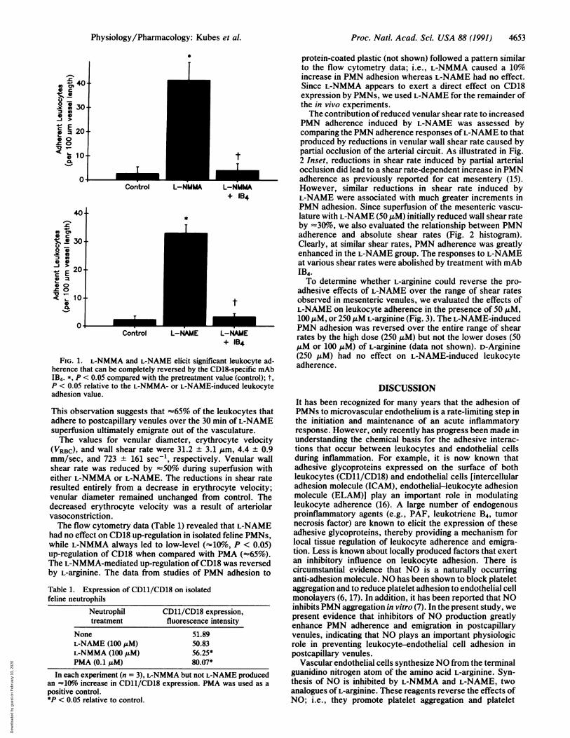
RESULTSFig. 1 illustrates the effects of the L-arginine analoguesL-NMMA (100 ,uM) and L-NAME (100 AM) on PMN adhe-sion in postcapillary venules. Both L-NMMA and L-NAMEelicited a dramatic increase in PMN adhesion (16- and21-fold, respectively) 30 min into the superfusion period.Following mAb IB4 administration, the number of adherentPMNs returned to pretreatment values within 10 min. PMNemigration from venules into the adjacent extravascularcompartment increased from 8.5 + 2.5 to 29.5 ± 1.5 per field.
Dow
nloa
ded
by g
uest
on
Feb
ruar
y 10
, 202
0
Proc. Natl. Acad. Sci. USA 88 (1991) 4653
I.
IControl L-NMMA L-NMMA
+ IB4
*
Control L-NAME L-NAME+ IB4
FIG. 1. L-NMMA and L-NAME elicit significant leukocyte ad-herence that can be completely reversed by the CD18-specific mAbIB4. *, P < 0.05 compared with the pretreatment value (control); t,P < 0.05 relative to the L-NMMA- or L-NAME-induced leukocyteadhesion value.
This observation suggests that -65% of the leukocytes thatadhere to postcapillary venules over the 30 min of L-NAMEsuperfusion ultimately emigrate out of the vasculature.The values for venular diameter, erythrocyte velocity
(VRBC), and wall shear rate were 31.2 + 3.1 A&m, 4.4 ± 0.9mm/sec, and 723 ± 161 sec1, respectively. Venular wallshear rate was reduced by 50%o during superfusion witheither L-NMMA or L-NAME. The reductions in shear rateresulted entirely from a decrease in erythrocyte velocity;venular diameter remained unchanged from control. Thedecreased erythrocyte velocity was a result of arteriolarvasoconstriction.The flow cytometry data (Table 1) revealed that L-NAME
had no effect on CD18 up-regulation in isolated feline PMNs,while L-NMMA always led to low-level (410%, P < 0.05)up-regulation of CD18 when compared with PMA (z65%).The L-NMMA-mediated up-regulation of CD18 was reversedby L-arginine. The data from studies of PMN adhesion to
Table 1. Expression of CD11/CD18 on isolatedfeline neutrophils
Neutrophil CD11/CD18 expression,treatment fluorescence intensity
None 51.89L-NAME (100 ZM) 50.83L-NMMA (100 .M) 56.25*PMA (0.1 AM) 80.07*
In each experiment (n = 3), L-NMMA but not L-NAME producedan -10% increase in CD11/CD18 expression. PMA was used as apositive control.*P < 0.05 relative to control.
protein-coated plastic (not shown) followed a pattern similarto the flow cytometry data; i.e., L-NMMA caused a 10%increase in PMN adhesion whereas L-NAME had no effect.Since L-NMMA appears to exert a direct effect on CD18expression by PMNs, we used L-NAME for the remainder ofthe in vivo experiments.The contribution ofreduced venular shear rate to increased
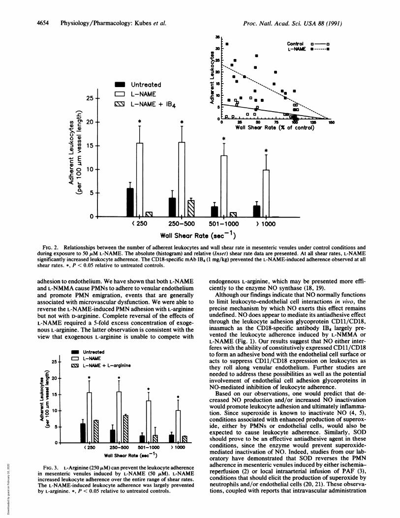
PMN adherence induced by L-NAME was assessed bycomparing the PMN adherence responses ofL-NAME to thatproduced by reductions in venular wall shear rate caused bypartial occlusion of the arterial circuit. As illustrated in Fig.2 Inset, reductions in shear rate induced by partial arterialocclusion did lead to a shear rate-dependent increase in PMNadherence as previously reported for cat mesentery (15).However, similar reductions in shear rate induced byL-NAME were associated with much greater increments inPMN adhesion. Since superfusion of the mesenteric vascu-lature with L-NAME (50 tM) initially reduced wall shear rateby -30%o, we also evaluated the relationship between PMNadherence and absolute shear rates (Fig. 2 histogram).Clearly, at similar shear rates, PMN adherence was greatlyenhanced in the L-NAME group. The responses to L-NAMEat various shear rates were abolished by treatment with mAbIB4.To determine whether L-arginine could reverse the pro-
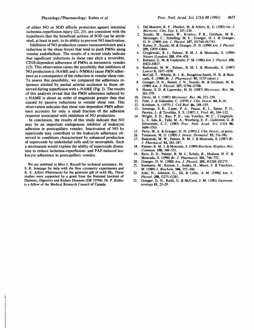
adhesive effects of L-NAME over the range of shear ratesobserved in mesenteric venules, we evaluated the effects ofL-NAME on leukocyte adherence in the presence of 50 ,uM,100 uM, or 250AM L-arginine (Fig. 3). The L-NAME-inducedPMN adhesion was reversed over the entire range of shearrates by the high dose (250 ,uM) but not the lower doses (50,uM or 100 ,uM) of L-arginine (data not shown). D-Arginine(250 AM) had no effect on L-NAME-induced leukocyteadherence.
DISCUSSIONIt has been recognized for many years that the adhesion ofPMNs to microvascular endothelium is a rate-limiting step inthe initiation and maintenance of an acute inflammatoryresponse. However, only recently has progress been made inunderstanding the chemical basis for the adhesive interac-tions that occur between leukocytes and endothelial cellsduring inflammation. For example, it is now known thatadhesive glycoproteins expressed on the surface of bothleukocytes (CD11/CD18) and endothelial cells [intercellularadhesion molecule (ICAM), endothelial-leukocyte adhesionmolecule (ELAM)] play an important role in modulatingleukocyte adherence (16). A large number of endogenousproinflammatory agents (e.g., PAF, leukotriene B4, tumornecrosis factor) are known to elicit the expression of theseadhesive glycoproteins, thereby providing a mechanism forlocal tissue regulation of leukocyte adherence and emigra-tion. Less is known about locally produced factors that exertan inhibitory influence on leukocyte adhesion. There iscircumstantial evidence that NO is a naturally occurringanti-adhesion molecule. NO has been shown to block plateletaggregation and to reduce platelet adhesion to endothelial cellmonolayers (6, 17). In addition, it has been reported that NOinhibits PMN aggregation in vitro (7). In the present study, wepresent evidence that inhibitors of NO production greatlyenhance PMN adherence and emigration in postcapillaryvenules, indicating that NO plays an important physiologicrole in preventing leukocyte-endothelial cell adhesion inpostcapillary venules.
Vascular endothelial cells synthesize NO from the terminalguanidino nitrogen atom of the amino acid L-arginine. Syn-thesis of NO is inhibited by L-NMMA and L-NAME, twoanalogues of L-arginine. These reagents reverse the effects ofNO; i.e., they promote platelet aggregation and platelet
'.0 40.o c0 c
O 0
0-
0 10
11 10i .
0-Q =ooYI a
C E0
< L=0
0.
0 1
Physiology/Pharmacology: Kubes et al.
f
Dow
nloa
ded
by g
uest
on
Feb
ruar
y 10
, 202
0
4654 Physiology/Pharmacology: Kubes et al.
_~ Untreated
25- IEI L-NAMEL-NAME + IB4
20 * *
15 -
5 - I.A a.u
301S
PS3 a0
-J4.15
.~0
5I
a
aU
a
.
< 250 250-500 501-1000 > 1000
Wall Shear Rate (sec-1)FIG. 2. Relationships between the number of adherent leukocytes and wall shear rate in mesenteric venules under control conditions and
during exposure to 50 ,uM L-NAME. The absolute (histogram) and relative (Inset) shear rate data are presented. At all shear rates, L-NAMEsignificantly increased leukocyte adherence. The CD18-specific mAb IB4 (1 mg/kg) prevented the L-NAME-induced adherence observed at allshear rates. *, P < 0.05 relative to untreated controls.
adhesion to endothelium. We have shown that both L-NAMEand L-NMMA cause PMNs to adhere to venular endotheliumand promote PMN emigration, events that are generallyassociated with microvascular dysfunction. We were able toreverse the L-NAME-induced PMN adhesion with L-argininebut not with D-arginine. Complete reversal of the effects ofL-NAME required a 5-fold excess concentration of exoge-nous L-arginine. The latter observation is consistent with theview that exogenous L-arginine is unable to compete with
_ Untreated
25 l L-NAMECO L-NAME + L-arginine
-
10NE
(250 250-500 501-1000 >1000
Wall Shear Rote (soc-1)
FIG. 3. L-Arginine (250 tuM) can prevent the leukocyte adherencein mesenteric venules induced by L-NAME (50 /uM). L-NAMEincreased leukocyte adherence over the entire range of shear rates.The L-NAME-induced leukocyte adherence was largely preventedby L-arginine. *, P < 0.05 relative to untreated controls.
endogenous L-arginine, which may be presented more effi-ciently to the enzyme NO synthase (18, 19).Although our findings indicate that NO normally functions
to limit leukocyte-endothelial cell interactions in vivo, theprecise mechanism by which NO exerts this effect remainsundefined. NO does appear to mediate its antiadhesive effectthrough the leukocyte adhesion glycoprotein CD11/CD18,inasmuch as the CD18-specific antibody IB4 largely pre-vented the leukocyte adherence induced by L-NMMA orL-NAME (Fig. 1). Our results suggest that NO either inter-feres with the ability of constitutively expressed CD11/CD18to form an adhesive bond with the endothelial cell surface oracts to suppress CD11/CD18 expression on leukocytes as
they roll along venular endothelium. Further studies are
needed to address these possibilities as well as the potentialinvolvement of endothelial cell adhesion glycoproteins inNO-mediated inhibition of leukocyte adherence.Based on our observations, one would predict that de-
creased NO production and/or increased NO inactivationwould promote leukocyte adhesion and ultimately inflamma-tion. Since superoxide is known to inactivate NO (4, 5),conditions associated with enhanced production of superox-ide, either by PMNs or endothelial cells, would also beexpected to cause leukocyte adherence. Similarly, SODshould prove to be an effective antiadhesive agent in theseconditions, since the enzyme would prevent superoxide-mediated inactivation of NO. Indeed, studies from our lab-oratory have demonstrated that SOD reverses the PMNadherence in mesenteric venules induced by either ischemia-reperfusion (2) or local intraarterial infusion of PAF (3),conditions that should elicit the production of superoxide byneutrophils and/or endothelial cells (20, 21). These observa-tions, coupled with reports that intravascular administration
Control o-oL-NAME --
a
s-
U) m0 C
0 -0 0-Y 0)
J0)L.X
00 -
< -
0.%-S1
.
a
of
*
*
Proc. Natl. Acad Sci. USA 88 (1991)
Dow
nloa
ded
by g
uest
on
Feb
ruar
y 10
, 202
0
Proc. Natl. Acad. Sci. USA 88 (1991) 4655
of either NO or SOD affords protection against intestinalischemia-reperfusion injury (22, 23), are consistent with thehypothesis that the beneficial actions of SOD can be attrib-uted, at least in part, to its ability to prevent NO inactivation.
Inhibition ofNO production causes vasoconstriction and areduction in the shear forces that tend to push PMNs alongvenular endothelium. The results of a recent study indicatethat significant reductions in shear rate elicit a reversible,CD18-dependent adherence of PMNs in mesenteric venules(15). This observation raises the possibility that inhibitors ofNO production (L-NAME and L-NMMA) cause PMN adher-ence as a consequence of the reduction in venular shear rate.To assess this possibility, we compared the adherence re-sponses elicited by partial arterial occlusion to those ob-served during superfusion with L-NAME (Fig. 2). The resultsof this analysis reveal that the PMN adherence induced byL-NAME is about an order of magnitude greater than thatcaused by passive reductions in venular shear rate. Thisobservation indicates that shear rate-dependent PMN adher-ence accounts for only a small fraction of the adhesionresponse associated with inhibition of NO production.
In conclusion, the results of this study indicate that NOmay be an important endogenous inhibitor of leukocyteadhesion in postcapillary venules. Inactivation of NO bysuperoxide may contribute to the leukocyte adherence ob-served in conditions characterized by enhanced productionof superoxide by endothelial cells and/or neutrophils. Sucha mechanism would explain the ability of superoxide dismu-tase to reduce ischemia-reperfusion- and PAF-induced leu-kocyte adherence in postcapillary venules.
We are indebted to Miss J. Russell for technical assistance, Dr.S. R. Jennings for help with the flow cytometry experiments andK. E. Arfors (Pharmacia) for the generous gift of mAb IB4. Thesestudies were supported by a grant from the National Institute ofDiabetes, Digestive and Kidney Diseases (DK 33594). Dr. P. Kubesis a fellow of the Medical Research Council of Canada.
1. Del Maestro, R. F., Planker, M. & Arfors, K. E. (1982) Int. J.Microcirc. Clin. Exp. 1, 105-120.
2. Suzuki, M., Inauen, W., Kvietys, P. R., Grisham, M. B.,Meininger, C., Schelling, M. E., Granger, H. J. & Granger,D. N. (1989) Am. J. Physiol. 257, H1740-H1745.
3. Kubes, P., Suzuki, M. & Granger, D. N. (1990) Am. J. Physiol.259, G859-G864.
4. Gryglewski, R. J., Palmer, R. M. J. & Moncada, S. (1986)Nature (London) 320, 454-456.
5. Rubanyi, G. M. & Vanhoutte, P. M. (1986) Am. J. Physiol. 250,H822-H827.
6. Radomski, M. W., Palmer, R. M. J. & Moncada, S. (1987)Lancet fi, 1057-1058.
7. McCall, T., Whittle, B. J. R., Boughton-Smith, N. K. & Mon-cada, S. (1988) Br. J. Pharmacol. 95, 517P (abstr.).
8. Granger, D. N., Benoit, J. N., Suzuki, M. & Grisham, M. B.(1989) Am. J. Physiol. 257, G704-G708.
9. House, S. D. & Lipowsky, H. H. (1987) Microvasc. Res. 34,363-379.
10. Davis, M. J. (1987) Microvasc. Res. 34, 223-230.11. Fehr, J. & Dahinden, C. (1979) J. Clin. Invest. 64, 8-16.12. Krisham, A. (1975) J. Cell Biol. 66, 188-193.13. Jennings, S. R., Lippe, P. A., Pauza, K. J., Spear, P. G.,
Pereira, L. & Tevethia, S. S. (1987) J. Virol. 61, 104-112.14. Wright, S. D., Rao, P. E., van Voorhis, W. C., Craigmyle,
L. S., lida, K., Talle, M. A., Westberg, E. F., Goldstein, G. &Silverstein, S. C. (1983) Proc. Natl. Acad. Sci. USA 80,5699-5703.
15. Perry, M. A. & Granger, D. N. (1991) J. Clin. Invest., in press.16. Tonneson, M. G. (1989) J. Invest. Dermatol. 93, 53s-58s.17. Radomski, M. W., Palmer, R. M. J. & Moncada, S. (1987) Br.
J. Pharmacol. 92, 181-187.18. Palmer, R. M. J. & Moncada, S. (1989) Biochem. Biophys. Res.
Commun. 158, 348-352.19. Rees, D. D., Palmer, R. M. J., Schulz, R., Hodson, H. F. &
Moncada, S. (1990) Br. J. Pharmacol. 101, 746-752.20. Granger, D. N. (1988) Am. J. Physiol. 255, H1269-H1275.21. Suematsu, M., Kurose, I., Asako, H., Miura, S. & Tsuchiya,
M. (1989) J. Biochem. 106, 355-360.22. Aoki, N., Johnson, G., III, & Lefer, A. M. (1990) Am. J.
Physiol. 258, G275-G281.23. Granger, D. N., Rutili, G. & McCord, J. M. (1981) Gastroen-
terology 81, 22-29.
Physiology/Pharmacology: Kubes et al.
Dow
nloa
ded
by g
uest
on
Feb
ruar
y 10
, 202
0