Embed Size (px)
Citation preview

UNIT 5
The reductionist approach to study of life forms resulted in increasinguse of physico-chemical concepts and techniques. Majority of thesestudies employed either surviving tissue model or straightaway cell-free systems. An explosion of knowledge resulted in molecular biology.Molecular physiology became almost synonymous with biochemistryand biophysics. However, it is now being increasingly realised thatneither a purely organismic approach nor a purely reductionisticmolecular approach would reveal the truth about biological processesor living phenomena. Systems biology makes us believe that all livingphenomena are emergent properties due to interaction amongcomponents of the system under study. Regulatory network of molecules,supra molecular assemblies, cells, tissues, organisms and indeed,populations and communities, each create emergent properties. In thechapters under this unit, major human physiological processes likedigestion, exchange of gases, blood circulation, locomotion andmovement are described in cellular and molecular terms. The last twochapters point to the coordination and regulation of body events at theorganismic level.
HUMAN PHYSIOLOGY
Chapter 16Digestion and Absorption
Chapter 17Breathing and Exchangeof Gases
Chapter 18Body Fluids andCirculation
Chapter 19Excretory Products andtheir Elimination
Chapter 20Locomotion and Movement
Chapter 21Neural Control andCoordination
Chapter 22Chemical Coordinationand Integration
2015-16(19/01/2015)

ALFONSO CORTI, Italian anatomist, was born in 1822. Corti beganhis scientific career studying the cardiovascular systems ofreptiles. Later, he turned his attention to the mammalianauditory system. In 1851, he published a paper describing astructure located on the basilar membrane of the cochleacontaining hair cells that convert sound vibrations into nerveimpulses, the organ of Corti. He died in the year 1888.
Alfonso Corti(1822 – 1888)
2015-16(19/01/2015)

Food is one of the basic requirements of all living organisms. The major
components of our food are carbohydrates, proteins and fats. Vitamins
and minerals are also required in small quantities. Food provides energy
and organic materials for growth and repair of tissues. The water we take
in, plays an important role in metabolic processes and also prevents
dehydration of the body. Biomacromolecules in food cannot be utilised
by our body in their original form. They have to be broken down and
converted into simple substances in the digestive system. This process of
conversion of complex food substances to simple absorbable forms is
called digestion and is carried out by our digestive system by mechanical
and biochemical methods. General organisation of the human digestive
system is shown in Figure 16.1.
16.1 DIGESTIVE SYSTEM
The human digestive system consists of the alimentary canal and the
associated glands.
16.1.1 Alimentary Canal
The alimentary canal begins with an anterior opening – the mouth, and it
opens out posteriorly through the anus. The mouth leads to the buccal
cavity or oral cavity. The oral cavity has a number of teeth and a muscular
tongue. Each tooth is embedded in a socket of jaw bone (Figure16.2).
This type of attachment is called thecodont. Majority of mammals
including human being forms two sets of teeth during their life, a set of
DIGESTION AND ABSORPTION
CHAPTER 16
16.1 Digestive
System
16.2 Digestion of
Food
16.3 Absorption of
Digested
Products
16.4 Disorders of
Digestive
System
2015-16(19/01/2015)

258 BIOLOGY
Pancreas
Jejunum
Ascending colon
Transverse colon
Duodenum
Gall bladder
Liver
Ileum
Caecum
Vermiform appendix
Anus
Rectum
Descending colon
Oral cavity
Mouth
Submaxillary andsublingual glands
Parotid gland
Pharynx
Oesophagus
Stomach
Figure 16.1 The human digestive system
temporary milk or deciduous teeth replaced by a set of permanent or
adult teeth. This type of dentition is called diphyodont. An adult human
has 32 permanent teeth which are of four different types (Heterodont
dentition), namely, incisors (I), canine (C), premolars (PM) and molars
(M). Arrangement of teeth in each half of the upper and lower jaw in the
order I, C, PM, M is represented by a dental formula which in human
is 2123
2123. The hard chewing surface of the teeth, made up of enamel, helps
in the mastication of food. The tongue is a freely movable muscular organ
attached to the floor of the oral cavity by the frenulum. The upper surface
of the tongue has small projections called papillae, some of which bear
taste buds.
The oral cavity leads into a short pharynx which serves as a common
passage for food and air. The oesophagus and the trachea (wind pipe)
2015-16(19/01/2015)

DIGESTION AND ABSORPTION 259
open into the pharynx. A cartilaginous flap
called epiglottis prevents the entry of food
into the glottis – opening of the wind pipe –
during swallowing. The oesophagus is a
thin, long tube which extends posteriorly
passing through the neck, thorax and
diaphragm and leads to a ‘J’ shaped bag like
structure called stomach. A muscular
sphincter (gastro-oesophageal) regulates the
opening of oesophagus into the stomach.
The stomach, located in the upper left
portion of the abdominal cavity, has three
major parts – a cardiac portion into which
the oesophagus opens, a fundic region and
a pyloric portion which opens into the first
part of small intestine (Figure 16.3). Small
intestine is distinguishable into three
regions, a ‘C’ shaped duodenum, a long
coiled middle portion jejunum and a highly
coiled ileum. The opening of the stomach
into the duodenum is guarded by the pyloric
sphincter. Ileum opens into the large
intestine. It consists of caecum, colon and
rectum. Caecum is a small blind sac which
hosts some symbiotic micro-organisms. A
narrow finger-like tubular projection, the
vermiform appendix which is a vestigial
organ, arises from the caecum. The caecum
opens into the colon. The colon is divided
into three parts – an ascending, a transverse
and a descending part. The descending part
opens into the rectum which opens out
through the anus.
The wall of alimentary canal from
oesophagus to rectum possesses four layers
(Figure 16.4) namely serosa, muscularis,
sub-mucosa and mucosa. Serosa is the
outermost layer and is made up of a thin
mesothelium (epithelium of visceral organs)
with some connective tissues. Muscularis is
formed by smooth muscles usually
arranged into an inner circular and an outer
longitudinal layer. An oblique muscle layer
may be present in some regions. The sub-
mucosal layer is formed of loose connective
Figure 16.3 Anatomical regions of human
stomach
Figure 16.2 Arrangement of different types of
teeth in the jaws on one side andthe sockets on the other side
2015-16(19/01/2015)

260 BIOLOGY
Figure 16.4 Diagrammatic representation of transverse section of gut
tissues containing nerves, blood and lymph
vessels. In duodenum, glands are also present in
sub-mucosa. The innermost layer lining the
lumen of the alimentary canal is the mucosa. This
layer forms irregular folds (rugae) in the stomach
and small finger-like foldings called villi in the
small intestine (Figure 16.5). The cells lining the
villi produce numerous microscopic projections
called microvilli giving a brush border
appearance. These modifications increase the
surface area enormously. Villi are supplied with
a network of capillaries and a large lymph vessel
called the lacteal. Mucosal epithelium has goblet
cells which secrete mucus that help in lubrication.
Mucosa also forms glands in the stomach (gastric
glands) and crypts in between the bases of villi in
the intestine (crypts of Lieberkuhn). All the four
layers show modifications in different parts of the
alimentary canal.
16.1.2 Digestive Glands
The digestive glands associated with the alimentary canal include the
salivary glands, the liver and the pancreas.
Saliva is mainly produced by three pairs of salivary glands, the parotids
(cheek), the sub-maxillary/sub-mandibular (lower jaw) and the sub-
linguals (below the tongue). These glands situated just outside the buccal
cavity secrete salivary juice into the buccal cavity.
Figure 16.5 A section of small intestinal
mucosa showing villi
VilliLacteal
Capillaries
Crypts
Artery
Vein
2015-16(19/01/2015)

DIGESTION AND ABSORPTION 261
Liver is the largest gland of the body weighing about 1.2 to 1.5 kg in
an adult human. It is situated in the abdominal cavity, just below the
diaphragm and has two lobes. The hepatic lobules are the structural and
functional units of liver containing hepatic cells arranged in the form of
cords. Each lobule is covered by a thin connective tissue sheath called
the Glisson’s capsule. The bile secreted by the hepatic cells passes through
the hepatic ducts and is stored and concentrated in a thin muscular sac
called the gall bladder. The duct of gall bladder (cystic duct) along with
the hepatic duct from the liver forms the common bile duct (Figure 16.6).
Figure 16.6 The duct systems of liver, gall bladder and pancreas
The bile duct and the pancreatic duct open together into the duodenum
as the common hepato-pancreatic duct which is guarded by a sphincter
called the sphincter of Oddi.
The pancreas is a compound (both exocrine and endocrine) elongated
organ situated between the limbs of the ‘U’ shaped duodenum. The
exocrine portion secretes an alkaline pancreatic juice containing enzymes
and the endocrine portion secretes hormones, insulin and glucagon.
16.2 DIGESTION OF FOOD
The process of digestion is accomplished by mechanical and chemical
processes.
The buccal cavity performs two major functions, mastication of food
and facilitation of swallowing. The teeth and the tongue with the help of
2015-16(19/01/2015)

262 BIOLOGY
saliva masticate and mix up the food thoroughly. Mucus in saliva helps
in lubricating and adhering the masticated food particles into a bolus.
The bolus is then conveyed into the pharynx and then into the oesophagus
by swallowing or deglutition. The bolus further passes down through
the oesophagus by successive waves of muscular contractions called
peristalsis. The gastro-oesophageal sphincter controls the passage of food
into the stomach.The saliva secreted into the oral cavity contains
electrolytes and enzymes, salivary amylase and
lysozyme. The chemical process of digestion is initiated in the oral cavity
by the hydrolytic action of the carbohydrate splitting enzyme, the salivary
amylase. About 30 per cent of starch is hydrolysed here by this enzyme
(optimum pH 6.8) into a disaccharide – maltose. Lysozyme present in
saliva acts as an antibacterial agent that prevents infections.
StarchSalivary
.
Amylase
pHMaltose
6 8 →
The mucosa of stomach has gastric glands. Gastric glands have three
major types of cells namely -
(i) mucus neck cells which secrete mucus;
(ii) peptic or chief cells which secrete the proenzyme pepsinogen; and
(iii) parietal or oxyntic cells which secrete HCl and intrinsic factor
(factor essential for absorption of vitamin B12
).
The stomach stores the food for 4-5 hours. The food mixes thoroughly
with the acidic gastric juice of the stomach by the churning movements
of its muscular wall and is called the chyme. The proenzyme pepsinogen,
on exposure to hydrochloric acid gets converted into the active enzyme
pepsin, the proteolytic enzyme of the stomach. Pepsin converts proteins
into proteoses and peptones (peptides). The mucus and bicarbonates
present in the gastric juice play an important role in lubrication and
protection of the mucosal epithelium from excoriation by the highly
concentrated hydrochloric acid. HCl provides the acidic pH (pH 1.8)
optimal for pepsins. Rennin is a proteolytic enzyme found in gastric juice
of infants which helps in the digestion of milk proteins. Small amounts of
lipases are also secreted by gastric glands.
Various types of movements are generated by the muscularis layer of
the small intestine. These movements help in a thorough mixing up of
the food with various secretions in the intestine and thereby facilitate
digestion. The bile, pancreatic juice and the intestinal juice are the
secretions released into the small intestine. Pancreatic juice and bile are
released through the hepato-pancreatic duct. The pancreatic juice
contains inactive enzymes – trypsinogen, chymotrypsinogen,
procarboxypeptidases, amylases, lipases and nucleases. Trypsinogen is
activated by an enzyme, enterokinase, secreted by the intestinal mucosa
2015-16(19/01/2015)

DIGESTION AND ABSORPTION 263
into active trypsin, which in turn activates the other enzymes in the
pancreatic juice. The bile released into the duodenum contains bile
pigments (bilirubin and bili-verdin), bile salts, cholesterol and
phospholipids but no enzymes. Bile helps in emulsification of fats, i.e.,
breaking down of the fats into very small micelles. Bile also activates lipases.
The intestinal mucosal epithelium has goblet cells which secrete mucus.
The secretions of the brush border cells of the mucosa alongwith the
secretions of the goblet cells constitute the intestinal juice or
succus entericus. This juice contains a variety of enzymes like
disaccharidases (e.g., maltase), dipeptidases, lipases, nucleosidases, etc.
The mucus alongwith the bicarbonates from the pancreas protects the
intestinal mucosa from acid as well as provide an alkaline medium (pH
7.8) for enzymatic activities. Sub-mucosal glands (Brunner’s glands) also
help in this.
Proteins, proteoses and peptones (partially hydrolysed proteins) in
the chyme reaching the intestine are acted upon by the proteolytic
enzymes of pancreatic juice as given below:
Proteins
Peptones
Proteoses
Trypsin/Chymotrypsin
Carboxyp
eeptidaseDipeptides →
Carbohydrates in the chyme are hydrolysed by pancreatic amylase
into disaccharides.
Polysaccharides starch DisaccharidesAmylase
( ) →
Fats are broken down by lipases with the help of bile into di-and
monoglycerides.
Fats Diglycerides MonoglyceridesLipases
→ →
Nucleases in the pancreatic juice acts on nucleic acids to form
nucleotides and nucleosides
Nucleic acids Nucleotides NucleosidesNucleases → →
The enzymes in the succus entericus act on the end products of the
above reactions to form the respective simple absorbable forms. These
final steps in digestion occur very close to the mucosal epithelial cells of
the intestine.
2015-16(19/01/2015)

264 BIOLOGY
DipeptidesDipeptidases
→ Amino acids
MaltoseMaltase → Glucose + Glucose
Lactose Glucose+ alactoseLactase → G
Sucrose FructoseSucrase → +Glucose
Nucleotides NucleosidesNucleotidases Nucleosidases → →→ +Sugars Bases
Diand Monoglycerides Fatty acids GlycerolLipases
→ +
The breakdown of biomacromolecules mentioned above occurs in the
duodenum region of the small intestine. The simple substances thus
formed are absorbed in the jejunum and ileum regions of the small
intestine. The undigested and unabsorbed substances are passed on to
the large intestine.
No significant digestive activity occurs in the large intestine. The
functions of large intestine are:
(i) absorption of some water, minerals and certain drugs;
(ii) secretion of mucus which helps in adhering the waste (undigested)
particles together and lubricating it for an easy passage.
The undigested, unabsorbed substances called faeces enters into the
caecum of the large intestine through ileo-caecal valve, which prevents
the back flow of the faecal matter. It is temporarily stored in the rectum
till defaecation.
The activities of the gastro-intestinal tract are under neural and
hormonal control for proper coordination of different parts. The sight,
smell and/or the presence of food in the oral cavity can stimulate the
secretion of saliva. Gastric and intestinal secretions are also, similarly,
stimulated by neural signals. The muscular activities of different parts of
the alimentary canal can also be moderated by neural mechanisms, both
local and through CNS. Hormonal control of the secretion of digestive
juices is carried out by local hormones produced by the gastric and
intestinal mucosa.
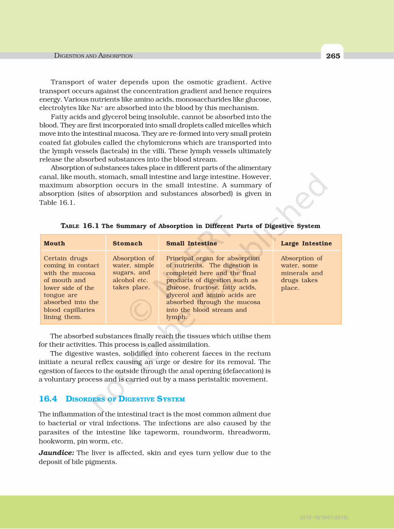
16.3 ABSORPTION OF DIGESTED PRODUCTS
Absorption is the process by which the end products of digestion pass
through the intestinal mucosa into the blood or lymph. It is carried out by
passive, active or facilitated transport mechanisms. Small amounts of
monosaccharides like glucose, amino acids and some electrolytes like
chloride ions are generally absorbed by simple diffusion. The passage of
these substances into the blood depends upon the concentration gradients.
However, some substances like glucose and amino acids are absorbed with
the help of carrier proteins. This mechanism is called the facilitated transport.
2015-16(19/01/2015)
DIGESTION AND ABSORPTION 265
Transport of water depends upon the osmotic gradient. Active
transport occurs against the concentration gradient and hence requires
energy. Various nutrients like amino acids, monosaccharides like glucose,
electrolytes like Na+ are absorbed into the blood by this mechanism.
Fatty acids and glycerol being insoluble, cannot be absorbed into the
blood. They are first incorporated into small droplets called micelles which
move into the intestinal mucosa. They are re-formed into very small protein
coated fat globules called the chylomicrons which are transported into
the lymph vessels (lacteals) in the villi. These lymph vessels ultimately
release the absorbed substances into the blood stream.
Absorption of substances takes place in different parts of the alimentary
canal, like mouth, stomach, small intestine and large intestine. However,
maximum absorption occurs in the small intestine. A summary of
absorption (sites of absorption and substances absorbed) is given in
Table 16.1.
The absorbed substances finally reach the tissues which utilise them
for their activities. This process is called assimilation.
The digestive wastes, solidified into coherent faeces in the rectum
initiate a neural reflex causing an urge or desire for its removal. The
egestion of faeces to the outside through the anal opening (defaecation) is
a voluntary process and is carried out by a mass peristaltic movement.
16.4 DISORDERS OF DIGESTIVE SYSTEM
The inflammation of the intestinal tract is the most common ailment due
to bacterial or viral infections. The infections are also caused by the
parasites of the intestine like tapeworm, roundworm, threadworm,
hookworm, pin worm, etc.
Jaundice: The liver is affected, skin and eyes turn yellow due to the
deposit of bile pigments.
Stomach
Absorption ofwater, simplesugars, and
alcohol etc.takes place.
Small Intestine
Principal organ for absorptionof nutrients. The digestion is
completed here and the finalproducts of digestion such asglucose, fructose, fatty acids,
glycerol and amino acids areabsorbed through the mucosa
into the blood stream andlymph.
Mouth
Certain drugscoming in contact
with the mucosaof mouth and
lower side of thetongue areabsorbed into the
blood capillarieslining them.
Large Intestine
Absorption ofwater, some
minerals anddrugs takes
place.
TABLE 16.1 The Summary of Absorption in Different Parts of Digestive System
2015-16(19/01/2015)
266 BIOLOGY
Vomiting: It is the ejection of stomach contents through the mouth.
This reflex action is controlled by the vomit centre in the medulla. A feeling
of nausea precedes vomiting.
Diarrhoea: The abnormal frequency of bowel movement and increased
liquidity of the faecal discharge is known as diarrhoea. It reduces the
absorption of food.
Constipation: In constipation, the faeces are retained within the rectum
as the bowel movements occur irregularly.
Indigestion: In this condition, the food is not properly digested leading to
a feeling of fullness. The causes of indigestion are inadequate enzyme
secretion, anxiety, food poisoning, over eating, and spicy food.
SUMMARY
The digestive system of humans consists of an alimentary canal and associated
digestive glands. The alimentary canal consists of the mouth, buccal cavity,
pharynx, oesophagus, stomach, small intestine, large intestine, rectum and the
anus. The accessory digestive glands include the salivary glands, the liver (with
gall bladder) and the pancreas. Inside the mouth the teeth masticates the food,
the tongue tastes the food and manipulates it for proper mastication by mixing
with the saliva. Saliva contains a starch digestive enzyme, salivary amylase that
digests the starch and converts it into maltose (disaccharide). The food then passes
into the pharynx and enters the oesophagus in the form of bolus, which is further
carried down through the oesophagus by peristalsis into the stomach. In stomach
mainly protein digestion takes place. Absorption of simple sugars, alcohol and
medicines also takes place in the stomach.
The chyme (food) enters into the duodenum portion of the small intestine
and is acted on by the pancreatic juice, bile and finally by the enzymes in the
succus entericus, so that the digestion of carbohydrates, proteins and fats is
completed. The food then enters into the jejunum and ileum portions of the small
intestine. Carbohydrates are digested and converted into monosaccharides like
glucose. Proteins are finally broken down into amino acids. The fats are converted
to fatty acids and glycerol. The digested end products are absorbed into the body
through the epithelial lining of the intestinal villi. The undigested food (faeces)
enters into the caecum of the large intestine through ileo-caecal valve, which
prevents the back flow of the faecal matter. Most of the water is absorbed in the
large intestine. The undigested food becomes semi-solid in nature and then enters
into the rectum, anal canal and is finally egested out through the anus.
2015-16(19/01/2015)

DIGESTION AND ABSORPTION 267
EXERCISES
1. Choose the correct answer among the following :
(a) Gastric juice contains
(i) pepsin, lipase and rennin
(ii) trypsin, lipase and rennin
(iii) trypsin, pepsin and lipase
(iv) trypsin, pepsin and renin
(b) Succus entericus is the name given to
(i) a junction between ileum and large intestine
(ii) intestinal juice
(iii) swelling in the gut
(iv) appendix
2. Match column I with column II
Column I Column II
(a) Bilirubin and biliverdin (i) Parotid
(b) Hydrolysis of starch (ii) Bile
(c) Digestion of fat (iii) Lipases
(d) Salivary gland (iv) Amylases
3. Answer briefly:
(a) Why are villi present in the intestine and not in the stomach?
(b) How does pepsinogen change into its active form?
(c) What are the basic layers of the wall of alimentary canal?
(d) How does bile help in the digestion of fats?
4. State the role of pancreatic juice in digestion of proteins.
5. Describe the process of digestion of protein in stomach.
6. Give the dental formula of human beings.
7. Bile juice contains no digestive enzymes, yet it is important for digestion. Why?
8. Describe the digestive role of chymotrypsin. Which two other digestive enzymes
of the same category are secreted by its source gland?
9. How are polysaccharides and disaccharides digested?
10. What would happen if HCl were not secreted in the stomach?
11. How does butter in your food get digested and absorbed in the body?
12. Discuss the main steps in the digestion of proteins as the food passes through
different parts of the alimentary canal.
13. Explain the term thecodont and diphyodont.
14. Name different types of teeth and their number in an adult human.
15. What are the functions of liver?
2015-16(19/01/2015)