Embed Size (px)
Citation preview
This content has been downloaded from IOPscience. Please scroll down to see the full text.
Download details:
IP Address: 140.112.77.175
This content was downloaded on 28/02/2016 at 21:55
Please note that terms and conditions apply.
NIR-assisted orchid virus therapy using urchin bimetallic nanomaterials in phalaenopsis
View the table of contents for this issue, or go to the journal homepage for more
2013 Adv. Nat. Sci: Nanosci. Nanotechnol. 4 045006
(http://iopscience.iop.org/2043-6262/4/4/045006)
Home Search Collections Journals About Contact us My IOPscience
IOP PUBLISHING ADVANCES IN NATURAL SCIENCES: NANOSCIENCE AND NANOTECHNOLOGY
Adv. Nat. Sci.: Nanosci. Nanotechnol. 4 (2013) 045006 (6pp) doi:10.1088/2043-6262/4/4/045006
NIR-assisted orchid virus therapy usingurchin bimetallic nanomaterials inphalaenopsisShin-Yu Chen1,6, Liang-Chien Cheng2,6, Chieh-Wei Chen2, Po-Han Lee2,Fengjiao Yu3, Wuzong Zhou3, Ru-Shi Liu2,4, Yi-Yin Do1
and Pung-Ling Huang1,5
1 Department of Horticulture and Landscape Architecture, National Taiwan University, Taipei 10617,Taiwan2 Department of Chemistry, National Taiwan University, Taipei 10617 , Taiwan3 Department of Chemistry, University of St Andrews, St Andrew, UK4 Genomics Research Center, Academia Sinica, Taipei 115, Taiwan5 Graduate Institute of Biotechnology, Chinese Culture University, No. 55, Hwa-Kang Road,Yang Ming Shan, Taipei 11114, Taiwan6 These authors contributed equally to this work.
E-mail: [email protected], [email protected] and [email protected]
Received 22 June 2013Accepted for publication 11 July 2013Published 14 August 2013Online at stacks.iop.org/ANSN/4/045006
AbstractThe use of nanoparticles has drawn special attention, particularly in the treatment of plantdiseases. Cymbidium mosaic virus (CymMV) and Odontoglossum ring spot virus (ORSV) arethe most prevalent and serious diseases that affect the development of the orchid industry. Inthis study we treated nanoparticles as a strategy for enhancing the resistance of orchids againstCymMV and ORSV. After chitosan-modified gold nanoparticles (Au NPs) were injected intoPhalaenopsis leaves, the injected leaves were exposed to 980 nm laser for light–heatconversion. To evaluate virus elimination in the treated Phalaenopsis leaves, the transcripts ofcoat protein genes and the production of viral proteins were assessed by reversetranscription-Polymerase chain reaction and enzyme-linked immunosorbent assay,respectively. The expression of coat protein genes for both CymMV and ORSV wassignificantly lower in the chitosan-modified Au NP-treated Phalaenopsis leaves than in thecontrol. Similarly, the amount of coat proteins for both viruses in the Phalaenopsis leaves waslower than that in the control (without nanoparticle injection). We propose that thetemperature increase in the chitosan-modified Au NP-treated Phalaenopsis tissues after laserexposure reduces the viral population, consequently conferring resistance against CymMVand ORSV. Our findings suggest that the application of chitosan-modified Au NPs is apromising new strategy for orchid virus therapy.
Keywords: cymbidium mosaic virus, odontoglossum ring spot virus, gold, nanoparticles,photothermal therapy
Classification numbers: 2.04, 4.02, 5.08
1. Introduction
Orchidaceae is one of the largest families of angiosperms,comprising approximately 20 000–25 000 species. Orchid
Content from this work may be used under the terms ofthe Creative Commons Attribution 3.0 licence. Any further
distribution of this work must maintain attribution to the author(s) and thetitle of the work, journal citation and DOI.
flowers are morphologically diverse, as in the patterns ofcolors in sepals, petals and lips. In addition, the column helpsfacilitate pollination [1]. The genus Phalaenopsis is a popularpot plant with high economic value in the flower marketworldwide because of its graceful appearance, long floweringage and wide variety of species.
Cymbidium mosaic virus (CymMV) and odontoglossumring spot virus (ORSV) are the most prevalent and serious
2043-6262/13/045006+06$33.00 1 © 2013 Vietnam Academy of Science & Technology

Adv. Nat. Sci.: Nanosci. Nanotechnol. 4 (2013) 045006 S-Y Chen et al
diseases that affect the development of the orchid industry [2].As indicated by statistical data, CymMV and ORSV infect atleast 80% of all virus-infected orchids [3]. The variability incoat protein gene sequences of CymMV and ORSV are highlyconserved, enabling these two viruses to naturally infectorchids worldwide [4]. CymMV and ORSV reduce yieldand diminish the quality of orchid flowers and plant vigor,thereby affecting the economic value of orchids. CymMVinduces floral and foliar necrosis and ORSV causes ring spotson leaves. Although the plant exhibits symptoms, such ascolor breakage in flowers, blossom brown necrotic streaksmay possibly result from mixed infection by CymMV andORSV [5]. These two viruses mostly infect cultivated orchidsbut very few wild orchids [2]. Nevertheless, no effectivemethod for protecting orchid plants from viral diseases hasbeen developed.
To date, the most popular method is to use plantgenetic transformation to solve the disease problem, but thisapproach presents certain disadvantages: it is time consuming,costly and involves lengthy culture procedures to producestable transgenic plants. Nanoparticles have recently beenextensively applied in plants. For example, researchers havedelivered nanoparticles into plant tissues, with the materialsremaining stable in plant cell or tissue [6–8]. Despite thisprogress, limited studies have been devoted to crop productionor quality improvement by nanoparticle application. No cleardirection has emerged as to establishing a direct relationshipbetween plant disease and nanoparticle function.
Nanomaterials have been widely investigated in manyfields, including energy and biology [9–11]. Investigationson bio-application, however, focus on animals. Given thisbackdrop, we used nanoparticles in CymMV and ORSV astwo types of orchid virus therapy in Phalaenopsis. To thebest of our knowledge, this study is the first to attempt theuse of gold nanoparticle (Au NP) injection for application inorchid virus therapy. This study provides a fast and convenientmethod for enhancing disease resistance in orchids.
2. Experimental
2.1. Synthesis of urchin-like Au/Ag nanomaterials
The synthesis route of urchin-like Au/Ag bimetallicnanomaterials was presented elsewhere [12]. In brief, thenanomaterials were fabricated by mixing 20 µl 10 mMchloroauric acid (HAuCl4), 2 µl 10 mM aqueous silvernitrate (AgNO3), and 1 ml milli-Q deionized water in a2 ml centrifugal tube. Then, 4 µl of 100 mM ascorbic acid(AA) was quickly added to the mixture, and vigorouslyshaken for 20 s. The mixture instantly changed from alight yellowish solution into a bluish solution, implyingthe fabrication of urchin-like Au/Ag nanomaterialsby reduction. After a 1 min reaction, 1 wt% aqueousO-carboxymethylchitosan or fluorescein isothiocyanate(FITC)-capped O-carboxymethylchitosan was chosen assurfactant and added into the solution for reacting one moreday. The mixture was centrifuged at 9279 g three times andredispersed into deionized water for further investigation.The morphologies of urchin-like Au/Ag nanomaterials can bealtered by tuning metal precursors.
2.2. Plant materials and growth conditions
Phalaenopsis ‘Sogo Yukidian’ was used in the analysis ofnear-infrared light (NIR)-assisted virus therapy. The plantswere kept in a greenhouse with natural light and a controlledtemperature of 27/22 ◦C (day/night). They were checked forthe absence of two prevalent orchid viruses, CymMV andORSV by detecting the coat protein through enzyme-linkedimmunoassay (ELISA) before experimentation.
2.3. Virus inoculation and nanoparticle injection
The virus-free Phalaenopsis plants were inoculated byagroinfiltration of Agrobacterium tumefaciens containingCymMV or ORSV viral vector before the nanoparticleinjection experiment. Agrobacterium was grown overnight at28 ◦C and diluted to optical density at 600 nm (OD600) = 0.5with an infiltration buffer (0.5% d-glucose, 50 mM MES,2 mM Na3PO4 · 12H2O, and 100 µM acetosyringone) [13].Successful systemic infection with the virus was confirmedby ELISA [14]. For leaf injection, nanoparticles were injectedinto the leaf surface with a 1 ml syringe in all treatments. After1 day, the targeted leaf was irradiated using a 980 nm laser andcollected 1, 3 and 7 days after treatment. The treated materialswere frozen in liquid nitrogen and stored at 80 ◦C for futureinvestigation.
2.4. Collection and processing of samples for microscopyanalysis
Approximately 1 mm thick, hand-cut cross-sections of thesamples were collected 48 and 72 h after application ofFITC-labeled nanoparticles. Then, the cross-sections wereobserved under a microscope (Leica MZ FL-III). The imagesof the sections were taken with a Zeiss AxioCam MRcmonochrome cooled-CCD camera. Transmission electronmicroscopic (TEM) and high resolution TEM (HRTEM)images were recorded on a Jeol JEM-2010 electronmicroscope operating at 200 kV
2.5. Quantitative reverse transcription polymerase chainreaction (RT-PCR)
Total ribonucleic acid (RNA) was extracted from thedissected leaves by using a MaestroZolTM RNA PLUSExtraction Reagent (Omics Biotechnology), and treatedwith RNase-free DNase (Stratagene) to remove residualDNA. For quantitative RT-PCR, the RNA template wasmixed with KAPA SYBR Fast One-step qRT-PCR Kit(ABI Prism, KAPA Biosystem) on a Biometra RT-PCRsystem (TAIGEN Bioscience Corporation, Taiwan). Theprimers for CymMV coat protein gene were as follows:5′-TACTACGCAAAAGTGGTGTGGAAT-3′ as forward pri-mer and 5′-AGAGCATAGAGAGTGTTGGTGGAG-3′ asreverse primer. The ORSV coat protein gene was ampli-fied using 5′-AATCAACCTTTGTACCAATTCTCTG-3′ asforward primer and 5′-AGACTTGATTGTACGTACCAGTTCC-3′ as reverse primer. The housekeeping geneactin (PACT4, AY134752) was amplified using5′-GCTGGCATATATTGCTCTTGATTAT-3′ as forwardprimer and 5′-ATCCAAACACTGTACTTCCTCTCTG-3′ as
2
Adv. Nat. Sci.: Nanosci. Nanotechnol. 4 (2013) 045006 S-Y Chen et al
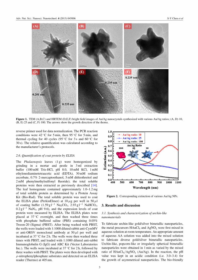
Figure 1. TEM (A,B,C) and HRTEM (D,E,F) bright field images of Au/Ag nanocrystals synthesized with various Au/Ag ratios; (A, D) 10,(B, E) 25 and (C, F) 100. The arrows show the growth direction of the thorns.
reverse primer used for data normalization. The PCR reactionconditions were 42 ◦C for 5 min, then 95 ◦C for 5 min, andthermal cycling for 40 cycles (95 ◦C for 3 s and 60 ◦C for30 s). The relative quantification was calculated according tothe manufacturer’s protocols.
2.6. Quantification of coat protein by ELISA
The Phalaenopsis leaves (1 g) were homogenized bygrinding in a mortar and pestle in 3 ml extractionbuffer (100 mM Tris-HCl, pH 8.0, 10 mM KCl, 1 mMethylenediaminetetraacetic acid (EDTA), 30 mM sodiumascorbate, 0.7% 2-mercaptoethanol, 5 mM dithiothreitol and2 mM phenylmethylsulfonyl fluoride); the total solubleproteins were then extracted as previously described [14].The leaf homogenate contained approximately 1.0–1.2 mgof total soluble protein as determined by a Protein AssayKit (Bio-Rad). The total soluble protein was used to coatthe ELISA plate (PerkinElmer) at 10 µg per well in 50 µlof coating buffer (1.59 g l−1 Na2CO3, 2.93 g l−1 NaHCO3,0.2 g l−1 NaN3, pH 9.6), and the expression levels of coatprotein were measured by ELISA. The ELISA plates wereplaced at 37 ◦C overnight, and then washed three timeswith phosphate buffered saline (PBS) containing 0.05%Tween-20 (Sigma) (PBST). After being washed with PBST,the wells were loaded with 1:1000 diluted rabbit anti-CymMVor anti-ORSV monoclonal antibody at 50 µl per well andincubated at 37 ◦C for 2 h. The wells were then washed threetimes with PBST, and loaded with 1:1000 diluted anti-rabbitImmunoglobulin G (IgG) and ABC Kit (Vector LaboratoriesInc.). The wells were incubated at 37 ◦C for 2 h, followed bythree washes with PBST. The plates were then developed withρ-nitrophenylphosphate substrates and detected on an ELISAreader (Thermo) at 405 nm.
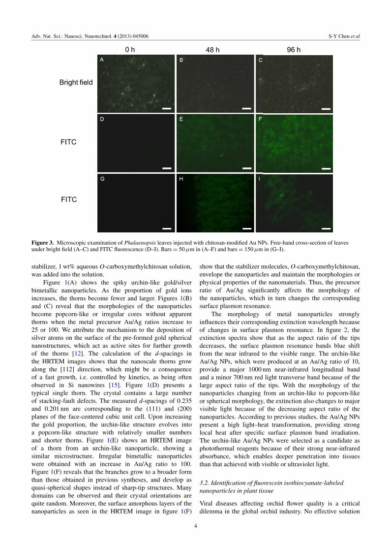
Figure 2. Corresponding extinction of various Au/Ag NPs.
3. Results and discussion
3.1. Synthesis and characterization of urchin-likenanomaterials
To fabricate urchin-like gold/silver bimetallic nanoparticles,the metal precursors HAuCl4 and AgNO3 were first mixed inaqueous solution at room temperature. An appropriate amountof aqueous AA solution was added into the mixed solutionto fabricate diverse gold/silver bimetallic nanoparticles.Urchin-like, popcorn-like or irregularly spherical bimetallicnanoparticles were obtained in 1 min as varied by the mixedratio of HAuCl4/AgNO3 (Au/Ag). In the reaction, the pHvalue was kept in an acidic condition (i.e. 3.0–3.4) forthe growth of asymmetrical nanoparticles. The bio-friendly
3
Adv. Nat. Sci.: Nanosci. Nanotechnol. 4 (2013) 045006 S-Y Chen et al
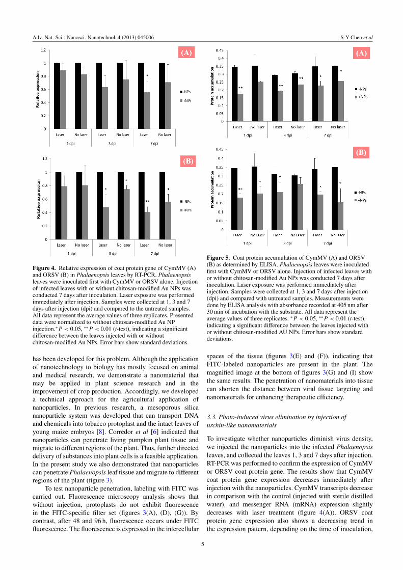
Figure 3. Microscopic examination of Phalaenopsis leaves injected with chitosan-modified Au NPs. Free-hand cross-section of leavesunder bright field (A–C) and FITC fluorescence (D–I). Bars = 50 µm in (A–F) and bars = 150 µm in (G–I).
stabilizer, 1 wt% aqueous O-carboxymethylchitosan solution,was added into the solution.
Figure 1(A) shows the spiky urchin-like gold/silverbimetallic nanoparticles. As the proportion of gold ionsincreases, the thorns become fewer and larger. Figures 1(B)and (C) reveal that the morphologies of the nanoparticlesbecome popcorn-like or irregular cores without apparentthorns when the metal precursor Au/Ag ratios increase to25 or 100. We attribute the mechanism to the deposition ofsilver atoms on the surface of the pre-formed gold sphericalnanostructures, which act as active sites for further growthof the thorns [12]. The calculation of the d-spacings inthe HRTEM images shows that the nanoscale thorns growalong the [112] direction, which might be a consequenceof a fast growth, i.e. controlled by kinetics, as being oftenobserved in Si nanowires [15]. Figure 1(D) presents atypical single thorn. The crystal contains a large numberof stacking-fault defects. The measured d-spacings of 0.235and 0.201 nm are corresponding to the (111) and (200)planes of the face-centered cubic unit cell. Upon increasingthe gold proportion, the urchin-like structure evolves intoa popcorn-like structure with relatively smaller numbersand shorter thorns. Figure 1(E) shows an HRTEM imageof a thorn from an urchin-like nanoparticle, showing asimilar microstructure. Irregular bimetallic nanoparticleswere obtained with an increase in Au/Ag ratio to 100.Figure 1(F) reveals that the branches grow to a broader formthan those obtained in previous syntheses, and develop asquasi-spherical shapes instead of sharp-tip structures. Manydomains can be observed and their crystal orientations arequite random. Moreover, the surface amorphous layers of thenanoparticles as seen in the HRTEM image in figure 1(F)
show that the stabilizer molecules, O-carboxymethylchitosan,envelope the nanoparticles and maintain the morphologies orphysical properties of the nanomaterials. Thus, the precursorratio of Au/Ag significantly affects the morphology ofthe nanoparticles, which in turn changes the correspondingsurface plasmon resonance.
The morphology of metal nanoparticles stronglyinfluences their corresponding extinction wavelength becauseof changes in surface plasmon resonance. In figure 2, theextinction spectra show that as the aspect ratio of the tipsdecreases, the surface plasmon resonance bands blue shiftfrom the near infrared to the visible range. The urchin-likeAu/Ag NPs, which were produced at an Au/Ag ratio of 10,provide a major 1000 nm near-infrared longitudinal bandand a minor 700 nm red light transverse band because of thelarge aspect ratio of the tips. With the morphology of thenanoparticles changing from an urchin-like to popcorn-likeor spherical morphology, the extinction also changes to majorvisible light because of the decreasing aspect ratio of thenanoparticles. According to previous studies, the Au/Ag NPspresent a high light–heat transformation, providing stronglocal heat after specific surface plasmon band irradiation.The urchin-like Au/Ag NPs were selected as a candidate asphotothermal reagents because of their strong near-infraredabsorbance, which enables deeper penetration into tissuesthan that achieved with visible or ultraviolet light.
3.2. Identification of fluorescein isothiocyanate-labelednanoparticles in plant tissue
Viral diseases affecting orchid flower quality is a criticaldilemma in the global orchid industry. No effective solution
4
Adv. Nat. Sci.: Nanosci. Nanotechnol. 4 (2013) 045006 S-Y Chen et al
Figure 4. Relative expression of coat protein gene of CymMV (A)and ORSV (B) in Phalaenopsis leaves by RT-PCR. Phalaenopsisleaves were inoculated first with CymMV or ORSV alone. Injectionof infected leaves with or without chitosan-modified Au NPs wasconducted 7 days after inoculation. Laser exposure was performedimmediately after injection. Samples were collected at 1, 3 and 7days after injection (dpi) and compared to the untreated samples.All data represent the average values of three replicates. Presenteddata were normalized to without chitosan-modified Au NPinjection.∗ P < 0.05, ∗∗ P < 0.01 (t-test), indicating a significantdifference between the leaves injected with or withoutchitosan-modified Au NPs. Error bars show standard deviations.
has been developed for this problem. Although the applicationof nanotechnology to biology has mostly focused on animaland medical research, we demonstrate a nanomaterial thatmay be applied in plant science research and in theimprovement of crop production. Accordingly, we developeda technical approach for the agricultural application ofnanoparticles. In previous research, a mesoporous silicananoparticle system was developed that can transport DNAand chemicals into tobacco protoplast and the intact leaves ofyoung maize embryos [8]. Corredor et al [6] indicated thatnanoparticles can penetrate living pumpkin plant tissue andmigrate to different regions of the plant. Thus, further directeddelivery of substances into plant cells is a feasible application.In the present study we also demonstrated that nanoparticlescan penetrate Phalaenopsis leaf tissue and migrate to differentregions of the plant (figure 3).
To test nanoparticle penetration, labeling with FITC wascarried out. Fluorescence microscopy analysis shows thatwithout injection, protoplasts do not exhibit fluorescencein the FITC-specific filter set (figures 3(A), (D), (G)). Bycontrast, after 48 and 96 h, fluorescence occurs under FITCfluorescence. The fluorescence is expressed in the intercellular
Figure 5. Coat protein accumulation of CymMV (A) and ORSV(B) as determined by ELISA. Phalaenopsis leaves were inoculatedfirst with CymMV or ORSV alone. Injection of infected leaves withor without chitosan-modified Au NPs was conducted 7 days afterinoculation. Laser exposure was performed immediately afterinjection. Samples were collected at 1, 3 and 7 days after injection(dpi) and compared with untreated samples. Measurements weredone by ELISA analysis with absorbance recorded at 405 nm after30 min of incubation with the substrate. All data represent theaverage values of three replicates. ∗ P < 0.05, ∗∗ P < 0.01 (t-test),indicating a significant difference between the leaves injected withor without chitosan-modified AU NPs. Error bars show standarddeviations.
spaces of the tissue (figures 3(E) and (F)), indicating thatFITC-labeled nanoparticles are present in the plant. Themagnified image at the bottom of figures 3(G) and (I) showthe same results. The penetration of nanomaterials into tissuecan shorten the distance between viral tissue targeting andnanomaterials for enhancing therapeutic efficiency.
3.3. Photo-induced virus elimination by injection ofurchin-like nanomaterials
To investigate whether nanoparticles diminish virus density,we injected the nanoparticles into the infected Phalaenopsisleaves, and collected the leaves 1, 3 and 7 days after injection.RT-PCR was performed to confirm the expression of CymMVor ORSV coat protein gene. The results show that CymMVcoat protein gene expression decreases immediately afterinjection with the nanoparticles. CymMV transcripts decreasein comparison with the control (injected with sterile distilledwater), and messenger RNA (mRNA) expression slightlydecreases with laser treatment (figure 4(A)). ORSV coatprotein gene expression also shows a decreasing trend inthe expression pattern, depending on the time of inoculation,
5

Adv. Nat. Sci.: Nanosci. Nanotechnol. 4 (2013) 045006 S-Y Chen et al
Figure 6. Pictured presentation content. Urchin-like nanomaterialscan convert light into heat via NIR irradiation and eliminate thecymbidium mosaic virus and Odontoglossum ring spot virus inPhalaenopsis.
and expression decreases in comparison with the control(injected with sterile distilled water) (figure 4(B)). Theresults reveal that laser is significantly related to decreasedCymMV or ORSV gene expression and protein accumulationby enhancing the effect of nanoparticles. As treatmentprogressed, however, the transcript level of CymMV andORSV transcripts gradually decreases. Consistent with thisresult, CymMV and ORSV coat proteins were also detectedby ELISA. We found that in the plant injected with thenanoparticles, CymMV and ORSV coat protein accumulationlevels are lower than that in the control. mRNA expressionslightly decreases with laser treatment (figure 5).
We found that CymMV and ORSV expression decreasestwo- to three-fold, and protein accumulation decreases one-to two-fold, suggesting that nanoparticle injection with lasertreatment effectively reduces CymMV and ORSV coat proteingene expression and protein accumulation (figures 4 and 5).Chitosan, which was selected as nanoparticle stabilizer, mayinhibit virus growth. Hirano [16] pointed out that chitinaseisoforms are one group of pathogenesis-related proteins inplants. As a chitinase isoform, chitosan may be used topromote tissue growth and differentiation in tissue culture,as well as to induce plant defense. Moreover, virus reductionmay be attributed to the ability of nanoparticles to convertabsorbed near-infrared irradiation into heat (figure 6). Localtemperature increases, thereby preventing virus differentiationand directly eliminating the virus. As presented in theresults, photo-induced virus elimination efficiently enhancestherapeutic behavior. In the future, nanomaterials may beconjugated with specific proteins to target CymMV andORSV for a more highly efficient treatment system.
4. Conclusion
We have demonstrated that urchin-like Au/Ag bimetallicnanoparticles can enter virus tissue via injection. Intercellularnanoparticles may decrease the gap between targeting virustissue and enhancing therapeutic behavior. Moreover, injectednanomaterials can kill viruses and increase resistance ofplants against orchid viruses; this approach is therefore aconvenient method for conferring resistance to orchids againstCymMV and ORSV. Finally, this study can be a particularlyessential reference for future research and lead to a betterunderstanding of the application of nanoparticles in enhancingvirus resistance in plants.
Acknowledgments
The authors would like to thank the Council of Agriculture(grant number 101AS-9.1.1-FD-Z1) of Taiwan andNational Science Council (contracts numbers NSC101-2113-M-002-014-MY3 and NSC 101-3113-P-002-021)for financially supporting this research.
References
[1] Yu H and Goh C J 2001 Plant Physiol. 127 1390[2] Zettler F W, Ko N J, Wisler G C, Elliott M S and Wong S M
1990 Plant Dis. 74 621[3] Wong S M, Chng C G, Lee Y H, Tan K and Zettler F W 1994
Crop. Prot. 13 235[4] Ajjikuttira P A, Lim-Ho C L, Woon M H, Ryu K H,
Chang C A, Loh C S and Wong S M 2002 Arch. Virol.147 1943
[5] Hu J S, Ferreira S, Wang M and Xu M Q 1993 Plant Dis.77 464
[6] Corredor E et al 2009 BMC Plant Biol. 9 1[7] Gonzalez-Melendi P, Fernandez-Pacheco R, Coronado M J,
Corredor E, Testillano P S, Risueno M C, Marquina C,Ibarra M R, Rubiales D and Perez-De-Luque A 2008 Ann.Bot. 101 187
[8] Torney F, Trewyn B G, Lin V S Y and Wang K 2007 NatureNanotechnol. 2 295
[9] Chen H M et al 2012 ACS Nano 6 7362[10] Cheng L C, Chen H M, Lai T C, Chan Y C, Liu R S, Sung J C,
Hsiao M, Chen C H, Her L J and Tsai D P 2013 Nanoscale5 3931
[11] Cheng L C, Jiang X M, Wang J, Chen C Y and Liu R S 2013Nanoscale 5 3547
[12] Cheng L C, Huang J H, Chen H M, Lai T C, Yang K Y,Liu R S, Hsiao M, Chen C H, Her L J and Tsai D P 2012J. Mater. Chem. 22 2244
[13] Sparkes I A, Runions J, Kearns A and Hawes C 2006 NatureProtocol. 1 2019
[14] Arakawa T, Chong D K X, Merritt J L and Langridge W H R1997 Transgenic Res. 6 403
[15] Su Z X, Dickinson C, Wan Y T, Wang Z L, Wang Y W, Sha Jand Zhou W Z 2010 Cryst. Eng. Commun. 12 2793
[16] Hirano S 1999 Polym. Int. 48 732
6