Embed Size (px)
Citation preview
Analytical tools for characterizing biopharmaceuticals and theimplications for biosimilars
Steven A. Berkowitz1, John R. Engen2, Jeffrey R. Mazzeo3, and Graham B. Jones2
1Analytical Development, Biogen Idec, 14 Cambridge Center, Cambridge, Massachusetts 02142,USA2Department of Chemistry & Chemical Biology, The Barnett Institute of Chemical & BiologicalAnalysis, Northeastern University, 360 Huntington Ave., Boston, Massachusetts 02115, USA3Waters Corporation, 34 Maple Street, Milford, Massachusetts 01757, USA
AbstractBiologics such as monoclonal antibodies are much more complex than small-molecule drugs,which raises challenging questions for the development and regulatory evaluation of follow-onversions of such biopharmaceutical products (also known as biosimilars) and their clinical useonce patent protection for the pioneering biologic has expired. With the recent introduction ofregulatory pathways for follow-on versions of complex biologics, the role of analyticaltechnologies in comparing biosimilars with the corresponding reference product is attractingsubstantial interest in establishing the development requirements for biosimilars. Here, we discussthe current state of the art in analytical technologies to assess three characteristics of proteinbiopharmaceuticals that regulatory authorities have identified as being important in developmentstrategies for biosimilars: post-translational modifications, three-dimensional structures andprotein aggregation.
The clinical and commercial success of biologics such as monoclonal antibodies andrecombinant versions of endogenous proteins is transforming the pharmaceutical industry. In2010, worldwide sales of all biologics approached the US$100 billion mark1, and by 2015 itis expected that more than 50% of new drug approvals will be biologics2, rising to morethan 70% by 2025 (REF. 3). As these drugs begin to come off patent, substantialopportunities exist for other companies to make copies or ‘generic’ versions of these drugs.
For small-molecule drugs, abbreviated regulatory pathways for the development andintroduction of generic versions of the drug (following the expiration of patent protection onthe original product) have been established for more than 25 years. Rather than requiringgeneric versions to undergo the same level of evaluation as the original drug, including
© 2012 Macmillan Publishers Limited. All rights reserved
Correspondence to G.B.J. [email protected].
Competing interests statementThe authors declare competing financial interests.
FURTHER INFORMATIONDraft guidances for biosimilars on FDA website: http://www.fda.gov/Drugs/DevelopmentApprovalProcess/HowDrugsareDevelopedandApproved/ApprovalApplications/TherapeuticBiologicApplications/Biosimilars/default.htmEMA website (scientific guidance documents on biosimilar medicines): http://www.ema.europa.eu/ema/index.jsp?curl=pages/regulation/general/general_content_000408.jsp&mid=WC0b01ac058002958cFDA website 27 October 2010 press release: http://www.fda.gov/Safety/Recalls/ucm231639.htmrap.ID Particle Systems website: http://www.rapid.comALL LINKS ARE ACTIVE IN THE ONLINE PDF
NIH Public AccessAuthor ManuscriptNat Rev Drug Discov. Author manuscript; available in PMC 2013 July 17.
Published in final edited form as:Nat Rev Drug Discov. ; 11(7): 527–540. doi:10.1038/nrd3746.
NIH
-PA Author Manuscript
NIH
-PA Author Manuscript
NIH
-PA Author Manuscript
clinical trials, abbreviated approval for the same purposes is generally based ondemonstrating that the generic drug is pharmaceutically equivalent (that is, it contains thesame active ingredient in the same purity, strength, dosage form and route of administration)and bioequivalent (that is, it is absorbed into the body at a similar rate and extent) to theoriginal drug4. Consequently, abbreviated approval is considerably less expensive toachieve, thus dramatically lowering the costs of generic drugs. This has led to thewidespread use of generic versions and substantial cost savings for health-care systems; arecent paper noted that in 2009 almost 75% of small-molecule drug prescriptions dispensedin the United States were for generics, and the approval of a generic drug resulted in averagesavings of 77% of the original product’s cost within 1 year5.
However, for biologics, establishing a regulatory pathway for the introduction of follow-onversions of the original product (once its patent protection has expired) is much morechallenging than for small molecules. Some simple biologics — for example, small peptidessuch as recombinant insulin and recombinant human growth hormone — can be wellcharacterized by established analytical approaches, which has facilitated the regulatoryapproval of follow-on versions under abbreviated pathways (based in part on data from theoriginal drug and in part on analytical data and limited clinical data in some cases)4;however, many biologics such as monoclonal antibodies and other recombinant therapeuticproteins are much larger and more complex. For such biologics, the extent to which existinganalytical technologies can be used to support the likelihood of clinical comparabilitybetween a follow-on version and the original product is much more limited than for small-molecule drugs, and it is not possible to demonstrate that the two products are absolutelyidentical.
Consequently, a key question for the development and regulation of follow-on biologics —also known as biosimilars — is how much and what kind of data are needed to establish thatthe differences between similar (although not identical) products are not clinicallyimportant4.
Clearly, the overall success of developing a biosimilar — as has been the case with genericsmall-molecule drugs — will depend on the ability of the biosimilar sponsor to offer ahighly similar, safe and efficacious drug product at a cost saving that will encourage health-care providers to purchase it over the original product while still allowing the biosimilarsponsor to make an adequate profit. If the bar of comparability or similarity is set too high,the economics of biosimilar development may not be sufficiently attractive for companies toparticipate. However, if the bar of comparability or similarity is set too low, the drug’sefficacy and the safety of patients could be in jeopardy. With the setting of this bar in thehands of government regulators (coupled with the recent or imminent expiration of patentprotection for a growing number of commercially successful biologics), regulatoryauthorities globally have been developing pathways for the introduction of biosimilars thatare intended to realize the ultimate desired benefits.
In Europe, the European Medicines Agency (EMA) introduced the first operatingframework in 2005 (REF. 6) for a path towards developing and marketing biosimilars. Sincethen, European biosimilar guidelines have been described7 (see the EMA website) and 13biosimilars have been approved and are still on the market8. In the United States, the 2009Biologics Price Competition and Innovation (BPCI) Act empowered the US Food and DrugAdministration (FDA) to develop a pathway to introduce biosimilars within the UnitedStates, and the draft guidelines were announced on 9 February 2012 (see the FDA website)(BOXES 1,2). In developing this draft, a hearing by the FDA9 was conducted in 2010 toseek input from stakeholders on four main areas related to biosimilars: “First, what scientificand technical factors should the agency consider in determining whether the biological
Berkowitz et al. Page 2
Nat Rev Drug Discov. Author manuscript; available in PMC 2013 July 17.
NIH
-PA Author Manuscript
NIH
-PA Author Manuscript
NIH
-PA Author Manuscript
product is highly similar to the reference product, notwithstanding minor differences inclinically inactive components? Second, what scientific and technical factors should theagency consider in determining the appropriate analytical, animal, and clinical study orstudies to assess the nature and impact of actual or potential structural differences betweenthe proposed biosimilar product and the reference product? Third, what range of structuraldifferences between a proposed biosimilar product and the reference product is consistentwith the standard “highly similar” and may be acceptable in a 351(k) application if theapplicant can demonstrate the absence of any clinically meaningful differences between theproposed biosimilar product and the reference product? Fourth, under what circumstancesshould the agency consider finding that animal studies or a clinical study or studies are‘unnecessary’ for submission of a 351(k) application?”
Box 1
Commentary on the FDA’s proposed regulatory pathway for biosimilars inthe United States
The 2009 Biologics Price Competition and Innovation (BPCI) Act empowered the USFood and Drug Administration (FDA) to develop a pathway to approve biosimilars. InFebruary 2012 the FDA released three draft documents (see the FDA website) in supportof developing the initial pathway that sponsors of biosimilars need to follow to achievedrug approval, and on 11 May 2012 the FDA held a 1 day public hearing to obtain inputon the draft guidance documents. The new pathway outlined by the FDA, summarizedpictorially in BOX 2, is based on a risk based approach using what the agency calls“totality of the evidence”.
In principle, at first sight this approach is not novel. The approval of anybiopharmaceutical product is based on the totality of the evidence provided to the FDA inthe data package filing that any drug sponsor normally provides to support drug approval(whether that is for a completely new drug or for a change to an existing drug, includinga second generation drug made by the same sponsor) or to gain approval to conduct aclinical trial. This “totality of the evidence” data package includes biochemical,biophysical, biological, toxicology and clinical data. However, for a biosimilar, it isalready known that the original drug is sufficiently safe and efficacious from the work ofthe innovator (along with the drug’s commercial history). The crucial question becomes:how similar or comparable to the innovator’s drug does the biosimilar have to be in orderto take advantage of the innovator’s experience and the drug’s long history (particularlywith regard to the extent of clinical data needed to support biosimilar approval)?
One key conclusion emphasized by the FDA in its recent guidance and in priorpublications4,5 is that the answer to this question is not unique. Although there are clearlycore features corresponding to information that is normally provided in a chemistry,manufacturing and controls (CMC) filing that will be needed in all biosimilar filings (forexample, information on aggregation, impurities, and so on), several paths could befollowed to achieve the goal of obtaining an abbreviated approval for a biosimilar,especially without the need to conduct clinical trials. The answer is therefore formulatedon a case by case basis. Minimal clinical studies (or perhaps even no clinical studies)might be sufficient for the approval of the biosimilar, if biochemical, biophysical andbiological data (structural and functional analysis) can demonstrate that the innovatordrug (also known as the reference product) and its biosimilar are identical (or similarenough) and that there is no effect of any difference in the mode of formulation,container closure as well as handling and administration, so that equivalence in clinicalperformance (pharmacokinetics/pharmacodynamics (PK/PD) and immunogenicity) canbe assured.
Berkowitz et al. Page 3
Nat Rev Drug Discov. Author manuscript; available in PMC 2013 July 17.
NIH
-PA Author Manuscript
NIH
-PA Author Manuscript
NIH
-PA Author Manuscript
An important feature in enabling the agency to make clear and accurate decisions as towhat is needed for the approval of a biosimilar will rest on delivering a robust andcomprehensive structural and functional analysis package for the molecule. Such a datapackage requires more extensive information than what is normally provided in drugfilings for innovator products. In stating the requirement for more information, theagency has referenced the use of orthogonal, state of the art and fingerprint methods that,although not yet validated (or even very difficult to validate), are based on sound science.
For biologics, proving that two separate manufactured lots are identical is virtuallyimpossible. Even an innovator company cannot manufacture its own biologic so that it isabsolutely identical on a lot to lot basis owing to the inherent level of complexity of thesemolecules and the way in which they are made. The best a manufacturer of a biologic cando is to demonstrate consistency in manufacture, with attributes that fall within a set ofacceptable specification criteria that regulators have agreed to through a history of testingand characterization. Such a history of information concerning the innovator’s drug is notknown by the sponsor of the biosimilar. However, as a result of filings and historical dataprovided to them by the innovator, known as “prior knowledge”, regulatory agencieshave this information or key subsets of this information. This prior knowledge mayinclude unique biochemical, biophysical, biological and maybe even toxicology andclinical (including PK/PD and immunogenicity) data that the sponsor of the biosimilarmay not be aware of, in addition to the normal standard filing information. Hence, toreduce development time and costs for the sponsor of the biosimilar, as well as achieveoptimum utilization of FDA resources, a new paradigm for interaction would be neededbetween sponsors of biosimilars and the FDA. In such a paradigm, biosimilar sponsorswould have early, effective and active dialogue (known as a stepwise approach) with theFDA to understand what this “totality of the evidence” data package should include,especially in terms of understanding what kind of toxicological and clinical data will berequired. Even after a biosimilar receives approval, pharmacovigilance studies will needto be implemented to mitigate any additional unknown potential risks associated withbiosimilars relative to the innovator drug.
Finally, the FDA documents indicate that clinical studies will be needed for a biosimilarto reach a level of identity — in terms of clinical performance and immunogenicity —that is considered to be close enough to that of the innovator drug for it to be used“interchangeably”. Such interchangeability could be established before the approval of abiosimilar via clinical studies, post approval through additional clinical trials or possiblythrough pharmacovigilance studies. It should be noted that at present neither the FDA northe European Medicines Agency (EMA) has provided a clear definition and draftguidance on “interchangeability” (see BOX 2 for the FDA’s statement oninterchangeability).
Box 2
Summary of the draft pathway for biosimilars in the United States
The figure below summarizes the key points in the draft documents released by the USFood and Drug Administration (FDA) (see the FDA website) that initiate its plans for thedevelopment of the path for obtaining approval of a biosimilar in the United States.Selected points of this process are listed below.
• Owing to the uniqueness of this process, the FDA recommends a stepwiseapproach involving a substantial level of interaction of the biosimilar sponsorwith the FDA (step a).
Berkowitz et al. Page 4
Nat Rev Drug Discov. Author manuscript; available in PMC 2013 July 17.
NIH
-PA Author Manuscript
NIH
-PA Author Manuscript
NIH
-PA Author Manuscript
• Part of the uniqueness resides in the level of prior knowledge of the innovatordrug (step b).
• Another part of the uniqueness of this process is in assessing the level ofcomparability or biosimilarity (usually referred to as ‘highly similar’) of thebiosimilar to the innovator drug (also known as the reference product) (step c).In conducting this assessment, several different lots (of both the biosimilar andthe reference product) should be used to understand the data space variability ofthe biosimilar and the innovator drug.
• The first experimental key step in the biosimilar approval process is to assessthe comparative structural and functional analysis between the biosimilar andthe reference product (step d). In performing this assessment, the FDAemphasizes the added use of “orthogonal methods” and “fingerprint likemethods”. These latter methods may represent more advanced or state of the artanalytical characterization methods that have not been validated but must bescientifically sound. The biosimilar sponsor should realize at this stage that themore extensive, comprehensive and robust the comparability process, the lowerthe likelihood of requiring data from animal (toxicology) and clinical (human)studies (see below).
• Using prior information coupled with the outcome from step d, the level oftoxicology and clinical data required will be assessed and determined (step e).At a minimum, pharmacokinetics/pharmacodynamics (PK/PD) andimmunogenicity data will need to be provided in support of biosimilarity;however, other more extensive toxicology and/or clinical studies may berequired, depending on prior knowledge (step b) and the results from step d,especially in reference to the need to address residual uncertainties regardingbiosimilarity.
• Biosimilarity, as defined by the FDA, is when: “The biological product is highlysimilar to the reference product notwithstanding minor differences in clinicallyinactive components … [and] there are no clinically meaningful differencesbetween the biological product and reference product in terms of safety, purityand potency of the product.” In making this assessment, the biosimilar sponsorneeds to realize that the assessment is made on a case by case basis (step f).
• A higher standard of biosimilarity is defined as “interchangeability” (step g).Although the FDA has not clearly defined this attribute, the agency did say that“an applicant must provide sufficient information to demonstrate biosimilarity,and also to demonstrate that the biological product can be expected to producethe same clinical result as the reference product in any given patient and, if thebiological product is administered more than once to an individual, the risk interms of safety or diminished efficacy of alternating or switching between theuse of the biological product and the reference product is not greater than therisk of using the reference product without such alternation or switch (seesection 351(i)(3) of the PHS [Public Health Service] Act)”.
• Following the approval of either a biosimilar or a biosimilar that can be usedunder the status of interchangeability, a pharmacovigilance programme needs tobe in place to ensure the safety and the effectiveness of the biosimilartherapeutic protein product, especially during the initial phase of its introductioninto the public setting (step h).
Berkowitz et al. Page 5
Nat Rev Drug Discov. Author manuscript; available in PMC 2013 July 17.
NIH
-PA Author Manuscript
NIH
-PA Author Manuscript
NIH
-PA Author Manuscript

Related to several of these areas, three properties of therapeutic proteins — in the opinion ofthe FDA10 — cannot be sufficiently measured but are deemed to be important forunderstanding the behaviour of protein drugs: post-translational modifications, three-dimensional structures and protein aggregation. Given the need to rapidly improve ourability to measure these three properties, it is an opportune moment to survey the currentlyavailable analytical techniques used to generate information about these properties, whichare discussed here in light of the guidance issued by the FDA and EMA. Before consideringthe three main areas identified for analytical improvement, however, it is important todiscuss some of the general challenges in assessing comparability.
General challenges in assessing comparabilityThe first challenge in assessing the comparability of biologics is understanding exactly whatis meant by the terms ‘comparable’, ‘similar’ and ‘highly similar’. During the developmentof innovative biologics, there are often numerous changes to the manufacturing process (forvarious reasons), sometimes including changes even after the drug is commercialized. As aresult, the innovator will need to conduct comparability studies to show regulators that drugproducts before and after process changes are ‘comparable’ in order to be able to use thesepost-change drug products in any subsequent clinical trials or in existing commerciallicensed products11. Here, we will refer to this type of comparison as an ‘internal’comparison. In the case of developing a biosimilar version of an originator’s biologic, themanufacturer of the biosimilar must execute a far more challenging task of comparability inorder to prove that its biosimilar is ‘similar’ (the term used by the EMA) or ‘highly similar’(the term used by the FDA) to the innovator’s biologic. Here, we refer to this type ofcomparability as an ‘external’ comparison. It should be noted that the terms comparable,similar and highly similar are specific terms used by the EMA and the FDA for the abovementioned purposes7 (see the FDA website), and so care should be taken in their use; seebelow.
The approach taken by the EMA to deal with the primary issues of comparability forbiosimilars — defined here as external comparability — is a logical extension of the conceptof internal comparability set forth initially by the FDA and conceptualized by theInternational Conference on Harmonisation (ICH) Q5E guidelines (BOX 3). In fact, theEuropean Generic medicines Association (EGA) indicated in its response to the FDA’spublic biosimilar meeting in 2010 (REF. 9) that: “It should be noted that the EMA neverlimited the application of comparability to a single company making manufacturing changesto its own product, instead it was held to be a scientific evaluation irrespective of the source(manufacturing change or biosimilar) to the two entities being compared. Thus, while we allrecognize that ICH Q5E defines comparable as having ‘highly similar quality attributes’,and limits its use to a single company with their own product, the European Unionguidelines use the terminology of similarity and comparability interchangeably within thesame regulatory documents, and recognize that the two terms refer to the same scientific
Berkowitz et al. Page 6
Nat Rev Drug Discov. Author manuscript; available in PMC 2013 July 17.
NIH
-PA Author Manuscript
NIH
-PA Author Manuscript
NIH
-PA Author Manuscript
principles.” The EGA further added: “As successfully used in the European Union, thescientific grounding of similarity and comparability is the same, and the regulatorsendeavour to apply these principles consistently to both, original biologics and biosimilars.A high degree of similarity forms the basis for abbreviated clinical programs, approvability,extrapolation of indications, interchangeability, and trust by patients and health-careproviders.”
Box 3
Existing characterization guidelines and relationship to comparabilitystudies
Perhaps the most relevant existing industry standards in place to determine thecomparability of biopharmaceuticals are the International Conference on Harmonisation(ICH)’s harmonized tripartite guidelines referred to as Q5E (Comparability ofBiotechnological/Biological Products Subject to Changes in their ManufacturingProcess), which were issued by the ICH Working Group in November 2004 (REF. 143).These guidelines were developed for innovators of biopharmaceutical products to helpthem establish internal comparability between products before and after process changesin order to implement process changes for their products. These guidelines focus on fourmain criteria described in Q5E section 2.2.2:
Physicochemical properties (as defined in ICH Q6B)
The ICH Q6B guidelines on physicochemical properties cover comparability exercises ofthe molecular entity to assess the degree of molecular homo and/or heterogeneity. Higherorder structural analyses are recommended, including the assessment of any changes insecondary, tertiary and quaternary structures. Dedicated guidelines are described in theICH Q6B document. If higher order structural information cannot be obtained, it issuggested that relevant biological assays could be used to confirm or supportconformational equivalency between the two products.
Biological activity/bold
Manufacturers are encouraged to provide meaningful and insightful bioassay data inorder to highlight or confirm the absence of any effects that might be attributed tochanges in process. These assays may also be used in some circumstances as surrogatesto confirm higher order structures. In cases where physicochemical or biological assaysare not sufficient to confirm that higher order structures of two products are maintained,non clinical and clinical studies may be necessary.
Immunochemical properties
In the case of antibodies and antibody based products, the manufacturer should confirmthat specific attributes of the (two) products are maintained and/or comparable byappropriate assays. Given that small differences in glycosylation and/or deglycosylationare known to have immunogenic consequences, this is an area of special interest in thedevelopment of guidelines for biosimilars and interchangeable biologics.
Purity, impurities and contaminants
The guidelines on purities, impurities and contaminants, which are intended to ensurethat isoforms and degradation products are detected, specify that a combination ofanalytical procedures should be used to confirm that the purity profile of the product (orproducts) has not changed. It is assumed that the manufacturer will take measures toprevent the formation of contaminants, and any that are discovered through this processare identified and characterized using appropriate methods.
Berkowitz et al. Page 7
Nat Rev Drug Discov. Author manuscript; available in PMC 2013 July 17.
NIH
-PA Author Manuscript
NIH
-PA Author Manuscript
NIH
-PA Author Manuscript
However, what is potentially the Achilles heel in the above EGA statement is that such anapproach implies that the manufacturer of a biosimilar has: first, the same extensiveknowledge base about the properties and behaviour of the innovator’s drug as the innovator;and second, the availability of appropriate reference material for making comparisons.Regarding the second point, although there may be a perception that the sourcing ofreference standard material could be solved by purchasing the innovator’s product on theopen market, this approach presents its own set of problems12. The formulation matrix of theinnovator’s product could interfere with necessary comparison studies between theinnovator’s drug and the biosimilar product. Some type of deformulation or extraction of theactive drug material from its original drug product formulation may be required13,14, andsuch sample processing could lead to alterations in the generated reference material thatcould in turn cause misleading comparison results15,16. This problem was recognized byboth the EMA and the FDA, and hence a validated deformulation and extraction process ofthe active drug substance from the commercial drug product will be required6 (see the FDAwebsite).
A possible approach for achieving such a valid deformulation and extraction process mayrequire the manufacturer of the biosimilar to place its biosimilar drug molecule into thesame formulation as the innovator drug product and co-process it with the innovator drugproduct in order to conduct a potentially more valid comparability study.
A further interesting point related to appropriate reference material is the source of thereference material relative to the jurisdiction of the regulatory body reviewing thebiosimilar. In the case of the EMA the reference material must be derived from itsjurisdiction, whereas in the case of the FDA reference material outside its jurisdiction can beused if an acceptable bridging study of this material to a US-licensed reference product canbe provided.
A second challenge is defining the acceptable level of comparability to confirm the claimthat two or more biologics are in fact comparable, similar or highly similar. As alreadystated, the use of the term ‘identical’ is inappropriate owing to the inherent complexity ofprotein bio-pharmaceuticals and their manufacturing processes. For protein drugs, simplechanges such as a single-amino-acid mutation or covalent modification (a post-translationalmodification (PTM)) may lead to small perturbations in higher-order structure. Such smallperturbations might result in the drug not functioning or, worse, cause a drug to malfunctionvia aggregation or immunogenicity. Therefore, defining the meaning of the termscomparable, similar and highly similar as acceptability levels is a real challenge. Inparticular, as noted earlier in this article, in the biosimilar area the term ‘similar’ isspecifically used by the EMA, whereas the term ‘highly similar’ is specifically used by theFDA. Such word usage unfortunately only further adds to the confusion, and hence it isworth repeating that care should be taken in determining how and when to apply theseterms. In the case of the EMA, various product-specific biosimilar guidelines have beengenerated (see the EMA website) (for example, for recombinant erythropoietins andrecombinant granulocyte colony-stimulating factor) and continue to be generated (forexample, for interferon-β and monoclonal antibodies) to help guide applicants of biosimilarsto more effectively formulate their comparability studies.
Below, we discuss the three properties of therapeutic proteins that, in the FDA’s view,cannot be sufficiently measured (as described above), as well as the analytical tools that areavailable and being developed to assess these properties. We also discuss key challenges forthe development of biosimilars in light of regulatory guidance related to these threeproperties.
Berkowitz et al. Page 8
Nat Rev Drug Discov. Author manuscript; available in PMC 2013 July 17.
NIH
-PA Author Manuscript
NIH
-PA Author Manuscript
NIH
-PA Author Manuscript
Post-translational modificationThere are many types of PTMs of proteins17,18. Common PTMs are glycosylation (whichincludes galactosylation, fucosylation, high mannose derivatives and sialylation), oxidation,phosphorylation, sulphation, lipidation, disulphide bond formation and deamidation. In mostcases these chemical changes occur in vivo but some chemical changes can also occur invitro: for example, during various stages of manufacture such as purification and storage.
Changes to proteins as a result of PTMs can have a role in protein activity19, and so there isa need to characterize and understand them when manufacturing biologics. In addition,PTMs may influence the immunogenicity of biologics. Several comprehensive reviews20,21
have addressed the immunogenicity of biologics and include discussions on the potentialcontribution (or contributions) of PTMs to immunogenicity. In the case of several types ofPTMs (for example, deamidation, oxidation and differential glycosylation), directconnections between the post-translationally modified biologics and immunogenicity havenot been clinically demonstrated22. Nevertheless, it is known that PTMs can alter proteinstructure and cause aggregation, and that such changes can cause immunogenicity problems(glycosylation and amino acid isomerization are also discussed below). Therefore, it iscrucial to be aware of PTMs and understand them for each biologic. Furthermore, todemonstrate the reproducible production of a biologic, the manufacturer needs to monitorPTMs at many steps during the manufacturing process23, which requires the identification ofPTMs, control of their levels and assessment of their impact on the protein. The levels ofvariability in PTMs that are allowed in a developmental or commercial biologic can varyand are generally determined by information concerning their importance and priormanufacturing history. In most cases, a definitive impact (on clinical outcome) of variabilityin the amount, level and forms of the different PTMs is not known24.
Although PTM characterization is a very daunting task in proteomics studies (in which thereare hundreds, if not thousands, of proteins to investigate all in the same sample)25, biologicsrepresent a much simpler case. For biologics, the sequence and identity of the protein isknown and the various modified forms can generally be isolated, sometimes in largequantities. As a result, characterization of PTMs is more straightforward than it is in a globalproteomics investigation. Software from several instrumentation vendors is designed to firstidentify whether modifications are present, in what quantity and, in some cases, where themodifications have occurred. The analysis of PTMs is not, however, without limitations.Sophisticated instrumentation, skilled analysts and time are all required and althoughcomputation and automation have eased the burden, there is still a long way to go.
Mass spectrometry (MS) is a valuable tool for detecting and investigating proteinmodifications (by monitoring mass differences), for determining where they occur on theprotein (by analysing peptide fragments and their mass alterations) and for elucidating whatcauses them (by comparing different types of samples, storage conditions and formulations)(FIG. 1; see REFS 26–28 for examples). Several detailed reviews17,29–33 on the topic ofapplying MS to PTM detection have highlighted a few trends. One recent trend is theincreasing emphasis on understanding the nature of the sugars involved in glycosylation.Given that various cell lines, expression hosts and protocols can result in differentglycosylation patterns, measuring and understanding glycosylation by MS is crucial.Furthermore, to understand batch-to-batch variability and to compare innovator andbiosimilar proteins, it is necessary to determine where glycosylation occurs (which isrelatively straightforward in peptide mapping experiments) as well as the structure andcontent of the individual sugars (which is much more challenging, even for MS).
Berkowitz et al. Page 9
Nat Rev Drug Discov. Author manuscript; available in PMC 2013 July 17.
NIH
-PA Author Manuscript
NIH
-PA Author Manuscript
NIH
-PA Author Manuscript

For example, a study comparing the glycosylation pattern of an innovator tissue-typeplasminogen activator (tPA) and its biosimilar demonstrated a ~2.5-fold greater amount ofglycosylation at one N-linked site in the innovator material versus the biosimilar material34.Differences between an innovator monoclonal antibody, trastuzumab (Herceptin;Genentech/Roche), and a bio-similar were readily detected with liquid chromatography–MS(LC–MS)35, including changes in glycosylation and amino acid mutations in the heavychain. Analysis of recombinant and human-derived factor IX glycans following enzymaticdigestion and LC–MS36 indicated that the fucosylation site on the human-derived proteinand the recombinant version was different (FIG. 1). The clinical ramifications of thesedifferences are not clear. It was recently reported24 that although changes in glycosylationlevels were observed for etanercept (Enbrel; Amgen/Pfizer), darbepoetin alfa (Aranesp;Amgen/Kyowa Hakko Kirin) and rituximab (Rituxan/Mabthera; Biogen Idec/Genentech/Roche) as a result of post-approval process changes, the clinical ramifications of thedifferences were unknown.
Differences involving one or two monosaccharides and/or their linkage specificity (mostlyfrom sugars that are not made in humans but are instead made in the mammalian cells usedto produce the recombinant protein) have been linked to immunological responses in only afew cases37. Galactose-α-1,3-galactose linkages or terminal-α-1,3-galactose have beenconnected to anaphylaxis associated with cetuximab (Erbitux; Bristol-Myers Squibb/Lilly/Merck Serono)38 and immune response to bovine thrombin39. The sialic acid N-glycolylneuraminic acid (Neu5Gc; also known as NGNA) is known to be associated withimmunogenicity issues40, and reduction or elimination of this sugar in recombinant proteinsis highly desirable41. A recent comparison42 of Neu5Gc in cetuximab and panitumumab(Vectibix; Amgen) showed the presence of Neu5Gc in cetuximab but not in panitumumab,and showed that addition of Neu5Ac (N-acetylneuraminic acid) to the culture media reducesthe incorporation of Neu5Gc. The ability to detect differences in glycans by MS is thereforevaluable for biologics43, including for the analysis of human glycosylation pathwaysthrough the discovery of non-consensus additions44 and for the assessment of whetherdifferent glycosylation patterns have clinical relevance19.
Another trend in the application of MS to the study of PTMs is the awareness that themodification itself could be altered by the analysis method. To correct this limitation, therehas been an increasing role for alternatives to classical collision-induced dissociation MSfragmentation methods in recent years. The value of the bottom-up approach of digesting theprotein into peptides and characterizing the mass of all the peptides is well established, butanalysis in a top-down fashion is now also proving to be useful29,45,46. In a top-downanalysis, characterization of the whole protein (rather than a collection of its digested pieces)is conducted inside the mass spectrometer, in which fragmentation methods such as electrontransfer dissociation (ETD) and electron capture dissociation (ECD)22,47,48 are used tofragment the protein into smaller pieces for more detailed analysis. ETD and ECD are notonly able to preserve labile PTMs, but common labile structural elements of a protein (suchas disulphide bonds) that may be scrambled in other fragmentation methods are alsopreserved49.
The detection of amino acid isomerization is another important form of PTM that isbenefiting from ongoing method development. One amino acid of particular concern isaspartic acid as it can isomerize readily to form isoaspartic acid (isoAsp), which has beenreported to have some undesirable immunogenic consequences50,51. The formation ofisoAsp can potentially occur either through direct isomerization of Asp or throughdeamidation of asparagine, proceeding through a common succinate intermediate. Suchsubtle isomeric differences (the mass difference is zero) obviously present a substantialchallenge for detection and analysis, and this issue is compounded in large proteins. MS can
Berkowitz et al. Page 10
Nat Rev Drug Discov. Author manuscript; available in PMC 2013 July 17.
NIH
-PA Author Manuscript
NIH
-PA Author Manuscript
NIH
-PA Author Manuscript
be used to detect and characterize isoAsp, particularly with ETD52,53 or ECD54–56. In onecase involving β-amyloid protein57, enzymatic cleavage at aspartic acid residues (using theendoproteinase AspN) allowed the isoAsp/Asp ratio to be quantified by ETD–MS to levelsas low as 0.5%. However, considerable expertise and time may be required to achieve suchanalyses.
A good way to localize, detect and characterize N-linked glycans on proteins is toenzymatically release them and then carry out MS analysis. However, in the case of O-linked glycans58 caution must be exercised as the chemical cleavage and release steps mayoften result in breakdown of the glycan itself, and so alternative strategies may need to beconsidered. Although the presence of O-glycosylated sites can be detected on intact proteinsusing new, softer ionizing MS techniques, information on the covalent structure at thelinkage site is typically difficult to obtain by MS. One method that is well suited to O-glycananalysis is nuclear magnetic resonance (NMR) spectroscopy. Indeed, this was thebreakthrough technique used in the well-publicized case of heparin contamination; here, thecontaminant — over-sulphated chondroitin sulphate — was detected by 600 MHz NMRspectroscopy, which revealed the location, identity and orientation of the O-glycan chains59.
One of the general limiting factors in traditional NMR analysis of proteins has been the largesample size required (up to ~20 mg) for meaningful data collection. However, recentadvances in NMR technologies — including flow NMR and most recently microcoil NMR— have changed this outlook. In the latter case, it is possible — with limits of detectionbelow 100 pmol — to obtain high-quality spectra. In a recent example60 of an analysis ofcyanobacterial cell extracts, it was possible to detect metabolic components representing 1%of a mixture (the total quantity injected was 30 μg) after LC separation. Coupling MS withNMR makes the analyses more powerful. LC–MS–NMR can accommodate the largedisparities in the requirements (such as sample mass and analysis time) of the twotechniques, and directs the more demanding NMR technique to ambiguities or gaps in theMS analysis60. An attractive extension of this for biologics could be to start with proteomicanalyses of mixtures separated by high-performance LC (HPLC) and ultra-performance LC(UPLC), and to then split the effluent for both MS and MS–MS, followed by microcoilNMR analysis of selected features. Future developments in LC–MS–NMR technology couldhopefully be applied to PTM characterization.
Higher-order structureAlthough major advances have been made in developing tools for primary structure analysis,as discussed above, what is missing in these studies is an understanding of the impact of thePTM on the three-dimensional structure of the biopharmaceutical product. The higher-orderstructure of proteins — that is, the secondary, tertiary and quaternary structure — is whatgives each protein its three-dimensional shape and ultimately affects the way that the proteinfunctions. So, the ability to monitor the higher-order structure of intact proteins has obviousimportance for the characterization of biologics61–63. Differences in higher-order structurecan not only provide potential clues to any observed biological and/or immunologicaldifferences between proteins and variant forms (that is, proteins with PTMs) but can alsoserve as a means for assessing the lack of comparability between versions of an innovatorproduct before and after process changes, as well as for establishing a lack of comparabilitybetween an innovator product and a biosimilar version.
Although characterization of the higher-order structure of biopharmaceutical proteinsrepresents a substantial challenge, progress is being made on various fronts using specializedanalytical methods64. The higher-order structure of proteins results from a collection offorces, many of which are weak when independently considered but strong when combined.
Berkowitz et al. Page 11
Nat Rev Drug Discov. Author manuscript; available in PMC 2013 July 17.
NIH
-PA Author Manuscript
NIH
-PA Author Manuscript
NIH
-PA Author Manuscript
These weak interactions have a major role in the overall conformation and conformationaldynamics of all proteins. During the manufacture of biopharmaceutical proteins (whichincludes steps such as cell culture overexpression, purification, transportation, storage andhandling), factors can be encountered that can perturb some of these weak forces in proteins,causing alterations to the three-dimensional structure without changing the primary structureof the protein. Such changes can effectively be referred to as ‘silent’ changes as they haveno chemical covalent signature that one could detect as a fingerprint of the change. Withoutanalytical tools to detect and characterize these conformational changes, their impact onstructure–function relationships remains unknown. Finally, protein dynamics in solution(involving protein structure ‘breathing’, polypeptide chain bending, flexing and local proteinstructure unfolding) is another important attribute of protein behaviour that has — until now— been virtually unknown in biopharmaceutical analysis owing to the lack of appropriatepractical routine analytical tools (as discussed below).
The two main techniques for protein structure determination are X-ray crystallography andNMR. Unfortunately, the application of these technologies for the purpose of higher-orderstructural comparability studies presents major problems. X-ray crystallography isimpractical for routine testing as the sample must first be crystallized — something that mayor may not be possible for structural reasons (that is, owing to the presence of PTMs and/ordisordered regions in the protein). Structural analysis via X-ray crystallography is alsogenerally too time-consuming and complex for routine biopharmaceutical analysis. In thecase of NMR, the large size of protein biopharmaceuticals (with their complex array ofstructural elements), the relatively low sensitivity of the NMR signals and the low naturalabundance of active nuclei (other than the 1H isotope) in biopharmaceuticals all make thistechnique impractical for routine higher-order structure comparability studies. However,under certain circumstances where smaller protein biopharmaceuticals are being developed,simple one-dimensional 1H NMR to produce NMR fingerprints may provide a very usefuland practical comparability assessment65.
The use of two-dimensional NMR for biopharmaceuticals has been reported66,67; however,these applications have again been for small protein biopharmaceuticals and have requiredlengthy data acquisition times just for one sample (especially when natural abundance levelsof active nuclei are being used), also making them impractical for routine application wheremany samples need to be compared. We note that although present opportunities for usingNMR in routine protein biopharmaceutical comparability analyses are limited, it appearsthat the use of this technology for polysaccharide biopharmaceuticals is more feasible68,69
(see the brief discussion below concerning NMR and heparin).
Other classical biophysical techniques such as circular dichroism, fluorescence, differentialscanning calorimetry, isothermal calorimetry, analytical ultracentrifugation (AUC), sizeexclusion chromatography (SEC) and various dye-binding assays can be used to characterizeprotein structure15,70,71. A major limitation of these methods is that they generally provideinformation that is derived from a sum of signal inputs that come from many different partsof the protein being probed. The information obtained from these types of measurementscorresponds to a global average over the entire structure of the biopharmaceutical. Forexample, circular dichroism measurements indicate only the average percentage of eachbasic type of major secondary structural element (α-helix, β-sheet and random coil) that ispresent in a protein. If a protein with several α-helices is analysed and only part of one α-helix is modified in one of the two samples being compared, the biopharmaceutical scientisthas the difficult task of trying to discriminate between two large signals that only differ by asmall amount. Furthermore, the ability to detect this difference is also a function of theinherent noise level, which in many cases is large in comparison with the amplitude of theactual difference.
Berkowitz et al. Page 12
Nat Rev Drug Discov. Author manuscript; available in PMC 2013 July 17.
NIH
-PA Author Manuscript
NIH
-PA Author Manuscript
NIH
-PA Author Manuscript
Classical biophysical tools are thus not well suited for detecting small, subtle differences inprotein conformation or even major changes in a biopharmaceutical product when thosealtered molecules represent only a small fraction (population) of the total ensemble ofmolecules present in solution. Even when changes are observed, these techniques providelittle to no information about the location in the molecule where the change has occurred.Hence, other methods that could provide more information in a practical and routine waywould be very desirable.
The use of protein labelling methods such as hydrogen deuterium exchange MS (HDX–MS)72 and covalent labelling strategies73 can be valuable in detecting small changes in thehigher-order structure of a biopharmaceutical. Importantly, when changes are detected, thesetechniques can provide information as to where in the biopharmaceutical molecule thechange has occurred. In HDX–MS, the protein is exposed to deuterated water (D2O) andexchangeable hydrogen atoms become labelled with deuterium, thereby adding oneadditional measurable mass unit per amino acid. The exchange is a function of proteinstructure and dynamics. By conducting the exchange and analysis in real time underphysiological conditions, information on the dynamics of the protein is obtained, as well ascomparative information on the higher-order structure of the protein. HDX–MS providesinformation-rich data, is highly sensitive (requiring only 1–2 nanomoles of material forcomplete characterization), can be automated74 and can localize where changes ordifferences exist in specific regions of the biopharmaceutical; furthermore, resolution at thesingle amino acid level is on the horizon75. Current limitations continue to be the analysistime required to interpret the data and the complexity introduced by solution conditions thatare incompatible with MS analyses (for example, detergent-containing buffers); however,these have been substantially improved in recent years72,76.
Recently, HDX–MS was used to study the conformation and conformational dynamics of arecombinant immunoglobulin G1 monoclonal antibody (FIG. 1), and was used to monitorthe changes to its higher-order structure after removing its glycans, altering itsoligosaccharide structure and following receptor interaction77,78. The ability to acquire theseand similar data could have an instrumental role in building a comprehensive map of thestructural aspects of a biopharmaceutical that are crucial for understanding its function,maximizing its stability and understanding how PTMs such as glycans influence local andglobal features and properties. Because HDX–MS can reveal details of the higher-orderstructure of proteins, as well as protein dynamics, the method has the potential to become animportant tool for studies assessing comparability between innovator products and theirbiosimilars79–81. It should also be mentioned that HDX–MS can be used to monitor theeffects of the interactions of biopharmaceuticals with target proteins that are deemed to beimportant to their biological function82–84, so this technique could also have a key role inthe discovery and development of biopharmaceuticals.
As many aspects of higher-order structure can be driven by the proper formation ofdisulphide bonds, knowing their location and verifying that they are formed correctly duringprotein manufacture and handling is crucial85. In recent examples86,87, comprehensivecharacterization of the disulphide bonding pattern was performed on the blood-clottingregulator tPA and on therapeutic monoclonal antibodies using LC–MS. Key to analyses ofthese biopharmaceuticals was the use of carefully controlled enzymatic digests followed bysoft ionization (electrospray ionization) and gentle fragmentation (ETD). Before thedevelopment of such methods, comparative homology models with existing crystallographicstructures were widely used. The ability to make the measurements on the actual agent, nomatter how complex, will become a standard tool in determining structural equivalencebetween protein samples involving disulphide bonds.
Berkowitz et al. Page 13
Nat Rev Drug Discov. Author manuscript; available in PMC 2013 July 17.
NIH
-PA Author Manuscript
NIH
-PA Author Manuscript
NIH
-PA Author Manuscript
Another method that can provide some information on higher-order structure is ion mobilityspectrometry (IMS)88. In IMS, information about protein conformation is generated byprobing the collisional cross-section of the molecule in the gas phase88. The utility of thisinformation, however, is dependent on preserving the native state or important attributes of aprotein’s native-like structure during ionization and transition into the gas phase; this issomething that is now well understood88,89, especially from work on native MS ofproteins90 and protein complexes91. IMS can be used to characterize, among other things,the effects of pegylation of protein therapeutics92, potential lead antibody products93 andother aspects of antibody parameters31, as well as diagnose the presence of aggregates94
(FIG. 1).
One area where current analytical methods fall short in assessing the higher-order structureof biopharmaceuticals is in detecting the presence of small, conformationally alteredpopulations of the active drug that represent about 10% (or less than 10%) of the totalpopulation of normal molecules in a given sample. Unfortunately, these minorconformational forms of the biopharmaceutical are part of a complex mixture of closelyrelated structures in a dynamic equilibrium, which makes the task of characterizing themvery challenging.
Using current technology, the best opportunities for addressing this problem could be basedon analytical techniques in which a separation method such as chromatography orelectrophoresis (conducted under conditions that maintain the native structural andconformational population distribution of the biopharmaceutical) is coupled with onlinestructure analysis methods or other orthogonal separation methods. Such possibilitiesinclude the coupling of ion exchange chromatography (IEC) separation to MS (using an MS-friendly buffer system) to conduct IEC-native or native-like MS64,90,91,94. Here, charge-statedistributions can be utilized to extract information on the various separated drug variants toassess their conformation and aggregation state. In addition, if the mass spectrometer hasIMS or HDX–MS capability, tandem approaches such as IEC–IMS–MS or IEC–IMS plusgas-phase HDX–MS95 could be conducted to dissect complex mixtures. Such separation–analysis systems should enable the quantification of biopharmaceutical variant components(resulting from covalent and non-covalent PTMs) even when these components are presentat very low levels.
AggregationA major concern in manufacturing protein biopharmaceuticals is their propensity to formaggregates. These undesirable associated states of the monomeric form can be reversible orirreversible, and can range in size from a dimer to particles that may contain trillions (ormore) of monomer units that can be visible to the naked eye. In general, aggregation can bea problem for any protein biopharmaceutical. Beyond the obvious detrimental impact ofreducing the actual dosing concentration of the drug (as most aggregates have little orsubstantially reduced drug activity in comparison with the monomeric form of the drug), byfar the greatest concern surrounding the presence of aggregates is their unpredictable abilityto give rise to adverse toxicological and immunological responses, which in extreme casescan result in severe responses that can be life-threatening96–98. As a result, the area ofaggregation has attracted considerable amounts of research attention. Weak evidence hasmounted over the years pointing to factors such as the amount, size and native-like repeatingarray structure of these aggregates as potential key attributes associated with the adverseeffects97. Hence, there is considerable scrutiny and interest in how the biopharmaceuticalindustry monitors and assesses protein biopharmaceutical aggregation in terms of itsdetection, quantification and characterization99,100.
Berkowitz et al. Page 14
Nat Rev Drug Discov. Author manuscript; available in PMC 2013 July 17.
NIH
-PA Author Manuscript
NIH
-PA Author Manuscript
NIH
-PA Author Manuscript
There are some excellent recent reviews that cover many of the challenges associated withmeasuring and characterizing biopharmaceutical aggregates100–105. Recently, an entire issueof Current Pharmaceutical Biotechnology (volume 10, June 2009) was devoted toaggregation. Lists of analytical techniques that can detect, quantify or characterizeaggregates have appeared in many technical papers, in which the advantages, limitations andspecifics of each technique have been discus sed20,96,99,100,102,103,106–111. Unfortunately,there are limitations in the ability of all of these techniques to detect and quantify proteinaggregates100,102,112–114.
SEC has been, and will probably continue to be, the major method used to characterizeprotein aggregates owing to its simplicity, low cost, low amount of sample required, ease ofuse, high speed and therefore high sample throughput capability100,102,115. Althoughadvances in SEC column development have led to a number of ultra-high-performance sizeexclusion columns with improved resolution, allowing measurements to be made with lesssample and in a shorter time116, other methods have exposed validity issues and limitationsin the use of SEC115,117,118. Orthogonal analytical methods such as AUC and asymmetricflow field flow fractionation (AFFFF (or AF4); also known as field flow fractionation(FFF)) can provide a certain additional level of assurance that the data generated by SECmethods are accurate115,118–120. In fact, regulatory agencies are now beginning to ask fordata from orthogonal aggregation assessment methods, such as AUC or AF4, to help assessSEC methods. Although AUC and AF4 may not be capable of quantifying aggregateformation as precisely and accurately as correctly functioning SEC103,105,109,110,114,119–124,their orthogonal nature in the detection and quantification of aggregates in combination withSEC data reduces the possibility of overlooking gross errors encountered when using SEC.These errors include the inability to detect aggregates because of their removal by the SECcolumn or their dissociation during the SEC process. Thus, it is clear that at present nomethod is the best for the analysis of all forms of aggregates102.
In light of immunogenicity concerns surrounding aggregation (as alluded to earlier in thissection), more recent crucial insight into the entire scope of aggregation has led to concernsregarding the specific lack of monitoring of aggregates that have a size that falls betweenthat which is analytically measured via SEC, AUC and AF4 (less than or equal to a fewmillion daltons) and that which is measured via light obscuration and direct visualmonitoring (greater than or equal to ~10 μm)99,112,125–127. Aggregates in this intermediatesize range (from about 0.1 μm to almost 10 μm) include subvisible and submicrometre-sized particles that border on the edge of solubility and are present at very lowconcentrations. Techniques to assess such particles include dynamic or static light scattering,or recent new flow-imaging instruments that use light scattering or direct microscopicimaging. There is also the ability to interrogate the chemical composition of theseintermediate aggregates using Raman spectroscopy128 (see the rap.ID Particle Systemswebsite) or Fourier transform infrared (FTIR)129 spectroscopy to confirm whether theyrepresent the actual protein drug product or some type of extraneous material100,126.
In conclusion, although there are analytical tools to address those aggregates that fall intothis range (approximately 0.1–10 μm), challenges still exist owing to inherent bias in thesize range limits of each specific analytical tool, particle properties (for example, particletransparency), particle concentration, sample concentration and the way data are reported(number of particles in a specific size range per unit volume in comparison with massconcentration of particles in a specific size range per unit volume). Alternative approachesthat improve or supplement appearance testing and light obscuration methods, which arecommonly used to attempt to investigate such aggregates as well as visible particles, havenot been agreed upon112.
Berkowitz et al. Page 15
Nat Rev Drug Discov. Author manuscript; available in PMC 2013 July 17.
NIH
-PA Author Manuscript
NIH
-PA Author Manuscript
NIH
-PA Author Manuscript
Impact, adoption and legal ramificationsThe methods described in this article provide powerful analytical tools for the analysis ofkey aspects of protein biopharmaceuticals. The difficulties facing regulatory agencies andmanufacturers of biopharmaceuticals will be to determine (for both external and internalcomparability studies) the measurements that should be conducted, the significance of thedata obtained and the level of difference that is unacceptable. The precision of the data andthe level of confidence in it are crucial in this endeavour. Ultimately, some correlation needsto be made between the parameters that are measured and the effects of the drug when it isadministered to a patient. In other words, is measuring all of these analytical attributes reallyrelevant to the drug’s stability, clinical outcome or the functional activity of the active drugsubstance? In the case of biosimilars, one possible future decision — which would haveobvious economic consequences — could be that the manufacturers are not required to runexhaustive clinical trials on the basis that their drug is deemed to be analytically highlysimilar to the innovator drug.
At some point in the process, it will also be important for both the innovators and thedevelopers of biosimilars to determine which analytical methods are appropriate forinclusion with regulatory filings, and to what degree these analytical measurementscontribute to assessing physicochemical stability and clinical relevance, especially given theconcerns stemming from adverse clinical events involving biopharmaceuticals4 (see the 27October 2010 press release on the FDA website) and the vexing problem of assessingcomparability in relation to immunogenicity. The properties of a potential biopharmaceuticalthat will elicit an immunological response in a patient are poorly understood and at presentcannot be adequately predicted from in vitro or in vivo (animal) testing, structuralknowledge or analytical measurements of the biopharmaceutical. It is known, however, thatvarious factors can influence immunogenicity20,130,131, including the drug itself22,130, theprocess by which it is made132–136, the genetic and health history of the patient22,137 and themode of drug delivery22. At present, only clinical trials can provide definitive data, andthese data may only appear after the later stages of drug development or even after drugapproval20.
It is anticipated that advances in bioanalytical methodologies will continue at a rapid pace,but this will also be accompanied by multiple challenges. First, regulators will need to keepabreast of developments with these methodologies and require submitters of products toinclude analytical methods that are information-rich. In so doing, however, regulators willneed to understand the capabilities and limitations of these advanced analytical tools andkeep track of their improvement as these new tools are further developed. A secondchallenge will be in making these more advanced analytical technologies available to thenumerous smaller biopharmaceutical companies that could have difficulty in covering thehigh costs associated with these methods, such as the necessary instrumentation and highlyskilled staff to perform the testing and interpret the resulting data. A third challenge is theassociated cost faced by the instrument manufacturers in the development, maintenance andimprovement of these tools. Here, a balance is needed between development costs, potentiallimited profit (when the number of units that can be sold may be relatively low) and theimportance of the characterization information that these tools are capable of providing forbiopharmaceuticals.
To remain most viable, instrument manufacturers will probably focus their resources onareas where the greatest profits can be achieved. Further difficulties arise when the task ofinstrument improvement is raised where market saturation has been nearly reached and anew instrument or improvement itself does not mandate the end user to buy an entirely newinstrument. For example, in AUC the present sole vendor of this very important technology
Berkowitz et al. Page 16
Nat Rev Drug Discov. Author manuscript; available in PMC 2013 July 17.
NIH
-PA Author Manuscript
NIH
-PA Author Manuscript
NIH
-PA Author Manuscript
(Beckman Coulter, which pioneered the commercial development of this instrument) has notprovided substantial improvements to this instrument in about two decades, irrespective ofmajor advancements in electronics, detectors, computer hardware and software as well as inthe detection of product quality issues121,122,124,138. Such a situation potentiallycompromises the full capability of this technology in terms of precision, accuracy andconfidence in what it can truly deliver.
An additional potential concern that can arise as these advanced instruments and methodsare developed and become commonplace is related to the due diligence on the part of thedrug manufacturers. The possibility of a safety-related issue being traced back to inadequateanalytical characterization of a product is very real, as illustrated in the case of heparincontamination. Here, simple one-dimensional 1H-NMR measurements were easily able toreveal the presence of the contaminant59,139. It is hoped that both the biopharmaceuticalindustry and regulatory agencies will fully realize and appreciate the ramifications of theemerging bioanalytical technologies available to them, as well as existing technologieswhose potential has not been fully exploited, to help monitor the safety of biopharmaceuticalproducts. Failure to adopt and/or incorporate such methods into future regulatory filingscould render corporations liable to product recalls and patient-initiated litigations, but moreimportantly it could cause needless harm to patients. Furthermore, it is expected that the useof these new characterization tools will prove to be definitive in intellectual property andpatent-related litigations regarding the equivalency of the composition of matter, for bothinnovators and the biosimilar industry.
Given the importance of effectively establishing the comparability and biosimilarity ofbiopharmaceuticals, it is expected that manufacturers of analytical instruments — workingwith industry, regulatory agencies and the academic community — will develop moresensitive and more specifically targeted analytical technologies. There are also merits to theestablishment of truly independent reference laboratories (within either the government orthe private sector) for conducting advanced bioanalytical testing. A recent US governmentaccountability office report cited potential conflict of interest concerns with a biotechnologycompany that had been engaged by the FDA to assist in solving the heparin contaminationissue140. Hence, this approach — if taken — needs to be carefully executed. In addition toreference laboratories, there is great merit in developing standardized assays141 that can beutilized, especially for comparison purposes, to address the complex issues associated withdeveloping biopharmaceuticals and biosimilars. The opportunity to obtain governmentfunding for reference laboratories, instrumentation companies and academics in order toencourage them to develop, maintain and improve reference standards, instrumentation andtechnologies that are identified to be of key importance might also merit seriousconsideration in dealing with some of the challenges discussed in this article.
In closing, modern analytical technology is rapidly advancing the characterization ofbiopharmaceuticals. However, despite gaining an ever-expanding knowledge ofbiopharmaceuticals and their effects in patients, our understanding of how living systemswork is still limited, as for every answered question there often seem to be many more newones to answer. A sensible path forward for the characterization of biopharmaceuticals is towork to close the gap between what we know and what we do not; however, we cannot waituntil these gaps are closed. We need to gather whatever information we can from the bestanalytical tools available to make the best decisions feasible. Using a “risk managementplan”20 that is understood and embraced by all stakeholders, including the public, we thenneed to move forward with the development process of biosimilars. The application ofanalytical technologies such as those described in this article will have an important role inthis process by enhancing our understanding and reducing some of the risks associated with
Berkowitz et al. Page 17
Nat Rev Drug Discov. Author manuscript; available in PMC 2013 July 17.
NIH
-PA Author Manuscript
NIH
-PA Author Manuscript
NIH
-PA Author Manuscript
biopharmaceuticals, an approach that is well reflected by a quote from the late GeorgeBernard Shaw142:
“The only man who behaved sensibly was my tailor; he took my measure anewevery time he saw me, while all the rest went on with their old measurements andexpected them to fit me.”
AcknowledgmentsJ.R.E. acknowledges research funding from the US National Institutes of Health (NIH) (R01-GM086507) and aresearch collaboration with the Waters Corporation. G.B.J. acknowledges research funding from the NIH (R01CA111985-04), US Department of Energy (DE-SC0001781) and the US National Science Foundation (HRM0811170). This is contribution 979 from the Barnett Institute.
References1. Walsh G. Biopharmaceutical benchmarks 2010. Nature Biotech. 2010; 28:917–924. This
comprehensive survey on the biopharmaceutical marketplace, carried out every 4 years, offers anoverview of the key trends in the industry and new biopharmaceutical approvals.
2. Lawrence S. Billion dollar babies — biotech drugs as blockbusters. Nature Biotech. 2007; 25:380–382.
3. Erickson BE. Untangling biosimilars. Chem Eng News. 2010; 88:25–27.
4. Woodcock J, et al. The FDA’s assessment of follow-on protein products: a historical perspective.Nature Rev Drug Discov. 2007; 6:437–442. [PubMed: 17633790]
5. Kozlowski S, Woodcock J, Midthun K, Sherman RB. Developing the nation’s biosimilars program.N Engl J Med. 2011; 365:385–388. References 4 and 5 are two papers that were written byregulators at the FDA; these two papers have summarized in a compact form the agency’s historicalperspective on biosimilars — a perspective that became mostly encapsulated by the draft guidelinesthat were later issued. [PubMed: 21812668]
6. European Medicines Agency. Guideline on similar biological medicinal products containingbiotechnology-derived proteins as an active substance: quality issues. EMA website. 2005. [online],http://www.ema.europa.eu/docs/en_GB/document_library/Scientific_guideline/2009/09/WC500003953.pdf
7. McCamish M, Woollett G. Worldwide experience with biosimilar development. MAbs. 2011;3:209–217. This is a detailed treatment on the opportunities, comparability, developmentrequirements and product attributes of biosimilars, and includes perspectives on how thesemolecules have been dealt with in the European Union. [PubMed: 21441787]
8. Dowlat HA. Perception & realities of clinical safety of biosimilars — EU & US perspectives: part 1.Regulatory Rapporteur. 2012; 9:20–25.
9. European Generic medicines Association. EGA Docket response: Docket No. FDA-2010-N-0477.EGA website. 2010. [online], http://www.egagenerics.com/doc/EGA_Docket_No_FDA-2010-N-0477.pdf
10. US Food and Drug Administration. Potential need for measurement standards to facilitate R&D ofbiologic drugs: statement of Steven Kozlowski, M.D. before the U.S. House of Representatives.FDA website. 2009. [online], http://www.fda.gov/NewsEvents/Testimony/ucm183596.htm
11. Lubiniecki A, et al. Comparability assessments of process and product changes made duringdevelopment of two different monoclonal antibodies. Biologicals. 2011; 39:9–22. [PubMed:20888784]
12. Skrlin A, et al. Comparison of the physicochemical properties of a biosimilar filgrastim with thoseof reference filgrastim. Biologicals. 2010; 38:557–566. [PubMed: 20637652]
13. Liu C, et al. Assessment of the quality and structural integrity of a complex glycoprotein mixturefollowing extraction from the formulated biopharmaceutical drug product. J Pharm Biomed Anal.2011; 54:27–36. [PubMed: 20800406]
Berkowitz et al. Page 18
Nat Rev Drug Discov. Author manuscript; available in PMC 2013 July 17.
NIH
-PA Author Manuscript
NIH
-PA Author Manuscript
NIH
-PA Author Manuscript
14. Panjwani N, Hodgson DJ, Sauve S, Aubin Y. Assessment of the effects of pH, formulation anddeformulation on the conformation of interferon alpha-2by NMR. J Pharm Sci. 2010; 99:3334–3342. [PubMed: 20186942]
15. Deechongkit S, Aoki KH, Park SS, Kerwin BA. Biophysical comparability of the same proteinfrom different manufacturers: a case study using epoetin alfa from Epogen and Eprex. J PharmSci. 2006; 95:1931–1943. [PubMed: 16850392]
16. Heavner GA, Arakawa T, Philo JS, Calmann MA, Labrenz S. Protein isolated frombiopharmaceutical formulations cannot be used for comparative studies: follow-up to “a case studyusing epoetin Alfa from Epogen and EPREX”. J Pharm Sci. 2007; 96:3214–3225. [PubMed:17721976]
17. Farley AR, Link AJ. Identification and quantification of protein posttranslational modifications.Methods Enzymol. 2009; 463:725–763. [PubMed: 19892200]
18. Walsh CT, Garneau-Tsodikova S, Gatto GJ Jr. Protein posttranslational modifications: thechemistry of proteome diversifications. Angew Chem Int Ed Engl. 2005; 44:7342–7372. [PubMed:16267872]
19. Walsh G, Jefferis R. Post-translational modifications in the context of therapeutic proteins. NatureBiotech. 2006; 24:1241–1252. References 17–19 provide a comprehensive overview of proteinPTMs; reference 19 also covers the impact that these modifications have on the structure–functionrelationships of therapeutic proteins, with particular emphasis on glycosylation.
20. Buttel IC, et al. Taking immunogenicity assessment of therapeutic proteins to the next level.Biologicals. 2011; 39:100–109. [PubMed: 21353596]
21. Baker MP, Reynolds HM, Lumicisi B, Bryson CJ. Immunogenicity of protein therapeutics: the keycauses, consequences and challenges. Self Nonself. 2010; 1:314–322. [PubMed: 21487506]
22. Singh SK. Impact of product-related factors on immunogenicity of biotherapeutics. J Pharm Sci.2011; 100:354–387. [PubMed: 20740683]
23. Wen D, et al. Discovery and investigation of misincorporation of serine at asparagine positions inrecombinant proteins expressed in Chinese hamster ovary cells. J Biol Chem. 2009; 284:32686–32694. [PubMed: 19783658]
24. Schiestl M, et al. Acceptable changes in quality attributes of glycosylated biopharmaceuticals.Nature Biotech. 2011; 29:310–312.
25. Han X, Aslanian A, Yates JR. Mass spectrometry for proteomics. Curr Opin Chem Biol. 2008;12:483–490. [PubMed: 18718552]
26. Chen G, Pramanik BN. LC-MS for protein characterization: current capabilities and future trends.Expert Rev Proteom. 2008; 5:435–444.
27. Jaisson S, Gillery P. Evaluation of nonenzymatic posttranslational modification-derived productsas biomarkers of molecular aging of proteins. Clin Chem. 2010; 56:1401–1412. [PubMed:20562349]
28. Morelle W, Michalski JC. Analysis of protein glycosylation by mass spectrometry. Nature Protoc.2007; 2:1585–1602. [PubMed: 17585300]
29. Chen G, et al. Characterization of protein therapeutics by mass spectrometry: recent developmentsand future directions. Drug Discov Today. 2011; 16:58–64. [PubMed: 21093608]
30. An HJ, Froehlich JW, Lebrilla CB. Determination of glycosylation sites and site-specificheterogeneity in glycoproteins. Curr Opin Chem Biol. 2009; 13:421–426. [PubMed: 19700364]
31. Zhang Z, Pan H, Chen X. Mass spectrometry for structural characterization of therapeuticantibodies. Mass Spectrom Rev. 2009; 28:147–176. [PubMed: 18720354]
32. Witze ES, Old WM, Resing KA, Ahn NG. Mapping protein post-translational modifications withmass spectrometry. Nature Methods. 2007; 4:798–806. [PubMed: 17901869]
33. Srebalus Barnes CA, Lim A. Applications of mass spectrometry for the structural characterizationof recombinant protein pharmaceuticals. Mass Spectrom Rev. 2007; 26:370–388. [PubMed:17410555]
34. Jiang H, Wu SL, Karger BL, Hancock WS. Characterization of the glycosylation occupancy andthe active site in the follow-on protein therapeutic: TNK-tissue plasminogen activator. Anal Chem.2010; 82:6154–6162. [PubMed: 20552988]
Berkowitz et al. Page 19
Nat Rev Drug Discov. Author manuscript; available in PMC 2013 July 17.
NIH
-PA Author Manuscript
NIH
-PA Author Manuscript
NIH
-PA Author Manuscript

35. Xie H, et al. Rapid comparison of a candidate biosimilar to an innovator monoclonal antibody withadvanced liquid chromatography and mass spectrometry technologies. MAbs. 2010; 2:379–394.
36. Yu, YQ., et al. Analysis of N-linked glycans from recombinant and human plasma derivedcoagulation factor IX using HILIC LC/FLR/QTof MS. Proceedings of the 58th ASMS Conferenceon Mass Spectrometry ThP 032; 23–27 May 2010; Salt Lake City, Utah, USA.
37. Kilgore BR, Lucka AW, Patel R, Andrien BA Jr, Dhume S. T Comparability and monitoringimmunogenic N-linked oligosaccharides from recombinant monoclonal antibodies from twodifferent cell lines using HPLC with fluorescence detection and mass spectrometry. Methods MolBiol. 2008; 446:333–346. [PubMed: 18373268]
38. Chung CH, et al. Cetuximab-induced anaphylaxis and IgE specific for galactose-α-1,3-galactose.N Engl J Med. 2008; 358:1109–1117. [PubMed: 18337601]
39. Schoenecker JG, Hauck RK, Mercer MC, Parker W, Lawson JH. Exposure to topical bovinethrombin during surgery elicits a response against the xenogeneic carbohydrate galactoseα1-3galactose. J Clin Immunol. 2000; 20:434–444. [PubMed: 11202233]
40. Hokke CH, et al. Sialylated carbohydrate chains of recombinant human glycoproteins expressed inChinese hamster ovary cells contain traces of N-glycolylneuraminic acid. FEBS Lett. 1990; 275:9–14. [PubMed: 2124546]
41. Borys MC, et al. Effects of culture conditions on N-glycolylneuraminic acid (Neu5Gc) content of arecombinant fusion protein produced in CHO cells. Biotechnol Bioeng. 2010; 105:1048–1057.[PubMed: 20039310]
42. Ghaderi D, Taylor RE, Padler-Karavani V, Diaz S, Varki A. Implications of the presence of N-glycolylneuraminic acid in recombinant therapeutic glycoproteins. Nature Biotech. 2010; 28:863–867.
43. Marino K, Bones J, Kattla JJ, Rudd PM. A systematic approach to protein glycosylation analysis: apath through the maze. Nature Chem Biol. 2010; 6:713–723. [PubMed: 20852609]
44. Valliere-Douglass JF, et al. Asparagine-linked oligosaccharides present on a non-consensus aminoacid sequence in the CH1 domain of human antibodies. J Biol Chem. 2009; 284:32493–32506.[PubMed: 19767389]
45. Kellie JF, et al. The emerging process of top down mass spectrometry for protein analysis:biomarkers, protein-therapeutics, and achieving high throughput. Mol Biosyst. 2010; 6:1532–1539. [PubMed: 20711533]
46. Borchers CH, et al. Combined top-down and bottom-up proteomics identifies a phosphorylationsite in stem-loop-binding proteins that contributes to high-affinity RNA binding. Proc Natl AcadSci USA. 2006; 103:3094–3099. [PubMed: 16492733]
47. Coon JJ. Collisions or electrons? Protein sequence analysis in the 21st century. Anal Chem. 2009;81:3208–3215. [PubMed: 19364119]
48. Siuti N, Kelleher NL. Decoding protein modifications using top-down mass spectrometry. NatureMethods. 2007; 4:817–821. [PubMed: 17901871]
49. Wu SL, et al. Mass spectrometric determination of disulfide linkages in recombinant therapeuticproteins using online LC-MS with electron-transfer dissociation. Anal Chem. 2009; 81:112–122.[PubMed: 19117448]
50. Mamula MJ, et al. Isoaspartyl post-translational modification triggers autoimmune responses toself-proteins. J Biol Chem. 1999; 274:22321–22327. [PubMed: 10428801]
51. Doyle HA, Gee RJ, Mamula MJ. Altered immunogenicity of isoaspartate containing proteins.Autoimmunity. 2007; 40:131–137. [PubMed: 17453712]
52. Chan WY, Chan TW, O’Connor PB. Electron transfer dissociation with supplemental activation todifferentiate aspartic and isoaspartic residues in doubly charged peptide cations. J Am Soc MassSpectrom. 2010; 21:1012–1015. [PubMed: 20304674]
53. Mukherjee R, Adhikary L, Khedkar A, Iyer H. Probing deamidation in therapeuticimmunoglobulin gamma (IgG1) by ‘bottom-up’ mass spectrometry with electron transferdissociation. Rapid Commun Mass Spectrom. 2010; 24:879–884. [PubMed: 20196189]
54. Li X, Lin C, O’Connor PB. Glutamine deamidation: differentiation of glutamic acid and γ-glutamic acid in peptides by electron capture dissociation. Anal Chem. 2010; 82:3606–3615.[PubMed: 20373761]
Berkowitz et al. Page 20
Nat Rev Drug Discov. Author manuscript; available in PMC 2013 July 17.
NIH
-PA Author Manuscript
NIH
-PA Author Manuscript
NIH
-PA Author Manuscript
55. Sargaeva NP, Lin C, O’Connor PB. Identification of aspartic and isoaspartic acid residues inamyloid β peptides, including Aβ1–42, using electron-ion reactions. Anal Chem. 2009; 81:9778–9786. [PubMed: 19873993]
56. Yang H, Fung EY, Zubarev AR, Zubarev RA. Toward proteome-scale identification andquantification of isoaspartyl residues in biological samples. J Proteome Res. 2009; 8:4615–4621.[PubMed: 19663459]
57. Ni W, Dai S, Karger BL, Zhou ZS. Analysis of isoaspartic acid by selective proteolysis with Asp-N and electron transfer dissociation mass spectrometry. Anal Chem. 2010; 82:7485–7491.[PubMed: 20712325]
58. An HJ, Lebrilla CB. Structure elucidation of native N- and O-linked glycans by tandem massspectrometry (tutorial). Mass Spectrom Rev. 2011; 30:560–578. [PubMed: 21656841]
59. Guerrini M, et al. Oversulfated chondroitin sulfate is a contaminant in heparin associated withadverse clinical events. Nature Biotech. 2008; 26:669–675.
60. Lin Y, Schiavo S, Orjala J, Vouros P, Kautz R. Microscale LC-MS-NMR platform applied to theidentification of active cyanobacterial metabolites. Anal Chem. 2008; 80:8045–8054. [PubMed:18834150]
61. Moffat K, Chait BT. Biophysical methods: doing more with less. Curr Opin Struct Biol. 2003;13:535–537. [PubMed: 14568606]
62. Denslow ND, Wingfield PT, Rose K. Overview of the characterization of recombinant proteins.Curr Protoc Protein Sci. 2001; Chapter 7(Unit 7.1)
63. Price NC. Conformational issues in the characterization of proteins. Biotechnol Appl Biochem.2000; 31:29–40. [PubMed: 10669400]
64. Kaltashov IA, et al. Advances and challenges in analytical characterization of biotechnologyproducts: mass spectrometry-based approaches to study properties and behavior of proteintherapeutics. Biotechnol Adv. 2012; 30:210–222. [PubMed: 21619926]
65. Sorgel F, Lerch H, Lauber T. Physicochemical and biologic comparability of a biosimilargranulocyte colony-stimulating factor with its reference product. BioDrugs. 2010; 24:347–357.[PubMed: 20873878]
66. Aubin Y, Gingras G, Sauve S. Assessment of the three-dimensional structure of recombinantprotein therapeutics by NMR fingerprinting: demonstration on recombinant human granulocytemacrophage-colony stimulation factor. Anal Chem. 2008; 80:2623–2627. [PubMed: 18321136]
67. Zuperl S, Pristovsek P, Menart V, Gaberc-Porekar V, Novic M. Chemometric approach inquantification of structural identity/similarity of proteins in biopharmaceuticals. J Chem InfModel. 2007; 47:737–743. [PubMed: 17458952]
68. Abeygunawardana C, Williams TC, Sumner JS, Hennessey JP Jr. Development and validation ofan NMR-based identity assay for bacterial polysaccharides. Anal Biochem. 2000; 279:226–240.[PubMed: 10706792]
69. Freedberg DI. Using nuclear magnetic resonance spectroscopy to characterize biologicals. DevBiol (Basel). 2005; 122:77–83. [PubMed: 16375252]
70. Lundblad, RL. Approaches to the Conformational Analysis of Biopharmaceuticals. Chapman Hall/CRC Press; 2009.
71. Jiskoot, W.; Crommelin, DJ., editors. Methods for Structural Analysis of Protein Pharmaceuticals.AAPS Press; 2005.
72. Engen JR. Analysis of protein conformation and dynamics by hydrogen/deuterium exchange MS.Anal Chem. 2009; 81:7870–7875. [PubMed: 19788312]
73. Takamoto K, Chance MR. Radiolytic protein footprinting with mass spectrometry to probe thestructure of macromolecular complexes. Annu Rev Biophys Biomol Struct. 2006; 35:251–276.References 72 and 73 summarize the main mass spectrometry-based techniques for assessing thehigher-order structure of proteins. [PubMed: 16689636]
74. Chalmers MJ, et al. Probing protein ligand interactions by automated hydrogen/deuteriumexchange mass spectrometry. Anal Chem. 2006; 78:1005–1014. [PubMed: 16478090]
75. Rand KD, Zehl M, Jensen ON, Jorgensen TJ. Protein hydrogen exchange measured at single-residue resolution by electron transfer dissociation mass spectrometry. Anal Chem. 2009;81:5577–5584. [PubMed: 19601649]
Berkowitz et al. Page 21
Nat Rev Drug Discov. Author manuscript; available in PMC 2013 July 17.
NIH
-PA Author Manuscript
NIH
-PA Author Manuscript
NIH
-PA Author Manuscript
76. Iacob RE, Engen JR. Hydrogen exchange mass spectrometry: are we out of the quicksand? J AmSoc Mass Spectrom. 2012; 23:1003–1010. [PubMed: 22476891]
77. Houde D, Arndt J, Domeier W, Berkowitz S, Engen JR. Characterization of IgG1 conformationand conformational dynamics by hydrogen/deuterium exchange mass spectrometry. Anal Chem.2009; 81:2644–2651. [PubMed: 19265386]
78. Houde D, Peng Y, Berkowitz SA, Engen JR. Post-translational modifications differentially affectIgG1 conformation and receptor binding. Mol Cell Proteom. 2010; 9:1716–1728.
79. Kaltashov IA, Bobst CE, Abzalimov RR, Berkowitz SA, Houde D. Conformation and dynamics ofbiopharmaceuticals: transition of mass spectrometry-based tools from academe to industry. J AmSoc Mass Spectrom. 2010; 21:323–337. [PubMed: 19963397]
80. Houde D, Berkowitz SA, Engen JR. The utility of hydrogen/deuterium exchange massspectrometry in biopharmaceutical comparability studies. J Pharm Sci. 2011; 100:2071–2086.[PubMed: 21491437]
81. Wei H, et al. Using hydrogen/deuterium exchange mass spectrometry to study conformationalchanges in granulocyte colony stimulating factor upon PEGylation. J Am Soc Mass Spectrom.2012; 23:498–504. [PubMed: 22227798]
82. Houde D, Demarest SJ. Fine details of IGF-1R activation, inhibition, and asymmetry determinedby associated hydrogen/deuterium-exchange and peptide mass mapping. Structure. 2011; 19:890–900. [PubMed: 21645859]
83. Gerhardt S, et al. Structure of IL-17A in complex with a potent, fully human neutralizing antibody.J Mol Biol. 2009; 394:905–921. [PubMed: 19835883]
84. Chalmers MJ, Busby SA, Pascal BD, West GM, Griffin PR. Differential hydrogen/deuteriumexchange mass spectrometry analysis of protein–ligand interactions. Expert Rev Proteom. 2011;8:43–59.
85. Bagal D, Valliere-Douglass JF, Balland A, Schnier PD. Resolving disulfide structural isoforms ofIgG2 monoclonal antibodies by ion mobility mass spectrometry. Anal Chem. 2010; 82:6751–6755.[PubMed: 20704363]
86. Wang Y, Lu Q, Wu SL, Karger BL, Hancock WS. Characterization and comparison of disulfidelinkages and scrambling patterns in therapeutic monoclonal antibodies: using LC-MS withelectron transfer dissociation. Anal Chem. 2011; 83:3133–3140. [PubMed: 21428412]
87. Wu SL, Jiang H, Hancock WS, Karger BL. Identification of the unpaired cysteine status andcomplete mapping of the 17 disulfides of recombinant tissue plasminogen activator using LC-MSwith electron transfer dissociation/collision induced dissociation. Anal Chem. 2010; 82:5296–5303. [PubMed: 20481521]
88. Benesch JL, Ruotolo BT, Simmons DA, Robinson CV. Protein complexes in the gas phase:technology for structural genomics and proteomics. Chem Rev. 2007; 107:3544–3567. [PubMed:17649985]
89. Scarff CA, Thalassinos K, Hilton GR, Scrivens JH. Travelling wave ion mobility massspectrometry studies of protein structure: biological significance and comparison with X-raycrystallography and nuclear magnetic resonance spectroscopy measurements. Rapid CommunMass Spectrom. 2008; 22:3297–3304. [PubMed: 18816489]
90. Zamani L, Lindholm J, Ilag LL, Jacobsson SP. Discrimination among IgG1-κ monoclonalantibodies produced by two cell lines using charge state distributions in nanoESI-TOF massspectra. J Am Soc Mass Spectrom. 2009; 20:1030–1036. [PubMed: 19251439]
91. Bobst CE, Kaltashov IA. Advanced mass spectrometry-based methods for the analysis ofconformational integrity of biopharmaceutical products. Curr Pharm Biotechnol. 2011; 12:1517–1529. [PubMed: 21542797]
92. Bagal D, Zhang H, Schnier PD. Gas-phase proton-transfer chemistry coupled with TOF massspectrometry and ion mobility-MS for the facile analysis of poly(ethylene glycols) and PEGylatedpolypeptide conjugates. Anal Chem. 2008; 80:2408–2418. [PubMed: 18324791]
93. Atmanene C, et al. Extending mass spectrometry contribution to therapeutic monoclonal antibodylead optimization: characterization of immune complexes using noncovalent ESI-MS. Anal Chem.2009; 81:6364–6373. [PubMed: 19588976]
Berkowitz et al. Page 22
Nat Rev Drug Discov. Author manuscript; available in PMC 2013 July 17.
NIH
-PA Author Manuscript
NIH
-PA Author Manuscript
NIH
-PA Author Manuscript
94. Kukrer B, et al. Mass spectrometric analysis of intact human monoclonal antibody aggregatesfractionated by size-exclusion chromatography. Pharm Res. 2010; 27:2197–2204. [PubMed:20680668]
95. Rand KD, et al. Gas-phase hydrogen/deuterium exchange in a traveling wave ion guide for theexamination of protein conformations. Anal Chem. 2009; 81:10019–10028. [PubMed: 19921790]
96. Bucciantini M, et al. Inherent toxicity of aggregates implies a common mechanism for proteinmisfolding diseases. Nature. 2002; 416:507–511. [PubMed: 11932737]
97. Rosenberg AS. Effects of protein aggregates: an immunologic perspective. AAPS J. 2006; 8:E501–E507. [PubMed: 17025268]
98. Filipe, V.; Hawe, A.; Schellekens, H.; Jiskoot, W. Aggregation of Therapeutic Proteins. Wang, W.;Roberts, CJ., editors. John Wiley and Sons; 2010. p. 400-433.
99. Carpenter JF, et al. Overlooking subvisible particles in therapeutic protein products: gaps that maycompromise product quality. J Pharm Sci. 2009; 98:1201–1205. [PubMed: 18704929]
100. Philo JS. A critical review of methods for size characterization of non-particulate proteinaggregates. Curr Pharm Biotechnol. 2009; 10:359–372. [PubMed: 19519411]
101. Wang, W.; Roberts, CJ., editors. Aggregation of Therapeutic Proteins. John Wiley and Sons;2010. Reference 100 covers the main issues in protein aggregation and most of the keymethodologies that have been developed to assess aggregation, whereas reference 101 provides acollection of articles that touch on all areas of therapeutic protein aggregation that are of greatconcern to the biopharmaceutical industry
102. Philo JS. Is any measurement method optimal for all aggregate sizes and types? AAPS J. 2006;8:E564–E571. [PubMed: 17025274]
103. Mahler HC, Friess W, Grauschopf U, Kiese S. Protein aggregation: pathways, induction factorsand analysis. J Pharm Sci. 2009; 98:2909–2934. [PubMed: 18823031]
104. Weiss WF, Young TM, Roberts CJ. Principles, approaches, and challenges for predicting proteinaggregation rates and shelf life. J Pharm Sci. 2009; 98:1246–1277. [PubMed: 18683878]
105. Gabrielson JP, Arthur KK, Kendrick BS, Randolph TW, Stoner MR. Common excipients impairdetection of protein aggregates during sedimentation velocity analytical ultracentrifugation. JPharm Sci. 2009; 98:50–62. [PubMed: 18425806]
106. Sharma, VK.; Kalonia, DS. Aggregates of Therapeutic Proteins. Wang, W.; Roberts, CJ., editors.John Wiley and Sons; 2010. p. 205-256.
107. Cordoba-Rodriquez RV. Aggregates in MAbs and recombinant therapeutic proteins: a regulatoryperspective. BioPharm Int. 2008; 21:44–53.
108. Arakawa T, Philo JS, Ejima D, Tsumoto K, Arisaka F. Aggregation analysis of therapeuticproteins, part I: general aspects and techniques for assessment. BioProcess International. 2006;4:32–42.
109. Arakawa T, Philo JS, Ejima D, Tsumoto K, Arisaka F. Aggregation analysis of therapeuticproteins, part II: analytical ultracentrifugation and dynamic light scattering. BioProcessInternational. 2007; 5:36–47.
110. Arakawa T, Philo JS, Ejima D, Tsumoto K, Arisaka F. Aggregation analysis of therapeuticproteins, part III: principles and optimization of field-flow fractionation (FFF). BioProcessInternational. 2007; 5:52–70.
111. Cromwell, MEM.; Felten, C.; Flores, H.; Lui, J.; Shire, SJ. Misbehaving Proteins: Protein (Mis)Folding, Aggregation, And Stability. Murphy, RM.; Tsai, AM., editors. Springer; 2006. p.316-318.
112. Demeule B, Messick S, Shire SJ, Liu J. Characterization of particles in protein solutions: reachingthe limits of current technologies. AAPS J. 2010; 12:708–715. [PubMed: 20953747]
113. den Engelsman J, et al. Strategies for the assessment of protein aggregates in pharmaceuticalbiotech product development. Pharm Res. 2011; 28:920–933. [PubMed: 20972611]
114. Gabrielson JP, Arthur KK. Measuring low levels of protein aggregation by sedimentationvelocity. Methods. 2011; 54:83–91. [PubMed: 21187149]
115. Berkowitz SA. Role of analytical ultracentrifugation in assessing the aggregation of proteinbiopharmaceuticals. AAPS J. 2006; 8:E590–E605. [PubMed: 17025277]
Berkowitz et al. Page 23
Nat Rev Drug Discov. Author manuscript; available in PMC 2013 July 17.
NIH
-PA Author Manuscript
NIH
-PA Author Manuscript
NIH
-PA Author Manuscript
116. Jeong J, Zhang T, Zhang J, Kao YH. Ultra-high pressure LC (UHPLC) for therapeutic proteincharacterization. Amer Pharma Rev. 2011; 14:44–51.
117. Arakawa T, Ejima D, Li T, Philo JS. The critical role of mobile phase composition in sizeexclusion chromatography of protein pharmaceuticals. J Pharm Sci. 2010; 99:1674–1692.[PubMed: 19894271]
118. Carpenter JF, et al. Potential inaccurate quantitation and sizing of protein aggregates by sizeexclusion chromatography: essential need to use orthogonal methods to assure the quality oftherapeutic protein products. J Pharm Sci. 2010; 99:2200–2208. [PubMed: 19918982]
119. Gabrielson JP, et al. Quantitation of aggregate levels in a recombinant humanized monoclonalantibody formulation by size-exclusion chromatography, asymmetrical flow field flowfractionation, and sedimentation velocity. J Pharm Sci. 2007; 96:268–279. [PubMed: 17080424]
120. Liu J, Andya JD, Shire SJ. A critical review of analytical ultracentrifugation and field flowfractionation methods for measuring protein aggregation. AAPS J. 2006; 8:E580–E589.[PubMed: 17025276]
121. Arthur KK, Gabrielson JP, Kendrick BS, Stoner MR. Detection of protein aggregates bysedimentation velocity analytical ultracentrifugation (SV-AUC): sources of variability and theirrelative importance. J Pharm Sci. 2009; 98:3522–3539. [PubMed: 19130472]
122. Gabrielson JP, et al. Precision of protein aggregation measurements by sedimentation velocityanalytical ultracentrifugation in biopharmaceutical applications. Anal Biochem. 2010; 396:231–241. [PubMed: 19782040]
123. Gabrielson JP, Randolph TW, Kendrick BS, Stoner MR. Sedimentation velocity analyticalultracentrifugation and SEDFIT/c(s): limits of quantitation for a monoclonal antibody system.Anal Biochem. 2007; 361:24–30. [PubMed: 17181992]
124. Pekar A, Sukumar M. Quantitation of aggregates in therapeutic proteins using sedimentationvelocity analytical ultracentrifugation: practical considerations that affect precision and accuracy.Anal Biochem. 2007; 367:225–237. [PubMed: 17548043]
125. Singh SK, et al. An industry perspective on the monitoring of subvisible particles as a qualityattribute for protein therapeutics. J Pharm Sci. 2010; 99:3302–3321. [PubMed: 20310025]
126. Narhi LO, Jiang Y, Cao S, Benedek K, Shnek D. A critical review of analytical methods forsubvisible and visible particles. Curr Pharm Biotechnol. 2009; 10:373–381. [PubMed: 19519412]
127. Carpenter J, et al. Meeting report on protein particles and immunogenicity of therapeutic proteins:filling in the gaps in risk evaluation and mitigation. Biologicals. 2010; 38:602–611. [PubMed:20702108]
128. Cao X, Wen ZQ, Vance A, Torraca G. Raman microscopic applications in the biopharmaceuticalindustry: in situ identification of foreign particulates inside glass containers with aqueousformulated solutions. Appl Spectrosc. 2009; 63:830–834. [PubMed: 19589222]
129. Li G, Torraca G, Jing W, Wen ZQ. Application of FTIR in identification of foreign materials forbiopharmaceutical clinical manufacturing. Vibrat Spectrosc. 2009; 50:152–159.
130. Rosenberg A, Worobec A. A risk-based approach to immunogenicity concerns of therapeuticprotein products, part I: considering consequences of the immune response to a protein.BioPharm Int. 2004; 17:22–26.
131. Schellekens H. Bioequivalence and the immunogenicity of biopharmaceuticals. Nature Rev DrugDiscov. 2002; 1:457–462. [PubMed: 12119747]
132. Rosenberg A, Worobec A. A risk-based approach to immunogenicity concerns of therapeuticprotein products, part III: effects of manufacturing changes in immunogenicity and the utility ofanimal immunogenicity studies. BioPharm Int. 2005; 18:32–36.
133. Sharma B. Immunogenicity of therapeutic proteins. Part 1: impact of product handling.Biotechnol Adv. 2007; 25:310–317. [PubMed: 17336479]
134. Sharma B. Immunogenicity of therapeutic proteins. Part 2: impact of container closures.Biotechnol Adv. 2007; 25:318–324. [PubMed: 17337336]
135. Sharma B. Immunogenicity of therapeutic proteins. Part 3: impact of manufacturing changes.Biotechnol Adv. 2007; 25:325–331. [PubMed: 17337334]
136. Richard J, Prang N. The formulation and immunogenicity of therapeutic proteins: product qualityas a key factor. IDrugs. 2010; 13:550–558. [PubMed: 20721826]
Berkowitz et al. Page 24
Nat Rev Drug Discov. Author manuscript; available in PMC 2013 July 17.
NIH
-PA Author Manuscript
NIH
-PA Author Manuscript
NIH
-PA Author Manuscript
137. Rosenberg A, Worobec A. A risk-based approach to immunogenicity concerns of therapeuticprotein products, part II: considering host-specific and product-specific factors impactingimmunogenicity. BioPharm Int. 2004; 17:34–42.
138. Colfen H, et al. The Open AUC Project. Eur Biophys J. 2010; 39:347–359. [PubMed: 19296095]
139. Zhang Z, et al. Analysis of pharmaceutical heparins and potential contaminants using 1H-NMRand PAGE. J Pharm Sci. 2009; 98:4017–4026. [PubMed: 19642166]
140. United States Government Accountability Office (GAO). Food and Drug Administration:response to heparin contamination helped protect public health; controls that were needed forworking with external entities were recently added. GAO website. 2010. [online], http://www.gao.gov/products/GAO-11-95
141. European Medicines Agency. Biologics Working Party report: beta-interferons and neutralisingantibodies (in multiple sclerosis). EMA website. 2008. [online], http://www.emea.europa.eu/docs/en_GB/document_library/Report/2009/11/WC500015395.pdf
142. Bernard Shaw, G. Man and Superman. Act I (Brentano’s, 1903)
143. International Conference on Harmonisation of Technical Requirements for Registration ofPharmaceuticals for Human Use (ICH). Comparability of biotechnological/biological productssubject to changes in their manufacturing process (ICH Q5E guidelines). ICH website. 2004.[online], http://www.ich.org/fileadmin/Public_Web_Site/ICH_Products/Guidelines/Quality/Q5E/Step4/Q5E_Guideline.pdf
144. Chen, W.; Chakraborty, A.; Skilton, SJ.; Berger, S.; Mazzeo, J. Characterizing biotherapeuticprotein 3D structures by electrospray ion-mobility mass spectrometry: biological significance andcomparison with X-ray crystallography and NMR measurements. Proceedings of the 58th ASMSConference on Mass Spectrometry MOD; 23–27 May 2010; Salt Lake City, Utah, USA. p. 10
Berkowitz et al. Page 25
Nat Rev Drug Discov. Author manuscript; available in PMC 2013 July 17.
NIH
-PA Author Manuscript
NIH
-PA Author Manuscript
NIH
-PA Author Manuscript
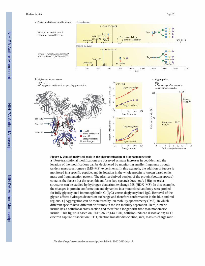
Figure 1. Uses of analytical tools in the characterization of biopharmaceuticalsa | Post-translational modifications are observed as mass increases in peptides, and thelocation of the modifications can be deciphered by monitoring smaller fragments throughtandem mass spectrometry (MS–MS) experiments. In this example, the addition of fucose ismonitored in a specific peptide, and its location in the whole protein is known based on itsmass and fragmentation pattern. The plasma-derived version of the protein (bottom spectra)contains the fucose but the recombinant form (top spectra) does not. b | Higher-orderstructures can be studied by hydrogen deuterium exchange MS (HDX–MS). In this example,the changes in protein conformation and dynamics in a monoclonal antibody were probedfor fully glycosylated immunoglobulin G (IgG) versus deglycosylated IgG. Removal of theglycan affects hydrogen deuterium exchange and therefore conformation in the blue and redregions. c | Aggregation can be monitored by ion mobility spectrometry (IMS), in whichdifferent species have different drift times in the ion mobility separation. Here, dimericinsulin has a collisional cross-section and therefore a longer drift time than monomericinsulin. This figure is based on REFS 36,77,144. CID, collision-induced dissociation; ECD,electron capture dissociation; ETD, electron transfer dissociation; m/z, mass-to-charge ratio.
Berkowitz et al. Page 26
Nat Rev Drug Discov. Author manuscript; available in PMC 2013 July 17.
NIH
-PA Author Manuscript
NIH
-PA Author Manuscript
NIH
-PA Author Manuscript