Embed Size (px)
Citation preview

Spatial localization of bacteria controls coagulation of humanblood by ‘quorum acting’
Christian J Kastrup1, James Q Boedicker1, Andrei P Pomerantsev2, Mahtab Moayeri2, YaoBian3, Rebecca R Pompano1, Timothy R Kline1, Patricia Sylvestre4,5, Feng Shen1, StephenH Leppla2, Wei-Jen Tang3, and Rustem F Ismagilov1
1Department of Chemistry and Institute for Biophysical Dynamics, The University of Chicago, 929 East 57thStreet, Chicago, Illinois 60637, USA
2Bacterial Toxins and Therapeutics Section, National Institute of Allergy and Infectious Diseases, NationalInstitutes of Health, 30 Convent Drive, Building 30, Room 303, Bethesda, Maryland 20892-4349, USA
3Ben-May Institute for Cancer Research, The University of Chicago, 929 East 57th Street, Chicago, Illinois60637, USA
4Unité Toxines et Pathogénie Bactériennes, Institut Pasteur, 28 rue du Dr Roux, 75724 Paris cédex 15, France
5Centre National de la Recherche Scientifique, Unité de Recherche Associée 2172, 28 rue du Dr Roux, 75724Paris cédex 15, France
AbstractBlood coagulation often accompanies bacterial infections and sepsis and is generally accepted as aconsequence of immune responses. Though many bacterial species can directly activate individualcoagulation factors, they have not been shown to directly initiate the coagulation cascade thatprecedes clot formation. Here we demonstrated, using microfluidics and surface patterning, that thespatial localization of bacteria substantially affects coagulation of human and mouse blood andplasma. Bacillus cereus and Bacillus anthracis, the anthrax-causing pathogen, directly initiatedcoagulation of blood in minutes when bacterial cells were clustered. Coagulation of human blood byB. anthracis required secreted zinc metalloprotease InhA1, which activated prothrombin and factorX directly (not via factor XII or tissue factor pathways). We refer to this mechanism as ‘quorumacting’ to distinguish it from quorum sensing—it does not require a change in gene expression, itcan be rapid and it can be independent of bacterium-to-bacterium communication.
This paper describes a physical and biochemical mechanism responsible for regulating theinitiation of human blood coagulation by bacteria. In vivo, coagulation often accompaniesbacterial infections of the blood and is believed to be a consequence of immune andinflammatory responses1-5. Immune and inflammatory responses cause upregulation of tissuefactor on the timescale of hours and lead to increased coagulation6,7. One of the few drugsavailable to treat septic shock, activated protein C, is also an anticoagulant8. This coagulationis believed to prevent dissemination of bacteria through the blood9,10 but also results in seriousvascular damage due to blockage and injury of blood vessels8. Coagulation accompanyingbacterial infections of the blood is particularly relevant for people infected with anthrax, whichinvolves sepsis and disseminated intravascular coagulation caused by the pathogen Bacillus
Correspondence should be addressed to R.F.I. ([email protected])..AUTHOR CONTRIBUTIONSC.J.K., J.Q.B., M.M., Y.B., R.R.P., T.R.K. and F.S. performed experiments; C.J.K., J.Q.B., M.M., Y.B., R.R.P., T.R.K., F.S., S.H.L.,W.-J.T. and R.F.I. designed experiments and analyzed data; C.J.K., W.-J.T. and R.F.I. wrote the paper; A.P.P. and P.S. provided reagents.
NIH Public AccessAuthor ManuscriptNat Chem Biol. Author manuscript; available in PMC 2009 June 1.
Published in final edited form as:Nat Chem Biol. 2008 December ; 4(12): 742–750.
NIH
-PA Author Manuscript
NIH
-PA Author Manuscript
NIH
-PA Author Manuscript

anthracis4. Here, we considered an alternative and complementary mechanism for thecoagulation that accompanies infection: direct activation of the human coagulation cascadethrough activation of coagulation factors by bacteria.
Many bacteria and bacterial components can directly activate individual human coagulationfactors. However, direct initiation of the coagulation cascade and the formation of apropagating clot are not typically observed11-17. These bacterial components usually activatelow levels of coagulation factors, which does not result in the amplification and positivefeedback necessary to form a clot that can grow and propagate. For example, Staphylococcusaureus produces coagulase, a protein that binds prothrombin stoichiometrically and leads tocleavage of fibrinogen to fibrin14. However, this conversion simply precipitates fibrin anddoes not result in production of thrombin, feedback or amplification of the coagulation cascade.Escherichia coli that express the protein Curli are also known to activate coagulation factors,such as factor XII (ref. 17). This process was shown to cause slower initiation of coagulationdue to depletion of factor XII (ref. 17). Bacteria are also well known to directly initiatecoagulation in some organisms, such as horseshoe crabs, but this mechanism of controllinginfection is believed to have been lost during the evolution of vertebrates18. All of these resultsprompt the following simple question: are bacteria capable of directly initiating the coagulationcascade and causing coagulation of human blood?
We hypothesized that initiation of coagulation by bacteria would be regulated by the spatiallocalization, not the total amount, of bacteria. In other words, for bacteria that activatecoagulation factors, coagulation would only occur when a cluster of bacteria forms. Thishypothesis was based on previous experiments with human blood and plasma that showed that(i) stimuli must exceed a local threshold concentration to initiate coagulation19,20, and (ii)this threshold response to concentration leads to a spatial threshold response, in whichcoagulation initiates on a patch of stimulus above but not below a threshold size21,22. Thethreshold to concentration for coagulation is due to competition between production andinhibition of activated coagulation proteases. The spatial threshold response to the size ofstimulus arises from the competition between the local production of activated coagulationproteases at the site of the stimulus and the diffusion of these proteases away from the stimulus.We hypothesized that individual bacteria as a stimulus of coagulation are below the thresholdstimulus size needed to initiate coagulation (Fig. 1a). Therefore, a solution of uniformlydispersed bacteria would not initiate coagulation if the bacteria are spaced far apart. However,a sufficiently large cluster of bacteria would generate a high local concentration of activatedcoagulation factors, which would exceed the threshold concentration and initiate thecoagulation cascade rapidly. In this paper we showed that coagulation can be controlled bychanging the spatial distribution, or clustering, of bacteria.
RESULTSSpatial localization of B. cereus controls coagulation
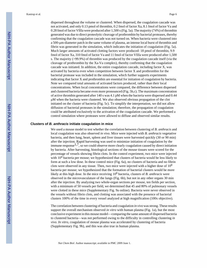
To test this hypothesis (Fig. 1a), we compared the clot time of human blood plasma exposedto bacteria dispersed in solution to the clot time of human blood plasma containing bacteriaclustered on the surface of the microfluidic chamber (Fig. 1b). Bacillus cereus spatiallylocalized to a surface cluster rapidly initiated coagulation. However, B. cereus dispersed insolution at concentrations of up to 107 colony-forming units (CFU) ml-1 did not initiatecoagulation (P < 0.01, for clustered versus dispersed B. cereus). The clot times of human bloodplasma exposed to the dispersed bacteria were not significantly different from the clot timesof the control samples of human blood plasma that did not contain bacteria (P > 0.5). In anothercontrol experiment, clusters of E. coli did not rapidly initiate coagulation, and there was nosignificant difference between the clot time of clustered E. coli and the samples of plasma thatdid not contain bacteria (P > 0.1). These results demonstrate that not all bacterial strains initiate
Kastrup et al. Page 2
Nat Chem Biol. Author manuscript; available in PMC 2009 June 1.
NIH
-PA Author Manuscript
NIH
-PA Author Manuscript
NIH
-PA Author Manuscript

coagulation in this experimental setup. The E. coli control strain, which does not produce theCurli protein, was used here because it was previously shown not to activate coagulationfactors17. In the solution-phase experiments (Fig. 1b), approximately 5 × 10⁵ CFU of B.cereus in 50 μl of human blood plasma did not initiate coagulation. However, significantlyfewer bacteria initiated coagulation when clustered—single clusters of approximately 4 ×10³ CFU were capable of initiating coagulation in 10 ml of human blood plasma. This numberwas also substantially lower than the number of bacteria (approximately 108 CFU) that couldnot initiate coagulation when dispersed in 10 ml of human blood plasma. Control experimentsconfirmed that fluorescence observed in coagulation by B. cereus corresponded to trueinitiation of the coagulation cascade and was not due to simple cleavage of the fluorogenicsubstrate or to an S. aureus-type coagulase activity (Supplementary Figs. 1 and 2 online). Forexample, both coagulation factor X and prothrombin are required for initiation of coagulationby B. cereus, which is not expected for S. aureus-type coagulase activity.
In a second experiment, we used microfluidics23,24 and micropatterned surfaces to controlthe spatial distribution of bacteria (Fig. 1c,d) and to demonstrate that the size of the cluster,rather than the amount of bacteria, can control the rate of initiation of coagulation of humanblood plasma. We patterned the surface of a microfluidic chamber with 90 μm patches of B.cereus expressing green fluorescent protein (GFP). We then monitored coagulation of humanblood plasma on these patches in the absence of flow. On smaller patches (90 μm) spaced farapart (400 μm), coagulation was slow, initiating on the first patch in 9 min ± 1 with clottingon all the patches in the array in 22 ± 3 min (mean ± s.e.), which indicates that the individual90 μm patches were below the size necessary to initiate coagulation rapidly (Fig. 1c). However,when the same number of bacteria were patterned closer together to form a large patch,coagulation initiated rapidly in 5 ± 1 min (mean ± s.e.) over the entire patch (Fig. 1d) (P < 0.01in comparison with initiation on the first 90 μm patch and P < 0.005 in comparison withinitiation on the entire set of patches). For this large patch, activated coagulation factorsaccumulated and exceeded the threshold concentration because diffusion of activatedcoagulation factors off of the patch was slower than the production of activated coagulationfactors.
B. cereus initiates coagulation of flowing whole bloodTo test whether B. cereus initiates coagulation in the presence of flow, human whole bloodwas flowed over localized colonies of B. cereus in microfluidic channels (Fig. 2). We wishedto test this effect because flow is important in maintaining hemostasis and could affectphenomena that rely on local concentration thresholds. We demonstrated previously thatthresholds to initiation and propagation of coagulation are preserved in the presence offlow25,26. In these experiments, several parameters known to contribute to the coagulationprocess in vivo were incorporated and carefully controlled, including flow and shear rates, thegeometry and surface chemistry of channels, and the presence of platelets and cells of the blood.Other components that contribute to coagulation in vivo were not tested here, including thepresence of membrane proteins and other components of the vessel wall. The components ofthe endothelium are known to greatly contribute to the coagulation process, and differencesare likely to exist between the microfluidic system described here and the in vivo setting.Clusters of bacteria in microfluidic channels were made by encapsulating bacteria in gelmicrodroplets (GMDs)27. GMDs consisted of colonies of bacteria and magnetic particles 1μm in diameter contained in agarose spheres approximately 50 μm in diameter; the magneticparticles allowed the GMDs to be trapped in the microfluidic channels by a magnet incorporatedinto the device near the channel (Fig. 2a)28. Clusters of B. cereus initiated coagulation offlowing human whole blood in 3-13 min (Fig. 2b,c), whereas coagulation did not occur until48-59 min in experiments with the control strain of E. coli (Fig. 2d; P < 0.001). Previous worksuggests that initiation and propagation of coagulation of human blood and plasma are sensitive
Kastrup et al. Page 3
Nat Chem Biol. Author manuscript; available in PMC 2009 June 1.
NIH
-PA Author Manuscript
NIH
-PA Author Manuscript
NIH
-PA Author Manuscript

to shear rate26. It predicts that coagulation induced by bacteria will be most pronounced inregions of low shear, such as in dead volumes in venous valves or in the extremities of peoplethat are immobilized and experiencing venous stasis (for example, in intensive care units). Thisprediction has not yet been tested.
Classical initiation points of the network are bypassedThere are two classical pathways to initiate coagulation (Fig. 3a). The principal pathway invivo is initiated by tissue factor, a high-affinity receptor for coagulation factor VII. The otherpathway, which is activated via factor XII, has been suggested as an initiation point for somebacteria17. We initially hypothesized that coagulation by B. cereus would occur by activationof either factor XII or factor VII. To determine which coagulation factors are essential, wetested blood plasmas that were depleted of specific coagulation factors. The clot times by B.cereus were compared to the clot times by reagents of the prothrombin time (PT) test or theactivated partial thromboplastin time (APPT) test (Fig. 3b,c). The PT test initiates coagulationvia the tissue factor/factor VII pathway, and the APTT test initiates coagulation via the factorXII pathway. Surprisingly, coagulation of plasma immunodepleted in either factor XII or factorVII was still rapidly initiated by B. cereus, which indicates that these factors are not essentialfor this process. However, coagulation did not occur within 30 min in plasma immunodepletedin either factor X or prothrombin. We performed further experiments with purified coagulationfactors and found that B. cereus is capable of directly activating prothrombin (factor II) andfactor X (Supplementary Figs. 3 and 4 online). The initiation point for B. cereus occurred atthe major hubs of the coagulation network. It remains to be seen whether further networkanalysis can be used to identify inhibition points of the network that could stop coagulationinitiated by bacteria while maintaining the ability of blood to initiate coagulation by tissuefactor.
Multiple Bacillus species initiate coagulationIn addition to B. cereus, we found that clusters of several other Bacillus species, including B.anthracis, the anthrax-causing pathogen, rapidly initiated coagulation of human blood plasma(Fig. 4a). The closely related species Bacillus thuringiensis and other species, includingBacillus subtilis and Bacillus licheniformis, also initiated coagulation. Experiments withpurified coagulation factors showed a strong correlation between the ability of these strains toinitiate coagulation and their ability to activate prothrombin and factor X (Supplementary Fig.4). Furthermore, components secreted from the bacteria into solution were also capable ofactivating purified coagulation factors, including prothrombin and factors X and XI, but notfactors VII and IX (Supplementary Fig. 5 online). It is surprising that an insect pathogen, B.thuringiensis, rapidly caused coagulation of human plasma. This result may be due to theconservation of protease cascades29,30. Further work is needed to understand thesephenomena.
Coagulation by B. anthracis requires metalloprotease InhA1To identify the molecular components responsible for activating coagulation factors andinitiating coagulation, we screened a small library of B. anthracis Ames 35 mutants; most ofthese mutants are deficient in secretion of a specific protease. We chose to investigate mutantsof B. anthracis for two reasons. First, B. anthracis secretes a much smaller number of proteasesthan B. cereus, resulting in a smaller screening library. Second, there is currently a demand foridentifying the essential molecular components responsible for the pathophysiology ofanthrax31,32. It has been shown previously that the B. anthracis protein lethal factor does notinitiate coagulation33. Inthe mutants used here, the genes corresponding to the proteases wereremoved by Cre recombinase (Supplementary Fig. 6 online)34. We hypothesized that secretedproteases were involved because solutions containing secreted components from B.
Kastrup et al. Page 4
Nat Chem Biol. Author manuscript; available in PMC 2009 June 1.
NIH
-PA Author Manuscript
NIH
-PA Author Manuscript
NIH
-PA Author Manuscript

anthracis cells were found to activate purified coagulation factors (Supplementary Fig. 5), butwith activities below the threshold needed for coagulation.
We found that bacteria that did not produce either metalloprotease NprB or InhA1 displayedreduced ability to activate purified human prothrombin or factor X and reduced ability to initiatecoagulation of human blood plasma compared with B. anthracis Ames 35 (Fig. 4b-e). NprBis highly homologous to bacillolysin proteases of other Bacillus species. InhA1 is a homologof the B. thuringiensis immune inhibitor A (ref. 16), and its expression has been observedduring growth in minimal aerobic medium35. InhA1 was essential for initiation of coagulationof human blood plasma by B. anthracis (P < 0.001 for Ames 35 versus ΔinhA1). To ensurethat InhA1 is capable of activating human coagulation factors, we purified this enzyme fromthe B. anthracis Ames 35 ΔnprB strain. As reported, InhA1 was purified as a 46 kDa and 18kDa complex (Supplementary Fig. 7 online)16. Purified InhA1 indeed cleaved prothrombinand generated active thrombin (Fig. 4d). In addition, control experiments showed that heat-inactivated InhA1 was not active, and the measured activity of purified InhA1 was due toactivation of prothrombin, not direct cleavage of the fluorescent substrate by InhA1. Recentexperiments have found that von Willebrand factor, a regulator of platelet aggregation, is alsoa substrate for InhA1 (ref. 15). Although B. anthracis could still rapidly initiate coagulation inhuman blood plasma deficient in von Willebrand factor and platelets (see SupplementaryMethods online), interactions with this factor further supported the notion that B. anthracismay target the coagulation process during infection.
The dependence of coagulation on the spatial arrangement of bacteria could suggest a quorum-sensing mechanism, and B. anthracis has been previously shown to exhibit quorumsensing36. However, two results contradict such a hypothesis. First, for the experimentsdescribed in Figure 1b, the bacteria in each sample were subjected to the same conditions untilthe human blood plasma was introduced. Then, within one minute, the bacteria were eitherdispersed into the plasma or clustered in the plasma, leading to the significant difference ininitiation of coagulation (Fig. 1b). It is not likely that quorum sensing could induce changes inphenotype within one minute. The production of InhA1 by B. anthracis was not stronglyinfluenced by clustering on the timescale that coagulation occurs (less than 2 h), as determinedby immunoblot analysis using mouse anti-InhA1 serum (Supplementary Fig. 8 online). In asecond experiment, mutants of B. anthracis that had reduced quorum-sensing ability stilltriggered rapid coagulation of human blood plasma. We used a previously characterizedquorum-sensing mutant strain of B. anthracis (34F2) that lacks functional luxS activity andproduction of autoinducer-2 (1)36.Similar to the B. anthracis Ames 35 control strain, thismutant strain rapidly initiated coagulation of human blood plasma in less than 3 min (Fig. 4f).The alternative mechanism that we are proposing here is that individual bacteria are below thecritical size necessary to initiate coagulation (Fig. 1a), but clusters of bacteria exceed thethreshold size necessary for initiation21,22,37. We used numerical simulations to test thefeasibility of this mechanism.
Numerical simulations reproduce the coagulation dynamicsTo examine the physical mechanism responsible for the initiation of coagulation on localizedclusters of bacteria, we used a two-dimensional numerical simulation that considered 40reactions of the human coagulation cascade, the activation of prothrombin and factor X by B.cereus or B. anthracis, the spatial localization of bacteria, and diffusion. The components andrate constants for the coagulation reactions were chosen based on an established numericalmodel for the human coagulation network. The rates of activation of prothrombin and factorX per bacterium were determined from kinetic assays that used B. cereus, purified prothrombinor factor X, and fluorogenic substrates for thrombin or factor Xa with known rates ofcleavage38. Simulations mimicked 2 × 10⁶ bacteria in 50 μl of human blood plasma, either
Kastrup et al. Page 5
Nat Chem Biol. Author manuscript; available in PMC 2009 June 1.
NIH
-PA Author Manuscript
NIH
-PA Author Manuscript
NIH
-PA Author Manuscript
dispersed throughout the volume or clustered. When dispersed, the coagulation cascade wasnot activated, and only 0.13 pmol of thrombin, 0.2 fmol of factor Xa, 8.1 fmol of factor Va and0.28 fmol of factor VIIIa were produced after 1,500 s (Fig. 5a). The majority (74%) of thrombingenerated was due to direct proteolytic cleavage of prothrombin by bacterial proteases, therebyconfirming that the coagulation cascade was not turned on. When bacteria were clustered intoa 500-μm-diameter patch in the same volume of plasma, an intense local burst of thrombin andfibrin was generated in the simulation, which indicates the initiation of coagulation (Fig. 5a).Much larger amounts of activated clotting factors were produced: 18 pmol of thrombin, 9.9fmol of factor Xa, 310 fmol of factor Va and 11 fmol of factor VIIIa were produced after 1,500s. The majority (>99.9%) of thrombin was produced by the coagulation cascade itself (via thecleavage of prothrombin by the Xa-Va complex), thereby confirming that the coagulationcascade was initiated. In addition, the entire coagulation cascade, including factor X, wasactivated by bacteria even when competition between factor X and prothrombin for thebacterial protease was included in the simulation, which further supports experimentsindicating that factor X and prothrombin are essential for initiation of coagulation by bacteria.Note we compared total amounts of activated factors produced, rather than their localconcentrations. When local concentrations were compared, the difference between dispersedand clustered bacteria became even more pronounced (Fig. 5b,c). The maximum concentrationof active thrombin generated after 140 s was 4.1 pM when the bacteria were dispersed and 0.44μM when the bacteria were clustered. We also observed obvious propagation of the clotinitiated on the cluster of bacteria (Fig. 5c). To simplify the interpretation, we did not allowdiffusion of bacterial proteases in the simulation; therefore, the propagation of coagulationcould be attributed exclusively to the activation of the coagulation cascade. We performed acontrol simulation where proteases were allowed to diffuse and observed similar results.
Clusters of B. anthracis initiate coagulation in miceWe used a mouse model to test whether the correlation between clustering of B. anthracis andlocal coagulation was also observed in vivo. Mice were injected with B. anthracis vegetativebacteria, and then lung, heart, spleen and liver tissues were harvested quickly (30 or 90 min)after the injection. Rapid harvesting was used to minimize initiation of coagulation by theimmune response1,7, so we could observe more clearly coagulation caused by direct initiationby bacteria. After harvesting, histological sections of the mouse tissues were scored for thepercentage of vessels showing fibrin clots. In the control experiment, two mice were injectedwith 104 bacteria per mouse; we hypothesized that clusters of bacteria would be less likely toform at such a low dose. In these control mice (Fig. 6a), no clusters of bacteria and no fibrinclots were observed in any tissue. Then, two mice were injected with a higher dose of 108
bacteria per mouse; we hypothesized that the formation of bacterial clusters would be morelikely at this high dose. In the mice receiving 108 bacteria, clusters of B. anthracis wereobserved in the microvasculature of the lungs (Fig. 6b), but not in any other organs 30 minafter the injection. By analyzing two whole-organ sections per mouse, ten fields per section,with a minimum of 50 vessels per field, we determined that 45 and 80% of pulmonary vesselswere clotted in these mice (Supplementary Fig. 9a online). Bacteria were never observed inthe vessels without fibrin clots, and clotting was associated with the presence of bacterialclusters 100% of the time in every vessel analyzed at high magnification (100x objective).
The correlation between clustering of bacteria and coagulation in vivo was strong. These resultssupport the overall mechanism observed in vitro with human plasma (Fig. 1a), but the mostconclusive experiment in this mouse model—comparing the same amount of dispersed bacteriato clustered bacteria—was not performed owing to the difficulty in controlling clustering invivo. In vitro, coagulation of mouse plasma was accelerated by clustering of bacteria(Supplementary Fig. 9b), and this was also true in human plasma.
Kastrup et al. Page 6
Nat Chem Biol. Author manuscript; available in PMC 2009 June 1.
NIH
-PA Author Manuscript
NIH
-PA Author Manuscript
NIH
-PA Author Manuscript

We emphasize that the characterization of the interaction of Bacillus cells and proteases (Fig.4 and Supplementary Figs. 4-6) with the coagulation cascade was performed by using human(not mouse) whole blood and plasma. Therefore, the details of the in vivo experiment performedwith the mouse model (Fig. 6) should be interpreted with caution. Mouse models have beenuseful for studying the pathophysiology of B. anthracis infection, but they have been shownto have major differences in response to B. anthracis infection compared with humans39. Inaddition, there are known differences between proteolytic activation of mouse and humanclotting cascades40,41. Rapid clotting in the mouse model strongly suggests that directactivation of coagulation occurs; however, we did not identify the key proteases responsiblefor initiation of coagulation by B. anthracis in mouse, rather than human, blood. The ΔnprBand ΔinhA1 strains of B. anthracis were still able to rapidly initiate coagulation in mice, whichindicates that coagulation directly induced by B. anthracis is a robust process in mice. Inaddition, this result implies either that clotting of the mouse blood is initiated by an unknownrapid mechanism that is sensitive to spatial distribution and clustering of bacteria, or thatproteases other than InhA1 and NprB are involved in the activation of the mouse coagulationcascade. In support of the latter (and simpler) argument, we found differences in the rates andspecificity of the B. anthracis proteases for human and mouse coagulation proteins. Forexample, we tested activation of human and mouse factor X under the same conditions by B.anthracis cells in vitro. Deletion of either InhA1 or NprB substantially decreased the rate ofhuman factor X activation by a factor of 20 to low levels, but these deletions decreased the rateof activation of mouse factor X by only a factor of 5 and 2.5, with substantial activationremaining, which suggests that other B. anthracis proteases can also activate mouse factor X.These proteases may be active in the B. anthracis Ames 35 strain or upregulated in responseto the loss of a protease in the mutant strains. Though the overall dynamics of rapid activationof clotting factors by the parent Ames 35 strain were reproducible, the exact amount of decreaseof activation in mutant strains varied with conditions. These results were consistent with thedifferences in the sequence of peptides of human and mouse factor X, which is substantiallydifferent in the region that is proteolysed to convert factor X to factor Xa (ref. 42). Thesedifferences make the mouse model less appropriate for the detailed biochemical studies thatare necessary to understand induction of coagulation in the human blood in vivo. In addition,these results suggest that, in addition to InhA1, other proteases could potentially affectinduction of coagulation of human blood (for example, by cross-activation). An important nextstep for understanding the role of coagulation induced in humans by bacterial proteases wouldbe identifying an animal model of blood coagulation that responds to bacterial proteases in amanner similar to human coagulation. Rabbit and primate models may be suitable, but theseexperiments are beyond the scope of this paper.
DISCUSSIONThe first area of interest related to this work is the connection between bacterial infections andblood coagulation1,4,43 and the hypothesis that clusters of bacteria may directly initiatecoagulation of human blood during infection, bypassing inflammation. This hypothesis shouldbe considered for immunosuppressed and immunocompromised people for whom infectionand septicemia by B. cereus is a threat44. It should also be considered in the context ofinflammatory responses that are triggered by activated coagulation factors2,5,43. Our resultsreported a physical and biochemical mechanism in support of this hypothesis and provide themotivation to test this mechanism further by using in vivo models of infection. This mechanismpredicts that inhibiting clustering of bacteria, inhibiting local accumulation of coagulationfactors on surfaces of bacteria17, and inhibiting expression, transport or processing of bacterialproteases may help reduce coagulation during infection. To choose the appropriate in vivomodel, one would have to characterize carefully the interactions between bacteria and thecoagulation cascade of the model organism to ensure relevance to the human coagulationsystem. In vivo bioluminescent imaging of B. anthracis45 should be a useful tool for
Kastrup et al. Page 7
Nat Chem Biol. Author manuscript; available in PMC 2009 June 1.
NIH
-PA Author Manuscript
NIH
-PA Author Manuscript
NIH
-PA Author Manuscript

understanding further the role of this mechanism in anthrax by characterizing the effects ofclusters, biofilms and local infection sites on coagulation.
The second area of interest related to this work is understanding the dynamics of groups orclusters of bacteria46,47. Quorum sensing is perhaps the best known example of suchdynamics46. In quorum sensing, bacteria send out a diffusible signal (Fig. 7a). At a lowconcentration of bacteria, the signal diffuses away and does not accumulate, and bacteria donot detect the ‘quorum’. As bacteria are brought together to a higher (quorum) concentration,the diffusible signal accumulates above a threshold concentration and is sensed by the bacteria.A linear system cannot exhibit a spatially dependent phenomenon such as a quorumsensing25, and a nonlinearity, such as a threshold response, must be present either in the systemitself or in its environment. In quorum sensing, the nonlinearity required for the thresholdresponse is provided by the bacterial regulatory network, and no nonlinearity in theenvironment is required. Once quorum sensing takes place, it drives changes in gene expressionto produce effector molecules required for bacteria to take appropriate actions (Fig. 7a).
Initiation of coagulation by clusters of bacteria (Fig. 1) can occur by a mechanism distinct fromthat of quorum sensing. In the proposed mechanism (Fig. 7b), a diffusible molecule is alsogenerated by an individual bacterium. For initiation of coagulation discussed in this paper, twoclasses of molecules satisfy the requirement of this diffusible molecule: (i) a diffusiblemolecule secreted directly by the bacterium—for example, protease InhA1—and (ii) adiffusible molecule from the environment that can be activated near or on the surface of thebacterium—for example, thrombin. The molecule does not accumulate at low concentrationsof bacteria. When bacteria are brought closer together into a cluster, the molecule reaches athreshold concentration. At this point, there are three critical differences between thismechanism and quorum sensing. First, the nonlinearity that established the threshold can comefrom the environment around the bacteria, not necessarily from the bacterial regulatorynetwork. In the example discussed here, the threshold comes from the nonlinearities of thecoagulation network. Second, sensing by bacteria is not required for this mechanism to operate,because ‘sensing’ is essentially performed by the environment via its threshold. This alsodiffers from the sensing mechanism used by bacteria, such as E. feacalis, to probe theirenvironment for host mammalian cells48. Third, this mechanism does not require eitherchanges in bacterial gene expression or any other bacterial mechanism for production of theeffector molecules. The secreted molecule is the effector molecule required for action, and ittriggers the response of the environment once bacteria reach a sufficiently high local density.To emphasize these three differences, we refer to this mechanism as ‘quorum acting’. Thisdistinction is supported by the rapid initiation of coagulation by the B. anthracis luxS mutantdeficient in quorum sensing (Fig. 4f). We predict that other bacterial species that activatecoagulation factors may demonstrate this quorum-acting mechanism, although this predictionremains to be tested. Porphyromonas gingivalis, a causative agent of gum disease, is one likelycandidate. Purified proteases of P. gingivalis are particularly potent and known to activatemany coagulation factors and reduce coagulation times in standard assays11. P. gingivalisinfections have also been linked to cardiovascular disease, although the nature of thisconnection is still under investigation49.
Further work is required to differentiate the connections between quorum sensing and quorumacting, as the two mechanisms are likely to be coupled and are likely to feedback to one another.Though the quorum-acting mechanism does not require a change in bacterial phenotype tofunction, it is not likely that it is constitutively turned on, independently of the phenotype. Whatregulates the ‘coagulation phenotype’ and secretion of proteases responsible for the initiationof coagulation? What is the role of the environment relative to the role of bacterialcommunication, for example by oligopeptides46, in this regulation? Other bacteria such asstreptococci and Yersinia pestis, the plague agent, are known to break apart clots3,41,43. Is
Kastrup et al. Page 8
Nat Chem Biol. Author manuscript; available in PMC 2009 June 1.
NIH
-PA Author Manuscript
NIH
-PA Author Manuscript
NIH
-PA Author Manuscript

avoiding entrapment by coagulation10 a better strategy? Or does initiation of coagulationbenefit B. anthracis by shielding it from the host’s immune system and, coincidentally, fromadministered antibiotics?
Although we do not know whether quorum acting is as widespread as quorum sensing, moreexamples are likely to be found in environments capable of nonlinear responses. Suchenvironments could range from interactions of communities of microorganisms in soils and inbiofilms, to secretion of toxins and virulence factors, to interactions of microorganisms withthe gut, the immune system and the coagulation cascade of a mammalian host. One may expectconfined environments to enhance quorum acting, in analogy to quorum sensing47. Quorumacting may be especially beneficial when a rapid response to aggregation of microorganismsis needed, either as a defensive response, or an opportunistic response. Quorum acting couldalso serve as a driving force for the evolution of cooperation within and among the bacterialgroups, by facilitating kin and group selection as well as reciprocity—the common themes forcollaborative selections50.
In conclusion, this work demonstrates that bacteria can directly initiate coagulation of humanblood and plasma, a process that was previously thought to be lost during vertebrate evolution.This process relies on a quorum-acting mechanism that is distinct from quorum-sensingprocesses. These results emphasize the importance of spatial distribution, rather than averageconcentration, in the function of nonlinear biochemical networks21,25. We expect spatialdistribution to also be critical in initiation of coagulation by other mechanisms that are distinctfrom proteolytic activation by bacterial proteases. These results may also have implicationsfor improving our understanding of coagulation during bacterial infections and of the role ofspatial organization of bacteria in their interactions with nonlinear environments.
METHODSGeneral methods
See Supplementary Methods for additional experimental protocols, description of thebacterial strains and details of the numerical simulation. A summary of the methods is givenbelow.
Patterning bacteria using microfluidic techniquesFor experiments measuring the initiation of coagulation of human blood and plasma on clustersof bacteria that were not spatially patterned (that is, not Fig. 1c,d), bacteria were concentratedto a pellet, and then droplets of the concentrated bacteria (∼50 nl) were deposited onto a plasticcoverslip in the bottom of a microfluidic chamber. The bacteria were either dispersed in humanblood plasma by mixing for ∼2 s or allowed to remain localized in a patch. Bacteria remainedlocalized owing to weak adhesive forces between themselves and the plastic coverslip.
To prepare spatially patterned bacteria (Fig. 1c,d), micropatterning techniques were used24.Bacteria were patterned on substrates consisting of alumina membranes (200 nm pore size)coated with patterned photoresist. A gentle vacuum was applied from under the substrate topull the bacteria to the open pores. Control experiments confirmed that both patterned andnonpatterned substrates were relatively inert; bacteria were able to grow on them, and, in theabsence of bacteria, these surfaces did not initiate coagulation of human blood for >30 min.
Measuring clot times of human whole blood and plasmaCitrated human platelet-poor plasma was obtained from George King Biomedical, Inc. Citratedimmunodepleted plasmas and measurements of their PT and APTT times were obtained fromHaematologic Technologies, Inc. Human whole blood was obtained from individual healthy
Kastrup et al. Page 9
Nat Chem Biol. Author manuscript; available in PMC 2009 June 1.
NIH
-PA Author Manuscript
NIH
-PA Author Manuscript
NIH
-PA Author Manuscript

donors in accordance with the guidelines set by the Institutional Review Board (protocol #12502A) at The University of Chicago. Written informed consent was obtained from donors.All human blood and plasma samples were incubated with corn trypsin inhibitor to inhibit thefactor XII pathway of initiation of coagulation, with the exception of the experiment testingclustered bacteria versus dispersed bacteria with human plasma (Fig. 1b) and the experimentwith immunodepleted plasmas (Fig. 3b,c). Human whole blood and plasma were recalcifiedby adding a solution containing CaCl2 and a thrombin-sensitive fluorescent substrate.Experiments were performed at 37 °C. In all experiments, clot times were determined bymonitoring the formation of thrombin and fibrin by fluorescence and brightfield microscopy,respectively.
Microfluidic device fabricationAll devices were fabricated by using rapid prototyping in polydimethylsiloxane (Dow CorningCorporation). Before adding the GMDs or human blood, the microfluidic channels were coatedwith inert phospholipids by flowing vesicles of L-α-phosphatidylcholine (Avanti) through thedevice. GMDs containing bacteria were flowed into the device, localized near magnets andgrown for 14-24 h. GMDs27 (Fig. 2) consisted of ∼50-μm-sized droplets of solid agarosecontaining bacteria and magnetic particles. They were prepared separately using a droplet-based microfluidic approach. After bacterial colonies were present, human whole blood wasflowed through the device and the formation of thrombin and fibrin was monitored.
Measuring coagulation by B. anthracis in miceAll animal experiments and protocols were approved by and conducted according to theguidelines of the US National Institute of Allergy and Infectious Diseases (NIAID) AnimalCare and Use Committee. Solutions containing B. anthracis (vegetative cells) were injectedintravenously into the tail vein of DBA/2J mice (Jackson Laboratories). After 30 or 90 min,organs were harvested and immediately fixed in a neutral-buffered 10% formalin solution forhematoxylin and eosin (H&E) staining.
Supplementary MaterialRefer to Web version on PubMed Central for supplementary material.
ACKNOWLEDGMENTSThis work was supported in part by the US National Institutes of Health (NIH) Director’s Pioneer Award (grant numberDP1OD003584), the US National Science Foundation CAREER Award (grant number CHE-0349034), the US Officeof Naval Research (grant number N000140610630), the Camille Dreyfus Teacher-Scholar Awards Program and theCottrell Scholar of Research Corporation Awards Program to R.F.I., by NIH grants (GM 62548 and GM 81539) toW.-J.T., and by the Intramural Research Program of the NIAID. We thank J. Alverdy, B. Bishop, S. Crosson, C.Esmon, M. Mock, M. Runyon, J. Shapiro, U. Spitz, T. Van Ha, D. Wiebel and O. Zaborina for helpful discussions;O. Zaborina (The University of Chicago) for the gift of the EGPF plasmid for E. coli and for assisting in thetransformation procedure; M. Mock (Institut Pasteur) for the gift of the mouse anti-InhA1 serum; L. Cheng and D.Crown for assisting in the animal studies; H. Herwald (Lund University) for the gift of the E. coli Ymel-1 strain; J.Handelsman (University of Wisconsin) for the gift of the B. cereus GFP strain; M. Blaser (New York University) forthe gift of the B. anthracis ΔluxS strain; and J. Price for contributions in writing and editing this manuscript. We thankC. Tallant (Institut de Biologia Molecular de Barcelona) for assistance in construction of the protease gene knockoutstrains.
References1. Opal SM, Esmon CT. Bench-to-bedside review: functional relationships between coagulation and the
innate immune response and their respective roles in the pathogenesis of sepsis. Crit. Care 2003;7:23–38. [PubMed: 12617738]
Kastrup et al. Page 10
Nat Chem Biol. Author manuscript; available in PMC 2009 June 1.
NIH
-PA Author Manuscript
NIH
-PA Author Manuscript
NIH
-PA Author Manuscript

2. Niessen F, et al. Dendritic cell PAR1-S1P3 signalling couples coagulation and inflammation. Nature2008;452:654–658. [PubMed: 18305483]
3. Sun H. The interaction between pathogens and the host coagulation system. Physiology (Bethesda)2006;21:281–288. [PubMed: 16868317]
4. Stearns-Kurosawa DJ, Lupu F, Taylor FB, Kinasewitz G, Kurosawa S. Sepsis and pathophysiology ofanthrax in a nonhuman primate model. Am. J. Pathol 2006;169:433–444. [PubMed: 16877346]
5. Esmon CT. The interactions between inflammation and coagulation. Br. J. Haematol 2005;131:417–430. [PubMed: 16281932]
6. Tang H, et al. Sepsis-induced coagulation in the baboon lung is associated with decreased tissue factorpathway inhibitor. Am. J. Pathol 2007;171:1066–1077. [PubMed: 17640967]
7. Pawlinski R, et al. Regulation of tissue factor and inflammatory mediators by Egr-1 in a mouseendotoxemia model. Blood 2003;101:3940–3947. [PubMed: 12543866]
8. Levi M, de Jonge E, van der Poll T. New treatment strategies for disseminated intravascular coagulationbased on current understanding of the pathophysiology. Ann. Med 2004;36:41–49. [PubMed:15000346]
9. Medzhitov R. Recognition of microorganisms and activation of the immune response. Nature2007;449:819–826. [PubMed: 17943118]
10. Mullarky IK, et al. Infection-stimulated fibrin deposition controls hemorrhage and limits hepaticbacterial growth during listeriosis. Infect. Immun 2005;73:3888–3895. [PubMed: 15972474]
11. Imamura T, Potempa J, Tanase S, Travis J. Activation of blood coagulation factor X by arginine-specific cysteine proteinases (gingipain-Rs) from Porphyromonas gingivalis. J. Biol. Chem1997;272:16062–16067. [PubMed: 9188512]
12. Narasaki R, et al. Bacillolysin MA, a novel bacterial metalloproteinase that produces angiostatin-likefragments from plasminogen and activates protease zymogens in the coagulation and fibrinolysissystems. J. Biol. Chem 2005;280:14278–14287. [PubMed: 15677446]
13. Smith SA, et al. Polyphosphate modulates blood coagulation and fibrinolysis. Proc. Natl. Acad. Sci.USA 2006;103:903–908. [PubMed: 16410357]
14. Friedrich R, et al. Structural basis for reduced staphylocoagulase-mediated bovine prothrombinactivation. J. Biol. Chem 2006;281:1188–1195. [PubMed: 16230338]
15. Chung MC, et al. Degradation of circulating von Willebrand factor and its regulator ADAMTS13implicates secreted Bacillus anthracis metalloproteases in anthrax consumptive coagulopathy. J.Biol. Chem 2008;283:9531–9542. [PubMed: 18263586]
16. Chung MC, et al. Secreted neutral metalloproteases of Bacillus anthracis as candidate pathogenicfactors. J. Biol. Chem 2006;281:31408–31418. [PubMed: 16926147]
17. Herwald H, et al. Activation of the contact-phase system on bacterial surfaces - a clue to seriouscomplications in infectious diseases. Nat. Med 1998;4:298–302. [PubMed: 9500602]
18. Muta T, Iwanaga S. The role of hemolymph coagulation in innate immunity. Curr. Opin. Immunol1996;8:41–47. [PubMed: 8729445]
19. Jesty J, Rodriguez J, Beltrami E. Demonstration of a threshold response in a proteolytic feedbacksystem: control of the autoactivation of factor XII. Pathophysiol. Haemost. Thromb 2005;34:71–79.[PubMed: 16432309]
20. van’t Veer C, Mann KG. Regulation of tissue factor initiated thrombin generation by thestoichiometric inhibitors tissue factor pathway inhibitor, antithrombin-III, and heparin cofactor-II. J.Biol. Chem 1997;272:4367–4377. [PubMed: 9020158]
21. Kastrup CJ, Runyon MK, Shen F, Ismagilov RF. Modular chemical mechanism predictsspatiotemporal dynamics of initiation in the complex network of hemostasis. Proc. Natl. Acad. Sci.USA 2006;103:15747–15752. [PubMed: 17043240]
22. Kastrup CJ, Shen F, Runyon MK, Ismagilov RF. Characterization of the threshold response ofinitiation of blood clotting to stimulus patch size. Biophys. J 2007;93:2969–2977. [PubMed:17586576]
23. Weibel DB, DiLuzio WR, Whitesides GM. Microfabrication meets microbiology. Nat. Rev.Microbiol 2007;5:209–218. [PubMed: 17304250]
Kastrup et al. Page 11
Nat Chem Biol. Author manuscript; available in PMC 2009 June 1.
NIH
-PA Author Manuscript
NIH
-PA Author Manuscript
NIH
-PA Author Manuscript

24. Whitesides GM, Ostuni E, Takayama S, Jiang XY, Ingber DE. Soft lithography in biology andbiochemistry. Annu. Rev. Biomed. Eng 2001;3:335–373. [PubMed: 11447067]
25. Pompano RR, Li HW, Ismagilov RF. Rate of mixing controls rate and outcome of autocatalyticprocesses—theory and microfluidic experiments with chemical reactions and blood coagulation.Biophys. J 2008;95:1531–1543. [PubMed: 18424502]
26. Runyon MK, Kastrup CJ, Johnson-Kerner BL, Ha TG, Ismagilov RF. The effects of shear rate onpropagation of blood clotting determined using microfluidics and numerical simulations. J. Am.Chem. Soc 2008;130:3458–3464. [PubMed: 18302373]
27. Weaver JC, Williams GB, Klibanov A, Demain AL. Gel microdroplets - rapid detection andenumeration of individual microorganisms by their metabolic-activity. Bio/Technology1988;6:1084–1089.
28. Siegel AC, et al. Cofabrication of electromagnets and microfluldic systems in poly(dimethylsiloxane).Angew. Chem. Int. Ed 2006;45:6877–6882.
29. Jiang Y, Doolittle RF. The evolution of vertebrate blood coagulation as viewed from a comparisonof puffer fish and sea squirt genomes. Proc. Natl. Acad. Sci. USA 2003;100:7527–7532. [PubMed:12808152]
30. Krem MM, Di Cera E. Evolution of enzyme cascades from embryonic development to bloodcoagulation. Trends Biochem. Sci 2002;27:67–74. [PubMed: 11852243]
31. Min DH, Tang WJ, Mrksich M. Chemical screening by mass spectrometry to identify inhibitors ofanthrax lethal factor. Nat. Biotechnol 2004;22:717–723. [PubMed: 15146199]
32. Bugge TH, Leppla SH. Anthrax target in macrophages unveiled. Nat. Genet 2006;38:137–138.[PubMed: 16444249]
33. Moayeri M, Haines D, Young HA, Leppla SH. Bacillus anthracis lethal toxin induces TNF-alpha-independent hypoxia-mediated toxicity in mice. J. Clin. Invest 2003;112:670–682. [PubMed:12952916]
34. Pomerantsev AP, Sitaraman R, Galloway CR, Kivovich V, Leppla SH. Genome engineering inBacillus anthracis using Cre recombinase. Infect. Immun 2006;74:682–693. [PubMed: 16369025]
35. Gat O, et al. Search for Bacillus anthracis potential vaccine candidates by a functional genomic-serologic screen. Infect. Immun 2006;74:3987–4001. [PubMed: 16790772]
36. Jones MB, Blaser MJ. Detection of a luxS-signaling molecule in Bacillus anthracis. Infect. Immun2003;71:3914–3919. [PubMed: 12819077]
37. Beltrami E, Jesty J. The role of membrane patch size and flow in regulating a proteolytic feedbackthreshold on a membrane: possible application in blood coagulation. Math. Biosci 2001;172:1–13.[PubMed: 11472773]
38. Kawabata SI, et al. Highly sensitive peptide-4-methylcoumaryl-7-amide substrates for blood-clottingproteases and trypsin. Eur. J. Biochem 1988;172:17–25. [PubMed: 3278905]
39. Loving CL, Kennett M, Lee GM, Grippe VK, Merkel TJ. Murine aerosol challenge model of anthrax.Infect. Immun 2007;75:2689–2698. [PubMed: 17353290]
40. Coughlin SR. Thrombin signalling and protease-activated receptors. Nature 2000;407:258–264.[PubMed: 11001069]
41. Sun HM, et al. Plasminogen is a critical host pathogenicity factor for group A streptococcal infection.Science 2004;305:1283–1286. [PubMed: 15333838]
42. Heidtmann HH, Kontermann RE. Cloning and recombinant expression of mouse coagulation factorX. Thromb. Res 1998;92:33–41. [PubMed: 9783672]
43. Degen JL, Bugge TH, Goguen JD. Fibrin and fibrinolysis in infection and host defense. J. Thromb.Haemost 2007;5:24–31. [PubMed: 17635705]
44. Akiyama N, et al. Fulminant septicemic syndrome of Bacillus cereus in a leukemic patient. Intern.Med 1997;36:221–226. [PubMed: 9144019]
45. Glomski IJ, Piris-Gimenez A, Huerre M, Mock M, Goossens PL. Primary involvement of pharynxand Peyer’s patch in inhalational and intestinal anthrax. PLoS Pathog 2007;3:e76. [PubMed:17542645]
46. Bassler BL, Losick R. Bacterially speaking. Cell 2006;125:237–246. [PubMed: 16630813]
Kastrup et al. Page 12
Nat Chem Biol. Author manuscript; available in PMC 2009 June 1.
NIH
-PA Author Manuscript
NIH
-PA Author Manuscript
NIH
-PA Author Manuscript

47. Redfield RJ. Is quorum sensing a side effect of diffusion sensing? Trends Microbiol 2002;10:365–370. [PubMed: 12160634]
48. Coburn PS, Pillar CM, Jett BD, Haas W, Gilmore MS. Enterococcus faecalis senses target cells andin response expresses cytolysin. Science 2004;306:2270–2272. [PubMed: 15618522]
49. Demmer RT, Desvarieux M. Periodontal infections and cardiovascular disease - the heart of thematter. J. Am. Dent. Assoc 2006;137:14S–20S. [PubMed: 17012731]
50. Nowak MA. Five rules for the evolution of cooperation. Science 2006;314:1560–1563. [PubMed:17158317]
Kastrup et al. Page 13
Nat Chem Biol. Author manuscript; available in PMC 2009 June 1.
NIH
-PA Author Manuscript
NIH
-PA Author Manuscript
NIH
-PA Author Manuscript
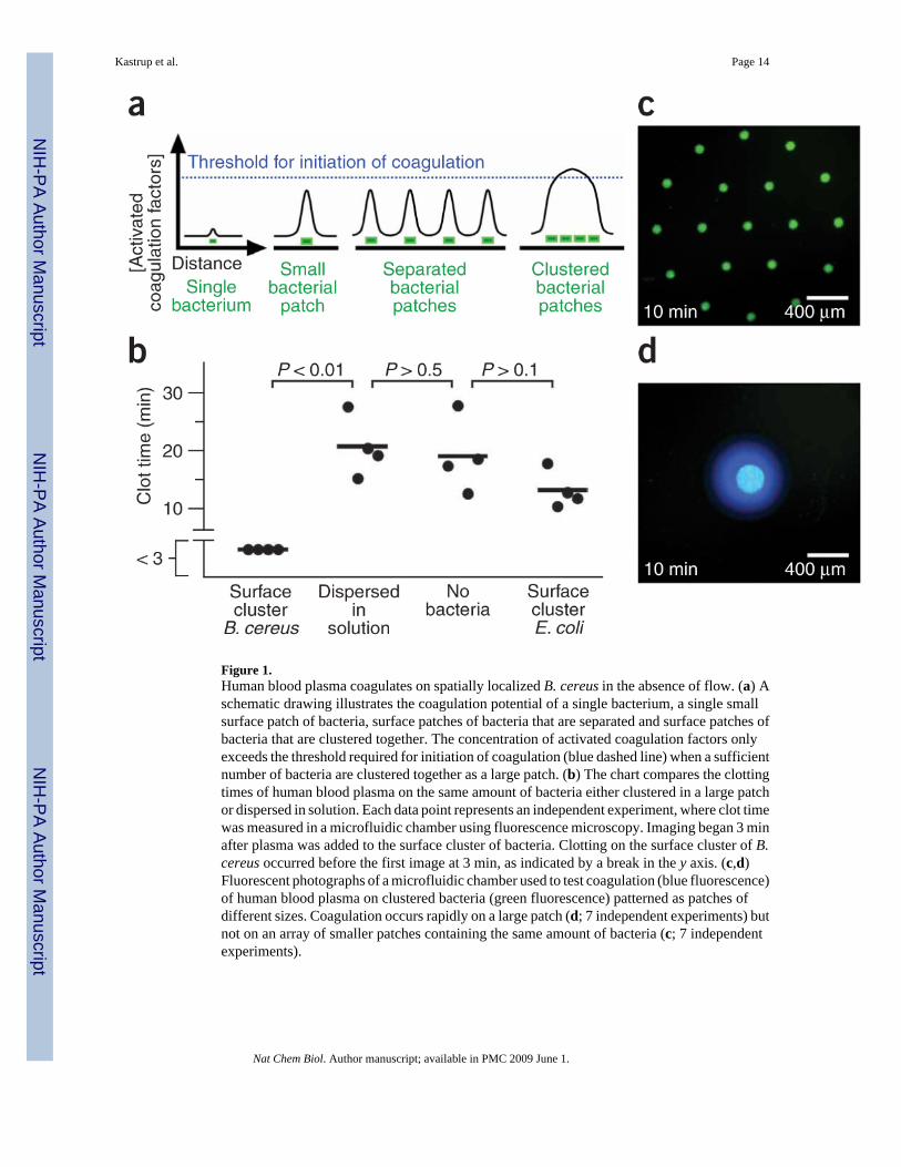
Figure 1.Human blood plasma coagulates on spatially localized B. cereus in the absence of flow. (a) Aschematic drawing illustrates the coagulation potential of a single bacterium, a single smallsurface patch of bacteria, surface patches of bacteria that are separated and surface patches ofbacteria that are clustered together. The concentration of activated coagulation factors onlyexceeds the threshold required for initiation of coagulation (blue dashed line) when a sufficientnumber of bacteria are clustered together as a large patch. (b) The chart compares the clottingtimes of human blood plasma on the same amount of bacteria either clustered in a large patchor dispersed in solution. Each data point represents an independent experiment, where clot timewas measured in a microfluidic chamber using fluorescence microscopy. Imaging began 3 minafter plasma was added to the surface cluster of bacteria. Clotting on the surface cluster of B.cereus occurred before the first image at 3 min, as indicated by a break in the y axis. (c,d)Fluorescent photographs of a microfluidic chamber used to test coagulation (blue fluorescence)of human blood plasma on clustered bacteria (green fluorescence) patterned as patches ofdifferent sizes. Coagulation occurs rapidly on a large patch (d; 7 independent experiments) butnot on an array of smaller patches containing the same amount of bacteria (c; 7 independentexperiments).
Kastrup et al. Page 14
Nat Chem Biol. Author manuscript; available in PMC 2009 June 1.
NIH
-PA Author Manuscript
NIH
-PA Author Manuscript
NIH
-PA Author Manuscript

Figure 2.Human whole blood coagulated on spatially localized B. cereus in the presence of flow. (a) Amicrophotograph shows the microfluidic device used to flow human whole blood over coloniesof bacteria. The bacteria were localized in GMDs that also contained magnetic particles.Magnets were used to localize the GMDs in the device. (b) A brightfield image showscoagulation of human whole blood on GMDs containing B. cereus expressing GFP (overlaidgreen fluorescence). (c,d) Microphotographs of fluorescence show coagulation of humanwhole blood (blue) on GMDs containing B. cereus (green) (c) but not on E. coli (green) (d).White dashed lines outline the channel walls. Images were taken 11 min after blood wasintroduced into the device. (e) A graph shows the clot times of flowing whole blood on coloniesof B. cereus and E. coli in microfluidic devices. Each data point represents an independentexperiment.
Kastrup et al. Page 15
Nat Chem Biol. Author manuscript; available in PMC 2009 June 1.
NIH
-PA Author Manuscript
NIH
-PA Author Manuscript
NIH
-PA Author Manuscript
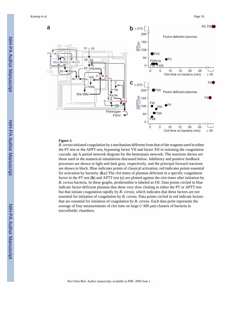
Figure 3.B. cereus initiated coagulation by a mechanism different from that of the reagents used in eitherthe PT test or the APPT test, bypassing factor VII and factor XII in initiating the coagulationcascade. (a) A partial network diagram for the hemostasis network. The reactions shown arethose used in the numerical simulations discussed below. Inhibitory and positive feedbackprocesses are shown in light and dark gray, respectively, and the principal forward reactionsare shown in black. Blue indicates points of classical activation; red indicates points essentialfor activation by bacteria. (b,c) The clot times of plasmas deficient in a specific coagulationfactor in the PT test (b) and APTT test (c) are plotted against the clot times after initiation byB. cereus bacteria. In these graphs, prothrombin is labeled as FII. Data points circled in blueindicate factor-deficient plasmas that show very slow clotting in either the PT or APTT testbut that initiate coagulation rapidly by B. cereus, which indicates that these factors are notessential for initiation of coagulation by B. cereus. Data points circled in red indicate factorsthat are essential for initiation of coagulation by B. cereus. Each data point represents theaverage of four measurements of clot time on large (>300 μm) clusters of bacteria inmicrofluidic chambers.
Kastrup et al. Page 16
Nat Chem Biol. Author manuscript; available in PMC 2009 June 1.
NIH
-PA Author Manuscript
NIH
-PA Author Manuscript
NIH
-PA Author Manuscript

Figure 4.Human blood plasma coagulates on surface clusters of many Bacillus species, including B.anthracis, but not on control species of E. coli and S. aureus. (a) Chart quantifying the clottimes of human blood plasma on clusters of bacteria in a microfluidic chamber in the absenceof flow. (b,c) B. anthracis strains that do not produce secreted zinc metalloproteases NprB(ΔnprB) or InhA1 (ΔinhA1) have reduced ability to activate purified human prothrombin (b)and purified human factor X (c). (d) Purified prothrombin is activated by InhA1 purified fromB. anthracis. (e) A graph quantifying the clot times of human blood plasma on Ames 35,ΔnprB and ΔinhA1 strains of B. anthracis shows that the Ames 35 control strain rapidly initiatedcoagulation. The ΔnprB strain also initiated coagulation, whereas the ΔinhA1 strain did notaccelerate coagulation relative to background clotting (P < 0.001 for Ames 35 versusΔinhA1). (f) A graph quantifying the clot times of human blood plasma on control Ames 35and a ΔluxS strain shows that the ΔluxS strain rapidly initiated coagulation despite its inabilityto secrete autoinducer-2 (1), a quorum-sensing signaling molecule. Each data point (a,e,f)represents the clot time on a single large (>300 μm) cluster of bacteria in a microfluidic chamberthat was measured by fluorescence microscopy. Clotting on some surface clusters occurredbefore the first images obtained at 3 min (a) or 2 min (e and f), as indicated by breaks in they axis.
Kastrup et al. Page 17
Nat Chem Biol. Author manuscript; available in PMC 2009 June 1.
NIH
-PA Author Manuscript
NIH
-PA Author Manuscript
NIH
-PA Author Manuscript

Figure 5.Two-dimensional simulations of the human blood coagulation cascade comparing thegeneration of activated coagulation factors by bacteria dispersed in solution versus bacteriaclustered in a surface patch. The overall number of bacteria was the same in all simulations.(a) Graphs of the total amount (not concentrations) of thrombin and factors Xa, Va and VIIIagenerated in the simulation by dispersed (blue line) and clustered (red dashed line) bacteria.The total amounts included both active (free) forms of the enzyme and active forms that havebeen inhibited after activation. (b,c) Two-dimensional plots show the thrombin concentrationin a simulated microfluidic chamber (represented in schematic drawings at the left). Plots at140 s show the concentration of active thrombin, and plots at 1,500 s show the total thrombinconcentration (both active and inhibited). When bacteria (green) were dispersed in solution,little thrombin was produced and coagulation did not initiate within 1,500 s (b). However,when the same number of bacteria were localized to a surface cluster (inside green outline),thrombin was generated at a high concentration, coagulation was initiated and the clotpropagated away from the cluster (c).
Kastrup et al. Page 18
Nat Chem Biol. Author manuscript; available in PMC 2009 June 1.
NIH
-PA Author Manuscript
NIH
-PA Author Manuscript
NIH
-PA Author Manuscript

Figure 6.Clusters of B. anthracis rapidly initiate coagulation in mice. (a,b) H&E-stained histologicalsections of mouse lung. (a) Pulmonary vessels from control mice injected with a low dose ofbacteria show no clusters and no coagulation. (b) Pulmonary vessels from mice injected witha higher dose of bacteria show clustering of bacteria. Coagulation in vessels (large magentaregions) occurred on clusters of bacteria (chains of rod-like bacteria are seen, blue) within 30min. Digitally magnified portions of a vessel are shown in images on the left and right. Twomice were sampled at each dose. See text for details.
Kastrup et al. Page 19
Nat Chem Biol. Author manuscript; available in PMC 2009 June 1.
NIH
-PA Author Manuscript
NIH
-PA Author Manuscript
NIH
-PA Author Manuscript

Figure 7.Even without quorum sensing, clustering of bacteria may elicit large-scale action. (a) Duringquorum sensing, the localization of bacteria (blue) at a high concentration elicits phenotypicchanges in the bacteria themselves (red). (b) When bacteria interact with an environmentcapable of nonlinear responses, upon clustering, bacteria may act without having to undergophenotypic changes. Action is illustrated here as activation of the environment (large red area).
Kastrup et al. Page 20
Nat Chem Biol. Author manuscript; available in PMC 2009 June 1.
NIH
-PA Author Manuscript
NIH
-PA Author Manuscript
NIH
-PA Author Manuscript