Embed Size (px)
Citation preview
Behavioral impact of neurotransmitter-activated GPCRs:Muscarinic and GABAB receptors regulate C. elegans locomotion
Jeremy S Dittman and Joshua M KaplanDepartment of Molecular Biology, Massachusetts General Hospital, Boston MA, 02114
AbstractNeurotransmitter released from presynaptic terminals activates both ligand-gated ion channels(ionotropic receptors) and a variety of G protein-coupled receptors (GPCRs). These neurotransmitterreceptors are expressed on both pre- and postsynaptic cells. Thus, each neurotransmitter acts onmultiple receptor classes, generating a large repertoire of physiological responses. The impact ofmany ionotropic receptors on neuronal activity and behavior has been clearly elucidated; however,much less is known about how neurotransmitter-gated GPCRs regulate neurons and circuits. In C.elegans, both Acetylcholine (ACh) and GABA are released in the nerve cord and mediate fastneuromuscular excitation and inhibition during locomotion. Here we identify a muscarinic receptor(GAR-2) and the GABAB receptor dimer (GBB-1/2) that detect synaptically released ACh andGABA, respectively. Both GAR-2 and GBB-1/2 inhibited cholinergic motor neurons when ACh andGABA levels were enhanced. Loss of either GPCR resulted in movement defects, suggesting thatthese receptors are activated during locomotion. When the negative feedback provided by GAR-2was replaced with positive feedback, animals became highly sensitive to ACh levels and locomotionwas severely impaired. Thus, conserved GPCRs act in the nematode motor circuit to provide negativefeedback and to regulate locomotory behaviors that underlie navigation.
KeywordsC. elegans; feedback; GABAB receptor; GPCR; locomotion; muscarinic
IntroductionDivergence is a widespread and important theme in the biology of signaling: the divergenceof one molecule to many receptor classes generates a large repertoire of responses to the releaseof a single chemical. All known transmitter molecules that mediate fast chemical synaptictransmission via ionotropic receptors also activate a variety of GPCRs thereby extendingneurotransmission into multiple intracellular signaling pathways. Ionotropic receptors andGPCRs have very distinct signaling properties. Fast synaptic transmission is mediated byligand-gated ion channels, which have rapid kinetics (operating on time scales of a fewmilliseconds), and relatively low affinity for neurotransmitter ligands (typically KD ~0.1–1mM), which restricts activation to only those receptors that are clustered adjacent to pre-synaptic release sites. These properties endow synapses with the ability to produce theextremely fast, local signals that underlie fast synaptic transmission.
Corresponding author with complete address, including an email address: Joshua M Kaplan, Department of Molecular Biology,Massachusetts General Hospital – Simches Research Bldg. 7th Floor, 185 Cambridge St. Boston MA 02114, E-mail:[email protected] Dittman’s current address: Department of Biochemistry, Weill Cornell Medical College, New York, NY 10021
NIH Public AccessAuthor ManuscriptJ Neurosci. Author manuscript; available in PMC 2009 May 8.
Published in final edited form as:J Neurosci. 2008 July 9; 28(28): 7104–7112. doi:10.1523/JNEUROSCI.0378-08.2008.
NIH
-PA Author Manuscript
NIH
-PA Author Manuscript
NIH
-PA Author Manuscript
By contrast, neurotransmitter-gated GPCRs have properties that typically limit their functionto slower, modulatory signals. Neurotransmitter-gated GPCRs regulate neuronal activity via acascade of cytoplasmic second messengers, giving these signals intrinsically slow kinetics(operating on timescales of seconds to minutes), although in some cases, direct binding of thebeta gamma subunit can modulate ion channels in tens to hundreds of milliseconds (Dittmanand Regehr, 1997). GPCRs have relatively high affinity for neurotransmitter ligands (KD ~0.1–1 μM), which permits activation of receptors that are relatively far-removed forneurotransmitter release sites (Hille, 1992). Thus, signals produced by neurotransmitter-gatedGPCRs act over longer timescales, and larger spatial domains.
Although a great deal is known about the contribution of neurotransmitter-gated ion channelsto fast synaptic transmission and behavior, much less is known about how neurotransmitter-gated GPCRs regulate circuit activity and behavior. In mammals, extrasynaptic GABA inhibitsneuronal activity via metabotropic GABA (GABAB) receptors (Isaacson et al., 1993; Dittmanand Regehr, 1997), whereas extrasynaptic glutamate can both stimulate and inhibit neighboringneurons by activating ionotropic and metabotropic receptors (Mitchell and Silver, 2000;DiGregorio et al., 2002; Szapiro and Barbour, 2007). Thus, the same neurotransmitters thatdirectly gate a postsynaptic ionotropic receptor can influence intracellular signaling in both thepre and postsynaptic cell as well as neighboring neurons.
The nematode C. elegans provides a simple model organism for investigating the behavioralimpact of neurotransmitter-gated GPCRs. Nematode propulsion arises from sinusoidal wavesof muscle contraction and relaxation driven by ACh and GABA respectively (Croll, 1975;Niebur and Erdos, 1993; Schuske et al., 2004). C. elegans locomotion is extensively regulatedby G protein signaling pathways, including antagonistic regulation of ACh secretion by Goand Gq (Hajdu-Cronin et al., 1999; Lackner et al., 1999; Miller et al., 1999; Nurrish et al.,1999; Robatzek and Thomas, 2000; Robatzek et al., 2001; Bastiani and Mendel, 2006). Whilethese studies suggest that Go and Gq play important roles in regulating locomotion, much lessis known about the upstream GPCRs regulating these pathways, and specifically about the roleof neurotransmitter-gated GPCRs. In this study, we identified two motor neuron GPCRs thatdetect ACh and GABA, and analyzed the locomotion behavior of mutants lacking thesereceptors.
Materials and MethodsStrains
Strains were maintained at 20 °C as described by Brenner (Brenner, 1974). The followingstrains were used in this study: N2 Bristol, gar-2(ok520), gbb-1(tm1406), gbb-2(tm1165),goa-1(sa734), KP3473: nuIs155 [Punc-17::Venus::GAR-2], KP3457: nuEx1076[Punc-17::Venus::GAR-3], KP3456: gar-2(ok520);nuEx1075 [Pgar-2::Venus::GAR-2],KP3526: nuEx1072 [Pgar-2::GFP];nuEx1067 [Punc-25::dsRed2], KP3447: nuEx1066[Pgbb-1::GFP];nuEx1067, KP4488: Punc-25::Venus::GAR-2;gar-2(ok520), eri-1(mg366);lin-15B(n744).
The eri-1;lin-15B strain was used for feeding RNA interference (RNAi) experiments becauseneurons of wild-type animals are refractory to RNAi. This strain was developed to provideenhanced sensitivity to RNA interference in neurons (Sieburth et al., 2005).
Constructs and TransgenescDNA encoding the splice form GAR-2a was subcloned into the fire lab vector pPD49.26flanked by NheI and KpnI. An in-frame NotI site was introduced after the start codon andYFPvenus was ligated to create an N-terminal fusion protein. The signal sequence from pat-3
Dittman and Kaplan Page 2
J Neurosci. Author manuscript; available in PMC 2009 May 8.
NIH
-PA Author Manuscript
NIH
-PA Author Manuscript
NIH
-PA Author Manuscript
beta integrin was included on the 5′ end of YFP-Venus to insure proper receptor insertion.Tissue-specific expression of this fusion protein was determined by including about 3 kb of 5′regulatory sequence from unc-17 VAChT (for all cholinergic neurons), gar-2 (for rescueconstruct), or unc-25 GAD (for rescue in GABAergic neurons only). cDNA encoding the spliceform GAR-3b was subcloned as described for GAR-2a. Transcriptional reporters for gar-2 andgbb-1 were made by subcloning 3 kb of 5′ regulatory sequence for each of these genes into theFire lab vector pPD95.75 which contains a cDNA encoding soluble GFP. The unc-25 reporterconstruct was made by replacing GFP in pPD95.75 with DsRed2 (Clontech) and thensubcloning 3 kb of 5′ regulatory sequence from the unc-25 gene.
Predictions for the gene structures of gbb-1 and gbb-2 were generated using GeneMark HMMroutines (http://opal.biology.gatech.edu/GeneMark/eukhmm.cgi), and cDNAs were amplifiedand sequenced from a C. elegans cDNA library. Two splice variants for gbb-1 were identified:2.5 and 2.6 kb whereas only one cDNA for gbb-2 was identified.
Fluorescence ImagingVenus::GAR-2 expressing animals were mounted on agarose pads and viewed on a ZeissAxiovert microscope, using an Olympus PlanApo 100x NA 1.4 objective, as describedpreviously (Dittman and Kaplan, 2006). Images were captured with a Hamamatsu ORCAdigital camera and line scans were analyzed with custom software in Igor Pro (Wavemetrics).Images of 500 nm fluorescein-conjugated beads (Molecular Probes) were captured during eachimaging session to provide a fluorescence standard for comparing absolute fluorescence levelsbetween animals. Background signal (CCD dark current and slide autofluorescence) wassubtracted before analysis.
Aldicarb and Levamisole AssaysSensitivity to aldicarb and levamisole was determined by analyzing the onset of paralysisfollowing treatment with either 1 mM aldicarb (Chem Services) or 200 μM levamisole (Sigma)as described previously (Nurrish et al., 1999). Each assay included between 2 and 4 plates ofat least 20 animals for each genotype being tested. The experimenter was blind to the genotypes.Each genotype was tested between 4 and 10 independent experiments and paralysis curveswere generated by averaging paralysis time courses for each plate.
Worm Tracking and AnalysisFor tracking measurements, worms were reared at 20 °C and moved to room temperature 30minutes prior to imaging. Young adult animals were picked to agar plates with no bacteriallawn (5–15 worms per plate). Imaging began 5–10 minutes after worms were removed fromfood. One minute digital videos of individual animals were captured at 8x magnification and4 Hz frame rate on a Zeiss Discovery Stereomicroscope using Axiovision software. If theanimal left the field of view, acquisition was paused while the plate was manually recenteredand after a few seconds, acquisition was restarted. The center of mass was recorded for eachanimal on each video frame using object tracking software in Axiovision. The trajectories werethen analyzed using custom software written in Igor Pro 5.0 (Wavemetrics). The raw data wasfiltered to remove artifactual discontinuities in the trajectory due to noise in the center-of-massestimates. Speed and angle values were calculated and from frame-by-frame changes inposition and angle (from −180° to +180°), with turns and reversals estimated as follows: turnswere counted as angle changes between 50° and 160° (taking the absolute value of the anglechange). Reversals were counted as angle changes between 170° and 180°. The accuracy ofthese criteria was assessed by visual inspection of the video. Averages for speed, turn number,and reversal number were determined for each animal and then pooled to form estimated valuesfor each genotype. Standard error was based on the animal-to-animal variation in parametervalues. Typically between 40 and 70 animals were assayed for each genotype. For Rmax
Dittman and Kaplan Page 3
J Neurosci. Author manuscript; available in PMC 2009 May 8.
NIH
-PA Author Manuscript
NIH
-PA Author Manuscript
NIH
-PA Author Manuscript
distributions, the maximal Euclidean distance from the starting point within a 40 second timeinterval was recorded for each animal. The cumulative distributions of Rmax values werecompared between genotypes using the Kolmogorov-Smirnov statistic.
Worm Curvature AnalysisImages were collected for 10 to 15 animals at 20x magnification using a Zeiss Discoverystereomicroscope. The animals selected were on or near the bacterial lawn and relativelystationary. The anterior-posterior axis of the animal was traced by hand in Axiovision and thex–y coordinates of the traced path were recorded and further analyzed using custom writtensoftware in Igor Pro 5.0 (Wavemetrics). Data were smoothed to remove extraneous curvaturedue to the tracing technique and the local curvature of the smoothed trace was calculatedaccording to the parametric equation of curvature:
The average curvature over the length of the animal was then calculated as: where Lworm is the length of the worm. This measure was averaged for all worms for eachgenotype and normalized to wild-type average.
ResultsGAR-2/M2 mediates inhibition of cholinergic neurons when ACh levels arepharmacologically enhanced
The worm genome contains numerous conserved neurotransmitter-gated GPCRs, includingthree muscarinic acetylcholine receptors (mAChRs), four dopamine (DARs), three serotonin(5-HTRs), two glutamate (mGluRs) receptors, and two predicted GABAB receptor subunits(Bargmann, 1998; Lee et al., 2000; Chase et al., 2004; Carre-Pierrat et al., 2006). We examinedthe effects of neuronal mAChRs on motor neuron function and locomotion using an M2receptor GAR-2 deletion mutant (gar-2(ok520)) and RNA interference. The ok520 deletionremoves 1.1 kb of the gene including most of the third intracellular (i3) loop, a region of thereceptor that is essential for coupling to G proteins (Kubo et al., 1988; Burstein et al., 1996),and consequently is likely to eliminate GAR-2 function. ACh levels are normally limited bysynaptic AChEs so inhibiting cholinesterase activity will elevate ACh (Hartzell et al., 1975).To experimentally enhance ACh levels, we used the AChE inhibitor aldicarb. Aldicarbtreatment induces paralysis with a characteristic time course due to enhanced muscle activation(Miller et al., 1996; Nurrish et al., 1999).
To test the effects of GAR-2 on cholinergic motor neuron synapses, we measured the timecourse of acute paralysis induced by aldicarb in gar-2 mutants. The gar-2 deletion mutationand gar-2 inactivation by RNAi both accelerated the time course of aldicarb-induced paralysis(Figure 1A and B). Muscle sensitivity to the ionotropic cholinergic agonist levamisole was notaltered in the gar-2 mutant (Figure 1C), indicating that intrinsic muscle sensitivity to ACh wasnot affected by loss of GAR-2. Wild-type aldicarb sensitivity was restored in gar-2 mutantsby expressing an N-terminal YFP(Venus)-tagged GAR-2 cDNA using the gar-2 promoter.These results suggest that GAR-2 regulates the activity of ventral cord motor neurons.
GAR-2 has been previously shown to be expressed throughout the nervous system includingventral cord neurons but not in muscle (Lee et al., 2000). Consistent with this study, we foundthat GAR-2 was expressed in some cholinergic motor neurons as well as GABAergic motor
Dittman and Kaplan Page 4
J Neurosci. Author manuscript; available in PMC 2009 May 8.
NIH
-PA Author Manuscript
NIH
-PA Author Manuscript
NIH
-PA Author Manuscript
neurons, the two major types of ventral cord motor neurons (Figure 2A). We examinedsubcellular localization of GAR-2 by expressing a YFP::GAR-2 fusion protein in cholinergicmotor neurons and imaged the dorsal cord (Figure 2Ad). The protein appeared to be diffuselydistributed in axons with no obvious spatial relationship to presynaptic terminals. A similarpattern was seen using a C-terminal fusion protein GAR-2::YFP (data not shown).
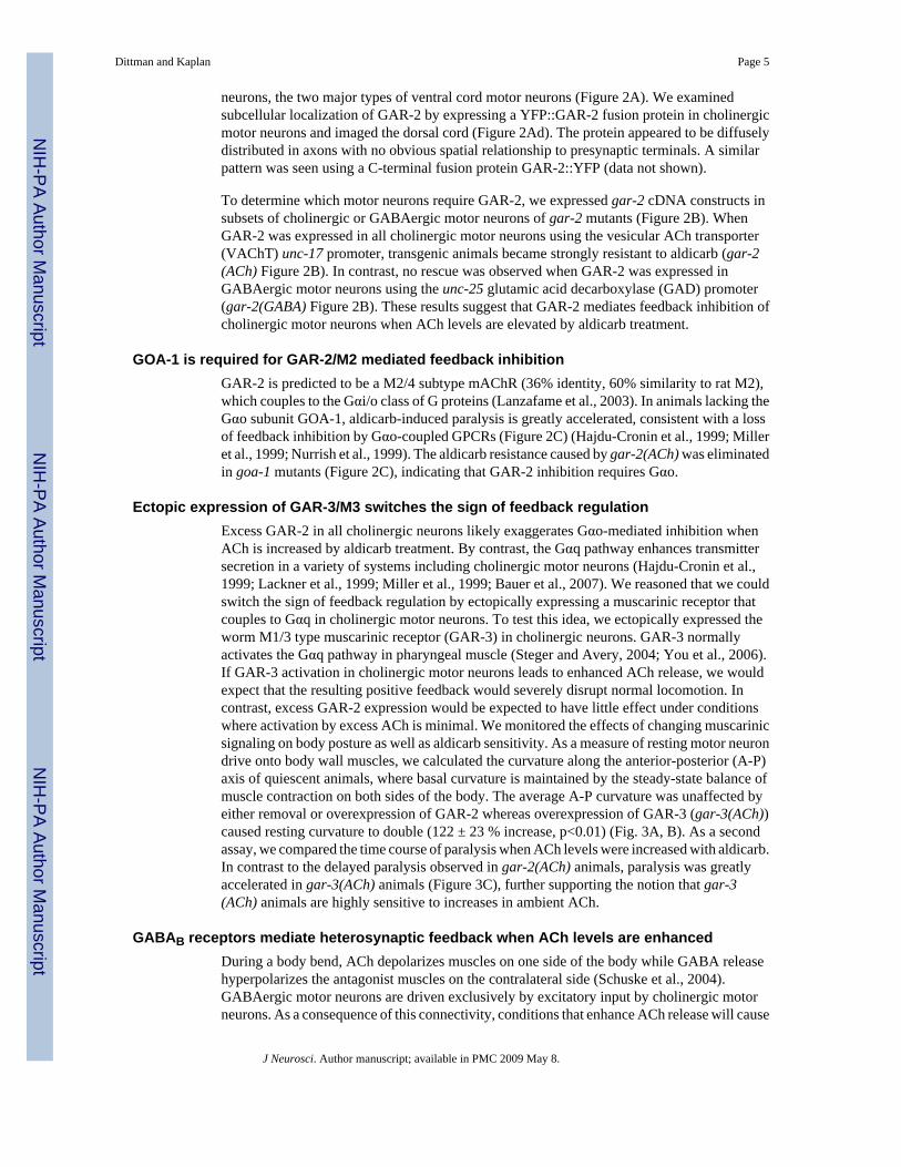
To determine which motor neurons require GAR-2, we expressed gar-2 cDNA constructs insubsets of cholinergic or GABAergic motor neurons of gar-2 mutants (Figure 2B). WhenGAR-2 was expressed in all cholinergic motor neurons using the vesicular ACh transporter(VAChT) unc-17 promoter, transgenic animals became strongly resistant to aldicarb (gar-2(ACh) Figure 2B). In contrast, no rescue was observed when GAR-2 was expressed inGABAergic motor neurons using the unc-25 glutamic acid decarboxylase (GAD) promoter(gar-2(GABA) Figure 2B). These results suggest that GAR-2 mediates feedback inhibition ofcholinergic motor neurons when ACh levels are elevated by aldicarb treatment.
GOA-1 is required for GAR-2/M2 mediated feedback inhibitionGAR-2 is predicted to be a M2/4 subtype mAChR (36% identity, 60% similarity to rat M2),which couples to the Gαi/o class of G proteins (Lanzafame et al., 2003). In animals lacking theGαo subunit GOA-1, aldicarb-induced paralysis is greatly accelerated, consistent with a lossof feedback inhibition by Gαo-coupled GPCRs (Figure 2C) (Hajdu-Cronin et al., 1999; Milleret al., 1999; Nurrish et al., 1999). The aldicarb resistance caused by gar-2(ACh) was eliminatedin goa-1 mutants (Figure 2C), indicating that GAR-2 inhibition requires Gαo.
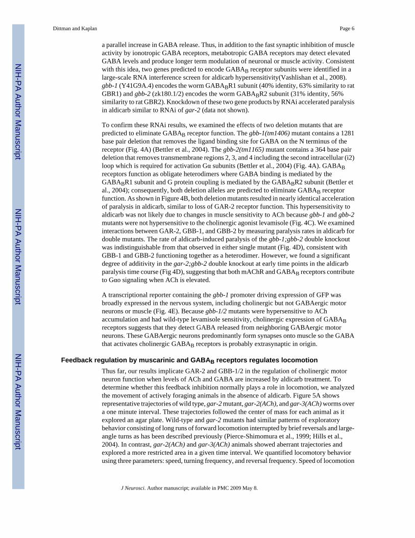
Ectopic expression of GAR-3/M3 switches the sign of feedback regulationExcess GAR-2 in all cholinergic neurons likely exaggerates Gαo-mediated inhibition whenACh is increased by aldicarb treatment. By contrast, the Gαq pathway enhances transmittersecretion in a variety of systems including cholinergic motor neurons (Hajdu-Cronin et al.,1999; Lackner et al., 1999; Miller et al., 1999; Bauer et al., 2007). We reasoned that we couldswitch the sign of feedback regulation by ectopically expressing a muscarinic receptor thatcouples to Gαq in cholinergic motor neurons. To test this idea, we ectopically expressed theworm M1/3 type muscarinic receptor (GAR-3) in cholinergic neurons. GAR-3 normallyactivates the Gαq pathway in pharyngeal muscle (Steger and Avery, 2004; You et al., 2006).If GAR-3 activation in cholinergic motor neurons leads to enhanced ACh release, we wouldexpect that the resulting positive feedback would severely disrupt normal locomotion. Incontrast, excess GAR-2 expression would be expected to have little effect under conditionswhere activation by excess ACh is minimal. We monitored the effects of changing muscarinicsignaling on body posture as well as aldicarb sensitivity. As a measure of resting motor neurondrive onto body wall muscles, we calculated the curvature along the anterior-posterior (A-P)axis of quiescent animals, where basal curvature is maintained by the steady-state balance ofmuscle contraction on both sides of the body. The average A-P curvature was unaffected byeither removal or overexpression of GAR-2 whereas overexpression of GAR-3 (gar-3(ACh))caused resting curvature to double (122 ± 23 % increase, p<0.01) (Fig. 3A, B). As a secondassay, we compared the time course of paralysis when ACh levels were increased with aldicarb.In contrast to the delayed paralysis observed in gar-2(ACh) animals, paralysis was greatlyaccelerated in gar-3(ACh) animals (Figure 3C), further supporting the notion that gar-3(ACh) animals are highly sensitive to increases in ambient ACh.
GABAB receptors mediate heterosynaptic feedback when ACh levels are enhancedDuring a body bend, ACh depolarizes muscles on one side of the body while GABA releasehyperpolarizes the antagonist muscles on the contralateral side (Schuske et al., 2004).GABAergic motor neurons are driven exclusively by excitatory input by cholinergic motorneurons. As a consequence of this connectivity, conditions that enhance ACh release will cause
Dittman and Kaplan Page 5
J Neurosci. Author manuscript; available in PMC 2009 May 8.
NIH
-PA Author Manuscript
NIH
-PA Author Manuscript
NIH
-PA Author Manuscript
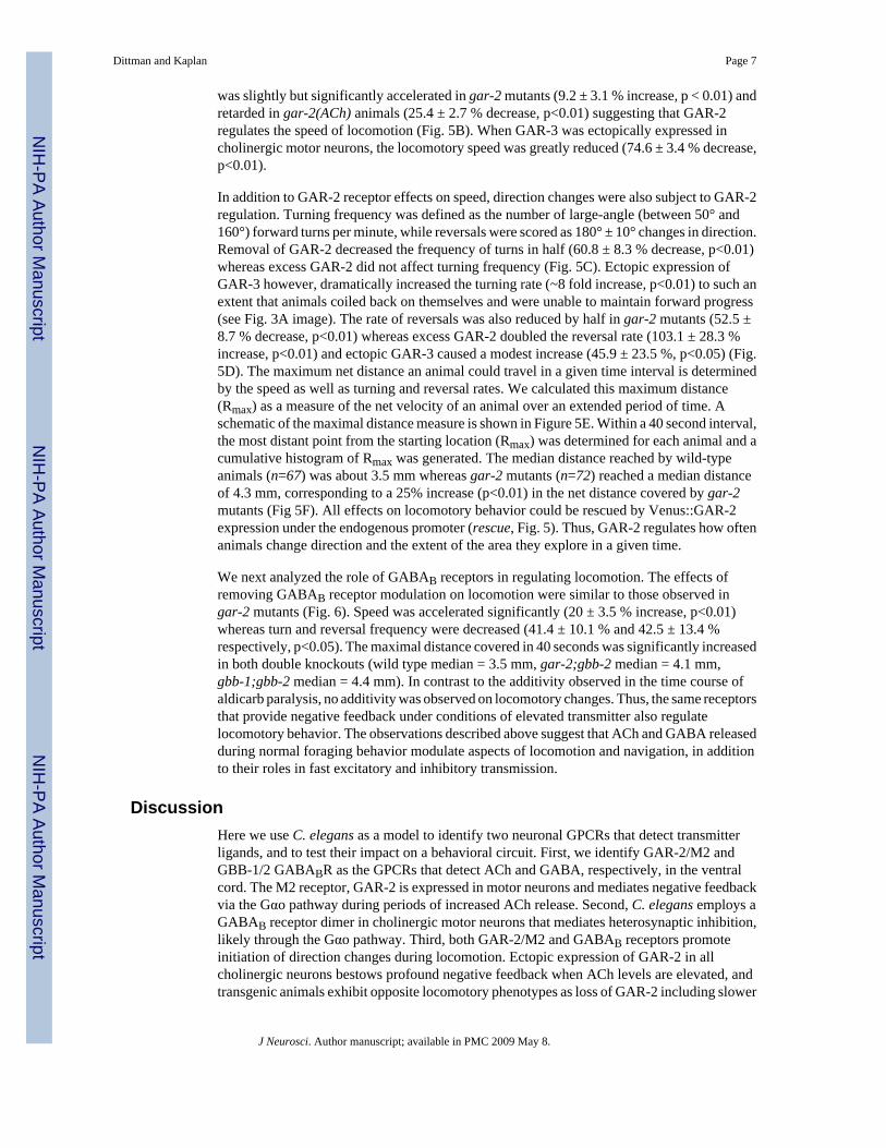
a parallel increase in GABA release. Thus, in addition to the fast synaptic inhibition of muscleactivity by ionotropic GABA receptors, metabotropic GABA receptors may detect elevatedGABA levels and produce longer term modulation of neuronal or muscle activity. Consistentwith this idea, two genes predicted to encode GABAB receptor subunits were identified in alarge-scale RNA interference screen for aldicarb hypersensitivity(Vashlishan et al., 2008).gbb-1 (Y41G9A.4) encodes the worm GABABR1 subunit (40% identity, 63% similarity to ratGBR1) and gbb-2 (zk180.1/2) encodes the worm GABABR2 subunit (31% identity, 56%similarity to rat GBR2). Knockdown of these two gene products by RNAi accelerated paralysisin aldicarb similar to RNAi of gar-2 (data not shown).
To confirm these RNAi results, we examined the effects of two deletion mutants that arepredicted to eliminate GABAB receptor function. The gbb-1(tm1406) mutant contains a 1281base pair deletion that removes the ligand binding site for GABA on the N terminus of thereceptor (Fig. 4A) (Bettler et al., 2004). The gbb-2(tm1165) mutant contains a 364 base pairdeletion that removes transmembrane regions 2, 3, and 4 including the second intracellular (i2)loop which is required for activation Gα subunits (Bettler et al., 2004) (Fig. 4A). GABABreceptors function as obligate heterodimers where GABA binding is mediated by theGABABR1 subunit and G protein coupling is mediated by the GABABR2 subunit (Bettler etal., 2004); consequently, both deletion alleles are predicted to eliminate GABAB receptorfunction. As shown in Figure 4B, both deletion mutants resulted in nearly identical accelerationof paralysis in aldicarb, similar to loss of GAR-2 receptor function. This hypersensitivity toaldicarb was not likely due to changes in muscle sensitivity to ACh because gbb-1 and gbb-2mutants were not hypersensitive to the cholinergic agonist levamisole (Fig. 4C). We examinedinteractions between GAR-2, GBB-1, and GBB-2 by measuring paralysis rates in aldicarb fordouble mutants. The rate of aldicarb-induced paralysis of the gbb-1;gbb-2 double knockoutwas indistinguishable from that observed in either single mutant (Fig. 4D), consistent withGBB-1 and GBB-2 functioning together as a heterodimer. However, we found a significantdegree of additivity in the gar-2;gbb-2 double knockout at early time points in the aldicarbparalysis time course (Fig 4D), suggesting that both mAChR and GABAB receptors contributeto Gαo signaling when ACh is elevated.
A transcriptional reporter containing the gbb-1 promoter driving expression of GFP wasbroadly expressed in the nervous system, including cholinergic but not GABAergic motorneurons or muscle (Fig. 4E). Because gbb-1/2 mutants were hypersensitive to AChaccumulation and had wild-type levamisole sensitivity, cholinergic expression of GABABreceptors suggests that they detect GABA released from neighboring GABAergic motorneurons. These GABAergic neurons predominantly form synapses onto muscle so the GABAthat activates cholinergic GABAB receptors is probably extrasynaptic in origin.
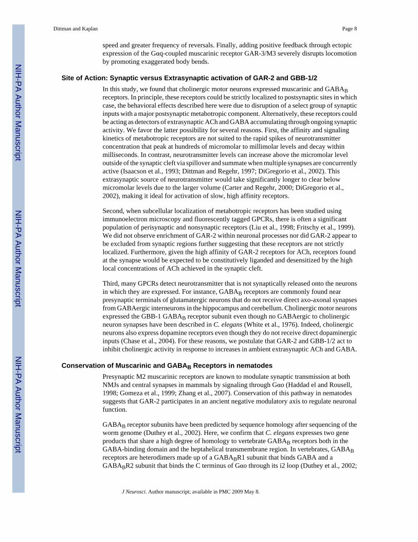
Feedback regulation by muscarinic and GABAB receptors regulates locomotionThus far, our results implicate GAR-2 and GBB-1/2 in the regulation of cholinergic motorneuron function when levels of ACh and GABA are increased by aldicarb treatment. Todetermine whether this feedback inhibition normally plays a role in locomotion, we analyzedthe movement of actively foraging animals in the absence of aldicarb. Figure 5A showsrepresentative trajectories of wild type, gar-2 mutant, gar-2(ACh), and gar-3(ACh) worms overa one minute interval. These trajectories followed the center of mass for each animal as itexplored an agar plate. Wild-type and gar-2 mutants had similar patterns of exploratorybehavior consisting of long runs of forward locomotion interrupted by brief reversals and large-angle turns as has been described previously (Pierce-Shimomura et al., 1999; Hills et al.,2004). In contrast, gar-2(ACh) and gar-3(ACh) animals showed aberrant trajectories andexplored a more restricted area in a given time interval. We quantified locomotory behaviorusing three parameters: speed, turning frequency, and reversal frequency. Speed of locomotion
Dittman and Kaplan Page 6
J Neurosci. Author manuscript; available in PMC 2009 May 8.
NIH
-PA Author Manuscript
NIH
-PA Author Manuscript
NIH
-PA Author Manuscript
was slightly but significantly accelerated in gar-2 mutants (9.2 ± 3.1 % increase, p < 0.01) andretarded in gar-2(ACh) animals (25.4 ± 2.7 % decrease, p<0.01) suggesting that GAR-2regulates the speed of locomotion (Fig. 5B). When GAR-3 was ectopically expressed incholinergic motor neurons, the locomotory speed was greatly reduced (74.6 ± 3.4 % decrease,p<0.01).
In addition to GAR-2 receptor effects on speed, direction changes were also subject to GAR-2regulation. Turning frequency was defined as the number of large-angle (between 50° and160°) forward turns per minute, while reversals were scored as 180° ± 10° changes in direction.Removal of GAR-2 decreased the frequency of turns in half (60.8 ± 8.3 % decrease, p<0.01)whereas excess GAR-2 did not affect turning frequency (Fig. 5C). Ectopic expression ofGAR-3 however, dramatically increased the turning rate (~8 fold increase, p<0.01) to such anextent that animals coiled back on themselves and were unable to maintain forward progress(see Fig. 3A image). The rate of reversals was also reduced by half in gar-2 mutants (52.5 ±8.7 % decrease, p<0.01) whereas excess GAR-2 doubled the reversal rate (103.1 ± 28.3 %increase, p<0.01) and ectopic GAR-3 caused a modest increase (45.9 ± 23.5 %, p<0.05) (Fig.5D). The maximum net distance an animal could travel in a given time interval is determinedby the speed as well as turning and reversal rates. We calculated this maximum distance(Rmax) as a measure of the net velocity of an animal over an extended period of time. Aschematic of the maximal distance measure is shown in Figure 5E. Within a 40 second interval,the most distant point from the starting location (Rmax) was determined for each animal and acumulative histogram of Rmax was generated. The median distance reached by wild-typeanimals (n=67) was about 3.5 mm whereas gar-2 mutants (n=72) reached a median distanceof 4.3 mm, corresponding to a 25% increase (p<0.01) in the net distance covered by gar-2mutants (Fig 5F). All effects on locomotory behavior could be rescued by Venus::GAR-2expression under the endogenous promoter (rescue, Fig. 5). Thus, GAR-2 regulates how oftenanimals change direction and the extent of the area they explore in a given time.
We next analyzed the role of GABAB receptors in regulating locomotion. The effects ofremoving GABAB receptor modulation on locomotion were similar to those observed ingar-2 mutants (Fig. 6). Speed was accelerated significantly (20 ± 3.5 % increase, p<0.01)whereas turn and reversal frequency were decreased (41.4 ± 10.1 % and 42.5 ± 13.4 %respectively, p<0.05). The maximal distance covered in 40 seconds was significantly increasedin both double knockouts (wild type median = 3.5 mm, gar-2;gbb-2 median = 4.1 mm,gbb-1;gbb-2 median = 4.4 mm). In contrast to the additivity observed in the time course ofaldicarb paralysis, no additivity was observed on locomotory changes. Thus, the same receptorsthat provide negative feedback under conditions of elevated transmitter also regulatelocomotory behavior. The observations described above suggest that ACh and GABA releasedduring normal foraging behavior modulate aspects of locomotion and navigation, in additionto their roles in fast excitatory and inhibitory transmission.
DiscussionHere we use C. elegans as a model to identify two neuronal GPCRs that detect transmitterligands, and to test their impact on a behavioral circuit. First, we identify GAR-2/M2 andGBB-1/2 GABABR as the GPCRs that detect ACh and GABA, respectively, in the ventralcord. The M2 receptor, GAR-2 is expressed in motor neurons and mediates negative feedbackvia the Gαo pathway during periods of increased ACh release. Second, C. elegans employs aGABAB receptor dimer in cholinergic motor neurons that mediates heterosynaptic inhibition,likely through the Gαo pathway. Third, both GAR-2/M2 and GABAB receptors promoteinitiation of direction changes during locomotion. Ectopic expression of GAR-2 in allcholinergic neurons bestows profound negative feedback when ACh levels are elevated, andtransgenic animals exhibit opposite locomotory phenotypes as loss of GAR-2 including slower
Dittman and Kaplan Page 7
J Neurosci. Author manuscript; available in PMC 2009 May 8.
NIH
-PA Author Manuscript
NIH
-PA Author Manuscript
NIH
-PA Author Manuscript
speed and greater frequency of reversals. Finally, adding positive feedback through ectopicexpression of the Gαq-coupled muscarinic receptor GAR-3/M3 severely disrupts locomotionby promoting exaggerated body bends.
Site of Action: Synaptic versus Extrasynaptic activation of GAR-2 and GBB-1/2In this study, we found that cholinergic motor neurons expressed muscarinic and GABABreceptors. In principle, these receptors could be strictly localized to postsynaptic sites in whichcase, the behavioral effects described here were due to disruption of a select group of synapticinputs with a major postsynaptic metabotropic component. Alternatively, these receptors couldbe acting as detectors of extrasynaptic ACh and GABA accumulating through ongoing synapticactivity. We favor the latter possibility for several reasons. First, the affinity and signalingkinetics of metabotropic receptors are not suited to the rapid spikes of neurotransmitterconcentration that peak at hundreds of micromolar to millimolar levels and decay withinmilliseconds. In contrast, neurotransmitter levels can increase above the micromolar leveloutside of the synaptic cleft via spillover and summate when multiple synapses are concurrentlyactive (Isaacson et al., 1993; Dittman and Regehr, 1997; DiGregorio et al., 2002). Thisextrasynaptic source of neurotransmitter would take significantly longer to clear belowmicromolar levels due to the larger volume (Carter and Regehr, 2000; DiGregorio et al.,2002), making it ideal for activation of slow, high affinity receptors.
Second, when subcellular localization of metabotropic receptors has been studied usingimmunoelectron microscopy and fluorescently tagged GPCRs, there is often a significantpopulation of perisynaptic and nonsynaptic receptors (Liu et al., 1998; Fritschy et al., 1999).We did not observe enrichment of GAR-2 within neuronal processes nor did GAR-2 appear tobe excluded from synaptic regions further suggesting that these receptors are not strictlylocalized. Furthermore, given the high affinity of GAR-2 receptors for ACh, receptors foundat the synapse would be expected to be constitutively liganded and desensitized by the highlocal concentrations of ACh achieved in the synaptic cleft.
Third, many GPCRs detect neurotransmitter that is not synaptically released onto the neuronsin which they are expressed. For instance, GABAB receptors are commonly found nearpresynaptic terminals of glutamatergic neurons that do not receive direct axo-axonal synapsesfrom GABAergic interneurons in the hippocampus and cerebellum. Cholinergic motor neuronsexpressed the GBB-1 GABAB receptor subunit even though no GABAergic to cholinergicneuron synapses have been described in C. elegans (White et al., 1976). Indeed, cholinergicneurons also express dopamine receptors even though they do not receive direct dopaminergicinputs (Chase et al., 2004). For these reasons, we postulate that GAR-2 and GBB-1/2 act toinhibit cholinergic activity in response to increases in ambient extrasynaptic ACh and GABA.
Conservation of Muscarinic and GABAB Receptors in nematodesPresynaptic M2 muscarinic receptors are known to modulate synaptic transmission at bothNMJs and central synapses in mammals by signaling through Gαo (Haddad el and Rousell,1998; Gomeza et al., 1999; Zhang et al., 2007). Conservation of this pathway in nematodessuggests that GAR-2 participates in an ancient negative modulatory axis to regulate neuronalfunction.
GABAB receptor subunits have been predicted by sequence homology after sequencing of theworm genome (Duthey et al., 2002). Here, we confirm that C. elegans expresses two geneproducts that share a high degree of homology to vertebrate GABAB receptors both in theGABA-binding domain and the heptahelical transmembrane region. In vertebrates, GABABreceptors are heterodimers made up of a GABABR1 subunit that binds GABA and aGABABR2 subunit that binds the C terminus of Gαo through its i2 loop (Duthey et al., 2002;
Dittman and Kaplan Page 8
J Neurosci. Author manuscript; available in PMC 2009 May 8.
NIH
-PA Author Manuscript
NIH
-PA Author Manuscript
NIH
-PA Author Manuscript
Bettler et al., 2004; Thuault et al., 2004). Interestingly, the GABABR1 subunit GBB-1 GABA-binding domain has substantially greater amino acid sequence identity with its rodent orthologthan GBB-2 compared to rodent GABABR2 (55% vs 31%, Fig 4A), whereas GBB-2 shareshigher amino acid identity with its ortholog in the heptahelical region, especially in the i2 loop(58% vs 25% for GBB-2 and GBB-1 respectively). Thus, phylogenetic conservation of thesedomains reflects their function in GABA binding and Gαo signaling.
Determinants of locomotory behaviors in C. elegansNeurotransmitter-activated GPCRs have been implicated in many functions of the mammaliansensory and motor systems. For instance, rodent knockouts of M2 and GABABR subunits havebeen associated with specific locomotory alterations (Gomeza et al., 1999; Schuler et al.,2001; Zhang et al., 2002; Gassmann et al., 2004). C. elegans possesses simple sensory andmotor circuits that are amenable to precise genetic changes in GPCR function. These nematodesnavigate their environment through long runs of forward locomotion interrupted by briefreversals and large-angle (omega turn) body bends that alter direction (Croll, 1975). Many ofthe neurons that initiate or control these stereotyped behaviors have been identified (Pierce-Shimomura et al., 1999; Hills et al., 2004; Gray et al., 2005). Because loss of either GAR-2 orGBB-1/2 decreases reversal and omega turn frequencies, and over-expression of GAR-2increases reversal frequency, it is possible that these inhibitory receptors act in neurons thatnormally suppress turns and reversals. The gustatory sensory neuron ASI, the interneuron AIY,as well as a head motor neuron, RIM have all been reported to inhibit reversals and omegaturns (Gray et al., 2005). Thus, these neurons would be candidates for targets of inhibitoryGPCR modulation. RIM in particular is cholinergic and therefore may employ homosynapticnegative feedback to regulate its suppression of reversals. Further studies elaborating on thespecific neurons that endogenously express GAR-2 will reveal how ACh signaling in thenavigation circuits use GAR-2 to trigger direction changes.
GPCRs generally modulate neuronal activity on a time scale of hundreds of milliseconds tomany seconds. Since the body bends that drive locomotion occur at a rate of about one persecond, it is unlikely that transmitter release and activation of GAR-2 and GBB-1/GBB-2 willmodulate locomotion within a body bend. Instead, these pathways may set the basal tone ofinhibition. In addition, the GPCR mutant data suggest that the transition between behaviors(i.e. reversal of direction) may itself be modulated. These transitions occur at a frequency of1 to 5 per minute so there is ample time for neurotransmitter to accumulate and activate theGαo pathway.
An in vivo analysis of feedback regulationNeurotransmitter spillover has been proposed to mediate homo- and hetero-synaptic depressionand, consequently, to play an important role in activity dependent modulation of circuitfunction (Dittman and Regehr, 1997; Mitchell and Silver, 2000). Here we describe a possiblerole for extrasynaptic ACh and GABA in regulating a locomotory circuit via metabotropicreceptors. In particular, we focused on a negative feedback loop created by the inhibitory actionof ACh and GABA on cholinergic neurons. Negative feedback typically provides a stabilizingforce that can contribute to the fine tuning of a network. Exchanging a negative feedback loopfor a positive feedback would likely abolish this stability since small perturbations would begreatly amplified. In the case of exchanging an M2 receptor for an M3 receptor, small increasesin extrasynaptic ACh would now drive further ACh release by coupling to a stimulatorypathway. Even under resting conditions, transgenic animals expressing the M3 receptor showedsevere dysfunction of coordinated locomotion. Body bend angles were greatly increased,compromising the ability of the animal to generate and propagate the sinusoidal wavesnecessary for normal locomotion.
Dittman and Kaplan Page 9
J Neurosci. Author manuscript; available in PMC 2009 May 8.
NIH
-PA Author Manuscript
NIH
-PA Author Manuscript
NIH
-PA Author Manuscript
In addition to the homosynaptic negative feedback provided by cholinergic GAR-2 receptors,GABA MNs also expressed GAR-2. Although GABAergic rescue of GAR-2 did not restorewild-type aldicarb sensitivity, it is possible that heterosynaptic effects of ACh on GABAneurons may be important for navigational behaviors.
Our results extend C. elegans as a genetic model to dissect the functional impact of GPCR-mediated feedback regulation on a behavioral circuit. Future studies will help elucidate theparticular neurons involved in modulating locomotory behavior as well as the spatial andtemporal domains over which the GAR-2 and GBB-1/2 receptors sense their ligands andmodulate the nervous system.
AcknowledgementsThis work was supported by a research grant (GM54728) from the National Institutes of Health (J.K.) and by a DamonRunyon CRF postdoctoral fellowship (J.D.). We thank the C. elegans Genetic Stock Center and S. Mitani for strains.We also thank members of the Kaplan lab for comments on this manuscript.
ReferencesBargmann CI. Neurobiology of the Caenorhabditis elegans genome. Science 1998;282:2028–2033.
[PubMed: 9851919]Bastiani, C.; Mendel, J. WormBook. 2006. Heterotrimeric G proteins in C. elegans; p. 1-25.Bauer CS, Woolley RJ, Teschemacher AG, Seward EP. Potentiation of exocytosis by phospholipase C-
coupled G-protein-coupled receptors requires the priming protein Munc13-1. J Neurosci 2007;27:212–219. [PubMed: 17202488]
Bettler B, Kaupmann K, Mosbacher J, Gassmann M. Molecular structure and physiological functions ofGABA(B) receptors. Physiol Rev 2004;84:835–867. [PubMed: 15269338]
Brenner S. The genetics of Caenorhabditis elegans. Genetics 1974;77:71–94. [PubMed: 4366476]Burstein ES, Spalding TA, Brann MR. Constitutive activation of chimeric m2/m5 muscarinic receptors
and delineation of G-protein coupling selectivity domains. Biochem Pharmacol 1996;51:539–544.[PubMed: 8619900]
Carre-Pierrat M, Baillie D, Johnsen R, Hyde R, Hart A, Granger L, Segalat L. Characterization of theCaenorhabditis elegans G protein-coupled serotonin receptors. Invert Neurosci 2006;6:189–205.[PubMed: 17082916]
Carter AG, Regehr WG. Prolonged synaptic currents and glutamate spillover at the parallel fiber to stellatecell synapse. J Neurosci 2000;20:4423–4434. [PubMed: 10844011]
Chase DL, Pepper JS, Koelle MR. Mechanism of extrasynaptic dopamine signaling in Caenorhabditiselegans. Nat Neurosci 2004;7:1096–1103. [PubMed: 15378064]
Croll NA. Behavioural analysis of nematode movement. Adv Parasitol 1975;13:71–122. [PubMed:1169872]
DiGregorio DA, Nusser Z, Silver RA. Spillover of glutamate onto synaptic AMPA receptors enhancesfast transmission at a cerebellar synapse. Neuron 2002;35:521–533. [PubMed: 12165473]
Dittman JS, Regehr WG. Mechanism and kinetics of heterosynaptic depression at a cerebellar synapse.J Neurosci 1997;17:9048–9059. [PubMed: 9364051]
Dittman JS, Kaplan JM. Factors regulating the abundance and localization of synaptobrevin in the plasmamembrane. Proc Natl Acad Sci U S A 2006;103:11399–11404. [PubMed: 16844789]
Duthey B, Caudron S, Perroy J, Bettler B, Fagni L, Pin JP, Prezeau L. A single subunit (GB2) is requiredfor G-protein activation by the heterodimeric GABA(B) receptor. J Biol Chem 2002;277:3236–3241.[PubMed: 11711539]
Fritschy JM, Meskenaite V, Weinmann O, Honer M, Benke D, Mohler H. GABAB-receptor splicevariants GB1a and GB1b in rat brain: developmental regulation, cellular distribution andextrasynaptic localization. Eur J Neurosci 1999;11:761–768. [PubMed: 10103070]
Gassmann M, Shaban H, Vigot R, Sansig G, Haller C, Barbieri S, Humeau Y, Schuler V, Muller M,Kinzel B, Klebs K, Schmutz M, Froestl W, Heid J, Kelly PH, Gentry C, Jaton AL, Van der Putten
Dittman and Kaplan Page 10
J Neurosci. Author manuscript; available in PMC 2009 May 8.
NIH
-PA Author Manuscript
NIH
-PA Author Manuscript
NIH
-PA Author Manuscript
H, Mombereau C, Lecourtier L, Mosbacher J, Cryan JF, Fritschy JM, Luthi A, Kaupmann K, BettlerB. Redistribution of GABAB(1) protein and atypical GABAB responses in GABAB(2)-deficientmice. J Neurosci 2004;24:6086–6097. [PubMed: 15240800]
Gomeza J, Shannon H, Kostenis E, Felder C, Zhang L, Brodkin J, Grinberg A, Sheng H, Wess J.Pronounced pharmacologic deficits in M2 muscarinic acetylcholine receptor knockout mice. ProcNatl Acad Sci U S A 1999;96:1692–1697. [PubMed: 9990086]
Gray JM, Hill JJ, Bargmann CI. A circuit for navigation in Caenorhabditis elegans. Proc Natl Acad SciU S A 2005;102:3184–3191. [PubMed: 15689400]
Haddad el B, Rousell J. Regulation of the expression and function of the M2 muscarinic receptor. TrendsPharmacol Sci 1998;19:322–327. [PubMed: 9745360]
Hajdu-Cronin YM, Chen WJ, Patikoglou G, Koelle MR, Sternberg PW. Antagonism between G(o)alphaand G(q)alpha in Caenorhabditis elegans: the RGS protein EAT-16 is necessary for G(o)alphasignaling and regulates G(q)alpha activity. Genes Dev 1999;13:1780–1793. [PubMed: 10421631]
Hartzell HC, Kuffler SW, Yoshikami D. Post-synaptic potentiation: interaction between quanta ofacetylcholine at the skeletal neuromuscular synapse. J Physiol 1975;251:427–463. [PubMed:171379]
Hille B. G protein-coupled mechanisms and nervous signaling. Neuron 1992;9:187–195. [PubMed:1353972]
Hills T, Brockie PJ, Maricq AV. Dopamine and glutamate control area-restricted search behavior inCaenorhabditis elegans. J Neurosci 2004;24:1217–1225. [PubMed: 14762140]
Isaacson JS, Solis JM, Nicoll RA. Local and diffuse synaptic actions of GABA in the hippocampus.Neuron 1993;10:165–175. [PubMed: 7679913]
Kubo T, Bujo H, Akiba I, Nakai J, Mishina M, Numa S. Location of a region of the muscarinicacetylcholine receptor involved in selective effector coupling. FEBS Lett 1988;241:119–125.[PubMed: 3197827]
Lackner MR, Nurrish SJ, Kaplan JM. Facilitation of synaptic transmission by EGL-30 Gqalpha andEGL-8 PLCbeta: DAG binding to UNC-13 is required to stimulate acetylcholine release. Neuron1999;24:335–346. [PubMed: 10571228]
Lanzafame AA, Christopoulos A, Mitchelson F. Cellular signaling mechanisms for muscarinicacetylcholine receptors. Receptors Channels 2003;9:241–260. [PubMed: 12893537]
Lee YS, Park YS, Nam S, Suh SJ, Lee J, Kaang BK, Cho NJ. Characterization of GAR-2, a novel Gprotein-linked acetylcholine receptor from Caenorhabditis elegans. J Neurochem 2000;75:1800–1809. [PubMed: 11032868]
Liu XB, Munoz A, Jones EG. Changes in subcellular localization of metabotropic glutamate receptorsubtypes during postnatal development of mouse thalamus. J Comp Neurol 1998;395:450–465.[PubMed: 9619499]
Miller KG, Emerson MD, Rand JB. Goalpha and diacylglycerol kinase negatively regulate the Gqalphapathway in C. elegans. Neuron 1999;24:323–333. [PubMed: 10571227]
Miller KG, Alfonso A, Nguyen M, Crowell JA, Johnson CD, Rand JB. A genetic selection forCaenorhabditis elegans synaptic transmission mutants. Proc Natl Acad Sci U S A 1996;93:12593–12598. [PubMed: 8901627]
Mitchell SJ, Silver RA. Glutamate spillover suppresses inhibition by activating presynaptic mGluRs.Nature 2000;404:498–502. [PubMed: 10761918]
Niebur E, Erdos P. Theory of the locomotion of nematodes: control of the somatic motor neurons byinterneurons. Math Biosci 1993;118:51–82. [PubMed: 8260760]
Nurrish S, Segalat L, Kaplan JM. Serotonin inhibition of synaptic transmission: Galpha(0) decreases theabundance of UNC-13 at release sites. Neuron 1999;24:231–242. [PubMed: 10677040]
Pierce-Shimomura JT, Morse TM, Lockery SR. The fundamental role of pirouettes in Caenorhabditiselegans chemotaxis. J Neurosci 1999;19:9557–9569. [PubMed: 10531458]
Robatzek M, Thomas JH. Calcium/calmodulin-dependent protein kinase II regulates Caenorhabditiselegans locomotion in concert with a G(o)/G(q) signaling network. Genetics 2000;156:1069–1082.[PubMed: 11063685]
Dittman and Kaplan Page 11
J Neurosci. Author manuscript; available in PMC 2009 May 8.
NIH
-PA Author Manuscript
NIH
-PA Author Manuscript
NIH
-PA Author Manuscript
Robatzek M, Niacaris T, Steger K, Avery L, Thomas JH. eat-11 encodes GPB-2, a Gbeta(5) orthologthat interacts with G(o)alpha and G(q)alpha to regulate C. elegans behavior. Curr Biol 2001;11:288–293. [PubMed: 11250160]
Schuler V, Luscher C, Blanchet C, Klix N, Sansig G, Klebs K, Schmutz M, Heid J, Gentry C, Urban L,Fox A, Spooren W, Jaton AL, Vigouret J, Pozza M, Kelly PH, Mosbacher J, Froestl W, Kaslin E,Korn R, Bischoff S, Kaupmann K, van der Putten H, Bettler B. Epilepsy, hyperalgesia, impairedmemory, and loss of pre- and postsynaptic GABA(B) responses in mice lacking GABA(B(1)).Neuron 2001;31:47–58. [PubMed: 11498050]
Schuske K, Beg AA, Jorgensen EM. The GABA nervous system in C. elegans. Trends Neurosci2004;27:407–414. [PubMed: 15219740]
Sieburth D, Ch’ng Q, Dybbs M, Tavazoie M, Kennedy S, Wang D, Dupuy D, Rual JF, Hill DE, VidalM, Ruvkun G, Kaplan JM. Systematic analysis of genes required for synapse structure and function.Nature 2005;436:510–517. [PubMed: 16049479]
Steger KA, Avery L. The GAR-3 muscarinic receptor cooperates with calcium signals to regulate musclecontraction in the Caenorhabditis elegans pharynx. Genetics 2004;167:633–643. [PubMed:15238517]
Szapiro G, Barbour B. Multiple climbing fibers signal to molecular layer interneurons exclusively viaglutamate spillover. Nat Neurosci 2007;10:735–742. [PubMed: 17515900]
Thuault SJ, Brown JT, Sheardown SA, Jourdain S, Fairfax B, Spencer JP, Restituito S, Nation JH, ToppsS, Medhurst AD, Randall AD, Couve A, Moss SJ, Collingridge GL, Pangalos MN, Davies CH, CalverAR. The GABA(B2) subunit is critical for the trafficking and function of native GABA(B) receptors.Biochem Pharmacol 2004;68:1655–1666. [PubMed: 15451409]
Vashlishan AB, Madison JM, Dybbs M, Bai J, Sieburth D, Ch’ng Q, Tavazoie M, Kaplan JM. An RNAiScreen Identifies Genes that Regulate GABA Synapses. Neuron 2008;58:346–361. [PubMed:18466746]
White JG, Southgate E, Thomson JN, Brenner S. The structure of the ventral nerve cord of Caenorhabditiselegans. Philos Trans R Soc Lond B Biol Sci 1976;275:327–348. [PubMed: 8806]
You YJ, Kim J, Cobb M, Avery L. Starvation activates MAP kinase through the muscarinic acetylcholinepathway in Caenorhabditis elegans pharynx. Cell Metab 2006;3:237–245. [PubMed: 16581001]
Zhang HM, Chen SR, Pan HL. Regulation of glutamate release from primary afferents and interneuronsin the spinal cord by muscarinic receptor subtypes. J Neurophysiol 2007;97:102–109. [PubMed:17050831]
Zhang W, Basile AS, Gomeza J, Volpicelli LA, Levey AI, Wess J. Characterization of central inhibitorymuscarinic autoreceptors by the use of muscarinic acetylcholine receptor knock-out mice. J Neurosci2002;22:1709–1717. [PubMed: 11880500]
Dittman and Kaplan Page 12
J Neurosci. Author manuscript; available in PMC 2009 May 8.
NIH
-PA Author Manuscript
NIH
-PA Author Manuscript
NIH
-PA Author Manuscript
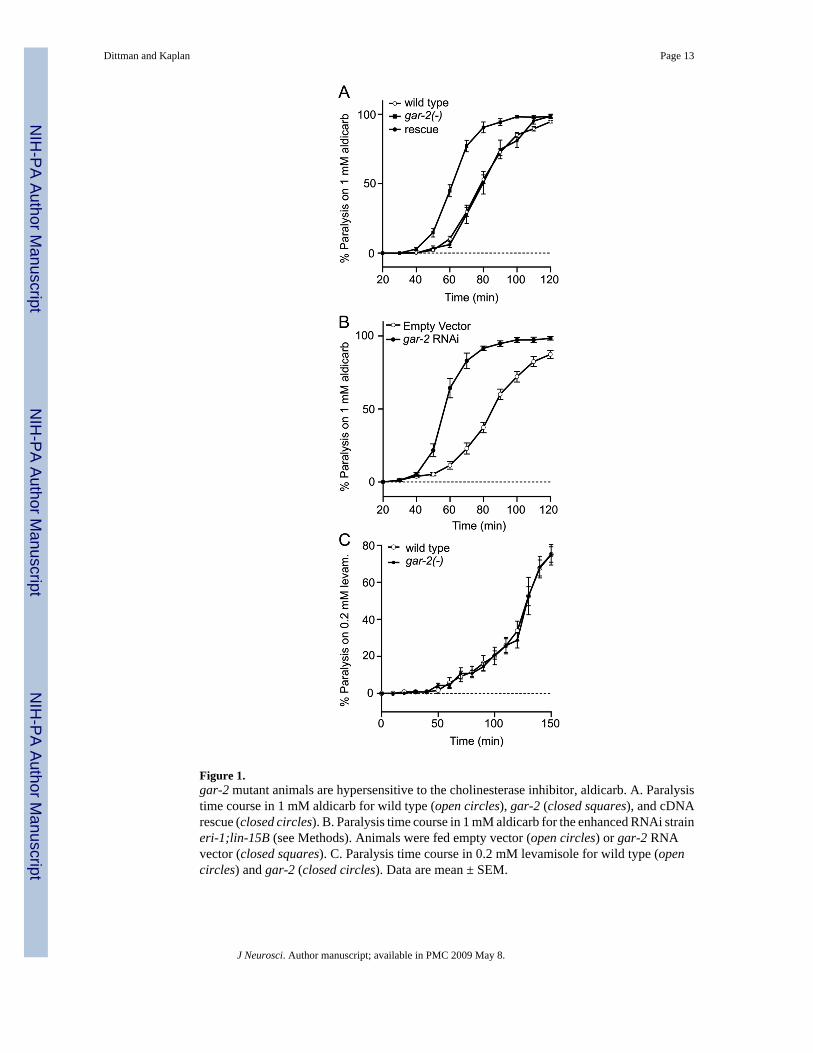
Figure 1.gar-2 mutant animals are hypersensitive to the cholinesterase inhibitor, aldicarb. A. Paralysistime course in 1 mM aldicarb for wild type (open circles), gar-2 (closed squares), and cDNArescue (closed circles). B. Paralysis time course in 1 mM aldicarb for the enhanced RNAi straineri-1;lin-15B (see Methods). Animals were fed empty vector (open circles) or gar-2 RNAvector (closed squares). C. Paralysis time course in 0.2 mM levamisole for wild type (opencircles) and gar-2 (closed circles). Data are mean ± SEM.
Dittman and Kaplan Page 13
J Neurosci. Author manuscript; available in PMC 2009 May 8.
NIH
-PA Author Manuscript
NIH
-PA Author Manuscript
NIH
-PA Author Manuscript
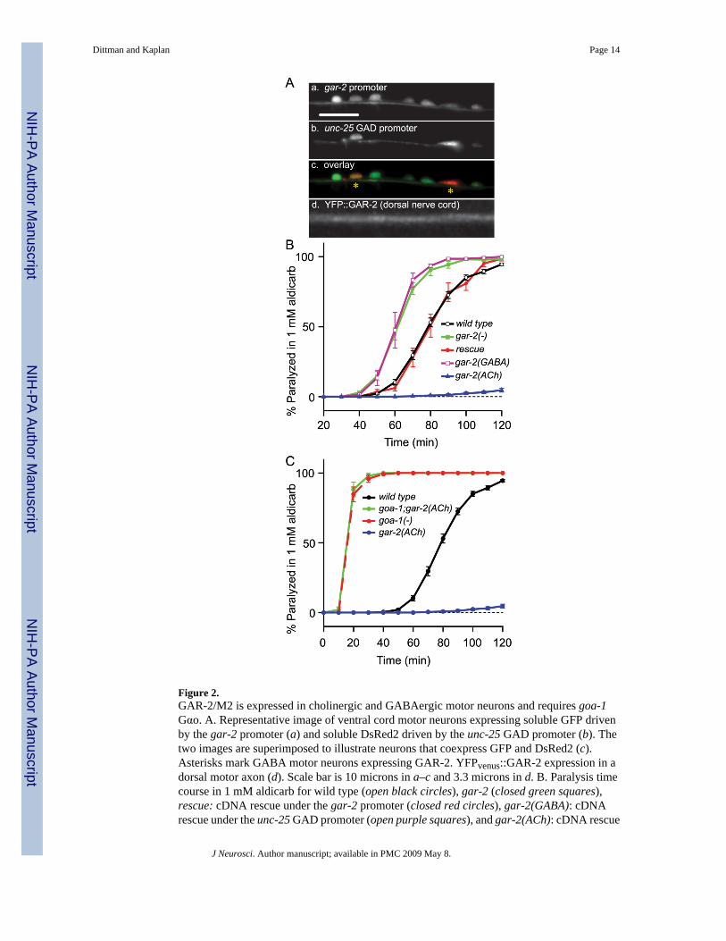
Figure 2.GAR-2/M2 is expressed in cholinergic and GABAergic motor neurons and requires goa-1Gαo. A. Representative image of ventral cord motor neurons expressing soluble GFP drivenby the gar-2 promoter (a) and soluble DsRed2 driven by the unc-25 GAD promoter (b). Thetwo images are superimposed to illustrate neurons that coexpress GFP and DsRed2 (c).Asterisks mark GABA motor neurons expressing GAR-2. YFPvenus::GAR-2 expression in adorsal motor axon (d). Scale bar is 10 microns in a–c and 3.3 microns in d. B. Paralysis timecourse in 1 mM aldicarb for wild type (open black circles), gar-2 (closed green squares),rescue: cDNA rescue under the gar-2 promoter (closed red circles), gar-2(GABA): cDNArescue under the unc-25 GAD promoter (open purple squares), and gar-2(ACh): cDNA rescue
Dittman and Kaplan Page 14
J Neurosci. Author manuscript; available in PMC 2009 May 8.
NIH
-PA Author Manuscript
NIH
-PA Author Manuscript
NIH
-PA Author Manuscript
under the unc-17 VAChT promoter (closed blue triangles). C. Paralysis time course in 1 mMaldicarb for wild type (closed black circles), goa-1Gαo (closed red circles), gar-2(ACh): cDNArescue under the unc-17 VAChT promoter (closed blue circles), and goa-1;gar-2(ACh) (closedgreen circles). Data are mean ± SEM.
Dittman and Kaplan Page 15
J Neurosci. Author manuscript; available in PMC 2009 May 8.
NIH
-PA Author Manuscript
NIH
-PA Author Manuscript
NIH
-PA Author Manuscript
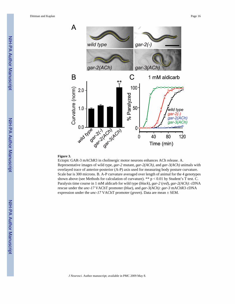
Figure 3.Ectopic GAR-3 mAChR3 in cholinergic motor neurons enhances ACh release. A.Representative images of wild type, gar-2 mutant, gar-2(ACh), and gar-3(ACh) animals withoverlayed trace of anterior-posterior (A-P) axis used for measuring body posture curvature.Scale bar is 300 microns. B. A-P curvature averaged over length of animal for the 4 genotypesshown above (see Methods for calculation of curvature). ** p < 0.01 by Student’s T test. C.Paralysis time course in 1 mM aldicarb for wild type (black), gar-2 (red), gar-2(ACh): cDNArescue under the unc-17 VAChT promoter (blue), and gar-3(ACh): gar-3 mAChR3 cDNAexpression under the unc-17 VAChT promoter (green). Data are mean ± SEM.
Dittman and Kaplan Page 16
J Neurosci. Author manuscript; available in PMC 2009 May 8.
NIH
-PA Author Manuscript
NIH
-PA Author Manuscript
NIH
-PA Author Manuscript
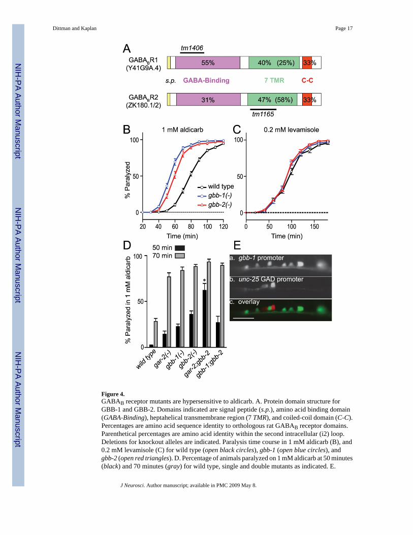
Figure 4.GABAB receptor mutants are hypersensitive to aldicarb. A. Protein domain structure forGBB-1 and GBB-2. Domains indicated are signal peptide (s.p.), amino acid binding domain(GABA-Binding), heptahelical transmembrane region (7 TMR), and coiled-coil domain (C-C).Percentages are amino acid sequence identity to orthologous rat GABAB receptor domains.Parenthetical percentages are amino acid identity within the second intracellular (i2) loop.Deletions for knockout alleles are indicated. Paralysis time course in 1 mM aldicarb (B), and0.2 mM levamisole (C) for wild type (open black circles), gbb-1 (open blue circles), andgbb-2 (open red triangles). D. Percentage of animals paralyzed on 1 mM aldicarb at 50 minutes(black) and 70 minutes (gray) for wild type, single and double mutants as indicated. E.
Dittman and Kaplan Page 17
J Neurosci. Author manuscript; available in PMC 2009 May 8.
NIH
-PA Author Manuscript
NIH
-PA Author Manuscript
NIH
-PA Author Manuscript
Representative image of ventral cord motor neurons expressing soluble GFP driven by thegbb-1 promoter (a) and soluble DsRed2 driven by a GABAergic neuron-specific promoter(b). The two images are superimposed to illustrate neurons do not coexpress GFP and DsRed2(c) Scale bar is 5 microns. Data are mean ± SEM. ** p < 0.01 compared to gar-2, gbb-1, andgbb-2 by Student’s T test.
Dittman and Kaplan Page 18
J Neurosci. Author manuscript; available in PMC 2009 May 8.
NIH
-PA Author Manuscript
NIH
-PA Author Manuscript
NIH
-PA Author Manuscript
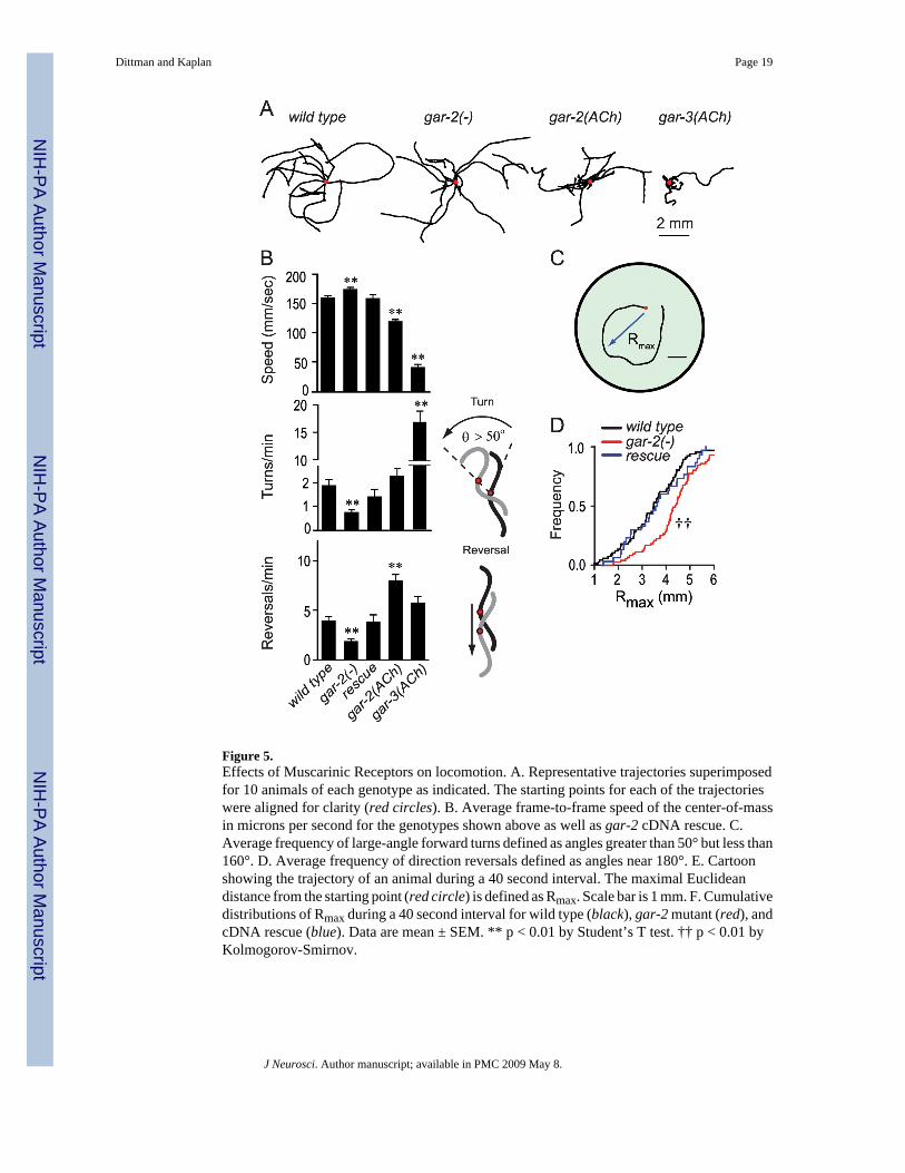
Figure 5.Effects of Muscarinic Receptors on locomotion. A. Representative trajectories superimposedfor 10 animals of each genotype as indicated. The starting points for each of the trajectorieswere aligned for clarity (red circles). B. Average frame-to-frame speed of the center-of-massin microns per second for the genotypes shown above as well as gar-2 cDNA rescue. C.Average frequency of large-angle forward turns defined as angles greater than 50° but less than160°. D. Average frequency of direction reversals defined as angles near 180°. E. Cartoonshowing the trajectory of an animal during a 40 second interval. The maximal Euclideandistance from the starting point (red circle) is defined as Rmax. Scale bar is 1 mm. F. Cumulativedistributions of Rmax during a 40 second interval for wild type (black), gar-2 mutant (red), andcDNA rescue (blue). Data are mean ± SEM. ** p < 0.01 by Student’s T test. †† p < 0.01 byKolmogorov-Smirnov.
Dittman and Kaplan Page 19
J Neurosci. Author manuscript; available in PMC 2009 May 8.
NIH
-PA Author Manuscript
NIH
-PA Author Manuscript
NIH
-PA Author Manuscript
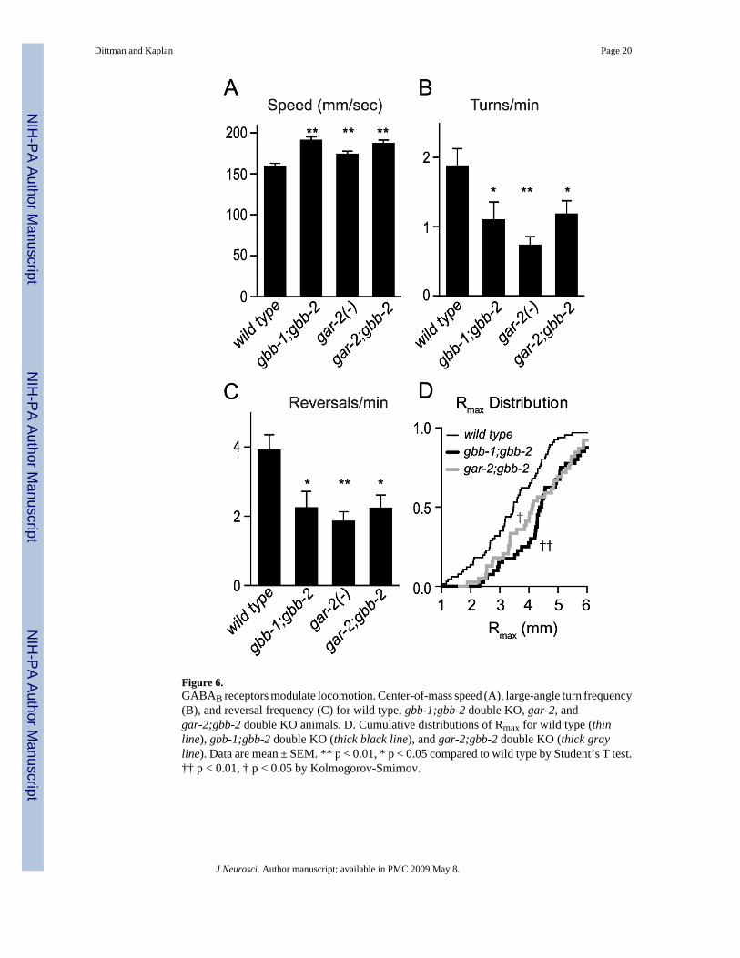
Figure 6.GABAB receptors modulate locomotion. Center-of-mass speed (A), large-angle turn frequency(B), and reversal frequency (C) for wild type, gbb-1;gbb-2 double KO, gar-2, andgar-2;gbb-2 double KO animals. D. Cumulative distributions of Rmax for wild type (thinline), gbb-1;gbb-2 double KO (thick black line), and gar-2;gbb-2 double KO (thick grayline). Data are mean ± SEM. ** p < 0.01, * p < 0.05 compared to wild type by Student’s T test.†† p < 0.01, † p < 0.05 by Kolmogorov-Smirnov.
Dittman and Kaplan Page 20
J Neurosci. Author manuscript; available in PMC 2009 May 8.
NIH
-PA Author Manuscript
NIH
-PA Author Manuscript
NIH
-PA Author Manuscript
![Sensory Neurons Arouse C. elegans Locomotion via Both ... · TRPV)RMG circuit activity are associated with locomotion arousal andquiescence respec-tively [11,14,17,18]. We previously](https://img.pdfslide.us/doc/110x75/5f097a987e708231d4270580/sensory-neurons-arouse-c-elegans-locomotion-via-both-trpvrmg-circuit-activity.jpg)
![A - Benzodiazepine-Chloride Receptor-Targeted Therapy for ......nisms through GABAA and GABAB receptors [12]. GABA is classified into two main categories: GABAA and GABAB. GABAA and](https://img.pdfslide.us/doc/110x75/60f82a0e0bab2d34196b5ccd/a-benzodiazepine-chloride-receptor-targeted-therapy-for-nisms-through.jpg)