Embed Size (px)
Citation preview
Atypical empathic responses in adolescents with aggressiveconduct disorder: a functional MRI investigation
Jean Decety1,2, Kalina J. Michalska1, Yuko Akitsuki1, and Benjamin B. Lahey3,21 Department of Psychology and Center for Cognitive and Social Neuroscience, The University ofChicago2 Department of Psychiatry, The University of Chicago3 Department of Health Studies, The University of Chicago
AbstractBecause youth with aggressive conduct disorder (CD) often inflict pain on others, it is important todetermine if they exhibit atypical empathic responses to viewing others in pain. In this initialfunctional magnetic resonance imaging (fMRI) study, 8 adolescents with aggressive CD and 8matched controls were scanned while watching animated visual stimuli depicting other peopleexperiencing pain or not experiencing pain. Furthermore, these situations involved either anindividual whose pain was caused by accident or an individual whose pain was inflicted on purposeby another person. After scanning, participants rated how painful the situations were. In both groupsthe perception of others in pain was associated with activation of the pain matrix, including the ACC,insula, somatosensory cortex, supplementary motor area and periaqueductal gray. The pain matrixwas activated to a significantly greater extent in participants with CD, who also showed strongamygdala, ventral striatum, and temporal pole activation. When watching situations in which painwas intentionally inflicted, control youth also exhibited signal increase in the medial prefrontal frontalcortex, lateral obitofrontal cortex, and temporoparietal junction, whereas youth with CD onlyexhibited activation in the insula. Furthermore, connectivity analyses demonstrated that youth withCD exhibited less amygdala/prefrontal coupling when watching pain inflicted by another than didcontrol youth. These preliminary findings suggest that youth with aggressive CD exhibit an atypicalpattern of neural response to viewing others in pain that should be explored in further studies.
KeywordsPerception of Pain; Empathy; Emotion Regulation; Conduct Disorder; Aggression
1. IntroductionConduct disorder (CD) is a serious mental disorder of childhood and adolescence that ischaracterized by a longstanding pattern of violations of rules and laws. Symptoms of CDinclude physical aggression, manipulative lying, theft, forced sex, bullying, running away fromhome overnight, and destruction of property. CD is a major public health problem becauseyouth with conduct disorder not only inflict serious physical and psychological harm on others,but they are at greatly increased risk for incarceration, injury, depression, substance abuse, anddeath by homicide and suicide themselves (Loeber et al., 1998). Furthermore, CD is important
Corresponding Author: Dr. Jean Decety, Departments of Psychology and Psychiatry, The University of Chicago, 5848 S. UniversityAvenue, 5848 S. University Avenue, Chicago, IL 60637, Phone: 773-834-3711, Fax: 773-702-0886, [email protected], Lab website: http://scnl.org.
NIH Public AccessAuthor ManuscriptBiol Psychol. Author manuscript; available in PMC 2010 February 10.
Published in final edited form as:Biol Psychol. 2009 February ; 80(2): 203. doi:10.1016/j.biopsycho.2008.09.004.
NIH
-PA
Author M
anuscriptN
IH-P
A A
uthor Manuscript
NIH
-PA
Author M
anuscript
because it is the major childhood precursor to antisocial personality disorder in adulthood(Lahey, Loeber, Burke, & Applegate, 2005). Thus there is a pressing need to understand thebiopsychological processes at multiple levels of analysis that give rise to CD. Biological studiesof antisocial behavior can lead to new approaches to the treatment of psychiatric conditio nsassociated with aggression (Van Goozen & Fairchild, 2008).
Empathy, the capacity to understand and appreciate the emotional states and needs of othersin reference to oneself, has been one psychological characteristic repeatedly proposed as a coredeficit in CD (see Lovett & Sheffield, 2007 for a review). Here we consider empathy as aconstruct accounting for a sense of similarity in feelings experienced by the self and the other,without confusion between the two individuals (Batson, Fultz & Schoenrade, 1987; Decety &Batson, 2007; Decety & Jackson, 2004; Decety & Moriguchi, 2007; Eisenberg, Spinrad &Sadovsky, 2006). The experience of empathy can lead to sympathy or empathic concern foranother based on the apprehension or comprehension of the other’s emotional state orcondition, or to personal distress, i.e., an aversive, self-focused emotional reaction to theemotional state or condition of another (Batson, Fultz & Schoenrade, 1987; Decety & Lamm,2008; Eisenberg & Eggum, 2008; Lamm, Batson & Decety, 2007).
Interestingly, some developmental psychologists have hypothesized that empathy andsympathetic concern for others is an essential factor inhibiting aggression toward others(Eisenberg 2005, Hoffman, 2000). Empathy may be regarded as a proximate factor motivatingprosocial rather than antisocial behavior (Batson, 1991). It is commonly defined as an affectivereaction that is appropriate to someone else’s situation rather than one’s own. Some researchershave theorized that there should be a relation between aggressive behavior and a lack ofempathy (e.g., Zahn-Waxler et al., 1995). Similarly, other scholars have proposed thatindividual differences in callous disregard for the welfare of others is an important risk factorfor CD (Frick et al., 2005; Lahey & Waldman, 2003).
The propensity for aggressive behavior has been hypothesized to reflect a blunted empathicresponse to the suffering of others (Blair, 2005). Such a lack of empathy in aggressiveindividuals may be a consequence of a failure to be aroused to the distress of others (Raine etal., 1997). In line with this hypothesis, it has been suggested that aggressive behavior arisesfrom an abnormal processing of affective information, resulting in a deficiency in experiencingfear, empathy, and guilt, which in normally inhibit the acting out of violent impulses (Davidsonet al., 2000; Herpertz & Sass, 2000). Consistent with this hypothesis, one functional MRI studyfound reduced left amygdala response in 13 adolescents with CD in response to the visualpresentation of pictures with strong negative emotional valence compared to 14 controladolescents (Sterzer et al., 2005).
An alternative hypothesis can be drawn from research on aggression that has shown thatnegative affect is generally associated with aggression (Anderson & Bushman, 2002;Berkowitz, 2003), suggesting that empathic mimicry in conjunction with poor emotionregulation might produce distress that increases aggression (Campbell, 1990; Gill & Calkins,2003). For instance, there are many empirical studies that documented that physical pain ofteninstigates aggressive inclinations (Berkowitz, 1983, 1993). This is particularly interesting inthe light of recent work in cognitive neuroscience of empathy for pain.
Indeed, a growing number of fMRI studies have demonstrated striking similarities in the neuralcircuits involved in the processing of both the first-hand experience of pain and the experienceof observing other individuals in pain (Jackson, Rainville & Decety, 2006). These studies haveconsistently shown that the perception of pain in others elicits activation of the neural circuitsubserving the processing of the affective and motivational dimension of pain in oneself(Botvinick et al., 2005; Cheng et al., 2007a; Gu & Han, 2007; Jackson et al., 2005, 2006; Lamm
Decety et al. Page 2
Biol Psychol. Author manuscript; available in PMC 2010 February 10.
NIH
-PA
Author M
anuscriptN
IH-P
A A
uthor Manuscript
NIH
-PA
Author M
anuscript
et al., 2007a, Moriguchi et al., 2007, Morrison et al., 2004; Ogino et al., 2007; Saarela et al.2007, Singer et al., 2004). This circuit includes the dorsal anterior cingulate cortex (ACC), theanterior midcingulate cortex (aMCC), the supplementary motor area (SMA), and anteriorinsula (Derbyshire, 2000; Price, 2000). In addition, somatosensory-evoked potentials (Bufalariet al., 2007), and fMRI studies (Cheng et al., 2007a; Lamm et al., 2007b; Moriguchi et al.,2007) have demonstrated that areas processing the sensory dimension of pain (posterior insula/somatosensory cortex) may also be elicited by the visual perception of pain in others.
Recently, one functional MRI study investigated empathy and intentionality in typicallydeveloping middle-school children (Decety, Michalska & Akitsuki, 2008) while they watcheddynamic visual stimuli depicting either a person whose pain was accidentally caused or a personwhose pain was intentionally inflicted by another individual. Interestingly, when watching aperson inflicting pain on another, regions that are engaged in representing social interactionand moral behavior including the temporo-parietal junction, the paracingulate, orbital medialfrontal cortices, and amygdala (see Moll et al., 2003, 2007) were additionally recruited, andincreased their connectivity with the frontoparietal attention network.
There also is evidence that specifically associates the amygdala and paralimbic prefrontalregions, including the dorsal and ventral/orbital medial prefrontal cortex (dMPFC and vMPFC/OFC, respectively), with human aggression (Coccarro et al., 2007; Davidson et al., 2000). Inhumans, amygdala atrophy and/or lesions have been associated with impulsively aggressivebehaviors (van Elst et al., 2000). Specific damage to the OFC is associated with impulsive andaggressive behavior (Izquierdo et al., 2005), and individuals with such damage show littlecontrol over their emotions as well as limited awareness of the moral implications of theiractions (Anderson et al., 1999). Since the amygdala and OFC are anatomically and functionallyconnected (Amaral and Price 1984), their interactions may be critical for interpretingemotionally significant information and guiding goal directed behaviors (Saddoris et al.,2005). Furthermore, the OFC is hypothesized to play a key role in modulating limbic reactivityto threat (Davidson et al., 2000; Izquierdo et al., 2005), and in general is important for theinterpretation of social cues.
Thus, there is evidence that perceiving others in pain triggers an automatic somatic andsensorimotor resonance between other and self, which activates almost the entire neural painmatrix including the periaqueductal gray (PAG) a major site in pain transmission and forprocessing fear and anxiety (Jenk et al., 1995), the SMA that programs defensive skeletomotorimpulses to avoid the stimulus in the context of nociceptive information (Morrison et al.,2006), and thalamus. Such a mechanism provides a functional bridge between first-person andthird person information, which allows for analogical reasoning, and offers a possible, yetpartial, route to understanding others (Decety & Sommerville, 2003). It also provides a clearsignal of the other’s distress that usually inhibits aggressive behavior.
So far, there is no published work on how youth with CD react to viewing others in pain. Ifthe blunted empathic emotional response hypothesis is correct, adolescents with aggressiveCD should react less to stimuli depicting others in pain than healthy controls. Furthermore,this lack of signal from the pain matrix and amygdala could account for impairment inrecognizing information about the distress of others. If the pain-aggression hypothesis iscorrect, youth with aggressive CD should exhibit greater activation than healthy controls inthe amygdala, PAG, and related areas in response to stimuli depicting others in pain. Thisstrong ac tivation of structures subserving negative emotion and less functional amygdala/OFCconnectedness could reflect a general tendency to experience personal distress that elicitsaggression in some circumstances. In addition, having a condition in which pain wasintentionally inflicted into another allows us to examine the respective contribution ofmechanisms that contribute to theory of mind and moral reasoning in the context of pain
Decety et al. Page 3
Biol Psychol. Author manuscript; available in PMC 2010 February 10.
NIH
-PA
Author M
anuscriptN
IH-P
A A
uthor Manuscript
NIH
-PA
Author M
anuscript
perception (see Decety, Michalska & Akitsuki, 2008). This is particularly interesting toinvestigate with respect to antisocial behavior given that these individuals have been reportedto lack guilt and empathic concern.
2. Methods and Materials2.1. Participants
Two groups of 16–18 year-old adolescents (exactly matched on age, sex, and race-ethnicity)were scanned. The participants were purposively selected from a 9-year longitudinal study of127 adolescents with attention-deficit/hyperactivity disorder and a matched healthycomparison group of 125 youth (Lahey et al., 2005). The study included 8 structured diagnosticassessments of CD using the DSM-IV field trials version of the Diagnostic Interview Schedulefor Children (Lahey et al., 1994; Shaffer, Fisher, Piacentini, Schwab-Stone, & Wicks, 1993)conducted over 9 years beginning at 4–6 years of age. Information on symptoms of thedisruptive behavior disorders was obtained from teachers using the DSM-IV version of theDBD Checklist (Pelham et al., 1992). Beginning in the 6th annual assessment, the DISC wasalso administered to the youth. Youth were considered to exhibit each symptom if anyinformant reported it (Piacentini, Cohen, & Cohen, 1992).
In the 6th annual assessment, parents also completed the Child and Adolescent DispositionsScale (CADS, Lahey et al, in press), which quantifies three socioemotional dispositions.Prosociality is defined by sympathetic concern for others, helping and sharing, respect forsocial rules, and guilt over misdeeds. It is similar to Eisenberg’s construct of dispositionalsympathy (Eisenberg et al., 1989), which is inversely related to CD. Daring is defined by thedescriptors of daring, brave, and adventurous, and by enjoyment of risky and loud activitiesand rough games and sports. It is based on Farrington and West’s (1993) observation that parentratings of the single item of ‘daring’ during childhood was a robust predictor of future criminaloffending and is similar to the construct of sensation seeking, which is correlated with CD(Russo et al., 1993). Children rated high on negative emotionality are easily and intensely upsetby frustrations, threats, and losses.
An ad hoc measure of sadism was created from items that did not factor onto the three mainfactors of the CADS. The sadism measure was composed of the items: enjoys bothering orhurting other children, thinks it’s funny when other children are upset; likes to scare otherchildren, and thinks it would be fun to watch two dogs fight.
2.2. Experimental groupsThe aggressive CD group was selected to be the 8 adolescents in the sample with the mostpersistent and aggressive histories of CD over the 9 years. They met criteria for CD 1–7 timesin the 8 assessments (mean = 2.25) and exhibited a total of 2–18 aggression symptoms (startingfights; bullying using a weapon; theft with confrontation of the victim; physical cruelty topeople; cruelty to animals and forced sex) over the 8 assessments (mean = 7.5). In contrast,the comparison group was selected to be 8 adolescents who never exhibited any symptoms ofCD or aggression during the 8 assessments and who matched the CD youth in age, sex, andrace-ethnicity. Parental written informed consent and adolescent oral assent were obtained.Participants were paid for their participation. The study was approved by the University ofChicago Institutional Review Board and conducted in accordance with the Declaration ofHelsinki.
2.3. Stimuli preparation and validationThe task consisted of the successive presentation of animated visual images of hands and feetin blocks depicting painful and non-painful situations. A series of 96 stimuli were created and
Decety et al. Page 4
Biol Psychol. Author manuscript; available in PMC 2010 February 10.
NIH
-PA
Author M
anuscriptN
IH-P
A A
uthor Manuscript
NIH
-PA
Author M
anuscript
validated for this study. Validation of the material was conducted with a group of 222 healthymale and female participants (age range 12–38 yrs; middle school to college educated) whowere shown these dynamic stimuli and asked to estimate how painful these situations were andwhether they believed that the pain was caused intentionally (Estabrook, 2007). Each animationconsisted of three digital color pictures, which were edited to the same size (600 × 480 pixels).The durations of the first, second and third pictures were 1000 ms, 200 ms and 1000 msrespectively. These animated stimuli contained scenes of various types of painful and non-painful everyday situations.
Each animation displayed one or two persons whose right hands or right feet are visible butnot their faces (see Figure 1). When presented, the two people are distinguished from oneanother in clothing or shoe type. These 96 stimuli belong to four categories (24 each) of painand involved person types, including:
1. Only one person is in a painful situation caused by accident, e.g., a person droppinga heavy bowl on her hand (PCA).
2. Only one person is involved in a non-painful situation, e.g., opening a door (NPS).
3. One person is in a painful situation caused by another, e.g., stepping purposely onsomeone’s toe (PCO).
4. One person is in a painful situation at first but this pain is alleviated by the other, e.g.,helping another get his or her hand out of a door (APO).
2.4. Training in the mock scannerPrior to MRI scanning, adolescents were acclimated to the procedures in a mock scanner. Theywere asked to lie in the mock scanner while a documentary movie was played (JacquesCousteau’s Pacific Explorations). When the adolescents felt comfortable, then they werepresented with 24 stimuli (6 per condition) depicting situations similar to, but not the same as,those they would watch in the actual scanning sessions. MRI noise was simulated through arecording played during the mock session, which lasted approximately 10–15 minutes withtheir parents remained in the room.
2.5. Scanning sessionStimuli were presented with E-prime software (Psychology Software Tools, Inc. Pittsburgh,PA, USA) and a back-projection system. A block-design paradigm was used with 9 baselineblocks (duration 17.6 s each) during which fixation cross was presented and 8 active blocks(duration 19.8 s each) during which stimuli from one of the four categories were presented.The presentation order was counterbalanced across runs and across subjects. Each blockconsisted of 6 stimuli (2200 ms each) with 5 inter-stimulus intervals (1100 ms each) duringwhich a black fixation cross was presented against a gray background. Adolescents were shownthe stimuli in two short sessions (6 min each) to maintain their attention. To avoid confoundingmotor-related activation in the ACC and pre-SMA/SMA, no overt response was required.Instead, adolescents were instructed to watch the stimuli carefully.
Magnetic resonance imaging was performed on a GE 3T magnet (Horizon LX). Functionalimages were obtained using T2*-weighted gradient echo spiral in/out pulse sequence (Gloverand Lai, 2001). Thirty six coronal slices of 5 mm slice thickness without spatial gap wereobtained for 160 repetitions (including 16 discarded acquisitions at the onset of each of tworuns) using the following parameters: TR = 2200 ms, TE = 26 ms, flip angle = 81°, FOV = 24cm, matrix = 64 × 64, and in-plane resolution = 3.75 × 3.75 mm. The spiral-in/out sequencewas shown to be effective in recovering blood oxygenation level-dependent (BOLD) signal infrontal regions important to this study (Preston et al., 2004). An axial T1-weighted 3D
Decety et al. Page 5
Biol Psychol. Author manuscript; available in PMC 2010 February 10.
NIH
-PA
Author M
anuscriptN
IH-P
A A
uthor Manuscript
NIH
-PA
Author M
anuscript
magnetization-prepared rapid acquisition gradient echo (MP-RAGE) anatomical scan was alsoacquired for 3D localization (TR = 8 ms, TE = 3.2 ms, flip angle = 6°, FOV = 24 cm, matrix= 256 × 192, slice thickness = 1.5 mm, 124 slices).
2.6. Image processing and analysisImage processing was carried out with SPM5 (Wellcome Department of ImagingNeuroscience, London, UK), implemented in MATLAB 7.0 (Mathworks Inc. Sherborn, MA).Preprocessing included slice-timing correction, correction for head motion, normalization tothe EPI template provided in SPM5, and smoothing using a 6-mm full-width half-maximumisotropic Gaussian kernel. Images were realigned and normalized using standard SPMprocedures. All 17 subjects had less than 0.5 voxels of in-plane motion. A two-level approachfor block-design fMRI data was adopted using SPM5. A voxel-by-voxel multiple regressionanalysis of expected signal changes for each of the four block categories, which wereconstructed using the hemodynamic response function provided by SPM5, was applied to thepreprocessed images for each subject. Individual subject data were analyzed using a fixed-effects model. Group data were analyzed using a random-effects model using one-sample ttests. Condition effects at the subject level were modeled by box-car regressors representingthe occurrence of each of the 4 block types. Except where noted, a voxel-level threshold ofP < 0.005 for group contrasts, uncorrected for multiple comparisons (with an extent thresholdof 10 contiguous voxels) was used for to identify significant activity changes in pain-relatedregions and other regions of a priori interest. These included regions associated with theory ofmind (TPJ, PCC), and emotion regulation (OFC, dACC). For other regions, a threshold of P< 0.005 corrected for multiple comparisons was used. Activations were overlaid on arepresentative high-resolution structural T1-weighted image from a single subject from theSPM5 canonical image set, co-registered to Montreal Neurological Institute (MNI) space.
Pain related activation was identified using the contrast between stimuli depicting “pain causedby accident vs. no pain stimulus” (PCA - NPS). Perception of agency-related activation wasidentified using the contrast between “pain caused by other vs. pain caused by accident” (PCO- PCA).
2.7. Analyses of effective connectivityTo investigate group difference in the context-dependent contributions of left amygdala activityduring painful and non-painful trials, psycho-physiological interaction (PPI; Friston et al.,1997) analyses were performed. The goal of PPI analyses is to assess whether the influencetwo neural networks exert over each other is modulated by certain psychological factors. ThePPI analysis is used to compare the functional ‘coupling’ of different brain regions (physicalcomponent) during different tasks (psychological component); it can capture the modulationof activity in one brain region by activity in another brain region dependent on specific activetasks. In a PPI analysis, a design matrix contains three columns of variables: (i) stimuli; (ii) atime-series ‘physiological’ variable that represents the time course of the source (seed) region;(iii) an interaction variable that represents the interaction of psychological and physiologicalvariables, (i) and (ii) respectively. In our case, we were interested in one specific modulation:(2) whether the pain was caused by another individual (PCO) or caused by accident (PCA).The regression coefficient for the interaction term provides a measure of PPI; a correlation inactivity between the seed region and the identified ‘coupled’ region (or regions) that issignificantly different between tasks (i.e., pain caused by other versus pain caused by accident)yields a significant PPI effect. As such, a PPI analysis examines differences in context-specificfunctional connectivity between regions of interest. Based upon a priori hypotheses, we werespecifically interested in amygdala–PFC/OFC interactions while viewing painful situations.We therefore chose the amygdala as a potential source region of interest. To obtain data forthe physiological variable, we extracted the individual time series from a 6-mm spherical region
Decety et al. Page 6
Biol Psychol. Author manuscript; available in PMC 2010 February 10.
NIH
-PA
Author M
anuscriptN
IH-P
A A
uthor Manuscript
NIH
-PA
Author M
anuscript
centered on the coordinates of subject-specific activations in the left amygdala. To perform anunbiased contrast analysis, we first performed a conjunction analysis with SPM5 to identifyamygdala activation that was present across all subjects in both PCO and PCA blocks. Theconjunction (i.e., [PCO > Baseline] and [PCA > Baseline]) analysis identified that the leftamygdala cluster was active during both.
PPI analyses were performed in the following way: 1) extraction of the time-series data of thefirst eigenvalue of the seed VOI (2) generating a vector contrasting the time-series of theestimated neural response for the targeted conditions (the PPI regressor), a second vectorrepresenting the main effect of the selected contrast (the psychological variable), and a thirdvector representing the VOI timecourse (the physiological variable); and 3) forward-convolving these regressors with the canonical hemodynamic response function in order toestimate the effects of the PPI regressor. The resulting Statistical Parametric Maps (SPMs)showed clusters for which connectivity differed in the chosen conditions.
As in the segregation analyses, the two target contrasts were Pain Caused by Accident > NoPain Self, and Pain Caused by Other > Pain Caused by Accident. The resulting individual PPI-SPMs were then entered into a random-effects group analysis (two-sample t-tests) to look atgroup differences. Results were second-level rfx analyses, thresholded at P = 0.001, k = 10uncorrected for areas with no a priori hypotheses, and P = 0.005, k = 10 uncorrected for areaswith a priori hypotheses (i.e., areas of the pain matrix, dmPFC, OFC and temporo-parietaljunction, which all have been associated with empathy for pain and mentalizing in previousstudies (Cheng et al., 2007a; Decety et al., 2008; Jackson et al., 2006; Lamm et al., 2007).
2.8. Post-scan pain ratingsAfter the fMRI sessions, the same stimuli that had been shown during scanning were presentedto the adolescents on a computer screen. They were asked to rate how painful the each situationwas using a computer-based visual analogue scale (VAS) ranging from “no pain” to “extremepain.” Adolescents were also asked to report what they felt when watching the stimuli in thescanner. Mean ratings for each of the four conditions were assessed by repeated measuresANOVA. A significance threshold of P < 0.05 was used for all comparisons.
2.9. Correlation analysesTo assess correlations between the youth’s post-scanning pain intensity ratings andhemodynamic responses, random-effects correlation analyses were performed. Eachindividual’s average scores for “pain caused by accident” on the VAS ratings was correlatedwith parameter estimates of the contrast “pain caused by accident” > Baseline. A significancethreshold of P < 0.005 (uncorrected) and k > 10 was selected for these analyses. To reducechances of false positives associated with the multitude of possible analyses, significantcorrelations were only interpreted if they were located in a priori defined regions of the painmatrix (Derbyshire, 2000).
Parametric analyses were also performed to determine the regions whose activity varied withof the 8 youths’ number of aggression symptoms. Each individual’s mean number of aggressivesymptoms, and mean score on each of the three CADS scores, negative emotionality, daring/sensation seeking and prosociality, as well as adolescents’ average for the sadism score werecorrelated with parameter estimates of the contrast PCO > PCA. A significance threshold ofP < 0.005 (uncorrected) and k > 10 was selected for these analyses.
Decety et al. Page 7
Biol Psychol. Author manuscript; available in PMC 2010 February 10.
NIH
-PA
Author M
anuscriptN
IH-P
A A
uthor Manuscript
NIH
-PA
Author M
anuscript
3. Results3.1. Post-scanning pain ratings
A repeated-measures ANOVA on the pain ratings indicated that both healthy adolescents andadolescents with CD rated the painful situations as depicting significantly greater pain (F1,14= 98.9) p < 0.001) (PCA: 57 ± 7, and PCO: 59 ± 7 for healthy adolescents; PCA: 61 ± 5 andPCO: 62 ± 4 for adolescents with CD) than the neutral situations (APO: 27 ± 8, and NPS: 0.4± 0.2 for healthy adolescents; APO: 29 ± 7 and NPS: 2 ± 1 for adolescents with CD). Althoughwatching hands and feet in painful situations resulted in the highest pain ratings for PCO thanthe other three conditions in both groups of adolescents, there was no statistical differencebetween this condition and the PCA condition (P > 0.26).
3.2. Functional MRI resultsWhen watching others in pain caused by accident versus others in non-painful situations, thepain matrix (including the anterior insula, aMCC, somatosensory cortex, and PAG), wasselectively activated in both groups (see Tables 1 and 2). In addition, in adolescents with CD,significant signal increase was detected bilaterally in the amygdala, ventral striatum, medialorbitofrontal cortex and temporal pole. A two-sample t-test for this contrast (PCA > NPS)showed a significant group effect in anterior midcingulate cortex, left amygdala, right caudate,and temporal pole bilaterally (see Table 3).
Watching painful situations intentionally caused by another individual versus pain caused byaccident was associated with different patterns of activation in the two groups. The PCC, lateralOFC, TPJ, and superior frontal gyrus were activated in the control group. The left temporalpole, medial OFC and middle temporal gyrus were activated in the CD group.
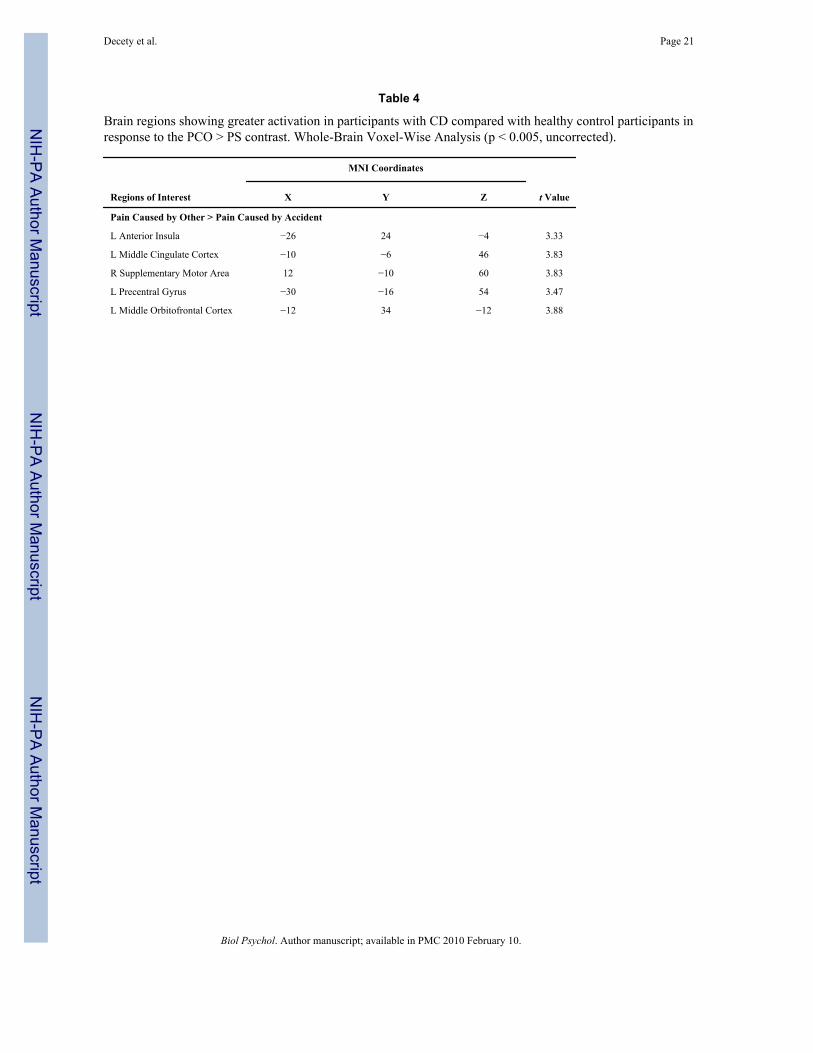
Direct comparison (two sample t test) between the control and CD groups when viewingsituations depicting an individual intentionally inflicting harm shows greater activation inadolescents with CD than controls in the left insula, SMA, precentral gyrus, and left medialOFC (see Table 4). Conversely, activation of the DLPFC [ 42, 42, 22] was greater in HC thanCD subjects. Participants with conduct disorder showed diminished BOLD response in theanterior PCC, TPJ and the lateral orbitofrontal cortex.
The degree of activation in the anterior TP in the CD group was linearly correlated with thepost-scanning VAS ratings of the pain experienced by the persons in the stimuli (r = 0.91, P <0.001). Furthermore, a similar and nearly perfect correlation (r = 0.93, P < 0.001) was foundwith hemodynamic activity in the aMCC and right anterior and middle insula (r = 0.89, P <0.001).
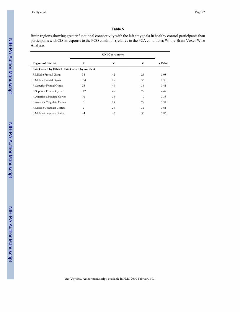
3.3. Effective connectivity analysisThe PPI analysis indicated that the CD group differed from the controls in the extent to whichactivity in the amygdala covaried with frontal cortical regions and the insula during the PCOcondition compared to the PCA condition. For the healthy adolescents, PPI analyses showedthat activity in the left amygdala was accompanied by condition-dependent (pain caused byother > pain caused by accident) functional interaction with a number of areas in the prefrontalcortex (see Table 5). The pattern of coupling was observed only in the pain caused by other >pain caused by accident contrast. In contrast, the CD group showed no significant effectiveconnectivity between amygdala and these frontal and parietal regions. However, in this group,the PCO condition did modulate the effective connectivity between the amygdala and the leftinsula [ 32, 12, 18], which was not the case for the healthy controls. Direct comparisons (2sample t tests) confirmed the above differences between these two groups (P < 0.005).Interestingly, the effective connectivity between the left amygdala and the temporal pole was
Decety et al. Page 8
Biol Psychol. Author manuscript; available in PMC 2010 February 10.
NIH
-PA
Author M
anuscriptN
IH-P
A A
uthor Manuscript
NIH
-PA
Author M
anuscript
stronger for PCA > NPS in participants with CD. No significant modulation was detected withfrontal regions.
3.4. Correlation analyses between hemodynamic response and traits related to psychopathyMean number of aggressive CD symptoms and ratings on the CADS daring dimension bothcorrelated with amygdala, inferior frontal gyrus, right PAG and right middle cingulate cortex[12, 38, 32] activations but prosociality did not. Interestingly, adolescents’ sadism scores werehighly correlated with activity in right amygdala [28, 12, 8], bilateral precuneus [0, 56, 46;4, 60, 64], and left fusiform gyrus [ 24, 52, 14] (P < 0.0001).
4. DiscussionThe goal of this study was to explore differences in neural response to empathy-eliciting stimuliin adolescents with and without aggressive CD. As predicted, when the control participantsobserved painful situations accidentally caused, regions of the pain matrix were selectivelyactivated, including the insula, aMCC, dorsal ACC, SMA, PAG and somatosensory cortex.This result fits well with previous functional neuroimaging studies on pain empathy (seeJackson, Rainville & Decety, 2006 for a review) that consistently showed that attending to theother people’ pain triggers an automatic somatic sensorimotor resonance mechanism betweenother and self, which activates almost the entire neural pain matrix in the observer includingthe periaqueductal gray (PAG) and the SMA.
Interestingly, the pattern of activation in the adolescents with CD showed both similarities aswell as striking differences when observing these painful situations. In the CD group,hemodynamic signal increase was detected in the insula, aMCC, SMA, PAG, andsomatosensory cortex (see Figure 1). In addition, strong activation was observed bilaterally inthe amygdala, ventral striatum, and temporal poles. The dorsal portion of the TP projects tothe hypothalamus, a neuromodulatory region important for autonomic regulation. Electricalstimulation of the TP produces changes in heart rate, respiration, and blood pressure (Gloor etal., 1982). These regions (i.e., amygdala, striatum and TP) were not activated in the controlparticipants. This result suggests that individuals with CD actually react to the pain of othersto a greater extent than youth without CD. Direct comparison of the two groups further indicatesthat participants with CD have a stronger signal response in the aMCC, striatum and leftamygdala than the control participants when viewing painful situations that have beenaccidentally caused.
The finding that aggressive adolescents with CD exhibit greater response in the pain matrixwhen viewing accidental pain than controls is interesting given the finding of previous fMRIstudies that reported reduced amygdala response in youth with CD during the viewing ofpictures with negative emotional valence (Marsh et al., 2008; Sterzer et al., 2005), as well asreduced amygdala volume (Sterzer et al., 2007). The present findings suggest that youth withCD do not exhibit reduced amygdala response to all negatively valenced stimuli; indeed, theyappear to exhibit enhanced response to images of people in pain, including a specific activationof the ventral striatum.
Our results suggest that there may be no deficit in neural response to distress of others (asreflected by the strong activation in the amygdala, temporal poles, and other structures in thepain matrix) in youth with CD. In fact, this somatic sensorimotor resonance was significantlygreater (P < 0.005) in participants with CD than without CD. We also observed that the extentof amygdala activation to painful situations in subjects with CD was significantly related in apositive direction to their number of aggressive acts and ratings of daring and sadism score onthe CADS (Lahey et al., in press).
Decety et al. Page 9
Biol Psychol. Author manuscript; available in PMC 2010 February 10.
NIH
-PA
Author M
anuscriptN
IH-P
A A
uthor Manuscript
NIH
-PA
Author M
anuscript
The present findings generate at least two hypotheses for testing in future studies. First, it isimportant to note that the amygdala is involved in the processing of more than just negativeaffect. Numerous studies point to a role for the amygdala in positive affect, and its couplingwith the ventral striatum enables a general arousing effect of reward (Murray, 2007). It ispossible, therefore, that the robust hemodynamic response in the amygdala/ventral striatum toviewing others in pain in youth with aggressive CD reflects a positive affective response (e.g.,“excitement”). That is, highly aggressive antisocial youth may enjoy hurting others and,coupled with diminished PFC/amygdala connectivity may not effectively regulate positivelyreinforced aggressive impulses. The finding that CADS ratings on the daring dimension (whichreflects sensation seeking) and sadism items correlate with amygdala response is consistentwith this hypothesis. The ventral striatum (nucleus accumbens) plays an important role inreward, pleasure, but also in fear. It is located at the head of the caudate nucleus and anteriorportion of the putamen and receives major input (excitatory fibers) from the amygdala and thehippocampal formation. It can be viewed as a functional interface between the limbic and motorsystems (Mogenson, Jones & Yim, 1980). In humans, the striatum is activated by stimuliassociated with reward, but also aversive, novel or intense stimuli. A common property linkingthese stimuli is saliency (Groenewegen, 2007). Attending to the pain of others may lead toeither approach or avoidance. The instrumental aspects of avoidance unlike the Pavlovianelicited responses require connections between the amygdala and the ventral striatum for theiracquisition and/or expression. In particular, the nucleus accumbens of the ventral striatum maybe a crucial area for the initiation and control of instrumental responses motivated by eitherappetitive or aversive responses, resulting from its innervation by dopaminergic pathways(LeDoux, 2002). It is thus difficult to decide whether the amygdala/ventral striatum responsein CD participants is associated with enjoyment or repulsion when watching the pain of others.The fact that this condition was also associated with strong activation of the PAG may be aclue. This region of the midbrain receives input from the amygdala and its stimulation triggersaversive or defensive responses and anxiety. It is also worth noting that lesion to the ventralstriatum is associated with selective impairment of anger processing, both in the recognitionof signals of anger and the experience of this emotion (Calder et al., 2004).
Second, many studies indicate that individuals with CD have a lower threshold for sensitivityto negative affect than other youth (Lahey & Waldman, 2003). This is potentially importantas their negative affect may increase the likelihood of aggression, especially in the absence ofeffective emotion regulation (Berkowitz, 1993, 2003). This interpretation fits well with thehypothesis of a dysfunction in the neural circuitry of emotion regulation (Davidson et al.,2000) and is consistent with our analyses of effective PFC/amygdala connectivity. Aggressionmay be related to affective instability and poor impulse control (Raine, 2002). Children withaggressive behavior problems have difficulties regulating negative emotions, which may resultin harmful patterns of interpersonal behavior (Lewis, Granic & Lamm, 2006). Often triggeredby hypersensitivity to specific stimuli, aggressive adults experience escalating agitationfollowed by an abrupt outburst of aggressive and threatening behavior (Gollan, Lee, & Coccaro,2005). Failure to discriminate between pain to others and to oneself may further lead to personaldistress. The fact that the anterior TP was specifically and highly activated in youth with CDprovides support for the distress interpretation. It has been suggested that the TP is part of asystem that modulates visceral functions in response to emotionally evocative stimuli basedon its anatomical connectivity (Kondo et al., 2003). A number of neuroimaging studies havereported activation in the left anterior TP in response to negative visual and auditory stimuli,such as aversive sounds (Olson et al., 2007). Importantly, one fMRI study found that TPactivation correlates with personal distress scores, a measure of how much one personally feelsupset when viewing another’s negative emotions (Moriguchi et al., 2006).
The stimuli that depicted pain intentionally caused by another individual were associated, inthe control group, with additional activation of temporoparietal junction, PCC and lateral OFC.
Decety et al. Page 10
Biol Psychol. Author manuscript; available in PMC 2010 February 10.
NIH
-PA
Author M
anuscriptN
IH-P
A A
uthor Manuscript
NIH
-PA
Author M
anuscript
The same pattern of activity was recently reported in a functional MRI study with typicallydeveloping children, and interpreted with relation to the perception of social interaction andintentionality (Decety, Michalska & Akitsuki, 2008). Functional neuroimaging studies haveconsistently supported the existence of a distributed neural network underlying the ability tounderstand other people as intentional and emotional agents (theory of mind mechanism). Thisnetwork comprises the superior temporal sulcus, the TPJ, and the medial prefrontal/anteriorparacingulate cortex (e.g., Ciaramidaro et al., 2007; Decety & Lamm, 2007). Specifically,research indicates that the anterior PCC is implicated in understanding the mental states of anagent involved in social interaction, regardless of whether this interaction is observed, takingplace online or even imagined (e.g., Walter et al., 2004).
Different patterns of response were detected in the orbitofrontal cortex across the two groups.While the lateral OFC was selectively activated in the control participants when observing paininflicted by another, activation of the medial OFC was found in the participants with CD. Directcomparison between the groups confirmed this finding. The OFC/MPFC has been specificallyimplicated in a variety of areas relevant to CD and aggression, including the regulation ofnegative affect (Phan et al., 2005). The finding that the response to these situations in the lateralOFC was attenuated in CD subjects relative to controls suggests an impairment in the CD groupin the regulation of negative affect. This interpretation supports the findings of a recent studythat observed an attenuated response in this region to angry faces in adults with intermittentexplosive disorder (Coccaro et al., 2007).
The PPI analyses corroborated the functional MRI findings, demonstrating an amygdala/PFCcoupling specifically while watching pain being intentionally caused by another individual andonly for the control group. In particular, in healthy adolescents, left amygdala activity covariedwith activity in the prefrontal cortex to a greater extent while watching situations of pain beingintentionally caused compared to viewing of pain caused by accident. Adolescents with CDshowed no functional connectivity between frontal regions and amygdala, which is in line witha recent study that demonstrated greater functional connectivity between the amygdala andPFC in comparison subjects than a group of aggressive youth (Marsh et al., 2008).
We posit that the condition in which another individual is inflicting pain intentionally elicitsin the normal controls a certain degree of regulation/inhibition. Such regulatory processdepends upon posterior STS and medial prefrontal cortex (Harenski & Hamann, 2006). A meta-analysis shows that frontal regions become active when subjects engage in cognitive strategies(such as reappraisal or detachment) to modulate negative affect (Ochsner & Gross, 2005). Thisis consistent with the role of the prefrontal cortex in cognitive inhibition and executive function,processes that are important for the regulation of affect (Fuster, 2001).
Overall, our results suggest a complex relation between the neural correlates of empathy andCD. The functional MRI data seem to indicate that adolescents with CD are at least asresponsive to the pain of others than the adolescents without CD. The fact that activation ofthe posterior insula, somatosensory cortex, and PAG are involved in the observation of othersin painful situations such an interpretation. However, unlike the adolescents without CD, andthe group of typically developing children in our first preliminary study (Decety, Michalska& Akitsuki, 2008), there was no activation in adolescents with CD in the neural regions thatcontribute to self-regulation and metacognition (including moral reasoning), such as theDLPFC, PCC, TPJ, dorsal and medial ACC and OFC.
Research with non-humans demonstrates that physical pain often elicits aggression (Berkowitz,2003). It has been hypothesized that aggressive persons are disposed to experience negativeaffect (Anderson & Bushman, 2002; Lahey & Waldman, 2003). This suggests that, in certainsituations, empathic mimicry might produce high levels of distress in youth predisposed to be
Decety et al. Page 11
Biol Psychol. Author manuscript; available in PMC 2010 February 10.
NIH
-PA
Author M
anuscriptN
IH-P
A A
uthor Manuscript
NIH
-PA
Author M
anuscript
aggressive that, ironically, increases their aggression. It is possible that strong activation ofneural circuits that underpin actual pain processing is associated with negative affect in youthwith CD. This, in conjunction with reduced activation in areas associated with emotionregulation, could result in a dysregulated negative affective state, which may instigateaggression under some circumstances (Berkowitz, 1983). For example, youth with CD whosee an injured friend (or fellow member of a gang) may be more likely to respond aggressivelythan other youth for this reason.
Finally, the strong and specific activation of the amygdala and ventral striatum in the aggressiveadolescents with CD during the perception of pain in others is an important and intriguingfinding, which necessitates additional research in order to understand its role in aggression andempathic dysfunction.
4.1. ConclusionThis study is to our knowledge the first functional neuroimaging investigation of brain responseto pain empathy-eliciting stimuli in aggressive adolescents with CD. In the future it will beimportant, given the limited size of our sample, to examine whether these findings replicatewith larger samples. We believe that such investigations are critical to move beyond self-reportmeasures of empathy. They will also provide a better understanding of the computational andneural mechanism underpinning empathy as well as their dysfunction in high-risk juvenilepopulations.
AcknowledgmentsThe study was supported by NSF (BCS-0718480), a seed grant to Jean Decety from the University of Chicago Centerfor Integrative Neuroscience and Neuroengineering Research, and grant R01 MH053554 to Benjamin Lahey.
ReferencesAmaral DG, Price JL. Amygdalo-cortical projections in the monkey (Macaca fascicularis). Journal of
Comparative Neurology 1984;230:465–496. [PubMed: 6520247]Anderson SW, Bechara A, Damasio H, Tranel D, Damasio AR. Impairment of social and moral behavior
related to early damage in human prefrontal cortex. Nature Neuroscience 1999;2:1032–1037.Anderson CA, Bushman BJ. Human aggression. Annual Review of Psychology 2002;53:27–51.Batson CD. Empathic joy and the empathy altruism hypothesis. Journal of Personality and Social
Psychology 1991;61:413–426. [PubMed: 1941512]Batson CD, Fultz J, Schoenrade PA. Distress and empathy: two quantitatively distinct vicarious emotions
with different motivational consequences. Journal of Personality 1987;55:19–39. [PubMed: 3572705]Berkowitz L. Aversively stimulated aggression: some parallels and differences in research with animals
and humans. American Psychologist 1983;38:1135–1144. [PubMed: 6359985]Berkowitz L. Pain and aggression: some findings and implications. Motivation and Emotion
1993;17:277–293.Berkowitz, L. Affect, aggression, and antisocial behavior. In: Davidson, RJ.; Scherer, KR.; Goldsmith,
HH., editors. Handbook of Affective Sciences. New York: Oxford University Press; 2003. p. 804-823.Blair RJR. Responding to the emotions of others: Dissociating forms of empathy through the study of
typical and psychiatric populations. Consciousness and Cognition 2005;14:698–718. [PubMed:16157488]
Botvinick M, Jha AP, Bylsma LM, Fabian SA, Solomon PE, Prkachin KM. Viewing facial expressionsof pain engages cortical areas involved in the direct experience of pain. NeuroImage 2005;25:312–319. [PubMed: 15734365]
Bufalari I, Aprile T, Avenanti A, Di Russo F, Aglioti SM. Empathy for pain and touch in the humansomatosensory cortex. Cerebral Cortex 2007;17:2553–2561. [PubMed: 17205974]
Decety et al. Page 12
Biol Psychol. Author manuscript; available in PMC 2010 February 10.
NIH
-PA
Author M
anuscriptN
IH-P
A A
uthor Manuscript
NIH
-PA
Author M
anuscript
Calder AJ, Keane J, Lawrence AD, Manes F. Impaired recognition of anger following damage of theventral striatum. Brain 2004;127:1958–1969. [PubMed: 15289264]
Campbell, SB. Behavior problems in preschool children: Clinical and developmental issues. New York:Guilford Press; 1990.
Cheng Y, Lin C, Liu HL, Hsu Y, Lim K, Hung D, Decety J. Expertise modulates the perception of painin others. Current Biology 2007a;17:1708–1713. [PubMed: 17900903]
Ciaramidaro A, Adenzato M, Enrici I, Erk S, Pia L, Bara BG, Walter H. The intentional network: Howthe brain reads varieties of intentions. Neuropsychologia 2007;45:3105–3113. [PubMed: 17669444]
Coccaro E, McCloskey M, Fitzgerald D, Phan K. Amygdala and Orbitofrontal Reactivity to Social Threatin Individuals with Impulsive Aggression. Biological Psychiatry 2007;62:168–178. [PubMed:17210136]
Davidson RJ, Putnam KM, Larson CL. Dysfunction in the neural circuitry of emotion regulation-apossible prelude to violence. Science 2000;289:591–594. [PubMed: 10915615]
Decety J, Michalska KJ, Akitsuki Y. Who caused the pain? A functional MRI investigation of empathyand intentionality in children. Neuropsychologia 2008;46:2607–2614. [PubMed: 18573266]
Decety J, Batson CD. Social neuroscience approaches to interpersonal sensitivity. Social Neuroscience2007;2(3–4):151–157. [PubMed: 18633813]
Decety J, Jackson PL. The functional architecture of human empathy. Behavioral and CognitiveNeuroscience Reviews 2004;3:71–100. [PubMed: 15537986]
Decety, J.; Lamm, C. Empathy versus personal distress - Recent evidence from social neuroscience. In:Decety, J.; Ickes, W., editors. The Social Neuroscience of Empathy. Cambridge: MIT press; 2008.in press
Decety J, Lamm C. The role of the right temporoparietal junction in social interaction: How low-levelcomputational processes contribute to meta-cognition. The Neuroscientist 2007;13:580–593.[PubMed: 17911216]
Decety J, Moriguchi Y. The empathic brain and its dysfunction in psychiatric populations: implicationsfor intervention across different clinical conditions. BioPsychoSocial Medicine 2007;1:22–65.[PubMed: 18021398]
Decety J, Sommerville JA. Shared representations between self and others: A social cognitiveneuroscience view. Trends in Cognitive Sciences 2003;7:527–533. [PubMed: 14643368]
Derbyshire SWG. Exploring the pain “neuromatrix”. Current Review of Pain 2000;4:467–477. [PubMed:11060593]
Eisenberg N, Miller PA, Schaller M, Fabes RA, Fultz J, Shell R. The role of sympathy and altruisticpersonality traits in helping: A reexamination. Journal of Personality 1989;57:41–67. [PubMed:2709301]
Eisenberg N. Age changes in prosocial responding and moral reasoning in adolescence and earlyadulthood. Journal of Research on Adolescence 2005;15:235–260.
Eisenberg, N.; Eggum, ND. Empathic responding: sympathy and personal distress. In: Decety, J.; Ickes,W., editors. The Social Neuroscience of Empathy. Cambridge: MIT press; 2008. in press
Eisenberg, N.; Spinrad, TL.; Sadovsky, A. Empathy-related responding in children. In: Killen, M.;Smetana, J., editors. Handbook of Moral Development. Mahwah, NJ: Lawrence Erlbaum Associates;2006. p. 517-549.
Estabrook, S. Master of Art Thesis under direction of Dr. Jean Decety. University of Chicago; 2007. Doescontext modulate empathy for pain?.
Farrington DP, West DJ. Criminal, penal and life histories of chronic offenders: Risk and protectivefactors and early identification. Criminal Behaviour and Mental Health 1993;3:492–523.
Frick PJ, Stickle TR, Dandreaux DM, Farrell JM, Kimonis ER. Callous-unemotional traits in predictingthe severity and stability of conduct problems and delinquency. Journal of Abnormal ChildPsychology 2005;33:471–487. [PubMed: 16118993]
Friston KJ, Buechel C, Fink GR, Morris J, Rolls E, Dolan RJ. Psychophysiological and modulatoryinteraction in neuroimaging. NeuroImage 1997;6:218–229. [PubMed: 9344826]
Fuster JM. The prefrontal cortex - An update: review time is of the essence. Neuron 2001;30:319–333.[PubMed: 11394996]
Decety et al. Page 13
Biol Psychol. Author manuscript; available in PMC 2010 February 10.
NIH
-PA
Author M
anuscriptN
IH-P
A A
uthor Manuscript
NIH
-PA
Author M
anuscript
Gill KL, Calkins SD. Do aggressive/destructive toddlers lack concern for others? Behavioral andphysiological indicators of empathic responding in 2-year-old children. Development andPsychopathology 2003;15:55–71. [PubMed: 12848435]
Glover GH, Lai TF. Spiral-in/out BOLD fMRI for increased SNR and reduced susceptibility artifacts.Magnetic Resonance Medicine 2001;46:515–522.
Gloor P, Olivier A, Quesney LF, Andermann F, Horowitz S. The role of the limbic system in experientialphenomena of temporal lobe epilepsy. Annals of Neurology 1982;12:129–144. [PubMed: 7125603]
Gollan JK, Lee R, Coccaro EF. Developmental psychopathology and neurobiology of aggression.Development and Psychopathology 2005;17:1151–1171. [PubMed: 16613435]
Groenewegen HJ. The ventral striatum as an interface between the limbic and motor systems. CNSSpectrums 2007;12:887–889. [PubMed: 18163034]
Gu X, Han S. Attention and reality constraints on the neural processes of empathy for pain. NeuroImage2007;36:256–267. [PubMed: 17400480]
Harenski CL, Hamann S. Neural correlates of regulating negative emotions related to moral violations.NeuroImage 2006;30:313–324. [PubMed: 16249098]
Herpertz SC, Sass H. Emotional deficiency and psychopathy. Behavioral Science and Law 2000;18:317–323.
Hoffman, ML. Empathy and Moral Development. New York: Cambridge University Press; 2000.Izquierdo A, Suda RK, Murray EA. Comparison of the effects of bilateral orbitofrontal cortex lesions
and amygdala lesions on emotional responses in rhesus monkeys. Journal of Neuroscience2005;25:8534–8542. [PubMed: 16162935]
Jackson PL, Brunet E, Meltzoff AN, Decety J. Empathy examined through the neural mechanismsinvolved in imagining how I feel versus how you feel pain: An event-related fMRI study.Neuropsychologia 2006;44:752–61. [PubMed: 16140345]
Jackson PL, Meltzoff AN, Decety J. How do we perceive the pain of others: a window into the neuralprocesses involved in empathy. NeuroImage 2005;24:771–779. [PubMed: 15652312]
Jackson PL, Rainville P, Decety J. To what extent do we share the pain of others? Insight from the neuralbases of pain empathy. Pain 2006;125:5–9. [PubMed: 16997470]
Jenck F, Moreau JL, Martin JR. Dorsal periaqueductal gray-induced aversion as a simulation of panicanxiety: Elements of face and predictive validity. Psychiatry Research 1995;57:181–191. [PubMed:7480384]
Kondo H, Saleem KS, Price JL. Differential connections of the temporal pole with the orbital and medialprefrontal networks in macaque monkeys. Journal of Comparative Neurology 2003;465:499–523.[PubMed: 12975812]
Lahey BB, Applegate B, Barkley RA, Garfinkel B, McBurnett K, Kerdyk L, Greenhill L, Hynd GW,Frick PJ, Newcorn J, Biederman J, Ollendick T, Hart EL, Perez D, Waldman I, Shaffer D. DSM-IVfield trials for oppositional defiant disorder and conduct disorder in children and adolescents.American Journal of Psychiatry 1994;151:1163–1171. [PubMed: 8037251]
Lahey, BB.; Waldman, ID. A developmental propensity model of the origins of conduct problems duringchildhood and adolescence. In: Lahey, BB.; Moffitt, TE.; Caspi, A., editors. Causes of conductdisorder and juvenile delinquency. New York: Guilford Press; 2003. p. 76-117.
Lahey BB, Loeber R, Burke JD, Applegate B. Predicting future antisocial personality disorder in malesfrom a clinical assessment in childhood. Journal of Consulting and Clinical Psychology 2005;73:389–399. [PubMed: 15982137]
Lahey BB, Applegate B, Chronis AM, Jones HA, Williams SH, Loney J, Waldman ID. Psychometriccharacteristics of a measure of emotional dispositions developed to test a developmental propensitymodel of conduct disorder. Journal of Clinical Child and Adolescent Psychology. in press.
Lamm C, Batson CD, Decety J. The neural substrate of human empathy: effects of perspective-takingand cognitive appraisal. Journal of Cognitive Neuroscience 2007a;19:42–58. [PubMed: 17214562]
Lamm C, Nusbaum H, Meltzoff AN, Decety J. What are you feeling? Using functional magneticresonance imaging to assess the modulation of sensory and affective responses during empathy forpain. PLoS ONE 2007b;12:e1292.
LeDoux, JE. Emotion: Clue from the brain. In: Cacioppo, JT., et al., editors. Foundations in SocialNeuroscience. Cambridge: MIT press; 2002. p. 389-410.
Decety et al. Page 14
Biol Psychol. Author manuscript; available in PMC 2010 February 10.
NIH
-PA
Author M
anuscriptN
IH-P
A A
uthor Manuscript
NIH
-PA
Author M
anuscript
Lewis MD, Granic I, Lamm C. Behavioral differences in aggressive children linked with neuralmechanisms of emotion regulation. Annals of the New York Academy of Science 2006;1094:164–177.
Loeber, R.; Farrington, DP.; Stouthamer-Loeber, M.; Van Kammen, WB. Antisocial behavior and mentalhealth problems: Explanatory factors in childhood and adolescence. Mahwah, NJ: Lawrence ErlbaumAssociates; 1998.
Lovett BJ, Sheffield RA. Affective empathy deficits in aggressive children and adolescents: A criticalreview. Clinical Psychology Review 2007;27:1–13. [PubMed: 16697094]
Marsh AA, Finger EC, Mitchell DGV, Reid ME, Sims C, Kosson D, Towbin KE, Leibenluft E, Pine DS,Blair RJR. Reduced amygdala response to fearful expressions in children and adolescents with callousunemotional traits and disruptive behavior disorders. American Journal of Psychiatry. 2008 Epubahead of time.
Mogenson GJ, Jones DL, Kim CY. From motivation to action: functional interface between the limbicsystem and the motor system. Progress in Neurobiology 1980;14:69–97. [PubMed: 6999537]
Moll J, de Oliviera-Souza R, Eslinger P. Morals and the human brain. NeuroReport 2003;14:299–305.[PubMed: 12634472]
Moll J, de Oliviera-Souza R, Garrido GJ, Bramati IE, Caparelli-Daquer EMA, Paiva ML, Zahn R,Grafman J. The self as a moral agent: Linking the neural bases of social agency and moral sensitivity.Social Neuroscience 2007;2(3–4):336–352. [PubMed: 18633822]
Moriguchi Y, Decety J, Ohnishi T, Maeda M, Matsuda H, Komaki G. Empathy and judging other’s pain:An fMRI study of alexithymia. Cerebral Cortex 2007;17:2223–2234. [PubMed: 17150987]
Moriguchi Y, Ohnishi T, Lane RD, Maeda M, Mori T, Nemoto K, Matsuda H, Komaki G. Impaired self-awareness and theory of mind: an fMRI study of mentalizing in alexithymia. NeuroImage2006;32:1472–1482. [PubMed: 16798016]
Morrison I, Lloyd D, di Pellegrino G, Roberts N. Vicarious responses to pain in anterior cingulate cortex:is empathy a multisensory issue? Cognitive and Affective Behavioral Neuroscience 2004;4:270–278.
Morrison I, Poliakoff E, Gordon L, Downing PE. Response specific effects of pain observation on motorbehavior. Cognition 2006;104:407–416. [PubMed: 16919254]
Murray EA. The amygdala, reward and emotion. Trends in Cognitive Sciences 2007;11:489–497.[PubMed: 17988930]
Ochsner KN, Gross JJ. The cognitive control of emotion. Trends in Cognitive Sciences 2005;9:242–249.[PubMed: 15866151]
Olson IR, Plotzker A, Ezzyat Y. The enigmatic temporal pole: a review of findings on social and emotionalprocessing. Brain 2007;130:1718–1731. [PubMed: 17392317]
Pelham WE, Gnagy EM, Greenslade KE, Milich R. Teacher ratings of DSM-III-R symptoms for thedisruptive behavior disorders. Journal of the American Academy of Child and Adolescent Psychiatry1992;31:210–218. [PubMed: 1564021]
Phan KL, Fitzgerald DA, Nathan PJ, Moore GJ, Uhde TW, Tancer ME. Neural substrates for voluntarysuppression of negative affect: a functional magnetic resonance imaging study. Biological Psychiatry2005;57:210–219. [PubMed: 15691521]
Phillips ML, Drevets WC, Rauch SL, Lane R. Neurobiology of emotion perception I: the neural basis ofnormal emotion perception. Biological Psychiatry 2003;54:504–14. [PubMed: 12946879]
Piacentini JC, Cohen P, Cohen J. Combining discrepant diagnostic information from multiple sources:Are complex algorithms better than simple ones? Journal of Abnormal Child Psychology1992;20:51–63. [PubMed: 1548394]
Preston AR, Thomason ME, Ochsner KN, Cooper JC, Glover GH. Comparison of spiral-in/out and spiral-out BOLD fMRI at 1.5 T and 3 T. NeuroImage 2004;21:291–301. [PubMed: 14741667]
Price DD. Psychological and neural mechanisms of the affective dimension of pain. Science2000;288:1769–1772. [PubMed: 10846154]
Raine A. Biosocial Studies of Antisocial and Violent Behavior in Children and Adults: A Review. Journalof Abnormal Child Psychology 2002;30:311–326. [PubMed: 12108763]
Raine A, Venables P, Mednick S. Low resting heart rate at age three years predisposes to aggression atage 11 years: Evidence from the Mauritius Child Health Project. Journal of the Academy of Childand Adolescent Psychiatry 1997;36:1457–1464.
Decety et al. Page 15
Biol Psychol. Author manuscript; available in PMC 2010 February 10.
NIH
-PA
Author M
anuscriptN
IH-P
A A
uthor Manuscript
NIH
-PA
Author M
anuscript
Russo MF, Stokes GS, Lahey BB, Christ MAG, McBurnett K, Loeber R. A sensation seeking scale forchildren: Further refinement and psychometric development. Journal of Psychopathology andBehavioral Assessment 1993;15:69–86.
Saarela MV, Hlushchuk Y, Williams AC, Schurmann M, Kalso E, Hari R. The compassionate brain:humans detect pain intensity from another’s face. Cerebral Cortex 2007;17:230–237. [PubMed:16495434]
Saddoris MP, Gallagher M, Schoenbaum G. Rapid associative encoding in basolateral amygdala dependson connections with orbitofrontal cortex. Neuron 2005;46:321–331. [PubMed: 15848809]
Shaffer, D.; Fisher, P.; Piacentini, J.; Schwab-Stone, M.; Wicks, J. Diagnostic Interview Schedule forChildren. New York: Columbia University; 1993.
Singer T, Seymour B, O’Doherty J, Kaube H, Dolan RJ, Frith CD. Empathy for pain involves the affectivebut not the sensory components of pain. Science 2004;303:1157–1161. [PubMed: 14976305]
Sterzer P, Stadler C, Krebs A, Kleinschmidt A, Poutska F. Abnormal neural responses to emotional visualstimuli in adolescents with conduct disorder. Biological Psychiatry 2005;57:7–15. [PubMed:15607294]
Sterzer P, Stadler C, Poustka F, Kleinschmidt A. A structural neural deficit in adolescents with conductdisorder and its association with lack of empathy. NeuroImage 2007;37:335–342. [PubMed:17553706]
van Elst LT, Woermann FG, Lemieux L, Thompson PJ, Trimble MR. Affective aggression in patientswith temporal lobe epilepsy: a quantitative MRI study of the amygdala. Brain 2000;123:234–243.[PubMed: 10648432]
Van Goozen SHM, Fairchild G. How can the study of biological processes help design new interventionsfor children with severe antisocial behavior? Development and Psychopathology 2008;20:941–973.[PubMed: 18606039]
Walter H, Adenzato M, Ciaramidaro A, Enrici I, Bara BG. Understanding intentions in social interaction:the role of the anterior paracingulate cortex. Journal of Cognitive Neuroscience 2004;16:1854–1863.[PubMed: 15701234]
Zahn-Waxler C, Cole PM, Welsh JD, Fox NA. Psychophysiological correlates of empathy and prosocialbehaviors in preschool children with behavior problems. Development and Psychopathology1995;7:27–48.
Decety et al. Page 16
Biol Psychol. Author manuscript; available in PMC 2010 February 10.
NIH
-PA
Author M
anuscriptN
IH-P
A A
uthor Manuscript
NIH
-PA
Author M
anuscript
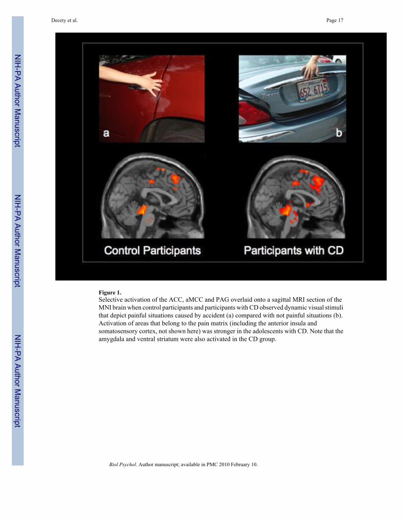
Figure 1.Selective activation of the ACC, aMCC and PAG overlaid onto a sagittal MRI section of theMNI brain when control participants and participants with CD observed dynamic visual stimulithat depict painful situations caused by accident (a) compared with not painful situations (b).Activation of areas that belong to the pain matrix (including the anterior insula andsomatosensory cortex, not shown here) was stronger in the adolescents with CD. Note that theamygdala and ventral striatum were also activated in the CD group.
Decety et al. Page 17
Biol Psychol. Author manuscript; available in PMC 2010 February 10.
NIH
-PA
Author M
anuscriptN
IH-P
A A
uthor Manuscript
NIH
-PA
Author M
anuscript
NIH
-PA
Author M
anuscriptN
IH-P
A A
uthor Manuscript
NIH
-PA
Author M
anuscript
Decety et al. Page 18
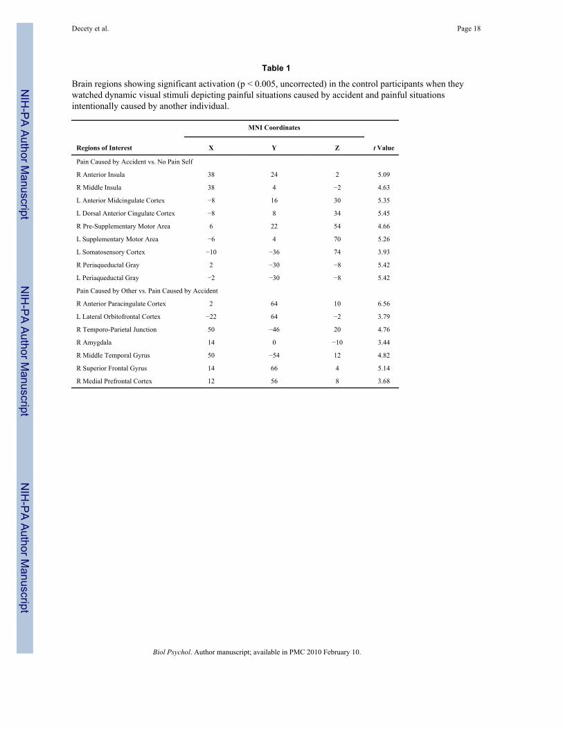
Table 1
Brain regions showing significant activation (p < 0.005, uncorrected) in the control participants when theywatched dynamic visual stimuli depicting painful situations caused by accident and painful situationsintentionally caused by another individual.
MNI Coordinates
Regions of Interest X Y Z t Value
Pain Caused by Accident vs. No Pain Self
R Anterior Insula 38 24 2 5.09
R Middle Insula 38 4 2 4.63
L Anterior Midcingulate Cortex 8 16 30 5.35
L Dorsal Anterior Cingulate Cortex 8 8 34 5.45
R Pre-Supplementary Motor Area 6 22 54 4.66
L Supplementary Motor Area 6 4 70 5.26
L Somatosensory Cortex 10 36 74 3.93
R Periaqueductal Gray 2 30 8 5.42
L Periaqueductal Gray 2 30 8 5.42
Pain Caused by Other vs. Pain Caused by Accident
R Anterior Paracingulate Cortex 2 64 10 6.56
L Lateral Orbitofrontal Cortex 22 64 2 3.79
R Temporo-Parietal Junction 50 46 20 4.76
R Amygdala 14 0 10 3.44
R Middle Temporal Gyrus 50 54 12 4.82
R Superior Frontal Gyrus 14 66 4 5.14
R Medial Prefrontal Cortex 12 56 8 3.68
Biol Psychol. Author manuscript; available in PMC 2010 February 10.

NIH
-PA
Author M
anuscriptN
IH-P
A A
uthor Manuscript
NIH
-PA
Author M
anuscript
Decety et al. Page 19
Table 2
Brain regions showing significant signal increase (p<0.005, uncorrected) in the CD participants for both paincaused by accident and pain caused by other conditions.
MNI Coordinates
Regions of Interest X Y Z t Value
Pain Caused by Accident vs. No Pain Self
R Anterior Insula 32 32 4 11.65
L Middle Insula 40 2 8 5.30
L Anterior Midcingulate Cortex 2 12 24 10.95
R Middle Cingulate Cortex 6 6 42 9.30
R Pre-Supplementary Motor Area 4 6 48 7.69
L Pre-Supplementary Motor Area 4 2 52 5.68
L Somatosensory Cortex 58 29 36 4.70
R Medial Orbital Gyrus 10 46 14 8.05
L Medial Orbital Gyrus 6 54 6 8.75
L Temporal Pole 56 10 2 7.71
R Temporal Pole 38 6 38 3.65
R Ventral Striatum (putamen) 26 12 4 8.12
R Ventral Striatum (head of caudate) 8 4 10 4.88
R Periaqueductal Gray 2 30 7 3.09
L Periaqueductal Gray 2 30 7 4.17
L Amygdala 18 8 10 11.60
R Amygdala 15 8 9 9.42
Pain Caused by Other vs. Pain Caused by Acccident
L Temporal Pole 34 16 28 6.64
L Middle Temporal Gyrus 54 68 14 5.60
R Middle Temporal Gyrus 62 34 0 5.13
R Precuneus 8 62 48 10.24
L Inferior Temporal Gyrus 32 2 44 9.01
R Superior Frontal Gyrus 16 42 34 5.97
L Medial Orbitofrontal Cortex 4 30 17 4.79
Biol Psychol. Author manuscript; available in PMC 2010 February 10.
NIH
-PA
Author M
anuscriptN
IH-P
A A
uthor Manuscript
NIH
-PA
Author M
anuscript
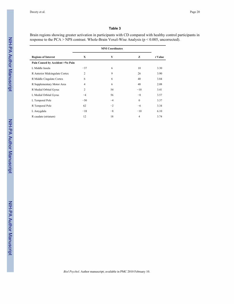
Decety et al. Page 20
Table 3
Brain regions showing greater activation in participants with CD compared with healthy control participants inresponse to the PCA > NPS contrast. Whole-Brain Voxel-Wise Analysis (p < 0.005, uncorrected).
MNI Coordinates
Regions of Interest X Y Z t Value
Pain Caused by Accident >No Pain
L Middle Insula 37 6 10 3.30
R Anterior Midcingulate Cortex 2 9 26 3.90
R Middle Cingulate Cortex 6 6 40 3.84
R Supplementary Motor Area 4 6 48 2.08
R Medial Orbital Gyrus 2 54 10 3.41
L Medial Orbital Gyrus 4 56 8 3.57
L Temporal Pole 50 4 0 3.37
R Temporal Pole 62 2 6 3.34
L Amygdala 18 8 10 6.10
R caudate (striatum) 12 18 4 3.74
Biol Psychol. Author manuscript; available in PMC 2010 February 10.
NIH
-PA
Author M
anuscriptN
IH-P
A A
uthor Manuscript
NIH
-PA
Author M
anuscript
Decety et al. Page 21
Table 4
Brain regions showing greater activation in participants with CD compared with healthy control participants inresponse to the PCO > PS contrast. Whole-Brain Voxel-Wise Analysis (p < 0.005, uncorrected).
MNI Coordinates
Regions of Interest X Y Z t Value
Pain Caused by Other > Pain Caused by Accident
L Anterior Insula 26 24 4 3.33
L Middle Cingulate Cortex 10 6 46 3.83
R Supplementary Motor Area 12 10 60 3.83
L Precentral Gyrus 30 16 54 3.47
L Middle Orbitofrontal Cortex 12 34 12 3.88
Biol Psychol. Author manuscript; available in PMC 2010 February 10.
NIH
-PA
Author M
anuscriptN
IH-P
A A
uthor Manuscript
NIH
-PA
Author M
anuscript
Decety et al. Page 22
Table 5
Brain regions showing greater functional connectivity with the left amygdala in healthy control participants thanparticipants with CD in response to the PCO condition (relative to the PCA condition): Whole-Brain Voxel-WiseAnalysis.
MNI Coordinates
Regions of Interest X Y Z t Value
Pain Caused by Other > Pain Caused by Accident
R Middle Frontal Gyrus 34 42 24 5.08
L Middle Frontal Gyrus 34 26 36 2.38
R Superior Frontal Gyrus 26 40 34 3.41
L Superior Frontal Gyrus 12 46 28 4.49
R Anterior Cingulate Cortex 10 38 10 3.38
L Anterior Cingulate Cortex 0 18 28 3.34
R Middle Cingulate Cortex 2 20 32 3.61
L Middle Cingulate Cortex 4 6 50 3.86
Biol Psychol. Author manuscript; available in PMC 2010 February 10.