Embed Size (px)
Citation preview
Porphyromonas gingivalis manipulates complement and TLRsignaling to uncouple bacterial clearance from inflammation andpromote dysbiosis
Tomoki Maekawa1,7, Jennifer L. Krauss2,7,8, Toshiharu Abe1, Ravi Jotwani2, MarthaTriantafilou3, Kathy Triantafilou3, Ahmed Hashim4, Shifra Hoch5, Michael A. Curtis4,Gabriel Nussbaum5, John D. Lambris6, and George Hajishengallis1,*
1University of Pennsylvania, School of Dental Medicine, Department of Microbiology,Philadelphia, PA 19104, USA
2University of Louisville, Center for Oral Health and Systemic Disease, Louisville, KY 40292, USA
3Cardiff University School of Medicine, Institute of Infection and Immunity, Cardiff CF14 4XN, UK
4Queen Mary University of London, Centre for Immunology and Infectious Disease, BlizardInstitute, Barts and The London School of Medicine and Dentistry, London E1 2AT, UK
5Hebrew University, Hadassah Dental School, Jerusalem 12272, Israel
6University of Pennsylvania, Perelman School of Medicine, Department of Pathology andLaboratory Medicine, Philadelphia, PA 19104, USA
SUMMARY
Certain low-abundance bacterial species, such as the periodontitis-associated oral bacterium
Porphyromonas gingivalis can subvert host immunity to remodel a normally symbiotic microbiota
into a dysbiotic, disease-provoking state. However, such pathogens also exploit inflammation to
thrive in dysbiotic conditions. How these bacteria evade immunity while maintaining
inflammation is unclear. As previously reported, P. gingivalis remodels the oral microbiota into a
dysbiotic state by exploiting complement. Now we show that in neutrophils P. gingivalis disarms
a host-protective TLR2-MyD88 pathway via proteasomal degradation of MyD88, whereas it
activates an alternate TLR2-Mal-PI3K pathway. This alternate TLR2-Mal-PI3K pathway blocks
© 2014 Elsevier Inc. All rights reserved.*Correspondence: [email protected] author.8Present address: Department of Pathology, Washington University School of Medicine, St. Louis, MO 63110.
Publisher's Disclaimer: This is a PDF file of an unedited manuscript that has been accepted for publication. As a service to ourcustomers we are providing this early version of the manuscript. The manuscript will undergo copyediting, typesetting, and review ofthe resulting proof before it is published in its final citable form. Please note that during the production process errors may bediscovered which could affect the content, and all legal disclaimers that apply to the journal pertain.
AUTHOR CONTRIBUTIONSCo-first authors T.M. and J.L.K. performed most of the in vivo and in vitro assays and analyzed the data. T.A., R.J., M.T., K.T., A.H.,S.H. performed research and data analysis. M.A.C. and G.N. performed data analysis and discussed the proposed model with G.H.J.D.L. and G.H. conceived of and designed research. GH supervised research and wrote the paper. All authors approved the finalmanuscript.
The authors declare no financial conflicts of interest.
NIH Public AccessAuthor ManuscriptCell Host Microbe. Author manuscript; available in PMC 2015 June 11.
Published in final edited form as:Cell Host Microbe. 2014 June 11; 15(6): 768–778. doi:10.1016/j.chom.2014.05.012.
NIH
-PA
Author M
anuscriptN
IH-P
A A
uthor Manuscript
NIH
-PA
Author M
anuscript
phagocytosis, provides ‘bystander’ protection to otherwise susceptible bacteria, and promotes
dysbiotic inflammation in vivo. This mechanism to disengage bacterial clearance from
inflammation required an intimate crosstalk between TLR2 and the complement receptor C5aR,
and can contribute to the persistence of microbial communities that drive dysbiotic diseases.
INTRODUCTION
Dysbiosis, a state characterized by imbalances in the relative abundance or influence of
microbial species within an ecosystem, is emerging as a potential trigger of mucosal
inflammatory disorders, such as inflammatory bowel disease, colorectal cancer, bacterial
vaginosis, and periodontitis (Eloe-Fadrosh and Rasko, 2013; Hajishengallis et al., 2012).
The mechanisms leading to dysbiosis are poorly understood and hence under intensive
investigation. In this regard, the keystone-pathogen hypothesis holds that certain low-
abundance pathogens can subvert host immunity in ways that favor the remodeling of a
normally symbiotic microbiota into a dysbiotic and disease-provoking state (Hajishengallis
et al., 2012; Stecher et al., 2013).
For instance, the oral asaccharolytic bacterium Porphyromonas gingivalis (Pg) fails to cause
periodontitis in germ-free mice despite colonizing this host; however, in conventional mice,
Pg transforms the periodontal microbiota into a dysbiotic community and co-opts it to
instigate destructive inflammation (Hajishengallis et al., 2011). In contrast, C5a receptor
(C5aR; CD88)-deficient mice are protected from Pg-induced dysbiosis and periodontitis
(Hajishengallis et al., 2011). The ability of Pg to instigate inflammatory disease through
community-wide supportive effects, while being a low-abundance constituent of
periodontitis-associated biofilms in humans and animal models (Abusleme et al., 2013;
Hajishengallis et al., 2011), has prompted its designation as a keystone pathogen
(Hajishengallis et al., 2012).
Organisms involved in dysbiotic diseases are burdened with the challenge of evading host
immunity in an inflammatory environment. In this respect, we hypothesized that the survival
and persistence of Pg is crucially dependent upon its capacity to selectively manipulate the
host response in ways that would suppress killing mechanisms but not inflammation. This is
because inflammation actually serves the nutritional needs of asaccharolytic dysbiotic
communities through the release of tissue breakdown products including peptides and heme-
containing compounds (Hajishengallis, 2014). Therefore, although immunosuppression is a
common immune evasion strategy of many pathogens (Cyktor and Turner, 2011), it should
not be a viable option for pathobionts that thrive on inflammation. However, the molecular
mechanism(s) by which immune-subversive bacteria such as Pg can selectively inhibit
immune elimination without suppressing inflammation have remained obscure. Since
neutrophils are heavily involved in the initiation and progression of human periodontitis and
comprise ≥95% of total leukocytes in periodontal pockets where they constantly encounter
Pg (Hajishengallis and Hajishengallis, 2014; Nussbaum and Shapira, 2011), we addressed
this issue in the context of Pg-neutrophil interactions in vitro and in vivo. Our study
dissected how P. gingivalis subverts complement and Toll-like receptor (TLR) signaling to
inhibit bacterial killing while preserving inflammation and promoting dysbiosis in vivo.
Maekawa et al. Page 2
Cell Host Microbe. Author manuscript; available in PMC 2015 June 11.
NIH
-PA
Author M
anuscriptN
IH-P
A A
uthor Manuscript
NIH
-PA
Author M
anuscript
RESULTS
Pg inhibits the killing but not the proinflammatory activity of neutrophils in vivo
The ability of Pg to persist and cause dysbiosis and periodontitis in mice requires intact
C5aR signaling (Abe et al., 2012; Hajishengallis et al., 2011). Since neutrophils constitute
the overwhelming majority of professional phagocytes recruited to periodontal pockets, we
hypothesized that Pg exploits C5aR to inhibit neutrophil killing but not neutrophil-mediated
inflammation. To address this hypothesis in a quantitative way, we used the mouse chamber
model (Burns et al., 2010; Mydel et al., 2006). This model involves injectable inoculation of
bacteria into the lumen of a subcutaneously implanted titanium-coil chamber, thereby
allowing quantitative assessment of bacterial interactions with recruited neutrophils (>97%
of recruited leukocytes; Figure 1A). Since innate pattern recognition of Pg is predominantly
mediated by TLR2 (Burns et al., 2010), which synergizes with C5aR in periodontal
inflammation (Abe et al., 2012), we examined the interactions of Pg with both C5aR and
TLR2 using knockout mice or specific inhibitors.
At 2h post-inoculation with Pg, chamber fluid aspirated from mice lacking C5aR (C5ar−/−)
or TLR2 (Tlr2−/−), or from wild-type (WT) mice treated with C5aR antagonist (C5aRA) or
anti-TLR2, contained significantly lower Pg viable counts (CFU) as compared with
corresponding controls (i.e., WT mice that were left untreated or treated with control
compounds) (Figure 1B). In contrast, Pg CFU counts recovered from the chambers of mice
lacking complement receptor 3 (CR3; CD11b−/−) did not differ from those recovered from
WT controls (Figure 1B). These data suggested that both C5aR and TLR2 are required for
enhanced Pg survival in the chambers. Consistent with this, a gingipain-deficient isogenic
mutant (KDP128), which cannot generate C5a to activate C5aR (Liang et al., 2011), was
extremely susceptible to killing unless C5a was added exogenously resulting in decreased
phagocytosis (Figure S1). Interestingly, despite their increased capacity to clear Pg, C5ar−/−
mice and C5aRA-treated WT mice recruited lower numbers of neutrophils (typically 2.5
[±0.8] ×106) as compared to those in untreated WT mice (typically 1.0 [±0.3] ×107),
whereas the recruitment of neutrophils in the chambers of Tlr2−/− mice and anti-TLR2-
treated WT mice was not significantly affected compared with untreated WT controls (not
shown). No Pg viable counts could be recovered from blood or internal organs (spleen, liver,
lungs, and kidney) from WT or knockout mice; similarly, quantitative real-time PCR of the
ISPg1 gene of Pg failed to detect Pg in those tissues.
Treatment of Tlr2−/− mice with C5aRA (or, conversely, treatment of C5ar−/− mice with anti-
TLR2) had no further inhibitory effect on Pg viability compared to untreated Tlr2−/− (or
C5ar−/−) mice (Figure 1B). This finding and the observation that the survival of Pg is
inhibited to the same extent regardless of which of the two receptors (C5aR or TLR2) is
lacking or blocked collectively suggest that C5aR and TLR2 engage in cooperative crosstalk
to prevent Pg killing. This C5aR/TLR2-dependent evasive mechanism is unlikely to involve
generalized neutrophil immunosuppression, since the induction of proinflammatory
cytokines in the chambers was higher when both C5aR and TLR2 were functional (WT
mice) than when C5aR or TLR2 signaling was ablated genetically (C5ar−/− and Tlr2−/−
mice) or pharmacologically (C5aRA-treated and anti-TLR2-treated WT mice) (Figure 1C).
Maekawa et al. Page 3
Cell Host Microbe. Author manuscript; available in PMC 2015 June 11.
NIH
-PA
Author M
anuscriptN
IH-P
A A
uthor Manuscript
NIH
-PA
Author M
anuscript
In contrast to Pg, the periodontal and systemic pathogen Fusobacterium nucleatum
(Hajishengallis and Lamont, 2012; Rubinstein et al., 2013) was susceptible to neutrophil
killing in the chambers of WT mice (Figure 1D, far left). Intriguingly, the survival of F.
nucleatum was remarkably enhanced by about 3 log10 units in co-infection experiments with
Pg in WT but not in C5ar−/− or Tlr2−/− mice (Figure 1D). Therefore, Pg can rescue F.
nucleatum in a C5aR- and TLR2-dependent manner.
Pg induces C5aR-TLR2 co-association in human neutrophils and inhibits their killingfunction
To test the potential human relevance of the C5aR/TLR2-dependent evasion mechanism of
Pg in mice, we performed receptor blockade experiments in human neutrophils. The ability
of human neutrophils to kill Pg was significantly enhanced by blocking C5aR or TLR2, but
not other receptors such as CR3 or CXCR4 (Figure 2A), suggesting that intact C5aR and
TLR2 signaling is required for maximal protection of Pg against also human neutrophils.
Unlike our previous findings with macrophages (Wang et al., 2010), the mechanism
underlying immune evasion of neutrophils did not involve cAMP-dependent protein kinase
A (PKA) signaling, since treatments with inhibitors of cAMP synthesis (SQ22536) or of
PKA (PKI 6-22 or H89) failed to influence the capacity of neutrophils to kill Pg (Figure
2A).
Although C5a is produced endogenously in the cell-bacterial cultures by direct enzymatic
action of the Pg gingipains on C5 (Liang et al., 2011; Wang et al., 2010), exogenous
addition of extra C5a further inhibited neutrophil killing of Pg in a C5aR (CD88)-dependent
manner (Figure 2B). On the other hand, exogenous C5a augmented the Pg-induced oxidative
burst in a C5aR-dependent manner (Figure 2C), suggesting that the mechanism whereby Pg
evades killing involves interference with a subset of neutrophil responses rather than
immune paralysis.
Confocal microscopy of Pg-stimulated human neutrophils revealed colocalization of Pg with
C5aR (Figure 2D), which came into molecular proximity with TLR2 (but not TLR5 or MHC
Class I) in Pg-stimulated (but not resting) neutrophils, as shown by FRET (Figure 2E). This
C5aR-TLR2 co-association probably takes place in lipid rafts, as there was significant
energy transfer between these receptors and the lipid raft marker GM1 (Figure 2E). In
summary, Pg interacts with and exploits C5aR and TLR2 in human neutrophils to enhance
its survival. We next set out to dissect the evasive mechanism(s) acting downstream of C5aR
and TLR2.
Pg-induced C5aR-TLR2 crosstalk leads to MyD88 degradation
Despite being a TLR2 signaling adaptor (O’Neill and Bowie, 2007), MyD88 is unlikely to
contribute to immune evasion mediated by the Pg-induced C5aR-TLR2 crosstalk; indeed,
MyD88 was shown to contribute to the clearance of Pg infection (Burns et al., 2010).
Accordingly, Pg exhibited enhanced survival in the chambers of Myd88−/− mice as
compared with WT mice (Figure 3A far left). We then investigated whether Pg can
counteract MyD88 and thereby achieve increased protection against neutrophils. Strikingly,
Pg caused time-dependent reduction in the levels of MyD88 protein but not mRNA (Figure
Maekawa et al. Page 4
Cell Host Microbe. Author manuscript; available in PMC 2015 June 11.
NIH
-PA
Author M
anuscriptN
IH-P
A A
uthor Manuscript
NIH
-PA
Author M
anuscript
3B), suggesting that Pg causes MyD88 degradation at least in vitro. The ability of Pg to
degrade MyD88 was completely reversed by C5aRA or anti-TLR2 (Figure 3C), suggesting
that degradation is mediated by C5aR-TLR2 crosstalk. A TLR2 agonist, the synthetic
lipopeptide Pam3CSK4, failed to induce MyD88 degradation (Figure 3B), suggesting that
TLR2 activation by itself is not sufficient for this effect. However, Pam3CSK4 together with
C5a did induce MyD88 degradation reproducing the phenotype of Pg-treated cells and
confirming dependence on concomitant activation of TLR2 and C5aR for MyD88
degradation (Figure S2A). Moreover, Pg induced ubiquitination of MyD88, which correlated
temporally with reduced MyD88 protein levels (Figure 3D). This observation suggested that
the reduction in MyD88 protein levels was mediated by ubiquitin-proteasome degradation, a
notion supported by the reversal of Pg-induced degradation of MyD88 by the proteasome
inhibitor epoxomycin (Figure 3E). Pg caused C5aR- and TLR2-mediated and ubiquitin-
proteasome–dependent degradation of MyD88 also in all-trans retinoic acid (ATRA)–
differentiated HL-60 neutrophils (Figure S2 B-G), thus validating the use of these cells as a
model of Pg exploitation of human neutrophils. Using a siRNA approach and ATRA–HL-60
cells, we have shown that Pg-induced ubiquitination and degradation of MyD88 requires the
E3 ubiquitin ligase Smad ubiquitin regulatory factor 1 (Smurf1). Indeed, when Smurf1
protein expression was diminished by siRNA to Smurf1, the ability of Pg to degrade MyD88
was suppressed (Figure 3F). Consistently, diminished expression of Smurf1 due to specific
siRNA treatment resulted in reduced ubiquitination of MyD88 in Pg-treated cells (Figure
3F). In contrast to Smurf1, other E3 ubiquitin ligases involved in the regulation of TLR
signaling pathways (Triad3A and Pellino-1) were shown to not be involved in Pg-induced
ubiquitination and degradation of MyD88 (Figure S2H). Our investigation of Smurf1 was
prompted by our findings that transforming growth factor-β1 (TGF-β1), a cytokine shown to
cause ubiquitin-proteasome degradation of MyD88 via Smurf1 (Lee et al., 2011), was
induced in Pg-challenged neutrophils in a C5aR/TLR2-dependent manner (Figure S2I).
Consistently, antibody-mediated neutralization of TGF-β abrogated the ability of Pg to
ubiquitinate and degrade MyD88 (Figure 3G).
Importantly, Pg (but not Pam3CSK4) induced degradation of MyD88 also in vivo, as shown
by examining neutrophils harvested from Pg-inoculated mouse chambers (Figure 3H). In
contrast, F. nucleatum, which is susceptible to neutrophil killing in the chambers (Figure
1D), failed to induce MyD88 degradation (Figure 3H). Neutrophils purified from the bone
marrow of Myd88−/− mice exhibited decreased killing activity against Pg compared to WT
controls, which in turn were less potent in killing Pg than Tlr2−/− or C5ar−/− neutrophils
(Figure 3I). Therefore, although MyD88 contributes to neutrophil control of Pg, the
pathogen can counteract this host defense mechanism by inducing degradation of MyD88.
MyD88-independent activation of PI3K inhibits Pg phagocytosis and induces inflammation
The data in figures 1-3 show that Pg evades both mouse and human neutrophils through a
common C5aR/TLR2-mediated mechanism that involves MyD88 degradation. Therefore,
findings in the mouse chamber model are relevant to and predictive of findings in the human
system of Pg-neutrophil interactions. We thus used the chamber model to obtain additional
mechanistic insights on how Pg manipulates neutrophils in vivo. Although Pg exhibited
enhanced survival in the genetic absence of MyD88, treatments with C5aRA or anti-TLR2
Maekawa et al. Page 5
Cell Host Microbe. Author manuscript; available in PMC 2015 June 11.
NIH
-PA
Author M
anuscriptN
IH-P
A A
uthor Manuscript
NIH
-PA
Author M
anuscript
promoted the killing of Pg in the chambers of Myd88−/− mice and diminished its viable
counts to levels similar to those seen in the chambers of C5ar−/− or Tlr2−/− mice (Figure
3A). This finding suggested that C5aR and TLR2 promote the survival of Pg also via
MyD88-independent mechanism(s). In this regard, treatment of mice with the
phosphatidylinositol-3-OH kinase (PI3K) inhibitor LY294002 (but not with the inactive
analog LY303511) resulted in significant reduction of Pg CFU counts (Figure 4A) to levels
comparable to those seen after inhibition of C5aR or TLR2 signaling (Figure 1B). The
increased killing of Pg in the presence of LY294002 was not associated with changes in the
numbers of recruited neutrophils (Figure 4B) and was attributed to promotion of
phagocytosis (Figure 4C). In contrast, LY294002 had no significant effect on the
phagocytosis of F. nucleatum (Figure S3A). C5aRA or anti-TLR2 treatment enhanced Pg
phagocytosis in vivo (in the chambers; Figure 4C) and in vitro (in Pg-neutrophil cultures;
Figure S3B) comparably to LY294002, suggesting that PI3K may be acting as an effector of
a signaling crosstalk between C5aR and TLR2. Indeed, maximal activation of PI3K (Tyr
phosphorylation of its p85 regulatory subunit) by Pg in purified mouse neutrophils required
the presence of both C5aR and TLR2, although it was essentially independent of MyD88
(Figure 4D, left). Stimulation of neutrophils with both Pam3CSK4 and C5a caused
considerably higher PI3K activation compared to each ligand alone, thereby reproducing the
phenotype obtained by Pg and confirming dependence on TLR2 and C5aR for synergistic
PI3K activation (Figure 4D, right). Consistent with the notion that PI3K is an effector of the
subversive C5aR-TLR2 crosstalk, the capacity of Pg to protect F. nucleatum against
neutrophil killing in co-infection experiments was abrogated in the presence of LY294002
(but not LY303511) (Figure 4E), as seen earlier in the absence of C5aR or TLR2 signaling
(Figure 1D).
Given that the in vivo host response to Pg is MyD88-independent (Burns et al., 2010), we
hypothesized that PI3K acts downstream of C5aR and TLR2 to promote inflammation. We
therefore assessed induction of proinflammatory cytokines (IL-1β, IL-6, and TNF) in the
chambers of Pg-inoculated WT mice in the presence of LY294002 or LY303511 control.
LY294002 (but not LY303511) inhibited the inflammatory responses, thereby implicating
PI3K in the process (Figure 4F). A similar but less pronounced PI3K-depedent effect was
observed when Pam3CSK4 was injected in the chambers in lieu of Pg (Figure S3C).
The mechanism by which the C5aR-TLR2-PI3K pathway inhibited Pg phagocytosis
involved inhibition of actin polymerization, a process required for phagocytosis. Indeed,
C5aRA, anti-TLR2, and LY294002 (but not their respective controls) promoted Pg-induced
actin polymerization (Figure 4G), suggesting that Pg suppresses actin polymerization when
the C5aR-TLR2-PI3K pathway remains functional. Pg also suppressed the activation of
RhoA (Figure 4H and Figure S3D), a key regulator of actin polymerization (Wiedemann et
al., 2006). The ability of Pg to prevent RhoA activation was counteracted by C5aRA, anti-
TLR2, and LY294002 (but not their respective controls) as shown by an ELISA-based assay
that measures the levels of GTP-loaded RhoA (Figure 4H) or a related pull-down assay
(Figure S3D). Taken together, Pg inhibits phagocytosis by exploiting C5aR/TLR2-
dependent PI3K signaling which suppresses RhoA activation and actin polymerization.
Consistent with in vivo mouse data (Figure 4F), LY294002 inhibited inflammatory cytokine
Maekawa et al. Page 6
Cell Host Microbe. Author manuscript; available in PMC 2015 June 11.
NIH
-PA
Author M
anuscriptN
IH-P
A A
uthor Manuscript
NIH
-PA
Author M
anuscript
responses by Pg-stimulated human neutrophils, as did treatments with C5aRA or anti-TLR2
(Figure 4I). In summary, Pg escapes phagocytic killing and induces inflammatory responses
in neutrophils by activating PI3K in a way dependent upon C5aR and TLR2 but not MyD88.
PI3K mediates periodontal dysbiosis and inflammation
The ability of Pg to cause dysbiosis and periodontitis requires intact C5aR signaling (Abe et
al., 2012; Hajishengallis et al., 2011). In contrast to WT Pg, the KDP128 mutant that lacks
C5a-generating capacity failed to cause changes to the amount and composition of the oral
microbiota (Figure S4). Interestingly, compositional shifts in the murine oral microbiota are
not necessarily associated with periodontal inflammation, unless they are accompanied by a
significant increase in the bacteria load (Eskan et al., 2012). To further characterize host
signaling pathways involved in Pg-induced elevation of the total microbiota counts and
periodontal inflammation, Pg-colonized mice were locally microinjected in the gingiva with
various inhibitors and their effects on the microbiota and host response were assessed two
days later. The main purpose was to determine the role of PI3K in dysbiotic inflammation in
relation to C5aR and TLR2. We found that LY294002 (but not LY303511) almost
eliminated Pg from the periodontal tissue (Pg numbers were decreased by ≈1.5 log10 units
corresponding to 97% reduction) and reversed the increase in the total microbial load
induced by Pg colonization; these effects were similar to those of C5aRA and anti-TLR2
(but not anti-TLR4 which had no effect) (Figure 5A). Moreover, pharmacological inhibition
of PI3K with LY294002 (but not with LY303511 control) inhibited Pg-induced expression
of inflammatory cytokines in the periodontal tissue (at both the mRNA and protein levels;
Figure 5 B and C, respectively), as seen earlier after genetic or pharmacological ablation of
C5aR or TLR2 (Abe et al., 2012; Hajishengallis et al., 2011; Liang et al., 2011). Taken
together, our findings are consistent with the notion that PI3K is an effector of the
subversive C5aR-TLR2 crosstalk that protects Pg and contributes to dysbiotic inflammation.
Mal is a component of the C5aR-TLR2 subversive pathway acting upstream of PI3K
MyD88-adapter-like (Mal), another adaptor molecule involved in TLR2 signaling, has been
reported to bridge MyD88 to the receptor complex but it can also interact with and activate
PI3K (Honda et al., 2012; Kagan and Medzhitov, 2006; O’Neill and Bowie, 2007; Santos-
Sierra et al., 2009). Therefore, it could not be predicted whether Mal would have a host-
protective role similar to MyD88, or whether it would cooperate with the C5aR-TLR2-PI3K
subversive pathway. To determine the role of Mal in neutrophil killing of Pg, we performed
siRNA-mediated Mal gene silencing in ATRA-differentiated HL-60 neutrophils. Treatment
with siRNA to Mal promoted the killing of Pg similarly to C5aRA, anti-TLR2, or
LY294002, whereas siRNA to MyD88 inhibited the killing of Pg relative to untreated cells
or cells treated with control siRNA (Figure 6A). Moreover, siRNA to Mal promoted Pg
phagocytosis (Figure 6B) further suggesting that Mal is a component of the immunoevasive
pathway of Pg. Treatments of ATRA–HL-60 cells with siRNA to Mal also inhibited Pg-
induced activation of PI3K (Figure 6C) and induction of proinflammatory cytokines (Figure
6D), suggesting that Mal acts upstream of PI3K. The immunoevasive and proinflammatory
role of Mal was confirmed by similar findings in mouse neutrophils treated with a cell-
permeable Mal inhibitor peptide (Figure S5 A-D), and also by genetic evidence using WT
and Mal-deficient neutrophils in the same assays (Figure S5 E-I). Moreover, MyD88−/−
Maekawa et al. Page 7
Cell Host Microbe. Author manuscript; available in PMC 2015 June 11.
NIH
-PA
Author M
anuscriptN
IH-P
A A
uthor Manuscript
NIH
-PA
Author M
anuscript

neutrophils responded to Pg (monitored by TNF production) in a strictly PI3K-dependent
manner, whereas MyD88−/− macrophages failed to respond at all (Figure S5K). These data
further support the existence of a Pg-induced MyD88-independent proinflammatory PI3K
pathway in neutrophils. Co-immunoprecipitation analysis showed that PI3K associated with
both TLR2 and Mal in Pg-activated ATRA–HL-60 cells or primary human neutrophils
(Figure 6E). In summary, Mal is an essential component of the TLR2-PI3K subversive
pathway that inhibits Pg phagocytosis and killing while promoting inflammatory responses.
DISCUSSION
Although neutrophils in the periodontal pockets of patients with chronic periodontitis form a
‘defense wall’ against the bacteria, they largely fail to control bacterial growth and prevent
dysbiosis despite being viable and capable of immune responses (Ryder, 2010). In fact,
periodontitis patients have a greater number of and longer-lived neutrophils in the oral
tissues compared with healthy controls (Lakschevitz et al., 2013). Our findings provide
molecular explanation for the inability of human (and mouse) neutrophils to control
periodontal bacteria and prevent dysbiotic inflammation. Specifically, we showed that Pg
instigates a crosstalk between C5aR and TLR2 to disarm and disassociate a host-protective
TLR2–MyD88 pathway from a proinflammatory and immune-evasive TLR2–Mal–PI3K
pathway (Figure 7). Neutrophils exposed to Pg were impaired also in the clearance of
bystander bacterial species as long as Pg had access to functional C5aR and TLR2,
consistent with the capacity of Pg to elevate the total microbiota counts in periodontal tissue
with intact C5aR and TLR2 signaling.
The subtle manipulation of the human neutrophil response by Pg is probably the result of
coevolution with the host, in which Pg could persist only if its ability to evade killing would
not interfere with the inflammatory response. This is because periodontal bacteria including
Pg depend critically on an inflammatory environment to obtain nutrients in the form of
tissue-breakdown products (e.g., peptides and heme-containing compounds) (Hajishengallis
and Lamont, 2014). Such bacteria are therefore “inflammo-philic” (from the Greek suffix
philic meaning attracted to or loving). Consistent with this concept, the bacterial biomass of
human periodontitis-associated biofilms increases with increasing periodontal inflammation
(Abusleme et al., 2013), and anti-inflammatory treatments in animal models suppress the
periodontal bacterial load (Eskan et al., 2012; Hasturk et al., 2007; Moutsopoulos et al.,
2014). This vital dependence on inflammation could account for the radically different
behavior of Pg as compared with certain other major immune-evasive bacteria such as
Staphylococcus aureus. Whereas Pg expresses virulence factors (gingipains) to proactively
generate C5a and activate C5aR (Liang et al., 2011; Wang et al., 2010), S. aureus expresses
a virulence factor (chemotaxis inhibitory protein) to block C5aR activation (de Haas et al.,
2004). Moreover, S. aureus expresses a leukotoxin that targets and kills neutrophils (Reyes-
Robles et al., 2013), whereas Pg manipulates but does not kill neutrophils. It could be
speculated that inflammophilic pathobionts in diverse disease settings (e.g., thriving in gut
dysbiosis and implicated in inflammatory bowel disease (Stecher et al., 2013)) may use
immune subversive strategies analogous to those of Pg.
Maekawa et al. Page 8
Cell Host Microbe. Author manuscript; available in PMC 2015 June 11.
NIH
-PA
Author M
anuscriptN
IH-P
A A
uthor Manuscript
NIH
-PA
Author M
anuscript
TGF-β1 is a pluripotent cytokine with complex and context-dependent regulatory effects on
inflammation (Wahl, 1994; Yang et al., 2010), including ubiquitination and negative
regulation of MyD88 signaling (Lee et al., 2011; Naiki et al., 2005) as well as promotion of
neutrophil recruitment and activation (Wahl, 1994). The augmented production of TGF-β1
by Pg-stimulated neutrophils in a C5aR/TLR2-dependent manner prompted us to
hypothesize and show that this cytokine mediated the C5aR/TLR2-dependent ubiquitination
and degradation of MyD88 via the E3 ubiquitin ligase Smurf1.
The mechanism by which MyD88 mediates neutrophil killing of Pg may not necessarily be
related to MyD88-dependent transcriptional programs. The MyD88-IRAK4 pathway was
shown to mediate TLR-triggered neutrophil exocytosis (Brzezinska et al., 2009) and to
control bacterial killing (Suzuki et al., 2002). Further research is warranted to determine
whether such mechanism also operates for the control of Pg. Although MyD88 also
participates in the induction of the neutrophil oxidative burst (Miletic et al., 2007), this
mechanism is unlikely to explain the antimicrobial effect of MyD88 against Pg. This is
because Pg is quite resistant to oxidative burst killing by neutrophils (Hajishengallis et al.,
2008; Mydel et al., 2006), although in gingival epithelial cells, where this bacterium can
reside intracellularly, Pg inhibits the long-term production of reactive oxygen species
despite an early activation of the oxidative response (Choi et al., 2013).
In stark contrast to MyD88, Mal acted as a critical component of an immune-evasive and
proinflammatory pathway acting downstream of the C5aR-TLR2 crosstalk and upstream of
PI3K (Figure 7). This MyD88-independent proinflammatory PI3K pathway could be
activated by Pg in neutrophils but not macrophages. Although Mal can mediate MyD88-
independent activation of PI3K also in macrophages, this activity strictly requires TLR2/6
signaling and cannot be mediated by TLR2/1 agonists (Santos-Sierra et al., 2009), such as
Pg (Jain et al., 2013). The involvement of PI3K in the evasion of phagocytosis by Pg was an
unexpected finding since PI3K is generally thought to promote phagocytic function
(Hannigan et al., 2004). However, PI3K activation by high concentrations of C5a (100 nM)
leads to inhibition of phagocytosis in neutrophils (Morris et al., 2011). Therefore, high C5a
levels, such as those generated by Pg through the enzymatic action of its gingipains (Liang
et al., 2011; Wang et al., 2010), could induce altered PI3K signaling that inhibits rather than
promotes phagocytosis. Indeed, PI3K signaling activated by Pg-induced C5aR/TLR2
crosstalk inhibited RhoA activation and actin polymerization, thereby accounting for the
observed inhibition of phagocytosis (Figure 7).
In the murine periodontal tissue, the capacity of Pg to colonize and mediate dysbiosis
required functional C5aR, TLR2, and PI3K, that is, the same key molecules involved in the
manipulation of neutrophils (Figure 7), although additional cell types in the periodontal
environment express the implicated molecules and might also be affected by Pg. For
instance, Pg was shown to activate and exploit PI3K signaling also in gingival epithelial
cells, where the pathogen suppresses apoptosis in a PI3K-dependent manner to enhance its
intracellular persistence (Yilmaz et al., 2004). Moreover, in co-infection experiments in the
chamber model, bystander F. nucleatum bacteria acquired resistance to neutrophil killing as
long as C5aR, TLR2, and PI3K were all functional. These findings attest to the predictive
power of the model proposed (Figure 7).
Maekawa et al. Page 9
Cell Host Microbe. Author manuscript; available in PMC 2015 June 11.
NIH
-PA
Author M
anuscriptN
IH-P
A A
uthor Manuscript
NIH
-PA
Author M
anuscript
It is increasingly acknowledged that periodontitis is caused by a synergistic and dysbiotic
community of pathobionts rather than by select ‘periodontal pathogens’ as traditionally
thought (Abusleme et al., 2013; Hajishengallis and Lamont, 2012). In such community, the
role of Pg is likely to tip the balance from homeostasis to dysbiosis, upon which commensal
bacteria can opportunistically mediate destructive inflammation as pathobionts
(Hajishengallis, 2014; Jiao et al., 2013). The dissected subversive mechanism of Pg is
therapeutically important as it offers potentially promising targets (C5aR, TLR2, Mal, PI3K)
for intervention against periodontal dysbiosis and inflammation.
In conclusion, the uncoupling of bacterial clearance from inflammation is a mechanism
whereby keystone pathogens can contribute to the persistence of inflammophilic
communities that drive dysbiotic inflammatory diseases. In general, the targeting of
pathobiotic microbial communities through host modulation therapy is likely to be a more
effective strategy to treat polymicrobial inflammatory diseases than traditional direct
antimicrobial approaches, especially since mucosal keystone pathogens can act at low levels
of abundance (Hajishengallis et al., 2012).
EXPERIMENTAL PROCEDURES
Reagents
All reagents used including mammalian cells, bacterial and mouse strains are described in
the Supplemental Experimental Procedures together with description of standard techniques
(e.g., real-time PCR, siRNA transfection, and immunoprecipitation).
Subcutaneous chamber model
The mouse chamber model was previously described (Burns et al., 2010; Mydel et al.,
2006). Briefly, a surgical-grade titanium coil chamber was implanted subcutaneously in the
dorsolumbar region of each mouse. After a 7-d healing period, bacteria (109 CFU in 0.1-ml
PBS) were injected into the chambers with or without inhibitors or control compounds, and
the mice were euthanized at different timepoints (2h to 24h post-inoculation). Chamber fluid
was aspirated and divided into aliquots that were used for enumeration of recovered CFU
(after anaerobic growth on blood agar plates), analysis of recruited cells, and assessment of
cytokine responses by ELISA. Kanamycin-Vancomycin blood agar plates were used to grow
bacteria in co-infection experiments using F. nucleatum (susceptible to kanamycin and
resistant to vancomycin) and Pg (resistant to kanamycin and susceptible to vancomycin). All
mouse experimental procedures described in this study have been approved by the
Institutional Animal Care and Use Committee, in compliance with established federal and
state policies.
Killing assay
Neutrophil-Pg suspensions (MOI=1:1) were incubated at 37°C and 5%CO2 for 2h, at which
time the neutrophils were lysed and viable CFU were enumerated after anaerobic culture on
blood agar plates. The killing index was calculated according to the formula: [(CFU in the
absence of neutrophils–CFU in the presence of neutrophils)/CFU in the absence of
neutrophils]×100.
Maekawa et al. Page 10
Cell Host Microbe. Author manuscript; available in PMC 2015 June 11.
NIH
-PA
Author M
anuscriptN
IH-P
A A
uthor Manuscript
NIH
-PA
Author M
anuscript
Oxidative burst assay
Neutrophils in glucose-containing HBSS (Life Technologies) were preincubated at 37°C for
15 min with 100 μM 2′,7′-dichlorofluorescin diacetate (Life Technologies) in microtiter
culture plates (2×10⁵ cells/well). The samples were then incubated with Pg (MOI=1:1) at
37°C for 15 min (Hajishengallis et al., 2008). The fluorescent signal resulting from the
oxidation of 2′,7′-dichlorofluorescin into dichlorofluorescein was measured in relative
fluorescence units on a microplate fluorescence reader (BioTek) with excitation/emission
wavelength settings of 485/530 nm.
Phagocytosis
ATRA-differentiated HL-60 neutrophils were incubated at 37°C with Syto9-labeled Pg
(MOI=10:1) for 30 min. After cell washing to remove nonadherent bacteria, extracellular
fluorescence (representing attached but not internalized bacteria) was quenched with 0.2%
trypan blue. The cells were washed again, fixed, and analyzed by flow cytometry (%
positive cells for Syto9-Pg and mean fluorescence intensity). The phagocytic index was
calculated using the formula (% positive cells × mean fluorescence intensity)/100. A similar
procedure was followed to measure in vivo phagocytosis of Pg, in which case Syto9-labeled
Pg was injected into the implanted chambers. The chamber contents were harvested 2h post-
inoculation for processing and flow cytometry as above.
Bacterial colonization
The levels of Pg colonization and the number of total bacteria in the periodontal tissue were
determined using quantitative real-time PCR of the ISPg1 gene (Pg) and the 16S rRNA gene
(total bacteria) (Hajishengallis et al., 2011; McIntosh and Hajishengallis, 2012). Details are
given in Supplemental Experimental Procedures.
Cell activation assays
The activation of PI3K (Tyr phosphorylation of p85 subunit) was determined using a
colorimetric cell-based ELISA kit (Active Motif). To determine RhoA activation, stimulated
neutrophil lysates (10 μg) were subjected to the G-LISA RhoA activation assay
(Cytoskeleton). In both assays, the cells were stimulated with Pg (MOI=10:1) for times
shown in figure legends.
Actin polymerization
Neutrophils were incubated with Pg (MOI=10:1) for up to 60 min and F-actin levels were
determined by phalloidin staining. Specifically, following washing in ice-cold PBS, the cells
were fixed with 4% formaldehyde (10 min), permeabilized with 0.1% Triton (5 min), and
incubated with rhodamine-conjugated phalloidin (2 μg/ml; 30 min). Rhodamine
fluorescence was measured spectrofluometrically.
Fluorescence resonance energy transfer (FRET)
Upon stimulation for 10 min at 37°C with Pg (MOI=10:1), neutrophils were labeled with a
mixture of Cy3-conjugated (donor) and Cy5-conjugated (acceptor) antibodies, as indicated
in Figure 2E. The cells were washed and fixed, and energy transfer between various donor-
Maekawa et al. Page 11
Cell Host Microbe. Author manuscript; available in PMC 2015 June 11.
NIH
-PA
Author M
anuscriptN
IH-P
A A
uthor Manuscript
NIH
-PA
Author M
anuscript
acceptor pairs was calculated from the increase in donor fluorescence after acceptor
photobleaching.
Statistical analysis
Data were evaluated by analysis of variance and the Dunnett multiple-comparison test using
the InStat program (GraphPad). Where appropriate (comparison of two groups only), two-
tailed t tests were performed. If SD were significantly different among groups, non-
parametric tests were used (Mann-Whitney test for comparison of two groups; Kruskal-
Wallis nonparametric ANOVA and multiple comparisons test for comparison of three
groups or more). P < 0.05 was taken as the level of significance.
Supplementary Material
Refer to Web version on PubMed Central for supplementary material.
Acknowledgments
This work was supported by grants from the National Institutes of Health (AI068730 to J.D.L.; DE015254 andDE021685 to G.H.), the European Commission (FP7-DIREKT 602699 to J.D.L.) and the MRC (UK) (G0900408 toM.A.C.).
REFERENCES
Abe T, Hosur KB, Hajishengallis E, Reis ES, Ricklin D, Lambris JD, Hajishengallis G. Localcomplement-targeted intervention in periodontitis: proof-of-concept using a C5a receptor (CD88)antagonist. J. Immunol. 2012; 189:5442–5448. [PubMed: 23089394]
Abusleme L, Dupuy AK, Dutzan N, Silva N, Burleson JA, Strausbaugh LD, Gamonal J, Diaz PI. Thesubgingival microbiome in health and periodontitis and its relationship with community biomassand inflammation. ISME J. 2013; 7:1016–1025. [PubMed: 23303375]
Brzezinska AA, Johnson JL, Munafo DB, Ellis BA, Catz SD. Signalling mechanisms for Toll-likereceptor-activated neutrophil exocytosis: key roles for interleukin-1-receptor-associated kinase-4and phosphatidylinositol 3-kinase but not Toll/IL-1 receptor (TIR) domain-containing adaptorinducing IFNβ (TRIF). Immunology. 2009; 127:386–397. [PubMed: 19019092]
Burns E, Eliyahu T, Uematsu S, Akira S, Nussbaum G. TLR2-dependent inflammatory response toPorphyromonas gingivalis is MyD88 independent, whereas MyD88 is required to clear infection. J.Immunol. 2010; 184:1455–1462. [PubMed: 20042569]
Choi CH, Spooner R, DeGuzman J, Koutouzis T, Ojcius DM, Yilmaz O. Porphyromonas gingivalis-nucleoside-diphosphate-kinase inhibits ATP-induced reactive-oxygen-species via P2X7 receptor/NADPH-oxidase signalling and contributes to persistence. Cell Microbiol. 2013; 15:961–976.[PubMed: 23241000]
Cyktor JC, Turner J. Interleukin-10 and immunity against prokaryotic and eukaryotic intracellularpathogens. Infect. Immun. 2011; 79:2964–2973. [PubMed: 21576331]
de Haas CJC, Veldkamp KE, Peschel A, Weerkamp F, Van Wamel WJB, Heezius ECJM, PoppelierMJJG, Van Kessel KPM, van Strijp JAG. Chemotaxis inhibitory protein of Staphylococcus aureus,a bacterial antiinflammatory agent. J. Exp. Med. 2004; 199:687–695. [PubMed: 14993252]
Eloe-Fadrosh EA, Rasko DA. The human microbiome: from symbiosis to pathogenesis. Annu. Rev.Med. 2013; 64:145–163. [PubMed: 23327521]
Eskan MA, Jotwani R, Abe T, Chmelar J, Lim JH, Liang S, Ciero PA, Krauss JL, Li F, Rauner M, etal. The leukocyte integrin antagonist Del-1 inhibits IL-17-mediated inflammatory bone loss. Nat.Immunol. 2012; 13:465–473. [PubMed: 22447028]
Hajishengallis E, Hajishengallis G. Neutrophil homeostasis and periodontal health in children andadults. J. Dent. Res. 2014; 93:231–237. [PubMed: 24097856]
Maekawa et al. Page 12
Cell Host Microbe. Author manuscript; available in PMC 2015 June 11.
NIH
-PA
Author M
anuscriptN
IH-P
A A
uthor Manuscript
NIH
-PA
Author M
anuscript
Hajishengallis G. Immunomicrobial pathogenesis of periodontitis: keystones, pathobionts, and hostresponse. Trends Immunol. 2014; 35:3–11. [PubMed: 24269668]
Hajishengallis G, Darveau RP, Curtis MA. The keystone-pathogen hypothesis. Nat. Rev. Microbiol.2012; 10:717–725. [PubMed: 22941505]
Hajishengallis G, Lamont RJ. Beyond the red complex and into more complexity: The PolymicrobialSynergy and Dysbiosis (PSD) model of periodontal disease etiology. Mol. Oral Microbiol. 2012;27:409–419. [PubMed: 23134607]
Hajishengallis G, Lamont RJ. Breaking bad: Manipulation of the host response by Porphyromonasgingivalis. Eur. J. Immunol. 2014; 44:328–338. [PubMed: 24338806]
Hajishengallis G, Liang S, Payne MA, Hashim A, Jotwani R, Eskan MA, McIntosh ML, Alsam A,Kirkwood KL, Lambris JD, et al. Low-abundance biofilm species orchestrates inflammatoryperiodontal disease through the commensal microbiota and complement. Cell Host Microbe. 2011;10:497–506. [PubMed: 22036469]
Hajishengallis G, Wang M, Bagby GJ, Nelson S. Importance of TLR2 in early innate immuneresponse to acute pulmonary infection with Porphyromonas gingivalis in mice. J. Immunol. 2008;181:4141–4149. [PubMed: 18768871]
Hannigan MO, Huang CK, Wu DQ. Roles of PI3K in neutrophil function. Curr. Top Microbiol.Immunol. 2004; 282:165–175. [PubMed: 14594217]
Hasturk H, Kantarci A, Goguet-Surmenian E, Blackwood A, Andry C, Serhan CN, Van Dyke TE.Resolvin E1 regulates inflammation at the cellular and tissue level and restores tissue homeostasisin vivo. J. Immunol. 2007; 179:7021–7029. [PubMed: 17982093]
Honda F, Kano H, Kanegane H, Nonoyama S, Kim ES, Lee SK, Takagi M, Mizutani S, Morio T. Thekinase Btk negatively regulates the production of reactive oxygen species and stimulation-inducedapoptosis in human neutrophils. Nat. Immunol. 2012; 13:369–378. [PubMed: 22366891]
Jain S, Coats SR, Chang AM, Darveau RP. A novel class of lipoprotein lipase-sensitive moleculesmediates Toll-like receptor 2 activation by Porphyromonas gingivalis. Infect. Immun. 2013;81:1277–1286. [PubMed: 23381996]
Jiao Y, Darzi Y, Tawaratsumida K, Marchesan JT, Hasegawa M, Moon H, Chen GY, Nunez G,Giannobile WV, Raes J, et al. Induction of bone loss by pathobiont-mediated nod1 signaling in theoral cavity. Cell Host Microbe. 2013; 13:595–601. [PubMed: 23684310]
Kagan JC, Medzhitov R. Phosphoinositide-mediated adaptor recruitment controls Toll-like receptorsignaling. Cell. 2006; 125:943–955. [PubMed: 16751103]
Lakschevitz FS, Aboodi GM, Glogauer M. Oral neutrophil transcriptome changes result in a pro-survival phenotype in periodontal diseases. PLoS One. 2013; 8:e68983. [PubMed: 23874838]
Lee YS, Park JS, Kim JH, Jung SM, Lee JY, Kim SJ, Park SH. Smad6-specific recruitment of SmurfE3 ligases mediates TGF-beta1-induced degradation of MyD88 in TLR4 signalling. Nat.Commun. 2011; 2:460. [PubMed: 21897371]
Liang S, Krauss JL, Domon H, McIntosh ML, Hosur KB, Qu H, Li F, Tzekou A, Lambris JD,Hajishengallis G. The C5a receptor impairs IL-12-dependent clearance of Porphyromonasgingivalis and is required for induction of periodontal bone loss. J. Immunol. 2011; 186:869–877.[PubMed: 21149611]
McIntosh ML, Hajishengallis G. Inhibition of Porphyromonas gingivalis-induced periodontal boneloss by CXCR4 antagonist treatment. Mol. Oral Microbiol. 2012; 27:449–457. [PubMed:23134610]
Miletic AV, Graham DB, Montgrain V, Fujikawa K, Kloeppel T, Brim K, Weaver B, Schreiber R,Xavier R, Swat W. Vav proteins control MyD88-dependent oxidative burst. Blood. 2007;109:3360–3368. [PubMed: 17158234]
Morris AC, Brittan M, Wilkinson TS, McAuley DF, Antonelli J, McCulloch C, Barr LC, McDonaldNA, Dhaliwal K, Jones RO, et al. C5a-mediated neutrophil dysfunction is RhoA-dependent andpredicts infection in critically ill patients. Blood. 2011; 117:5178–5188. [PubMed: 21292772]
Moutsopoulos NM, Konkel J, Sarmadi M, Eskan MA, Wild T, Dutzan N, Abusleme L, Zenobia C,Hosur KB, Abe T, et al. Defective neutrophil recruitment in leukocyte adhesion deficiency type Idisease causes local IL-17–driven inflammatory bone loss. Sci. Transl. Med. 2014; 6:229ra240.
Maekawa et al. Page 13
Cell Host Microbe. Author manuscript; available in PMC 2015 June 11.
NIH
-PA
Author M
anuscriptN
IH-P
A A
uthor Manuscript
NIH
-PA
Author M
anuscript

Mydel P, Takahashi Y, Yumoto H, Sztukowska M, Kubica M, Gibson FC 3rd, Kurtz DM Jr. Travis J,Collins LV, Nguyen KA, et al. Roles of the host oxidative immune response and bacterialantioxidant rubrerythrin during Porphyromonas gingivalis infection. PLoS Pathog. 2006; 2:e76.[PubMed: 16895445]
Naiki Y, Michelsen KS, Zhang W, Chen S, Doherty TM, Arditi M. Transforming growth factor-betadifferentially inhibits MyD88-dependent, but not TRAM- and TRIF-dependent,lipopolysaccharide-induced TLR4 signaling. J. Biol. Chem. 2005; 280:5491–5495. [PubMed:15623538]
Nussbaum G, Shapira L. How has neutrophil research improved our understanding of periodontalpathogenesis? J. Clin. Periodontol. 2011; 38:49–59. [PubMed: 21323704]
O’Neill LA, Bowie AG. The family of five: TIR-domain-containing adaptors in Toll-like receptorsignalling. Nat. Rev. Immunol. 2007; 7:353–364. [PubMed: 17457343]
Reyes-Robles T, Alonzo F, Kozhaya L, Lacy DB, Unutmaz D, Torres VJ. Staphylococcus aureusleukotoxin ED targets the chemokine receptors CXCR1 and CXCR2 to kill leukocytes andpromote infection. Cell Host Microbe. 2013; 14:453–459. [PubMed: 24139401]
Rubinstein MR, Wang X, Liu W, Hao Y, Cai G, Han YW. Fusobacterium nucleatum promotescolorectal carcinogenesis by modulating E-cadherin/beta-catenin signaling via its FadA adhesin.Cell Host Microbe. 2013; 14:195–206. [PubMed: 23954158]
Ryder MI. Comparison of neutrophil functions in aggressive and chronic periodontitis. Periodontol.2000. 2010; 53:124–137. [PubMed: 20403109]
Santos-Sierra S, Deshmukh SD, Kalnitski J, Kuenzi P, Wymann MP, Golenbock DT, Henneke P. Malconnects TLR2 to PI3Kinase activation and phagocyte polarization. EMBO J. 2009; 28:2018–2027. [PubMed: 19574958]
Stecher B, Maier L, Hardt WD. ‘Blooming’ in the gut: how dysbiosis might contribute to pathogenevolution. Nat. Rev. Microbiol. 2013; 11:277–284. [PubMed: 23474681]
Suzuki N, Suzuki S, Duncan GS, Millar DG, Wada T, Mirtsos C, Takada H, Wakeham A, Itie A, Li S,et al. Severe impairment of interleukin-1 and Toll-like receptor signalling in mice lacking IRAK-4.Nature. 2002; 416:750–756. [PubMed: 11923871]
Wahl SM. Transforming growth factor β: the good, the bad, and the ugly. J. Exp. Med. 1994;180:1587–1590. [PubMed: 7964446]
Wang M, Krauss JL, Domon H, Hosur KB, Liang S, Magotti P, Triantafilou M, Triantafilou K,Lambris JD, Hajishengallis G. Microbial hijacking of complement-toll-like receptor crosstalk. Sci.Signal. 2010; 3:ra11. [PubMed: 20159852]
Wiedemann A, Patel JC, Lim J, Tsun A, van Kooyk Y, Caron E. Two distinct cytoplasmic regions ofthe β2 integrin chain regulate RhoA function during phagocytosis. J. Cell Biol. 2006; 172:1069–1079. [PubMed: 16567504]
Yang L, Pang Y, Moses HL. TGFβ and immune cells: an important regulatory axis in the tumormicroenvironment and progression. Trends Immunol. 2010; 31:220–227. [PubMed: 20538542]
Yilmaz O, Jungas T, Verbeke P, Ojcius DM. Activation of the phosphatidylinositol 3-kinase/Aktpathway contributes to survival of primary epithelial cells infected with the periodontal pathogenPorphyromonas gingivalis. Infect. Immun. 2004; 72:3743–3751. [PubMed: 15213114]
Maekawa et al. Page 14
Cell Host Microbe. Author manuscript; available in PMC 2015 June 11.
NIH
-PA
Author M
anuscriptN
IH-P
A A
uthor Manuscript
NIH
-PA
Author M
anuscript

HIGHLIGHTS
• P. gingivalis degrades MyD88 and interferes with the neutrophil killing function
• P. gingivalis activates PI3K which blocks phagocytosis and promotes
inflammation
• These mechanisms require crosstalk between complement receptor C5aR and
TLR2
• This crosstalk protects P. gingivalis and bystander bacteria and promotes
dysbiosis
Maekawa et al. Page 15
Cell Host Microbe. Author manuscript; available in PMC 2015 June 11.
NIH
-PA
Author M
anuscriptN
IH-P
A A
uthor Manuscript
NIH
-PA
Author M
anuscript
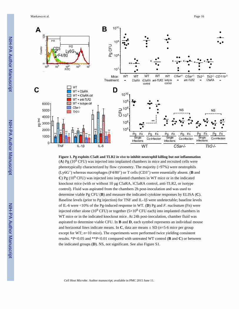
Figure 1. Pg exploits C5aR and TLR2 in vivo to inhibit neutrophil killing but not inflammation(A) Pg (109 CFU) was injected into implanted chambers in mice and recruited cells were
phenotypically characterized by flow cytometry. The majority (>97%) were neutrophils
(Ly6G+) whereas macrophages (F4/80+) or T cells (CD3+) were essentially absent. (B and
C) Pg (109 CFU) was injected into implanted chambers in WT mice or in the indicated
knockout mice (with or without 10 μg C5aRA, iC5aRA control, anti-TLR2, or isotype
control). Fluid was aspirated from the chambers 2h post-inoculation and was used to
determine viable Pg CFU (B) and measure the indicated cytokine responses by ELISA (C).
Baseline levels (prior to Pg injection) for TNF and IL-1β were undetectable; baseline levels
of IL-6 were <10% of the Pg-induced response in WT. (D) Pg and F. nucleatum (Fn) were
injected either alone (109 CFU) or together (5×10⁸ CFU each) into implanted chambers in
WT mice or in the indicated knockout mice. At 24h post-inoculation, chamber fluid was
aspirated to determine viable CFU. In B and D, each symbol represents an individual mouse
and horizontal lines indicate means. In C, data are means ± SD (n=5-6 mice per group
except for WT; n=10 mice). The experiments were performed twice yielding consistent
results. *P<0.05 and **P<0.01 compared with untreated WT control (B and C) or between
the indicated groups (D). NS, not significant. See also Figure S1.
Maekawa et al. Page 16
Cell Host Microbe. Author manuscript; available in PMC 2015 June 11.
NIH
-PA
Author M
anuscriptN
IH-P
A A
uthor Manuscript
NIH
-PA
Author M
anuscript
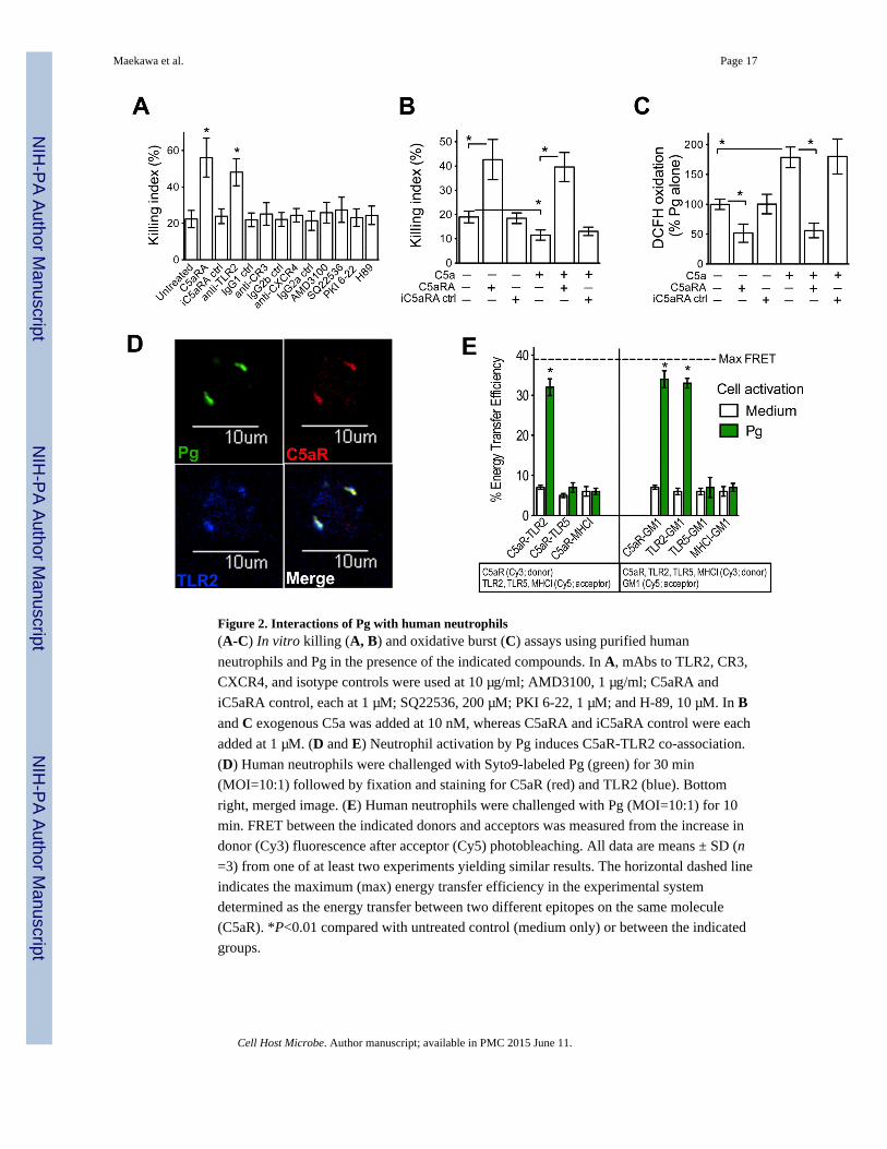
Figure 2. Interactions of Pg with human neutrophils(A-C) In vitro killing (A, B) and oxidative burst (C) assays using purified human
neutrophils and Pg in the presence of the indicated compounds. In A, mAbs to TLR2, CR3,
CXCR4, and isotype controls were used at 10 μg/ml; AMD3100, 1 μg/ml; C5aRA and
iC5aRA control, each at 1 μM; SQ22536, 200 μM; PKI 6-22, 1 μM; and H-89, 10 μM. In Band C exogenous C5a was added at 10 nM, whereas C5aRA and iC5aRA control were each
added at 1 μM. (D and E) Neutrophil activation by Pg induces C5aR-TLR2 co-association.
(D) Human neutrophils were challenged with Syto9-labeled Pg (green) for 30 min
(MOI=10:1) followed by fixation and staining for C5aR (red) and TLR2 (blue). Bottom
right, merged image. (E) Human neutrophils were challenged with Pg (MOI=10:1) for 10
min. FRET between the indicated donors and acceptors was measured from the increase in
donor (Cy3) fluorescence after acceptor (Cy5) photobleaching. All data are means ± SD (n
=3) from one of at least two experiments yielding similar results. The horizontal dashed line
indicates the maximum (max) energy transfer efficiency in the experimental system
determined as the energy transfer between two different epitopes on the same molecule
(C5aR). *P<0.01 compared with untreated control (medium only) or between the indicated
groups.
Maekawa et al. Page 17
Cell Host Microbe. Author manuscript; available in PMC 2015 June 11.
NIH
-PA
Author M
anuscriptN
IH-P
A A
uthor Manuscript
NIH
-PA
Author M
anuscript
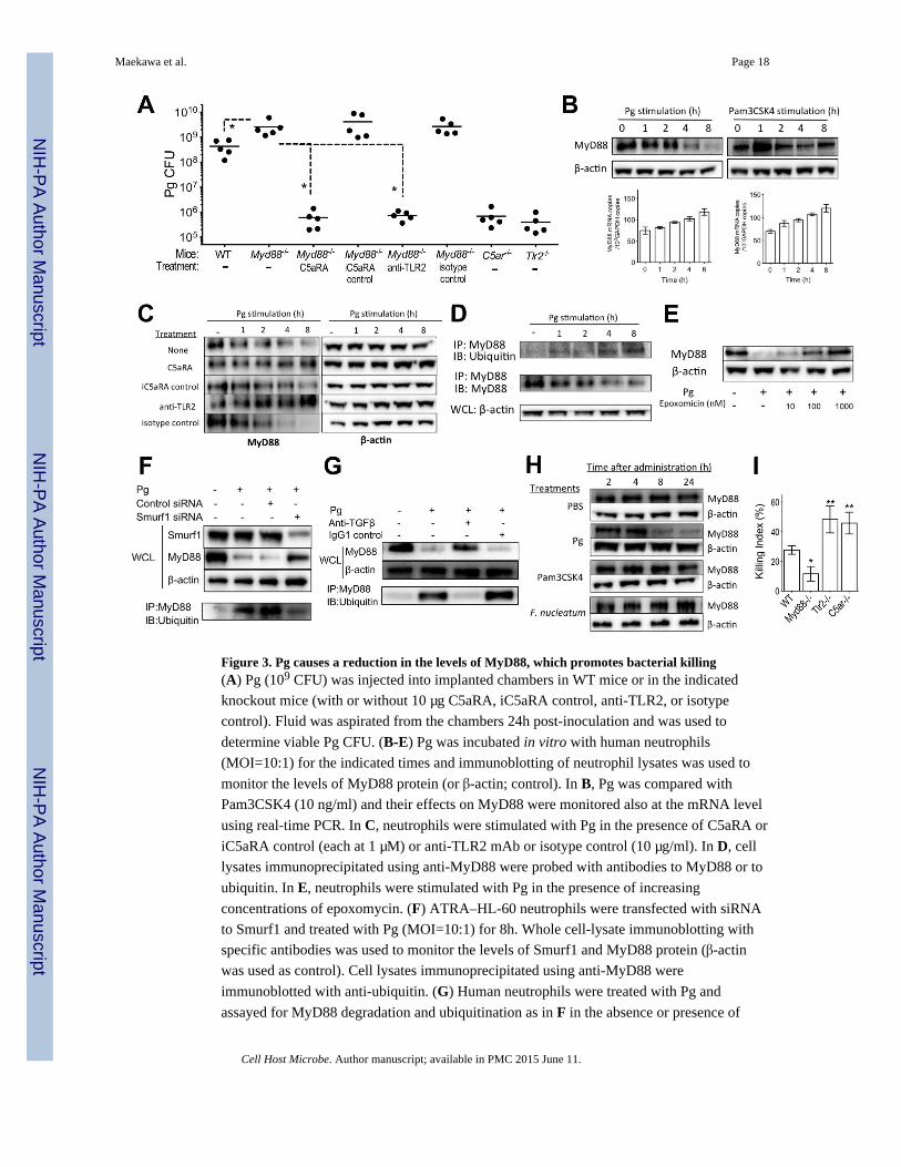
Figure 3. Pg causes a reduction in the levels of MyD88, which promotes bacterial killing(A) Pg (109 CFU) was injected into implanted chambers in WT mice or in the indicated
knockout mice (with or without 10 μg C5aRA, iC5aRA control, anti-TLR2, or isotype
control). Fluid was aspirated from the chambers 24h post-inoculation and was used to
determine viable Pg CFU. (B-E) Pg was incubated in vitro with human neutrophils
(MOI=10:1) for the indicated times and immunoblotting of neutrophil lysates was used to
monitor the levels of MyD88 protein (or β-actin; control). In B, Pg was compared with
Pam3CSK4 (10 ng/ml) and their effects on MyD88 were monitored also at the mRNA level
using real-time PCR. In C, neutrophils were stimulated with Pg in the presence of C5aRA or
iC5aRA control (each at 1 μM) or anti-TLR2 mAb or isotype control (10 μg/ml). In D, cell
lysates immunoprecipitated using anti-MyD88 were probed with antibodies to MyD88 or to
ubiquitin. In E, neutrophils were stimulated with Pg in the presence of increasing
concentrations of epoxomycin. (F) ATRA–HL-60 neutrophils were transfected with siRNA
to Smurf1 and treated with Pg (MOI=10:1) for 8h. Whole cell-lysate immunoblotting with
specific antibodies was used to monitor the levels of Smurf1 and MyD88 protein (β-actin
was used as control). Cell lysates immunoprecipitated using anti-MyD88 were
immunoblotted with anti-ubiquitin. (G) Human neutrophils were treated with Pg and
assayed for MyD88 degradation and ubiquitination as in F in the absence or presence of
Maekawa et al. Page 18
Cell Host Microbe. Author manuscript; available in PMC 2015 June 11.
NIH
-PA
Author M
anuscriptN
IH-P
A A
uthor Manuscript
NIH
-PA
Author M
anuscript
anti-TGF-β mAb. (H) The indicated bacteria or compounds were injected (Pg, 109 CFU;
Pam3SK4, 10 μg; F. nucleatum, 109 CFU) into implanted chambers in mice for the
indicated times and immunoblotting of neutrophil lysates was used to monitor the levels of
MyD88 protein. (I) Assay for in vitro killing of Pg by purified mouse neutrophils of the
indicated genotypes. All experiments were performed two (A) or three times (B-I) yielding
similar findings. In A, each symbol represents an individual mouse and horizontal lines
indicate means. In I, data are means ± SD (n=4). *P<0.05 and **P<0.01 compared with WT
control or between the indicated groups. IB, immunoblotting; IP, immunoprecipitation;
WCL, whole cell lysates. See also Figure S2.
Maekawa et al. Page 19
Cell Host Microbe. Author manuscript; available in PMC 2015 June 11.
NIH
-PA
Author M
anuscriptN
IH-P
A A
uthor Manuscript
NIH
-PA
Author M
anuscript
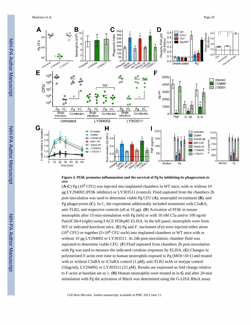
Figure 4. PI3K promotes inflammation and the survival of Pg by inhibiting its phagocytosis invivo(A-C) Pg (109 CFU) was injected into implanted chambers in WT mice, with or without 10
μg LY294002 (PI3K inhibitor) or LY303511 (control). Fluid aspirated from the chambers 2h
post-inoculation was used to determine viable Pg CFU (A), neutrophil recruitment (B), and
Pg phagocytosis (C). In C, the experiment additionally included treatments with C5aRA,
anti-TLR2, and respective controls (all at 10 μg). (D) Activation of PI3K in mouse
neutrophils after 15-min-stimulation with Pg (left) or with 10 nM C5a and/or 100 ng/ml
Pam3CSK4 (right) using FACE PI3Kp85 ELISA. In the left panel, neutrophils were from
WT or indicated knockout mice. (E) Pg and F. nucleatum (Fn) were injected either alone
(109 CFU) or together (5×10⁸ CFU each) into implanted chambers in WT mice with or
without 10 μg LY294002 or LY303511. At 24h post-inoculation, chamber fluid was
aspirated to determine viable CFU. (F) Fluid aspirated from chambers 2h post-inoculation
with Pg was used to measure the indicated cytokine responses by ELISA. (G) Changes in
polymerized F-actin over time in human neutrophils exposed to Pg (MOI=10:1) and treated
with or without C5aRA or iC5aRA control (1 μM), anti-TLR2 mAb or isotype control
(10μg/ml), LY294002 or LY303511 (25 μM). Results are expressed as fold change relative
to F-actin at baseline set as 1. (H) Human neutrophils were treated as in G and after 20-min
stimulation with Pg the activation of RhoA was determined using the G-LISA RhoA assay
Maekawa et al. Page 20
Cell Host Microbe. Author manuscript; available in PMC 2015 June 11.
NIH
-PA
Author M
anuscriptN
IH-P
A A
uthor Manuscript
NIH
-PA
Author M
anuscript
kit. The dashed line indicates baseline RhoA activation (in cells not exposed to Pg). (I)
Human neutrophils were treated as in G and after 6h-stimulation with Pg the indicated
cytokine responses were measured by ELISA. In A and E, each symbol represents an
individual mouse and horizontal lines indicate means. In bar graphs, data are means ± SD
(B, C, and F, n=5; D and G-I, n=3). The experiments were performed two (A-C and E-H)
or three times (D,I) yielding consistent results. *P <0.01 compared with control (WT or
untreated) or between the indicated groups. NS, not significant. See also Figure S3.
Maekawa et al. Page 21
Cell Host Microbe. Author manuscript; available in PMC 2015 June 11.
NIH
-PA
Author M
anuscriptN
IH-P
A A
uthor Manuscript
NIH
-PA
Author M
anuscript
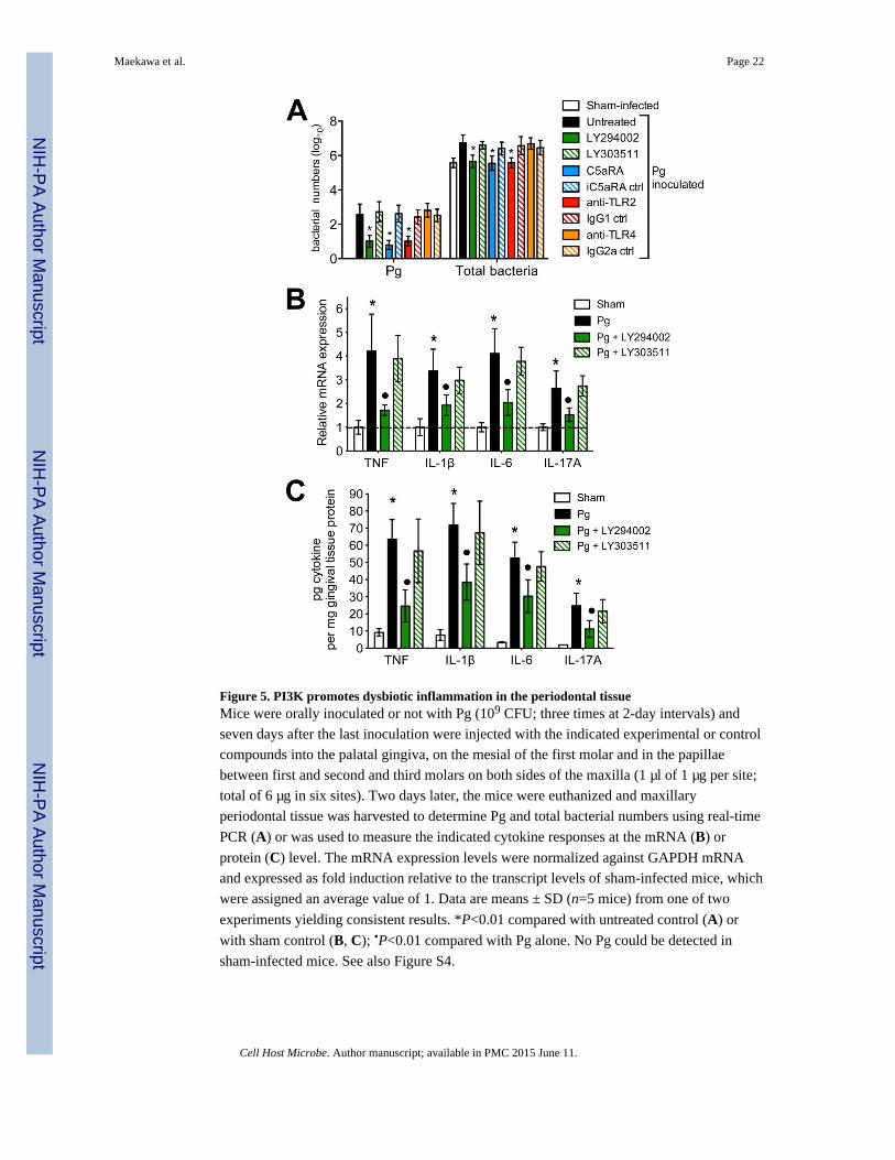
Figure 5. PI3K promotes dysbiotic inflammation in the periodontal tissueMice were orally inoculated or not with Pg (109 CFU; three times at 2-day intervals) and
seven days after the last inoculation were injected with the indicated experimental or control
compounds into the palatal gingiva, on the mesial of the first molar and in the papillae
between first and second and third molars on both sides of the maxilla (1 μl of 1 μg per site;
total of 6 μg in six sites). Two days later, the mice were euthanized and maxillary
periodontal tissue was harvested to determine Pg and total bacterial numbers using real-time
PCR (A) or was used to measure the indicated cytokine responses at the mRNA (B) or
protein (C) level. The mRNA expression levels were normalized against GAPDH mRNA
and expressed as fold induction relative to the transcript levels of sham-infected mice, which
were assigned an average value of 1. Data are means ± SD (n=5 mice) from one of two
experiments yielding consistent results. *P<0.01 compared with untreated control (A) or
with sham control (B, C); •P<0.01 compared with Pg alone. No Pg could be detected in
sham-infected mice. See also Figure S4.
Maekawa et al. Page 22
Cell Host Microbe. Author manuscript; available in PMC 2015 June 11.
NIH
-PA
Author M
anuscriptN
IH-P
A A
uthor Manuscript
NIH
-PA
Author M
anuscript
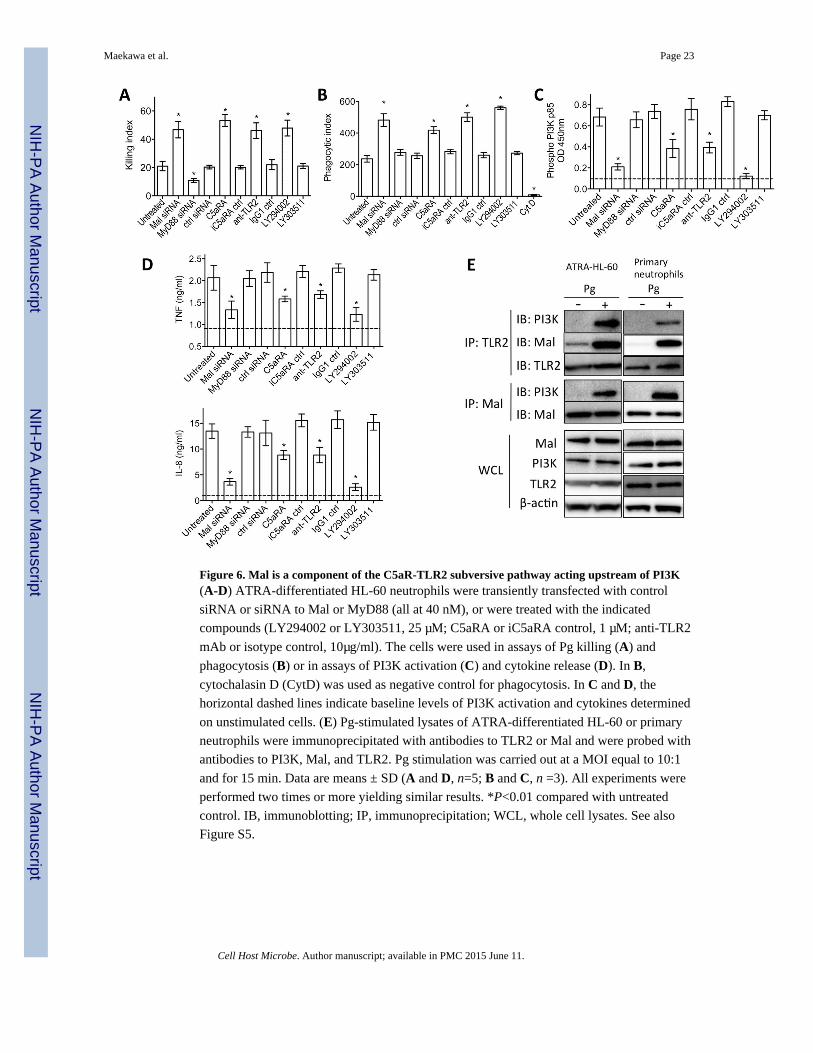
Figure 6. Mal is a component of the C5aR-TLR2 subversive pathway acting upstream of PI3K(A-D) ATRA-differentiated HL-60 neutrophils were transiently transfected with control
siRNA or siRNA to Mal or MyD88 (all at 40 nM), or were treated with the indicated
compounds (LY294002 or LY303511, 25 μM; C5aRA or iC5aRA control, 1 μM; anti-TLR2
mAb or isotype control, 10μg/ml). The cells were used in assays of Pg killing (A) and
phagocytosis (B) or in assays of PI3K activation (C) and cytokine release (D). In B,
cytochalasin D (CytD) was used as negative control for phagocytosis. In C and D, the
horizontal dashed lines indicate baseline levels of PI3K activation and cytokines determined
on unstimulated cells. (E) Pg-stimulated lysates of ATRA-differentiated HL-60 or primary
neutrophils were immunoprecipitated with antibodies to TLR2 or Mal and were probed with
antibodies to PI3K, Mal, and TLR2. Pg stimulation was carried out at a MOI equal to 10:1
and for 15 min. Data are means ± SD (A and D, n=5; B and C, n =3). All experiments were
performed two times or more yielding similar results. *P<0.01 compared with untreated
control. IB, immunoblotting; IP, immunoprecipitation; WCL, whole cell lysates. See also
Figure S5.
Maekawa et al. Page 23
Cell Host Microbe. Author manuscript; available in PMC 2015 June 11.
NIH
-PA
Author M
anuscriptN
IH-P
A A
uthor Manuscript
NIH
-PA
Author M
anuscript
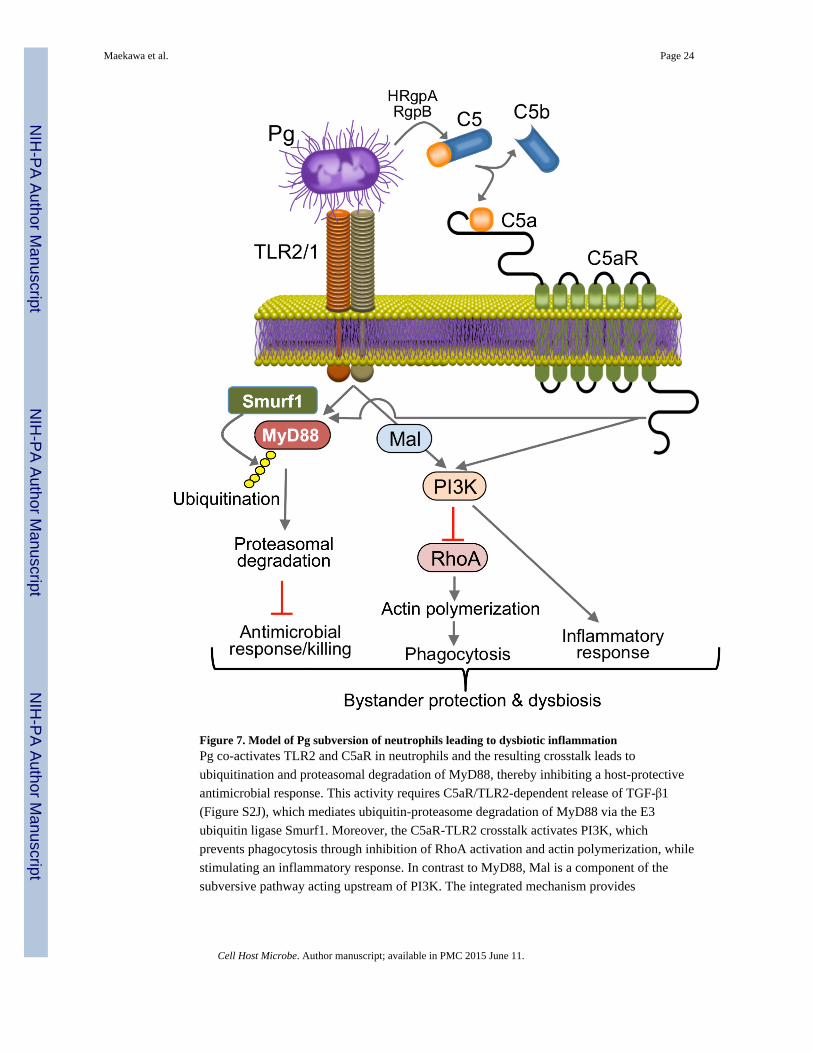
Figure 7. Model of Pg subversion of neutrophils leading to dysbiotic inflammationPg co-activates TLR2 and C5aR in neutrophils and the resulting crosstalk leads to
ubiquitination and proteasomal degradation of MyD88, thereby inhibiting a host-protective
antimicrobial response. This activity requires C5aR/TLR2-dependent release of TGF-β1
(Figure S2J), which mediates ubiquitin-proteasome degradation of MyD88 via the E3
ubiquitin ligase Smurf1. Moreover, the C5aR-TLR2 crosstalk activates PI3K, which
prevents phagocytosis through inhibition of RhoA activation and actin polymerization, while
stimulating an inflammatory response. In contrast to MyD88, Mal is a component of the
subversive pathway acting upstream of PI3K. The integrated mechanism provides
Maekawa et al. Page 24
Cell Host Microbe. Author manuscript; available in PMC 2015 June 11.
NIH
-PA
Author M
anuscriptN
IH-P
A A
uthor Manuscript
NIH
-PA
Author M
anuscript
‘bystander’ protection to otherwise susceptible bacterial species and promotes polymicrobial
dysbiotic inflammation in vivo.
Maekawa et al. Page 25
Cell Host Microbe. Author manuscript; available in PMC 2015 June 11.
NIH
-PA
Author M
anuscriptN
IH-P
A A
uthor Manuscript
NIH
-PA
Author M
anuscript