Embed Size (px)
Citation preview

THURSDAY, JUNE 29, 2017 8:00 a.m. Registration and Check-In 8:30 a.m. Welcome & Presentation of Award(s) for the NIH “Follow that Cell Challenge”
James Anderson, Ph.D., M.D., Director of the NIH Division of Program Coordination, Planning, and Strategic Initiatives (DPCPSI) Roderic Pettigrew, Ph.D., M.D., Director of the National Institute of Biomedical Imaging and Bioengineering (NIBIB) Joshua A. Gordon, Ph.D. M.D., Director of the National Institute of Mental Health (NIMH)
9:00 a.m. Presentation by Follow that Cell Winner(s) Winner(s) 9:30 a.m. Keynote Address Integrated learnings across modalities, technologies, and species for single cell genomics Rahul Satija, Ph.D. (New York University) 10:00 a.m. Theories of Cellular Phenotype – Multimodal Analysis of in vivo and in vitro cells James Eberwine, Ph.D. (University of Pennsylvania) 10:20 a.m. Break 10:45 a.m. Keynote Address Having fun with single cell RNA-seq: imputation, manifolds and trajectories Dana Pe’er, Ph.D. (Memorial Sloan Kettering Cancer Center) 11:15 a.m. Multiplex and Multimodal Analysis of RNA Expression by HCR and SeqFISH Scott Fraser, Ph.D. (University of Southern California) 11:35 a.m. Single Cell Imaging of Epigenetic Dynamics Peter Yingxiao Wang, Ph.D. (University of California San Diego) 11:55 am. Single Cell Genomics: When Stochasticity Meet Precision Xiaoliang Sunney Xie, Ph.D. (Harvard University) 12:15 p.m. Lunch on Your Own 1:00 p.m. Poster Session Location: FAES Terrace 1:00 – 2:00 Presenters for odd numbered posters 2:00 – 3:00 Presenters for even numbered posters 3:00 p.m. Keynote Address Genes, cells, and behavior: lessons from C. elegans Cori Bargmann Ph.D. (The Rockefeller University; The Chan Zuckerberg Initiative)
NIH Common Fund 5th Annual Single Cell Analysis Investigators Meeting
Masur Auditorium, NIH Campus, Bethesda, MD

3:30 p.m. Breakout Sessions
Single-cell approaches to infectious disease Moderator: John Yin, Ph.D. (University of Wisconsin–Madison) Location: Classroom 1&2 Is Heterogeneity Regulated? Moderator: Suraj Bhat, Ph.D. (University of California Los Angeles) Location: Classroom 4 Immune Cell Diversity Moderator: William Lu, Ph.D. (National Cancer Institute) Location: Classroom 6 5:00 p.m. General Meeting Adjourns FRIDAY, JUNE 30, 2017 8:00 a.m. Registration and Check-In 8:30 a.m. Keynote Address Imaging Single Molecules of mRNA in Single Living Cells Robert Singer, Ph.D. (Albert Einstein College of Medicine) 9:00 a.m. Breakout Sessions Moving single cell technologies out of the lab for wider Moderator: Navin Varadarajan, Ph.D. (University of Houston) Location: Classroom 3 Public sharing of resources and data: Lessons from other trans-NIH Programs Moderator: Grace Shen, Ph.D. (National Eye Institute) Location: Classroom 7 Birds of a Feather Location: TBD 10:30 a.m. Break 11:00 a.m. Keynote Address Illuminating Cellular Diversity in the Nervous System John Ngai, Ph.D. (University of California, Berkeley) 11:30 a.m. Comprehensive and integrated DNA, RNA and protein profiling in single cells in situ with cleavable
fluorescent probes Jia Guo, Ph.D. (Arizona State University) 11:50 a.m. Multimodal imaging of single cell populations by mass spectrometry, immunocytochemistry, and
vibrational spectroscopy for uncovering chemical heterogeneity within the brain Elizabeth Neumann (University of Illinois at Urbana-Champaign) 12:10 p.m. Automating the Optical Manipulation of Single Cells in Complex Tissues Pavak Shah, Ph.D. (Memorial Sloan-Kettering Cancer Center) 12:30 p.m. Resistance to targeted cancer therapy arises from pre-existing rare-cell expression variability
followed by drug-induced epigenetic reprogramming Sydney Shaffer (University of Pennsylvania) 12:50 p.m. Wrap-Up 1:00 p.m. General Meeting Adjourns
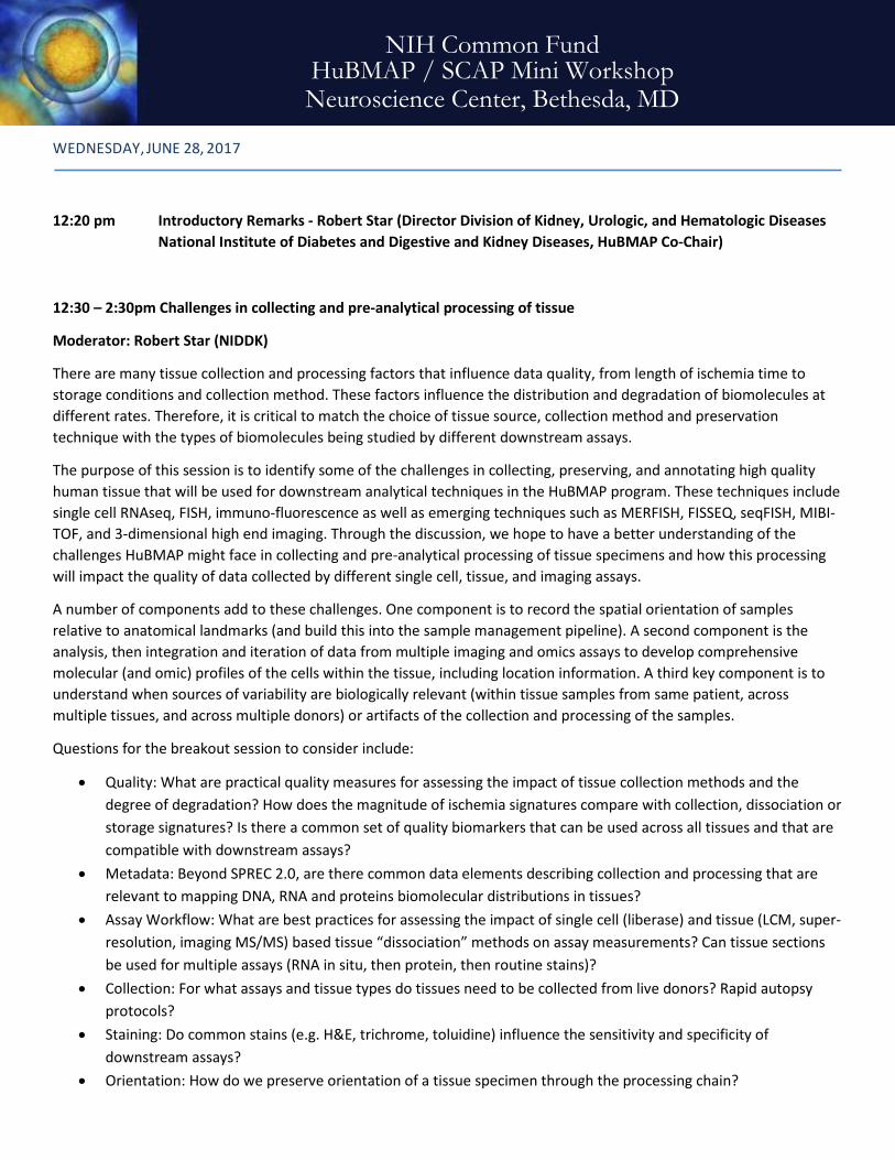
WEDNESDAY, JUNE 28, 2017
12:20 pm Introductory Remarks - Robert Star (Director Division of Kidney, Urologic, and Hematologic Diseases National Institute of Diabetes and Digestive and Kidney Diseases, HuBMAP Co-Chair)
12:30 – 2:30pm Challenges in collecting and pre-analytical processing of tissue
Moderator: Robert Star (NIDDK)
There are many tissue collection and processing factors that influence data quality, from length of ischemia time to storage conditions and collection method. These factors influence the distribution and degradation of biomolecules at different rates. Therefore, it is critical to match the choice of tissue source, collection method and preservation technique with the types of biomolecules being studied by different downstream assays.
The purpose of this session is to identify some of the challenges in collecting, preserving, and annotating high quality human tissue that will be used for downstream analytical techniques in the HuBMAP program. These techniques include single cell RNAseq, FISH, immuno-fluorescence as well as emerging techniques such as MERFISH, FISSEQ, seqFISH, MIBI-TOF, and 3-dimensional high end imaging. Through the discussion, we hope to have a better understanding of the challenges HuBMAP might face in collecting and pre-analytical processing of tissue specimens and how this processing will impact the quality of data collected by different single cell, tissue, and imaging assays.
A number of components add to these challenges. One component is to record the spatial orientation of samples relative to anatomical landmarks (and build this into the sample management pipeline). A second component is the analysis, then integration and iteration of data from multiple imaging and omics assays to develop comprehensive molecular (and omic) profiles of the cells within the tissue, including location information. A third key component is to understand when sources of variability are biologically relevant (within tissue samples from same patient, across multiple tissues, and across multiple donors) or artifacts of the collection and processing of the samples.
Questions for the breakout session to consider include:
• Quality: What are practical quality measures for assessing the impact of tissue collection methods and the degree of degradation? How does the magnitude of ischemia signatures compare with collection, dissociation or storage signatures? Is there a common set of quality biomarkers that can be used across all tissues and that are compatible with downstream assays?
• Metadata: Beyond SPREC 2.0, are there common data elements describing collection and processing that are relevant to mapping DNA, RNA and proteins biomolecular distributions in tissues?
• Assay Workflow: What are best practices for assessing the impact of single cell (liberase) and tissue (LCM, super-resolution, imaging MS/MS) based tissue “dissociation” methods on assay measurements? Can tissue sections be used for multiple assays (RNA in situ, then protein, then routine stains)?
• Collection: For what assays and tissue types do tissues need to be collected from live donors? Rapid autopsy protocols?
• Staining: Do common stains (e.g. H&E, trichrome, toluidine) influence the sensitivity and specificity of downstream assays?
• Orientation: How do we preserve orientation of a tissue specimen through the processing chain?
NIH Common Fund HuBMAP / SCAP Mini Workshop Neuroscience Center, Bethesda, MD
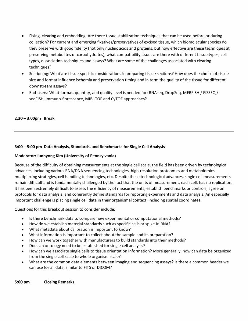
• Fixing, clearing and embedding: Are there tissue stabilization techniques that can be used before or during collection? For current and emerging fixatives/preservatives of excised tissue, which biomolecular species do they preserve with good fidelity (not only nucleic acids and proteins, but how effective are these techniques at preserving metabolites or carbohydrates), what compatibility issues are there with different tissue types, cell types, dissociation techniques and assays? What are some of the challenges associated with clearing techniques?
• Sectioning: What are tissue-specific considerations in preparing tissue sections? How does the choice of tissue size and format influence ischemia and preservation timing and in term the quality of the tissue for different downstream assays?
• End-users: What format, quantity, and quality level is needed for: RNAseq, DropSeq, MERFISH / FISSEQ / seqFISH, immuno-florescence, MIBI-TOF and CyTOF approaches?
2:30 – 3:00pm Break
3:00 – 5:00 pm Data Analysis, Standards, and Benchmarks for Single Cell Analysis
Moderator: Junhyong Kim (University of Pennsylvania)
Because of the difficulty of obtaining measurements at the single cell scale, the field has been driven by technological advances, including various RNA/DNA sequencing technologies, high-resolution proteomics and metabolomics, multiplexing strategies, cell handling technologies, etc. Despite these technological advances, single cell measurements remain difficult and is fundamentally challenged by the fact that the units of measurement, each cell, has no replication. It has been extremely difficult to assess the efficiency of measurements, establish benchmarks or controls, agree on protocols for data analysis, and coherently define standards for reporting experiments and data analysis. An especially important challenge is placing single cell data in their organismal context, including spatial coordinates.
Questions for this breakout session to consider include:
• Is there benchmark data to compare new experimental or computational methods? • How do we establish material standards such as specific cells or spike-in RNA? • What metadata about calibration is important to know? • What information is important to collect about the sample and its preparation? • How can we work together with manufacturers to build standards into their methods? • Does an ontology need to be established for single cell analysis? • How can we associate single cells to tissue orientation information? More generally, how can data be organized
from the single cell scale to whole organism scale? • What are the common data elements between imaging and sequencing assays? Is there a common header we
can use for all data, similar to FITS or DICOM?
5:00 pm Closing Remarks
Is heterogeneity regulated? 3:30 – 5pm, Thursday June 29 Moderator: Suraj Bhat, Ph.D. (University of California Los Angeles) Location: Classroom 4 The heterogeneity of gene expression in single cells is well established however it is unclear if this heterogeneity has any relationship to the morphological and/or molecular phenotype of a tissue or an organ. In this session we will ask questions to elucidate the challenges, both technical as well as conceptual, in understanding the role of the cellular variability (as assessed by gene expression) in the context of multicellularity of tissues and organs. We will explore the possible role of cellular heterogeneity in terminal differentiation. At the current state of our knowledge, we do not have a handle on whether the variation in the abundance of a gene transcript from cell to cell is because of the fluctuations intrinsic to the gene activity or whether it is the other cellular components that determine the variability between cells. In either case the question remains – what is regulated? Some of the questions that we will address (not necessarily in the order listed):
• Is heterogeneity causal or a result of the gene activity? • Is heterogeneity functional? • What is the relationship between the tissue /organ phenotype to the single cell? • Is heterogeneity the pathway to terminal differentiation? • Do different developmental programs entail specific states (stages) of heterogeneity? • What are the deterministic sources of cellular variability and how are they maintained? • Is deterministic variability important and what purpose does it serve? • Do we understand the link between molecular variability and the phenotypic variability between individual cells?
(Technically how do we study heterogeneity in single cells?)
Immune Cell Diversity 3:30 – 5pm, Thursday June 29 Moderator: Y. William Lu, Ph.D. (National Cancer Institute) Location: Classroom 6 Immunologists usually rely on flow cytometry and other traditional tools to conduct immune-related research. In the past several years, new single-cell technologies, such as single-cell transcriptomics and mass cytometry (CyTOF), have enabled researchers to ask scientific questions that could not be addressed previously. The theme for this session is to discuss the opportunities and potential challenges in immune-related studies, such as cell type diversity and phenotypic analysis. Questions for this breakout session to consider include:
• What immunological questions can we ask from the CyTOF analysis? • Similarly, what immunological questions can we ask from the single-cell transcriptome analysis? • Several technologies are available for single-cell transcriptome analysis. What are the pros and cons for these
technologies? Which technology is suitable for immune-related studies? • What kind of data quality should we expect in order to generate reproducible results? Should we validate the data by
additional assays? • One of the potential challenges is the communication between scientists with different expertise. Sometimes the
experiment may not perform well as expected. As an immunologist, a molecular biologist or a bioinformatician, what is the challenges to communicate with scientists with other disciplines?
• What kind of community resources can we establish to help immunologists to understand and utilize new single-cell technologies?
5th Annual Single Cell Analysis Investigators Meeting Masur Auditorium, NIH Campus, Bethesda, MD
Breakout Sessions

Single-cell approaches to infectious disease 3:30 – 5:00pm, Thursday June 29 Moderator: John Yin (University of Wisconsin–Madison) Location: Classroom 1 & 2 Transmission of infectious diseases from ailing to healthy hosts occurs through a cough or sneeze, handshake, faucet handle, or mosquito’s bite. Typically, the process transfers a few bacterial cells or virus particles. Although a small number of cells or particles encounter a few susceptible host tissues or cells, the resulting infection initiates a battle --- with potentially critical outcomes. The behavior of a few host cells infected by a small number of bacteria or virus particles can give rise to large 'noise' and significant variability in gene expression by the pathogen to amplify itself or by the host to set innate immune blockades, which then influence how further cycles of host cell or tissue infection amplify or inhibit the pathogen. The result can often be a diversity of symptoms and disease severities for patients, from mild to serious, or even deadly. Questions for the breakout session to consider include: • To what extent do genetic, environmental, or other (stochastic) factors contribute to extremely heterogeneous
distributions of virus production (yield) from single cells? • What are key challenges and opportunities for advancing innovative technologies to enable routine high-throughput
single-cell measurements? • How can the intrinsic heterogeneity of single-cell readouts be exploited to extract new insights into virus-cell
interactions? • How can systems biology approaches (mathematical modeling, computer simulations) add value to enable
mechanistic interpretation of single-cell data? • What features of virus or cellular behaviors at the single-cell level most impact the severity of infection in natural
hosts? (Note: Most infections in nature are initiated by small numbers of host cells initially becoming infected. From an evolutionary perspective, transmission can create genetic bottlenecks for the pathogen.)
Public sharing of resources and data: Lessons from other trans-NIH Programs 9:00 – 10:30am, Friday June 30 Moderator: Grace Shen, Ph.D. (National Eye Institute) Location: Classroom 7 Resource and data sharing is essential to speed translation of research results into knowledge, therapies, and procedures to improve our understanding of biological processes and human health. NIH is committed to sharing data from its research and supports a variety of resources and tools for researchers. These resources include tissue banks and repositories, datasets and databases, model organisms, genome and DNA sequences, and resource libraries. The panelists in this session will describe some of the resources and data that are available from programs they are involved with across NIH. [add details] Panelists for this session:
• Andrea Beckel-Mitchener, Ph.D. (National Institute of Mental Health) • Susan Gregurick, Ph.D. (National Institute of General Medical Sciences) • Shannon Hughes, Ph.D. (National Cancer Institute) • Halonna Kelly, Ph.D. (National Institute of Allergy and Infectious Diseases)
Moving single cell technologies out of the lab for wider adoption 9:00 – 10:30am, Friday June 30 Moderator: Navin Varadarajan, Ph.D. (University of Houston) Location: Classroom 3 The last few years have seen a dramatic increase in the number and complexity of single-cell technologies. A large number of these advances have been pioneered through individual laboratories and the scope of the biological questions interrogated has primarily been dictated by collaborations with these laboratories. In order for single-cell technologies to mature, they must become standardized tools that enable any biologist/clinician can access to test hypotheses. Mass cytometry is an example of one such tool that has made the successful transition. The scope of this discussion is to understand the challenges behind moving the technologies from the labs of the inventors and to make them commercially available tools/techniques. Questions for the breakout session to consider include: • Validation. One of the major challenges with single-cell technologies is cross-platform validation. While single-cell
technologies provide the ability to deliver insight that would not be available based on population analyses, it is important to be able to identify technical errors, limitations and how to implement confidence metrics in single cell results.
• Standardization of statistics, bioinformatics and visualization. Another major challenge with single-cell data is to have robust methods that perform normalization, discretization etc. in a standardized manner. Good practices in defining thresholds, how they are determined and adhering to a minimal set of standards that will be published (e.g. see Minimal Information About T-cell Assays, MIATA) comprise an essential framework. Similarly, there is a need to develop to visualization packages that better describe the complexity of multi-dimensional, time-resolved single-cell data. E.g. viSNE is good for single time points but what about time series?
• Commercialization. There are many layers to commercialization including IP framework, hurdles to manufacturing, ease of implementation and market adoption. What programs can help PIs understand these challenges: NSF ICorps? Institutional help?
Birds of a Feather 9:00 – 10:30am, Friday June 30 Moderator: You? Location: TBD Lead or attend a Birds of a Feather session! Meet with your peers! Discuss questions that have been on your mind or the next big things! This Birds-of-a-Feather session is an opportunity for very informal gatherings of people interested in a particular topic that we are not covering otherwise at the meeting. The goal is to have audience-driven discussion, grassroots participation and networking. How it will work: There will be a poster board at the registration desk for the meeting. If you are interested in leading a Birds of a Feather session, please use one of the postcards to write the topic for discussion on Thursday. If you are interested in participating in one of the sessions please add your name to the postcard. We will help each of the BOF groups find space to hold their discussion during the breakout session on Friday morning.

Poster Session
Poster
Number Authors Affiliations Poster Title
1
Prithvijit Mukherjee, Lingqian Chang, Eric Berns, S. Shiva P. Nathamgari, Milan Mrksich, Horacio D. Espinosa
Infinitesimal LLC, Skokie, IL Department of Mechanical Engineering, Northwestern University, Evanston, IL
A Localized Cell Analysis Device for Temporal Cell Analysis - Measuring Protein Tyrosine Phosphatase Activity in Live Cancer Cells
2 Yi Lu and Pak Kin Wong Department of Biomedical Engineering, The Pennsylvania State University, University Park, PA
A Multispectral Single Molecule Nanobiosensor for Dynamic Multigene Analysis during Collective Cell Migration
3
Masahiro Hitomi, Anastasia Chumakova, Stephanie Jarvis, Neha Anand, Bridget Corrigan, Peter Yoo, Upashruti Agrawal, Vid Yogeswaran, Malini Kamineni, Sunghyun Kim, and Justin D. Lathia
Dept. Cellular and Molecular Medicine, Lerner Research Institute, Cleveland Clinic, Cleveland, OH
Asymmetric cell division regulates fate decision of glioblastoma cancer stem cells
4 Pavak K. Shah, Anthony Santella, Adrian Jacobo, Kimberly Siletti, A. James Hudspeth, Zhirong Bao
Developmental Biology Program, Sloan Kettering Institute, New York, NY Howard Hughes Medical Institute and Laboratory of Sensory Neuroscience, The Rockefeller University, New York, New York
Automating the Optical Manipulation of Single Cells in Complex Tissues
5
Lin Han, Hua-Jun Wu, Haiying Zhu, Kun-Yong Kim, Sadie L. Marjani, Markus Riester, Ghia Euskirchen, Xiaoyuan Zi, Jennifer Yang, Jasper Han, Michael Snyder, In-Hyun Park, Rafael Irizarry, Sherman M. Weissman, Franziska Michor, Rong Fan, Xinghua Pan
Department of Biology Chemistry and Molecular Biology, School of Basic Medical Sciences, Southern Medical University, Guangzhou, Guangdong Province, China Department of Genetics, Yale University School of Medicine, New Haven, CT 06519
Bisulfite-independent analysis of CpG island methylation enables genome-scale stratification of single cells
6 Sachiko Sato, Ann Rancourt, Yukiko Sato, and Masahiko S. Satoh
Glycobiology and Bioimaging Laboratory of Research Center for Infectious Diseases Laboratory of DNA Damage Responses and Bioimaging, CHU de Québec, Faculty of Medicine, Laval University, 2705 Boulevard Laurier, Quebec, Quebec G1V 4G2, Canada Department of Physiology, McGill University, Montreal, Canada
Characterization of cultured cell lines using single-cell lineage tracking analysis
7 Jia Guo, Manas Mondal, Renjie Liao, Lu Xiao
Biodesign Institute & School of Molecular Sciences, Arizona State University, Tempe, AZ
Comprehensive and integrated DNA, RNA and protein profiling in single cells in situ with cleavable fluorescent probes
8 Erika P. Portero, Rosemary M. Onjiko, Sally A. Moody, and Peter Nemes
Department of Chemistry & Department of Anatomy and Regenerative Biology, The George Washington University, Washington, DC
Discovery Single-cell Mass Spectrometry Profiles Metabolic Gradients in the 16-cell Vertebrate (Frog) Embryo
NIH Common Fund 5th Annual Single Cell Analysis Investigators Meeting
Poster Session

NIH Common Fund 5th Annual Single Cell Analysis Investigators Meeting
Masur Auditorium, NIH Campus, Bethesda, MD
9
Tania Konry, Saheli Sarkar, Pooja Sabhachandani, Dina Stroopinksi, Kristen Palmer, Noa Cohen, Jacalyn Rosenblatt, David Avigan
Department of Pharmaceutical Sciences, Northeastern University, 360 Huntington Avenue, Boston, MA, 02115 Beth Israel Deaconess Medical Center, Harvard Medical School, Boston, MA 02115
Dynamic analysis of immune and cancer cell interaction at single cell level in microfluidic droplets
10 Tianyi Yuan, Diane S. Krause, and Oleg Denisenko
Yale University, New Haven, CT 06520 Department of Medicine, University of Washington, Seattle, WA 98109
Epigenetic analysis of gene activation in a single cell
11
P.A. Osmulski, Y.-T. Hsu, G. Huang, S.R. Polusani, C.-L. Chen, D. Mahalingam, N.B. Kirma, M.E. Gaczynska, and T. Hui-Ming Huang
Department of Molecular Medicine & Department of Medicine, University of Texas Health, San Antonio, TX
Guilty by adhesion – assessment of cells grip with atomic force microscopy
12 Stephen M. Anthony, Bryan Carson, Jerilyn A. Timlin
Bioenergy and Defense Technologies Department, Sandia National Laboratories, Albuquerque, NM
Hyperspectral Imaging Analysis of Cellular Heterogeneity Between and Across Populations
13 Peter Nemes, Rosemary M. Onjiko, Erika Portero, and Sally A. Moody
Department of Chemistry, The George Washington University, Washington, DC Department of Anatomy and Regenerative Biology, The George Washington University, Washington, DC
In Situ Optoguided Microsampling Single-cell Mass Spectrometry for Elucidating Cell Heterogeneity in the Developing Xenopus laevis (frog) Embryo
14
Dipjyoti Das, Dörthe Jülich, Jamie Schwendinger-Schreck, Andrew Lawton, Nicolas Dray, Thierry Emonet, Corey S. O’Hern, Mark D. Shattuck and Scott A. Holley
Department of Molecular, Cellular and Developmental Biology, Department of Physics, Department of Mechanical Engineering and Materials Science, Department of Applied Physics & Department of Physics, Yale University, New Haven, CT Benjamin Levich Institute, City College of the City University of New York, NY.
Long-range mechanical orchestration by the vertebrate tail organizer
15
Luke Stevens, Tanaya Pande, Hongru Hu, Aravindan Krishnan, Claudia Mizutani, Rui Sousa-Neves,
Department of Biology & Department of Genetics and Genome Sciences, Case Western Reserve University, Cleveland, OH
Multidimensional analyses of whole brain aging with single cell resolution
16
Elizabeth K. Neumann, Troy J. Comi, Stanislav S. Rubakhin, Sanghamitra Deb, Nicholas Spegazzini, Jennifer W. Mitchell, Collin Kaufman, Rohit Bhargava, Martha U. Gillette, Jonathan V. Sweedler
Department of Chemistry, Beckman Institute for Advanced Science and Technology, Department of Bioengineering, Department of Cell and Developmental Biology & Neuroscience Program, University of Illinois at Urbana-Champaign, Urbana, IL.
Multimodal imaging of single cell populations by mass spectrometry, immunocytochemistry, and vibrational spectroscopy for uncovering chemical heterogeneity within the brain
17 Scott E. Fraser, Long Cai
University of Southern California, Translational Imaging Center, Molecular and Computational Biology, Los Angeles, CA California Institute of Technology, Biology and Biological Engineering, Pasadena, CA
Multiplex and Multimodal Analysis of RNA Expression by HCR and SeqFISH
NIH Common Fund 5th Annual Single Cell Analysis Investigators Meeting
Masur Auditorium, NIH Campus, Bethesda, MD
18 Jimmie Ye, Hyun Min Kang
Institute for Human Genetics, Department of Medicine, Institute for Computational Health Sciences, Department of Epidemiology and Biostatistics & Department of Bioengineering and Therapeutic Science, UCSF, San Francisco, CA Department of Biostatistics, School of Public Health, University of Michigan, Ann Arbor, MI
Multiplexing droplet-based single cell RNA-sequencing using natural genetic barcodes
19 X. Nancy Xu Department of Chemistry and Biochemistry, Old Dominion University, Norfolk, VA 23529
Photostable Multiplexing NanoAssays for Real-time Molecular Imaging of Single Live Cells
20
Qin Zhu, Stephen A Fisher, Hannah Dueck, Sarah Middleton, Mugdha Khaladkar and Junhyong Kim
Department of Biology, University of Pennsylvania, Philadelphia, PA, USA.
PIVOT: Platform for Interactive Analysis and Visualization of Transcriptomics Data
21 Jennifer L. Geldart, Stephanie M. Schubert, Stephanie R. Walter, Mael Manesse, and David R. Walt
Department of Chemistry, Tufts University, Medford, Massachusetts 02155, United States
Protein Quantification in Single Cancer Cells using Simoa
22 A. Chumakova, M. Hitomi and J. D. Lathia
Dept. of Cellular and Molecular Medicine, Lerner Research Institute, Cleveland Clinic
Quantitative fluorescent microscopy as a tool for protein expression analysis in heterogeneous glioblastoma cancer stem cell population
23
Sydney Shaffer, Margaret Dunagin, Stefan Torborg, Eduardo Torre, Benjamin Emert, Clemens Krepler, Marilda Beqiri, Katrin Sproesser, Patricia Brafford, Elliott Eggan, Meenhard Herlyn, Arjun Raj
Department of Bioengineering, University of Pennsylvania, Philadelphia, PA The Wistar Institute, Molecular and Cellular Oncogenesis Program, Melanoma Research Center, Philadelphia, PA
Resistance to targeted cancer therapy arises from pre-existing rare-cell expression variability followed by drug-induced epigenetic reprogramming
24 Steffen K. Cornwell, Vipul Periwal
National Institute of Diabetes & Digestive & Kidney Diseases, National Institutes of Health, Bethesda, MD
scMatri-seq: simulation of heterogeneous single-cell RNA-seq data using a gene-gene developmental matrix
25 Xiaoliang Sunney Xie, Alec Chapman, David Lee, Dong Xing and Longzhi Tan
Single Cell Genomics: When Stochasticity Meet Precision
Single Cell Genomics: When Stochasticity Meet Precision
26 Qin Peng, Yuanliang Wang, Shu Chien, Yingxiao Wang
Department of Bioengineering, University of California, San Diego, CA 92093, USA Bioengineering College, Chongqing University, Chongqing 400030, China.
Single Cell Imaging of Epigenetic Dynamics
27 Karolyn A. Oetjen, Efthymia Papalexi, Rahul Satija, Christopher S. Hourigan
Myeloid Malignancy Section, National Heart, Lung and Blood Institute, National Institutes of Health, Bethesda, MD New York Genome Center, New York, NY Center for Genomics and Systems Biology, New York University, NY
Single Cell RNA Sequencing Analysis of Healthy Donor Bone Marrow Populations

NIH Common Fund 5th Annual Single Cell Analysis Investigators Meeting
Masur Auditorium, NIH Campus, Bethesda, MD 28 Yue J. Wang, Dana Avrahami-
Tzfati, Klaus H. Kaestner
Department of Genetics and Institute for Diabetes, Obesity, and Metabolism, University of Pennsylvania Perelman School of Medicine, Philadelphia, PA Endocrinology and Metabolism Service, Hadassah-Hebrew University Medical Center, Jerusalem, Israel
Single-cell analyses of the endocrine pancreas from a neonatal donor
29 Camille Lombard-Banek, Aparna Baxi, Sally A. Moody, and Peter Nemes
Department of Chemistry & Department of Anatomy & Regenerative Biology, The George Washington University, Washington DC, 20052
Single-cell Proteomics in the Developing Frog (Xenopus) Embryo
30 Long Cai, Michael Elowitz and Scott Fraser
Department of Biology and Biological Engineering, Caltech
Spatial genomics and single cell lineage dynamics by seqFISH and MEMOIR
31 Rosemary M. Onjiko, Erika P. Portero, Sally A. Moody, and Peter Nemes
Department of Chemistry & Department of Anatomy & Regenerative Biology, The George Washington University, Washington DC, 20052
Spatiotemporal Investigation of the Metabolic Architecture in Neuronal Cell Clones in the Developing Vertebrate (Frog) Embryo
32
James Eberwine, Jacqueline Morris, Young-Ji Na, Jaehee Lee, Hua Zhu, Eun-Hee Shim, Jinhui Wang, Kevin Miyashiro, Alexandra V. Ulyanova, Thomas Bell, John Wolf, Sean Grady, Jai Yoon Sul and Junhyong Kim
Department of Systems Pharmacology, Department of Neurosurgery & Department of Biology, University of Pennsylvania, Philadelphia, PA, USA.
Theories of Cellular Phenotype – Multimodal Analysis of in vivo and in vitro cells
33 Aaron Streets, Markita P. Landry
Department of Bioengineering, Department of Chemical and Biomolecular Engineering, California Institute for Quantitative Biosciences, QB3, University of California, Berkeley, CA Chan-Zuckerberg Biohub, San Francisco, CA
Toward Label-Free Single-Cell Profiling: Single-Molecule Detection of Protein Efflux and Raman Mapping of Intracellular Metabolites in Isolated Microorganisms and Brain Tissue
34 Juan Guan, Harrison Liu, Xiaoyu Shi, Siyu Feng, Bo Huang
Department of Pharmaceutical Chemistry, Department of Biochemistry and Biophysics & Department of Bioengineering, University of California San Francisco, San Francisco, CA
Tracking multiple genomic elements in single cell nuclei using correlative CRISPR imaging and sequential DNA FISH
35 Wen Zhou, Mary A. Yui, Brian A. Williams, Barbara J. Wold, Long Cai, Ellen V. Rothenberg
Division of Biology and Biological Engineering, California Institute of Technology, Pasadena, CA Division of Chemistry and Chemical Engineering, California Institute of Technology, Pasadena, CA
Transcriptional profiling with scRNAseq and SeqFISH on early T cell precursors reveals fine developmental steps
36
Robert S. Welner, Sam Wolock, Indira Krishnan, Danielle Tenen, Puneet Agarwal, Victoria McClearn, Ravi Bhatia, Daniel G Tenen, and Allon Klein
Division of Hematology/Oncology, Dept of Medicine, University of Alabama at Birmingham, Birmingham, AL Department of Systems Biology, Harvard Medical School, Boston, MA Division of Hematology/Oncology, Dept of Medicine, Beth Israel Deaconess Medical Center, Boston, MA
Unbiased Single-cell Analysis Reveals Hierarchy of the Bone Marrow Niche

NIH Common Fund 5th Annual Single Cell Analysis Investigators Meeting
Masur Auditorium, NIH Campus, Bethesda, MD
37 Kushal K Dey, Chiaowen Joyce Hsiao, Matthew Stephens
Department of Statistics, University of Chicago, Chicago, Illinois 60637, USA Department of Human Genetics, University of Chicago, Chicago, Illinois 60637, USA
Visualizing the Structure of Single Cell RNA-seq Expression Data using Grade of Membership Models
40 Nick Trotta, Rob McLellan, Nicholas Dobes, Steve Gebhart
Cell Microsystems, Inc. Research Triangle Park, North Carolina
The CellRaft AIR™ System: Automated Imaging, Sorting and Isolation of Single Cells
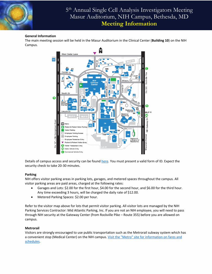
General Information The main meeting session will be held in the Masur Auditorium in the Clinical Center (Building 10) on the NIH Campus.
Details of campus access and security can be found here. You must present a valid form of ID. Expect the security check to take 20-30 minutes. Parking NIH offers visitor parking areas in parking lots, garages, and metered spaces throughout the campus. All visitor parking areas are paid areas, charged at the following rates:
• Garages and Lots: $2.00 for the first hour, $4.00 for the second hour, and $6.00 for the third hour. Any time exceeding 3 hours, will be charged the daily rate of $12.00.
• Metered Parking Spaces: $2.00 per hour. Refer to the visitor map above for lots that permit visitor parking. All visitor lots are managed by the NIH Parking Services Contractor: Mid Atlantic Parking, Inc. If you are not an NIH employee, you will need to pass through NIH security at the Gateway Center (from Rockville Pike – Route 355) before you are allowed on campus. Metrorail Visitors are strongly encouraged to use public transportation such as the Metrorail subway system which has a convenient stop (Medical Center) on the NIH campus. Visit the "Metro" site for information on fares and schedules.
5th Annual Single Cell Analysis Investigators Meeting Masur Auditorium, NIH Campus, Bethesda, MD
Meeting Information

Kiss and Ride Visitors can be dropped off and picked up from the Kiss and Ride park located at 9000 Rockville Pike, Bethesda, Maryland 20892 On-Campus Shuttle Shuttle services are provided throughout the day on the NIH Campus for employees, patients, and visitors. Click here for Shuttle routes and schedules Directions to Masur Auditorium from the Clinical Center
North lobby entrance: From the lobby, go down the right side, passing Admissions on your right. Continue straight through the sliding glass doors, following posted signs to the Masur. Continue following the “Detour” signs to the Masur. The auditorium is just past the main elevators. From the South lobby entrance: From the lobby, take either the left or right hallway up a slight incline until you come to the entrance of the Masur Auditorium. When the two hallways converge, you are standing in front of Masur Auditorium. Food & Beverages Food and beverages must be purchased. A full cafeteria is open from 6:30 a.m. - 2:30 p.m. located on the B1 level of the Clinical Center. More selections including Au Bon Pain is located on the SE side of the Clinical Center near the Main Lobby. Three concession/coffee stands are also available. The concession stand is located on the B1 level near the cafeteria and is open from 7:00 a.m. - 6:00 p.m. Two coffee stands are open from 7:00 a.m. - 4:00 p.m. and are located on the 1st floor in the CRC and the FAES corridor. Additionally, downtown Bethesda offers a fine selection of restaurants. Click Here for More Information
Meeting Information Poster Session The poster session on June 29th will be held in the Terrace located near the FAES classrooms. Directional signs will be posted in the registration area. Breakout Sessions The breakout sessions will be held on June 29th and June 30th. The breakout sessions will be in the FAES classrooms. Specific information regarding each session can be found on the meeting agenda.