Embed Size (px)
Citation preview
UNCO
RRECTED P
RO
OF
1 Nicotine effect on inflammatory and growth factor responses in murine cutaneous2 wound healing
3 SofiaQ1 Xanthoulea a,c,⁎, An Deliaert a, Andrea Romano b, Sander S. Rensen c,4 Wim A. Buurman c, Rene' RWJ van der Hulst a
5 a Department of Plastic Surgery, Maastricht University Medical Center, P.O. Box 5800, 6202 AZ, Maastricht, The Netherlands6 b GROW, School for Oncology and Developmental Biology, Maastricht University Medical Centre, Maastricht, The Netherlands7 c Department of Surgery, NUTRIM School for Nutrition, Toxicology & Metabolism, Maastricht University Medical Centre, Maastricht, The Netherlands
8
9
a b s t r a c ta r t i c l e i n f o
10 Article history:11 Received 14 June 201312 Received in revised form 3 September 201313 Accepted 15 October 201314 Available online xxxx15161718 Keywords:19 Cytokines20 Growth factors21 Inflammation22 Macrophages23 Nicotine24 Wound repair
25The aim of the current study was to investigate the effect of nicotine in an experimental mouse model of cutane-26ous injury and healing responses, during the inflammatory phase of repair. Nicotine injection in full-thickness27excisional skin wounds minimally affected inflammatory mediators like TNF, IL-6 and IL-12 while it induced28a down-regulation in the expression of growth factors like VEGF, PDGF, TGF-β1 and TGF-β2, and the anti-29inflammatory cytokine IL-10. Analysis of wound closure rate indicated no significant differences between nico-30tine and saline injected controls. In-vitro studies using bone marrow derived macrophages, resident peritoneal31macrophages and RAW 264.7 macrophages, indicated that nicotine down-regulates TNF production. Moreover,32nicotine was shown to down-regulate VEGF, PDGF and TGF-β1 in both bone marrow derived macrophages33and RAW 264.7 cells. Using an NF-κB luciferase reporter RAW 264.7 cell line, we show that nicotine effects are34minimally dependent on NF-κB inhibition. Moreover, nicotinic acetylcholine receptor (nAChR) subunit expres-35sion analyses indicated that while β2 nAChR subunit is expressed in mouse macrophages, α7 nAChR is not.36In conclusion, while skin inflammatory parameters were not significantly affected by nicotine, a down-regulation37of growth factor expression in bothmouse skin andmacrophageswas observed. Reduced growth factor expression38by nicotinemight contribute, at least in part, to the overall detrimental effects of tobacco use inwound healing and39skin diseases.40© 2013 Published by Elsevier B.V.
4142
43
44
45 1. Introduction
46 Cutaneouswound healing after an injury is a complex andhighly dy-47 namic process that involves interaction of different players like resident48 cells of the skin, inflammatory leukocytes, extracellular matrix compo-49 nents and soluble mediators. The healing process can be divided in50 three consecutive and partially overlapping phases i.e. inflammatory51 phase, proliferative phase and remodelling phase. The repair process52 finally results in the formation of a mass of fibrotic tissue known as53 scar [1,2].54 Numerous experimental and clinical studies have determined that55 inflammation plays a crucial although still not completely clear role dur-56 ing cutaneous wound healing and influences the quality of the resulting57 scar. Inflammatory cells and particularly macrophages appear to be58 essential for proper healing by, among other mechanisms, stimulating
59growth factor and anti-inflammatory cytokine production that are60necessary for repair [3]. Moreover, pathological functioning of macro-61phages and excessive inflammation in the wound healing process can62result in derailed healing, like the formation of ulcers, chronic wounds,63hypertrophic scars and keloids [4].64Nicotine, a major constituent of tobacco smoke, has been shown to65exert anti-inflammatory effects on different cell types and to be benefi-66cial in disorders where inflammation-related mechanisms are involved67like in ulcerative colitis and obesity [5]. Nicotine actions are mediated68through binding to cholinergic receptors termed nicotinic acetylcholine69receptors (nAChR) that are expressed inmany different tissues and cells70in the body, including immune cells. In both human and mouse macro-71phages, nicotine was shown to inhibit the release of pro-inflammatory72cytokines through a specific “nicotinic anti-inflammatory pathway”73that involves signalling through the α7 nAChR and prevents activation74of the NF-κB pathway [6–9]. In human microvascular endothelial cells,75nicotinewas found to inhibit TNF-inducedNF-κB activation, to suppress76adhesion molecule and chemokine expression and to reduce adhesion77of leukocytes to activated endothelium and consequent inflammation78[10]. Moreover, nicotine was shown to have angiogenic effects and to79increase endothelial cell proliferation and vascular growth in different80in-vitro and in-vivo models [11,12].
International Immunopharmacology xxx (2013) xxx–xxx
Abbreviations: TNF, tumor necrosis factor; IL-6, interleukin 6; IL-12, interleukin 12;VEGF, vascular endothelial growth factor; PDGF, platelet derived growth factor; TGF-β1,transforming growth factor beta 1; TGF-β2, transforming growth factor beta 2.⁎ Corresponding author at: Maastricht University Medical Centre, Universiteitssingel
50, Maastricht 6229 ER, The Netherlands. Tel.: +31 433882128; fax: +31 433884154.E-mail address: [email protected] (S. Xanthoulea).
INTIMP-03064; No of Pages 10
1567-5769/$ – see front matter © 2013 Published by Elsevier B.V.http://dx.doi.org/10.1016/j.intimp.2013.10.022
Contents lists available at ScienceDirect
International Immunopharmacology
j ourna l homepage: www.e lsev ie r .com/ locate / in t imp
Please cite this article as: Xanthoulea S, et al, Nicotine effect on inflammatory and growth factor responses in murine cutaneous wound healing,Int Immunopharmacol (2013), http://dx.doi.org/10.1016/j.intimp.2013.10.022
UNCO
RRECTED P
RO
OF
81 In the skin, effects of nicotine are ambivalent and often unclear, but82 they generally appear more negative than positive. Although cigarette83 smoking has been reported to negatively affect cutaneous wound84 healing [13], the angiogenic action of nicotine prompted for studies85 for a potential beneficial role of nicotine in this process. It was reported86 that in either normal C57BL/6 [14] or genetically diabetic mice [15],87 nicotine promotes wound healing due to increased angiogenesis. Yet88 the effect of nicotine administration onwound inflammatory responses89 was not evaluated in these studies. Moreover, we have observed that90 scar formation, a process largely dependent on the extent of the preced-91 ing inflammatory process [16], appears to be improved in smokers,92 which tend to have faster and less erythemateous scar healing com-93 pared to non-smokers [17].94 In the present study, we aimed to evaluate the effect of nicotine95 during the inflammatory phase of cutaneous wound healing responses96 in mice. Quantification of wound closure rate indicated that nicotine97 did not significantly affect the wound healing process. However,98 nicotine administration in wounds was found to negatively regulate99 the production of growth factors like VEGF, PDGF, TGF-β1 and TGF-β2100 but to only minimally affect wound inflammatory parameters. In-vitro,101 in both primary mouse macrophages and in the macrophage cell line102 RAW 264.7, nicotine was also found to induce a down-regulation of103 growth factor expression and to decrease TNF production. Nicotine104 mediated effects were found to be independent of NF-κB inhibition105 and the expression of the α7 nAChR.
106 2. Materials and methods
107 2.1. Animals and wound model
108 Ten to twelve weeks old C57BL/6 female mice (n = 3–4 mice/109 group) were used for experiments. To generate wounds, mice were110 anesthetized by isofluorane inhalation and the dorsal surface was111 shaved and cleaned with 70% ethanol. Full-thickness excisional skin112 wounds were generated on either side of the dorsal midline using a113 3 mm biopsy punch (Kai medical). Mice were housed individually in114 special paper beddingmaterial (7089 Harlan Teklad Diamond Soft Bed-115 ding), to avoid bedding particles interfering with the healing wounds.116 Two wounds were generated on the same animal and mice were divid-117 ed in groups. Mice in each group received only one treatment in both118 wounds (i.e. saline or nicotine solutions). Solutions were injected with119 3 injections of 20 μl (total 60 μl) around the wounds once daily for120 3 days. Wounds were photographed daily and wound area was quanti-121 fied using Image J. The percentage of wound closure was calculated122 using the following formula: Wound closure (%) = [(wound area on123 day 0 − wound area on indicated day)/wound area on day 0] × 100.124 At sacrifice, wounds were excised and snap frozen in liquid N2.
125Experiments were performed twice. Mice weremaintained under stan-126dard pathogen-free conditions and all experiments were approved by127the Committee for Animal Welfare of Maastricht University. The inves-128tigation conforms to theGuide for the Care and Use of Laboratory Animals129published by the US National Institutes of Health (NIH Publication130No. 85-23, revised 1996).
1312.2. Reagents
132Nicotine (N3876) and LPS (L2630) were from Sigma–Aldrich133Chemie BV (Zwijndrecht, The Netherlands). Nicotine solutions were134prepared in culture medium or PBS and were made fresh prior to each135experiment. ELISA kits for murine IL-6, IL-10 and IL-12 were purchased136from Invitrogen Life Technologies, Inc. (Carlsbad, CA, USA) and per-137formed according to manufacturer's protocol. For murine TNF ELISA a138hamster anti-murine TNF monoclonal (TN3) was used as capture anti-139body and a polyclonal rabbit anti-murine TNF (Genzyme Corporation,140Cambridge, MA, USA) was used as a second antibody. The ELISA had a141lower detection limit of 50 pg/ml. Quantikine TGF-β1 and PDGF-AB142ELISA kits (R&D systems) and VEGF ELISA kit (Life Technologies) were143performed according to manufacturer's protocol.
1442.3. Primary cell isolation and cell lines
145For generation of bone marrow derived macrophages (BMDM),146bone marrow was isolated from femur and tibia bones of C57BL/6147mice and cultured in 15-cm bacteriologic plastic petri dishes in RPMI1481640 supplemented with 10% heat inactivated fetal bovine serum, 100149U/ml penicillin, 100 μg/ml streptomycin, 2 mM L-glutamine, 10 mM150Hepes and 15% L-929 cell conditioned medium (LCM) for 8 days.151Resident peritoneal macrophages (RPM) were collected from C57BL/6152mice by flushing the peritoneal cavity with 5 ml of ice cold medium.153Peritoneal cells were plated in RPMI medium supplemented with anti-154biotics, L-glutamine and 10% FCS and macrophages were left to adhere155for 4 h. Cells were washed and the remaining macrophages were cul-156tured overnight and stimulated the next day with nicotine and/or LPS157for the indicated times. RAW264.7 cells (American Type Culture Collec-158tion, number TIB-71) were cultured in RPMI medium supplemented159with antibiotics, L-glutamine and 10% FCS. 3 T3-L1 murine fibroblasts160(American Type Culture Collection, number CL-173) were cultured in161DMEM supplemented with antibiotics, L-glutamine and 10% FCS and162SVEC4-10 small-vessel murine endothelial cells (ATCC number CRL-1632181) were cultured in F12 medium supplemented with antibiotics164and 10% FCS. RAW 264.7 cells stably transfected with the 3x-κB-luc165plasmid [18] were a generous gift from Dr. M. de Winther (AMC,166Amsterdam).
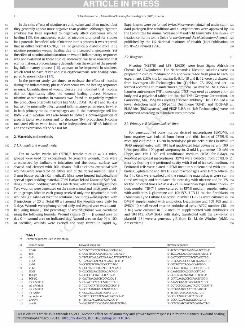
Table 1t1:1
t1:2 Primer sequences used in this study.
t1:3 Primer name Forward sequence Reverse sequence
t1:4 CD-68 5′-TGACCTGCTCTCTCTAAGGCTACA-3′ 5′-TCACGGTTGCAAGAGAAACATG-3′t1:5 TNF 5′-CATCTTCTCAAAATTCGAGTGACAA-3′ 5′-TGGGAGTAGACAAGGTACAACCC-3′t1:6 IL-6 5′-TTCAACCAAGAGGTAAAAGATTTACATAA-3′ 5′-CACTCCTTCTGTGACTCCAGCTT-3′t1:7 IL-12 5′-TGAGAACTACAGCACCAGCTTCTT-3′ 5′-CTTCAAAGGCTTCATCTGCAAGT-3′t1:8 IL-10 5′-GCTCTTACTGACTGGCATGAG-3′ 5′-CGCAGCTCTAGGAGCATGTG-3′t1:9 VEGF 5′-GCTTTACTGCTGTACCTCCACCA-3′ 5′-GGGACTTCTGCTCTCCTTCTGTC-3′t1:10 PDGF 5′-CGCCTGCAAGTGTGAGACAG-3′ 5′-GAATGGTCACCCGAGCTTGA-3′t1:11 TGF-β1 5′-GCCCTTCCTGCTCCTCATG-3′ 5′-CCGCACACAGCAGTTCTTCTC-3′t1:12 TGF-β2 5′-GACTTAACATCTCCCACCCA-3′ 5′-CCATCAATACCTGCAAATCTCG-3′t1:13 α7 nAchR-1 5′-CACATTCCACACCAACGTCTT-3′ 5′-AAAAGGGAACCAGCGTACATC-3′t1:14 α7 nAchR-2 5′-TGCTGGTATTCTTGCTGCCTGC-3′ 5′-GGTGCTGGCGAAGTACTGTGCTAT-3′t1:15 α7 nAchR-3 5′-GCCTAAGTGGACCAGGATCA-3′ 5′-CTCGGAAGCCAATGTAGAGC-3′t1:16 β2 nAchR 5′-GGGCAGGCACACTATTCTTC-3′ 5′-TCCAATCCTCCCTCACACTC-3′t1:17 cyclophilin 5′-TTCCTCCTTTCACAGAATTATTCCA-3′ 5′-CCGCCAGTGCCATTATGG-3′t1:18 GAPDH 5′-TTCACCACCATGGAGAAGGC-3′ 5′-GGCATGGACTGTGGTCATGA-3′t1:19 β-actin 5′-GACAGGATGCAGAAGGAGATTACTG-3′ 5′-CCACCGATCCACACAGAGTACTT-3′
2 S. Xanthoulea et al. / International Immunopharmacology xxx (2013) xxx–xxx
Please cite this article as: Xanthoulea S, et al, Nicotine effect on inflammatory and growth factor responses in murine cutaneous wound healing,Int Immunopharmacol (2013), http://dx.doi.org/10.1016/j.intimp.2013.10.022
UNCO
RRECTED P
RO
OF
NMS CD-68 IL-12 TNF IL-6 IL-100
2
4
6
25
50
*
** *
**
wounded
rela
tive
expr
essi
on (a
.u.)
rela
tive
expr
essi
on (a
.u.)
rela
tive
expr
essi
on (a
.u.)
rela
tive
expr
essi
on (a
.u.)
NMS VEGF PDGF TGF- 1 TGF- 20
1
2
3
wounded
*
saline 10-4M10-8M
d0
d1
d2
d3
sal. M
-810
M-4
10
sal. M
-81 0
M-4
1 0
sal M
-810
M-4
10
sal . M
-810
M-4
10
sal . M
-810
M-4
10
0
2
4
6
30
60
90
CD-68 IL-6 IL-10IL-12 TNF
NMS
*
sal. M
-810
M-4
10
sal . M
-810
M-4
10
sal M
-810
M-4
1 0
sal. M
-810
M-4
10
0.0
0.5
1.5
2.0
2.5
VEGF TGF- 2PDGF TGF- 1
NMS *
*
* **
0.07
0.08
time after wounding (days)
wou
nd c
losu
re (%
)
0
20
40
60
80
100saline
nic. 10-8 M
nic. 10-4 M
0 1 2 3
A B
C D
E F
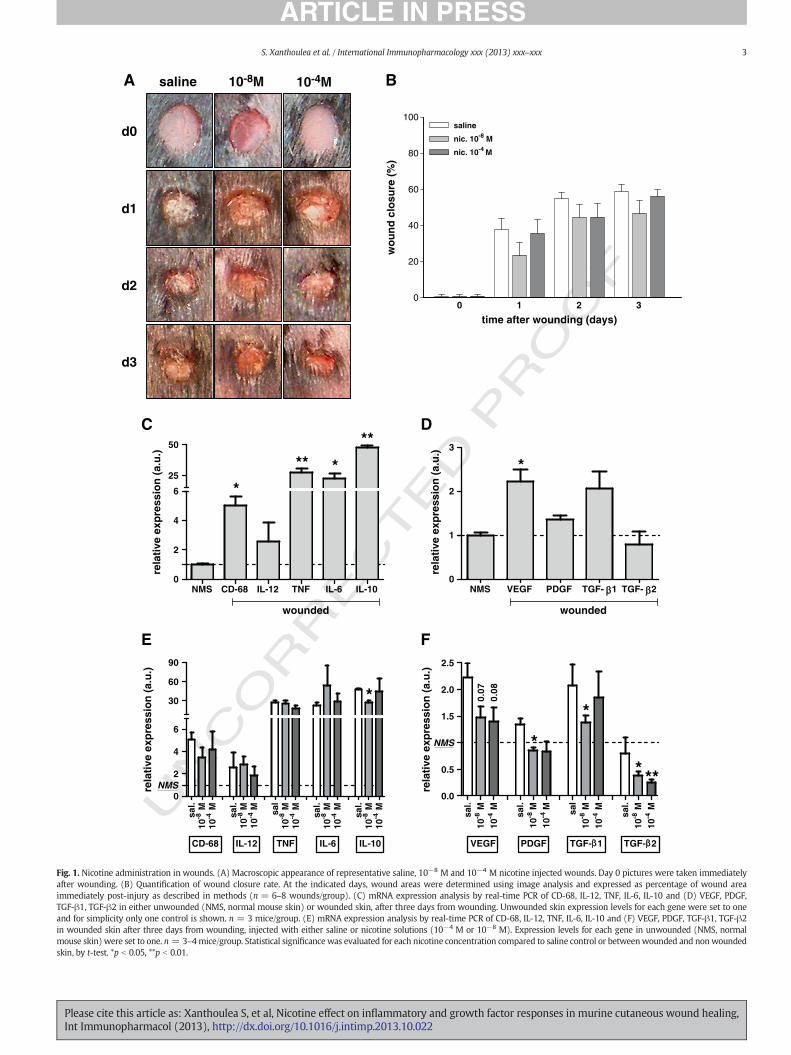
Fig. 1. Nicotine administration in wounds. (A) Macroscopic appearance of representative saline, 10−8 M and 10−4 M nicotine injected wounds. Day 0 pictures were taken immediatelyafter wounding. (B) Quantification of wound closure rate. At the indicated days, wound areas were determined using image analysis and expressed as percentage of wound areaimmediately post-injury as described in methods (n = 6–8 wounds/group). (C) mRNA expression analysis by real-time PCR of CD-68, IL-12, TNF, IL-6, IL-10 and (D) VEGF, PDGF,TGF-β1, TGF-β2 in either unwounded (NMS, normal mouse skin) or wounded skin, after three days from wounding. Unwounded skin expression levels for each gene were set to oneand for simplicity only one control is shown. n = 3 mice/group. (E) mRNA expression analysis by real-time PCR of CD-68, IL-12, TNF, IL-6, IL-10 and (F) VEGF, PDGF, TGF-β1, TGF-β2in wounded skin after three days from wounding, injected with either saline or nicotine solutions (10−4 M or 10−8 M). Expression levels for each gene in unwounded (NMS, normalmouse skin) were set to one. n = 3–4mice/group. Statistical significance was evaluated for each nicotine concentration compared to saline control or betweenwounded and nonwoundedskin, by t-test. *p b 0.05, **p b 0.01.
3S. Xanthoulea et al. / International Immunopharmacology xxx (2013) xxx–xxx
Please cite this article as: Xanthoulea S, et al, Nicotine effect on inflammatory and growth factor responses in murine cutaneous wound healing,Int Immunopharmacol (2013), http://dx.doi.org/10.1016/j.intimp.2013.10.022
UNCO
RRECTED P
RO
OF
167 2.4. RNA isolation and quantitative gene expression
168 RNA was isolated either with the RNeasy Fibrous Tissue kit (Qiagen169 GmbH, Hilden, Germany) for mouse skin tissue or with Trizol (Sigma–170 Aldrich Chemie BV) for cell monolayers. Residual DNA was digested171 with the RQ1 RNase-free DNase (Promega GmbH, Mannheim,172 Germany) and cDNA synthesis was performed using the iScriptTM
173 cDNA synthesis kit (Bio-Rad, Hercules, CA, USA). Quantitative PCR was174 performed in 10 ng of cDNA, with 1 × Absolute qPCR SYBR Green Fluo-175 rescein Mix (Westburg, Leusden, The Netherlands) and 150 nM of gene176 specific forward and reverse primers. Cyclophilin A and β-actin were177 used as housekeeping genes. Primer sequences are indicated in Table 1.
178 2.5. MTT assay, luciferase activity and ELISA
179 For MTT (3-(4,5-dimethylthiazol-2-yl)-2,5-diphenyl tetrazolium180 bromide; Sigma–Aldrich Chemie BV) assay, 105 cells/well of BMDM,181 RAW 264.7, PEC or 5 × 10⁴ cells/well of SVEC4-10 or 104 cells/well of182 3 T3-L1 cells were plated in 96-well plates overnight and stimulated183 the following day with nicotine and/or LPS (100 ng/ml) for 24 h.184 Nicotine was added to cell cultures 30 min before LPS. MTT assay was185 performed after 24 h by addition of MTT solution to a final concentra-186 tion of 0.5 mg/ml, for 2 h. Dye was solubilized with DMSO (Sigma–187 Aldrich Chemie BV) and absorbance was measured at 570 nm. Data188 represent mean ± SEM of 4 independent experiments performed in189 triplicate.190 For luciferase activity, RAW 264.7 cells stably transfected with the191 3x-κB-luc plasmid were plated at a density of 105 cells/well in 96 well192 plates and stimulated with the indicated compounds for the indicated
193times. Cells were lysed in lysis buffer (Promega GmbH) for 20 min194and 10 μl lysate was added to 50 μl luciferin (Steady-Glo Luciferase195assay system, Promega GmbH). Luciferase activity was measured196with a Lumac Biocounter M1500 luminometer (Promega GmbH). Data197represent mean ± SEM of 2 independent experiments performed in198quadruplicate.199For ELISA, 2 × 10⁵ 3 T3-L1 fibroblast cells or 5 × 10⁵macrophages or200SVEC4-10 endothelial cells were plated in 500 μl medium in triplicate/201condition in 24-well plates and stimulated for 6 or 24 h with nicotine202and/or LPS. Supernatantswere analyzed by ELISA. Since significant levels203of growth factors are present in bovine serum used in tissue culture204medium, for quantification of VEGF, PDGF-AB and TGF-β1 in macro-205phage supernatants, medium was changed to Optimem-1 (Gibco-BRL)206overnight and cellswere stimulated the followingmorningwith nicotine207for 6 or 24 h. Experiments were performed in triplicate and data208represent mean ± SEM of 3 independent experiments.
2092.6. Statistical analysis
210Statistical analyseswere performed using Graphpad Prism (Graphpad211Software) or SigmaPlot statistical tests. Data are expressed as means ±212SEM. A p b 0.05 is considered statistically significant.
2133. Results
2143.1. Nicotine down-regulates growth factor expression in skin wounds
215The initial response upon a cutaneous injury is characterized by a216strong inflammatory reaction with induction of different inflammatory
0
2
4
6
0
25
50
no nic. 10-8 10-6 10-4
*
BMDMRPMRAW
**
***
**
**
LPS (100 ng/ml)
nicotine concentration (M)
no nic. 10-8 10-6 10-4
LPS (100 ng/ml)
nicotine concentration (M)
no nic. 10-8 10-6 10-4
LPS (100 ng/ml)
nicotine concentration (M)
no nic. 10-8 10-6 10-4
LPS (100 ng/ml)
nicotine concentration (M)
TNF-
α (n
g/m
l)
for
BM
DM
and
RP
M TNF-α
(ng/ml)
for RA
W
0
10
20
30
40
50
0.0
2.5
5.0
7.5
RPMRAW
BMDM
IL-6
(ng/
ml)
for
BM
DM
and
RA
W
IL-1
2 (p
g/m
l)fo
r B
MD
M a
nd R
AW
IL-6 (ng/ml)
for RP
MIL-12 (pg/m
l)for R
PM
0
500
1000
1500
0
100
200
RPMRAW
BMDMIL-1
0 (p
g/m
l)fo
r B
MD
M a
nd R
PM
IL-10 (pg/ml)
for RA
W
0
750
1500
0
100
200
10000
14000
RPMRAW
BMDM
A B
C D
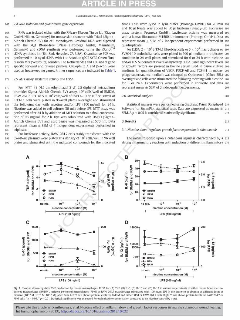
Fig. 2. Nicotine down-regulates TNF production by mouse macrophages. ELISA for (A) TNF, (B) IL-6, (C) IL-10 and (D) IL-12 in culture supernatants of either mouse bone marrowderived macrophages (BMDM), resident peritoneal macrophages (RPM) or RAW 264.7 macrophages stimulated with 100 ng/ml LPS in the presence or absence of different doses ofnicotine (10−4 M, 10−6 M, 10−8 M), after 24 h. Left Y axis shows protein levels for BMDM and either RPM or RAW 264.7 cells. Right Y axis shows protein levels for RAW 264.7 orRPM cells. * p b 0.05, **p b 0.01. Statistical significance was evaluated for each nicotine concentration compared to no nicotine control by t-test.
4 S. Xanthoulea et al. / International Immunopharmacology xxx (2013) xxx–xxx
Please cite this article as: Xanthoulea S, et al, Nicotine effect on inflammatory and growth factor responses in murine cutaneous wound healing,Int Immunopharmacol (2013), http://dx.doi.org/10.1016/j.intimp.2013.10.022
UNCO
RRECTED P
RO
OF
217 mediators. Since nicotine was previously shown to negatively regulate218 inflammation [6–9], and to promote wound healing in normal [14]219 and genetically diabetic mice [15], we aimed to examine the effect of220 nicotine administration during the inflammatory phase of cutaneous221 wound healing in mice. Full thickness excisional wounds were generat-222 ed on the dorsumof C57BL/6mice andmicewere divided in groups that223 received either saline or nicotine solutions. Saline or nicotine solutions224 at two different concentrations (10−4 M or 10−8 M) were injected225 around the wounds daily for three days, similar to other studies [14]226 and as described in the methods section. Quantification of wound area227 indicated a not significant delay in wound closure in the nicotine228 injected wounds compared to saline controls (Fig. 1 A and B). After229 three days, real time PCR analysis of RNA isolated from either wounded230 mice or non-wounded controls (normal mouse skin, NMS) was per-231 formed. As shown in Fig. 1 C, wounded skin exhibits a strong inflamma-232 tory response which is absent in non-wounded controls. CD-68 was233 over five-fold increased indicating extensive macrophage infiltration234 and expression of different inflammatory mediators (TNF, IL-6, IL-10)235 was also found several fold increased (Fig. 1C). A milder effect of236 wounding on expression of growth factors was observed (Fig. 1D).237 Nicotine injections resulted in significant mild down-regulation in238 the expression of IL-10 in the wounds of 10−8 M nicotine injected239 wounds compared to saline controls (Fig. 1E). Interestingly, nicotine ad-240 ministration indicated a clear down-regulation in themRNA expression241 of growth factors, with PDGF, TGF-β1 and TGF-β2 showing significant242 differences while VEGF expression had a similar trend but differences243 were borderline not significant (Fig. 1F).
2443.2. Nicotine down-regulates TNF and growth factor expression in245mouse macrophages
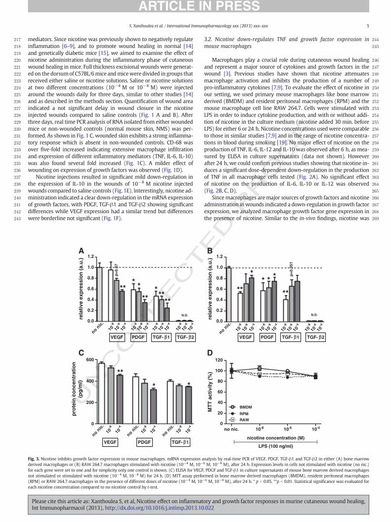
246Macrophages play a crucial role during cutaneous wound healing247and represent a major source of cytokines and growth factors in the248wound [3]. Previous studies have shown that nicotine attenuates249macrophage activation and inhibits the production of a number of250pro-inflammatory cytokines [7,9]. To evaluate the effect of nicotine in251our setting, we used primary mouse macrophages like bone marrow252derived (BMDM) and resident peritoneal macrophages (RPM) and the253mouse macrophage cell line RAW 264.7. Cells were stimulated with254LPS in order to induce cytokine production, and with or without addi-255tion of nicotine in the culture medium (nicotine added 30 min. before256LPS) for either 6 or 24 h. Nicotine concentrations usedwere comparable257to those in similar studies [7,9] and in the range of nicotine concentra-258tions in blood during smoking [19]. No major effect of nicotine on the259production of TNF, IL-6, IL-12 and IL-10 was observed after 6 h, as mea-260sured by ELISA in culture supernatants (data not shown). However261after 24 h, we could confirm previous studies showing that nicotine in-262duces a significant dose-dependent down-regulation in the production263of TNF in all macrophage cells tested (Fig. 2A). No significant effect264of nicotine on the production of IL-6, IL-10 or IL-12 was observed265(Fig. 2B, C, D).266Sincemacrophages aremajor sources of growth factors and nicotine267administration inwounds indicated a down-regulation in growth factor268expression, we analyzed macrophage growth factor gene expression in269the presence of nicotine. Similar to the in-vivo findings, nicotine was
0
20
40
60
80
100
120
10-8 10-6no nic. 10-4
BMDM
RAWRPM
LPS (100 ng/ml)
nicotine concentration (M)
MTT
act
ivity
(%)
0.0
0.2
0.4
0.6
0.8
1.0
1.2
nonic.
10-8
VEGF
10-6
10-4
10-8
PDGF
10-6
10-4
10-8
TGF- 1
10-6
10-4
10-8
TGF- 2
10-6
10-4
N.D.
p=0.
07
*** *
*****
**
rela
tive
expr
essi
on (a
.u.)
0.0
0.2
0.4
0.6
0.8
1.0
1.2
nonic.
10-8
VEGF
10-6
10-4
10-8
PDGF
10-6
10-4
10-8
TGF- 1
10-6
10-4
10-8
TGF- 2
10-6
10-4
N.D.
**
*
**
* * * p=0.
051
rela
tive
expr
essi
on (a
.u.)
0
200
400
600
10-8
VEGF
10-4
PDGF TGF- 1no
nic.10
-8
10-4
nonic.
10-8
10-4
nonic.
**
* *
prot
ein
conc
entr
atio
n(p
g/m
l)
A B
C D
Fig. 3. Nicotine inhibits growth factor expression in mouse macrophages. mRNA expression analysis by real-time PCR of VEGF, PDGF, TGF-β1 and TGF-β2 in either (A) bone marrowderived macrophages or (B) RAW 264.7 macrophages stimulated with nicotine (10−4 M, 10−6 M, 10−8 M), after 24 h. Expression levels in cells not stimulated with nicotine (no nic.)for each gene were set to one and for simplicity only one control is shown. (C) ELISA for VEGF, PDGF and TGF-β1 in culture supernatants of mouse bone marrow derived macrophagesnot stimulated or stimulated with nicotine (10−4 M, 10−8 M) for 24 h. (D) MTT assay performed in bone marrow derived macrophages (BMDM), resident peritoneal macrophages(RPM) or RAW 264.7 macrophages in the presence of different doses of nicotine (10−4 M, 10−6 M, 10−8 M), after 24 h. * p b 0.05, **p b 0.01. Statistical significance was evaluated foreach nicotine concentration compared to no nicotine control by t-test.
5S. Xanthoulea et al. / International Immunopharmacology xxx (2013) xxx–xxx
Please cite this article as: Xanthoulea S, et al, Nicotine effect on inflammatory and growth factor responses in murine cutaneous wound healing,Int Immunopharmacol (2013), http://dx.doi.org/10.1016/j.intimp.2013.10.022
UNCO
RRECTED P
RO
OF
270 found to down-regulate mRNA levels of VEGF, PDGF and TGF-β1 in271 BMDM and RAW 264.7 macrophages (Fig. 3A and B respectively)272 (TGF-β2 levels were undetectable). A comparable down-regulation in273 the protein levels of VEGF, PDGF and TGF-β1 was measured in BMDM274 culture supernatants after 24 h of incubation with nicotine (Fig. 3C).275 Expression levels were below detection limit in supernatants of RAW276 264.7 cells. Cell viability measurement by tetrazolium salt MTT assay277 showed that concentrations of nicotine used did not result in significant278 differences in cell survival and therefore the observed effects were not279 due to cytotoxicity (Fig. 3D).
280 3.3. Nicotine does not significantly affect NF-κB transcriptional activity in281 mouse macrophages
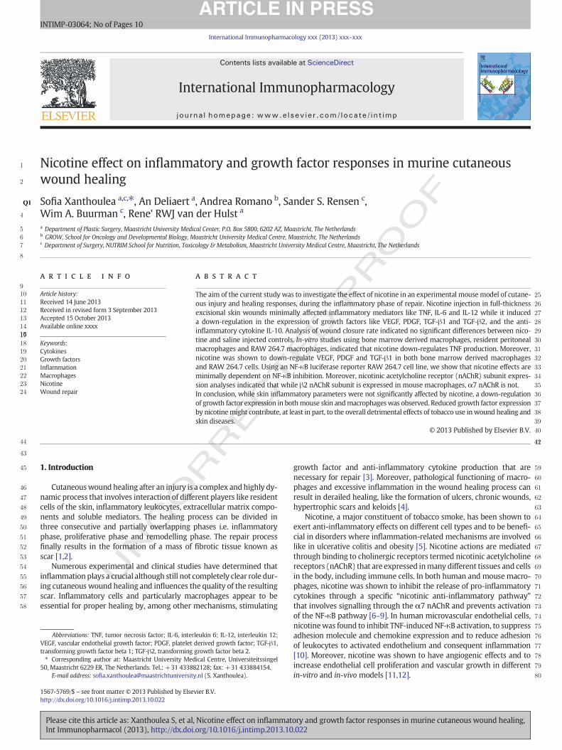
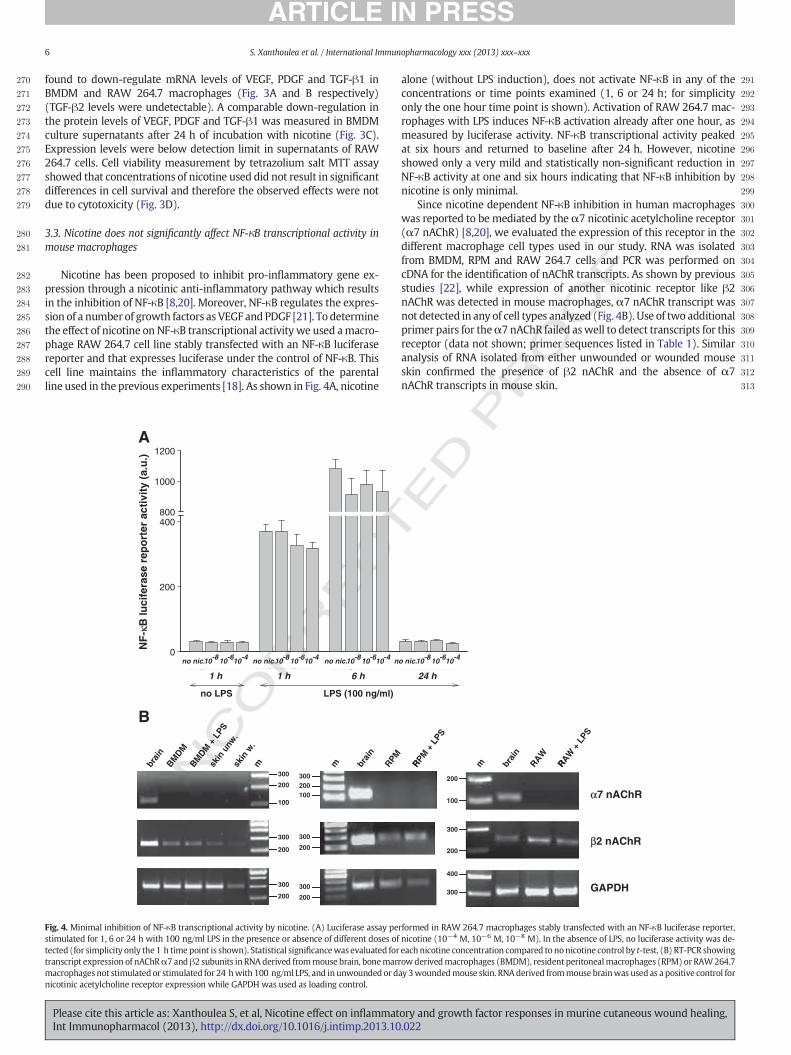
282 Nicotine has been proposed to inhibit pro-inflammatory gene ex-283 pression through a nicotinic anti-inflammatory pathway which results284 in the inhibition of NF-κB [8,20]. Moreover, NF-κB regulates the expres-285 sion of a number of growth factors as VEGF and PDGF [21]. To determine286 the effect of nicotine on NF-κB transcriptional activity we used amacro-287 phage RAW 264.7 cell line stably transfected with an NF-κB luciferase288 reporter and that expresses luciferase under the control of NF-κB. This289 cell line maintains the inflammatory characteristics of the parental290 line used in the previous experiments [18]. As shown in Fig. 4A, nicotine
291alone (without LPS induction), does not activate NF-κB in any of the292concentrations or time points examined (1, 6 or 24 h; for simplicity293only the one hour time point is shown). Activation of RAW 264.7 mac-294rophages with LPS induces NF-κB activation already after one hour, as295measured by luciferase activity. NF-κB transcriptional activity peaked296at six hours and returned to baseline after 24 h. However, nicotine297showed only a very mild and statistically non-significant reduction in298NF-κB activity at one and six hours indicating that NF-κB inhibition by299nicotine is only minimal.300Since nicotine dependent NF-κB inhibition in human macrophages301was reported to be mediated by the α7 nicotinic acetylcholine receptor302(α7 nAChR) [8,20], we evaluated the expression of this receptor in the303different macrophage cell types used in our study. RNA was isolated304from BMDM, RPM and RAW 264.7 cells and PCR was performed on305cDNA for the identification of nAChR transcripts. As shown by previous306studies [22], while expression of another nicotinic receptor like β2307nAChR was detected in mouse macrophages, α7 nAChR transcript was308not detected in any of cell types analyzed (Fig. 4B). Use of two additional309primer pairs for theα7 nAChR failed aswell to detect transcripts for this310receptor (data not shown; primer sequences listed in Table 1). Similar311analysis of RNA isolated from either unwounded or wounded mouse312skin confirmed the presence of β2 nAChR and the absence of α7313nAChR transcripts in mouse skin.
NF-
κB lu
cife
rase
rep
orte
r ac
tivity
(a.u
.)
0
200
400800
1000
1200
no LPS LPS (100 ng/ml)
10-410-610-8no nic.10-410-610-8no nic. 10-410-610-8no nic.10-410-610-8no nic.
1 h 1 h 6 h 24 h
m
α7 nAChR
β2 nAChR
GAPDH
brai
n
RAW
Rmbrai
nBM
DMBM
DM +
LPS
skin
unw
.sk
in w
.
200
300
300
200
200
300
100
200
300
200
300
200300
100
RAW +
LPS
brai
n
RPM
Rm RPM +
LPS
300
400
200
300
100
200
A
B
Fig. 4. Minimal inhibition of NF-κB transcriptional activity by nicotine. (A) Luciferase assay performed in RAW 264.7 macrophages stably transfected with an NF-κB luciferase reporter,stimulated for 1, 6 or 24 h with 100 ng/ml LPS in the presence or absence of different doses of nicotine (10−4 M, 10−6 M, 10−8 M). In the absence of LPS, no luciferase activity was de-tected (for simplicity only the 1 h time point is shown). Statistical significancewas evaluated for each nicotine concentration compared to nonicotine control by t-test. (B) RT-PCR showingtranscript expression of nAChRα7 andβ2 subunits in RNA derived frommouse brain, bonemarrowderivedmacrophages (BMDM), resident peritonealmacrophages (RPM) or RAW264.7macrophages not stimulated or stimulated for 24 hwith 100 ng/ml LPS, and inunwounded orday 3woundedmouse skin. RNAderived frommouse brainwas used as a positive control fornicotinic acetylcholine receptor expression while GAPDH was used as loading control.
6 S. Xanthoulea et al. / International Immunopharmacology xxx (2013) xxx–xxx
Please cite this article as: Xanthoulea S, et al, Nicotine effect on inflammatory and growth factor responses in murine cutaneous wound healing,Int Immunopharmacol (2013), http://dx.doi.org/10.1016/j.intimp.2013.10.022
UNCO
RRECTED P
RO
OF
314 3.4. Nicotine effect on endothelial and fibroblast cells
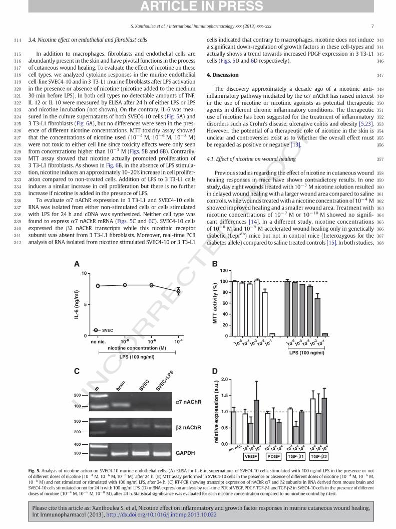
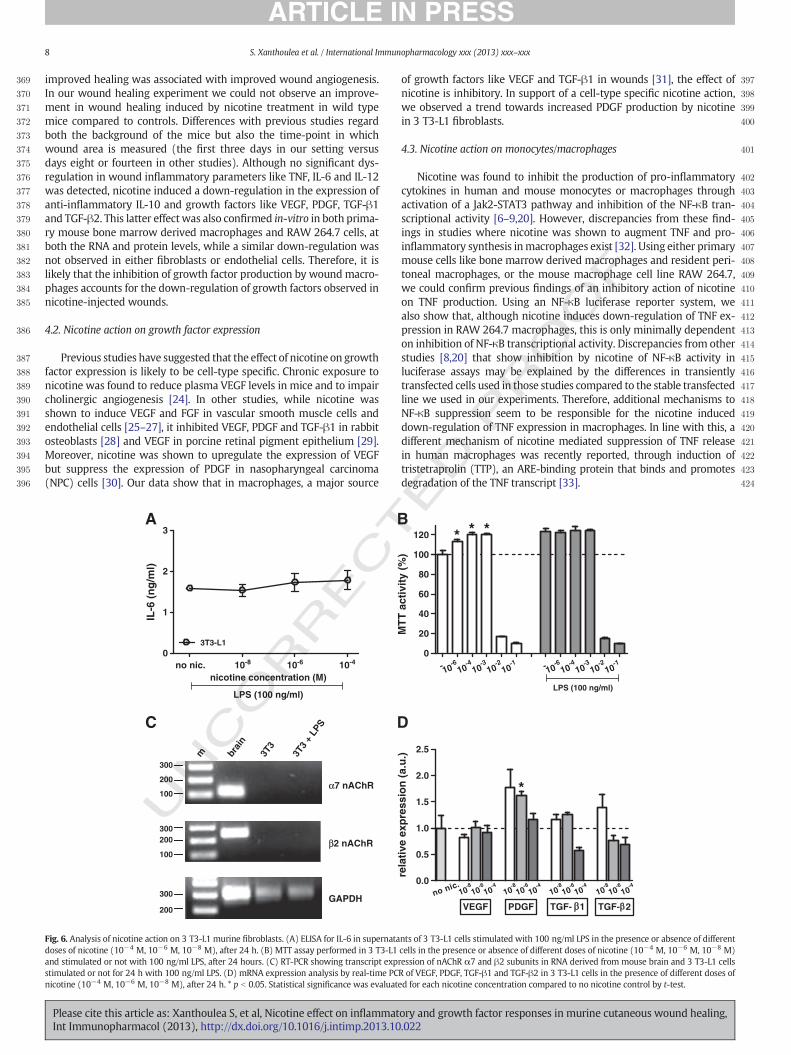
315 In addition to macrophages, fibroblasts and endothelial cells are316 abundantly present in the skin and have pivotal functions in the process317 of cutaneous wound healing. To evaluate the effect of nicotine on these318 cell types, we analyzed cytokine responses in the murine endothelial319 cell-line SVEC4-10 and in 3 T3-L1murinefibroblasts after LPS activation320 in the presence or absence of nicotine (nicotine added to the medium321 30 min before LPS). In both cell types no detectable amounts of TNF,322 IL-12 or IL-10 were measured by ELISA after 24 h of either LPS or LPS323 and nicotine incubation (not shown). On the contrary, IL-6 was mea-324 sured in the culture supernatants of both SVEC4-10 cells (Fig. 5A) and325 3 T3-L1 fibroblasts (Fig. 6A), but no differences were seen in the pres-326 ence of different nicotine concentrations. MTT toxicity assay showed327 that the concentrations of nicotine used (10−4 M, 10−6 M, 10−8 M)328 were not toxic to either cell line since toxicity effects were only seen329 from concentrations higher than 10−3 M (Figs. 5B and 6B). Contrarily,330 MTT assay showed that nicotine actually promoted proliferation of331 3 T3-L1 fibroblasts. As shown in Fig. 6B, in the absence of LPS stimula-332 tion, nicotine induces an approximately 10–20% increase in cell prolifer-333 ation compared to non-treated cells. Addition of LPS to 3 T3-L1 cells334 induces a similar increase in cell proliferation but there is no further335 increase if nicotine is added in the presence of LPS.336 To evaluate α7 nAChR expression in 3 T3-L1 and SVEC4-10 cells,337 RNA was isolated from either non-stimulated cells or cells stimulated338 with LPS for 24 h and cDNA was synthesized. Neither cell type was339 found to express α7 nAChR mRNA (Figs. 5C and 6C). SVEC4-10 cells340 expressed the β2 nAChR transcripts while this nicotinic receptor341 subunit was absent from 3 T3-L1 fibroblasts. Moreover, real-time PCR342 analysis of RNA isolated from nicotine stimulated SVEC4-10 or 3 T3-L1
343cells indicated that contrary to macrophages, nicotine does not induce344a significant down-regulation of growth factors in these cell-types and345actually shows a trend towards increased PDGF expression in 3 T3-L1346cells (Figs. 5D and 6D respectively).
3474. Discussion
348The discovery approximately a decade ago of a nicotinic anti-349inflammatory pathway mediated by the α7 nAChR has raised interest350in the use of nicotine or nicotinic agonists as potential therapeutic351agents in different chronic inflammatory conditions. The therapeutic352use of nicotine has been suggested for the treatment of inflammatory353disorders such as Crohn's disease, ulcerative colitis and obesity [5,23].354However, the potential of a therapeutic role of nicotine in the skin is355unclear and controversies exist as to whether the overall effect must356be regarded as positive or negative [13].
3574.1. Effect of nicotine on wound healing
358Previous studies regarding the effect of nicotine in cutaneouswound359healing responses in mice have shown contradictory results. In one360study, day eightwounds treatedwith 10−3 Mnicotine solution resulted361in delayed wound healing with a larger wound area compared to saline362controls, whilewounds treatedwith a nicotine concentration of 10−4 M363showed improved healing and a smaller wound area. Treatment with364nicotine concentrations of 10−7 M or 10−10 M showed no signifi-365cant differences [14]. In a different study, nicotine concentrations366of 10−8 M and 10−9 M accelerated wound healing only in genetically367diabetic (Leprdb) mice but not in control mice (heterozygous for the368diabetes allele) compared to saline treated controls [15]. In both studies,
0
5
10
no nic. 10-8 10-6 10-4
SVEC
LPS (100 ng/ml)
nicotine concentration (M)
IL-6
(ng/
ml)
- -610
-410
-310
-210
-110 - -6
10-4
10-3
10-2
10-1
10
0
20
40
60
80
100
120
LPS (100 ng/ml)
MTT
act
ivity
(%)
α7 nAChR
β2 nAChR
GAPDH
b S Sm brai
n
SVEC
SVEC+LPS
300
400
200
300
100
200
m
0.0
0.5
1.0
1.5
2.0
no nic.10
-8
VEGF
10-6
10-4
PDGF TGF- 1 TGF- 2
10-8
10-6
10-4
10-8
10-6
10-4
10-8
10-6
10-4
rela
tive
expr
essi
on (a
.u.)
A B
C D
Fig. 5. Analysis of nicotine action on SVEC4-10 murine endothelial cells. (A) ELISA for IL-6 in supernatants of SVEC4-10 cells stimulated with 100 ng/ml LPS in the presence or notof different doses of nicotine (10−4 M, 10−6 M, 10−8 M), after 24 h. (B) MTT assay performed in SVEC4-10 cells in the presence or absence of different doses of nicotine (10−4 M, 10−6 M,10−8 M) and not stimulated or stimulated with 100 ng/ml LPS, after 24 h. (C) RT-PCR showing transcript expression of nAChR α7 and β2 subunits in RNA derived from mouse brain andSVEC4-10 cells stimulated or not for 24 hwith 100 ng/ml LPS. (D)mRNA expression analysis by real-time PCR of VEGF, PDGF, TGF-β1 and TGF-β2 in SVEC4-10 cells in the presence of differentdoses of nicotine (10−4 M, 10−6 M, 10−8 M), after 24 h. Statistical significance was evaluated for each nicotine concentration compared to no nicotine control by t-test.
7S. Xanthoulea et al. / International Immunopharmacology xxx (2013) xxx–xxx
Please cite this article as: Xanthoulea S, et al, Nicotine effect on inflammatory and growth factor responses in murine cutaneous wound healing,Int Immunopharmacol (2013), http://dx.doi.org/10.1016/j.intimp.2013.10.022
UNCO
RRECTED P
RO
OF
369 improved healing was associated with improved wound angiogenesis.370 In our wound healing experiment we could not observe an improve-371 ment in wound healing induced by nicotine treatment in wild type372 mice compared to controls. Differences with previous studies regard373 both the background of the mice but also the time-point in which374 wound area is measured (the first three days in our setting versus375 days eight or fourteen in other studies). Although no significant dys-376 regulation in wound inflammatory parameters like TNF, IL-6 and IL-12377 was detected, nicotine induced a down-regulation in the expression of378 anti-inflammatory IL-10 and growth factors like VEGF, PDGF, TGF-β1379 and TGF-β2. This latter effect was also confirmed in-vitro in both prima-380 ry mouse bone marrow derived macrophages and RAW 264.7 cells, at381 both the RNA and protein levels, while a similar down-regulation was382 not observed in either fibroblasts or endothelial cells. Therefore, it is383 likely that the inhibition of growth factor production by woundmacro-384 phages accounts for the down-regulation of growth factors observed in385 nicotine-injected wounds.
386 4.2. Nicotine action on growth factor expression
387 Previous studies have suggested that the effect of nicotine on growth388 factor expression is likely to be cell-type specific. Chronic exposure to389 nicotine was found to reduce plasma VEGF levels in mice and to impair390 cholinergic angiogenesis [24]. In other studies, while nicotine was391 shown to induce VEGF and FGF in vascular smooth muscle cells and392 endothelial cells [25–27], it inhibited VEGF, PDGF and TGF-β1 in rabbit393 osteoblasts [28] and VEGF in porcine retinal pigment epithelium [29].394 Moreover, nicotine was shown to upregulate the expression of VEGF395 but suppress the expression of PDGF in nasopharyngeal carcinoma396 (NPC) cells [30]. Our data show that in macrophages, a major source
397of growth factors like VEGF and TGF-β1 in wounds [31], the effect of398nicotine is inhibitory. In support of a cell-type specific nicotine action,399we observed a trend towards increased PDGF production by nicotine400in 3 T3-L1 fibroblasts.
4014.3. Nicotine action on monocytes/macrophages
402Nicotine was found to inhibit the production of pro-inflammatory403cytokines in human and mouse monocytes or macrophages through404activation of a Jak2-STAT3 pathway and inhibition of the NF-κB tran-405scriptional activity [6–9,20]. However, discrepancies from these find-406ings in studies where nicotine was shown to augment TNF and pro-407inflammatory synthesis inmacrophages exist [32]. Using either primary408mouse cells like bone marrow derived macrophages and resident peri-409toneal macrophages, or the mouse macrophage cell line RAW 264.7,410we could confirm previous findings of an inhibitory action of nicotine411on TNF production. Using an NF-κB luciferase reporter system, we412also show that, although nicotine induces down-regulation of TNF ex-413pression in RAW 264.7 macrophages, this is only minimally dependent414on inhibition of NF-κB transcriptional activity. Discrepancies from other415studies [8,20] that show inhibition by nicotine of NF-κB activity in416luciferase assays may be explained by the differences in transiently417transfected cells used in those studies compared to the stable transfected418line we used in our experiments. Therefore, additional mechanisms to419NF-κB suppression seem to be responsible for the nicotine induced420down-regulation of TNF expression in macrophages. In line with this, a421different mechanism of nicotine mediated suppression of TNF release422in human macrophages was recently reported, through induction of423tristetraprolin (TTP), an ARE-binding protein that binds and promotes424degradation of the TNF transcript [33].
0
1
2
3
no nic. 10-8 10-6 10-4
3T3-L1
LPS (100 ng/ml)
nicotine concentration (M)
IL-6
(ng/
ml)
- -610
-410
-310
-210
-110 - -6
10-4
10-3
10-2
10-1
10
0
20
40
60
80
100
120
LPS (100 ng/ml)
* * *
MTT
act
ivity
(%)
0.0
0.5
1.0
1.5
2.0
2.5
no nic.10
-8
VEGF
10-6
10-4
PDGF TGF- 1 TGF- 2
*
10-8
10-6
10-4
10-8
10-6
10-4
10-8
10-6
10-4
rela
tive
expr
essi
on (a
.u.)
α7 nAChR
β2 nAChR
GAPDH200
300
brai
n
3T3
3T3
+ LP
S
m
200300
100
200
300
100
A B
C D
Fig. 6. Analysis of nicotine action on 3 T3-L1 murine fibroblasts. (A) ELISA for IL-6 in supernatants of 3 T3-L1 cells stimulated with 100 ng/ml LPS in the presence or absence of differentdoses of nicotine (10−4 M, 10−6 M, 10−8 M), after 24 h. (B) MTT assay performed in 3 T3-L1 cells in the presence or absence of different doses of nicotine (10−4 M, 10−6 M, 10−8 M)and stimulated or not with 100 ng/ml LPS, after 24 hours. (C) RT-PCR showing transcript expression of nAChR α7 and β2 subunits in RNA derived from mouse brain and 3 T3-L1 cellsstimulated or not for 24 h with 100 ng/ml LPS. (D) mRNA expression analysis by real-time PCR of VEGF, PDGF, TGF-β1 and TGF-β2 in 3 T3-L1 cells in the presence of different doses ofnicotine (10−4 M, 10−6 M, 10−8 M), after 24 h. * p b 0.05. Statistical significance was evaluated for each nicotine concentration compared to no nicotine control by t-test.
8 S. Xanthoulea et al. / International Immunopharmacology xxx (2013) xxx–xxx
Please cite this article as: Xanthoulea S, et al, Nicotine effect on inflammatory and growth factor responses in murine cutaneous wound healing,Int Immunopharmacol (2013), http://dx.doi.org/10.1016/j.intimp.2013.10.022
UNCO
RRECTED P
RO
OF
425 4.4. Nicotinic acetylcholine receptor expression
426 The anti-inflammatory effect of nicotine is considered to bemediated427 by the α7 nAChR expressed by many different cell types ranging from428 neurons to immune cells. Regarding the skin, theα7 nAChR has been de-429 tected in the upper spinous and granular layers of human scalp epider-430 mis [34] and in skin of BALB/c mice, where α7 nAChR positive staining431 was observed in epidermis, hair follicles, sebaceous glands, endothelial432 cells, resident dermal fibroblasts, but also in inflammatory cells likemac-433 rophages and PMNs during skin wound healing [35]. However, concerns434 have been raised regarding the specificity of anti-α7 nAChR antibodies435 due to the discrepancies in the results between immunodetection436 data and mRNA or genotyping results in α7 nAChR deficient mice437 [22,36,37]. Using three different primer pairs, we were unable to detect438 mRNA of α7 nAChR in either unwounded or wounded skin in C57BL/6439 mice or in primary mouse cells or cell-lines. Our results are supported440 by similar studies that show presence of other nAChR transcripts like441 the β2 nAChR but absence of the α7 nAChR mRNA in mouse alveolar442 [38,39], intestinal, splenic or peritoneal macrophages [22]. Since α7443 nAChR has been detected in human monocytes, macrophages, endothe-444 lial cells [6,10,12] but not in the corresponding mouse cells, it is likely445 that expression patterns of nicotinic acetylcholine receptors may differ446 between human and mouse tissues or between different mouse strains,447 and comparison of different studies or translation of mouse studies to448 humans should be done with caution.449 Finally, to evaluate the effect of nicotine on additional cell types450 present in the skin, we have used the mouse vascular endothelial451 cell line SVEC4-10 and the 3 T3-L1 murine fibroblast cell line. In452 both cell types nicotine was found to have no effect on IL-6 secretion,453 the only cytokine among TNF, IL-12 and IL-10 that was produced454 after LPS stimulation. However, we observed that nicotine induced455 a mild increase in cell proliferation in 3 T3-L1 fibroblasts under456 non-LPS stimulated conditions. This effect is potentially mediated457 by nicotinic acetylcholine receptor subunits expressed by fibroblasts458 that are different fromα7 nAChR or β2 nAChR since expression anal-459 ysis indicated absence of these subunits in mouse 3 T3-L1 cells. Sim-460 ilar nicotine-induced stimulation in cell proliferation was noted by461 others in endothelial cells [40,41], bone cells [42], epithelial cells462 [43], and chondrocytes [44].463 Taken together, our results show that during the inflammatory464 phase of murine cutaneous wound healing, the main effect of nicotine465 administration was a negative regulation of growth factor expression,466 an effect which is likely to be due to reduced growth factor expression467 bywoundmacrophages. The inhibitory effect of nicotine on growth fac-468 tor production may reflect, to a certain degree, the damaging effects of469 smoking on the skin vasculature and oxygenation andmay provide crit-470 ical insight into the overall detrimental effects of tobacco use in wound471 healing and general skin diseases.
472 Funding
473 This studywas supported by internal funds of theMaastricht Univer-474 sity Medical Centre.
475 Acknowledgements
476 Wewish to thankDr.M.P.J. deWinther (AMC, AmsterdamUniversity)477 for providing us with the RAW 264.7 NF-κB luciferase reporter line and478 Dr. P.J. Lindsey (MUMC, Maastricht University) for help with statistical479 analysis.
480 References
481 [1] Singer AJ, Clark RA. Cutaneous wound healing. N Engl J Med 1999;341:738–46.482 [2] Gurtner GC, Werner S, Barrandon Y, Longaker MT. Wound repair and regeneration.483 Nature 2008;453:314–21.
484[3] Eming SA, Krieg T, Davidson JM. Inflammation in wound repair: molecular and485cellular mechanisms. J Invest Dermatol 2007;127:514–25.486[4] Mahdavian Delavary B, van der Veer WM, van Egmond M, Niessen FB, Beelen RH.487Macrophages in skin injury and repair. Immunobiology 2011;216:753–62.488[5] Lakhan SE, Kirchgessner A. Anti-inflammatory effects of nicotine in obesity and489ulcerative colitis. J Transl Med 2011;9:129.490[6] Wang H, Yu M, Ochani M, Amella CA, Tanovic M, Susarla S, et al. Nicotinic acetyl-491choline receptor alpha7 subunit is an essential regulator of inflammation. Nature4922003;421:384–8.493[7] Borovikova LV, Ivanova S, Zhang M, Yang H, Botchkina GI, Watkins LR, et al. Vagus494nerve stimulation attenuates the systemic inflammatory response to endotoxin.495Nature 2000;405:458–62.496[8] Wang H, Liao H, Ochani M, Justiniani M, Lin X, Yang L, et al. Cholinergic agonists497inhibit HMGB1 release and improve survival in experimental sepsis. Nat Med4982004;10:1216–21.499[9] de Jonge WJ, van der Zanden EP, The FO, Bijlsma MF, van Westerloo DJ, Bennink RJ,500et al. Stimulation of the vagus nerve attenuates macrophage activation by activating501the Jak2-STAT3 signaling pathway. Nat Immunol 2005;6:844–51.502[10] Saeed RW, Varma S, Peng-Nemeroff T, Sherry B, Balakhaneh D, Huston J, et al.503Cholinergic stimulation blocks endothelial cell activation and leukocyte recruitment504during inflammation. J Exp Med 2005;201:1113–23.505[11] Heeschen C, Jang JJ, Weis M, Pathak A, Kaji S, Hu RS, et al. Nicotine stimulates angio-506genesis and promotes tumor growth and atherosclerosis. Nat Med 2001;7:833–9.507[12] Heeschen C, Weis M, Aicher A, Dimmeler S, Cooke JP. A novel angiogenic path-508way mediated by non-neuronal nicotinic acetylcholine receptors. J Clin Invest5092002;110:527–36.510[13] Misery L. Nicotine effects on skin: are they positive or negative? Exp Dermatol5112004;13:665–70.512[14] Morimoto N, Takemoto S, Kawazoe T, Suzuki S. Nicotine at a low concentration513promotes wound healing. J Surg Res 2008;145:199–204.514[15] Jacobi J, Jang JJ, Sundram U, Dayoub H, Fajardo LF, Cooke JP. Nicotine accelerates515angiogenesis and wound healing in genetically diabetic mice. Am J Pathol5162002;161:97–104.517[16] Larson BJ, Longaker MT, Lorenz HP. Scarless fetal wound healing: a basic science518review. Plast Reconstr Surg 2010;126:1172–80.519[17] Deliaert AE, Van den Kerckhove E, Tuinder S, Noordzij SM, Dormaar TS, van der Hulst520RR. Smoking and its effect on scar healing. Eur J Plast Surg 2012;35:421–4.521[18] Carlsen H, Moskaug JO, Fromm SH, Blomhoff R. In vivo imaging of NF-kappa B522activity. J Immunol 2002;168:1441–6.523[19] Henningfield JE, Stapleton JM, Benowitz NL, Grayson RF, London ED. Higher levels524of nicotine in arterial than in venous blood after cigarette smoking. Drug Alcohol525Depend 1993;33:23–9.526[20] Yoshikawa H, KurokawaM, Ozaki N, Nara K, Atou K, Takada E, et al. Nicotine inhibits527the production of proinflammatorymediators in humanmonocytes by suppression of528I-kappaB phosphorylation and nuclear factor-kappaB transcriptional activity through529nicotinic acetylcholine receptor alpha7. Clin Exp Immunol 2006;146:116–23.530[21] Gilmore L. NF-kB Target Genes. Available: http://www.bu.edu/nf-kb/gene-resources/531target-genes/.532[22] van der Zanden EP, Snoek SA, Heinsbroek SE, Stanisor OI, Verseijden C, Boeckxstaens533GE, et al. Vagus nerve activity augments intestinal macrophage phagocytosis via534nicotinic acetylcholine receptor alpha4beta2. Gastroenterology 2009;137:1029–39535(39 e1–4).536[23] Ulloa L. The vagus nerve and the nicotinic anti-inflammatory pathway. Nat Rev Drug537Discov 2005;4:673–84.538[24] Konishi H, Wu J, Cooke JP. Chronic exposure to nicotine impairs cholinergic angio-539genesis. Vasc Med 2010;15:47–54.540[25] Conklin BS, Zhao W, Zhong DS, Chen C. Nicotine and cotinine up-regulate541vascular endothelial growth factor expression in endothelial cells. Am J Pathol5422002;160:413–8.543[26] Zhen Y, Ruixing Y, Qi B, JinzhenW. Nicotine potentiates vascular endothelial growth544factor expression in balloon-injured rabbit aortas. Growth Factors 2008;26:284–92.545[27] Kanda Y, Watanabe Y. Nicotine-induced vascular endothelial growth factor546release via the EGFR-ERK pathway in rat vascular smooth muscle cells. Life Sci5472007;80:1409–14.548[28] Ma L, Zwahlen RA, Zheng LW, Sham MH. Influence of nicotine on the biological549activity of rabbit osteoblasts. Clin Oral Implants Res 2011;22:338–42.550[29] Klettner AK, Doths J, Roider J. Nicotine reduces VEGF-secretion and phagocytotic551activity in porcine RPE. Graefes Arch Clin Exp Ophthalmol 2011;250:33–8.552[30] Shi D, Guo W, ChenW, Fu L, Wang J, Tian Y, et al. Nicotine promotes proliferation of553human nasopharyngeal carcinoma cells by regulating alpha7AChR, ERK, HIF-1alpha554and VEGF/PEDF signaling. PLoS One 2012;7:e43898.555[31] Lucas T, Waisman A, Ranjan R, Roes J, Krieg T, Muller W, et al. Differential roles of556macrophages in diverse phases of skin repair. J Immunol 2010;184:3964–77.557[32] Lau PP, Li L, Merched AJ, Zhang AL, Ko KW, Chan L. Nicotine induces proinflammato-558ry responses inmacrophages and the aorta leading to acceleration of atherosclerosis559in low-density lipoprotein receptor(-/-) mice. Arterioscler Thromb Vasc Biol5602006;26:143–9.561[33] Joe Y, Kim HJ, Kim S, Chung J, Ko MS, Lee WH, et al. Tristetraprolin mediates anti-562inflammatory effects of nicotine in lipopolysaccharide-stimulated macrophages.563J Biol Chem 2011;286:24735–42.564[34] Kurzen H, Berger H, Jager C, Hartschuh W, Naher H, Gratchev A, et al. Phenotypical565and molecular profiling of the extraneuronal cholinergic system of the skin. J Invest566Dermatol 2004;123:937–49.567[35] Fan YY, Yu TS, Wang T, Liu WW, Zhao R, Zhang ST, et al. Nicotinic acetylcholine568receptor alpha7 subunit is time-dependently expressed in distinct cell types during569skin wound healing in mice. Histochem Cell Biol 2011;135:375–87.
9S. Xanthoulea et al. / International Immunopharmacology xxx (2013) xxx–xxx
Please cite this article as: Xanthoulea S, et al, Nicotine effect on inflammatory and growth factor responses in murine cutaneous wound healing,Int Immunopharmacol (2013), http://dx.doi.org/10.1016/j.intimp.2013.10.022
UNCO
RRECTED P
RO
OF
570 [36] Moser N, Mechawar N, Jones I, Gochberg-Sarver A, Orr-Urtreger A, Plomann571 M, et al. Evaluating the suitability of nicotinic acetylcholine receptor572 antibodies for standard immunodetection procedures. J Neurochem573 2007;102:479–92.574 [37] Herber DL, Severance EG, Cuevas J, Morgan D, Gordon MN. Biochemical and histo-575 chemical evidence of nonspecific binding of alpha7nAChR antibodies to mouse576 brain tissue. J Histochem Cytochem 2004;52:1367–76.577 [38] Matsunaga K, Klein TW, Friedman H, Yamamoto Y. Involvement of nicotinic acetyl-578 choline receptors in suppression of antimicrobial activity and cytokine responses of579 alveolar macrophages to Legionella pneumophila infection by nicotine. J Immunol580 2001;167:6518–24.581 [39] Galvis G, Lips KS, Kummer W. Expression of nicotinic acetylcholine receptors on582 murine alveolar macrophages. J Mol Neurosci 2006;30:107–8.
583[40] Zimmerman M, McGeachie J. The effect of nicotine on aortic endothelial cell turn-584over. An autoradiographic study. Atherosclerosis 1985;58:39–47.585[41] Villablanca AC. Nicotine stimulates DNA synthesis and proliferation in vascular586endothelial cells in vitro. J Appl Physiol 1998;84:2089–98.587[42] RampWK, Lenz LG, Galvin RJ. Nicotine inhibits collagen synthesis and alkaline phos-588phatase activity, but stimulates DNA synthesis in osteoblast-like cells. Proc Soc Exp589Biol Med 1991;197:36–43.590[43] Mazhari NJ, Mandal AK, Thusoo TK. Carcinogenic effect of nicotine on normal mam-591mary ductal epithelial cells and the protective role of beta-carotene. Indian J Pathol592Microbiol 2003;46:24–7.593[44] Ying X, Cheng S, Shen Y, Cheng X, An Rompis F, Wang W, et al. Nicotine promotes594proliferation and collagen synthesis of chondrocytes isolated from normal human595and osteoarthritis patients. Mol Cell Biochem 2012;359:263–9.596
597
10 S. Xanthoulea et al. / International Immunopharmacology xxx (2013) xxx–xxx
Please cite this article as: Xanthoulea S, et al, Nicotine effect on inflammatory and growth factor responses in murine cutaneous wound healing,Int Immunopharmacol (2013), http://dx.doi.org/10.1016/j.intimp.2013.10.022