Embed Size (px)
Citation preview
© 2014 Dental Press Journal of Orthodontics Dental Press J Orthod. 2014 Mar-Apr;19(2):96-10796
original article
Nicotine effect on bone remodeling during orthodontic
tooth movement: Histological study in rats
Ricardo Lima Shintcovsk1, Luégya Knop1, Orlando Motohiro Tanaka2, Hiroshi Maruo3
How to cite this article: Shintcovsk RL, Knop L, Tanaka OM, Maruo H. Nic-otine effect on bone remodeling during orthodontic tooth movement: Histologi-cal study in rats. Dental Press J Orthod. 2014 Mar-Apr;19(2):96-107. doi: http://dx.doi.org/10.1590/2176-9451.19.2.096-107.oar
Contact address: Ricardo Lima ShintcovskRua Magno Valente, 110 – Apt 1401A – Pituba – Salvador/BA – Brazil — CEP: 41810-620E-mail: [email protected]
» The authors report no commercial, proprietary or financial interest in the products or companies described in this article.
1 Professor, Brazilian Dental Association.2 Full professor, Catholic Univeristy of Paraná (PUC-PR). 3 PhD in Orthodontics, State Univeristy of Campinas (UNICAMP).
Submitted: September 17, 2012 - Revised and accepted: March 02, 2013
Introduction: Nicotine is harmful to angiogenesis, osteogenesis and synthesis of collagen. Objective: The aim of this study was to investigate the effect of nicotine on bone remodeling during orthodontic movement in rats. Methods: Eighty male Wistar rats were randomly divided into three groups: Group C (control), group CM (with orthodontic movement) and group NM (nicotine with orthodontic movement) groups. The animals comprising groups C and CM received 0.9% saline solution while group NM received nicotine solution (2 mg/kg). A nickel-titanium closed-coil spring was used to induce tooth movement. The animals were euthanized and tissue specimens were processed histologically. We quantified blood vessels, Howship’s lacunae and osteoclast-like cells present in the tension and compression areas of periodontal ligaments. The extent of bone formation was evaluated under polarized light to determine the percentage of immature/mature collagen. Results: We observed lower blood vessel densities in the NM group in comparison to the CM group, three (p < 0.001) and seven (p < 0.05) days after force application. Osteoclast-like cells and Howship’s lacunae in the NM group presented lower levels of expression in comparison to the CM group, with significant differences on day 7 (p < 0.05 for both variables) and day 14 (p < 0.05 for osteoclast-like cells and p < 0.01 for Howship’s lacunae). The percentage of immature collagen increased in the NM group in comparison to the CM group with a statistically significant difference on day 3 (p < 0.05), day 7 (p < 0.001), day 14 (p < 0.001) and day 21 (p < 0.001). Conclusions: Nicotine affects bone remodeling during orthodontic movement, reducing angiogenesis, osteoclast-like cells and Howship’s lacunae, thereby delaying the collagen maturation process in developed bone matrix.
Keywords: Tooth movement. Bone resorption. Bone formation. Blood vessels. Nicotine.
DOI: http://dx.doi.org/10.1590/2176-9451.19.2.096-107.oar
Introdução: a nicotina apresenta efeito prejudicial sobre a angiogênese, osteogênese e síntese de colágeno. Objetivo: investigar a ação da nicotina sobre a remodelação óssea durante o movimento dentário induzido em ratos. Métodos: oitenta ratos machos Wis-tar foram divididos em três grupos: grupo C (sem indução de movimento dentário e sem a ação da nicotina – controle); grupo CM (indução de movimento dentário) e grupo NM (indução de movimento dentário associado à ação da nicotina). Os animais dos gru-pos C e CM receberam solução salina a 0,9% e os animais do grupo NM receberam nicotina (solução PA a 98% diluída em solução salina a 0,9% estéril) por via subcutânea (2mg/kg). Após a eutanásia dos animais, com 3, 7, 14 e 21 dias de uso da mola ortodôntica, os espécimes teciduais foram processados histologicamente e quantificou-se o número de vasos sanguíneos, lacunas de Howship e células osteoclásticas nos lados de tração e compressão do ligamento periodontal. A neoformação óssea foi avaliada por meio de luz polarizada, para determinar a porcentagem de colágeno maduro e imaturo. Resultados: observou-se que a quantidade de va-sos sanguíneos diminuiu no grupo NM, quando comparado ao grupo CM, nos períodos de três (p < 0,001) e sete (p < 0,05) dias. Quanto às células osteoclásticas e lacunas de Howship, o grupo NM apresentou menores níveis de expressão em relação ao grupo CM, com diferença estatisticamente significativa nos períodos de 7 e 14 dias. A porcentagem de colágeno imaturo apresentou-se aumentada no grupo NM, quando comparado ao grupo CM, em todos os períodos analisados, com diferença estatisticamente sig-nificativa. Conclusão: a nicotina interferiu no processo de remodelação óssea durante o movimento dentário induzido, reduzindo a angiogênese, células osteoclásticas e lacunas de Howship, e atrasando a maturação do colágeno da matriz óssea neoformada.Palavras-chave: Movimento dentário. Reabsorção óssea. Formação óssea. Vasos sanguíneos. Nicotina.
© 2014 Dental Press Journal of Orthodontics Dental Press J Orthod. 2014 Mar-Apr;19(2):96-10797
original articleShintcovsk RL, Knop L, Tanaka OM, Maruo H
introductionSmoking and other forms of tobacco use are major
risk factors for cardiovascular disease. The effect of cigarette smoking on cardiovascular health is evident even at the lowest levels of exposure.1 Nicotine (Ni-cotiana tabacum) is the most pharmacologically active component present in tobacco and directly or indi-rectly affects cellular metabolism, reducing angio-genesis;2 osteogenesis;3,4 fibroblasts proliferation and adhesion as well as collagen synthesis.5
According to Hollinger et al6 and Feitelson et al,7 nicotine exhibits broad pharmacological action, of which the biggest effect is vasoconstriction. Zhu and Parmley8 reported that the mechanisms that pro-duce this vasoconstriction are associated with local or systemic catecholamine release, sympathetic neural stimulation, and endothelial dysfunction. Nicotine induces norepinephrine release from postgangli-onic sympathetic nerves, innervating blood vessels through a direct action on the nerve terminals.9 Con-sequently, systemic and local actions occur, which can influence the biological processes that require higher metabolic activity.2,3
The early phase of orthodontic tooth movement always involves an acute inflammatory response, char-acterized by periodontal vasodilatation and migration of leucocytes out of the capillaries. These migratory cells produce various cytokines, the local biome-chanical signal molecules that interact with the entire population of native paradental cells. Cytokines evoke the synthesis and secretion of numerous mediators by target cells, including growth factors, prostaglandins and other cytokines. Subsequent biological events oc-cur and result in bone remodeling to accommodate movement of the tooth.10,11
Bone resorption and bone formation are parts of the remodeling process during orthodontic tooth movement. Bone is deposited on the alveolar wall on the tension side of the tooth with both heavy and light forces, and newly formed bone spicules fol-low the orientation of the periodontal fiber bundles. On the pressure side, with light forces, alveolar bone is directly resorbed by numerous osteoclasts in How-ship’s lacunae.11
Although there are more than 1.3 billion of smokers in the world12 and the acceptance of the fact that nico-tine acts on cellular and tissue metabolism is widespread,
there are no reports in the literature demonstrating the action of nicotine on orthodontic movement.
Thus, the aim of this study was to investigate the effect of nicotine on bone remodeling during orth-odontic movement induced in rats.
Material and MethodsThe present study was approved by the Commit-
tee on Animal Research and Ethics of the Catho-lic University of Paraná (PUCPR), under protocol number 199/07.
A total of 80 male Wistar albino rats, 12 weeks old and weighing from 250 to 300 g, were used in this study. Animals were kept in polycarbonate box-es at temperatures ranging between 19°C and 22°C, with a standard 12-hour light-dark cycle. They were fed a diet of finely ground laboratory food ad libitum to minimize any discomfort to the animal follow-ing orthodontic appliance placement. The animals weight decreased during the experiment, but without significantly statistic differences (p > 0.05).
The rats were divided into three groups: Group C (control), group CM (with orthodon-tic movement) and group NM (nicotine with orth-odontic movement). The study focused on the me-sio-buccal roots of maxillary first molars. The right hemi-maxillae comprised groups CM and NM (40 rats in each) while the left hemi-maxillae comprised group C. The use of contralateral molars as control was according to Ong et al,13 Kalia et al,14 Bletsa et al,15 and Ren et al.16
The animals of groups C and CM received 0.9% saline solution at 0.5 ml/kg every 24 hours so as to simulate stress. Group NM received daily dos-es of 2 mg/kg nicotine solution (98% PA solution diluted in 0.9% saline solution), subcutaneously. The dosage was based on the study conducted by Chen et al.17 The applications began one day before orthodontic appliance placement and were reap-plied once a day during the experimental periods of three, seven, 14 and 21 days.
Orthodontic movement was induced by nickel-titanium closed-coil springs (G&H® Wire Company REF CCOF9XL Lote 103946 Hanover, Germany) that applied a reciprocal force between the maxillary right first molar and central incisors (Fig 1) of 30 g/f
© 2014 Dental Press Journal of Orthodontics Dental Press J Orthod. 2014 Mar-Apr;19(2):96-10798
Nicotine effect on bone remodeling during orthodontic tooth movement: Histological study in ratsoriginal article
magnitude, measured by a Dynamometer gauge (Den-taurum model stress and tension gauge, 25-250 g/f). The coil spring was inserted while the animal was sedated with intramuscular injection of 1.8 mg/kg ketamine (Vetanarcol®, Konig, Avellaneda, Argen-tina) and 1.1 mg/kg xylazine (Rompun®, Bayer, Lote 00404, São Paulo, Brazil). During the experiment, all coil springs were evaluated and should any of them fail, the animal would be replaced by another one.
Animals were euthanized three, seven, 14 and 21 days after the orthodontic appliance was placed, with ketamine (5.4 mg/kg) and xylazine (3.3 mg/kg), via intraperitoneal injection. After euthanasia, the maxil-lae were immediately removed and fixed in 10% neu-tral formalin for 72 hours. The resulting blocks were decalcified for approximately 12 weeks in a 4.13% EDTA aqueous solution. 5-μm-thick transversal cuts were obtained and stained by means of convention-al methods. From each maxillary block, 16 cuts were obtained from the alveolar crest up to the apices; 12 of which were stained with hematoxylin-eosin (HE) and four with picrosirius.
The histological study was performed by one op-erator, blinded to treatment allocation. Osteoclast-like cells, active Howship’s lacunae and blood vessels were
quantified under 400 x magnification using a light mi-croscope. The histologic criterion used to identify the osteoclast-like cells was the presence of multinuclear and eosinophilic cells on the bone surface.18
Histological sections stained by the picrosirius method were viewed under 100 x magnification with a polarized light microscope. This method allows an indirect evaluation of the stage of bone matrix orga-nization based on birefringence of the collagen fiber bundles.19 The analysis was performed by 4.5 Image Pro-Plus® software (Media Cybernetics, Silver Spring, MD, USA) which calculated the percentages of immature and mature collagen present on bone matrix proximal to the tension area.
The mean values obtained were statistically ana-lyzed by means of the SPSS 15.0 (SPSS Inc, Chica-go, IL, USA) and Statistica 8.0 (StatSoft, Inc, Tulsa, OK, USA) software. The Kolmogorov-Smirnov test and Levene test were used to evaluate normality for each treatment and the homogeneity of variance be-tween treatments. When the tests indicated non-nor-mal distribution and heterogeneity of variance between treatments, we applied the non-parametric test and the non-parametric Kruskal-Wallis test for multiple com-parisons. When the tests indicated normal distribution
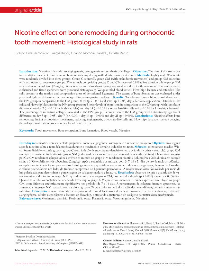
Figure 1 - First molar mesiobuccal roots photomicrographs from group C (AB= alveolar bone; CEM= cementum; PL= periodontal ligament; BV= blood vessels).
Group C (3, 7, 14 and 21 days) 400x, HE
© 2014 Dental Press Journal of Orthodontics Dental Press J Orthod. 2014 Mar-Apr;19(2):96-10799
original articleShintcovsk RL, Knop L, Tanaka OM, Maruo H
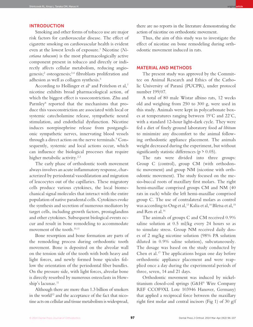
Group CMIn the sections of these samples, on day three, the
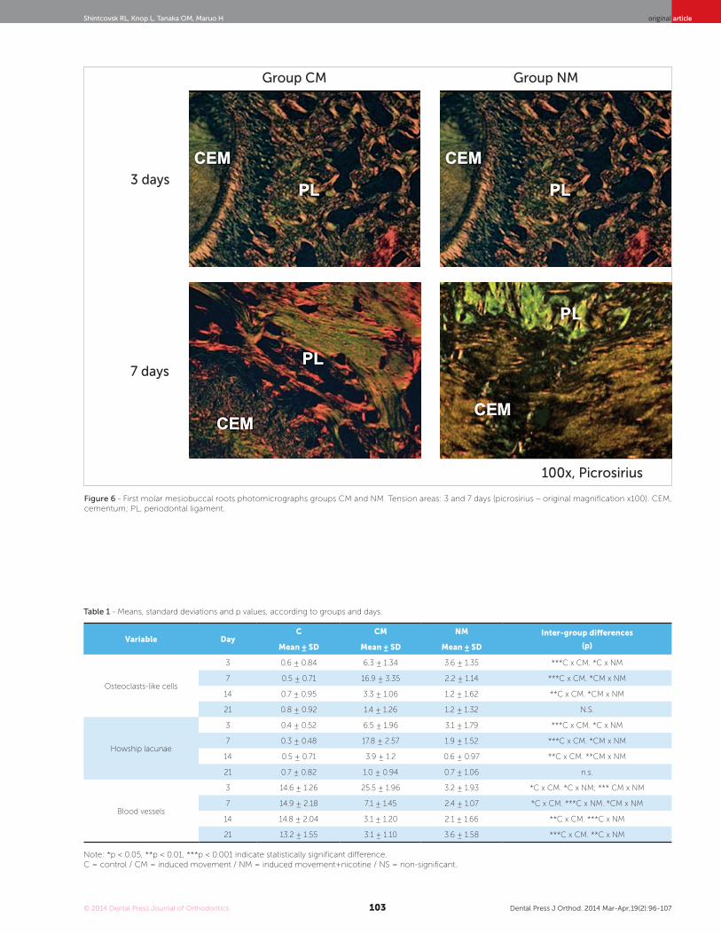
collagen fibers were elongated on the tension areas, compressed and completely disorganized on the compression areas. We also observed angiogenesis with congested vessels. The compression area showed irregular alveolar bone due to a small population of osteoclast-like cells localized on Howship’s lacunae. Hyalinized areas were not present (Fig 2). The bone matrix under polarized light demonstrated a predom-inance of greenish, immature fibers with irregular bi-refringence and few yellowish-orange fibers (Fig 6).
On day seven, the compression area showed ac-tive resorption, with accumulation of osteoclast-like cells associated with numerous Howship’s lacunae. In the tension area, the fibers were elongated and better organized (Fig 3). We observed deposition of thicker and yellowish-orange collagen fibers, forming
and homogeneity of variance, we used ANOVA and the Student’s t-test for independent samples. The level of significance was p < 0.05 when comparing the aver-ages between treatments and groups.
resultshistology
The histological study demonstrated characteristic structural aspects among different groups throughout the experimental period.
Group CThe periodontal ligament showed moderate vas-
cularization, with uniform width and irregular shape. The collagen fibers ran parallel and inserted perpendicular to the cementum and bone surface. On the bone surface, rare Howship’s lacunae associ-ated with osteoclast-like cells were observed (Fig 1).
Compression
Group CM Group NM
Tension
400x, HE
3 days
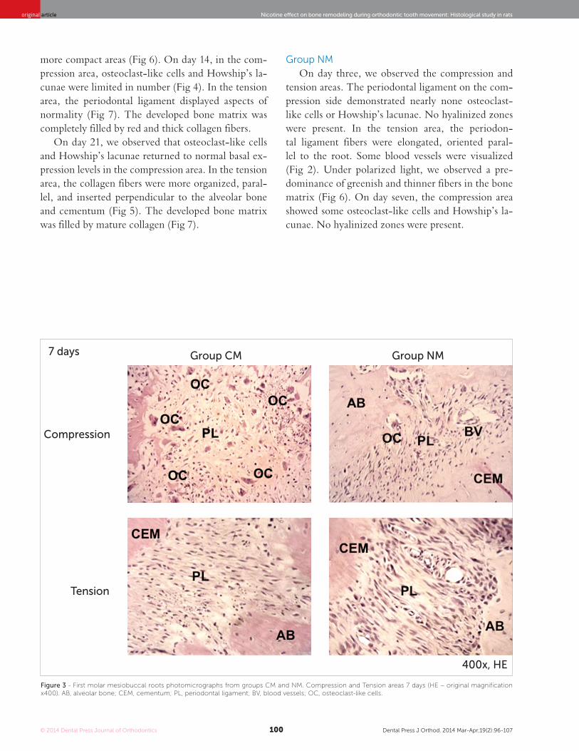
Figure 2 - First molar mesiobuccal roots photomicrographs from groups CM and NM. Compression and Tension areas 3 days (HE – original magnification x400). AB, alveolar bone; CEM, cementum; PL, periodontal ligament; BV, blood vessels.
© 2014 Dental Press Journal of Orthodontics Dental Press J Orthod. 2014 Mar-Apr;19(2):96-107100
Nicotine effect on bone remodeling during orthodontic tooth movement: Histological study in ratsoriginal article
Compression
Group CM Group NM
Tension
7 days
Figure 3 - First molar mesiobuccal roots photomicrographs from groups CM and NM. Compression and Tension areas 7 days (HE – original magnification x400). AB, alveolar bone; CEM, cementum; PL, periodontal ligament; BV, blood vessels; OC, osteoclast-like cells.
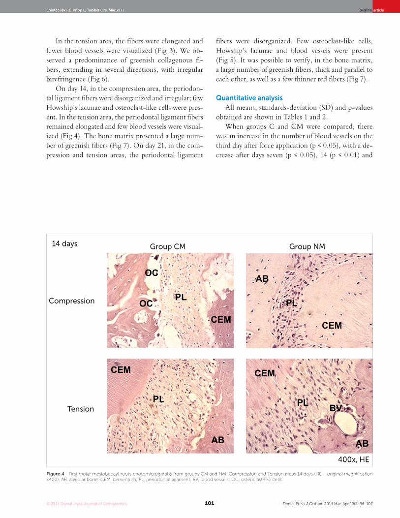
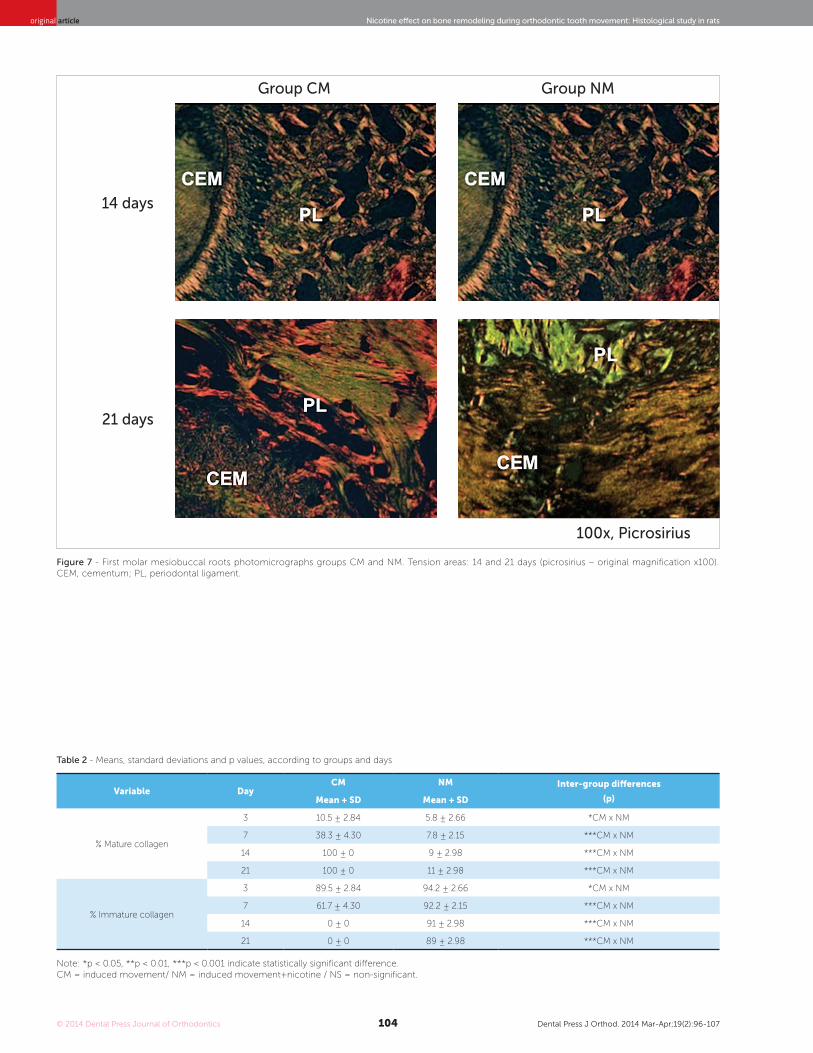
more compact areas (Fig 6). On day 14, in the com-pression area, osteoclast-like cells and Howship’s la-cunae were limited in number (Fig 4). In the tension area, the periodontal ligament displayed aspects of normality (Fig 7). The developed bone matrix was completely filled by red and thick collagen fibers.
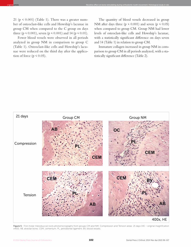
On day 21, we observed that osteoclast-like cells and Howship’s lacunae returned to normal basal ex-pression levels in the compression area. In the tension area, the collagen fibers were more organized, paral-lel, and inserted perpendicular to the alveolar bone and cementum (Fig 5). The developed bone matrix was filled by mature collagen (Fig 7).
Group NMOn day three, we observed the compression and
tension areas. The periodontal ligament on the com-pression side demonstrated nearly none osteoclast-like cells or Howship’s lacunae. No hyalinized zones were present. In the tension area, the periodon-tal ligament fibers were elongated, oriented paral-lel to the root. Some blood vessels were visualized (Fig 2). Under polarized light, we observed a pre-dominance of greenish and thinner fibers in the bone matrix (Fig 6). On day seven, the compression area showed some osteoclast-like cells and Howship’s la-cunae. No hyalinized zones were present.
400x, HE
© 2014 Dental Press Journal of Orthodontics Dental Press J Orthod. 2014 Mar-Apr;19(2):96-107101
original articleShintcovsk RL, Knop L, Tanaka OM, Maruo H
Compression
Group CM Group NM
Tension
14 days
Figure 4 - First molar mesiobuccal roots photomicrographs from groups CM and NM. Compression and Tension areas 14 days (HE – original magnification x400). AB, alveolar bone; CEM, cementum; PL, periodontal ligament; BV, blood vessels; OC, osteoclast-like cells.
In the tension area, the fibers were elongated and fewer blood vessels were visualized (Fig 3). We ob-served a predominance of greenish collagenous fi-bers, extending in several directions, with irregular birefringence (Fig 6).
On day 14, in the compression area, the periodon-tal ligament fibers were disorganized and irregular; few Howship’s lacunae and osteoclast-like cells were pres-ent. In the tension area, the periodontal ligament fibers remained elongated and few blood vessels were visual-ized (Fig 4). The bone matrix presented a large num-ber of greenish fibers (Fig 7). On day 21, in the com-pression and tension areas, the periodontal ligament
fibers were disorganized. Few osteoclast-like cells, Howship’s lacunae and blood vessels were present (Fig 5). It was possible to verify, in the bone matrix, a large number of greenish fibers, thick and parallel to each other, as well as a few thinner red fibers (Fig 7).
Quantitative analysisAll means, standards-deviation (SD) and p-values
obtained are shown in Tables 1 and 2.When groups C and CM were compared, there
was an increase in the number of blood vessels on the third day after force application (p < 0.05), with a de-crease after days seven (p < 0.05), 14 (p < 0.01) and
400x, HE
© 2014 Dental Press Journal of Orthodontics Dental Press J Orthod. 2014 Mar-Apr;19(2):96-107102
Nicotine effect on bone remodeling during orthodontic tooth movement: Histological study in ratsoriginal article
21 (p < 0.001) (Table 1). There was a greater num-ber of osteoclast-like cells and Howship’s lacunae in group CM when compared to the C group on days three (p < 0.001), seven (p < 0.001) and 14 (p < 0.01).
Fewer blood vessels were observed in all periods analyzed in group NM in comparison to group C (Table 1). Osteoclast-like cells and Howship’s lacu-nae were reduced on the third day after the applica-tion of force (p < 0.05).
The quantity of blood vessels decreased in group NM after days three (p < 0.001) and seven (p < 0.05) when compared to group CM. Group NM had lower levels of osteoclast-like cells and Howship’s lacunae, with a statistically significant difference on days seven and 14 (Table 1) in relation to group CM.
Immature collagen increased in group NM in com-parison to group CM in all periods analyzed, with a sta-tistically significant difference (Table 2).
Compression
Group CM Group NM
Tension
21 days
Figure 5 - First molar mesiobuccal roots photomicrographs from groups CM and NM. Compression and Tension areas: 21 days (HE – original magnification x400). AB, alveolar bone; CEM, cementum; PL, periodontal ligament; BV, blood vessels.
400x, HE
© 2014 Dental Press Journal of Orthodontics Dental Press J Orthod. 2014 Mar-Apr;19(2):96-107103
original articleShintcovsk RL, Knop L, Tanaka OM, Maruo H
Group CM
3 days
7 days
Group NM
Variable DayC CM NM Inter-group differences
(p)Mean ± SD Mean ± SD Mean ± SD
Osteoclasts-like cells
3 0.6 ± 0.84 6.3 ± 1.34 3.6 ± 1.35 ***C x CM. *C x NM
7 0.5 ± 0.71 16.9 ± 3.35 2.2 ± 1.14 ***C x CM. *CM x NM
14 0.7 ± 0.95 3.3 ± 1.06 1.2 ± 1.62 **C x CM. *CM x NM
21 0.8 ± 0.92 1.4 ± 1.26 1.2 ± 1.32 N.S.
Howship lacunae
3 0.4 ± 0.52 6.5 ± 1.96 3.1 ± 1.79 ***C x CM. *C x NM
7 0.3 ± 0.48 17.8 ± 2.57 1.9 ± 1.52 ***C x CM. *CM x NM
14 0.5 ± 0.71 3.9 ± 1.2 0.6 ± 0.97 **C x CM. **CM x NM
21 0.7 ± 0.82 1.0 ± 0.94 0.7 ± 1.06 n.s.
Blood vessels
3 14.6 ± 1.26 25.5 ± 1.96 3.2 ± 1.93 *C x CM. *C x NM; *** CM x NM
7 14.9 ± 2.18 7.1 ± 1.45 2.4 ± 1.07 *C x CM. ***C x NM. *CM x NM
14 14.8 ± 2.04 3.1 ± 1.20 2.1 ± 1.66 **C x CM. ***C x NM
21 13.2 ± 1.55 3.1 ± 1.10 3.6 ± 1.58 ***C x CM. **C x NM
Table 1 - Means, standard deviations and p values, according to groups and days.
Note: *p < 0.05, **p < 0.01, ***p < 0.001 indicate statistically significant difference.C = control / CM = induced movement / NM = induced movement+nicotine / NS = non-significant.
Figure 6 - First molar mesiobuccal roots photomicrographs groups CM and NM. Tension areas: 3 and 7 days (picrosirius – original magnification x100). CEM, cementum; PL, periodontal ligament.
100x, Picrosirius
© 2014 Dental Press Journal of Orthodontics Dental Press J Orthod. 2014 Mar-Apr;19(2):96-107104
Nicotine effect on bone remodeling during orthodontic tooth movement: Histological study in ratsoriginal article
Group CM
14 days
21 days
Group NM
Variable DayCM NM Inter-group differences
(p)Mean + SD Mean + SD
% Mature collagen
3 10.5 ± 2.84 5.8 ± 2.66 *CM x NM
7 38.3 ± 4.30 7.8 ± 2.15 ***CM x NM
14 100 ± 0 9 ± 2.98 ***CM x NM
21 100 ± 0 11 ± 2.98 ***CM x NM
% Immature collagen
3 89.5 ± 2.84 94.2 ± 2.66 *CM x NM
7 61.7 ± 4.30 92.2 ± 2.15 ***CM x NM
14 0 ± 0 91 ± 2.98 ***CM x NM
21 0 ± 0 89 ± 2.98 ***CM x NM
Table 2 - Means, standard deviations and p values, according to groups and days
Note: *p < 0.05, **p < 0.01, ***p < 0.001 indicate statistically significant difference.CM = induced movement/ NM = induced movement+nicotine / NS = non-significant.
Figure 7 - First molar mesiobuccal roots photomicrographs groups CM and NM. Tension areas: 14 and 21 days (picrosirius – original magnification x100). CEM, cementum; PL, periodontal ligament.
100x, Picrosirius
© 2014 Dental Press Journal of Orthodontics Dental Press J Orthod. 2014 Mar-Apr;19(2):96-107105
original articleShintcovsk RL, Knop L, Tanaka OM, Maruo H
discussionNicotine is a major cytotoxic and vasoactive sub-
stance present in tobacco which causes peripheral va-soconstriction, tissue ischemia and decreased oxygen tension by reducing the infusion of oxygen to tissue. Furthermore, this substance decreases osteoblastic activity, revascularization and bone healing.5,20
In group NM, there were lower numbers of blood vessels when compared to group C for all periods analyzed (Table 1). When comparing groups NM x CM, we observed fewer blood vessels in group NM on days three (p < 0.001) and seven (p < 0.05), with a statistically significant difference (Table 1).
Pinto et al3 reported that nicotine delays angiogen-esis, and as a consequence, delays the organization of connective tissue and osteogenesis. Saldanha et al20 dem-onstrated that administration of nicotine to jaw bone defects in dogs changes the density of newly formed bone tissue due to the inhibition of revascularization. Adversely, Zheng et al4 concluded that nicotine expo-sure enhances angiogenesis in rabbit model, but cannot compensate the adverse effect of vasoconstriction.
There are controversies regarding the effect of nic-otine on osteoclasts. Heremyre et al21 observed that nicotine stimulates osteoclast-like cell differentia-tion in cell cultures derived from pigs; Tanaka et al22 also observed increased formation of osteoclast-like
cells in vitro. We observed that nicotine reduced the expression of osteoclast-like cells and Howship’s la-cunae in group NM (Table 1). This result is in accor-dance with the findings by Yuhara et al23 who dem-onstrated that nicotine inhibits differentiation and activation of osteoclast-like cells and regulates bone metabolism in rats. Adler et al24 concluded that nico-tine did not stimulate the formation of osteoclasts in bone marrow at doses of 1 or 2 mg / kg in rats.
In this study, we evaluated structural changes in the newly developed bone matrix when nicotine and orthodontic force were applied simultaneously. The picrosirius-polarization method allows the detec-tion of mature and immature collagen and correlates the three-dimensional distribution of collagen fibers with the stage of bone formation.19 The collagen color and birefringence vary according to polymerization degree, which reflects fibers’ age and diameter. First, the collagen is deposited in the form of thin fibrils that aggregate to form larger fibers or bundles.25
The organic matrix of alveolar bone is composed fundamentally of type I collagen (95%), proteogly-cans and glycoproteins.26 Bone formation results from complex and inter-dependent processes, which involve osteoblast differentiation from primitive mes-enchymal cells, organic matrix synthesis and matura-tion, until complete mineralization.27
© 2014 Dental Press Journal of Orthodontics Dental Press J Orthod. 2014 Mar-Apr;19(2):96-107106
Nicotine effect on bone remodeling during orthodontic tooth movement: Histological study in ratsoriginal article
The process of bone formation is associated with the formation of new capillaries from existing blood vessels.28 Orthodontic movement results in a rapid formation of immature bone, and later, the bone is remodeled.29
Immature collagen percentage in group NM in-creased in all analyzed periods. On day 21, there were still immature fibers, although they were thicker and more parallel. Nicotine delayed the collagen maturation process in the developed bone matrix. It was not pos-sible to assess whether nicotine was able to inhibit the
synthesis of collagen, although Theiss et al2 showed that during bone healing in rabbits subjected to nicotine, there were lower levels of type I and II collagen mRNA.
conclusionIn conclusion, nicotine affects bone remodeling
mechanism during orthodontic movement, reduc-ing angiogenesis, osteoclast-like cells and Howship’s lacunae, thereby, delaying the collagen maturation process in developed bone matrix.
© 2014 Dental Press Journal of Orthodontics Dental Press J Orthod. 2014 Mar-Apr;19(2):96-107107
original articleShintcovsk RL, Knop L, Tanaka OM, Maruo H
1. Erhardt L. Cigarette smoking: an undertreated risk factor for
cardiovascular disease. Atherosclerosis. 2009;205(1):23-32.
2. Theiss SM, Boden SD, Hair G, Titus L, Morone MA, Ugbo J. The effect of
nicotine on gene expression during spine fusion. Spine (Phila Pa 1976).
2000;25(20):2588-94.
3. Pinto JR, Bosco AF, Okamoto T, Guerra JB, Piza IG. Effects of nicotine on
the healing of extraction sockets in rats. A histological study. Braz Dent J.
2002;13(1):3-9.
4. Zheng LW, MA L, Cheung LK. Changes in blood perfusion and bone
healing induced by nicotine during distraction osteogenesis. Bone.
2008;43(2):355-61.
5. Zhou J, Olson BL, Windsor LJ. Nicotine increases the collagen-degrading
ability of human gingival fibroblasts. J Periodontal Res. 2007;42(3):228-35.
6. Hollinger JO, Schmitt JM, Hwang K, Soleymani P, Buck D. Impact of
nicotine on bone healing. J Biomed Mater Res. 1999;45(4):294-301.
7. Feitelson JBA, Rowell PP, Roberts CS, Fleming JT. Two week nicotine
treatment selectively increases bone vascular constriction in response to
norepinephrine. J Orthop Res. 2003;21(3):497-502.
8. Zhu B, Parmley WW. Hemodynamic and vascular effects of active and
passive smoking. Am Heart J. 1995;130(6):1270-5.
9. Winniford MD, Wheelan KR, Kremers MS, Ugolini V, Van Den Berg E
Jr, Niggemann EH, et al. Smoking induced coronary vasoconstriction
in patients with atherosclerotic coronary artery disease: Evidence for
adrenergically mediated alterations in coronary artery tone. Circulation.
1986;73:662-7.
10. Krishnan V, Davidovitch Z. Cellular, molecular, and tissue-level reactions to
orthodontic force. Am J Orthod Dentofacial Orthop. 2006;129(4):469.e1-32.
11. Meikle MC. The tissue, cellular, and molecular regulation of orthodontic
tooth movement: 100 years after Carl Sandstedt. Eur J Orthod.
2006;28(3):221-40.
12. American Lung Association. Trends in tobacco use. 2009 [Access 2009
October 26]. Available from: http://www.lungusa.org.
13. Ong CK, Walsh LJ, Harbrow D, Taverne AA, Symons AL. Orthodontic
tooth movement in the prednisolone-treated rat. Angle Orthod.
2000;70(2):118-25.
14. Kalia S, Melsen B, Verna C. Tissue reaction to orthodontic tooth
movement in acute and chronic corticosteroid treatment. Orthod
Craniofac Res. 2004;7(1):26-34.
15. Bletsa A, Berggreen E, Brudvik P. Interleukin-1α and tumor necrosis
factor-α expression during the early phases of orthodontic tooth
movement in rats. Eur J Oral Sci. 2006;114(5):423-9.
16. Ren Y, Maltha JC, Stokroos I, Liem RS, Kuijpers-jagtman, AM. Effect of
duration of force application on blood vessels in young and adult rats.
Am J Orthod Dentofacial Orthop. 2008;133(5):752-7
REFERENCEs
17. Chen M, Wang T, Liao Z, Pan X, Feng Y, Wang H. Nicotine-induced
prenatal overexposure to maternal glucocorticoid and intrauterine
growth retardation in rat. Exp Toxicol Pathol. 2007;59(3-4):245-51.
18. Arias OR, Marquez-Orozco MC. Aspirin, acetaminophen, and ibuprofen:
their effects on orthodontic tooth movement. Am J Orthod Dentofacial
Orthop. 2006;130(3):364-70.
19. Garavello-Freitas I, Baranauskas V, Joazeiro PP, Padovani CR, Dal
Pai-Silva M, Höfling MAC. Low-power laser irradiation improves
histomorphometrical parameters and bone matrix organization during
tibia wound healing in rats. J Photochem Photobiol B. 2003;70(2):81-9.
20. Saldanha JB, Pimentel, SP, Casati MZ, Sallum AW, Sallum EA, Nociti
FH. Histologic evaluation of effect of nicotine administration on bone
regeneration, a study in dogs. Braz Oral Res. 2004;18(4):345-9.
21. Henemyre CL, Scales DK, Hokett SD, Cuenin MF, Peacock ME, Parker MH,
et al. Nicotine stimulated osteoclast resorption in a porcine marrow cell
model. J Periodontol. 2003;74(10):1440-6.
22. Tanaka H, Tanabe H, Shoji M, Suzuki N, Katono T, Sato S, et al. Nicotine
and lipopolysaccharide stimulate the formation of osteoclast-like cells by
increasing macrophage colony-stimulating factor and prostaglandin E2
production by osteoblasts. Life Sci. 2006;78(15):1733-40.
23. Yuhara S, Kasagi S, Inoue A, Otsuka E, Hirose S, Hagiwara H. Effects of
nicotine on cultured cells suggest that it can influence the formation and
resorption of bone. Eur J Pharmacol. 1999;383(3):387-93.
24. Adler ID, Attia SM. Nicotine is not clastogenic at doses of 1 or 2 mg/kg
body weight given orally to male mice. Mutat Res. 2003;542(1-2):139-42.
25. Junqueira LCU, Bignolas G, Brentani RR. Picrosirius staining plus
polarization microscopy, a specific method for collagen detection in
tissue sections. Histochem J. 1979;11(4):447-55.
26. Alberts B, Johnson A, Lewis J, Raff M, Roberts K, Walter P. Biology
molecular of the cell. London: IRL Press; 2002.
27. Martin TJ, Ng K. Mechanisms by which cells of the osteoblast
lineage control osteoclast formation and function. J Cell Biochem.
1994;56(3):357-66.
28. Roberts WE, Hartsfield JK. Bone development and function: genetic and
environmental mechanisms. Semin Orthod. 2004;10(2):100-22.
29. Graber TM, Vanarsdall RL. Orthodontics: Current principles and
techniques. St. Louis: Mosby; 2000.