Embed Size (px)
Citation preview

MOLPHARM/2015/101444
1
Nicotine Dependence Reveals Distinct Responses from Neurons and
their Resident Nicotinic Receptors in Medial Habenula
Pei-Yu Shih, J. Michael McIntosh, and Ryan M. Drenan
Department of Medicinal Chemistry and Molecular Pharmacology, Purdue University,
West Lafayette, IN 47907, USA (P.-Y.S. and R.M.D.)
George E. Wahlen Veterans Affairs Medical Center and Departments of Psychiatry and
Biology, University of Utah, Salt Lake City, UT 84148, USA (J.M.M.)
This article has not been copyedited and formatted. The final version may differ from this version.Molecular Pharmacology Fast Forward. Published on October 1, 2015 as DOI: 10.1124/mol.115.101444
at ASPE
T Journals on D
ecember 6, 2021
molpharm
.aspetjournals.orgD
ownloaded from
MOLPHARM/2015/101444
2
Running Title: Medial Habenula, Nicotine Dependence, and nAChRs
Corresponding author:
Ryan M. Drenan
Purdue University
Department of Medicinal Chemistry and Molecular Pharmacology
575 Stadium Mall Dr.
West Lafayette, IN 47907
Phone: 765-494-1403
Fax: 765-494-1414
# of text pages: 40
# of figures: 7
# of tables: 0
# of references: 54
# of words (Abstract): 243
# of words (Introduction): 747
# of words (Discussion): 1341
Nonstandard Abbreviations: ACh, acetylcholine; choline acetyltransferase (ChAT);
dihydro--erythroidine (DHE); interpeduncular nucleus (IPN); medial habenula (MHb);
ventrolateral medial habenula (MHbVL); ventroinferior medial habenula (MHbVI); nAChR,
nicotinic acetylcholine receptor; phosphate buffered saline (PBS); VTA, ventral
tegmental area;
This article has not been copyedited and formatted. The final version may differ from this version.Molecular Pharmacology Fast Forward. Published on October 1, 2015 as DOI: 10.1124/mol.115.101444
at ASPE
T Journals on D
ecember 6, 2021
molpharm
.aspetjournals.orgD
ownloaded from
MOLPHARM/2015/101444
3
Abstract
Nicotinic acetylcholine receptors (nAChRs) are the molecular target of nicotine. nAChRs
in the medial habenula (MHb) have recently been shown to play a role in nicotine
dependence, but it is not clear which nAChR subtypes or MHb neuron types are most
important. To identify MHb nAChRs and/or cell types that play a role in nicotine
dependence, we studied these receptors and cells with brain slice electrophysiology
using both acute and chronic nicotine application. Cells in ventroinferior and ventrolateral
MHb (MHbVI and MHbVL) subregions expressed functional nAChRs with different
pharmacology. Further, application of nicotine to cells in these subregions led to different
action potential firing patterns. The latter result was correlated with a differing ability of
nicotine to induce nAChR desensitization. Chronic nicotine caused functional up-
regulation of nAChRs selectively in MHbVI cells but did not change nAChR function in
MHbVL. Importantly, firing responses were also differentially altered in these subregions
following chronic nicotine. MHbVI neurons treated chronically with nicotine exhibited
enhanced basal pacemaker firing but a blunted nicotine-induced firing response. MHbVL
neurons did not change their firing properties in response to chronic nicotine. Together,
these results suggest that acute and chronic nicotine differentially affect nAChR function
and output of cells in MHb subregions. Because the MHb extensively innervates the
interpeduncular nucleus (IPN), an area critical for both affective and somatic signs of
withdrawal, these results could reflect some of the neurophysiological changes thought
to occur in the MHb to IPN circuit in human smokers.
This article has not been copyedited and formatted. The final version may differ from this version.Molecular Pharmacology Fast Forward. Published on October 1, 2015 as DOI: 10.1124/mol.115.101444
at ASPE
T Journals on D
ecember 6, 2021
molpharm
.aspetjournals.orgD
ownloaded from
MOLPHARM/2015/101444
4
Introduction
Chronic exposure to nicotine in tobacco products results in numerous health
consequences (lung cancer, emphysema, hypertension, etc.) and accounts for over 6
million deaths per year (WHO, 2011). Nicotine is the primary psychoactive substance in
tobacco products, and it is addictive due to its ability to strongly activate neuronal
nicotinic acetylcholine receptors (nAChRs) (Steinsland and Furchgott, 1975). It has
recently been appreciated that long-term nicotine use is maintained via a balanced
activation of brain circuits mediating both positive (rewarding) and negative (aversive)
motivational signals (Bromberg-Martin et al., 2010; Hikosaka, 2010; Matsumoto and
Hikosaka, 2007; Matsumoto and Hikosaka, 2009). It is also established that chronically
exposing the brain to nicotine changes nAChR number and/or function in key brain
circuits (Lester et al., 2009; Marks et al., 1983). These alterations in nAChR function,
and the consequent changes in neuronal circuit activity where these receptors reside,
are among the key events important for establishing and maintaining addiction to
nicotine. Positive motivational signals generated by nicotine utilize the mesolimbic
dopamine pathway, and much has been learned regarding this system (Dani and
Bertrand, 2007; Dani and Harris, 2005; Laviolette and van der Kooy, 2004). By contrast,
relatively little is known about the nAChRs and brain circuits mediating nicotine’s
aversive quality. In this study, we sought to study one of the key brain areas mediating
negative motivational signals after nicotine use: the medial habenula (MHb).
The MHb is a small bilateral structure immediately ventral to the hippocampus and
adjacent to the 3rd ventricle. The MHb sends a prominent projection via the fasciculus
retroflexus to a midline structure in the ventral midbrain, the interpeduncular nucleus
(IPN). Via the MHb’s connection to areas such as the septum and the nucleus of
diagonal band (Herkenham and Nauta, 1977; Qin and Luo, 2009), and the IPN’s
This article has not been copyedited and formatted. The final version may differ from this version.Molecular Pharmacology Fast Forward. Published on October 1, 2015 as DOI: 10.1124/mol.115.101444
at ASPE
T Journals on D
ecember 6, 2021
molpharm
.aspetjournals.orgD
ownloaded from
MOLPHARM/2015/101444
5
connection to the raphe (Montone et al., 1988; Shibata and Suzuki, 1984), the MHb to
IPN pathway is a key circuit connecting forebrain structures with midbrain areas
important for motivation and reward. The MHb to IPN pathway is a crucial mediator of
nicotine withdrawal following chronic exposure to nicotine, and specific nAChRs play a
key role. For example, mice lacking 5 and 4 subunits, which are expressed in the
MHb or IPN, exhibit reduced withdrawal responses when chronic nicotine is removed
(Salas et al., 2004; Salas et al., 2009). Similarly, antagonizing nAChRs via infusion of
pharmacological blockers into the MHb also reduces the severity of nicotine withdrawal
(Salas et al., 2009). Mice lacking 5 subunits self-administer greater amounts of nicotine
(i.e. they have reduced aversive signals at high doses of nicotine), and self-
administration is normalized when 5 subunits are re-introduced into the MHb (Fowler et
al., 2011). Conversely, transgenic mice overexpressing nAChRs in MHb show enhanced
sensitivity to the aversive aspects of nicotine (Frahm et al., 2011).
Although these early and seminal studies clearly demonstrate the importance of MHb
nAChRs in the motivational response to nicotine, a detailed picture elucidating specific
nAChRs in specific MHb neurons has not yet emerged. Both classic and recent studies
have determined that the MHb is not a homogeneous structure, but is rather a collection
of diverse cell types that could have varied physiological actions. For example, several
MHb subnuclei exist that each contain neuron types expressing various biochemical
markers (Aizawa et al., 2012). Cells in the dorsal 1/3 of the MHb preferentially express
substance P whereas cells in the ventral 2/3 of the MHb preferentially express choline
acetyltransferase (ChAT), a cholinergic marker (Contestabile et al., 1987; Yamaguchi et
al., 2013). The dorsal and ventral MHb project to anatomically divergent components of
the IPN (Contestabile and Flumerfelt, 1981; Shih et al., 2014; Yamaguchi et al., 2013),
and different regions of IPN have recently been appreciated to mediate diverse aspects
This article has not been copyedited and formatted. The final version may differ from this version.Molecular Pharmacology Fast Forward. Published on October 1, 2015 as DOI: 10.1124/mol.115.101444
at ASPE
T Journals on D
ecember 6, 2021
molpharm
.aspetjournals.orgD
ownloaded from
MOLPHARM/2015/101444
6
of nicotine withdrawal. For example, the dorsal IPN – which receives projections from
the ventrolateral MHb (MHbVL) – is a key mediator of the somatic aspect of nicotine
withdrawal (Zhao-Shea et al., 2013). Further, areas in the ventral IPN – which receive
projections preferentially from ventroinferior MHb (MHbVI) – appear to more strongly
control affective signs of nicotine withdrawal (Zhao-Shea et al., 2015). These recent data
point to the need for a more thorough description of the nAChRs in different subnuclei of
MHb. In this study, we used brain slice patch clamp electrophysiology, pharmacology,
and genetics to examine nAChR function in MHbVI and MHbVL. We present evidence
demonstrating that nAChRs in different MHb subregions respond differently to acute and
chronic nicotine, which in turn differentially affect neuronal activity in these areas.
This article has not been copyedited and formatted. The final version may differ from this version.Molecular Pharmacology Fast Forward. Published on October 1, 2015 as DOI: 10.1124/mol.115.101444
at ASPE
T Journals on D
ecember 6, 2021
molpharm
.aspetjournals.orgD
ownloaded from
MOLPHARM/2015/101444
7
Materials and methods
Animals
All procedures involving the use of live animals conformed to the guidelines provided by
the National Institutes of Health Office of Laboratory Animal Welfare. All protocols were
approved by the Purdue University Institutional Animal Care and Use Committee. Mice
were housed at 22°C with alternating 12 h light/dark cycle. Food and water was available
ad libitum. Mice were weaned and group-housed with the same sex littermates at
postnatal day 21. Adult (2-3 month old) mice were used for this study. No gender-based
differences were noted, so male and female data were pooled for subsequent analyses.
Most studies were conducted with C57BL/6 WT mice obtained from Jackson
Laboratories. Select experiments utilized genetically modified mice. Mice lacking 4
nAChR subunits (4KO) were a generous gift of Drs. Michael Marks and Jerry Stitzel
(University of Colorado, Boulder, CO), and were produced by mating mice heterozygous
for the 4KO allele. 6L9S mice were generated as previously described (Drenan et al.,
2008). A Leu “9-prime” to Ser mutation was introduced into a bacterial artificial
chromosome (BAC) at the 6 nAChR subunit gene. Mutant BAC DNA was injected into
FVB/N embryos and the embryos were implanted into pseudopregnant Swiss-Webster
surrogates. The transgene insertion site in the mouse genome is unknown. Founder
animals were identified and back-crossed to C57BL/6 mice for 12 or more generations.
As a result, 90-95% of the genome of the 6L9S strain is expected to be C57BL/6.
FVB/N allelic DNA close to the insertion site is likely to persist. The L9S mutation leaves
6* nAChRs 10- to 100- fold more sensitive to ligands compared to non�6* nAChRs
(Cohen et al., 2012; Drenan et al., 2010; Drenan et al., 2008). Previous studies have
confirmed that 6* nAChRs are not overexpressed, nor expressed in ectopic brain
regions in 6L9S mice (Drenan et al., 2010; Drenan et al., 2008). Littermate mice not
This article has not been copyedited and formatted. The final version may differ from this version.Molecular Pharmacology Fast Forward. Published on October 1, 2015 as DOI: 10.1124/mol.115.101444
at ASPE
T Journals on D
ecember 6, 2021
molpharm
.aspetjournals.orgD
ownloaded from

MOLPHARM/2015/101444
8
harboring the mutant BAC served as controls for 6L9S mice in electrophysiology
experiments. Mutant mouse lines were genotyped by PCR analysis of isolated tail DNA
as previously described (Drenan et al., 2008).
Brain slice preparation
Mice were anesthetized with sodium pentobarbital (100 mg/kg, i.p.) and transcardially
perfused with 4°C N-methyl-D-glucamine - based recovery solution (in mM: 93 N-methyl-
D-glucamine, 2.5 KCl, 1.2 NaH2PO4, 30 NaHCO3, 20 HEPES, 25 glucose, 5 Na-
ascorbate, 2 thiourea, 3 Na-pyruvate, 10 MgSO4·7H2O, and 0.5 CaCl2·2H2O). Brains
were removed and 200-μm-thick coronal slices containing the medial habenula (MHb)
were prepared with a vibrating microslicer (DTK Zero 1; Dosaka). Slices were allowed to
recover for 12 min at 33°C and then at room temperature for 1-8 h in HEPES-holding
solution (in mM: 92 NaCl, 2.5 KCl, 1.2 NaH2PO4, 30 NaHCO3, 20 HEPES, 25 glucose, 5
Na-ascorbate, 2 thiourea, 3 Na-pyruvate, 2 MgSO4·7H2O, 2 CaCl2·2H2O). All solutions
were saturated with 95 % O2 and 5 % CO2 with osmolarity adjusted to 300 – 310 mOsm.
Patch clamp electrophysiology
Each individual slice was transferred to a recording chamber and was continuously
superfused at a rate of 2-3 ml/min at 30 – 32°C (Warner Instrument, USA) with standard
recording artificial cerebrospinal fluid (ACSF) (in mM: 124 NaCl, 2.5 KCl, 1.2 NaH2PO4,
24 NaHCO3, 12.5 glucose, 2 MgSO4·7H2O, 2 CaCl2·2H2O). MHb neurons were visually
identified under infrared illumination using a Nikon FN-1 microscope equipped with
differential interference contrast optics and a water-immersion objective lens (40x, NA =
0.8; Nikon, Japan). MHb cell recordings were obtained from either the MHbVI area close
to the 3rd ventricle or the MHbVL area adjacent to the lateral habenula (Fig. 1A). Whole-
cell recordings and cell-attached recordings were obtained with patch pipettes filled with
internal solution (in mM: 135 K-gluconate, 5 EGTA, 10 HEPES, 2 MgATP, 0,1 NaGTP,
This article has not been copyedited and formatted. The final version may differ from this version.Molecular Pharmacology Fast Forward. Published on October 1, 2015 as DOI: 10.1124/mol.115.101444
at ASPE
T Journals on D
ecember 6, 2021
molpharm
.aspetjournals.orgD
ownloaded from
MOLPHARM/2015/101444
9
0.5 CaCl2, 2 MgCl2, pH adjusted to 7.3 with Tris base). In a subset of experiments, 2.5
mg/ml biocytin was added to the internal solution in order to mark the recorded neuron
for later cytochemical characterization. Data were collected using a Multiclamp 700B
amplifier (Molecular Devices), filtered at 2 kHz, and sampled at 10 kHz by a Digidata
1440A digitizer. Series resistance was measured by injection of hyperpolarizing pulses (-
5 mV, 100 ms) and was not compensated. To examine functional nAChR response, ACh
was locally applied (250 ms, 12 psi) at different concentrations (1-100 M) using a glass
pipette connected to a PV-820 Pneumatic Picopump (WPI, USA); this pipette was
moved to within 20-40 μm of the recorded cell with a piezoelectric manipulator (PA-
100/12; Piezosystem Jena) for drug application and retracted after the end of the puff to
minimize nAChR desensitization. All other drugs were bath perfused at final
concentrations indicated by diluting aliquots of stock solution in ACSF.
Immunohistochemistry
To identify the location of recorded cells, slices containing biocytin-loaded cells were
fixed by immersion in 4% paraformaldehyde in phosphate buffered saline (PBS)
overnight at 4°C. Sections were washed 2 times for 10 min per wash in PBS, blocked,
and permeabilized for 60 min in PBS containing 10% normal horse serum, 2% bovine
serum albumin and 0.3% Triton X-100 (PBST). Primary antibody incubations were done
overnight at 4°C in PBST containing goat anti-ChAT (1:500; Millipore) and streptavidin
conjugated Alexa-488 (1:1000; Invitrogen). After 3 PBS washes, secondary antibody
incubations were performed for 2 h at room temperature in PBST containing Alexa-555
donkey-anti-goat (1:500; Invitrogen). Sections were finally washed 3 times for 10 min per
wash, mounted with anti-fade mounting medium (Vectashield; Vector Laboratories), and
coverslipped for confocal microscopy.
Chronic nicotine treatment
This article has not been copyedited and formatted. The final version may differ from this version.Molecular Pharmacology Fast Forward. Published on October 1, 2015 as DOI: 10.1124/mol.115.101444
at ASPE
T Journals on D
ecember 6, 2021
molpharm
.aspetjournals.orgD
ownloaded from

MOLPHARM/2015/101444
10
Nicotine dependence was induced with osmotic minipumps (model 2004; Alzet) as
previously reported (Shih et al., 2014). These minipumps, implanted subcutaneously
under brief isoflurane anesthesia, were filled with either sterile saline or (-)-nicotine
hydrogen-tartrate salt (Glentham Life Sciences) dissolved in sterile saline. Mice received
a mean dose of 1 mg/kg/h of nicotine for 14 days. Nicotine dose used in this study to
develop physical dependence was chosen based on the results obtained in previous
studies (Hilario et al., 2012; Matta et al., 2007; Shih et al., 2014). This dose yields
plasma levels of ~0.3 M, a concentration similar to that observed in human smokers
consuming an average of 17 cigarettes a day (Matta et al., 2007).
Data analysis and statistical methods
Off-line data analysis was performed using Clampfit 10 (Molecular Devices, USA),
GraphPad Prism 6 (GraphPad Software, USA) and Origin 9.0 (Originlab, USA).
Summary data are reported as mean ± SEM. Statistical significance was assessed by
two-tailed paired t-tests to assess the effect of a drug within a recorded neuron and
unpaired t-test to compare across neurons. One-way ANOVA followed by Tukey post
hoc tests was applied for multiple comparisons. Differences were considered statistically
significant at p < 0.05.
Chemicals
All reagents were made from 1000x stock solutions kept frozen at -20°C in 50-100 μl
aliquots. 6-cyano-7-nitroquinoxaline-2,3-dione (CNQX) was purchased from Tocris
(Bristol, UK). -conotoxin MII was synthesized as described previously (Azam et al.,
2010). Unless specified otherwise, all other chemicals were purchased from Sigma-
Aldrich (St. Louis, USA).
This article has not been copyedited and formatted. The final version may differ from this version.Molecular Pharmacology Fast Forward. Published on October 1, 2015 as DOI: 10.1124/mol.115.101444
at ASPE
T Journals on D
ecember 6, 2021
molpharm
.aspetjournals.orgD
ownloaded from
MOLPHARM/2015/101444
11
Results
In our previous study, we developed a map of nAChR subunit expression and
localization within MHb. We noted that 4, 6, 2, and 3 subunits exhibit a localization
gradient from inferior to lateral across the ventral portion of MHb. In particular, 4
subunits are concentrated in MHbVL and 6 subunits are preferentially confined to
MHbVI (Shih et al., 2014). Given that the patterns seen for various nAChR subunits
suggest that specific subdivisions exist in MHb, we first sought to determine whether
nAChRs in MHbVL and MHbVI exhibit functional differences. To compare the ACh-
sensitivity in MHbVL and MHbVI, we performed whole-cell patch recordings from visually
identified cells in these two subregions. Our definition of MHbVI and MHbVL follows that
of a recent study (Aizawa et al., 2012). MHbVL cells are located in the lateral one-third of
the ventral MHb, mostly along the border between MHb and lateral habenula. MHbVI
cells are located in the inferior one-third of the ventral MHb, adjacent to the 3rd ventricle
(Fig. 1A). To further verify the location of recorded cells, a subset of cells were filled with
biocytin during recordings, followed by post hoc identification using immunostaining and
confocal microscopy. ChAT costaining was used to label the boundary of the ventral
MHb (Fig. 1B) (Contestabile et al., 1987).
To study nAChR function, local pressure application was used to apply brief (250 ms)
pulses of ACh from glass micropipettes positioned approximately 20-40 m from the
recorded cells using a piezoelectric translator as previously described (Engle et al.,
2013). A low concentration of ACh (1 M) evoked small inward currents or zero current
at a holding potential of -60 mV (Fig. 1C and D), irrespective of the location of the cells
(MHbVL: 9.1 ± 3.1 pA, n = 6; MHbVI: 7.5 ± 3.4 pA, n = 4, p = 0.7460). Application of 100
M ACh induced currents of similar peak amplitude in MHbVL and MHbVI neurons (Fig.
1C and D) (MHbVL: 489.0 ± 118.8 pA, n = 6; MHbVI: 597.9 ± 172.9 pA, n = 4, p =
This article has not been copyedited and formatted. The final version may differ from this version.Molecular Pharmacology Fast Forward. Published on October 1, 2015 as DOI: 10.1124/mol.115.101444
at ASPE
T Journals on D
ecember 6, 2021
molpharm
.aspetjournals.orgD
ownloaded from

MOLPHARM/2015/101444
12
0.6310). These findings indicate that neurons in MHbVL and MHbVI have roughly equal
sensitivity to puff-applied 1 M and 100 M ACh. MHb neurons are highly enriched in
mRNAs for most nAChR subunits (Marks et al., 1992).
Immunoprecipitation and immunopurification studies revealed a wealth of uncommon
nAChR subtypes in MHb (Grady et al., 2009). The nAChRs expressed in MHb can be
grouped to two major and distinct nAChR populations: 2-containing and 4-containing
nAChRs�(2* and 4*��the asterisk indicates other unidentified subunits may co-
assemble with the indicated subunits). Next, we sought to determine which nAChR
subtype(s) is/are responsible for the ACh-induced currents we measured in MHbVL and
MHbVI. First, we tested whether ACh-induced currents in MHbVL and MHbVI could be
blocked by the 34* nAChR antagonist SR16584 (20 M). Application of SR16584
resulted in a significant blockade of ACh-induced currents in both MHbVL (Fig. 1E and F;
n = 6, 44.9 ± 8.1 % remaining response, p = 0.0223) and MHbVI areas (Fig. 1E and F; n
= 5, 51.0 ± 7.4 % remaining response, p = 0.0141). Next, we tested dihydro--
erythroidine (DHE), which is traditionally used to discriminate 2* nAChRs from
homomeric 7 nAChRs. We applied ACh (100 M) with a puff electrode for 250 ms and
repeated this procedure after a 15 min exposure to DHE (500 nM). ACh-induced
currents in MHbVL were not influenced by DHE exposure (Fig. 1G and H; n = 5, 108.0
± 10.2 % remaining response, p = 0.5568). In contrast, ACh-induced currents in MHbVI
neurons were partially inhibited after application of DHE (Fig. 1G and H; n = 5, 78.7 ±
2.9 % remaining response, p = 0.0196). These data show that pharmacologically distinct
nAChRs exist in MHbVL and MHbVI.
Our previous studies strongly suggest that functional 6* nAChRs are expressed in
MHbVI (Henderson et al., 2014; Shih et al., 2014). We conducted further
This article has not been copyedited and formatted. The final version may differ from this version.Molecular Pharmacology Fast Forward. Published on October 1, 2015 as DOI: 10.1124/mol.115.101444
at ASPE
T Journals on D
ecember 6, 2021
molpharm
.aspetjournals.orgD
ownloaded from
MOLPHARM/2015/101444
13
pharmacological experiments to test this hypothesis. -conotoxin MII (CtxMII), a cone
snail toxin, is an antagonist with selectivity for 6* and 3* nAChRs. In particular,
CtxMII is 5.6-fold more selective for 6* versus 3* nAChRs, with little activity at other
nAChR subtypes (Cartier et al., 1996; McIntosh et al., 2004). We measured ACh (100
M)-evoked current amplitudes in MHbVL and MHbVI neurons before and after bath
application of 100 nM CtxMII. Interestingly, CtxMII did not significantly affect
responses to 100 M ACh in either MHb subregion (Fig. 2A and B; MHbVL: 102.3 ± 5.6
% remaining response, n = 4, p = 0.8332; MHbVI: 108.7 ± 14.2 % remaining response, n
= 8, p = 0.8793). 6* nAChRs in the dopamine pathway are known to exhibit high
sensitivity to ACh and nicotine (Salminen et al., 2007), so we reasoned that if functional
MHb 6* nAChRs exist, they may also exhibit high ACh sensitivity. We therefore tested
the ability of CtxMII to block currents evoked by lower concentrations of ACh, as the
large currents evoked by 100 M ACh (~500 pA) could preclude us from effectively
isolating a small, CtxMII-sensitive response. In MHbVI neurons, 10 M ACh reliably
evoked inward currents of 50-75 pA (Fig. 2C; 63.0 ± 13.1 pA, n = 9). CtxMII bath
application resulted in a significant inhibition of inward currents evoked by 10 M ACh in
7 of 9 MHbVI cells tested (Fig. 2C and D; 51.6 ± 4.7 % remaining response, p = 0.0085).
In MHbVL neurons, 10 M ACh was insufficient to evoke workable inward currents, but
30 M ACh application did result in measurable currents (Fig. 2C; 79.8 ± 10.4 pA, n = 5).
However, CtxMII was unable to block or attenuate inward currents evoked by 30 M
ACh in any of the MHbVL cells tested (Fig. 2C and D; 103.7 ± 13.9 % remaining
response, p = 0.8286). These results suggest that MHbVI neurons express a high-
sensitivity nAChR subtype with sensitivity to CtxMII.
This article has not been copyedited and formatted. The final version may differ from this version.Molecular Pharmacology Fast Forward. Published on October 1, 2015 as DOI: 10.1124/mol.115.101444
at ASPE
T Journals on D
ecember 6, 2021
molpharm
.aspetjournals.orgD
ownloaded from
MOLPHARM/2015/101444
14
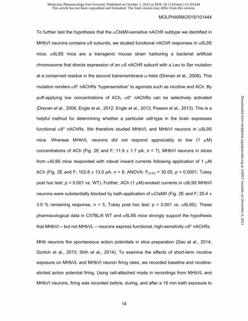
To further test the hypothesis that the CtxMII-sensitive nAChR subtype we identified in
MHbVI neurons contains 6 subunits, we studied functional nAChR responses in 6L9S
mice. 6L9S mice are a transgenic mouse strain harboring a bacterial artificial
chromosome that directs expression of an 6 nAChR subunit with a Leu to Ser mutation
at a conserved residue in the second transmembrane -helix (Drenan et al., 2008). This
mutation renders 6* nAChRs “hypersensitive” to agonists such as nicotine and ACh. By
puff-applying low concentrations of ACh, 6* nAChRs can be selectively activated
(Drenan et al., 2008; Engle et al., 2012; Engle et al., 2013; Powers et al., 2013). This is a
helpful method for determining whether a particular cell-type in the brain expresses
functional 6* nAChRs. We therefore studied MHbVL and MHbVI neurons in 6L9S
mice. Whereas MHbVL neurons did not respond appreciably to low (1 M)
concentrations of ACh (Fig. 2E and F; 11.9 ± 1.7 pA, n = 7), MHbVI neurons in slices
from 6L9S mice responded with robust inward currents following application of 1 M
ACh (Fig. 2E and F; 102.8 ± 13.0 pA, n = 8, ANOVA: F(4,25) = 30.05, p < 0.0001; Tukey
post hoc test: p < 0.001 vs. WT). Further, ACh (1 M)-evoked currents in 6L9S MHbVI
neurons were substantially blocked by bath-application of CtxMII (Fig. 2E and F; 20.4 ±
3.0 % remaining response, n = 5, Tukey post hoc test: p < 0.001 vs. 6L9S). These
pharmacological data in C57BL/6 WT and 6L9S mice strongly support the hypothesis
that MHbVI – but not MHbVL – neurons express functional, high-sensitivity 6* nAChRs.
MHb neurons fire spontaneous action potentials in slice preparation (Dao et al., 2014;
Gorlich et al., 2013; Shih et al., 2014). To examine the effects of short-term nicotine
exposure on MHbVL and MHbVI neuron firing rates, we recorded baseline and nicotine-
elicited action potential firing. Using cell-attached mode in recordings from MHbVL and
MHbVI neurons, firing was recorded before, during, and after a 16 min bath exposure to
This article has not been copyedited and formatted. The final version may differ from this version.Molecular Pharmacology Fast Forward. Published on October 1, 2015 as DOI: 10.1124/mol.115.101444
at ASPE
T Journals on D
ecember 6, 2021
molpharm
.aspetjournals.orgD
ownloaded from

MOLPHARM/2015/101444
15
nicotine (1 M). Although typical nicotine concentrations in the cerebrospinal fluid of
smokers is ~300 nM, higher concentrations have been reported (Malkawi et al., 2009).
Cell-attached recording was chosen to avoid disturbing the intracellular milieu. In
addition, action potentials were recorded in the presence of a cocktail of inhibitors to
block muscarinic receptors, type A gamma aminobutyric acid receptors, and glutamate
receptors (1 M atropine, 100 M picrotoxin, and 10 M CNQX) to better isolate the
actions of nAChRs. In 3 MHb neurons, firing frequency at rest was not altered by these
inhibitors (data not shown), suggesting that tonic action potential firing is driven by
intrinsic factors and is independent of inputs releasing glutamate, gamma aminobutyric
acid, or ACh. A recent study reached a similar conclusion regarding spontaneous firing
in MHb neurons (Gorlich et al., 2013).
Neurons from MHbVL and MHbVI fired at similar frequency at rest (MHbVL, 5.5 ± 0.6 Hz,
n = 9; MHbVI, 4.5 ± 0.8 Hz, n = 8, p = 0.3366). MHbVL neurons increased their firing
rate quickly in response to 1 M nicotine, followed subsequently by attenuation of this
increased firing in the continued presence of 1 M nicotine (Fig. 3A and B). In MHbVI,
the pattern was different. In response to 1 M nicotine, firing increased slower than in VL
neurons (Fig. 3A and B). In contrast to the attenuated firing seen in VL neurons, VI
neurons exhibited sustained firing in the continued presence of 1 M nicotine for the
duration of the experiment (Fig. 3A and B). We quantified action potential firing at 3
different time points during the experiment, and the results indicate a sustained increase
for MHbVI cells at the end of the experiment (Fig. 3C; 8.2 ± 0.6 Hz, p = 0.0002 vs.
baseline). In contrast, in MHbVL cells the firing frequency was not different at the end of
the experiment compared to pre-nicotine firing rate (Fig. 3C; 6.1 ± 0.9 Hz, p = 0.5252 vs.
baseline). Together, these experiments reveal that nicotine has a differential ability to
This article has not been copyedited and formatted. The final version may differ from this version.Molecular Pharmacology Fast Forward. Published on October 1, 2015 as DOI: 10.1124/mol.115.101444
at ASPE
T Journals on D
ecember 6, 2021
molpharm
.aspetjournals.orgD
ownloaded from
MOLPHARM/2015/101444
16
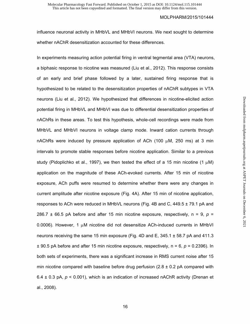
influence neuronal activity in MHbVL and MHbVI neurons. We next sought to determine
whether nAChR desensitization accounted for these differences.
In experiments measuring action potential firing in ventral tegmental area (VTA) neurons,
a biphasic response to nicotine was measured (Liu et al., 2012). This response consists
of an early and brief phase followed by a later, sustained firing response that is
hypothesized to be related to the desensitization properties of nAChR subtypes in VTA
neurons (Liu et al., 2012). We hypothesized that differences in nicotine-elicited action
potential firing in MHbVL and MHbVI was due to differential desensitization properties of
nAChRs in these areas. To test this hypothesis, whole-cell recordings were made from
MHbVL and MHbVI neurons in voltage clamp mode. Inward cation currents through
nAChRs were induced by pressure application of ACh (100 M, 250 ms) at 3 min
intervals to promote stable responses before nicotine application. Similar to a previous
study (Pidoplichko et al., 1997), we then tested the effect of a 15 min nicotine (1 M)
application on the magnitude of these ACh-evoked currents. After 15 min of nicotine
exposure, ACh puffs were resumed to determine whether there were any changes in
current amplitude after nicotine exposure (Fig. 4A). After 15 min of nicotine application,
responses to ACh were reduced in MHbVL neurons (Fig. 4B and C, 449.5 ± 79.1 pA and
286.7 ± 66.5 pA before and after 15 min nicotine exposure, respectively, n = 9, p =
0.0006). However, 1 M nicotine did not desensitize ACh-induced currents in MHbVI
neurons receiving the same 15 min exposure (Fig. 4D and E, 345.1 ± 58.7 pA and 411.3
± 90.5 pA before and after 15 min nicotine exposure, respectively, n = 6, p = 0.2396). In
both sets of experiments, there was a significant increase in RMS current noise after 15
min nicotine compared with baseline before drug perfusion (2.8 ± 0.2 pA compared with
6.4 ± 0.3 pA, p < 0.001), which is an indication of increased nAChR activity (Drenan et
al., 2008).
This article has not been copyedited and formatted. The final version may differ from this version.Molecular Pharmacology Fast Forward. Published on October 1, 2015 as DOI: 10.1124/mol.115.101444
at ASPE
T Journals on D
ecember 6, 2021
molpharm
.aspetjournals.orgD
ownloaded from
MOLPHARM/2015/101444
17
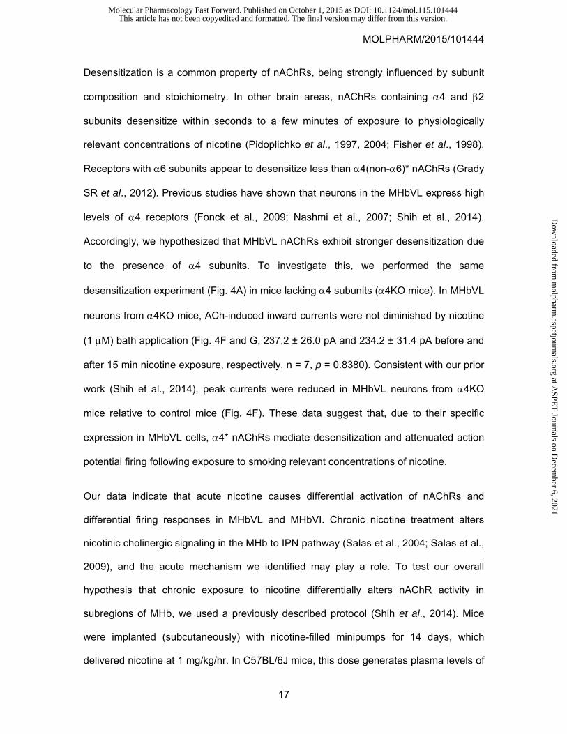
Desensitization is a common property of nAChRs, being strongly influenced by subunit
composition and stoichiometry. In other brain areas, nAChRs containing 4 and 2
subunits desensitize within seconds to a few minutes of exposure to physiologically
relevant concentrations of nicotine (Pidoplichko et al., 1997, 2004; Fisher et al., 1998).
Receptors with 6 subunits appear to desensitize less than 4(non-6)* nAChRs (Grady
SR et al., 2012). Previous studies have shown that neurons in the MHbVL express high
levels of 4 receptors (Fonck et al., 2009; Nashmi et al., 2007; Shih et al., 2014).
Accordingly, we hypothesized that MHbVL nAChRs exhibit stronger desensitization due
to the presence of 4 subunits. To investigate this, we performed the same
desensitization experiment (Fig. 4A) in mice lacking 4 subunits (4KO mice). In MHbVL
neurons from 4KO mice, ACh-induced inward currents were not diminished by nicotine
(1 M) bath application (Fig. 4F and G, 237.2 ± 26.0 pA and 234.2 ± 31.4 pA before and
after 15 min nicotine exposure, respectively, n = 7, p = 0.8380). Consistent with our prior
work (Shih et al., 2014), peak currents were reduced in MHbVL neurons from 4KO
mice relative to control mice (Fig. 4F). These data suggest that, due to their specific
expression in MHbVL cells, 4* nAChRs mediate desensitization and attenuated action
potential firing following exposure to smoking relevant concentrations of nicotine.
Our data indicate that acute nicotine causes differential activation of nAChRs and
differential firing responses in MHbVL and MHbVI. Chronic nicotine treatment alters
nicotinic cholinergic signaling in the MHb to IPN pathway (Salas et al., 2004; Salas et al.,
2009), and the acute mechanism we identified may play a role. To test our overall
hypothesis that chronic exposure to nicotine differentially alters nAChR activity in
subregions of MHb, we used a previously described protocol (Shih et al., 2014). Mice
were implanted (subcutaneously) with nicotine-filled minipumps for 14 days, which
delivered nicotine at 1 mg/kg/hr. In C57BL/6J mice, this dose generates plasma levels of
This article has not been copyedited and formatted. The final version may differ from this version.Molecular Pharmacology Fast Forward. Published on October 1, 2015 as DOI: 10.1124/mol.115.101444
at ASPE
T Journals on D
ecember 6, 2021
molpharm
.aspetjournals.orgD
ownloaded from
MOLPHARM/2015/101444
18
nicotine ~40 ng/ml (Sparks and Pauly, 1999). This concentration is comparable with
nicotine plasma levels measured in the afternoon in smokers (Matta et al., 2007) (10-50
ng/ml of nicotine, or 60-310 nM of nicotine). Control mice were implanted with
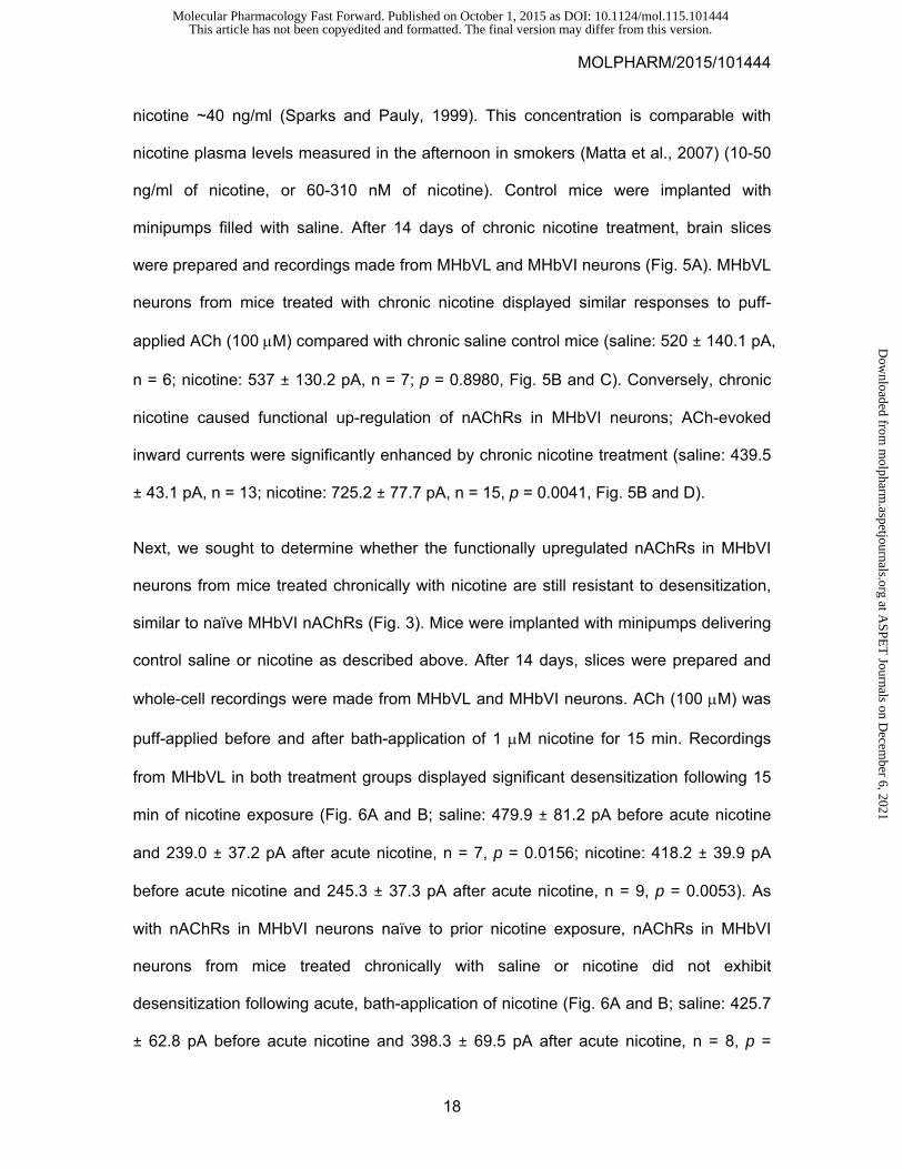
minipumps filled with saline. After 14 days of chronic nicotine treatment, brain slices
were prepared and recordings made from MHbVL and MHbVI neurons (Fig. 5A). MHbVL
neurons from mice treated with chronic nicotine displayed similar responses to puff-
applied ACh (100 M) compared with chronic saline control mice (saline: 520 ± 140.1 pA,
n = 6; nicotine: 537 ± 130.2 pA, n = 7; p = 0.8980, Fig. 5B and C). Conversely, chronic
nicotine caused functional up-regulation of nAChRs in MHbVI neurons; ACh-evoked
inward currents were significantly enhanced by chronic nicotine treatment (saline: 439.5
± 43.1 pA, n = 13; nicotine: 725.2 ± 77.7 pA, n = 15, p = 0.0041, Fig. 5B and D).
Next, we sought to determine whether the functionally upregulated nAChRs in MHbVI
neurons from mice treated chronically with nicotine are still resistant to desensitization,
similar to naïve MHbVI nAChRs (Fig. 3). Mice were implanted with minipumps delivering
control saline or nicotine as described above. After 14 days, slices were prepared and
whole-cell recordings were made from MHbVL and MHbVI neurons. ACh (100 M) was
puff-applied before and after bath-application of 1 M nicotine for 15 min. Recordings
from MHbVL in both treatment groups displayed significant desensitization following 15
min of nicotine exposure (Fig. 6A and B; saline: 479.9 ± 81.2 pA before acute nicotine
and 239.0 ± 37.2 pA after acute nicotine, n = 7, p = 0.0156; nicotine: 418.2 ± 39.9 pA
before acute nicotine and 245.3 ± 37.3 pA after acute nicotine, n = 9, p = 0.0053). As
with nAChRs in MHbVI neurons naïve to prior nicotine exposure, nAChRs in MHbVI
neurons from mice treated chronically with saline or nicotine did not exhibit
desensitization following acute, bath-application of nicotine (Fig. 6A and B; saline: 425.7
± 62.8 pA before acute nicotine and 398.3 ± 69.5 pA after acute nicotine, n = 8, p =
This article has not been copyedited and formatted. The final version may differ from this version.Molecular Pharmacology Fast Forward. Published on October 1, 2015 as DOI: 10.1124/mol.115.101444
at ASPE
T Journals on D
ecember 6, 2021
molpharm
.aspetjournals.orgD
ownloaded from
MOLPHARM/2015/101444
19
0.4171; nicotine: 696.9 ± 82.5 pA before acute nicotine and 579.9 ± 60.6 pA after acute
nicotine, n = 11, p = 0.1429).
Finally, we sought to identify the functional consequences of chronic nicotine treatment
on neuronal activity in MHbVL and MHbVI. Experiments were performed as described in
Fig. 5A; cell-attached patch clamp recordings were made in MHbVL and MHbVI neurons
from mice treated chronically with nicotine or saline for 14 days. Recordings in MHbVL
neurons revealed that basal firing was indistinguishable between chronic nicotine-treated
and chronic saline-treated groups (Fig. 7A and C; saline: 4.2 ± 0.8 Hz, n = 6; nicotine:
4.1 ± 0.3 Hz, n = 17; p = 0.9359) and that bath application of acute nicotine (1 M)
resulted in a similar increase of firing frequency in both groups (Fig. 7D; saline: 2.0 ± 0.2
fold-increase, n = 6; nicotine: 1.8 ± 0.2 fold-increase, n = 6; p = 0.4679). Also, the fold-
increase in MHbVL neuron action potential firing was the same as in untreated (no
minipump implanted) mice (ANOVA: p = 0.3461). Remarkably, MHbVI neurons from
mice treated chronically with nicotine displayed sustained elevation in their baseline
firing rates relative to MHbVI neurons from mice treated chronically with saline (Fig. 7B
and C; saline: 4.5 ± 0.7 Hz, n = 9; nicotine: 7.4 ± 0.6 Hz, n = 14; p = 0.0064). In contrast
to MHbVL neurons, when MHbVI neurons from mice treated chronically with nicotine
were exposed to acute nicotine (1 M), there was no additional gain in action potential
firing compared with MHbVI neurons from mice treated chronically with saline (Fig. 7D;
saline: 2.1 ± 0.3 fold-increase, n = 6; nicotine: 1.1 ± 0.1 fold-increase, n = 7; p = 0.0188).
Together, these results indicate that chronic nicotine selectively alters nAChR function
and neuronal activity in different subregions of MHb.
This article has not been copyedited and formatted. The final version may differ from this version.Molecular Pharmacology Fast Forward. Published on October 1, 2015 as DOI: 10.1124/mol.115.101444
at ASPE
T Journals on D
ecember 6, 2021
molpharm
.aspetjournals.orgD
ownloaded from
MOLPHARM/2015/101444
20
Discussion
In this study, we sought to investigate subregions of MHb with regard to nAChR
pharmacology and neuronal excitability using brain slice patch clamp electrophysiology.
We determined that MHbVL and MHbVI subregions express pharmacologically distinct
nAChRs. These distinct nAChRs respond differentially to both acute and chronic nicotine,
and these differences directly translate into divergent action potential firing patterns in
these MHb subregions.
Differential MHb nAChR Pharmacology
Our group and others have demonstrated high levels of 3 and 4 nAChR subunit
expression and function in all areas of ventral MHb (Quick et al., 1999; Shih et al., 2014).
SR16584 antagonizes 34 nAChRs and has little activity at 42 and 7 nAChRs
(Zaveri et al., 2010). Thus, our data demonstrating that bath-applied SR16584 was
effective at blocking ACh-evoked currents in both MHbVL and MHbVI are consistent with
the prevailing notion that 34* nAChRs play a dominant role in mediating nicotinic
cholinergic signaling in MHb (Quick et al., 1999). Future pharmacological studies of
SR16584’s action at other nAChR subtypes will help confirm this suggestion. Although
inward current responses attributed to MHbVL and MHbVI nAChRs were similar in
overall sensitivity to puff-applied ACh (Fig. 1C and D), there was a differential sensitivity
to DHE (Fig. 1G and H). Although generally used to discriminate 7 from 2* nAChRs
in native preparations, DHE has moderate antagonist activity at uncommon nAChR
subtypes that may be present in MHb. For example, experiments using human nAChRs
expressed in Xenopus oocytes showed that DHE is a potent antagonist at 22 and
44 nAChRs (Chavez-Noriega et al., 1997). A wide array of nAChR subunits that are
expressed in MHb cells, and responses evoked by 100 M ACh are likely aggregate
responses of multiple nAChR subtypes. Our DHE data could reflect a slight sensitivity
This article has not been copyedited and formatted. The final version may differ from this version.Molecular Pharmacology Fast Forward. Published on October 1, 2015 as DOI: 10.1124/mol.115.101444
at ASPE
T Journals on D
ecember 6, 2021
molpharm
.aspetjournals.orgD
ownloaded from
MOLPHARM/2015/101444
21
of multiple of these putative subtypes, or it could reflect complete block of a minor
subtype and no antagonism of the others. More studies will be required to probe this
question.
Our experiments (Fig. 2) utilizing CtxMII, ACh applications, and 6L9S transgenic mice
collectively support the idea that functional 6* nAChRs are expressed in MHbVI
neurons but excluded from MHbVL neurons. These findings are in line with our previous
study using mouse strains expressing GFP-tagged 6 subunits, where 6 subunit
immunoreactivity was demonstrated in MHbVI cells (Shih et al., 2014). Our findings are
similarly consistent with (Henderson et al., 2014), which also demonstrated 6 subunits
in a subregion of MHb consistent with MHbVI.
Although nAChRs in MHbVL and MHbVI exhibited modest differences in response to
acutely applied ACh, differences in their pharmacology was more pronounced when
nicotine application was carried out. Nicotine-induced action potential firing was more
rapid to reach peak but attenuated in MHbVL cells. In contrast, firing developed slower
and was more sustained in MHbVI cells (Fig. 3). The ability of the same nicotine
treatment protocol to preferentially desensitize nAChRs in MHbVL (Fig. 4) may account
for the differences in action potential firing we observed. 34 nAChRs, although not
especially sensitive to the desensitizing properties of nicotine (Fenster et al., 1997), are
expressed throughout the ventral MHb and therefore may not explain the difference we
observe. Rather, 4 subunits in MHbVL may confer sensitivity to desensitization by
nicotine whereas the specific expression of 6 subunits in MHbVI may confer resistance
to desensitization. This supposition is supported by our data showing that MHbVL
nAChRs lacking 4 subunits no longer desensitize in response to a 15 min exposure to
nicotine (Fig. 4F and G). It is also supported by similar studies in VTA, another brain
This article has not been copyedited and formatted. The final version may differ from this version.Molecular Pharmacology Fast Forward. Published on October 1, 2015 as DOI: 10.1124/mol.115.101444
at ASPE
T Journals on D
ecember 6, 2021
molpharm
.aspetjournals.orgD
ownloaded from
MOLPHARM/2015/101444
22
region expressing 4 and 6 subunits. Tapper and colleagues recently demonstrated
that 6-expressing neurons in VTA exhibit more sustained firing when exposed to
nicotine in the bath (Liu et al., 2012). Marks and colleagues also demonstrated that 6
subunits confer resistance to desensitization by nicotine (Grady et al., 2012). It should
be noted that these cited studies concern 4* and 6* nAChRs that assemble with 2
subunits. It is not yet clear whether 2 subunits play a role in the differential
desensitization that we observe in MHb. 2 subunits are expressed in most areas of
ventral MHb, and account for a substantial fraction of nAChRs in both mouse and rat
(Grady et al., 2009; Shih et al., 2014).
Responses of MHb nAChRs and Neurons to Chronic Nicotine
In mice, chronic systemic nicotine treatment induces functional changes to nAChRs
and/or neuronal circuits in the MHb to IPN pathway that is revealed by spontaneous or
precipitated withdrawal approaches. Blockade of ongoing nicotinic cholinergic signaling
in the MHb of mice chronically exposed to nicotine triggers withdrawal, and specific
nAChR subtypes play a role in this response (Salas et al., 2009). Our goal was to further
study this activity of nicotine, and we identified the MHbVI as a specific site that
undergoes functional upregulation of nAChR activity in response to chronic nicotine (Fig.
5). These data are consistent with recent findings indicating that MHb 6 subunits, which
are concentrated in MHbVI (Shih et al., 2014), upregulate following chronic nicotine
treatment (Henderson et al., 2014). Conversely, we did not find nAChR activity to be
functionally enhanced in MHbVL after chronic nicotine treatment (Fig. 5). This result is
consistent with previous work showing that 4 subunits, which are concentrated in
MHbVL (Shih et al., 2014), do not upregulate in response to chronic nicotine (Nashmi et
al., 2007). Indeed, after 24 h of withdrawal from chronic nicotine, responses attributed to
4 subunits exhibit downregulation (Shih et al., 2014). Although these data and
This article has not been copyedited and formatted. The final version may differ from this version.Molecular Pharmacology Fast Forward. Published on October 1, 2015 as DOI: 10.1124/mol.115.101444
at ASPE
T Journals on D
ecember 6, 2021
molpharm
.aspetjournals.orgD
ownloaded from
MOLPHARM/2015/101444
23
observations from the literature suggest that 6 upregulates in the MHbVI in our
experiments, further electrophysiology studies will be necessary to probe this question.
This is particularly important given that our pharmacological experiments suggest that
nAChRs containing 6 subunits are a minority, though high-sensitivity, subtype in MHbVI.
These results demonstrate the importance of considering MHb subregions
independently when studying nAChRs and chronic nicotine, as several recent studies
did not identify changes in the response of MHb neurons to nicotine when the MHb was
analyzed without considering subregions (Dao et al., 2014; Gorlich et al., 2013; Hsu et
al., 2013).
In addition to identifying changes in nAChR activity following chronic nicotine, we also
connected these changes with alterations in action potential firing in MHb neurons
exposed to nicotine. Chronic nicotine, but not chronic saline, caused an elevation in
baseline action potential firing in MHbVI neurons (Fig. 7B). Likely as a consequence of
enhanced baseline firing/excitability, acute nicotine was no longer able to further
increase firing in our experiments (Fig. 7D). Thus, we have identified a specific neuron
type that exhibits tolerance to acute nicotine exposure as a result of prior chronic
exposure to nicotine. If our slice experiments are representative of MHbVI firing patterns
in vivo, these data describe a potentially important change in MHb/IPN pathway circuit
activity. Our recent data describing nAChR subunit expression/localization in the
MHb/IPN pathway (Shih et al., 2014) confirm what was previously seen in anatomical
studies (Contestabile and Flumerfelt, 1981): MHbVI projects to ventral IPN areas
whereas MHbVL projects more preferentially to dorsal IPN. It has recently become
apparent that dorsal IPN neurons control somatic withdrawal responses (Zhao-Shea et
al., 2013), and ventral IPN neurons control affective responses (Zhao-Shea et al., 2015).
Moreover, during precipitated withdrawal from chronic nicotine exposure, MHb-derived
This article has not been copyedited and formatted. The final version may differ from this version.Molecular Pharmacology Fast Forward. Published on October 1, 2015 as DOI: 10.1124/mol.115.101444
at ASPE
T Journals on D
ecember 6, 2021
molpharm
.aspetjournals.orgD
ownloaded from
MOLPHARM/2015/101444
24
inputs onto ventral IPN neurons control anxiety responses (Zhao-Shea et al., 2015). In
our study, enhanced baseline firing and reduced nicotine-elicited firing in MHbVI neurons
chronically exposed to nicotine is likely involved in activation of this affective withdrawal
circuit in ventral IPN. Exerting specific control over MHbVI nAChRs and/or neuronal
activity could be useful in modulating anxiety during nicotine withdrawal/cessation in
human tobacco users.
Conclusion & Impact
The data we present here advance our knowledge of a key brain circuit involved in
human nicotine intake, and future studies in rats or non-human primates should test our
present conclusions in mouse models. We have shown previously (Shih et al., 2014) and
in this study that MHb neurons and nAChRs are diverse in their pharmacology and
activity patterns/excitability, respectively. As such, future mechanistic studies on the role
of the MHb in nicotine addiction/withdrawal should discriminate between the various
subregions that exist within the MHb. Because these different MHb subregions are
known to project to different areas of IPN, and because these different areas of IPN
mediate different aspects of nicotine withdrawal, these results may lead to a better
understanding of nicotine dependence.
This article has not been copyedited and formatted. The final version may differ from this version.Molecular Pharmacology Fast Forward. Published on October 1, 2015 as DOI: 10.1124/mol.115.101444
at ASPE
T Journals on D
ecember 6, 2021
molpharm
.aspetjournals.orgD
ownloaded from

MOLPHARM/2015/101444
25
Acknowledgements
We thank members of the Drenan laboratory for helpful insight and discussion.
This article has not been copyedited and formatted. The final version may differ from this version.Molecular Pharmacology Fast Forward. Published on October 1, 2015 as DOI: 10.1124/mol.115.101444
at ASPE
T Journals on D
ecember 6, 2021
molpharm
.aspetjournals.orgD
ownloaded from
MOLPHARM/2015/101444
26
Authorship Contributions
Participated in research design: R.M.D. and P.-Y.S.
Conducted experiments: P.-Y.S.
Contributed new reagents or analytic tools: J.M.M.
Performed data analysis: R.M.D. and P.-Y.S.
Wrote or contributed to the writing of the manuscript: R.M.D. and P.-Y.S.
This article has not been copyedited and formatted. The final version may differ from this version.Molecular Pharmacology Fast Forward. Published on October 1, 2015 as DOI: 10.1124/mol.115.101444
at ASPE
T Journals on D
ecember 6, 2021
molpharm
.aspetjournals.orgD
ownloaded from
MOLPHARM/2015/101444
27
References
Aizawa H, Kobayashi M, Tanaka S, Fukai T and Okamoto H (2012) Molecular
characterization of the subnuclei in rat habenula. J Comp Neurol 520(18): 4051-
4066.
Azam L, Maskos U, Changeux JP, Dowell CD, Christensen S, De Biasi M and McIntosh
JM (2010) -Conotoxin BuIA[T5A;P6O]: a novel ligand that discriminates
between 64 and 62 nicotinic acetylcholine receptors and blocks nicotine-
stimulated norepinephrine release. FASEB J 24(12): 5113-5123.
Bromberg-Martin ES, Matsumoto M and Hikosaka O (2010) Dopamine in motivational
control: rewarding, aversive, and alerting. Neuron 68(5): 815-834.
Cartier GE, Yoshikami D, Gray WR, Luo S, Olivera BM and McIntosh JM (1996) A new
-conotoxin which targets 32 nicotinic acetylcholine receptors. J Biol Chem
271(13): 7522-7528.
Chavez-Noriega LE, Crona JH, Washburn MS, Urrutia A, Elliott KJ and Johnson EC
(1997) Pharmacological characterization of recombinant human neuronal
nicotinic acetylcholine receptors hα2β2, hα2β4, hα3β2, hα3β4, hα4β2, hα4β4
and hα7 expressed in Xenopus oocytes. J Pharmacol Exp Ther 280(1): 346-356.
Cohen BN, Mackey ED, Grady SR, McKinney S, Patzlaff NE, Wageman CR, McIntosh
JM, Marks MJ, Lester HA and Drenan RM (2012) Nicotinic cholinergic
mechanisms causing elevated dopamine release and abnormal locomotor
behavior. Neuroscience 200: 31-41.
Contestabile A and Flumerfelt BA (1981) Afferent connections of the interpeduncular
nucleus and the topographic organization of the habenulo-interpeduncular
pathway: an HRP study in the rat. J Comp Neurol 196(2): 253-270.
This article has not been copyedited and formatted. The final version may differ from this version.Molecular Pharmacology Fast Forward. Published on October 1, 2015 as DOI: 10.1124/mol.115.101444
at ASPE
T Journals on D
ecember 6, 2021
molpharm
.aspetjournals.orgD
ownloaded from
MOLPHARM/2015/101444
28
Contestabile A, Villani L, Fasolo A, Franzoni MF, Gribaudo L, Oktedalen O and Fonnum
F (1987) Topography of cholinergic and substance P pathways in the habenulo-
interpeduncular system of the rat. An immunocytochemical and microchemical
approach. Neuroscience 21(1): 253-270.
Dani JA and Bertrand D (2007) Nicotinic acetylcholine receptors and nicotinic cholinergic
mechanisms of the central nervous system. Annu Rev Pharmacol Toxicol 47:
699-729.
Dani JA and Harris RA (2005) Nicotine addiction and comorbidity with alcohol abuse and
mental illness. Nature neuroscience 8(11): 1465-1470.
Dao DQ, Perez EE, Teng Y, Dani JA and De Biasi M (2014) Nicotine Enhances
Excitability of Medial Habenular Neurons via Facilitation of Neurokinin Signaling.
J Neurosci 34(12): 4273-4284.
Drenan RM, Grady SR, Steele AD, McKinney S, Patzlaff NE, McIntosh JM, Marks MJ,
Miwa JM and Lester HA (2010) Cholinergic modulation of locomotion and striatal
dopamine release is mediated by 64* nicotinic acetylcholine receptors. J
Neurosci 30(29): 9877-9889.
Drenan RM, Grady SR, Whiteaker P, McClure-Begley T, McKinney S, Miwa JM, Bupp S,
Heintz N, McIntosh JM, Bencherif M, Marks MJ and Lester HA (2008) In vivo
activation of midbrain dopamine neurons via sensitized, high-affinity 6* nicotinic
acetylcholine receptors. Neuron 60(1): 123-136.
Engle SE, Broderick HJ and Drenan RM (2012) Local application of drugs to study
nicotinic acetylcholine receptor function in mouse brain slices. J Vis Exp(68):
e50034.
Engle SE, Shih PY, McIntosh JM and Drenan RM (2013) α4α6β2* nicotinic acetylcholine
receptor activation on ventral tegmental area dopamine neurons is sufficient to
This article has not been copyedited and formatted. The final version may differ from this version.Molecular Pharmacology Fast Forward. Published on October 1, 2015 as DOI: 10.1124/mol.115.101444
at ASPE
T Journals on D
ecember 6, 2021
molpharm
.aspetjournals.orgD
ownloaded from
MOLPHARM/2015/101444
29
stimulate a depolarizing conductance and enhance surface AMPA receptor
function. Mol Pharmacol 84(3): 393-406.
Fenster CP, Rains MF, Noerager B, Quick MW and Lester RA (1997) Influence of
subunit composition on desensitization of neuronal acetylcholine receptors at low
concentrations of nicotine. J Neurosci 17(15): 5747-5759.
Fonck C, Nashmi R, Salas R, Zhou C, Huang Q, De Biasi M, Lester RA and Lester HA
(2009) Demonstration of functional 4-containing nicotinic receptors in the medial
habenula. Neuropharmacology 56(1): 247-253.
Fowler CD, Lu Q, Johnson PM, Marks MJ and Kenny PJ (2011) Habenular 5 nicotinic
receptor subunit signalling controls nicotine intake. Nature 471(7340): 597-601.
Frahm S, Slimak MA, Ferrarese L, Santos-Torres J, Antolin-Fontes B, Auer S, Filkin S,
Pons S, Fontaine JF, Tsetlin V, Maskos U and Ibanez-Tallon I (2011) Aversion to
Nicotine Is Regulated by the Balanced Activity of β4 and α5 Nicotinic Receptor
Subunits in the Medial Habenula. Neuron 70(3): 522-535.
Gorlich A, Antolin-Fontes B, Ables JL, Frahm S, Slimak MA, Dougherty JD and Ibanez-
Tallon I (2013) Reexposure to nicotine during withdrawal increases the
pacemaking activity of cholinergic habenular neurons. Proc Natl Acad Sci U S A.
Grady SR, Moretti M, Zoli M, Marks MJ, Zanardi A, Pucci L, Clementi F and Gotti C
(2009) Rodent habenulo-interpeduncular pathway expresses a large variety of
uncommon nAChR subtypes, but only the α3β4* and α3β3β4* subtypes mediate
acetylcholine release. J Neurosci 29(7): 2272-2282.
Grady SR, Wageman CR, Patzlaff NE and Marks MJ (2012) Low concentrations of
nicotine differentially desensitize nicotinic acetylcholine receptors that include α5
or α6 subunits and that mediate synaptosomal neurotransmitter release.
Neuropharmacology 62(5-6): 1935-1943.
This article has not been copyedited and formatted. The final version may differ from this version.Molecular Pharmacology Fast Forward. Published on October 1, 2015 as DOI: 10.1124/mol.115.101444
at ASPE
T Journals on D
ecember 6, 2021
molpharm
.aspetjournals.orgD
ownloaded from
MOLPHARM/2015/101444
30
Henderson BJ, Srinivasan R, Nichols WA, Dilworth CN, Gutierrez DF, Mackey ED,
McKinney S, Drenan RM, Richards CI and Lester HA (2014) Nicotine exploits a
COPI-mediated process for chaperone-mediated up-regulation of its receptors.
The Journal of general physiology 143(1): 51-66.
Herkenham M and Nauta WJ (1977) Afferent connections of the habenular nuclei in the
rat. A horseradish peroxidase study, with a note on the fiber-of-passage problem.
J Comp Neurol 173(1): 123-146.
Hikosaka O (2010) The habenula: from stress evasion to value-based decision-making.
Nat Rev Neurosci 11(7): 503-513.
Hilario MR, Turner JR and Blendy JA (2012) Reward sensitization: effects of repeated
nicotine exposure and withdrawal in mice. Neuropsychopharmacology : official
publication of the American College of Neuropsychopharmacology 37(12): 2661-
2670.
Hsu YW, Tempest L, Quina LA, Wei AD, Zeng H and Turner EE (2013) Medial Habenula
Output Circuit Mediated by α5 Nicotinic Receptor-Expressing GABAergic
Neurons in the Interpeduncular Nucleus. J Neurosci 33(46): 18022-18035.
Laviolette SR and van der Kooy D (2004) The neurobiology of nicotine addiction:
bridging the gap from molecules to behaviour. Nat Rev Neurosci 5(1): 55-65.
Lester HA, Xiao C, Srinivasan R, Son CD, Miwa J, Pantoja R, Banghart MR, Dougherty
DA, Goate AM and Wang JC (2009) Nicotine is a selective pharmacological
chaperone of acetylcholine receptor number and stoichiometry. Implications for
drug discovery. Aaps J 11(1): 167-177.
Liu L, Zhao-Shea R, McIntosh JM, Gardner P and Tapper A (2012) Nicotine Persistently
Activates Ventral Tegmental Area Dopaminergic Neurons Via Nicotinic
Acetylcholine Receptors Containing a4 and a6 subunits. Mol Pharmacol 81(4):
541-548.
This article has not been copyedited and formatted. The final version may differ from this version.Molecular Pharmacology Fast Forward. Published on October 1, 2015 as DOI: 10.1124/mol.115.101444
at ASPE
T Journals on D
ecember 6, 2021
molpharm
.aspetjournals.orgD
ownloaded from

MOLPHARM/2015/101444
31
Malkawi AH, Al-Ghananeem AM, de Leon J and Crooks PA (2009) Nicotine exposure
can be detected in cerebrospinal fluid of active and passive smokers. J Pharm
Biomed Anal 49(1): 129-132.
Marks MJ, Burch JB and Collins AC (1983) Effects of chronic nicotine infusion on
tolerance development and nicotinic receptors. J Pharmacol Exp Ther 226(3):
817-825.
Marks MJ, Pauly JR, Gross SD, Deneris ES, Hermans-Borgmeyer I, Heinemann SF and
Collins AC (1992) Nicotine binding and nicotinic receptor subunit RNA after
chronic nicotine treatment. J Neurosci 12(7): 2765-2784.
Matsumoto M and Hikosaka O (2007) Lateral habenula as a source of negative reward
signals in dopamine neurons. Nature 447(7148): 1111-1115.
Matsumoto M and Hikosaka O (2009) Representation of negative motivational value in
the primate lateral habenula. Nature neuroscience 12(1): 77-84.
Matta SG, Balfour DJ, Benowitz NL, Boyd RT, Buccafusco JJ, Caggiula AR, Craig CR,
Collins AC, Damaj MI, Donny EC, Gardiner PS, Grady SR, Heberlein U, Leonard
SS, Levin ED, Lukas RJ, Markou A, Marks MJ, McCallum SE, Parameswaran N,
Perkins KA, Picciotto MR, Quik M, Rose JE, Rothenfluh A, Schafer WR,
Stolerman IP, Tyndale RF, Wehner JM and Zirger JM (2007) Guidelines on
nicotine dose selection for in vivo research. Psychopharmacology 190(3): 269-
319.
McIntosh JM, Azam L, Staheli S, Dowell C, Lindstrom JM, Kuryatov A, Garrett JE, Marks
MJ and Whiteaker P (2004) Analogs of -Conotoxin MII are selective for 6-
containing nicotinic acetylcholine receptors. Mol Pharmacol 65(4): 944-952.
Montone KT, Fass B and Hamill GS (1988) Serotonergic and nonserotonergic
projections from the rat interpeduncular nucleus to the septum, hippocampal
formation and raphe: a combined immunocytochemical and fluorescent
This article has not been copyedited and formatted. The final version may differ from this version.Molecular Pharmacology Fast Forward. Published on October 1, 2015 as DOI: 10.1124/mol.115.101444
at ASPE
T Journals on D
ecember 6, 2021
molpharm
.aspetjournals.orgD
ownloaded from

MOLPHARM/2015/101444
32
retrograde labelling study of neurons in the apical subnucleus. Brain research
bulletin 20(2): 233-240.
Nashmi R, Xiao C, Deshpande P, McKinney S, Grady SR, Whiteaker P, Huang Q,
McClure-Begley T, Lindstrom JM, Labarca C, Collins AC, Marks MJ and Lester
HA (2007) Chronic nicotine cell specifically upregulates functional 4* nicotinic
receptors: basis for both tolerance in midbrain and enhanced long-term
potentiation in perforant path. J Neurosci 27(31): 8202-8218.
Pidoplichko VI, DeBiasi M, Williams JT and Dani JA (1997) Nicotine activates and
desensitizes midbrain dopamine neurons. Nature 390(6658): 401-404.
Powers MS, Broderick HJ, Drenan RM and Chester JA (2013) Nicotinic acetylcholine
receptors containing 6 subunits contribute to alcohol reward-related behaviours.
Genes Brain Behav 12(5): 543-553.
Qin C and Luo M (2009) Neurochemical phenotypes of the afferent and efferent
projections of the mouse medial habenula. Neuroscience 161(3): 827-837.
Quick MW, Ceballos RM, Kasten M, McIntosh JM and Lester RA (1999) α3β4 subunit-
containing nicotinic receptors dominate function in rat medial habenula neurons.
Neuropharmacology 38(6): 769-783.
Salas R, Pieri F and De Biasi M (2004) Decreased signs of nicotine withdrawal in mice
null for the β4 nicotinic acetylcholine receptor subunit. J Neurosci 24(45): 10035-
10039.
Salas R, Sturm R, Boulter J and De Biasi M (2009) Nicotinic receptors in the habenulo-
interpeduncular system are necessary for nicotine withdrawal in mice. J Neurosci
29(10): 3014-3018.
Salminen O, Drapeau JA, McIntosh JM, Collins AC, Marks MJ and Grady SR (2007)
Pharmacology of -Conotoxin MII-Sensitive Subtypes of Nicotinic Acetylcholine
This article has not been copyedited and formatted. The final version may differ from this version.Molecular Pharmacology Fast Forward. Published on October 1, 2015 as DOI: 10.1124/mol.115.101444
at ASPE
T Journals on D
ecember 6, 2021
molpharm
.aspetjournals.orgD
ownloaded from
MOLPHARM/2015/101444
33
Receptors Isolated by Breeding of Null Mutant Mice. Mol Pharmacol 71(6): 1563-
1571.
Shibata H and Suzuki T (1984) Efferent projections of the interpeduncular complex in the
rat, with special reference to its subnuclei: a retrograde horseradish peroxidase
study. Brain research 296(2): 345-349.
Shih PY, Engle SE, Oh G, Deshpande P, Puskar NL, Lester HA and Drenan RM (2014)
Differential expression and function of nicotinic acetylcholine receptors in
subdivisions of medial habenula. J Neurosci 34(29): 9789-9802.
Sparks JA and Pauly JR (1999) Effects of continuous oral nicotine administration on
brain nicotinic receptors and responsiveness to nicotine in C57Bl/6 mice.
Psychopharmacology 141(2): 145-153.
Steinsland OS and Furchgott RF (1975) Desensitization of the adrenergic neurons of the
isolated rabbit ear artery to nicotinic agonists. J Pharmacol Exp Ther 193(1): 138-
148.
WHO (2011) Department of Mental Health and Substance Abuse, Global Status Report
on Alcohol 2011. World Health Organization Department of Mental Health and
Substance Abuse Geneva.
Yamaguchi T, Danjo T, Pastan I, Hikida T and Nakanishi S (2013) Distinct roles of
segregated transmission of the septo-habenular pathway in anxiety and fear.
Neuron 78(3): 537-544.
Zaveri N, Jiang F, Olsen C, Polgar W and Toll L (2010) Novel α3β4 nicotinic
acetylcholine receptor-selective ligands. Discovery, structure-activity studies, and
pharmacological evaluation. Journal of medicinal chemistry 53(22): 8187-8191.
Zhao-Shea R, DeGroot SR, Liu L, Vallaster M, Pang X, Su Q, Gao G, Rando OJ, Martin
GE, George O, Gardner PD and Tapper AR (2015) Increased CRF signalling in a
This article has not been copyedited and formatted. The final version may differ from this version.Molecular Pharmacology Fast Forward. Published on October 1, 2015 as DOI: 10.1124/mol.115.101444
at ASPE
T Journals on D
ecember 6, 2021
molpharm
.aspetjournals.orgD
ownloaded from

MOLPHARM/2015/101444
34
ventral tegmental area-interpeduncular nucleus-medial habenula circuit induces
anxiety during nicotine withdrawal. Nature communications 6: 6770.
Zhao-Shea R, Liu L, Pang X, Gardner PD and Tapper AR (2013) Activation of
GABAergic neurons in the interpeduncular nucleus triggers physical nicotine
withdrawal symptoms. Current biology : CB 23(23): 2327-2335.
This article has not been copyedited and formatted. The final version may differ from this version.Molecular Pharmacology Fast Forward. Published on October 1, 2015 as DOI: 10.1124/mol.115.101444
at ASPE
T Journals on D
ecember 6, 2021
molpharm
.aspetjournals.orgD
ownloaded from

MOLPHARM/2015/101444
35
Footnotes
This work was supported by the National Institutes of Health [DA030396], the Brain and
Behavior Research Foundation (via a NARSAD Young Investigator Award), the Ralph W.
and Grace M. Showalter Research Trust, and Purdue University.
Reprint requests should be directed to Ryan M. Drenan (575 Stadium Mall Dr., West
Lafayette, IN 47907; [email protected]).
This article has not been copyedited and formatted. The final version may differ from this version.Molecular Pharmacology Fast Forward. Published on October 1, 2015 as DOI: 10.1124/mol.115.101444
at ASPE
T Journals on D
ecember 6, 2021
molpharm
.aspetjournals.orgD
ownloaded from
MOLPHARM/2015/101444
36
Figure Legends
Figure 1. Functional differences in nAChRs between MHbVL and MHbVI neurons.
(A) Schematic diagram defining a third of the ventrolateral portion of MHb as MHbVL
and a third of the ventroinferior portion of MHb as MHbVI.
(B) Locations of recorded cells in MHbVL and MHbVI were visualized by intracellular
biocytin staining (in green) and immunostaining for choline acetyltransferase (ChAT;
red). Yellow cells (white arrow) are double-labeled cells. Representative data from
labeled cells in MHbVL (left panel) and MHbVI (right panel) are shown. Scale bar:
100 m.
(C) Examples of currents evoked by 1 μM and 100 μM acetylcholine (250 ms puffs) in
MHbVL. Scale bar: 10 pA (1 M ACh) and 200 pA (100 M ACh), 500 ms.
(D) Summary plot of peak ACh-evoked currents at the indicated concentrations in
MHbVL and MHbVI neurons.
(E) Representative traces showing 100 μM ACh-induced current before (black trace) and
15 min after (gray trace; offset for clarity) application of SR16584 (20 M). Arrows
indicate ACh applications. Scale bar: 250 pA, 350 ms (MHbVL); 150 pA, 350 ms
(MHbVI).
(F) Summary bar graphs are plotted to indicate the degree of inhibition (i.e., the fraction
of control current remaining after application of SR16584). *p < 0.05 (paired t-test).
(G) Representative traces showing 100 μM ACh-induced current before (black trace) and
15 min after (gray trace; offset for clarity) application of DHE (500 nM). Arrows
indicate ACh applications. Scale bar: 250 pA, 350 ms (MHbVL); 200 pA, 350 ms
(MHbVI).
(H) Summary bar graphs are plotted to indicate the degree of inhibition (i.e., the fraction
of control current remaining after application of DHE). *p < 0.05 (paired t-test).
This article has not been copyedited and formatted. The final version may differ from this version.Molecular Pharmacology Fast Forward. Published on October 1, 2015 as DOI: 10.1124/mol.115.101444
at ASPE
T Journals on D
ecember 6, 2021
molpharm
.aspetjournals.orgD
ownloaded from

MOLPHARM/2015/101444
37
Figure 2. Functional 6* nAChR expression is specific to MHbVI
(A) Representative traces showing 100 M-induced currents in MHbVL and MHbVI
neurons before (black trace) and 15 min after (gray trace; offset for clarity)
application of CtxMII (100 nM). Arrows indicate ACh applications. Scale bar: 100
pA, 500 ms (MHbVL); 90 pA, 500 ms (MHbVI).
(B) Summary bar graphs to indicate the degree of inhibition by CtxMII (i.e. the fraction
of control current remaining after application of CtxMII) for ACh-evoked responses
in MHbVL and MHbVI neurons.
(C) Representative traces showing 30 M (for MHbVL neurons) or 10 M (for MHbVI
neurons) ACh-evoked currents before (black trace) and after (grey trace; offset for
clarity) application of CtxMII (100 nM). Different ACh concentrations were used in
MHbVL vs. MHbVI based on the minimum ACh concentration that evoked workable
inward currents. Scale bar: 50 pA, 500 ms (MHbVL); 60 pA, 500 ms (MHbVI).
(D) Summary bar graphs to indicate the degree of inhibition by CtxMII (i.e. the fraction
of control current remaining after application of CtxMII) for 30 M (MHbVL) or 10
M (MHbVI) ACh-evoked responses. *p < 0.05 (paired t-test)
(E) Representative traces for 1 M ACh-evoked currents in MHbVL and MHbVI cells in
slices from 6L9S mice. MHbVI cells, which responded robustly to 1 M ACh (black
trace), were subsequently exposed to CtxMII (100 nM), and 1 M ACh-evoked
currents were measured in the presence of CtxMII (grey trace). Scale bar: 70 pA,
500 ms.
(F) Summary bar graphs to indicate the average 1 M ACh-evoked current response
amplitude in MHbVL and MHbVI neurons in slices from 6L9S and non-transgenic
control littermate (WT) mice. ***p < 0.001 (one-way ANOVA, Tukey post hoc test)
This article has not been copyedited and formatted. The final version may differ from this version.Molecular Pharmacology Fast Forward. Published on October 1, 2015 as DOI: 10.1124/mol.115.101444
at ASPE
T Journals on D
ecember 6, 2021
molpharm
.aspetjournals.orgD
ownloaded from

MOLPHARM/2015/101444
38
Figure 3. Effect of prolonged nicotine application on firing rate as measured in cell-
attached recordings in MHbVL and MHbVI.
(A) Cell-attached recordings from MHbVL and MHbVI neurons were conducted. Baseline
firing was recorded for several minutes, followed by superfusion of nicotine (1 μM)
for 16 min.
(B) Representative traces obtained in cell-attached mode in MHbVL and MHbVI cells.
Each experiment comprised a recording taken at -60 mV, and the firing rates were
calculated at time point i (t = 2 min, baseline), ii (t = 8 min for MHbVL, t = 10 min for
MHbVI, peak response in 1 μM nicotine), and iii (t = 18 min, prolonged exposure).
(C) Quantification of summary data shown in (B). Average firing rates at the indicated
time points are shown. **p < 0.01, ***p < 0.001
(paired t-test)
Figure 4. Effects of acute nicotine on ACh-induced currents in MHbVL and MHbVI
neurons.
(A) Diagram of the experimental protocol. MHb neurons were held at -60 mV. Three ACh
(100 μM) applications (each arrow is one application) prior to nicotine (1 M) and
three after 15-min exposure to nicotine were conducted.
(B) Representative inward currents induced by ACh application before (black trace) and
after (gray trace) nicotine application to MHbVL neurons. Scale bar: 250 pA, 500 ms.
(C) Summary data showing nicotine-induced changes of nAChR-mediated peak currents
in MHbVL neurons. ***p < 0.001 (paired t-test)
(D) Representative inward currents induced by ACh application before (black trace) and
after (gray trace) nicotine application to MHbVI neurons. Scale bar: 250 pA, 500 ms.
(E) Summary data showing nicotine-induced changes of nAChR-mediated peak currents
in MHbVI neurons.
This article has not been copyedited and formatted. The final version may differ from this version.Molecular Pharmacology Fast Forward. Published on October 1, 2015 as DOI: 10.1124/mol.115.101444
at ASPE
T Journals on D
ecember 6, 2021
molpharm
.aspetjournals.orgD
ownloaded from
MOLPHARM/2015/101444
39
(F) Representative inward currents induced by ACh application before (black trace) and
after (gray trace) nicotine application to MHbVL neurons of 4KO mice. Scale bar:
250 pA, 500 ms.
(G) Summary data showing nicotine-induced changes of nAChR-mediated peak currents
in MHbVL neurons from 4KO mice.
Figure 5. Effects of chronic nicotine on ACh-induced currents in MHbVL and MHbVI
neurons.
(A) Nicotine dependence procedure. Mice were implanted with osmotic minipumps
delivering either saline (SAL) or nicotine (NIC; 1 mg/kg/h) for 14 days. On day 14,
slices were prepared for electrophysiology.
(B) Representative traces of ACh-evoked currents (100 μM, arrow) in MHbVL or MHbVI
neurons from animals treated either with chronic SAL or chronic NIC. Inward current
waveforms are an average of > 2 traces. Scale bar: 250 pA, 500 ms.
(C) Scatter plot and grouped data of peak amplitude of ACh-evoked currents from
MHbVL neurons from SAL- or NIC-treated mice.
(D) Scatter plot and grouped data of peak amplitude of ACh-evoked currents from
MHbVI neurons from SAL- or NIC-treated mice. *p < 0.05 (unpaired t-test)
Figure 6. Effects of chronic nicotine on desensitization of nAChRs in MHbVL and MHbVI
cells exposed to acute nicotine.
(A) Representative traces of ACh-evoked currents (100 M, arrow) in MHbVL and
MHbVI neurons from mice exposed to chronic saline (SAL) or chronic nicotine (NIC).
Experiments were conducted as described in Figure 4A: ACh (100 M) was puff-
applied three times, nicotine (1 M) was bath-applied for 16 min, and ACh was
applied three times again to the recorded cell. Scale bar: 200 pA, 500 ms.
This article has not been copyedited and formatted. The final version may differ from this version.Molecular Pharmacology Fast Forward. Published on October 1, 2015 as DOI: 10.1124/mol.115.101444
at ASPE
T Journals on D
ecember 6, 2021
molpharm
.aspetjournals.orgD
ownloaded from
MOLPHARM/2015/101444
40
(B) Summary data plot showing average current amplitude for 100 M ACh-evoked
responses in MHbVL neurons from mice treated chronically with SAL or NIC before
(control) and after bath-application of nicotine (NIC) (1 M). *p < 0.05, **p < 0.01
(paired t-test)
(C) Summary data plot showing average current amplitude for 100 M ACh-evoked
responses in MHbVI neurons from mice treated chronically with SAL or NIC before
(control) and after bath-application of nicotine (NIC) (1 M).
Figure 7. Excitability changes in MHb neurons from mice exposed to chronic nicotine or
saline.
(A) Baseline and nicotine-elicited changes in MHbVL neurons after chronic NIC
treatment. Representative cell-attached firing traces for MHbVL neurons showing the
typical change in firing are shown for the four indicated conditions (chronic SAL,
baseline; chronic SAL, 1 M nicotine; chronic NIC, baseline; chronic NIC, 1 M
nicotine).
(B) Baseline and nicotine-elicited changes in MHbVI neurons after chronic NIC treatment.
Representative cell-attached firing traces for MHbVI neurons showing the typical
change in firing are shown for the four indicated conditions (chronic SAL, baseline;
chronic SAL, 1 M nicotine; chronic NIC, baseline; chronic NIC, 1 M nicotine).
(C) Summary bar graph showing mean action potential firing rate at baseline for MHbVL
and MHbVI neurons from mice treated with chronic SAL or chronic NIC. **p < 0.01.
(unpaired t-test)
(D) Summary bar graph showing mean fold-change in action potential firing for nicotine-
exposed (1 M in bath for 15 min) MHbVL and MHbVI neurons from mice treated
with chronic SAL or chronic NIC. *p < 0.05 (unpaired t-test)
This article has not been copyedited and formatted. The final version may differ from this version.Molecular Pharmacology Fast Forward. Published on October 1, 2015 as DOI: 10.1124/mol.115.101444
at ASPE
T Journals on D
ecember 6, 2021
molpharm
.aspetjournals.orgD
ownloaded from

This article has not been copyedited and formatted. The final version may differ from this version.Molecular Pharmacology Fast Forward. Published on October 1, 2015 as DOI: 10.1124/mol.115.101444
at ASPE
T Journals on D
ecember 6, 2021
molpharm
.aspetjournals.orgD
ownloaded from
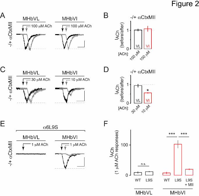
This article has not been copyedited and formatted. The final version may differ from this version.Molecular Pharmacology Fast Forward. Published on October 1, 2015 as DOI: 10.1124/mol.115.101444
at ASPE
T Journals on D
ecember 6, 2021
molpharm
.aspetjournals.orgD
ownloaded from

This article has not been copyedited and formatted. The final version may differ from this version.Molecular Pharmacology Fast Forward. Published on October 1, 2015 as DOI: 10.1124/mol.115.101444
at ASPE
T Journals on D
ecember 6, 2021
molpharm
.aspetjournals.orgD
ownloaded from
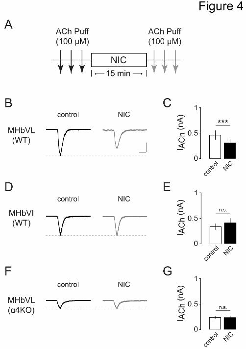
This article has not been copyedited and formatted. The final version may differ from this version.Molecular Pharmacology Fast Forward. Published on October 1, 2015 as DOI: 10.1124/mol.115.101444
at ASPE
T Journals on D
ecember 6, 2021
molpharm
.aspetjournals.orgD
ownloaded from

This article has not been copyedited and formatted. The final version may differ from this version.Molecular Pharmacology Fast Forward. Published on October 1, 2015 as DOI: 10.1124/mol.115.101444
at ASPE
T Journals on D
ecember 6, 2021
molpharm
.aspetjournals.orgD
ownloaded from
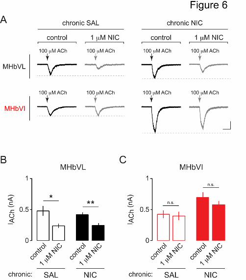
This article has not been copyedited and formatted. The final version may differ from this version.Molecular Pharmacology Fast Forward. Published on October 1, 2015 as DOI: 10.1124/mol.115.101444
at ASPE
T Journals on D
ecember 6, 2021
molpharm
.aspetjournals.orgD
ownloaded from
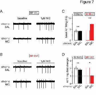
This article has not been copyedited and formatted. The final version may differ from this version.Molecular Pharmacology Fast Forward. Published on October 1, 2015 as DOI: 10.1124/mol.115.101444
at ASPE
T Journals on D
ecember 6, 2021
molpharm
.aspetjournals.orgD
ownloaded from