Embed Size (px)
Citation preview
PostertemplatecourtesyFaculty&CurriculumSupport(FACS),GeorgetownUniversitySchoolofMedicine
Nicole K. Cates, DPM1, John D. Miller, DPM2, Vineela M. Ayyagari, DPM2, Jacob Wynes, DPM, MS, FACFAS43
1:Fellow,2:PGY-3,3:Resident,3:AssistantProfessor&FellowshipProgramDirector,UniversityofMarylandSchoolofMedicineDepartmentofOrthopaedics,
• Tibia fractures are often cited as the primary reason to use solely thin wire fixation with multi-planar static external fixators
• The rate of tibia fractures with use of half pins has been reported as 16.7%, compared to 1.5-4.7% with the use of solely thin wires1,2
• The larger diameter pins have been associated with increased cortical stress and fracture1
• Hydroxyapatite (HA) coated half pins promote greater stability and are protective against osseous deformation and infection due to the similar modulus of elasticity, osseous incorporation, and tapered design3-7
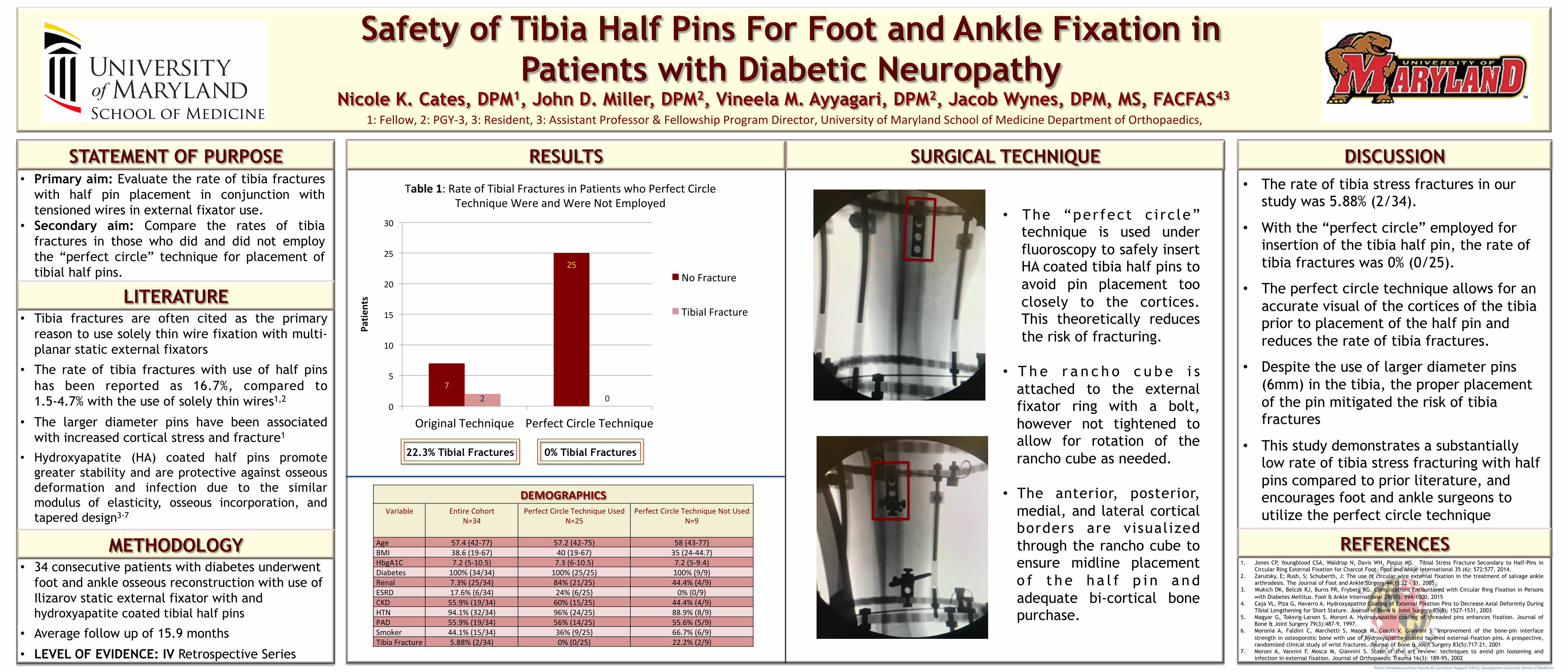
7
25
2 00
5
10
15
20
25
30
OriginalTechnique PerfectCircleTechnique
Patie
nts
Table1:RateofTibialFracturesinPatientswhoPerfectCircleTechniqueWereandWereNotEmployed
NoFracture
TibialFracture
DEMOGRAPHICSVariable EntireCohort
N=34PerfectCircleTechniqueUsed
N=25PerfectCircleTechniqueNotUsed
N=9
Age 57.4(42-77) 57.2(42-75) 58(43-77)BMI 38.6(19-67) 40(19-67) 35(24-44.7)HbgA1C 7.2(5-10.5) 7.3(6-10.5) 7.2(5-9.4)Diabetes 100%(34/34) 100%(25/25) 100%(9/9)Renal 7.3%(25/34) 84%(21/25) 44.4%(4/9)ESRD 17.6%(6/34) 24%(6/25) 0%(0/9)CKD 55.9%(19/34) 60%(15/25) 44.4%(4/9)HTN 94.1%(32/34) 96%(24/25) 88.9%(8/9)PAD 55.9%(19/34) 56%(14/25) 55.6%(5/9)Smoker 44.1%(15/34) 36%(9/25) 66.7%(6/9)TibiaFracture 5.88%(2/34) 0%(0/25) 22.2%(2/9)
• Primary aim: Evaluate the rate of tibia fractures with half pin placement in conjunction with tensioned wires in external fixator use.
• Secondary aim: Compare the rates of tibia fractures in those who did and did not employ the “perfect circle” technique for placement of tibial half pins.
• 34 consecutive patients with diabetes underwent foot and ankle osseous reconstruction with use of Ilizarov static external fixator with and hydroxyapatite coated tibial half pins
• Average follow up of 15.9 months
• LEVEL OF EVIDENCE: IV Retrospective Series
• The “perfect c i rc le” technique is used under fluoroscopy to safely insert HA coated tibia half pins to avoid pin placement too closely to the cortices. This theoretically reduces the risk of fracturing.
• T h e r a n c h o c u b e i s
attached to the external fixator ring with a bolt, however not tightened to allow for rotation of the rancho cube as needed.
• The anterior, posterior,
medial, and lateral cortical borders are visualized through the rancho cube to ensure midline placement o f t h e h a l f p i n a n d adequate bi-cortical bone purchase.
• The rate of tibia stress fractures in our study was 5.88% (2/34).
• With the “perfect circle” employed for
insertion of the tibia half pin, the rate of tibia fractures was 0% (0/25).
• The perfect circle technique allows for an
accurate visual of the cortices of the tibia prior to placement of the half pin and reduces the rate of tibia fractures.
• Despite the use of larger diameter pins
(6mm) in the tibia, the proper placement of the pin mitigated the risk of tibia fractures
• This study demonstrates a substantially
low rate of tibia stress fracturing with half pins compared to prior literature, and encourages foot and ankle surgeons to utilize the perfect circle technique
22.3% Tibial Fractures
1. Jones CP, Youngblood CSA, Waldrop N, Davis WH, Pinzur MS. Tibial Stress Fracture Secondary to Half-Pins in Circular Ring External Fixation for Charcot Foot. Foot and Ankle International 35 (6): 572:577, 2014.
2. Zarutsky, E; Rush, S; Schuberth, J: The use of circular wire external fixation in the treatment of salvage ankle arthrodesis. The Journal of Foot and Ankle Surgery 44(1):22 – 31, 2005.
3. Wukich DK, Belczk RJ, Burns PR, Fryberg RG. Complications Encountered with Circular Ring Fixation in Persons with Diabetes Mellitus. Foot & Ankle International 29(10): 994-1000, 2015
4. Caja VL, Piza G, Navarro A. Hydroxyapatite Coating of External Fixation Pins to Decrease Axial Deformity During Tibial Lengthening for Short Stature. Journal of Bone & Joint Surgery 85(8): 1527-1531, 2003
5. Magyar G, Toksvig-Larsen S, Moroni A. Hydroxyapatite coating of threaded pins enhances fixation. Journal of Bone & Joint Surgery 79(3):487-9, 1997.
6. Moronia A, Faldini C, Marchetti S, Manca M, Cosoli V, Giannini S. Improvement of the bone-pin interface strength in osteoporotic bone with use of hydroxyapatite-coated tapered external-fixation pins. A prospective, randomized clinical study of wrist fractures. Journal of Bone & Joint Surgery 83(5):717-21, 2001
7. Moroni A, Vannini F, Mosca M, Giannini S. State of the art review: techniques to avoid pin loosening and infection in external fixation. Journal of Orthopaedic Trauma 16(3): 189-95, 2002
0% Tibial Fractures

![[XLS] · Web viewPuliveerthi Vineela A Meena Chowdary Syed Mohammed Peershavali K S Gunashekar D Sandhya Sri Gunta Upendra R Ranganath Reddy Siva Prasad V Mythri E C Samba Siva Reddy](https://img.pdfslide.us/doc/110x75/5ae9d4f17f8b9a0877917579/xls-viewpuliveerthi-vineela-a-meena-chowdary-syed-mohammed-peershavali-k-s-gunashekar.jpg)