Embed Size (px)
Citation preview
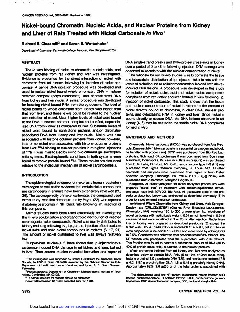
[CANCER RESEARCH 44, 3892-3897, September 1984]
Nickel-bound Chromatin, Nucleic Acids, and Nuclear Proteins from Kidneyand Liver of Rats Treated with Nickel Carbonate in ViVo1
Richard B. Ciccare!»2and Karen E. Wetterhahn3
Department of Chemistry, Dartmouth College, Hanover, New Hampshire 03755
ABSTRACT
The in vivo binding of nickel to chromatin, nucleic acids, andnuclear proteins from rat kidney and liver was investigated.Evidence is presented for the direct interaction of nickel withchromatin from rat tissues following i.p. injection of nickel carbonate. A gentle DNA isolation procedure was developed andused to isolate nickel-bound whole chromatin, DNA + histone
octamer complex (polynucleosomes), and deproteinized DNAfrom kidney and liver nuclei. A similar procedure was developedfor isolating nickel-bound RNA from the cytoplasm. The level of
nickel bound to whole chromatin from kidney was higher thanthat from liver, and these levels could be related to the nuclearconcentration of nickel. Much higher levels of nickel were boundto the DNA + histone octamer complex and purified, deproteinized DNA from kidney as compared to liver. Substantial levels ofnickel were bound to nonhistone proteins and/or chromatin-
associated RNA from kidney and liver nuclei. Nickel was alsoassociated with histone octamer proteins from kidney; however,little or no nickel was associated with histone octamer proteinsfrom liver. MNi binding to nuclear proteins in rats given injectionsof MNi(ll) was investigated by using two different gel electropho-
retic systems. Electrophoretic conditions in both systems werefound to remove protein-bound ^Ni. These results are discussed
relative to the molecular mechanism of nickel carcinogenesis.
INTRODUCTION
The epidemiological evidence for nickel as a human respiratorycarcinogen as well as the evidence that certain nickel compoundsare carcinogens in animals have been extensively reviewed (25,26). The carcmogenicity of nickel carbonate, the compound usedin this study, was first demonstrated by Payne (22), who reportedrhabdomyosarcomas in NIH black rats following i.m. injection ofthis compound.
Animal studies have been used extensively for investigatingthe in vivo solubilization and organotropic distribution of injectedcarcinogenic nickel compounds. Nickel was mainly distributed tokidney and lung following i.v., i.p., or s.c. injection of both solublenickel salts and solid nickel compounds in rodents (6, 17, 21).The amount of nickel distributed to liver was always relativelylow.
Our previous studies (4,5) have shown that ¡.p.-injectednickel
carbonate induced DNA damage in rat kidney and lung, but notin liver. Time course studies revealed formation and repair of
1This investigation was supported by Grant BC-320 from the American Cancer
Society, by USPHS Grant CA34869 awarded by the National Cancer Institute,Department of Health and Human Services, and by an A. P. Sloan ResearchFellowship.
2 Present address: Department of Chemistry, Massachusetts Institute of Tech
nology, Cambridge, MA 02139.3To whom requests for reprints should be addressed.
Received September 12.1983; accepted June 12,1984.
DNA single-strand breaks and DNA-protein cross-links in kidney
over a period of 0 to 48 hr following injection. DNA damage wasobserved to correlate with the nuclear concentration of nickë.
The rationale for our in vivo studies was to correlate the tissueand intracellular distribution of i.p.-injected nickel in rats with thelevels of nickel bound to cellular macromolecules and with nickel-
induced DNA lesions. A procedure was developed in this studyfor isolation of nickehnucleic acid and nickelmucleic acid:proteincomplexes from rat kidney and liver formed in vivo following i.p.injection of nickel carbonate. This study shows that the tissueand nuclear concentration of nickel is related to the amount ofnickel directly bound to chromatin, nuclear DNA, nuclear proteins, and cytoplasmic RNA in kidney and liver. Since nickel isbound directly to nuclear DNA, the DNA lesions observed in ratkidney (4, 5) may be related to the stable nickel:DNA complexesformed in vivo.
MATERIALS AND METHODS
Chemicals. Nickel carbonate (NiCOa) was purchased from Alfa Products, Danvers, MA (nickel carbonate is a potential carcinogen and shouldbe handled with proper care); 80S4 was purchased from Bio-Rad Lab
oratories, Richmond, CA; proteinase K was purchased from BoehringerMannheim, Indianapolis, IN; cesium sulfate (suprapure) was purchasedfrom E. M. Labs, Elmsford, NY. Calf thymus histone type III-S (H1) was
purchased from Sigma Chemical Company, St. Louis, MO. All otherchemicals and enzymes were purchased from Sigma or from FisherScientific Company, Pittsburgh, PA. ^NiClz (11.9 pC\/iig nickel) was
purchased from Amersham, Arlington Heights, IL.Reagents. All buffers/reagents for nickel analytical procedures were
prepared "metal free" by treatment with sodium-equilibrated cation-
exchange resin (AG 50W-X2; Bio-Rad). All glassware used in the pro
cedures described below was pretreated as described previously (5) inorder to avoid external metal contamination.
Isolation of Whole Chromatin from Kidney and Liver. Male Sprague-
Dawley rats |CRL:CD(SD)BR| (Charles River Breeding Laboratories,Wilmington, MA) weighing 160 to 200 g were given i.p. injections ofnickel carbonate (40 mg/kg body weight; 0.34 mmol nickel/kg) in 0.5 mlsesame oil and were sacrificed at 3 or 20 hr after injection. Nuclei fromliver or kidney were prepared as described previously (5) except thebuffer was 0.05 M Tris-HCI:0.25 M sucrose:0.15 M NaCI, pH 7.5. Nucleiwere suspended in ice-cold 0.15 M NaCI and were lysed by adding SDS
to 0.5%. Chromatin was collected after precipitation in 63% ethanol. TheNP fraction was precipitated from the supernatant with 76% ethanol.This fraction was found to contain a substantial amount of RNA (30 to40% of protein mass ratio) in addition to the nuclear proteins.
Whole chromatin isolated from rat kidney and liver was analyzed asdescribed below to contain DNA, RNA [5 to 10% of DNA mass ratio],histone proteins [1.0 g protein/g DNA (13)], and nonhistone proteins [2.8±0.2 (S.E.) g protein/g liver DNA; 1.6 ±0.15 g protein/g kidney DNA].Approximately 62% (1.6 g/2.6 g) of the total proteins associated with
4The abbreviations used are: NP fraction, nucieopiasm protein fraction; NHO
fraction, nonhistone-histone H1 octamer fraction; PAGE, polyacryiamide gel eiec-trophoresis; RNP. ribonucleoprotein complex; SOS, sodium dodecyl sulfate.
3892 CANCER RESEARCH VOL. 44
on April 4, 2019. © 1984 American Association for Cancer Research. cancerres.aacrjournals.org Downloaded from
Nickel-bound Chromatin
kidney chromatin and 74% (2.8 g/3.8 g) of the total proteins associatedwith liver chromatin were nonhistone proteins. Liver nonhistone proteinswere in approximately 40% excess mass ratio over kidney nonhistoneproteins (1.6 g kidney/2.8 g liver). The mass ratios for nonhistone proteinswere calculated as the difference between total chromatin-associated
protein and historie protein.Preparation of the ONA + Histone Octamer Complex (Polynucleo-
somes) from Chromatin. Chromatin prepared as described above fromkidney or liver nuclei was incubated with RNase A, and extracted withchlorofomrisoamyl alcohol (24:1). The DNA + historie octamer complexfrom kidney or liver was found to contain 0.48 to 0.54 g histone protein/g DNA and was analyzed by SDS and acidic polyacrylamide gel electro-
phoresis as described below. Both electrophoretic systems confirmedthe presence of only the 4 core histories (H2A, H2B, H3, and H4). HistoneH1 and RNA (<0.5% mass ratio) were not associated with this complex.
The NHO fraction was completely removed by this procedure. Thisfraction was composed of nonhistone proteins, histone H1, and approximately 33% of the histone octamer proteins from chromatin. For liverchromatin, this fraction was composed of (mass ratios) 85% nonhistoneprotein, 6% histone protein, and 9% DNA. For kidney chromatin, thisfraction was composed of 77% nonhistone protein, 9% histone protein,and 14% DNA. The mass ratios were determined from the weightpercentage of DNA and the total protein in the fraction and by calculatingthe mass contributed by the histone octamers (33% of 0.8 g protein/gDNA) and the known mass ratio of H1 (0.2 g H1 protein/g DNA).
Weight percentage calculations on the DNA + octamer complex basedupon the histone core octamer molecular weight of 110,000 g/mol anddouble-stranded DNA-phosphate repeating unit of 660 g/mol give 1
octamer/330 ±30 base pairs, which is lower than the ratio on nativechromatin of 1 octamer/200 base pairs (14).
Preparation of Deproteinized DNA. Whole chromatin or the DNA +histone octamer complex was incubated with proteinase K and extractedwith 95% ethanol:(24:1 chloroform:isoamyl alcohol), 3:2. This preparationconsistently yielded DNA which was >99.5% protein and RNA free.
Preparation of RNA from Kidney and Liver Cytosol. Following thepelleting of nuclei, the supernatant (cytoplasmic fraction) was centrifugedat 6500 x g for 10 min to remove mitochondria. RNA and protein wereprecipitated from the supernatant with 76% ethanol. The RNA andprotein fractions were digested with DNase(ll) and extracted with chlo-roforrtrisoamyl alcohol (24:1). Alternatively, the DNase(ll)-digested solu
tions were then treated with proteinase K and extracted with 95%ethanol:(24:1 chlorofoimisoamyl alcohol), 3:2. The amount of proteinassociated with both rat kidney and liver RNP was determined to be 0.4to 0.5 g protein/g RNA. Proteinase K treatment of RNP removed >99%of the protein, resulting in RNA. RNP and RNA were determined to be>99.6% DNA free.
DNA, Protein, RNA, and Nickel Assays. DNA was assayed by amicrofluorometric technique as reported previously (5). RNA and proteinwere found not to interfere with this assay.
Protein was determined by the modified procedure of Peterson (23)as described in Ref. 15 (range, 0 to 20 ng protein) or by the procedureof Lowry ef al. (16) (range, 20 to 250 ^/protein).
RNA was analyzed by the orcinol procedure of Albaum and Umbreit(2). Quantities of DNA up to 500 ^g did not interfere with the RNAdeterminations.
Nickel concentration was determined by electrothermal atomic absorption spectroscopy as described previously (5).
Fluorography of Kidney Chromatin and Constituents from RatsTreated with 63NiCI2in Vivo. Male Sprague-Dawley rats (0.18 kg) weregiven i.p. injections of 0.5 mCi ^NiClz (11.9 ¿iCi/Mgnickel; 0.23 mg nickel/
kg) administered as the Tris:nickel(ll) complex in 0.05 M Tris, pH 8.0, ina volume of 0.55 ml. After 4 hr, rats were sacrificed, and kidneys wereexcised. Nuclei, whole chromatin, and NP fraction were prepared asdescribed above. Whole chromatin and NP fraction proteins (~25 ng
protein) were separated by both acid:PAGE and SDS:PAGE as describedbelow. Lanes containing whole chromatin had approximately 2 x 10⁴
cpm loaded, and lanes containing NP fractions had approximately 1.2 x
10" cpm loaded. Following electrophoresis, gels were immediately
stained with silver as described below in order to locate all protein bandsor soaked for 15 min in Enlightning and exposed to Kodak X-Omat ARfilm. Fluorography was carried out at -70° for 6 weeks.
Acid:PAGE. PAGE of histories was carried out using modifications ofthe procedures described by Panyim and Chalkley (20) and Zweidler(33). Gels were prepared with the following conditions: acrylamide (20%,w/v); bisacrylamide (1%, w/v); acetic acid (0.9 M); urea (3.0 M); Triton X-100 (0.1%, v/v); ammonium persulfate (0.15%, w/v); A/,A/,A/',A/'-tetra-
methylethylenediamine (1 .5%, v/v), pH 2.3. The electrode buffer was 0.9M acetic acid:1% Triton X-100, pH 2.3. The sample buffer was 0.9 Macetic acid:1% Triton X-100:3.0 M urea:15% sucrose, pH 2.3. Gel elec
trophoresis was carried out at 200 V.SDS:PAGE Electrophoresis. PAGE of histone and nuclear proteins
was also carried out by a modification of the procedure of Thomas andKornberg (29). Gels were prepared with the following conditions: acrylamide (18%, w/v); bisacrylamide (0.15%, w/v); Tris base (0.75 M); SDS(0.1%, w/v); ammonium persulfate (0.1%, w/v); W,A/,A/',N'-tetramethyl-
ethylenediamine (0.04%, v/v), pH 8.6 (adjusted with HCI). The electrodebuffer was 0.20 M Tris base:0.24 M glycine:0.1% SDS, pH 8.8. Thesample buffer was 0.75 M Tris base:2.0% SDS:15% sucrose, pH 8.6.Electrophoresis was carried out at 50 V for 22 hr.
Silver Stain for Proteins. Silver staining of proteins in polyacrylamideslab gels was carried out by a modification of the procedure of Wray etal. (32). This procedure was used for increased sensitivity over theCoomassie blue procedure, since the extinction coefficients of Coomas-
sie blue:histone complexes vary drastically for individual histories (33).
RESULTS
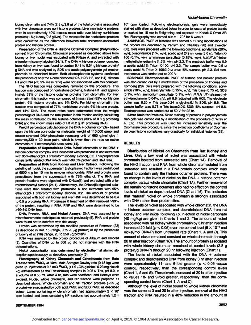
Distribution of Nickel on Chromatin from Rat Kidney andLiver. Only a low level of nickel was associated with wholechromatin isolated from untreated rats (Chart 1A). Removal ofthe NHO fraction and RNA from whole chromatin isolated fromuntreated rats resulted in a DNA:protein complex which wasfound to contain only the histone octamer proteins. There wasno change in the levels of nickel on the DNA + histone octamercomplex versus whole chromatin (Chart 1/1). Further removal ofthe remaining histone octamers also had no effect on the controllevels of nickel on deproteinized DNA (Chart ~\A).This indicatesthat "natural" nickel on whole chromatin is strongly associated
with DNA rather than protein sites.The levels of nickel associated with whole chromatin, the DNA
+ histone octamer complex, and deproteinized DNA from ratkidney and liver nuclei following i.p. injection of nickel carbonate(40 mg/kg) are given in Charts 1 and 2. The amount of nickelassociated with rat kidney whole chromatin by 3 hr after injectionincreased 20-fold (p < 0.05) over the control level (5 x 10~5 mol
nickel/mol DNA-P) from untreated rats (Chart 1, A and B). This
amount of nickel remained constant on whole chromatin through20 hr after injection (Chart 1C). The amount of protein associatedwith whole kidney chromatin remained at control levels (2.8 gprotein/g DNA-P) through 20 hr following injection (Chart 1).
The levels of nickel associated with the DNA + octamercomplex and deproteinized DNA from kidney 3 hr after injectionwere approximately 11- and 6-fold greater (p < 0.05 versus
control), respectively, than the corresponding control levels(Chart 1, A and B). These levels increased at 20 hr after injectionto values 18- and 9-fold greater, respectively, than the corre
sponding control levels (Chart 1, A and C).Although the level of nickel bound to whole kidney chromatin
was the same at 3 and 20 hr after injection, removal of the NHOfraction and RNA resulted in a 48% reduction in the amount of
SEPTEMBER 1984 3893
on April 4, 2019. © 1984 American Association for Cancer Research. cancerres.aacrjournals.org Downloaded from
R. B. Ciccarelli and K. E. Wetterhahn
RATKIDNEY
CHROMATINHjCONTROL(N=6)
mol Ni /mol DNA-Pxü5 (x±SE) (I D
20 3D 40 50 60 TO 80 90 100 IIP 120 130 140-
DNACONTROL
(N=8)
CHROMATIN3HR
(N=6)DNA
OCTAMER3HR(N=IO)DNA
3HR(N=4)UH—BZZHJ
1 B
CHROMATIN20 HRIN=6)DNA
OCTAMER20 HR(N=I2)DNA
20 HR(N=41••»,,H1—
1HC-40
80 120 1^0 200240 280
g Protein /g DNA »IO2320360 400 440 480
'(mm) (S* SE)520560-
Chart 1. Amount of nickel (D) and protein (•)associated with rat kidney chro-matm and constituents. Rats were given i.p. injections of nickel carbonate (40 mg/kg) and were sacrificed at 3 hr (S) and 20 hr (C) after injection: A. untreatedcontrols. The amount of nickel [mol nickel/mol DNA-nucleotide (P)| and protein (gprotein/g DNA) associated with whole chromatin, the DNA + historie octamercomplex, and deproteinized DNA were determined by standard procedures asdescribed in "Materials and Methods." Columns, mean values determined using 4
to 12 rats; bars, S.E.
nickel which was bound to the DNA + histone octamer complexat 3 hr and only a 22% reduction at 20 hr (Chart 1, B and C).The amount of protein associated with these complexes was notsignificantly increased (p < 0.2) over control levels. Furtherremoval of the remaining histone octamers resulted in a 45%reduction in the amount of nickel which was bound to deproteinized DNA at both 3 and 20 hr after injection (Chart 1, B and C).These results imply that redistribution of the nickel bound tochromatin occurred over the period of 3 through 20 hr, since theamount of nickel bound to the DNA + histone octamer complexand to deproteinized DNA increased, while the amount bound towhole chromatin remained constant. Therefore, nickel on kidneychromatin was redistributed essentially from nonhistone proteinsites (NHO fraction is 77% nonhistone proteins) and/or fromchromatin-associated RNA to DNA and histone-DNA sites.
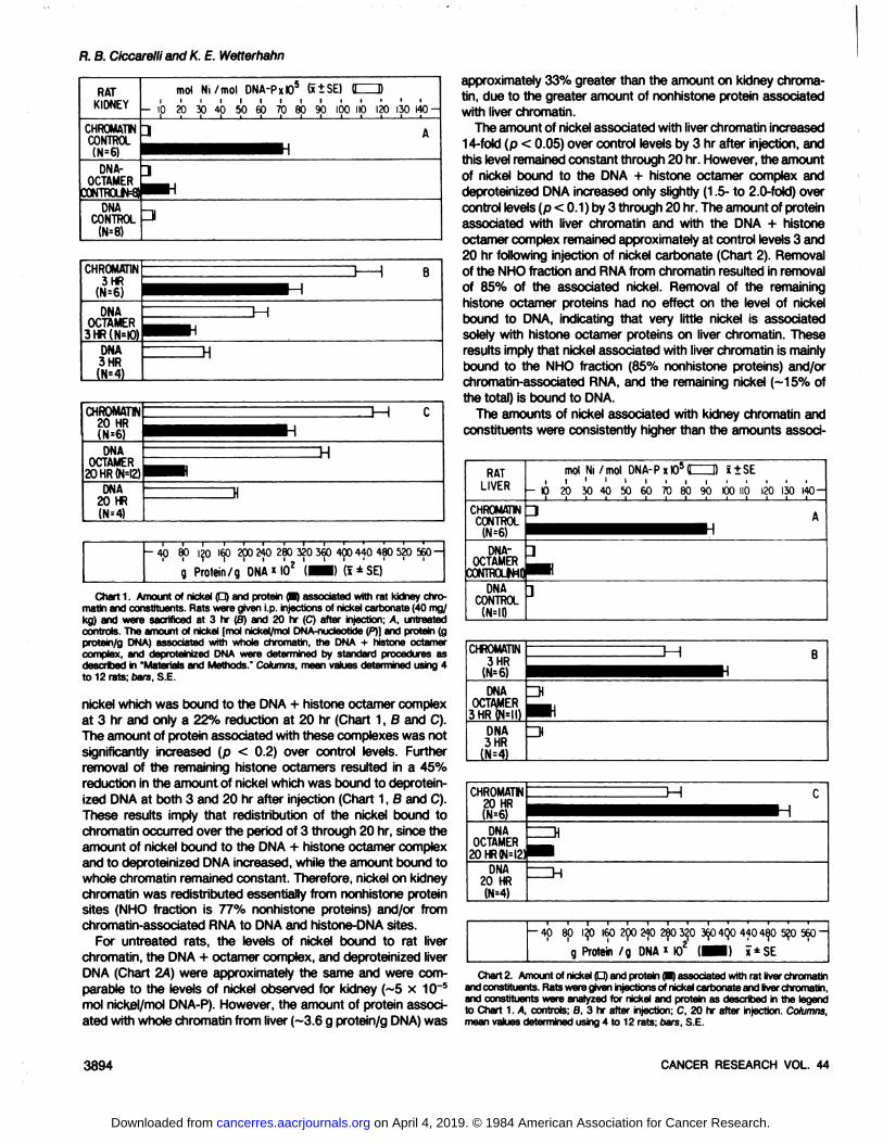
For untreated rats, the levels of nickel bound to rat liverchromatin, the DNA 4- octamer complex, and deproteinized liver
DNA (Chart 2A) were approximately the same and were comparable to the levels of nickel observed for kidney (~5 x 10~5
mol nickel/mol DNA-P). However, the amount of protein associated with whole chromatin from liver (~3.6 g protein/g DNA) was
approximately 33% greater than the amount on kidney chroma-
tin, due to the greater amount of nonhistone protein associatedwith liver chromatin.
The amount of nickel associated with liver chromatin increased14-fold (p < 0.05) over control levels by 3 hr after injection, and
this level remained constant through 20 hr. However, the amountof nickel bound to the DNA + histone octamer complex anddeproteinized DNA increased only slightly (1.5- to 2.0-fold) over
control levels (p < 0.1 ) by 3 through 20 hr. The amount of proteinassociated with liver chromatin and with the DNA + histoneoctamer complex remained approximately at control levels 3 and20 hr following injection of nickel carbonate (Chart 2). Removalof the NHO fraction and RNA from chromatin resulted in removalof 85% of the associated nickel. Removal of the remaininghistone octamer proteins had no effect on the level of nickelbound to DNA, indicating that very little nickel is associatedsolely with histone octamer proteins on liver chromatin. Theseresults imply that nickel associated with liver chromatin is mainlybound to the NHO fraction (85% nonhistone proteins) and/orchromatin-associated RNA, and the remaining nickel (~15% of
the total) is bound to DNA.The amounts of nickel associated with kidney chromatin and
constituents were consistently higher than the amounts associ-
RATLIVER
CHROMATINDCONTROL
(N=6)
mol Ni /mol DNA-Pxt05tIIID »±SE
- 10 20 30 40 50
CHROMATIN3HR!N=6)DNA
OCTAMER3HR(N=II)DNA
3HR(111=4)l-H1=>B
CHROMATW20 HR
IH
DNAOCTAMER
20HRWM2DNA
20 HR
-408p 120 160290 2f) 280 320 3604QO440480 520 560~
g Protein /g DNA * IO2 (••! ï±SE
Chart2. Amount of nickel (D) and protein (•)associated with rat liver chromatinandconstituents. Ratswere given injectionsof nickel carbonateand liverchromatin,and constituents were analyzed for nickel and protein as described in the legendto Chart 1. A, controls; B, 3 hr after injection; C, 20 hr after injection. Columns,mean values determined using 4 to 12 rats; bars, S.E.
3894 CANCER RESEARCH VOL. 44
on April 4, 2019. © 1984 American Association for Cancer Research. cancerres.aacrjournals.org Downloaded from
Nickel-bound Chromatin
ated with liver chromatin and constituents at 3 and 20 hr afterinjection.
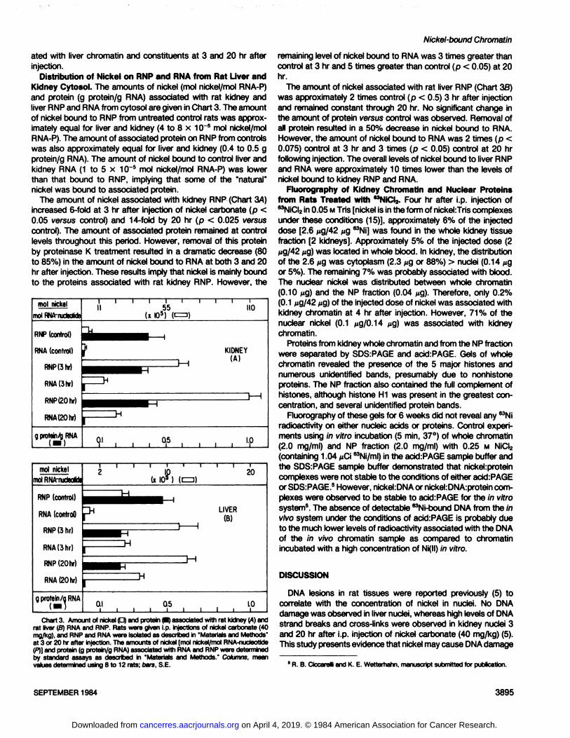
Distribution of Nickel on RNP and RNA from Rat Liver andKidney Cytosol. The amounts of nickel (mol nickel/mol RNA-P)
and protein (g protein/g RNA) associated with rat kidney andliver RNP and RNA from cytosol are given in Chart 3. The amountof nickel bound to RNP from untreated control rats was approximately equal for liver and kidney (4 to 8 x 10~5 mol nickel/mol
RNA-P). The amount of associated protein on RNP from controls
was also approximately equal for liver and kidney (0.4 to 0.5 gprotein/g RNA). The amount of nickel bound to control liver andkidney RNA (1 to 5 x 10~5 mol nickel/mol RNA-P) was lowerthan that bound to RNP, implying that some of the "natural"
nickel was bound to associated protein.The amount of nickel associated with kidney RNP (Chart 3A)
increased 6-fold at 3 hr after injection of nickel carbonate (p <0.05 versus control) and 14-fold by 20 hr (p < 0.025 versus
control). The amount of associated protein remained at controllevels throughout this period. However, removal of this proteinby proteinase K treatment resulted in a dramatic decrease (80to 85%) in the amount of nickel bound to RNA at both 3 and 20hr after injection. These results imply that nickel is mainly boundto the proteins associated with rat kidney RNP. However, the
mol nickelmolRNA-rudeoMel
RNP(control)
RNA(control)
RNPßhr)
RNA(3hr)
RNP(20hr)
RNA(20hr)
gprotein/R̂NA
molnickelmolRNA-nucteoMe
RNP(control)
RNA(control)
RNP(3hr)
RNA(3hr)
RNP(20hr)
RNA(20hr)
g protein/aRNA
20
LIVER(B)
H
LOiChart 3. Amount of nickel (D) and protein (•)associated with rat kidney (A) and
rat liver (B) RNA and RNP. Rats were given i.p. injections of nickel carbonate (40mg/kg), and RNP and RNA were isolated as described in 'Materials and Methods"
at 3 or 20 hr after injection. The amounts of nickel [mol nickel/mol RNA-nudeotide(P)] and protein (g protein/g RNA) associated with RNA and RNP were determinedby standard assays as described in "Materials and Methods." Columns, mean
values determined using 8 to 12 rats; oars, S.E.
remaining level of nickel bound to RNA was 3 times greater thancontrol at 3 hr and 5 times greater than control (p < 0.05) at 20hr.
The amount of nickel associated with rat liver RNP (Chart 3S)was approximately 2 times control (p < 0.5) 3 hr after injectionand remained constant through 20 hr. No significant change inthe amount of protein versus control was observed. Removal ofall protein resulted in a 50% decrease in nickel bound to RNA.However, the amount of nickel bound to RNA was 2 times (p <0.075) control at 3 hr and 3 times (p < 0.05) control at 20 hrfollowing injection. The overall levels of nickel bound to liver RNPand RNA were approximately 10 times lower than the levels ofnickel bound to kidney RNP and RNA.
Fluorography of Kidney Chromatin and Nuclear Proteinsfrom Rats Treated with MNiCI2. Four hr after i.p. injection ofMNiCI2in 0.05 MTris [nickel is in the form of nickehTris complexes
under these conditions (15)], approximately 6% of the injecteddose [2.6 /ug/42 ¿tg63Ni| was found in the whole kidney tissue
fraction [2 kidneys]. Approximately 5% of the injected dose (2ug/42 fig) was located in whole blood. In kidney, the distributionof the 2.6 ng was cytoplasm (2.3 ng or 88%) > nuclei (0.14 ngor 5%). The remaining 7% was probably associated with blood.The nuclear nickel was distributed between whole chromatin(0.10 ng) and the NP fraction (0.04 ^g). Therefore, only 0.2%(0.1 ug/42 ng) of the injected dose of nickel was associated withkidney chromatin at 4 hr after injection. However, 71% of thenuclear nickel (0.1 MQ/0.14 ¿ig)was associated with kidneychromatin.
Proteins from kidney whole chromatin and from the NP fractionwere separated by SDS:PAGE and acid:PAGE. Gels of wholechromatin revealed the presence of the 5 major histones andnumerous unidentified bands, presumably due to nonhistoneproteins. The NP fraction also contained the full complement ofhistones, although historie H1 was present in the greatest concentration, and several unidentified protein bands.
Fluorography of these gels for 6 weeks did not reveal any ^Ni
radioactivity on either nucleic acids or proteins. Control experiments using In vitro incubation (5 min, 37°)of whole chromatin
(2.0 mg/ml) and NP fraction (2.0 mg/ml) with 0.25 M NiCI2(containing 1.04 ¿¿Ci"Ni/ml) in the acid:PAGE sample buffer and
the SDS:PAGE sample buffer demonstrated that nickel:proteincomplexes were not stable to the conditions of either acid:PAGEor SDS-.PAGE.5However, nickel:DNA or nickel:DNA:protein com
plexes were observed to be stable to acid:PAGE for the in vitrosystem5. The absence of detectable ^Ni-bound DNA from the in
vivo system under the conditions of acid:PAGE is probably dueto the much lower levels of radioactivity associated with the DNAof the in vivo chromatin sample as compared to chromatinincubated with a high concentration of Ni(ll) in vitro.
DISCUSSION
DNA lesions in rat tissues were reported previously (5) tocorrelate with the concentration of nickel in nuclei. No DNAdamage was observed in liver nuclei, whereas high levels of DNAstrand breaks and cross-links were observed in kidney nuclei 3
and 20 hr after i.p. injection of nickel carbonate (40 mg/kg) (5).This study presents evidence that nickel may cause DNA damage
6 R. B. Ciccareiii and K. E. Wetterhahn, manuscript submitted for publication.
SEPTEMBER 1984 3895
on April 4, 2019. © 1984 American Association for Cancer Research. cancerres.aacrjournals.org Downloaded from
R. B. Ciccarelli and K. E. Wetterhahn
through a direct interaction with chromatin.Following injection of nickel carbonate (40 mg/kg), the amount
of nickel bound at 3 and 20 hr to whole liver chromatin wasapproximately 10 times greater than control; however, most ofthis nickel (85%) was bound to chromatin-associated RNA and/
or the NHO fraction composed mainly (85% mass ratio) ofnonhistone proteins. The amount of nickel bound to deprotein-
ized DNA from liver under these conditions increased only slightlyover control levels.
The levels of nickel bound to whole kidney chromatin underthese conditions were only 1.5 times greater than those of wholeliver chromatin. However, much more of this nickel was boundto DNA and histones (only 22% was bound to kidney NHOfraction and/or RNA at 20 hr). Furthermore, the distribution ofnickel on whole kidney chromatin was observed to changebetween 3 and 20 hr, as more nickel shifted from the NHOfraction and/or RNA sites to DNA sites over this period. Theseobserved differences in the binding of nickel to kidney chromatinversus liver chromatin may be due to the 40% greater nonhistoneprotein mass ratio found on liver chromatin. Since less nonhistone protein is associated with kidney chromatin, nickel probably has greater access to kidney DNA and histones. In liverchromatin, the excess nonhistone protein apparently binds themajority of the nickei(ll) and may protect histones from nickel,since very little nickel is bound to histones on liver chromatin,yet significant amounts of nickel are bound to histones on kidneychromatin.
Nickel was also found to be bound to RNP in the cytoplasmof kidney and liver cells following i.p. injection of nickel carbonate(40 mg/kg). The amount of nickel bound to kidney RNP was atleast 6 times greater at 3 hr and 10 times greater at 20 hr thanthe amount bound to liver RNP. This effect is most probably dueto the 6 times greater concentration of nickel in kidney cytoplasmversus liver cytoplasm through this time period. Removal of thetightly associateci protein by proteinase K digestion resulted inan 80 to 85% reduction in the nickel bound to kidney RNA. Thisnickel-bound protein was not characterized.
Many in vivo studies have characterized cytop'asmic nickel-
binding proteins in kidney, lung and liver (1, 9, 28). In most ofthese studies, nickel-binding proteins have yet to be further
characterized. It is possible that some of these cytoplasmicproteins may be capable of transporting nickel to and from thenucleus. Abdulwajid and Sarkar (1) have recently isolated anickel-sequestering M. 15,000 glycoprotein from renal cytosolfollowing i.p. injection of "NiCI,,. This glycoprotein was postu
lated to be part of the renal basement membrane and may berelated to excretion of toxic metals from the cell.
Wacker and Vallee (30) reported a level of nickel on rat liverRNA (64 jtg/g or 3.8 x 10"4 mol nickel/mol RNA-P) which ishigher than our control level (7 nQ/Q or 4 x 10~5 mol nickel/mol
RNA-P). The control level of nickel bound to mammalian DNA or
chromatin from the rat has not to our knowledge been reportedpreviously. This level (5.0 ±1.2 x 10~s mol nickel/mol DNA-P or
8.9 ±0.2 /¿g/g)was the same for both rat kidney and liver DNAand is approximately the same level as that observed for controlRNA from these tissues. Andronikashvili et al. (3) measuredcontrol levels of other metals on rat liver DNA (zinc, 0.54 ng/gor 2.7 x 10"6 mol/mol DNA-P; iron, 16 ng/g or 9.5 x 10~5 mol/mol DNA-P; cobalt, <0.05 /ig/g or <2.8 x 10~7 mol/mol DNA-P;manganese, 25 ng/g or 1.5 x 10~4 mol/mol DNA-P; magnesium,1 /ig/g or 1.4 x 10~s md/mol DNA-P, and our value for nickel is
in the same range as these metals.Other investigators have reported the level of nickel bound to
DNA isolated from nickel-exposed rats, nickel-induced tumors,or nickel-exposed cells. Webb er al. (31) measured the amountof nickel on "nucleolar" DNA to be 25.1 to 34.6 /ig nickel/mg
DNA (0.141 to 0.195 mol nickel/mol DNA-P) in nucleoli of primary
tumors induced in rat muscle by i.m. injection of metallic nickel.Hui and Sunderman (11) calculated the amount of '"Ni bound to
DNA from rat liver (0.29 to 0.52 mol nickel/mol DNA-P) and ratkidney (1.26 to 2.20 mol nickel/mol DNA-P) 4 hr following i.v.administration of MNi(CO)4 (20 mg/kg) and i.m. administration of^NiClz (20 mg/kg). Although these results show greater levels
of nickel bound to kidney versus liver DNA, which correlates withour results, the overall levels of nickel bound are much higherthan the nickel levels reported in our study. Their study did notreport the DNA isolation procedure in detail, and it is possiblethat a considerable level of protein was still associated with theDNA isolated in their studies. Harriett et al. (B) measured theincorporation of ^Ni in DNA from Chinese hamster ovary cellsto be 0.7 x 10~3 mol nickel/mol DNA-P after cells were exposed
to crystalline MNiS (10 ^g/ml) for 3 days. However, the DNA
isolated in this study was contaminated with both RNA andprotein.
The induction of specific nephrotoxic effects by nickel compounds has been observed in animals (7,10-12,18) and corre
lated to carcinogenicity (27). Induction of renal cancers and lungtumors in animals by nickel compounds has been reported (12,19, 24). However, nickel compounds have not induced cancersof the liver (25, 26).
The ability of nickel carbonate to induce lesions in DNA fromspecific rat tissues in vivo may be related to the nuclear concentration of nickel(ll) in these tissues, which is a major factor in thecontrol of the amount of nickel(ll) bound to chromatin. Thedifferent levels of nonhistone proteins associated with chromatinfrom different tissues may be a factor in the amount of nickel(ll)which has access to and binds to DNA. The stability ofnickel:DNA:nuclear protein complexes in the nucleus ornickekprotem complexes in the cytoplasm may be related to theDNA-protein cross-links observed in kidney in vivo. The levels of
nickel(ll) bound directly to DNA may be related to the number ofDNA lesions observed. If nickel(ll) acts as a chemical mutagenin somatic cells, the inability of cellular systems to repair nickel-induced lesions (DNA-protein cross-links) may be related to the
carcinogenicity of nickel compounds. Since animal studies indicate that nickel compounds act as complete carcinogens, promotional effects may occur through nickel-histone or nickel-nonhistone protein interactions and may be important to generegulation.
REFERENCES
1. Abdulwaiid. A. W.. and Sarkar, B. Nickel-sequestering renal glycoprotein. Proc.Nati. Acad. Sci. USA, 80:4509-4512,1983.
2. Albaum, H. G., and Umbreit. W. W. Differentiation between ribose 3-phosphateand ribose 5-phosphate. J. Biol. Chem., 767:369-376,1947.
3. Andronikashvili, E. L . Mosulishvili, L. M., Beiokobilski. A. I., Kharabadze, N.E . Tevzieva. T. K., and Efremova, E. Y. Content of some trace elements inSarcoma M-1 DNA in dynamics of malignant growth. Cancer Res., 34: 271-274,1974.
4. Ciccare!»,R. B., Hampton, T. H., and Jennette, K. W. Nickel carbonate inducesDNA-protein cross-links and DNA strand breaks in rat kidney. Cancer Lett.,72:349-354,1981.
5. Ciccareiii. R. B., and Wetterhahn, K. E. Nickel distribution and DNA lesionsinduced in rat tissues by the carcinogen nickel carbonate. Cancer Res., 42:3544-3549,1982.
3896 CANCER RESEARCH VOL. 44
on April 4, 2019. © 1984 American Association for Cancer Research. cancerres.aacrjournals.org Downloaded from
6. Clary, J. J. Nickel chloride induced metabolic changes in the rat and guineapig. Toxico!. Appi. Pharmacol., 31: 55-65,1975.
7. Gitlitz. P. H., Sunderman, F. W., Jr., and Goldblatt, P. J. Aminoadduria andprotemuria in rats after a single intraperitoneal injection of nickel(il). ToxicolAppi. Pharmacol., 34: 430-440,1975.
8. Harriett, P. B., Robison, S. H., Swartzendruber, D. E., and Costa, M. Comparison of protein. RNA and DMA binding and cell-cycle-specific growth inhibitoryeffects of nickel compounds in cultured cells. Toxicol. Appi. Pharmacol., 64:20-30,1982.
9. HerÃant-Peers, M. C., Hildebrand, H. F., and Kerckaert, J. P. In vitro and invivo incorporation of 6:'Ni(ll) into lung and liver subcellular fractions of BALB/c
mice. Carcinogenesis (Lond.), 4:387-392,1983.10. Hopfer, S. M., Sunderman, F. W., Jr., Morse, E. E., and Fredrickson, T. N.
Effects of intrarenal injection of nickel subsulfkJe in rodents. Arm. Clin. Lab.Sci., 10: 54-64,1980.
11. Hui, G., and Sunderman, F. W., Jr. Effects of nickel compounds on incorporation of [3H]thymidine into DNA in rat liver and kidney. Carcinogenesis (Lond.),
1: 297-303, 1980.12. Jasmin, G., and Solymoss, B. The topical effects of nickel subsuffide on renal
parenchyma. In: G. N. Schrauzer (ed.). Inorganic and Nutritional Aspects ofCancer, pp. 69-83. New York: Plenum Publishing Corp., 1978.
13. Johns, E. W. The isolation and purification of histories. Methods Cell Biol.. i6183-203,1977.
14. Kornberg. R. D. Chromatin structure: a repeating unit of histories and DNA.Science (Wash. DC), J84: 868-871,1974.
15. Lee, J. E., Ciccarelh, R. B., and Wetterhahn-Jennette. K. E. Solubilization ofthe carcinogen nickel subsulfide and its interaction with DNA and protein.Biochemistry, 21: 771-778,1982.
16. Lowry, O. H., Rosebrough, N. J., Fair, A. L. and Randall, R. J. Proteinmeasurement with the Folin phenol reagent. J. Biol. Chem., 793: 265-275,
1951.17. Mathur, A. K., Dikshtth, T. S. S., Lai, M. M., and Tandon, S. K. Distribution of
nickel and cytogenetic changes in poisoned rats. Toxicology. 10: 105-113,
1978.18. O Dell. G. D., Miller, W. J., King, W. A., Moore, S. L., and Blackmon. D. M.
Nickel toxicity in the young bovine. J. Nutr., 100:1447-1453,1970.
Nickel-bound Chromatin
19. Ottotenghi, A. D., Hasemna, J. K., Payne, W. W., Falk, H. L., and MacFariand,H. N. Inhalation studies of nickel sulfide in pulmonary Carcinogenesis of rats.J. Nati. Cancer Inst., 54.-1165-1172,1974.
20. Panyim, S., and Chalkley, R. High resolution acrylamide gel electrophoresis ofhistories. Arch. Biochem. Biophys., Õ30:337-346,1969.
21. Parker, K., and Sunderman, F. W., Jr. Distribution of "Ni in rabbit tissuesfollowing intravenous injections of "NiClj. Res. Commun. Chem. Pathol.Pharmacol., 7: 755-762,1974.
22. Payne, W. W. Carcmogemcity of nickel compounds in experimental animals.Proc. Am. Assoc Cancer Res., 5:50,1964.
23. Peterson, G. L. A simplification of the protein assay method of Lowry et al.which is more generally applicable. Anal. Biochem., 83:346-356,1977.
24. Stoner, G. D., Shimkin, M. B., Troxell, M. C., Thompson, T. L, and Terry, L.S. Test for carcinogenicity of metallic compounds by the pulmonary tumorresponse in strain A mice. Cancer Res., 36:1744-1747,1976.
25. Sunderman, F. W., Jr. In: Nickel, Chap. 6. Washington, DC: National Academyof Sciences, 1975.
26. Sunderman, F. W., Jr. Recent research on nickel Carcinogenesis. Environ.Health Perspect., 40:131 -141,1981.
27. Sunderman, F. W., Jr. In: Nickel in the Human Environment, (ARC Monograph,Vol. 53, in press, 1984.
28. Sunderman, F. W., Jr., Mangold, B. L. K., Wong, S. H. Y . Shen, S. K., Reid,M. C., and Jansson, I. High-performance size-exclusion chromatography of"Ni-constituents in renal cytosol and microsomes from wNiCI?-treated rats.
Res. Commun. Chem. Pathol. Pharmacol., 39:477-492,1983.29. Thomas, J. 0., and Kornberg, R. D. The study of histone-histone associations
by chemical cross-linking. Methods Cell Biol., 18:429-440,1978.30. Wacker, W. E. C.. and Vallee, B. L. Nucleic adds and metals. J. Biol. Chem..
234; 3257-3262,1959.
31. Webb, M., Heath, J. C., and Hopkins, T. Intramuscular distribution of theinducing metal in primary rhabdomyosarcomata induced in the rat by nickel,cobalt and cadmium. Br. J. Cancer, 26: 274-278,1972.
32. Wray, W., BouUokas, T.. Wray, V. P., and Hancock, R. Silver staining ofproteins in polyacrylamide gels. Anal. Biochem., n8:197-203,1981.
33. Zweidler, A. Resolution of histories by polyacrylamide gel electrophoresis inpresence of nonionic detergents. Methods Cell Biol., 17:223-233,1978.
SEPTEMBER 1984 3897
on April 4, 2019. © 1984 American Association for Cancer Research. cancerres.aacrjournals.org Downloaded from
1984;44:3892-3897. Cancer Res Richard B. Ciccarelli and Karen E. Wetterhahn Vivo
infrom Kidney and Liver of Rats Treated with Nickel Carbonate Nickel-bound Chromatin, Nucleic Acids, and Nuclear Proteins
Updated version
http://cancerres.aacrjournals.org/content/44/9/3892
Access the most recent version of this article at:
E-mail alerts related to this article or journal.Sign up to receive free email-alerts
Subscriptions
Reprints and
To order reprints of this article or to subscribe to the journal, contact the AACR Publications
Permissions
Rightslink site. Click on "Request Permissions" which will take you to the Copyright Clearance Center's (CCC)
.http://cancerres.aacrjournals.org/content/44/9/3892To request permission to re-use all or part of this article, use this link
on April 4, 2019. © 1984 American Association for Cancer Research. cancerres.aacrjournals.org Downloaded from