Embed Size (px)
Citation preview
ORIGINAL RESEARCH ARTICLEpublished: 25 July 2014
doi: 10.3389/fnint.2014.00061
Niche convergence suggests functionality of the nocturnalfoveaGillian L. Moritz1*, Amanda D. Melin 2 , Fred Tuh Yit Yu 3, Henry Bernard 4, Perry S. Ong 5 and
Nathaniel J. Dominy 1,6
1 Department of Biological Sciences, The Class of 1978 Life Sciences Center, Dartmouth College, Hanover, NH, USA2 Department of Anthropology, Washington University, St. Louis, MO, USA3 Research and Education Division, Zoology and Entomology, Kota Kinabalu, Malaysia4 Institute for Tropical Biology and Conservation, Universiti Malaysia Sabah, Kota Kinabalu, Malaysia5 Institute of Biology, University of the Philippines Diliman, Quezon City, Philippines6 Department of Anthropology, Dartmouth College, Hanover, NH, USA
Edited by:
Sharif A. Taha, University of UtahMedical School, USA
Reviewed by:
Andreas Reichenbach, University ofLeipzig, GermanyLuiz Carlos L. Silveira, UniversidadeFederal do Pará, Brazil
*Correspondence:
Gillian L. Moritz, Department ofBiological Sciences, The Class of 1978Life Sciences Center, DartmouthCollege, 78 College Street, Hanover,NH 03755, USAe-mail: [email protected]
The fovea is a declivity of the retinal surface associated with maximum visual acuity. Foveaeare widespread across vertebrates, but among mammals they are restricted to haplorhineprimates (tarsiers, monkeys, apes, and humans), which are primarily diurnal.Thus primateshave long contributed to the view that foveae are functional adaptations to diurnality.The foveae of tarsiers, which are nocturnal, are widely interpreted as vestigial traits andtherefore evidence of a diurnal ancestry. This enduring premise is central to adaptivehypotheses on the origins of anthropoid primates; however, the question of whether tarsierfoveae are functionless anachronisms or nocturnal adaptations remains open. To explorethis question, we compared the diets of tarsiers (Tarsius) and scops owls (Otus), taxa unitedby numerous anatomical homoplasies, including foveate vision. A functional interpretationof these homoplasies predicts dietary convergence. We tested this prediction by analyzingstable isotope ratios that integrate dietary information. In Borneo and the Philippines, thestable carbon isotope compositions of Tarsius and Otus were indistinguishable, whereasthe stable nitrogen isotope composition of Otus was marginally higher than that ofTarsius.Our results indicate that species in both genera consumed mainly ground-dwelling prey.Taken together, our findings support a functional interpretation of the many homoplasiesshared by tarsiers and scops owls, including a retinal fovea. We suggest that the foveamight function similarly in tarsiers and scops owls by calibrating the auditory localizationpathway. The integration of auditory localization and visual fixation during prey detectionand acquisition might be critical at low light levels.
Keywords: fovea centralis, stable isotopes, Otus lempiji, Otus megalotis, Tarsius bancanus, Tarsius syrichta, diet,
visual predation
INTRODUCTIONThe fovea centralis, or fovea, is an avascular declivity of theretinal surface. It is aligned with the visual axis of the eyeand contains a disproportionately high density of photorecep-tors. The optics of foveae are an enduring interest (Walls, 1937;Weale, 1966; Locket, 1992; Ross, 2004) because the fovea hasgreater spatial resolving power than other retinal specialization(Inzunza et al., 1989; Moore et al., 2012). A fovea is thereforethe site of maximal visual acuity among vertebrates (Walls, 1942;Polyak, 1957; Provis et al., 2013). The energetic cost of high-acuity vision is presumed to be high due to the large volumeof cortical tissue devoted to foveal vision (Perry and Cowey,1985; Silveira et al., 1989; Hendrickson, 2005). Indeed, thetandem concept of sensory specialization and cortical overrep-resentation, or magnification, is now practically idiomatic: gym-notid and mormyrid fish have electrosensory “foveas”; (Castellóet al., 2000; Bacelo et al., 2008); echolocating bats have acous-tic “foveas” (Neuweiler, 2003); and some haptic species havetactile or somatosensory “foveas” (Pettigrew and Frost, 1985;
Catania and Remple, 2004; Hoffmann et al., 2004; Mancini et al.,2013).
Foveal vision is assumed to serve a vital adaptive function andthe comparative biology of foveate taxa has proven instructive(review: Ross, 2004). Foveae are widespread among diurnal ver-tebrates, but among mammals they are restricted to haplorhineprimates (tarsiers, monkeys, apes, and humans). This taxonomicdistribution suggests that foveae are an adaptation to diurnal orphotopic conditions. The strongest support for this view stemsfrom taxa that shifted or reversed their primary activity pattern.For example, geckos are secondarily nocturnal and a fovea is nor-mally absent (Ross, 2004); however, some 15 genera have revertedto diurnality and regained foveate vision (Tansley, 1960; Röll,2001). Multiple tertiary origins of foveae within Gekkonidae sug-gest that the selective advantages of high-acuity vision are strongestunder photopic conditions. Yet some nocturnal birds and manydeep-sea fish possess rod-dominant foveae (Bowmaker and Mar-tin, 1978; Collin, 1999; Collin et al., 2000), raising the possibilitythat a nocturnal fovea is not always a scotopic anachronism.
Frontiers in Integrative Neuroscience www.frontiersin.org July 2014 | Volume 8 | Article 61 | 1
Moritz et al. Functional ecology of nocturnal foveae
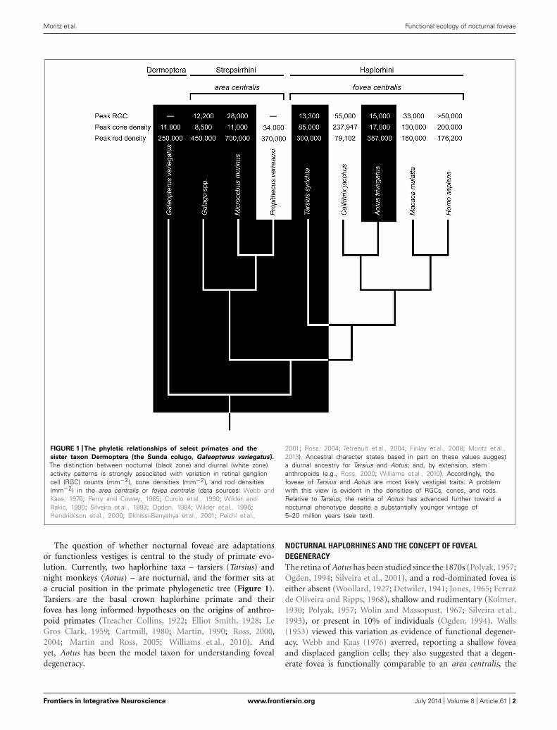
FIGURE 1 |The phyletic relationships of select primates and the
sister taxon Dermoptera (the Sunda colugo, Galeopterus variegatus).
The distinction between nocturnal (black zone) and diurnal (white zone)activity patterns is strongly associated with variation in retinal ganglioncell (RGC) counts (mm−2), cone densities (mm−2), and rod densities(mm−2) in the area centralis or fovea centralis (data sources: Webb andKaas, 1976; Perry and Cowey, 1985; Curcio et al., 1990; Wikler andRakic, 1990; Silveira et al., 1993; Ogden, 1994; Wilder et al., 1996;Hendrickson et al., 2000; Dkhissi-Benyahya et al., 2001; Peichl et al.,
2001; Ross, 2004; Tetreault et al., 2004; Finlay et al., 2008; Moritz et al.,2013). Ancestral character states based in part on these values suggesta diurnal ancestry for Tarsius and Aotus; and, by extension, stemanthropoids (e.g., Ross, 2000; Williams et al., 2010). Accordingly, thefoveae of Tarsius and Aotus are most likely vestigial traits. A problemwith this view is evident in the densities of RGCs, cones, and rods.Relative to Tarsius, the retina of Aotus has advanced further toward anocturnal phenotype despite a substantially younger vintage of5–20 million years (see text).
The question of whether nocturnal foveae are adaptationsor functionless vestiges is central to the study of primate evo-lution. Currently, two haplorhine taxa – tarsiers (Tarsius) andnight monkeys (Aotus) – are nocturnal, and the former sits ata crucial position in the primate phylogenetic tree (Figure 1).Tarsiers are the basal crown haplorhine primate and theirfovea has long informed hypotheses on the origins of anthro-poid primates (Treacher Collins, 1922; Elliot Smith, 1928; LeGros Clark, 1959; Cartmill, 1980; Martin, 1990; Ross, 2000,2004; Martin and Ross, 2005; Williams et al., 2010). Andyet, Aotus has been the model taxon for understanding fovealdegeneracy.
NOCTURNAL HAPLORHINES AND THE CONCEPT OF FOVEALDEGENERACYThe retina of Aotus has been studied since the 1870s (Polyak, 1957;Ogden, 1994; Silveira et al., 2001), and a rod-dominated fovea iseither absent (Woollard, 1927; Detwiler, 1941; Jones, 1965; Ferrazde Oliveira and Ripps, 1968), shallow and rudimentary (Kolmer,1930; Polyak, 1957; Wolin and Massopust, 1967; Silveira et al.,1993), or present in 10% of individuals (Ogden, 1994). Walls(1953) viewed this variation as evidence of functional degener-acy. Webb and Kaas (1976) averred, reporting a shallow foveaand displaced ganglion cells; they also suggested that a degen-erate fovea is functionally comparable to an area centralis, the
Frontiers in Integrative Neuroscience www.frontiersin.org July 2014 | Volume 8 | Article 61 | 2
Moritz et al. Functional ecology of nocturnal foveae
retinal specialization of strepsirrhine primates (Rohen and Cas-tenholz, 1967; Wolin and Massopust, 1970). Indeed, the densitiesof rods and cones in the foveae of Aotus azarae and Aotus trivirga-tus resemble those in the area centralis of Galago garnetti, a lorisidprimate (Wikler and Rakic, 1990; Finlay et al., 2008). The notion offoveal degeneracy in Aotus, together with the absence of a tapetumlucidum, is widely interpreted as evidence of a diurnal ancestry, asillustrated in Figure 1.
A shift to nocturnality could have occurred ∼20 Ma on the basisof phylogenetic affinities with Tremacebus, which was plausiblynocturnal (Kay and Kirk, 2000; Kay et al., 2004; Ross et al., 2007).Recent molecular phylogenies are compatible with this view, sug-gesting that the stem ancestor of Aotus diverged from diurnalCebidae ∼19.3 Ma (Perelman et al., 2011), whereas crown Aotusdiversified ∼5.5 to 4.6 Ma (Menezes et al., 2010; Ruiz-García et al.,2011). Thus, the antiquity of nocturnality in the aotine lineage isbetween ∼5 and 20 million years. This span was evidently suffi-cient to favor degenerate foveae among other distinctive attributes,such as relatively enlarged eyes and orbits (Kirk, 2006; Rossand Kirk, 2007), disabling mutations of the short-wavelength-sensitive-1 (SWS1) opsin gene (Jacobs et al., 1996; Levenson et al.,2007), rod photoreceptors with an inverted nuclear architecture(Joffe et al., 2014), and large numbers of P retinal ganglion cells(Silveira et al., 1994) with high rod convergence to both M and Pcells (Yamada et al., 2001). These traits differentiate Aotus from allother monkeys and are strongly convergent with nocturnal mam-mals; hence, the aotine visual system is almost certainly a nocturnalderivation.
The functional anatomy of the tarsier retina is more challeng-ing to interpret (Ross, 2004). Early studies of spectral tarsiers(Tarsius spectrum) failed to detect a fovea (Woollard, 1925, 1926),whereas recent investigations report the uniform presence of rod-dominant, concave-sided (concaviclivate) foveae (Hendricksonet al., 2000; Hendrickson cited in Ross, 2004). Similar foveaeare present in Philippine tarsiers (Tarsius syrichta; Polyak, 1957;Wolin and Massopust, 1967), but variable among Bornean tar-siers (Tarsius bancanus; Castenholz, 1965; Castenholz, 1984).On the surface, these findings point to an Aotus-like state offoveal degeneracy; however, the fovea of Tarsius is deeper, lessvariable, and associated with much higher cone densities (50,000–85,000 mm−2; Hendrickson et al., 2000; Hendrickson cited inRoss, 2004) than that of Aotus (5000–17,000 mm−2; Wikler andRakic, 1990; Finlay et al., 2008). Another difference concerns theSWS1 opsin gene; it is intact among tarsiers (Tan et al., 2005) and alow rate of non-synonymous to synonymous substitutions is con-sistent with strict purifying selection (Kawamura and Kubotera,2004).
Modest foveal degeneracy and a functional SWS1 opsin genehave been interpreted as evidence of a recent transition to noctur-nality (Tan et al., 2005). Indeed, two recent findings support thispremise. First, the rods of T. spectrum have a nuclear architecturethat is strongly associated with diurnality (Joffe et al., 2014). Sec-ond, molecular evidence suggests that the ancestral crown tarsierpossessed a cone opsin polymorphism that enabled trichromaticvision (Melin et al., 2013). The antiquity of this character trait isuncertain, with crown divergence dates ranging from ∼18.6 Ma(Springer et al., 2012) to ∼13 to 9 Ma (Melin et al., 2013), but
multiple independent losses of trichromatic vision appear to haveoccurred in the past 5 million years (Melin et al., 2013). Suchfindings suggest a relatively recent history of diurnality; and yet,the fossil record is a testament to committed nocturnality. Thehyperenlarged orbits of Tarsius eoceanus (Middle Eocene), Tarsiussirindhornae (Middle Miocene), and living tarsiers are most par-simoniously interpreted as evidence of continuous nocturnalityfor at least 45 million years (Rossie et al., 2006; Chaimanee et al.,2011). These discrepant lines of evidence are difficult to reconcile.
The foveae and rod architecture of tarsiers could be adaptationsto non-photopic conditions; and, hence not necessarily vestigesof a diurnal ancestor. Melin et al. (2013) hypothesized that thehyperenlarged eyes and foveate color vision of ancestral crowntarsiers (and potentially stem tarsiers and anthropoid primates),evolved to support visual predation under dim (mesopic) lightlevels such as twilight or bright moonlight. These light conditionsare predicted to support cone-mediated color vision (Melin et al.,2012) and favor enlarged eyes for greater visual sensitivity in theabsence of a tapetum lucidum (Cartmill, 1980). This attempt atconsilience is laudable but difficult to test.
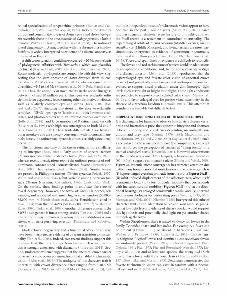
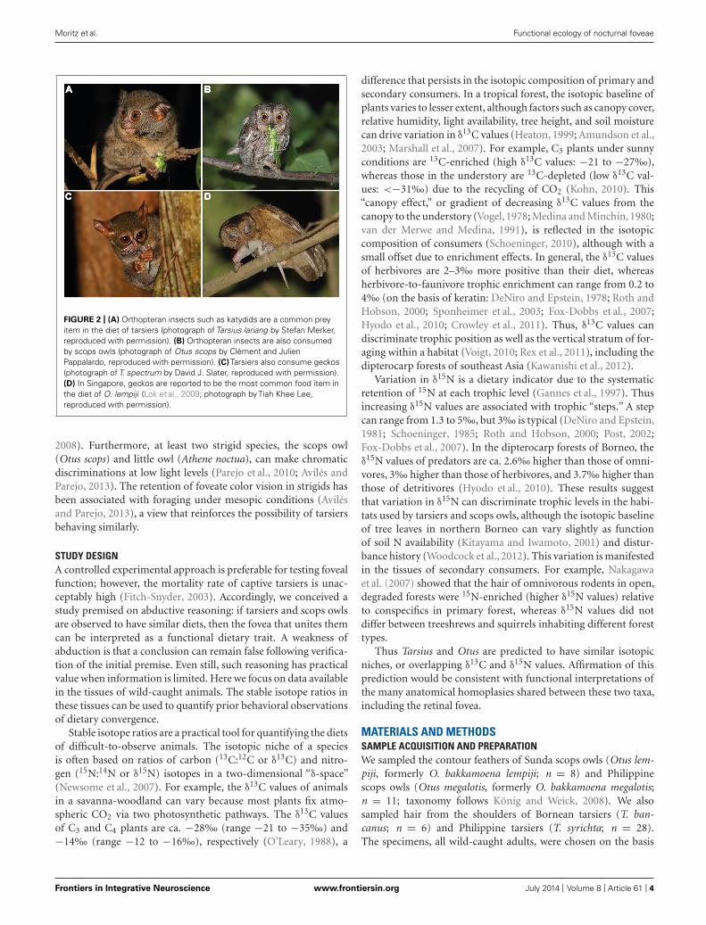
COMPARATIVE FUNCTIONAL ECOLOGY OF THE NOCTURNAL FOVEAIt is challenging for humans to observe how tarsiers discern verte-brate and invertebrate prey; they appear to integrate and alternatebetween auditory and visual cues depending on ambient con-ditions and prey type (Niemitz, 1979, 1984; MacKinnon andMacKinnon, 1980; Gursky, 2000, 2002; Dagosto et al., 2003). Sucha specialized niche is assumed to have few competitors, a conceptthat reinforces the perception of tarsiers as “living fossils” in astate of ecological stasis (Jablonski, 2003). However, observationsof the Sunda scops owl (Otus lempiji), a tarsier-sized faunivore(90–140 g), suggest a comparable niche (König and Weick, 2008;Figure 2). Potential niche convergence has attracted attention dueto the many homoplasies that unite tarsiers and scops owls, such as(i) hyperenlarged eyes that protrude from the orbit (Figures 3A,B);(ii) orbit-induced displacement of the olfactory tract, which itselfis unusually long; (iii) a loss of ocular mobility that correspondswith increased cervical mobility (Figures 3C,D); (iv) acute direc-tional hearing; (v) enlarged semicircular canals; and, (vi) derivedfeeding morphologies for perforating prey (Niemitz, 1985, 2010;Menegaz and Kirk, 2009). Niemitz (1985) interpreted this suite ofcharacter traits as an adaptation to sit-and-wait ambush preda-tion at low light levels. Evidence of dietary overlap would supportthis hypothesis and potentially shed light on yet another sharedhomoplasy, the fovea.
Within Strigiformes, there is mixed evidence for foveae in thefamily Tytonidae (barn and bay owls). For example, a fovea canbe present (Oehme, 1961) or absent in barn owls (Tyto alba;Wathey and Pettigrew, 1989; Lisney et al., 2012). In the fam-ily Strigidae (“typical” owls) rod-dominant, concaviclivate foveaeare uniformly present (Wood, 1917; Rochon-Duvigneaud, 1943;Oehme, 1961; Fite, 1973; Fite and Rosenfield-Wessels, 1975; Lis-ney et al., 2012); and at least one species, the tawny owl (Strixaluco), has a fovea with three cone classes (Martin and Gordon,1974; Bowmaker and Martin, 1978). Strix aluco demonstrates thatfoveate trichromatic vision can exist in tandem with a noctur-nal eye and orbit (Hall and Ross, 2007; Ross et al., 2007; Hall,
Frontiers in Integrative Neuroscience www.frontiersin.org July 2014 | Volume 8 | Article 61 | 3
Moritz et al. Functional ecology of nocturnal foveae
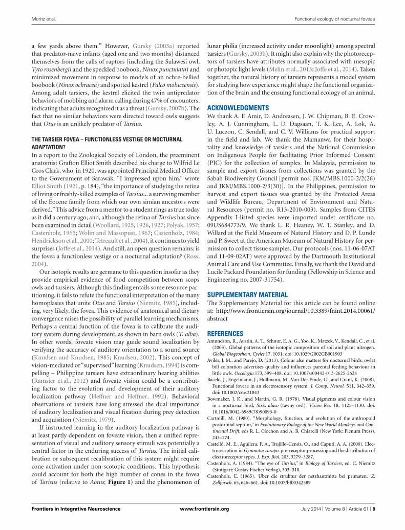
FIGURE 2 | (A) Orthopteran insects such as katydids are a common preyitem in the diet of tarsiers (photograph of Tarsius lariang by Stefan Merker,reproduced with permission). (B) Orthopteran insects are also consumedby scops owls (photograph of Otus scops by Clément and JulienPappalardo, reproduced with permission). (C) Tarsiers also consume geckos(photograph of T. spectrum by David J. Slater, reproduced with permission).(D) In Singapore, geckos are reported to be the most common food item inthe diet of O. lempiji (Lok et al., 2009; photograph by Tiah Khee Lee,reproduced with permission).
2008). Furthermore, at least two strigid species, the scops owl(Otus scops) and little owl (Athene noctua), can make chromaticdiscriminations at low light levels (Parejo et al., 2010; Avilés andParejo, 2013). The retention of foveate color vision in strigids hasbeen associated with foraging under mesopic conditions (Avilésand Parejo, 2013), a view that reinforces the possibility of tarsiersbehaving similarly.
STUDY DESIGNA controlled experimental approach is preferable for testing fovealfunction; however, the mortality rate of captive tarsiers is unac-ceptably high (Fitch-Snyder, 2003). Accordingly, we conceived astudy premised on abductive reasoning: if tarsiers and scops owlsare observed to have similar diets, then the fovea that unites themcan be interpreted as a functional dietary trait. A weakness ofabduction is that a conclusion can remain false following verifica-tion of the initial premise. Even still, such reasoning has practicalvalue when information is limited. Here we focus on data availablein the tissues of wild-caught animals. The stable isotope ratios inthese tissues can be used to quantify prior behavioral observationsof dietary convergence.
Stable isotope ratios are a practical tool for quantifying the dietsof difficult-to-observe animals. The isotopic niche of a speciesis often based on ratios of carbon (13C:12C or δ13C) and nitro-gen (15N:14N or δ15N) isotopes in a two-dimensional “δ-space”(Newsome et al., 2007). For example, the δ13C values of animalsin a savanna-woodland can vary because most plants fix atmo-spheric CO2 via two photosynthetic pathways. The δ13C valuesof C3 and C4 plants are ca. −28‰ (range −21 to −35‰) and−14‰ (range −12 to −16‰), respectively (O’Leary, 1988), a
difference that persists in the isotopic composition of primary andsecondary consumers. In a tropical forest, the isotopic baseline ofplants varies to lesser extent, although factors such as canopy cover,relative humidity, light availability, tree height, and soil moisturecan drive variation in δ13C values (Heaton, 1999; Amundson et al.,2003; Marshall et al., 2007). For example, C3 plants under sunnyconditions are 13C-enriched (high δ13C values: −21 to −27‰),whereas those in the understory are 13C-depleted (low δ13C val-ues: <−31‰) due to the recycling of CO2 (Kohn, 2010). This“canopy effect,” or gradient of decreasing δ13C values from thecanopy to the understory (Vogel, 1978; Medina and Minchin, 1980;van der Merwe and Medina, 1991), is reflected in the isotopiccomposition of consumers (Schoeninger, 2010), although with asmall offset due to enrichment effects. In general, the δ13C valuesof herbivores are 2–3‰ more positive than their diet, whereasherbivore-to-faunivore trophic enrichment can range from 0.2 to4‰ (on the basis of keratin: DeNiro and Epstein, 1978; Roth andHobson, 2000; Sponheimer et al., 2003; Fox-Dobbs et al., 2007;Hyodo et al., 2010; Crowley et al., 2011). Thus, δ13C values candiscriminate trophic position as well as the vertical stratum of for-aging within a habitat (Voigt, 2010; Rex et al., 2011), including thedipterocarp forests of southeast Asia (Kawanishi et al., 2012).
Variation in δ15N is a dietary indicator due to the systematicretention of 15N at each trophic level (Gannes et al., 1997). Thusincreasing δ15N values are associated with trophic “steps.” A stepcan range from 1.3 to 5‰, but 3‰ is typical (DeNiro and Epstein,1981; Schoeninger, 1985; Roth and Hobson, 2000; Post, 2002;Fox-Dobbs et al., 2007). In the dipterocarp forests of Borneo, theδ15N values of predators are ca. 2.6‰ higher than those of omni-vores, 3‰ higher than those of herbivores, and 3.7‰ higher thanthose of detritivores (Hyodo et al., 2010). These results suggestthat variation in δ15N can discriminate trophic levels in the habi-tats used by tarsiers and scops owls, although the isotopic baselineof tree leaves in northern Borneo can vary slightly as functionof soil N availability (Kitayama and Iwamoto, 2001) and distur-bance history (Woodcock et al., 2012). This variation is manifestedin the tissues of secondary consumers. For example, Nakagawaet al. (2007) showed that the hair of omnivorous rodents in open,degraded forests were 15N-enriched (higher δ15N values) relativeto conspecifics in primary forest, whereas δ15N values did notdiffer between treeshrews and squirrels inhabiting different foresttypes.
Thus Tarsius and Otus are predicted to have similar isotopicniches, or overlapping δ13C and δ15N values. Affirmation of thisprediction would be consistent with functional interpretations ofthe many anatomical homoplasies shared between these two taxa,including the retinal fovea.
MATERIALS AND METHODSSAMPLE ACQUISITION AND PREPARATIONWe sampled the contour feathers of Sunda scops owls (Otus lem-piji, formerly O. bakkamoena lempiji; n = 8) and Philippinescops owls (Otus megalotis, formerly O. bakkamoena megalotis;n = 11; taxonomy follows König and Weick, 2008). We alsosampled hair from the shoulders of Bornean tarsiers (T. ban-canus; n = 6) and Philippine tarsiers (T. syrichta; n = 28).The specimens, all wild-caught adults, were chosen on the basis
Frontiers in Integrative Neuroscience www.frontiersin.org July 2014 | Volume 8 | Article 61 | 4
Moritz et al. Functional ecology of nocturnal foveae
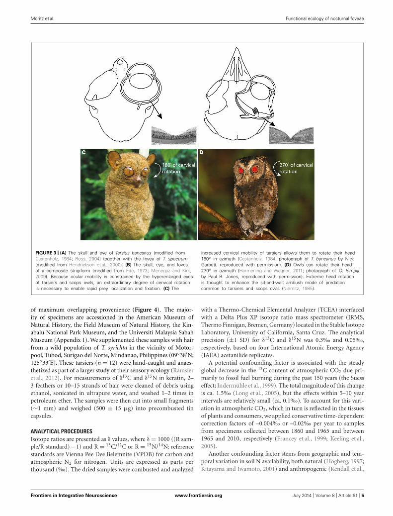
FIGURE 3 | (A) The skull and eye of Tarsius bancanus (modified fromCastenholz, 1984; Ross, 2004) together with the fovea of T. spectrum(modified from Hendrickson et al., 2000). (B) The skull, eye, and foveaof a composite strigiform (modified from Fite, 1973; Menegaz and Kirk,2009). Because ocular mobility is constrained by the hyperenlarged eyesof tarsiers and scops owls, an extraordinary degree of cervical rotationis necessary to enable rapid prey localization and fixation. (C) The
increased cervical mobility of tarsiers allows them to rotate their head180◦ in azimuth (Castenholz, 1984; photograph of T. bancanus by NickGarbutt, reproduced with permission). (D) Owls can rotate their head270◦ in azimuth (Harmening and Wagner, 2011; photograph of O. lempijiby Paul B. Jones, reproduced with permission). Extreme head rotationis thought to enhance the sit-and-wait ambush mode of predationcommon to tarsiers and scops owls (Niemitz, 1985).
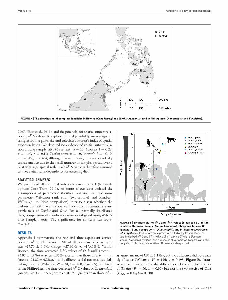
of maximum overlapping provenience (Figure 4). The major-ity of specimens are accessioned in the American Museum ofNatural History, the Field Museum of Natural History, the Kin-abalu National Park Museum, and the Universiti Malaysia SabahMuseum (Appendix 1). We supplemented these samples with hairfrom a wild population of T. syrichta in the vicinity of Motor-pool, Tubod, Surigao del Norte, Mindanao, Philippines (09◦38′N;125◦33′E). These tarsiers (n = 12) were hand-caught and anaes-thetized as part of a larger study of their sensory ecology (Ramsieret al., 2012). For measurements of δ13C and δ15N in keratin, 2–3 feathers or 10–15 strands of hair were cleaned of debris usingethanol, sonicated in ultrapure water, and washed 1–2 times inpetroleum ether. The samples were then cut into small fragments(∼1 mm) and weighed (500 ± 15 μg) into precombusted tincapsules.
ANALYTICAL PROCEDURESIsotope ratios are presented as δ values, where δ = 1000 ((R sam-ple/R standard) – 1) and R = 13C/12C or R = 15N/14N; referencestandards are Vienna Pee Dee Belemnite (VPDB) for carbon andatmospheric N2 for nitrogen. Units are expressed as parts perthousand (‰). The dried samples were combusted and analyzed
with a Thermo-Chemical Elemental Analyzer (TCEA) interfacedwith a Delta Plus XP isotope ratio mass spectrometer (IRMS,Thermo Finnigan, Bremen, Germany) located in the Stable IsotopeLaboratory, University of California, Santa Cruz. The analyticalprecision (±1 SD) for δ13C and δ15N was 0.3‰ and 0.05‰,respectively, based on four International Atomic Energy Agency(IAEA) acetanilide replicates.
A potential confounding factor is associated with the steadyglobal decrease in the 13C content of atmospheric CO2 due pri-marily to fossil fuel burning during the past 150 years (the Suesseffect; Indermühle et al., 1999). The total magnitude of this changeis ca. 1.5‰ (Long et al., 2005), but the effects within 5–10 yearintervals are relatively small (ca. 0.1‰). To account for this vari-ation in atmospheric CO2, which in turn is reflected in the tissuesof plants and consumers, we applied conservative time-dependentcorrection factors of –0.004‰ or –0.02‰ per year to samplesfrom specimens collected between 1860 and 1965 and between1965 and 2010, respectively (Francey et al., 1999; Keeling et al.,2005).
Another confounding factor stems from geographic and tem-poral variation in soil N availability, both natural (Högberg, 1997;Kitayama and Iwamoto, 2001) and anthropogenic (Kendall et al.,
Frontiers in Integrative Neuroscience www.frontiersin.org July 2014 | Volume 8 | Article 61 | 5
Moritz et al. Functional ecology of nocturnal foveae
FIGURE 4 |The distribution of sampling localities in Borneo (Otus lempiji andTarsius bancanus) and in Philippines (O. megalotis andT. syrichta).
2007; Hietz et al., 2011), and the potential for spatial autocorrela-tion of δ15N values. To explore this first possibility, we averaged allsamples from a given site and calculated Moran’s index of spatialautocorrelation. We detected no evidence of spatial autocorrela-tion among sample sites (Otus sites: n = 13, Moran’s I = 0.23,z = 1.60, p = 0.11; Tarsius sites: n = 10, Moran’s I = –0.19,z = –0.45, p = 0.65), although the semivariograms are potentiallyuninformative due to the small number of samples spread over arelatively large spatial scale. Each δ15N value is therefore assumedto have statistical independence for assessing diet.
STATISTICAL ANALYSESWe performed all statistical tests in R version 2.14.1 (R Devel-opment Core Team, 2011). As some of our data violated theassumptions of parametric statistical analysis, we used non-parametric Wilcoxon rank sum (two-sample) and Kruskal–Wallis χ2 (multiple comparison) tests to assess whether thecarbon and nitrogen isotope compositions differentiate sym-patric taxa of Tarsius and Otus. For all normally distributeddata, comparisons of significance were investigated using Welch’sTwo Sample t-tests. The significance for all tests was set atα = 0.05.
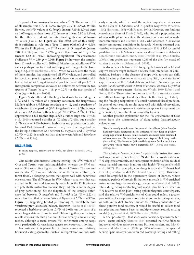
RESULTSAppendix 1 summarizes the raw and time-dependent correc-tions to δ13C. The mean ± SD of all time-corrected sampleswas –23.76 ± 1.6‰ (range: –27.80‰ to –17.41‰). WithinBorneo, the time-corrected δ13C values of O. lempiji (mean: -22.87 ± 1.7‰) were ca. 1.95‰ greater than those of T. bancanus(mean: –24.82 ± 0.2‰), but the difference did not reach statisti-cal significance (Wilcoxon W = 38; p = 0.08; Figure 5). Similarly,in the Philippines, the time-corrected δ13C values of O. megalotis(mean: –23.33 ± 2.5‰) were ca. 0.62‰ greater than those of T.
FIGURE 5 | Bivariate plot of δ13C and δ15N values (mean ± 1 SD) in the
keratin of Bornean tarsiers (Tarsius bancanus), Philippine tarsiers (T.
syrichta), Sunda scops owls (Otus lempiji ), and Philippine scops owls
(O. megalotis). To illustrate an approximate full dietary trophic step, thekeratin-derived δ13C and δ15N values of a frugivore (Müller’s Borneangibbon, Hylobates muelleri ) and a predator of vertebrates (leopard cat, Felisbengalensis) from Sabah, northern Borneo are also plotted.
syrichta (mean: –23.95 ± 1.1‰), but the difference did not reachsignificance (Wilcoxon W = 196; p = 0.198; Figure 5). Intra-generic comparisons revealed differences between the two speciesof Tarsius (W = 36, p = 0.03) but not the two species of Otus(t16.82 = 0.46, p = 0.648).
Frontiers in Integrative Neuroscience www.frontiersin.org July 2014 | Volume 8 | Article 61 | 6
Moritz et al. Functional ecology of nocturnal foveae
Appendix 1 summarizes the raw values δ15N. The mean ± SDof all samples was 5.79 ± 2.2‰ (range: 2.39–11.37‰). WithinBorneo, the δ15N values of O. lempiji (mean: 7.45 ± 1.7‰) wereca. 1.65‰ greater than those of T. bancanus (mean: 5.80 ± 1.8‰),but the difference did not reach statistical significance (WilcoxonW = 36; p = 0.142; Figure 5). The effect size of this analy-sis is sufficient to rule out a Type II error (Cohen’s d = 0.95).Within the Philippines, the δ15N values of O. megalotis (mean:7.04 ± 2.3‰) were ca. 2.22‰ greater than those of T. syrichta(mean: 4.82 ± 1.9‰), indicating significant 15N-enrichment(Wilcoxon W = 239, p = 0.008; Figure 5); however, the samplesfrom T. syrichta collected in 2010 exhibited systematically low δ15Nvalues, perhaps due to recent anthropogenic changes to the land-scape (e.g., Fox-Dobbs et al., 2012). When we calculated the meanof these samples, log-transformed all δ15N values, and controlledfor specimen year in a general model, there was no statistical dif-ference between O. megalotis and T. syrichta (t = –0.28, p = 0.781).Intrageneric comparisons revealed no differences between the twospecies of Tarsius (t7.43 = 1.19, p = 0.271) or the two species ofOtus (t17 = 0.44, p = 0.664).
Figure 5 also illustrates the larger food web by including theδ13C and δ15N values of a primary consumer, the frugivorousMüller’s gibbon (Hylobates muelleri; n = 1), and a predator ofvertebrates, the leopard cat (Felis bengalensis; n = 1). The isotopicdifferences (�) between these taxa (�13C: 2.36‰; �15N: 4.93‰)approximate a full trophic step, albeit a rather large one. Hyodoet al. (2010) reported a similar �13C value of 2.4‰, but a smaller�15N value of 3.0‰ between herbivores and predators in LambirNational Park, Sarawak, Borneo. In any case, the magnitude ofthe isotopic difference (�) between O. megalotis and T. syrichta(�15N = 2.22) is much less than that between Felis and Hylobates(�15N = 4.93‰).
DISCUSSIONIn many respects, tarsiers are not owls, but almost (Niemitz, 2010,p. 953)
Our results demonstrate isotopic overlap: the δ13C values ofOtus and Tarsius were indistinguishable, whereas the δ15N val-ues of Otus were often higher than those of Tarsius. The low andcomparable δ13C values indicate use of the same stratum (theforest floor), a foraging pattern that agrees well with behavioralobservations. The differences in δ15N values – a pattern that wasa trend in Borneo and temporally variable in the Philippines –are potentially instructive because they indicate a subtle degreeof prey partitioning. Yet the magnitude of the isotopic differ-ence (�) between O. megalotis and T. syrichta (�15N = 2.22) ismuch less than that between Felis and Hylobates (�15N = 4.93‰;Figure 5), suggesting limited partitioning of invertebrate andvertebrate prey (discussed below). However, Hyodo et al. (2010)reported a herbivore–predator �15N of 3.0‰ on the basis of amuch larger data set from Sarawak. Taken together, our isotopicresults demonstrate that Otus and Tarsius occupy similar dietaryniches, although a trend toward 15N-enrichment among scopsowls, particularly O. megalotis, suggests some prey partitioning.
For instance, it is plausible that tarsiers consume relativelyfew insect-eating squamates. Such an interpretation conflicts with
early accounts, which stressed the central importance of geckosto the diets of T. bancanus and T. syrichta (captivity: Wharton,1950; Harrison, 1963; wild: Fogden, 1974). However, our findingscorroborate those of Davis (1962), who found a preponderanceof large orthopteran insects in the stomachs of seven wild-caughtBornean tarsiers and Niemitz (1984), who observed T. bancanusunder seminatural conditions in Sarawak. Niemitz reported thatvertebrates (squamates, birds) represented <11% of 133 successfulpredation events. In Sulawesi, tarsiers seldom consume vertebrates(MacKinnon and MacKinnon, 1980; Tremble et al., 1993; Gursky,2007a), but geckos can represent 4.2% of the diet (by mass) oftarsiers in captivity (Dahang et al., 2008).
A discrepancy between the foraging behaviors of wild andcaptive tarsiers might indicate a release from predation or com-petition. Perhaps in the absence of scops owls, tarsiers can shifttheir foraging preference to vertebrate prey. Still, recent studies ofcaptive tarsiers in the United States report that T. bancanus ignoresanoles (Anolis carolinensis) in favor of crickets, whereas T. syrichtaexhibits the reverse pattern (Haring and Wright, 1989; Roberts andKohn, 1993). These mixed responses to a North American anoleare difficult to interpret, and they illustrate the challenge of study-ing the foraging adaptations of a small nocturnal visual predator.In general, our isotopic results agree well with field observations,although these are sparse – invertebrates appear to represent thegreat majority prey objects consumed by tarsiers.
Another possible explanation for the 15N-enrichment of Otusstems from the consumption of dung-eating (scatophagous)coleopterans:
“Food is usually sought near the ground... in villages (Otus lempiji)habitually hunts nocturnal insects attracted to cow dung or poultrydroppings around houses. Some stomachs examined were crammedwith cockroaches (Blattidae) and a particular type of black dung beetle(Scarabidae). The Sumatran (Minangklabau) name for this owl is kuascirit ayam, which means ‘fowl’s-excrement owl”’ (König and Weick,2008, p. 274).
The sobriquet “excrement owl” is potentially instructive. Ani-mal waste is often enriched in 15N due to the volatilization of15N-depleted ammonia, and subsequent oxidation of the residualwaste material can result in nitrate with high δ15N values (Kendallet al., 2007). For example, cow dung is typically 15N-enriched(∼2.3‰) relative to diet (Steele and Daniel, 1978). This effectcould be amplified in the dipterocarp forests of Borneo, whereextended periods of protein limitation can result in 15N-enrichedurine among large mammals, e.g., orangutans (Vogel et al., 2012).Thus, dung-eating (scatophagous) insects should be enriched in15N relative to their plant-eating (phytophagous) counterparts,and the relative 15N-enrichment of Otus could reflect a greaterproportion of scatophagous coleopterans or nocturnal squamates,or both, in the diet. To discriminate the relative contributions ofthese putative food sources, it would be useful to collect foodsamples and perform a Bayesian multiple source isotopic mixingmodel (e.g., Yeakel et al., 2009; Rutz et al., 2010).
A final possibility – that scops owls occasionally consume tar-siers – seems unlikely. Niemitz (1984) reported that Otus failed toinduce an obvious response among Bornean tarsiers, and MacK-innon and MacKinnon (1980, p. 375) observed that spectraltarsiers “paid no attention to an owl Ninox sp. sitting and calling
Frontiers in Integrative Neuroscience www.frontiersin.org July 2014 | Volume 8 | Article 61 | 7
Moritz et al. Functional ecology of nocturnal foveae
a few yards above them.” However, Gursky (2003a) reportedthat predator-naive infants (aged one and two months) distancedthemselves from the calls of raptors (including the Sulawesi owl,Tyto rosenbergii and the speckled boobook, Ninox punctulata) andminimized movement in response to models of an ochre-belliedboobook (Ninox ochracea) and spotted kestrel (Falco moluccensis).Among adult tarsiers, the kestrel elicited the twin antipredatorbehaviors of mobbing and alarm calling during 47% of encounters,indicating that adults recognized it as a threat (Gursky,2007b). Thefact that no similar behaviors were directed toward owls suggeststhat Otus is an unlikely predator of Tarsius.
THE TARSIER FOVEA – FUNCTIONLESS VESTIGE OR NOCTURNALADAPTATION?In a report to the Zoological Society of London, the preeminentanatomist Grafton Elliot Smith described his charge to Wilfrid LeGros Clark, who, in 1920, was appointed Principal Medical Officerto the Government of Sarawak. “I impressed upon him,” wroteElliot Smith (1921, p. 184), “the importance of studying the retinaof living or freshly-killed examples of Tarsius... a surviving memberof the Eocene family from which our own simian ancestors werederived.” This advice from a mentor to a student rings as true todayas it did a century ago; and, although the retina of Tarsius has sincebeen examined in detail (Woollard, 1925, 1926, 1927; Polyak, 1957;Castenholz, 1965; Wolin and Massopust, 1967; Castenholz, 1984;Hendrickson et al., 2000; Tetreault et al., 2004), it continues to yieldsurprises (Joffe et al., 2014). And still, an open question remains: isthe fovea a functionless vestige or a nocturnal adaptation? (Ross,2004).
Our isotopic results are germane to this question insofar as theyprovide empirical evidence of food competition between scopsowls and tarsiers. Although this finding entails some resource par-titioning, it fails to refute the functional interpretation of the manyhomoplasies that unite Otus and Tarsius (Niemitz, 1985), includ-ing, very likely, the fovea. This evidence of anatomical and dietaryconvergence raises the possibility of parallel learning mechanisms.Perhaps a central function of the fovea is to calibrate the audi-tory system during development, as shown in barn owls (T. alba).In other words, foveate vision may guide sound localization byverifying the accuracy of auditory orientation to a sound source(Knudsen and Knudsen, 1985; Knudsen, 2002). This concept ofvision-mediated or“supervised” learning (Knudsen, 1994) is com-pelling – Philippine tarsiers have extraordinary hearing abilities(Ramsier et al., 2012) and foveate vision could be a contribut-ing factor to the evolution and development of their auditorylocalization pathway (Heffner and Heffner, 1992). Behavioralobservations of tarsiers have long stressed the dual importanceof auditory localization and visual fixation during prey detectionand acquisition (Niemitz, 1979).
If instructed learning in the auditory localization pathway isat least partly dependent on foveate vision, then a unified repre-sentation of visual and auditory sensory stimuli was potentially acentral factor in the enduring success of Tarsius. The initial cali-bration or subsequent recalibration of this system might requirecone activation under non-scotopic conditions. This hypothesiscould account for both the high number of cones in the foveaof Tarisus (relative to Aotus; Figure 1) and the phenomenon of
lunar philia (increased activity under moonlight) among spectraltarsiers (Gursky, 2003b). It might also explain why the photorecep-tors of tarsiers have attributes normally associated with mesopicor photopic light levels (Melin et al., 2013; Joffe et al., 2014). Takentogether, the natural history of tarsiers represents a model systemfor studying how experience might shape the functional organiza-tion of the brain and the ensuing functional ecology of an animal.
ACKNOWLEDGMENTSWe thank A. F. Amir, D. Andreasen, J. W. Chipman, B. E. Crow-ley, A. J. Cunningham, L. D. Dagsaan, T. K. Lee, A. Lok, A.U. Luczon, C. Sendall, and C. V. Williams for practical supportin the field and lab. We thank the Mamanwa for their hospi-tality and knowledge of tarsiers and the National Commissionon Indigenous People for facilitating Prior Informed Consent(PIC) for the collection of samples. In Malaysia, permission tosample and export tissues from collections was granted by theSabah Biodiversity Council [permit nos. JKM/MBS.1000-2/2(26)and JKM/MBS.1000-2/3(30)]. In the Philippines, permission toharvest and export tissues was granted by the Protected Areasand Wildlife Bureau, Department of Environment and Natu-ral Resources (permit no. R13-2010-003). Samples from CITESAppendix I-listed species were imported under certificate no.09US684773/9. We thank L. R. Heaney, W. T. Stanley, and D.Willard at the Field Museum of Natural History and D. P. Lundeand P. Sweet at the American Museum of Natural History for per-mission to collect tissue samples. Our protocols (nos. 11-06-07ATand 11-09-02AT) were approved by the Dartmouth InstitutionalAnimal Care and Use Committee. Finally, we thank the David andLucile Packard Foundation for funding (Fellowship in Science andEngineering no. 2007-31754).
SUPPLEMENTARY MATERIALThe Supplementary Material for this article can be found onlineat: http://www.frontiersin.org/journal/10.3389/fnint.2014.00061/abstract
REFERENCESAmundson, R., Austin, A. T., Schuur, E. A. G., Yoo, K., Matzek, V., Kendall, C., et al.
(2003). Global patterns of the isotopic composition of soil and plant nitrogen.Global Biogeochem. Cycles 17, 1031. doi: 10.1029/2002GB001903
Avilés, J. M., and Parejo, D. (2013). Colour also matters for nocturnal birds: owletbill coloration advertises quality and influences parental feeding behaviour inlittle owls. Oecologia 173, 399–408. doi: 10.1007/s00442-013-2625-2628
Bacelo, J., Engelmann, J., Hollmann, M., Von Der Emde, G., and Grant, K. (2008).Functional foveae in an electrosensory system. J. Comp. Neurol. 511, 342–359.doi: 10.1002/cne.21843
Bowmaker, J. K., and Martin, G. R. (1978). Visual pigments and colour visionin a nocturnal bird, Strix aluco (tawny owl). Vision Res. 18, 1125–1130. doi:10.1016/0042-6989(78)90095-0
Cartmill, M. (1980). “Morphology, function, and evolution of the anthropoidpostorbital septum,” in Evolutionary Biology of the New World Monkeys and Con-tinental Drift, eds R. L. Ciochon and A. B. Chiarelli (New York: Plenum Press),243–274.
Castelló, M. E., Aguilera, P. A., Trujillo-Cenóz, O., and Caputi, A. A. (2000). Elec-troreception in Gymnotus carapo: pre-receptor processing and the distribution ofelectroreceptor types. J. Exp. Biol. 203, 3279–3287.
Castenholz, A. (1984). “The eye of Tarsius,” in Biology of Tarsiers, ed. C. Niemitz(Stuttgart: Gustav Fischer Verlag), 303–318.
Castenholz, E. (1965). Über die struktur der netzhautmitte bei primaten. Z.Zellforsch. 65, 646–661. doi: 10.1007/bf00342589
Frontiers in Integrative Neuroscience www.frontiersin.org July 2014 | Volume 8 | Article 61 | 8
Moritz et al. Functional ecology of nocturnal foveae
Catania, K. C., and Remple, F. E. (2004). Tactile foveation in the star-nosed mole.Brain Behav. Evol. 63, 1–12. doi: 10.1159/000073755
Chaimanee, Y., Lebrun, R., Yamee, C., and Jaeger, J.-J. (2011). A new MiddleMiocene tarsier from Thailand and the reconstruction of its orbital morphol-ogy using a geometric–morphometric method. Proc. Biol. Sci. 278, 1956–1963.doi: 10.1098/rspb.2010.2062
Collin, S. P. (1999). “Behavioural ecology and retinal cell topography,” in AdaptiveMechanisms in the Ecology of Vision, eds S. Archer, M. B. Djamgoz, E. Loew, J. C.Partridge, and S. Vallerga. (Dordrecht: Kluwer Academic Publishers), 509–535.doi: 10.1007/978-94-017-0619-3_17
Collin, S. P., Lloyd, D. J., and Wagner, H.-J. (2000). Foveate vision in deep-seateleosts: a comparison of primary visual and olfactory inputs. Phil. Trans. R. Soc.Lond. B 355, 1315–1320. doi: 10.1098/rstb.2000.0691
Crowley, B. E., Thorén, S., Rasoazanabary, E., Vogel, E. R., Barrett, M. A., Zohdy,S., et al. (2011). Explaining geographical variation in the isotope compositionof mouse lemurs (Microcebus). J. Biogeogr. 38, 2106–2121. doi: 10.1111/j.1365-2699.2011.02551.x
Curcio, C. A., Sloan, K. R., Kalina, R. E., and Hendrickson, A. E. (1990).Human photoreceptor topography. J. Comp. Neurol. 292, 497–523. doi:10.1002/cne.902920402
Dagosto, M., Gebo, D. L., and Dolino, C. N. (2003). “The natural history of thePhilippine tarsier (Tarsius syrichta),” in Tarsiers: Past, Present, and Future, edsP. C. Wright, E. L. Simons, and S. Gursky (New Brunswick: Rutgers UniversityPress), 237–259.
Dahang, D., Severn, K., and Shekelle, M. (2008). “Eastern tarsiers in captivity, partII: A preliminary assessment of diet,” in Primates of the Oriental Night, eds M.Shekelle, I. Maryanto, C. Groves, H. Schulze, and H. Fitch-Snyder (Jakarta: LIPIPress), 97–103.
Davis, D. D. (1962). Mammals of the lowland rain-forest of North Borneo. Bull.Natl. Mus. Singapore 31, 1–129.
DeNiro, M. J., and Epstein, S. (1978). Influence of diet on the distribution of carbonisotopes in animals. Geochim. Cosmochim. Acta 42, 495–506. doi: 10.1016/0016-7037(78)90199-90190
DeNiro, M. J., and Epstein, S. (1981). Influence of diet on the distribution of nitrogenisotopes in animals. Geochim. Cosmochim. Acta 45, 341–351. doi: 10.1016/0016-7037(81)90244-90241
Detwiler, S. R. (1941). The eye of the owl monkey (Nyctipithecus). Anat. Rec. 80,233–241. doi: 10.1002/ar.1090800209
Dkhissi-Benyahya, O., Szel, A., Degrip, W. J., and Cooper, H. M. (2001).Short and mid-wavelength cone distribution in a nocturnal Strepsirrhine pri-mate (Microcebus murinus). J. Comp. Neurol. 438, 490–504. doi: 10.1002/cne.1330
Elliot Smith, G. (1921). On examples of Tarsius from Sarawak. Proc. Zool. Soc. Lond.91, 184–186. doi: 10.1111/j.1096-3642.1921.tb03257.x
Elliot Smith, G. (1928). The Bowman Lecture, 1928: the new vision. Trans.Ophthalmol. Soc. UK 48, 64–85.
Ferraz de Oliveira, L., and Ripps, H. (1968). The “area centralis” of the owl monkey(Aotes trivirgatus). Vision Res. 8, 223–228. doi: 10.1016/0042-6989(68)90010-90012
Finlay, B. L., Franco, E. C. S., Yamada, E. S., Crowley, J. C., Parsons, M., Muniz,J. A. P. C., et al. (2008). Number and topography of cones, rods and opticnerve axons in New and Old World primates. Vis. Neurosci. 25, 289–299. doi:10.1017/S0952523808080371
Fitch-Snyder, H. M. (2003). “History of captive conservation of tarsiers,” in Tarsiers:Past, Present, and Future, eds P. C. Wright, E. L. Simons, and S. Gursky (NewBrunswick: Rutgers University Press), 277–295.
Fite, K. V. (1973). Anatomical and behavioral correlates of visual acuity in the greathorned owl. Vision Res. 13, 219–230. doi: 10.1016/0042-6989(73)90101-6
Fite, K. V., and Rosenfield-Wessels, S. (1975). A comparative study of deep avianfoveas. Brain Behav. Evol. 12, 97–115. doi: 10.1159/000124142
Fogden, M. P. L. (1974). “A preliminary field study of the western tarsier, Tarsiusbancanus Horsfield,” in Prosimian Biology, eds R. D. Martin, G. A. Doyle, and A.C. Walker (London: Gerald Duckworth), 151–165.
Fox-Dobbs, K., Bump, J. K., Peterson, R. O., Fox, D. L., and Koch, P. L.(2007). Carnivore-specific stable isotope variables and variation in the forag-ing ecology of modern and ancient wolf populations: case studies from IsleRoyale, Minnesota, and La Brea. Can. J. Zool. 85, 458–471. doi: 10.1139/Z07-018
Fox-Dobbs, K., Nelson, A. A., Koch, P. L., and Leonard, J. A. (2012). Faunal isotoperecords reveal trophic and nutrient dynamics in twentieth century Yellowstonegrasslands. Biol. Lett. 8, 838–841. doi: 10.1098/rsbl.2012.0321
Francey, R. J., Allison, C. E., Etheridge, D. M., Trudinger, C. M., Enting, I. G.,Leuenberger, M., et al. (1999). A 1000-year high precision record of δ13C inatmospheric CO2. Tellus B 51, 170–193. doi: 10.1034/j.1600-0889.1999.t01-1-00005.x
Gannes, L. Z., O’Brien, D. M., and Martínez Del Rio, C. (1997). Sta-ble isotopes in animal ecology: assumptions, caveats, and a call formore laboratory experiments. Ecology 78, 1271–1276. doi: 10.1890/0012-9658(1997)078[1271:SIIAEA]2.0.CO;2
Gursky, S. (2000). Effect of seasonality on the behavior of an insectivorous primate,Tarsius spectrum. Int. J. Primatol. 21, 477–495. doi: 10.1023/A:1005444020059
Gursky, S. (2002). The behavioral ecology of the spectral tarsier, Tarsius spectrum.Evol. Anthropol. 11, 226–234. doi: 10.1002/evan.10035
Gursky, S. (2003a). Predation experiments on infant spectral tarsiers (Tarsiusspectrum). Folia Primatol. 74, 272–284. doi: 10.1159/000073314
Gursky, S. (2003b). Lunar philia in a nocturnal primate. Int. J. Primatol. 24, 351–367.doi: 10.1023/A:1023053301059
Gursky, S. L. (2007a). The Spectral Tarsier. Upper Saddle River: Pearson Education.Gursky, S. L. (2007b). “The response of spectral tarsiers toward avian and terrestrial
predators,” in Primate Anti-predator Strategies, eds S. L. Gursky and K. A. I. Nekaris(New York: Springer), 241–252.
Hall, M. I. (2008). The anatomical relationships between the avian eye, orbit andsclerotic ring: implications for inferring activity patterns in extinct birds. J. Anat.212, 781–794. doi: 10.1111/j.1469-7580.2008.00897.x
Hall, M. I., and Ross, C. F. (2007). Eye shape and activity pattern in birds. J. Zool.271, 437–444. doi: 10.1111/j.1469-7998.2006.00227.x
Haring, D. M., and Wright, P. C. (1989). Hand-raising a Philippine tarsier, Tarsiussyrichta. Zoo Biol. 8, 265–274. doi: 10.1002/zoo.1430080307
Harmening, W. M., and Wagner, H. (2011). From optics to attention: visualperception in barn owls. J. Comp. Physiol. A 197, 1031–1042. doi: 10.1007/s00359-011-0664-663
Harrison, B. (1963). Trying to breed Tarsius. Malay. Nat. J. 17, 218–231.Heaton, T. H. E. (1999). Spatial, species, and temporal variations in the 13C/12C
ratios of C3 plants: implications for palaeodiet studies. J. Archaeol. Sci. 26, 637–649. doi: 10.1006/jasc.1998.0381
Heffner, R. S., and Heffner, H. E. (1992). “Evolution of sound localization in mam-mals,” in The Evolutionary Biology of Hearing, eds D. B. Webster, R. R. Fay, and A.N. Popper (New York: Springer), 691–715. doi: 10.1007/978-1-4612-2784-7_43
Hendrickson, A. (2005). “Organization of the adult primate fovea,” in MacularDegeneration, eds P. L. Penfold and J. M. Provis (Berlin: Springer), 1–23. doi:10.1007/3-540-26977-0_1
Hendrickson, A., Djajadi, H. R., Nakamura, L., Possin, D. E., and Sajuthi, D. (2000).Nocturnal tarsier retina has both short and long/medium-wavelength cones inan unusual topography. J. Comp. Neurol. 424, 718–730. doi: 10.1002/1096-9861(20000904)424:4<718::aid-cne12>3.0.co;2-z
Hietz, P., Turner, B. L., Wanek, W., Richter, A., Nock, C. A., and Wright, S. J. (2011).Long-term change in the nitrogen cycle of tropical forests. Science 334, 664–666.doi: 10.1126/science.1211979
Hoffmann, J. N., Montag, A. G., and Dominy, N. J. (2004). Meissner corpusclesand somatosensory acuity: the prehensile appendages of primates and elephants.Anat. Rec. A 281, 1138–1147. doi: 10.1002/ar.a.20119
Högberg, P. (1997). Tansley Review No. 95: 15N natural abundance in soil-plantsystems. New Phytol. 137, 179–203. doi: 10.1046/j.1469-8137.1997.00808.x
Hyodo, F., Matsumoto, T., Takematsu, Y., Kamoi, T., Fukuda, D., Nakagawa, M.,et al. (2010). The structure of a food web in a tropical rain forest in Malaysiabased on carbon and nitrogen stable isotope ratios. J. Trop. Ecol. 26, 205–214. doi:10.1017/S0266467409990502
Indermühle, A., Stocker, T. F., Joos, F., Fischer, H., Smith, H. J., Wahlen, M., et al.(1999). Holocene carbon-cycle dynamics based on CO2 trapped in ice at TaylorDome, Antarctica. Nature 398, 121–126. doi: 10.1038/18158
Inzunza, O., Bravo, H., and Smith, R. L. (1989). Foveal regions of bird retinascorrelate with the aster of the inner nuclear layer. Anat. Rec. 223, 342–346. doi:10.1002/ar.1092230313
Jablonski, N. G. (2003). “The evolution of the tarsiid niche,” in Tarsiers: Past, Present,and future, eds P. C. Wright, E. L. Simons, and S. Gursky. (New Brunswick: RutgersUniversity Press), 35–49.
Frontiers in Integrative Neuroscience www.frontiersin.org July 2014 | Volume 8 | Article 61 | 9
Moritz et al. Functional ecology of nocturnal foveae
Jacobs, G. H., Neitz, M., and Neitz, J. (1996). Mutations in S-cone pigment genesand the absence of colour vision in two species of nocturnal primate. Proc. R. Soc.Lond. B 263, 705–710. doi: 10.1098/rspb.1996.0105
Joffe, B., Peichl, L., Hendrickson, A., Leonhardt, H., and Solovei, I. (2014). Diurnalityand nocturnality in primates: an analysis from the rod photoreceptor nucleiperspective. Evol. Biol. 41, 1–11. doi: 10.1007/s11692-013-9240-9249
Jones, A. E. (1965). The retinal structure of (Aotes trivirgatus) the owl monkey. J.Comp. Neurol. 125, 19–27. doi: 10.1002/cne.901250104
Kawamura, S., and Kubotera, N. (2004). Ancestral loss of short wave-sensitive conevisual pigment in lorisiform prosimians, contrasting with its strict conserva-tion in other prosimians. J. Mol. Evol. 58, 314–321. doi: 10.1007/s00239-003-2553-z
Kawanishi, K., Liang, S. H. N., Darimont, C., Reimchen, T. E., and Sunquist, M.E. (2012). Isotopic niche differentiation among mammals from a rainforest inPeninsular Malaysia. Raffles Bull. Zool. 60, 233–239.
Kay, R. F., Campbell, V. M., Rossie, J. B., Colbert, M. W., and Rowe, T. B. (2004).Olfactory fossa of Tremacebus harringtoni (Platyrrhini, early Miocene, Sacanana,Argentina): implications for activity pattern. Anat. Rec. A 281, 1157–1172. doi:10.1002/ar.a.20121
Kay, R. F., and Kirk, E. C. (2000). Osteological evidence for the evolution of activitypattern and visual acuity in primates. Am. J. Phys. Anthropol. 113, 235–262. doi:10.1002/1096-8644(200010)113:2<235::aid-ajpa7>3.0.co;2–9
Keeling, C. D., Bollenbacher, A. F., and Whorf, T. P. (2005). “Monthly atmospheric13C/12C isotopic ratios for 10 SIO stations,” in Trends – A Compendium of Dataon Global Change. Oak Ridge, TN. Available at: cdiac.ornl.gov/trends/trends.htm
Kendall, C., Elliott, E. M., and Wankel, S. D. (2007). “Tracing anthropogenic inputsof nitrogen to ecosystems,” in Stable Isotopes in Ecology and Environmental Science,2nd Edn, eds R. Michener and K. Lajtha (Malden, MA: Blackwell), 375–449. doi:10.1002/9780470691854.ch12
Kirk, E. C. (2006). Effects of activity pattern on eye size and orbital aperture size inprimates. J. Hum. Evol. 51, 159–170. doi: 10.1016/j.jhevol.2006.02.004
Kitayama, K., and Iwamoto, K. (2001). Patterns of natural 15N abundance inthe leaf-to-soil continuum of tropical rain forests differing in N availability onMount Kinabalu, Borneo. Plant Soil 229, 203–212. doi: 10.1023/A:1004853915544
Knudsen, E. I. (1994). Supervised learning in the brain. J. Neurosci. 14, 3985–3997.Knudsen, E. I. (2002). Instructed learning in the auditory localization pathway of
the barn owl. Nature 417, 322–328. doi:10.1038/417322aKnudsen, E. I., and Knudsen, P. F. (1985). Vision guides the adjustment of
auditory localization in young barn owls. Science 230, 545–548. doi: 10.1126/sci-ence.4048948
Kohn, M. J. (2010). Carbon isotope compositions of terrestrial C3 plants as indi-cators of (paleo)ecology and (paleo)climate. Proc. Natl. Acad. Sci. U.S.A. 107,19691–19695. doi: 10.1073/pnas.1004933107
Kolmer, W. (1930). Zur kenntnis des auges der primaten. Z. Anat. Entwicklungs. 93,679–722. doi: 10.1007/bf02118055
König, C., and Weick, F. (2008). Owls of the World, 2nd Edn. New Haven: YaleUniversity Press.
Le Gros Clark, W. E. (1959). The Antecedents of Man. Edinburgh: EdinburghUniversity Press.
Levenson, D. H., Fernandez-Duque, E., Evans, S., and Jacobs, G. H. (2007). Muta-tional changes in S-cone opsin genes common to both nocturnal and cathemeralAotus monkeys. Am. J. Primatol. 69, 757–765. doi: 10.1002/ajp.20402
Lisney, T. J., Iwaniuk, A. N., Bandet, M. V., and Wylie, D. R. (2012). Eye shape andretinal topography in owls (Aves: Strigiformes). Brain Behav. Evol. 79, 218–236.doi: 10.1159/000337760
Locket, N. A. (1992). Problems of deep foveas. Aust. N. Z. J. Ophthalmol. 20,281–295. doi: 10.1111/j.1442-9071.1992.tb00740.x
Lok, A. F. S. L., Lee, T. K., and Lim, K. C. (2009). The biology of Otus lempijicnephaues Deignan, the Sunda scops-owl in Singapore. Nat. Singapore 2, 31–38.
Long, E., Sweitzer, R., Diefenbach, D., and Ben-David, M. (2005). Controllingfor anthropogenically induced atmospheric variation in stable carbon isotopestudies. Oecologia 146, 148–156. doi: 10.1007/s00442-005-0181-186
MacKinnon, J., and MacKinnon, K. (1980). The behavior of wild spectral tarsiers.Int. J. Primatol. 1, 361–379. doi: 10.1007/bf02692280
Mancini, F., Sambo, C. F., Ramirez, J. D., Bennett, D. L. H., Haggard, P., and Iannetti,G. D. (2013). A fovea for pain at the fingertips. Curr. Biol. 23, 496–500. doi:10.1016/j.cub.2013.02.008
Marshall, J. D., Brooks, J. R., and Lajtha, K. (2007). “Sources of variation in the stableisotopic composition of plants,” in Stable Isotopes in Ecology and EnvironmentalScience, 2nd Edn, eds R. Michener and K. Lajtha (Boston: Wiley-Blackwell),22–60. doi: 10.1002/9780470691854.ch2
Martin, G. R., and Gordon, I. E. (1974). Visual acuity in the tawny owl (Strix aluco).Vision Res. 14, 1393–1397. doi: 10.1016/0042-6989(74)90014-5
Martin, R. D. (1990). Primate Origins and Evolution: A Phylogenetic Reconstruction.Princeton: Princeton University Press.
Martin, R. D., and Ross, C. F. (2005). “The evolutionary and ecological context ofprimate vision,” in The Primate Visual System: A Comparative Approach, ed. J.Kremers (Chichester: John Wiley & Sons), 1–36.
Medina, E., and Minchin, P. (1980). Stratification of δ13C values of leaves inAmazonian rain forests. Oecologia 45, 377–378. doi: 10.1007/BF00540209
Melin, A. D., Matsushita, Y., Moritz, G. L., Dominy, N. J., and Kawamura, S.(2013). Inferred L/M cone opsin polymorphism of ancestral tarsiers sheds dimlight on the origin of anthropoid primates. Proc. R. Soc. B 280, 20130189. doi:10.1098/rspb.2013.0189
Melin, A. D., Moritz, G. L., Fosbury, R. A. E., Kawamura, S., and Dominy, N. J. (2012).Why aye-ayes see blue. Am. J. Primatol. 74, 185–192. doi: 10.1002/ajp.21996
Menegaz, R. A., and Kirk, E. C. (2009). Septa and processes: convergent evolution ofthe orbit in haplorhine primates and strigiform birds. J. Hum. Evol. 57, 672–687.doi: 10.1016/j.jhevol.2009.04.010
Menezes, A. N., Bonvicino, C. R., and Seuánez, H. N. (2010). Identification, clas-sification and evolution of owl monkeys (Aotus, Illiger 1811). BMC Evol. Biol.10:248. doi: 10.1186/1471-2148-10-248
Moore, B. A., Kamilar, J. M., Collin, S. P., Bininda-Emonds, O. R. P., Dominy, N.J., Hall, M. I., et al. (2012). A novel method for comparative analysis of retinalspecialization traits from topographic maps. J. Vis. 12, 1–24. doi: 10.1167/12.12.13
Moritz, G. L., Lim, N. T.-L., Neitz, M., Peichl, L., and Dominy, N. J. (2013). Expres-sion and evolution of short wavelength sensitive opsins in colugos: a nocturnallineage that informs debate on primate origins. Evol. Biol. 40, 542–553. doi:10.1007/s11692-013-9230-y
Nakagawa, M., Hyodo, F., and Nakashizuka, T. (2007). Effect of forest use ontrophic levels of small mammals: an analysis using stable isotopes. Can. J. Zool.85, 472–478. doi: 10.1139/Z07-026
Neuweiler, G. (2003). Evolutionary aspects of bat echolocation. J. Comp. Physiol. A189, 245–256. doi: 10.1007/s00359-003-0406-402
Newsome, S. D., Martinez Del Rio, C., Bearhop, S., and Phillips, D. L. (2007). Aniche for isotopic ecology. Front. Ecol. Environ. 5:429–436. doi: 10.1890/060150.1
Niemitz, C. (1979). “Outline of the behavior of Tarsius bancanus,” in The Study ofProsimian Behaviour, eds G. A. Doyle and R. D. Martin (New York: AcademicPress), 631–660.
Niemitz, C. (1984). “Synecological relationships and feeding behaviour of the genusTarsius,” in Biology of Tarsiers, ed. C. Niemitz (Stuttgart: Gustav Fischer Verlag),59–75.
Niemitz, C. (1985). Can a primate be an owl? – convergences in the same ecologicalniche. Forts. Zool. 30, 666–670.
Niemitz, C. (2010). Progreditur ordinara saltando et retrorsum... Normally pro-ceeds in a leaping fashion, and backwards... Int. J. Primatol. 31, 941–957. doi:10.1007/s10764-010-9454-y
Oehme, H. (1961). Vergleichend-histologische untersuchungen an der retina voneulen. Zoo Jb. Anat. 79, 439–478.
Ogden, T. E. (1994). “Ophthalmologic research in the owl monkey,” in Aotus: TheOwl Monkey, eds J. F. Baer, R. E. Weller, and I. Kakoma (San Diego: AcademicPress), 263–286. doi: 10.1016/B978-0-12-072405-5.50015-9
O’Leary, M. H. (1988). Carbon isotopes in photosynthesis. Bioscience 38, 328–336.doi: 10.2307/1310735
Parejo, D., Avilés, J. M., and Rodríguez, J. (2010). Visual cues and parentalfavouritism in a nocturnal bird. Biol. Lett. 6, 171–173. doi: 10.1098/rsbl.2009.0769
Peichl, L., Rakotondraparany, F., and Kappeler, P. (2001). Photoreceptor types anddistributions in nocturnal and diurnal Malagasy primates. Invest. Ophthalmol.Vis. Sci. 42:S48.
Perelman, P., Johnson, W. E., Roos, C., Seuánez, H. N., Horvath, J. E., Moreira,M. A. M., et al. (2011). A molecular phylogeny of living primates. PLoS Genet.7:e1001342. doi: 10.1371/journal.pgen.1001342
Perry, V. H., and Cowey, A. (1985). The ganglion cell and cone distributions in themonkey’s retina: implications for central magnification factors. Vision Res. 25,1795–1810. doi: 10.1016/0042-6989(85)90004-5
Frontiers in Integrative Neuroscience www.frontiersin.org July 2014 | Volume 8 | Article 61 | 10
Moritz et al. Functional ecology of nocturnal foveae
Pettigrew, J. D., and Frost, B. J. (1985). A tactile fovea in the Scolopacidae? BrainBehav. Evol. 26, 185–195. doi: 10.1159/000118775
Polyak, S. (1957). The Vertebrate Visual System. Chicago: University of ChicagoPress.
Post, D. M. (2002). Using stable isotopes to estimate trophic position: mod-els, methods, and assumptions. Ecology 83, 703–718. doi: 10.1890/0012-9658(2002)083[0703:USITET]2.0.CO;2
Provis, J. M., Dubis, A. M., Maddess, T., and Carroll, J. (2013). Adaptation of thecentral retina for high acuity vision: cones, the fovea and the avascular zone. Prog.Retin. Eye Res. 35, 63–81. doi: 10.1016/j.preteyeres.2013.01.005
Ramsier, M. A., Cunningham, A. J., Moritz, G. L., Finneran, J. J., Williams, C. V.,Ong, P. S., et al. (2012). Primate communication in the pure ultrasound. Biol.Lett. 8, 508–511. doi: 10.1098/rsbl.2011.1149
R Development Core Team. (2011). R: A Language and Environment for StatisticalComputing. Vienna: R Foundation for Statistical Computing.
Rex, K., Michener, R., Kunz, T. H., and Voigt, C. C. (2011). Vertical stratifica-tion of neotropical leaf-nosed bats (Chiroptera: Phyllostomidae) revealed bystable carbon isotopes. J. Trop. Ecol. 27, 211–222. doi: 10.1017/S0266467411000022
Roberts, M., and Kohn, F. (1993). Habitat use, foraging behavior, and activ-ity patterns in reproducing western tarsiers, Tarsius bancanus, in captivity:a management synthesis. Zoo Biol. 12, 217–232. doi: 10.1002/zoo.1430120207
Rochon-Duvigneaud, A. (1943). Les Yeux et la Vision des Vertébrés. Paris: ElsevierMasson.
Rohen, J. W., and Castenholz, A. (1967). Über die zentralisation der retina beiprimaten. Folia Primatol. 5, 92–147. doi: 10.1159/000161941
Röll, B. (2001). Gecko vision – retinal organization, foveae and implicationsfor binocular vision. Vision Res. 41, 2043–2056. doi: 10.1016/S0042-6989(01)00093-91
Ross, C. F. (2000). Into the light: the origin of Anthropoidea. Annu. Rev. Anthropol.29, 147–194. doi: 10.1146/annurev.anthro.29.1.147
Ross, C. F. (2004). “The tarsier fovea: functionless vestige on nocturnal adaptation?,”in Anthropoid Origins: New Visions, eds C. F. Ross and R. F. Kay (New York: KluwerAcademic), 477–537.
Ross, C. F., Hall, I. M., and Heesy, C. P. (2007). “Were basal primates nocturnal?Evidence from eye and orbit shape,” in Primate origins: Adaptations and Evolution,eds M. J. Ravosa and M. Dagosto (New York: Springer), 233–256.
Ross, C. F., and Kirk, E. C. (2007). Evolution of eye size and shape in primates. J.Hum. Evol. 52, 294–313. doi: 10.1016/j.jhevol.2006.09.006
Rossie, J. B., Ni, X., and Beard, K. C. (2006). Cranial remains of an Eocene tarsier.Proc. Natl. Acad. Sci. U.S.A. 103, 4381–4385. doi: 10.1073/pnas.0509424103
Roth, J. D., and Hobson, K. A. (2000). Stable carbon and nitrogen isotopic frac-tionation between diet and tissue of captive red fox: implications for dietaryreconstruction. Can. J. Zool. 78, 848–852. doi: 10.1139/z00-008
Ruiz-García, M., Vásquez, C., Camargo, E., Leguizamón, N., Gálvez, H., Vallejo,A., et al. (2011). Molecular phylogenetics of Aotus (Platyrrhini, Cebidae). Int. J.Primatol. 32, 1218–1241. doi: 10.1007/s10764-011-9539-9532
Rutz, C., Bluff, L. A., Reed, N., Troscianko, J., Newton, J., Inger, R., et al. (2010).The ecological significance of tool use in New Caledonian crows. Science 329,1523–1526. doi: 10.1126/science.1192053
Schoeninger, M. J. (1985). Trophic level effects on 15N/14N and 13C/12C ratios inbone collagen and strontium levels in bone mineral. J. Hum. Evol. 14, 515–525.doi: 10.1016/S0047-2484(85)80030-0
Schoeninger, M. J. (2010). “Toward a δ13C isoscape for primates,” in Isoscapes:Understanding Movement, Pattern, and Process on Earth through Isotope Mapping,eds J. B. West, G. J. Bowen, T. E. Dawson, and K. P. Tu (New York: Springer),319–333.
Silveira, L. C. L., Perry, V. H., and Yamada, E. S. (1993). The retinal ganglion celldistribution and the representation of the visual field in area 17 of the owl monkey,Aotus trivirgatus. Vis. Neurosci. 10, 887–897. doi: 10.1017/S095252380000609X
Silveira, L. C. L., Picanço-Diniz, C. W., Sampaio, L. F. S., and Oswaldo-Cruz, E.(1989). Retinal ganglion cell distribution in the cebus monkey: a comparison withthe cortical magnification factors. Vision Res. 29, 1471–1483. doi: 10.1016/0042-6989(89)90131-4
Silveira, L. C. L., Yamada, E. S., Franco, E. C. S., and Finlay, B. L. (2001). Thespecialization of the owl monkey retina for night vision. Color Res. Appl. 26,S118–S122. doi: 10.1002/1520-6378(2001)26:1++<::AID-COL26>3.0.CO;2-9
Silveira, L. C. L., Yamada, E. S., Perry, V. H., and Picanço-Diniz, C. W.(1994). M and P retinal ganglion cells of diurnal, and nocturnal New-World monkeys. Neuroreport 5, 2077–2081. doi: 10.1097/00001756-199410270-00022
Sponheimer, M., Robinson, T., Ayliffe, L., Roeder, B., Hammer, J., Passey, B., et al.(2003). Nitrogen isotopes in mammalian herbivores: hair δ15N values from acontrolled feeding study. Int. J. Osteoarchaeol. 13, 80–87. doi: 10.1002/oa.655
Springer, M. S., Meredith, R. W., Gatesy, J., Emerling, C. A., Park, J., Rabosky,D. L., et al. (2012). Macroevolutionary dynamics and historical biogeography ofprimate diversification inferred from a species supermatrix. PLoS ONE 7:e49521.doi: 10.1371/journal.pone.0049521
Steele, K. W., and Daniel, R. M. (1978). Fractionation of nitrogen isotopes byanimals: a further complication to the use of variations in the natural abun-dance of 15N for tracer studies. J. Agric. Sci. 90, 7–9. doi: 10.1017/S002185960004853X
Tan, Y., Yoder, A. D., Yamashita, N., and Li, W.-H. (2005). Evidence from opsingenes rejects nocturnality in ancestral primates. Proc. Natl. Acad. Sci. U.S.A. 102,14712–14716. doi: 10.1073/pnas.0507042102
Tansley, K. (1960). The retina of a diurnal gecko, Phelsuma madagascariensis lon-ginsulae. Pflügers Arch. Gesamte Physiol. Menschen Tiere 272, 262–269. doi:10.1007/BF00363014
Tetreault, N., Hakeem, A., and Allman, J. M. (2004). “The distribution and sizeof retinal ganglion cells in Microcebus murinus, Cheirogaleus medius, and Tar-sius syrichta: implications for the evolution of sensory systems in primates,” inAnthropoid Origins: New Visions, eds C. F. Ross and R. F. Kay (New York: KluwerAcademic/Plenum Publishers), 463–475.
Treacher Collins, E. (1922). Arboreal Life and the Evolution of the Human Eye.Philadelphia: Lea and Febiger.
Tremble, M., Muskita, Y., and Supriatna, J. (1993). Field observations of Tarsiusdianae at Lore Lindu National Park, Central Sulawesi, Indonesia. Trop. Biodivers.1, 67–76.
van der Merwe, N.J., and Medina, E. (1991). The canopy effect, carbon isotope ratiosand foodwebs in Amazonia. J. Archaeol. Sci. 18, 249–259. doi: 10.1016/0305-4403(91)90064-V
Vogel, E. R., Knott, C. D., Crowley, B. E., Blakely, M. D., Larsen, M. D., and Dominy,N. J. (2012). Bornean orangutans on the brink of protein bankruptcy. Biol. Lett.8, 333–336. doi: 10.1098/rsbl.2011.1040
Vogel, J. C. (1978). Recycling of CO2 in a forest environment. Oecolog. Plantar. 13,89–94.
Voigt, C. C. (2010). Insights into strata use of forest animals using the ‘canopy effect’.Biotropica 42, 634–637. doi: 10.1111/j.1744-7429.2010.00703.x
Walls, G. L. (1937). Significance of the foveal depression. Arch. Ophthalmol. 18,912–919. doi: 10.1001/archopht.1937.00850120046005
Walls, G. L. (1942). The Vertebrate Eye and its Adaptive Radiation. Bloomfield Hills,MI: Cranbrook Institute of Science. doi: 10.5962/bhl.title.7369
Walls, G. L. (1953). The Lateral Geniculate Nucleus and Visual Histophysiology.Berkeley: University of California Press.
Wathey, J. C., and Pettigrew, J. D. (1989). Quantitative analysis of the retinal ganglioncell layer and optic nerve of the barn owl Tyto alba. Brain Behav. Evol. 33, 279–292.doi: 10.1159/000115936
Weale, R. A. (1966). Why does the human retina possess a fovea? Nature 212,255–256. doi: 10.1038/212255a0
Webb, S. V., and Kaas, J. H. (1976). The sizes and distribution of ganglion cells inthe retina of the owl monkey, Aotus trivirgatus. Vision Res. 16, 1247–1254. doi:10.1016/0042-6989(76)90049-3
Wharton, C. H. (1950). The tarsier in captivity. J. Mammal. 31, 260–268. doi:10.2307/1375291
Wikler, K. C., and Rakic, P. (1990). Distribution of photoreceptor subtypes in theretina of diurnal and nocturnal primates. J. Neurosci. 10, 3390–3401.
Wilder, H. D., Grünert, U., Lee, B. B., and Martin, P. R. (1996). Topography ofganglion cells and photoreceptors in the retina of a New World monkey: the mar-moset Callithrix jacchus. Vis. Neurosci. 13, 335–352. doi: 10.1017/S0952523800007586
Williams, B. A., Kay, R. F., and Kirk, E. C. (2010). New perspectives onanthropoid origins. Proc. Natl. Acad. Sci. U.S.A. 107, 4797–4804. doi:10.1073/pnas.0908320107
Wolin, L. R., and Massopust, L. C. (1967). Characteristics of the ocular fundus inprimates. J. Anat. 101, 693–699.
Frontiers in Integrative Neuroscience www.frontiersin.org July 2014 | Volume 8 | Article 61 | 11
Moritz et al. Functional ecology of nocturnal foveae
Wolin, L. R., and Massopust, L. C. (1970). “Morphology of the primate retina,” inThe Primate Brain, eds C. R. Noback and W. Montagna (New York: Appleton-Century-Crofts), 1–27.
Wood, C. A. (1917). The Fundus Oculi of Birds Especially as Viewed by theOphthalmoscope. Chicago: Lakeside Press. doi: 10.5962/bhl.title.63624
Woodcock, P., Edwards, D. P., Newton, R. J., Edwards, F. A., Khen, C. V., Bottrell, S.H., et al. (2012). Assessing trophic position from nitrogen isotope ratios: effectivecalibration against spatially varying baselines. Naturwissenschaften 99, 275–283.doi: 10.1007/s00114-012-0896-892
Woollard, H. H. (1925). The anatomy of Tarsius spectrum. Proc. Zool. Soc. Lond. 95,1071–1184. doi: 10.1111/j.1469-7998.1925.tb07117.x
Woollard, H. H. (1926). Notes on the retina and lateral geniculate body inTupaia, Tarsius, Nycticebus and Hapale. Brain 49, 77–104. doi: 10.1093/brain/49.1.77
Woollard, H. H. (1927). The differentiation of the retina in the primates. Proc. Zool.Soc. Lond. 97, 1–18. doi: 10.1111/j.1096-3642.1927.tb02243.x
Yamada, E. S., Silveira, L. C. L., Perry, V. H., and Franco, E. C. (2001). Mand P retinal ganglion cells of the owl monkey: morphology, size and pho-toreceptor convergence. Vision Res. 41, 119–131. doi: 10.1016/S0042-6989(00)00244-3
Yeakel, J. D., Patterson, B. D., Fox-Dobbs, K., Okumura, M. M., Cerling, T. E., Moore,J. W., et al. (2009). Cooperation and individuality among man-eating lions. Proc.Natl. Acad. Sci. U.S.A. 106, 19040–19043. doi: 10.1073/pnas.0905309106
Conflict of Interest Statement: The authors declare that the research was conductedin the absence of any commercial or financial relationships that could be construedas a potential conflict of interest.
Received: 25 March 2014; accepted: 08 July 2014; published online: 25 July 2014.Citation: Moritz GL, Melin AD, Tuh Yit Yu F, Bernard H, Ong PS and Dominy NJ(2014) Niche convergence suggests functionality of the nocturnal fovea. Front. Integr.Neurosci. 8:61. doi: 10.3389/fnint.2014.00061This article was submitted to the journal Frontiers in Integrative Neuroscience.Copyright © 2014 Moritz, Melin, Tuh Yit Yu, Bernard, Ong and Dominy. This is anopen-access article distributed under the terms of the Creative Commons AttributionLicense (CC BY). The use, distribution or reproduction in other forums is permit-ted, provided the original author(s) or licensor are credited and that the originalpublication in this journal is cited, in accordance with accepted academic practice.No use, distribution or reproduction is permitted which does not comply with theseterms.
Frontiers in Integrative Neuroscience www.frontiersin.org July 2014 | Volume 8 | Article 61 | 12