Embed Size (px)
Citation preview
NFIA Haploinsufficiency Is Associatedwith a CNS Malformation Syndromeand Urinary Tract DefectsWeining Lu
1,2[, Fabiola Quintero-Rivera
3[¤, Yanli Fan
1[, Fowzan S. Alkuraya
1, Diana J. Donovan
4, Qiongchao Xi
1,
Annick Turbe-Doan1
, Qing-Gang Li2
, Craig G. Campbell5
, Alan L. Shanske6
, Elliott H. Sherr7
, Ayesha Ahmad8
,
Roxana Peters1
, Benedict Rilliet9
, Paloma Parvex10
, Alexander G. Bassuk11
, David J. Harris12
, Heather Ferguson13
,
Chantal Kelly13
, Christopher A. Walsh12,14,15
, Richard M. Gronostajski16
, Koenraad Devriendt17
, Anne Higgins4
,
Azra H. Ligon4
, Bradley J. Quade4
, Cynthia C. Morton4,13
, James F. Gusella3
, Richard L. Maas1*
1 Genetics Division, Brigham and Women’s Hospital and Harvard Medical School, Boston, Massachusetts, United States of America, 2 Renal Section, Boston University
Medical Center, Boston, Massachusetts, United States of America, 3 Center for Human Genetic Research, Massachusetts General Hospital and Harvard Medical School,
Boston, Massachusetts, United States of America, 4 Department of Pathology, Brigham and Women’s Hospital and Harvard Medical School, Boston, Massachusetts, United
States of America, 5 Division of Neurology, Children’s Hospital of Western Ontario, London, Ontario, Canada, 6 Children’s Hospital at Montefiore, Albert Einstein College of
Medicine, Bronx, New York, United States of America, 7 Department of Neurology, University of California San Francisco, San Francisco, California, United States of America,
8 Division of Genetic and Metabolic Disorders, Department of Pediatrics, Wayne State University, Detroit, Michigan, United States of America,
9 Department of Neurosurgery, University Hospital, Geneva, Switzerland, 10 Department of Nephrology, University Hospital, Geneva, Switzerland, 11 Departments of
Pediatrics and Neurology, Northwestern University Feinberg School of Medicine, Chicago, Illinois, United States of America, 12 Genetics Division, Children’s Hospital Boston
and Harvard Medical School, Boston, Massachusetts, United States of America, 13 Department of Obstetrics, Gynecology and Reproductive Biology, Brigham and Women’s
Hospital and Harvard Medical School, Boston, Massachusetts, United States of America, 14 Department of Neurology, Beth Israel Deaconess Medical Center and Harvard
Medical School, Boston, Massachusetts, United States of America, 15 Howard Hughes Medical Institute, Beth Israel Deaconess Medical Center and Harvard Medical School,
Boston, Massachusetts, United States of America, 16 Department of Biochemistry, State University of New York at Buffalo, Buffalo, New York, United States of America,
17 Centre for Human Genetics, University of Leuven, Leuven, Belgium
Complex central nervous system (CNS) malformations frequently coexist with other developmental abnormalities, but whetherthe associated defects share a common genetic basis is often unclear. We describe five individuals who share phenotypicallyrelated CNS malformations and in some cases urinary tract defects, and also haploinsufficiency for the NFIA transcription factorgene due to chromosomal translocation or deletion. Two individuals have balanced translocations that disrupt NFIA. A thirdindividual and two half-siblings in an unrelated family have interstitial microdeletions that include NFIA. All five individualsexhibit similar CNS malformations consisting of a thin, hypoplastic, or absent corpus callosum, and hydrocephalus orventriculomegaly. The majority of these individuals also exhibit Chiari type I malformation, tethered spinal cord, and urinarytract defects that include vesicoureteral reflux. Other genes are also broken or deleted in all five individuals, and may contributeto the phenotype. However, the only common genetic defect is NFIA haploinsufficiency. In addition, previous analyses of Nfia�/�
knockout mice indicate that Nfia deficiency also results in hydrocephalus and agenesis of the corpus callosum. Furtherinvestigation of the mouse Nfiaþ/� and Nfia�/� phenotypes now reveals that, at reduced penetrance, Nfia is also required in adosage-sensitive manner for ureteral and renal development. Nfia is expressed in the developing ureter and metanephricmesenchyme, and Nfiaþ/� and Nfia�/�mice exhibit abnormalities of the ureteropelvic and ureterovesical junctions, as well as bifidand megaureter. Collectively, the mouse Nfia mutant phenotype and the common features among these five human casesindicate that NFIA haploinsufficiency contributes to a novel human CNS malformation syndrome that can also include ureteraland renal defects.
Citation: Lu W, Quintero-Rivera F, Fan Y, Alkuraya FS, Donovan DJ, et al (2007) NFIA haploinsufficiency is associated with a CNS malformation syndrome and urinary tractdefects. PLoS Genet 3(5): e80. doi:10.1371/journal.pgen.0030080
Introduction
Complex human developmental phenotypes represent anespecially difficult problem in human genetics. In many cases,congenital birth defects are believed to result from thecombined effect of many genes, often with an environmentalcontribution, and frequently culminate in perinatal demise.Thus, for many cases, extended families do not exist, andapproaches to disease gene identification based on linkageanalysis are not possible. In addition, many developmentaldisorders are genetically heterogeneous, making the ascer-tainment of single contributory genes difficult.
The analysis of human balanced chromosome rearrange-ments offers a potential approach to this problem. Although
Editor: Veronica van Heyningen, Medical Research Council Human Genetics Unit,United Kingdom
Received September 8, 2006; Accepted April 5, 2007; Published May 25, 2007
Copyright: � 2007 Lu et al. This is an open-access article distributed under theterms of the Creative Commons Attribution License, which permits unrestricteduse, distribution, and reproduction in any medium, provided the original authorand source are credited.
Abbreviations: ACC, agenesis of the corpus callosum; aCGH, array comparativegenomic hybridization; BAC, bacterial artificial chromosome; CNS, central nervoussystem; FISH, fluorescence in situ hybridization; OMIM, Online MendelianInheritance in Man; UPJ, ureteropelvic junction; UVJ, ureterovesical junction;VCUG, voiding cystourethrogram; VUR, vesicoureteral reflux
* To whom correspondence should be addressed. E-mail: [email protected]
[ These authors contributed equally to this work.
¤ Current address: David Geffen School of Medicine and Department of Pathology& Laboratory Medicine, University of California Los Angeles, Los Angeles, California,United States of America
PLoS Genetics | www.plosgenetics.org May 2007 | Volume 3 | Issue 5 | e800830
unforeseen rearrangements and position effects may super-vene [1,2], and a background rate of birth defects exists,human translocations provide powerful tools to identifygenes that are essential to human development. Trans-locations may result in haploinsufficiency, the generation offusion transcripts, or position effects, or act in combinationwith a second loss-of-function allele. The DevelopmentalGenome Anatomy Project, DGAP (http://dgap.harvard.edu),has as its specific goal the ascertainment, recruitment, andanalysis of individuals with chromosomal rearrangements anddevelopmental disorders. A conspicuous class of suchdisorders is that involving the formation of the CNS andvisceral organs.
Within the CNS, the corpus callosum is the largestinterconnecting white matter tract in the brain, and itconnects the association fibers of both hemispheres. Agenesisof the corpus callosum (ACC) is among the most commonbrain malformations in humans, with an incidence of 1 per4,000 live births [3–5] and a prevalence as high as 3%–5% inindividuals with neurodevelopmental disabilities [6,7]. Inhuman embryos, the corpus callosum begins to develop at11–12 wk gestation when the first fibers cross the midline toform the genu in the region of the commissural plate, andsubsequent development proceeds from anterior to posteri-or, with formation of the anterior body, posterior body, andsplenium, followed by a progressive enlargement that reflectsthe rapid expansion of the cerebral hemispheres [8].Abnormalities of the corpus callosum can occur through anumber of mechanisms, including defects in the genesis orsurvival of neuronal cells whose axons form the corpuscallosum, and defects in axonal outgrowth, pathfinding, andmidline crossing [9]. The etiology of ACC is thus heteroge-neous and multifactorial, and both autosomal recessive andX-linked recessive mechanisms have been described [9] (seealso Online Mendelian Inheritance in Man [OMIM, http://www.ncbi.nlm.nih.gov/entrez/query.fcgi?db¼OMIM]). ACC isassociated with certain chromosomal rearrangements [9] andoccurs as a component of other genetic syndromes [10] and
metabolic conditions [11], but its genetic heterogeneity andphenotypic pleiotropy have limited identification of theresponsible genes to only a few of the more than 20 distinctloci that are associated with ACC, including one on the shortarm of Chromosome 1 [12].In addition to other CNS defects with which ACC is
frequently associated, ACC may occur in conjunction withvisceral organ malformations. For example, ACC and itsassociated brain and spinal cord lesions have been linked tovesicoureteral reflux (VUR), cystic kidney disease, renalagenesis and insufficiency, and neurogenic lower urinarytract dysfunction, a condition that includes neurogenicbladder and VUR [13–16]. Alternatively, single gene muta-tions may produce developmental defects in both the CNSand urinary tract. We show here that a novel humansyndrome involving both CNS and urinary tract defects isassociated with disruption or deletion of the NFIA gene at1p31.3, which encodes a member of the Nuclear Factor I (NFI)family of transcription factors [17]. Disruption of Nfia in miceresults in perinatal lethality, hydrocephalus, and ACC [18],and a recent study shows that NFIA controls the transitionfrom neurogenesis to gliogenesis in the developing spinalcord [19]. Similar studies indicate that two other Nfi familymembers, Nfib and Nfic, are essential for lung, brain and toothdevelopment [20,21]. However, the role that NFI transcrip-tion factors play in human disease has been unknown. Ourresults establish that NFIA haploinsufficiency is a likelycontributor to a range of CNS defects, including ACC,hydrocephalus, ventriculomegaly, Chiari type I malformation,and tethered spinal cord, and that renal defects can alsoresult from a disturbance in ureteral development.
Results
CNS and Urinary Tract Defects in Individuals with 1p31RearrangementsWe investigated five individuals enrolled in DGAP (i.e.,
DGAP104, 089, 174, 205–1, and 205–1s), each with a similarspectrum of CNS defects, and in three cases, of urinary tractdefects. All five individuals had chromosomal rearrangementsthat variously involved 1p31. DGAP104 is a 6-y-old femalediagnosed at birth with congenital hydrocephalus, a thincorpus callosum, Chiari I malformation, tethered spinal cord,and a low vertebral deformity (Figure 1A, 1B, and 1K). Shewas also found to have congenital bilateral dysplastic kidneys,and subsequently developed bilateral VUR, pyelonephritis, aureterovesical junction diverticulum, and hydronephrosis,and required ureteral reimplantation surgery at age 2 y(Figure 1L; Tables 1 and S1). Chromosome analysis in theneonatal period revealed an apparent de novo balancedchromosome translocation between 1p31 and 20q13.A second individual, DGAP089, is an 8-y-old male whose
clinical profile was described previously (Table 1, S1) [22]. Hehas both an interstitial deletion on 2q and a balancedtranslocation involving 1p and 2q [22]. As an infant, he hadpoor fetal movement, and a CT scan revealed a verticalorientation of the ventricles consistent with a primary defectof the corpus callosum. At age 2 y, a CT scan revealedventriculomegaly, and at age 6.5 y a brain MRI showed ahypoplastic corpus callosum, nonprogressive ventriculome-galy, and a gray matter heterotopia (Figure 1C and 1D; Tables1 and S1). Renal ultrasound revealed no major abnormalities,
PLoS Genetics | www.plosgenetics.org May 2007 | Volume 3 | Issue 5 | e800831
NFIA Haploinsufficiency in Human and Mouse
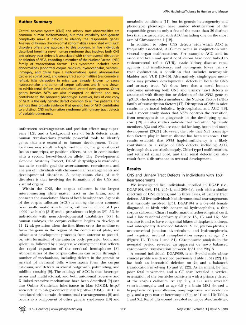
Author Summary
Central nervous system (CNS) and urinary tract abnormalities arecommon human malformations, but their variability and geneticcomplexity make it difficult to identify the responsible genes.Analysis of human chromosomal abnormalities associated with suchdisorders offers one approach to this problem. In five individualsdescribed herein, a novel human syndrome that involves both CNSand urinary tract defects is associated with chromosomal disruptionor deletion of NFIA, encoding a member of the Nuclear Factor I (NFI)family of transcription factors. This syndrome includes brainabnormalities (abnormal corpus callosum, hydrocephalus, ventricu-lomegaly, and Chiari type I malformation), spinal abnormalities(tethered spinal cord), and urinary tract abnormalities (vesicoureteralreflux). Nfia disruption in mice was already known to causehydrocephalus and abnormal corpus callosum, and is now shownto exhibit renal defects and disturbed ureteral development. Othergenes besides NFIA are also disrupted or deleted and maycontribute to the observed phenotype. However, loss of one copyof NFIA is the only genetic defect common to all five patients. Theauthors thus provide evidence that genetic loss of NFIA contributesto a distinct CNS malformation syndrome with urinary tract defectsof variable penetrance.
but a definitive evaluation for urinary reflux (i.e., a voidingcystourethrogram, or VCUG) was not performed.
A third individual, DGAP174, who exhibited complete ACCand enlarged ventricles by second trimester ultrasound, hadboth a de novo translocation of 1p31.1 and 3q25.1 and aninterstitial deletion of 1p31. A postnatal brain CT scanconfirmed these findings and also revealed ventriculomegalyand a tethered spinal cord. A brain MRI at age 3 y revealed aChiari type I malformation and dysplasia of the anterior lefttemporal fossa, in addition to ventriculomegaly and ACC(Figure 1E and 1F; Tables 1 and S1). As with DGAP089, nomajor renal abnormalities were detected by ultrasound, andVCUG was not performed.
Lastly, we ascertained two previously described half-siblings who shared CNS and renal phenotypes similar tothose in DGAP104 [23]. Both individuals, DGAP205–1 andDGAP205-1s, have a maternally inherited, unbalanced inter-stitial microdeletion, del(1)(p31.3p32.3), and exhibited an
abnormal corpus callosum, congenital hydrocephalus, syrin-gomyelia, tethered spinal cord, and urinary tract phenotypesincluding VUR (Figure 1G–1J and 1M–1O) [23].
Cytogenetic and Genetic Analyses of the Chromosomal
Rearrangements
To determine whether a common genetic defect underlaythe phenotypes of these individuals, we analyzed the 1p31region in all five patients and found that the NFIA gene waseither disrupted or deleted in each case. DGAP104 has a denovo balanced translocation, 46,XX,t(1;20)(p31.3;q13.31)dn(Figure 2A, 2B). By metaphase fluorescence in situ hybrid-ization (FISH), we identified a bacterial artificial chromosome(BAC) clone (RP4-802A10) that mapped to 1p31.3 andhybridized to the breakpoints of the der(1) and der(20)chromosomes (Figure 2C). The translocation breakpoint at1p31.3 disrupts intron 2 of NFIA, which is composed of 11
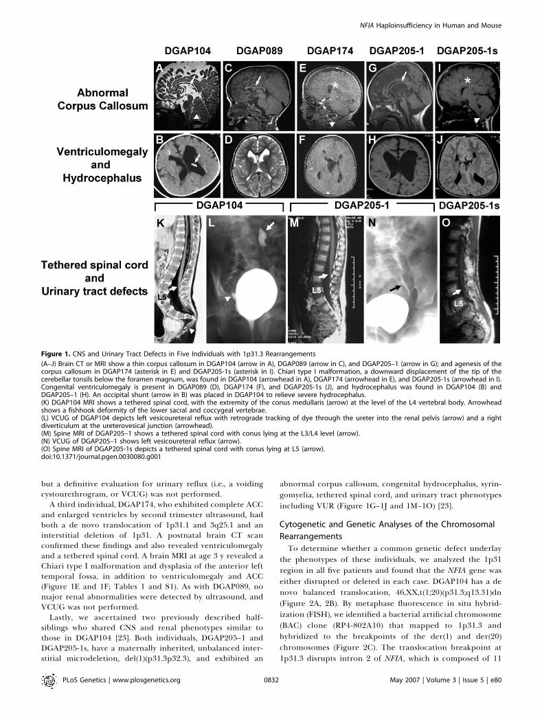
Figure 1. CNS and Urinary Tract Defects in Five Individuals with 1p31.3 Rearrangements
(A–J) Brain CT or MRI show a thin corpus callosum in DGAP104 (arrow in A), DGAP089 (arrow in C), and DGAP205–1 (arrow in G); and agenesis of thecorpus callosum in DGAP174 (asterisk in E) and DGAP205-1s (asterisk in I). Chiari type I malformation, a downward displacement of the tip of thecerebellar tonsils below the foramen magnum, was found in DGAP104 (arrowhead in A), DGAP174 (arrowhead in E), and DGAP205-1s (arrowhead in I).Congenital ventriculomegaly is present in DGAP089 (D), DGAP174 (F), and DGAP205-1s (J), and hydrocephalus was found in DGAP104 (B) andDGAP205–1 (H). An occipital shunt (arrow in B) was placed in DGAP104 to relieve severe hydrocephalus.(K) DGAP104 MRI shows a tethered spinal cord, with the extremity of the conus medullaris (arrow) at the level of the L4 vertebral body. Arrowheadshows a fishhook deformity of the lower sacral and coccygeal vertebrae.(L) VCUG of DGAP104 depicts left vesicoureteral reflux with retrograde tracking of dye through the ureter into the renal pelvis (arrow) and a rightdiverticulum at the ureterovesical junction (arrowhead).(M) Spine MRI of DGAP205–1 shows a tethered spinal cord with conus lying at the L3/L4 level (arrow).(N) VCUG of DGAP205–1 shows left vesicoureteral reflux (arrow).(O) Spine MRI of DGAP205-1s depicts a tethered spinal cord with conus lying at L5 (arrow).doi:10.1371/journal.pgen.0030080.g001
PLoS Genetics | www.plosgenetics.org May 2007 | Volume 3 | Issue 5 | e800832
NFIA Haploinsufficiency in Human and Mouse
exons and spans ;374 kb of genomic DNA (Figure 2I). Thisresult was confirmed by Southern blot analysis (Figure S1).
In addition to disruption of NFIA at 1p31.3 in DGAP104,C20orf32 was also disrupted by the 20q13.31 breakpoint. Acontribution of C20orf32 disruption to the spinal and kidneyphenotypes in DGAP104 is unlikely, however, becauseC20orf32 expression was not detected in the developingspinal cord or kidney by in situ hybridization (Figure S2). Byarray comparative genomic hybridization (aCGH), we ex-cluded any additional chromosome abnormalities inDGAP104 at ;1 Mb resolution.
For DGAP089, sequential FISH led to identification of a1p31.3 breakpoint-spanning BAC, RP5-902P15, which hybri-dized to Chromosome 1 and to both der(1) and der(2)chromosomes (Figure 2D). Subsequent FISH and Southernblot analyses (Figure S3) refined the breakpoint to ;3.9 kbbetween exons 7 and 8 of NFIA (Figure 2I). Similar analyses ofthe 2q breakpoint revealed a split signal for BAC RP11-745P9,thus localizing the breakpoint to ;138 kb in 2q22.1, whichcontains no annotated genes. However, metaphase FISHfollowed by aCGH at ;1 Mb resolution revealed a ;12-Mbinterstitial deletion in 2q proximal to the 2q translocationbreakpoint, for which the karyotype is 46,XY,t(1;2)(p31.3;q22.1),del(2)(q14.3q21)dn. The 39 genes within thisdeletion interval may thus also contribute to the DGAP089phenotype [22].
To establish further whether disruption of NFIA isprimarily associated with the congenital CNS anomaliesobserved in DGAP104 and DGAP089, we next investigatedDGAP174, who has both a t(1;3)(p31.1;q25.1)dn and aninterstitial deletion, del(1)(p31.3p32.1)dn. The 1p31.1 and3q25.1 translocation breakpoints were refined to 150 and 180kb, respectively. The only other potentially relevant gene inthese intervals is NEGR1, which is disrupted by the 1p31.1breakpoint. In rat, NEGR1 protein is expressed only afterE16, with peak expression occurring postnatally, after corpuscallosum formation [24]. However, by RT-PCR, NEGR1transcript is expressed in human cerebral cortex, hippo-campus, corpus callosum, and cerebellum (unpublished data);hence, a contribution to the DGAP174 CNS phenotype ispossible. On the other hand, the chromosome deletion in
DGAP174, which was delimited by FISH and aCGH to 2.2 Mbat 1p31.3–1p32.1 (Figures 2E and S4), also results in thecomplete deletion of NFIA and of eight additional genes(Figure 2I).Lastly, we performed metaphase FISH analyses on chro-
mosomes isolated from the two half-siblings DGAP205–1 andDGAP205-1s and their mother DGAP205–2 (Figure 2F–2I).The 1p31.3–1p32.3 region, encompassing ;12 Mb andcontaining the entire NFIA gene and ;47 additional genes,is deleted in both DGAP205–1 and DGAP205–2 (Figure 2G–2I). The mother, DGAP205–2, is phenotypically normal buthas an apparent balanced rearrangement in which1p31.3p32.3 is inserted into Chromosome 4 with no loss ofgenetic material, and her karyotype is therefore designated46,XX,ins(4;1)(q35;p31.3p32.3) (Figure 2H) [23].
NFIA Haploinsufficiency Is Common to All Five CasesAll five individuals share strikingly similar CNS pheno-
types, including abnormalities of the corpus callosum,hydrocephalus, and ventriculomegaly. All also share disrup-tion or deletion of NFIA, and in each case, the nonrearrangedor nondeleted NFIA allele was subjected to DNA sequencingand no mutations were identified (unpublished data). There-fore, all five cases have NFIA haploinsufficiency in common.NFIA is highly expressed in multiple regions of the humanbrain, including the embryonic and adult corpus callosum(Figure S5) [25], and ACC and hydrocephalus were observedin Nfia�/� mutant mice [18].In each DGAP case, one or more additional genes were also
directly affected as a consequence of either the translocationor deletion. These additional genes may contribute to ormodify the nature of the phenotype attributable to NFIAdisruption or deletion. However, the identities of these genesdiffer among the five DGAP cases, except for 205–1 and 205–1s, who share the same 12-Mb deletion; and 205–1, 205–1s,and 174, who share a common 2.2-Mb deletion region (Figure2I). C20orf32 is disrupted in addition to NFIA in DGAP104,whereas in DGAP089 disruption of NEGR1 and deletion of 39genes in del(2)(q14.3q21) occurred. Besides the disruption ordeletion of NFIA, none of the other of these geneticaberrations is shared by more than three cases. Therefore,
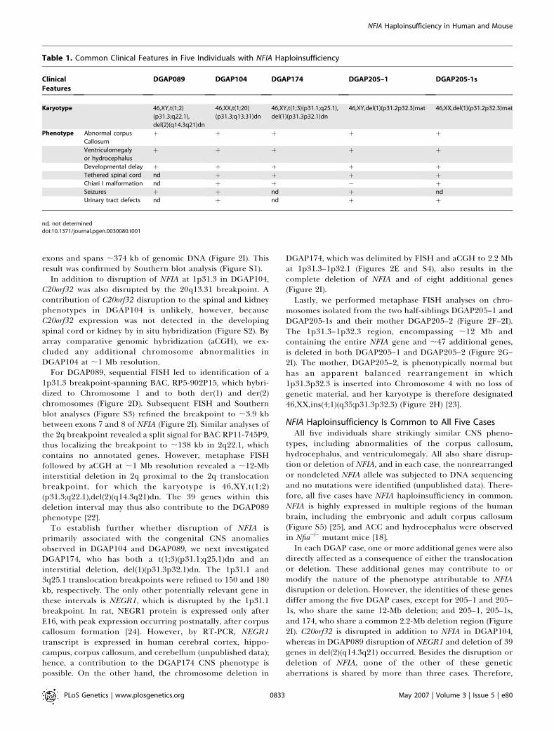
Table 1. Common Clinical Features in Five Individuals with NFIA Haploinsufficiency
Clinical
Features
DGAP089 DGAP104 DGAP174 DGAP205–1 DGAP205-1s
Karyotype 46,XY,t(1;2)
(p31.3;q22.1),
del(2)(q14.3q21)dn
46,XX,t(1;20)
(p31.3;q13.31)dn
46,XY,t(1;3)(p31.1;q25.1),
del(1)(p31.3p32.1)dn
46,XY,del(1)(p31.2p32.3)mat 46,XX,del(1)(p31.2p32.3)mat
Phenotype Abnormal corpus
Callosum
þ þ þ þ þ
Ventriculomegaly
or hydrocephalus
þ þ þ þ þ
Developmental delay þ þ þ þ þTethered spinal cord nd þ þ þ þChiari I malformation nd þ þ � þSeizures þ þ nd þ nd
Urinary tract defects nd þ nd þ þ
nd, not determineddoi:10.1371/journal.pgen.0030080.t001
PLoS Genetics | www.plosgenetics.org May 2007 | Volume 3 | Issue 5 | e800833
NFIA Haploinsufficiency in Human and Mouse
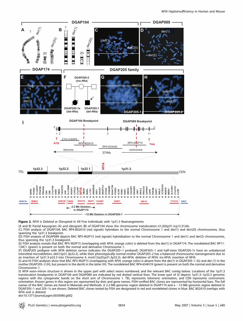
Figure 2. NFIA is Deleted or Disrupted in All Five Individuals with 1p31.3 Rearrangements
(A and B) Partial karyogram (A) and ideogram (B) of DGAP104 show the chromosome translocation t(1;20)(p31.3;q13.31)dn.(C) FISH analysis of DGAP104; BAC RP4-802A10 (red signals) hybridizes to the normal Chromosome 1 and der(1) and der(20) chromosomes, thusspanning the 1p31.3 breakpoint.(D) FISH analysis of DGAP089 depicts BAC RP5-902P15 (red signals) hybridization to the normal Chromosome 1 and der(1) and der(2) chromosomes,thus spanning the 1p31.3 breakpoint.(E) FISH analysis reveals that BAC RP5-902P15 (overlapping with NFIA, orange color) is deleted from the der(1) in DGAP174. The nondeleted BAC RP11-134C1 (green) is present on both the normal and derivative Chromosome 1.(F) DGAP205 pedigree with NFIA deletion (arrow indicates the DGAP205–1 proband). DGAP205–1 and half-sister DGAP205-1s have an unbalancedinterstitial microdeletion, del(1)(p31.3p32.3), while their phenotypically normal mother DGAP205–2 has a balanced chromosome rearrangement due toan insertion of 1p31.3-p32.3 into Chromosome 4, ins(4;1)(q35;p31.3p32.3). del-NFIA, deletion of NFIA; ins-NFIA, insertion of NFIA.(G and H) FISH analyses show that BAC RP5-902P15 (overlapping with NFIA, orange color) is absent from the der(1) in DGAP205–1 (G) and der (1) in themother DGAP205–2 (H), but present in the der(4) in the latter (H). The nondeleted BAC RP4-654H19 (green) is present on both the normal and derivativeChromosome 1.(I) NFIA exon–intron structure is shown in the upper part with select exons numbered, and the relevant BAC contig below. Locations of the 1p31.3translocation breakpoints in DGAP104 and DGAP089 are indicated by red dotted vertical lines. The lower part of (I) depicts 1p31.3–1p32.3 genomicregions with the cytogenetic bands on the short arm of Chromosome 1. TEL represents telomeric orientation, and CEN represents centromericorientation. Known genes in this region are represented by dots and gene names. FISH-verified BAC clones are represented by horizontal bars. The fullnames of the BAC clones are listed in Materials and Methods. A 2.2-Mb genomic region deleted in DGAP174 and a ;12-Mb genomic region deleted inDGAP205–1 and 205–1s are shown. Deleted BAC clones tested by FISH are designated in red and nondeleted clones in blue. BAC 802A10 overlaps withNFIA and is deleted.doi:10.1371/journal.pgen.0030080.g002
PLoS Genetics | www.plosgenetics.org May 2007 | Volume 3 | Issue 5 | e800834
NFIA Haploinsufficiency in Human and Mouse
the most parsimonious explanation for the observed CNSphenotypes is NFIA haploinsufficiency, which is the onlycommon genetic defect shared by all five individuals.
To test whether intragenic mutations in NFIA are associ-ated with abnormal callosal development and other CNSphenotypes, we sequenced the 11 exons and intron-exonboundaries of NFIA in 84 patients with various combinationsof syndromic CNS phenotypes, including abnormal corpuscallosum, tethered spinal cord, Chiari I malformation,
hydrocephalus, and urinary tract defects (Table S2). Sequenceanalysis was also performed on a group of 96 individuals thatincluded both syndromic and nonsyndromic ACC, and onanother group of 39 individuals with nonsyndromic tetheredcord syndrome. Although several known SNPs were detected,no intragenic mutations were identified. Thus, intragenicmutation in NFIA is not a frequent cause of the CNS defectsdescribed here.
Further Analysis of Mouse Nfia Mutants Reveals AdditionalPhenotypesTo gain further evidence for the assignment of NFIA as the
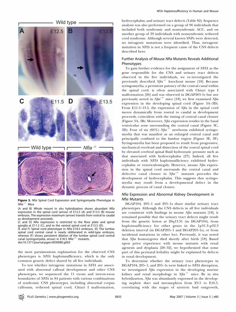
gene responsible for the CNS and urinary tract defectsobserved in the five individuals, we re-investigated thepreviously described Nfia�/� knockout mouse [18]. Becausesyringomyelia, a persistent patency of the central canal withinthe spinal cord, is often associated with Chiari type Imalformation [26] and was observed in DGAP205-1s but notpreviously noted in Nfia�/� mice [18], we first examined Nfiaexpression in the developing spinal cord (Figure 3A–3D).From E11.5–13.5, the expression of Nfia in the spinal cordmoves dynamically from rostral to caudal as developmentproceeds, coincident with the timing of central canal closure(Figure 3A, 3B). Moreover, Nfia expression resides in the basalventricular zone surrounding the central canal (Figure 3C,3D). Four of six (66%) Nfia�/� newborns exhibited syringo-myelia that was manifest as an enlarged central canal andprincipally confined to the lumbar region (Figure 3E, 3F).Syringomyelia has been proposed to result from progressive,mechanical overload and dissection of the rostral spinal cordby elevated cerebral spinal fluid hydrostatic pressure such asthat associated with hydrocephalus [27]. Indeed, all fiveindividuals with NFIA haploinsufficiency exhibited hydro-cephalus or ventriculomegaly. However, mouse Nfia expres-sion in the spinal cord surrounds the central canal anddefective canal closure in Nfia�/� mutants precedes thedevelopment of hydrocephalus. This suggests that syringo-myelia may result from a developmental defect in thedynamic process of canal closure.
Nfia Expression and Abnormal Kidney Development inNfia MutantsDGAP104, 205–1 and 205–1s share similar urinary tract
phenotypes. Although the CNS defects in all five individualsare consistent with findings in mouse Nfia mutants [18], itremained possible that the urinary tract defects might resultfrom the genetic lesion at 20q13.31 (in DGAP104), fromhaploinsufficiency for other genes in the 1p31.3-p32.3deletion interval (in DGAP205–1 and DGAP205-1s), or fromincidental mutations in other loci. Previously, it was notedthat Nfia homozygotes died shortly after birth [18]. Basedupon prior experience with mouse mutants with renalagenesis and dysplasia [28–32], we hypothesized that somepart of this perinatal lethality might be explained by defectsin renal development.To determine whether the urinary tract phenotypes in
DGAP104, 205–1, and 205–1s were linked to NFIA disruption,we investigated Nfia expression in the developing murinekidney and renal morphology in Nfia�/� mice. By in situhybridization, Nfia was abundantly expressed in the develop-ing nephric duct and metanephros from E9.5 to E16.5,correlating with the stages of ureteric bud outgrowth,
Figure 3. Nfia Spinal Cord Expression and Syringomyelia Phenotype in
Nfia�/� Mice
(A and B) Whole mount in situ hybridization shows abundant Nfiaexpression in the spinal cord (arrow) of E12.5 (A) and E13.5 (B) mouseembryos. The expression maximum (arrow) transits from rostral to caudalas development proceeds.(C and D) Nfia expression is restricted to the floor plate and spinalganglia at E11.5 (C), and to the ventral spinal cord at E13.5 (D).(E and F) Spinal cord phenotype in Nfia E18.5 embryos. (E) The lumbarspinal cord central canal is nearly obliterated in wild-type embryos,whereas (F) shows persistent dilation of the lumbar spinal cord centralcanal (syringomyelia, arrow) in E18.5 Nfia�/� mutants.doi:10.1371/journal.pgen.0030080.g003
PLoS Genetics | www.plosgenetics.org May 2007 | Volume 3 | Issue 5 | e800835
NFIA Haploinsufficiency in Human and Mouse
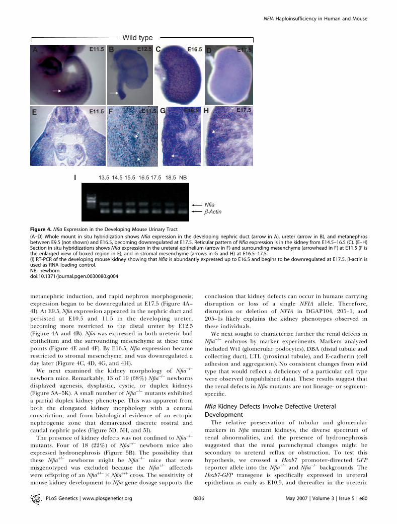
metanephric induction, and rapid nephron morphogenesis;expression began to be downregulated at E17.5 (Figure 4A–4I). At E9.5, Nfia expression appeared in the nephric duct andpersisted at E10.5 and 11.5 in the developing ureter,becoming more restricted to the distal ureter by E12.5(Figure 4A and 4B). Nfia was expressed in both ureteric budepithelium and the surrounding mesenchyme at these timepoints (Figure 4E and 4F). By E16.5, Nfia expression becamerestricted to stromal mesenchyme, and was downregulated aday later (Figure 4C, 4D, 4G, and 4H).
We next examined the kidney morphology of Nfia�/�
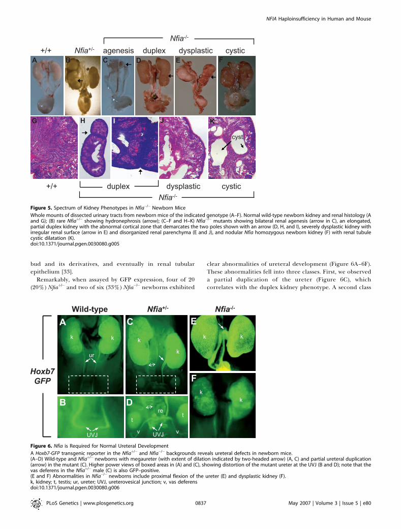
newborn mice. Remarkably, 13 of 19 (68%) Nfia�/� newbornsdisplayed agenesis, dysplastic, cystic, or duplex kidneys(Figure 5A–5K). A small number of Nfia�/�mutants exhibiteda partial duplex kidney phenotype. This was apparent fromboth the elongated kidney morphology with a centralconstriction, and from histological evidence of an ectopicnephrogenic zone that demarcated discrete rostral andcaudal nephric poles (Figure 5D, 5H, and 5I).
The presence of kidney defects was not confined to Nfia�/�
mutants. Four of 18 (22%) of Nfiaþ/� newborn mice alsoexpressed hydronephrosis (Figure 5B). The possibility thatthese Nfiaþ/� newborns might be Nfia�/� mice that weremisgenotyped was excluded because the Nfiaþ/� affectedswere offspring of an Nfiaþ/�3 Nfiaþ/þ cross. The sensitivity ofmouse kidney development to Nfia gene dosage supports the
conclusion that kidney defects can occur in humans carryingdisruption or loss of a single NFIA allele. Therefore,disruption or deletion of NFIA in DGAP104, 205–1, and205–1s likely explains the kidney phenotypes observed inthese individuals.We next sought to characterize further the renal defects in
Nfia�/� embryos by marker experiments. Markers analyzedincluded Wt1 (glomerular podocytes), DBA (distal tubule andcollecting duct), LTL (proximal tubule), and E-cadherin (celladhesion and aggregation). No consistent changes from wildtype that would reflect a deficiency of a particular cell typewere observed (unpublished data). These results suggest thatthe renal defects in Nfia mutants are not lineage- or segment-specific.
Nfia Kidney Defects Involve Defective UreteralDevelopmentThe relative preservation of tubular and glomerular
markers in Nfia mutant kidneys, the diverse spectrum ofrenal abnormalities, and the presence of hydronephrosissuggested that the renal parenchymal changes might besecondary to ureteral reflux or obstruction. To test thishypothesis, we crossed a Hoxb7 promoter-directed GFPreporter allele into the Nfiaþ/� and Nfia�/� backgrounds. TheHoxb7-GFP transgene is specifically expressed in ureteralepithelium as early as E10.5, and thereafter in the ureteric
Figure 4. Nfia Expression in the Developing Mouse Urinary Tract
(A–D) Whole mount in situ hybridization shows Nfia expression in the developing nephric duct (arrow in A), ureter (arrow in B), and metanephrosbetween E9.5 (not shown) and E16.5, becoming downregulated at E17.5. Reticular pattern of Nfia expression is in the kidney from E14.5–16.5 (C). (E–H)Section in situ hybridizations shows Nfia expression in the ureteral epithelium (arrow in F) and surrounding mesenchyme (arrowhead in F) at E11.5 (F isthe enlarged view of boxed region in E), and in stromal mesenchyme (arrows in G and H) at E16.5–17.5.(I) RT-PCR of the developing mouse kidney showing that Nfia is abundantly expressed up to E16.5 and begins to be downregulated at E17.5. b-actin isused as RNA loading control.NB, newborn.doi:10.1371/journal.pgen.0030080.g004
PLoS Genetics | www.plosgenetics.org May 2007 | Volume 3 | Issue 5 | e800836
NFIA Haploinsufficiency in Human and Mouse
bud and its derivatives, and eventually in renal tubularepithelium [33].
Remarkably, when assayed by GFP expression, four of 20(20%) Nfiaþ/� and two of six (33%) Nfia�/� newborns exhibited
clear abnormalities of ureteral development (Figure 6A–6F).These abnormalities fell into three classes. First, we observeda partial duplication of the ureter (Figure 6C), whichcorrelates with the duplex kidney phenotype. A second class
Figure 5. Spectrum of Kidney Phenotypes in Nfia�/� Newborn Mice
Whole mounts of dissected urinary tracts from newborn mice of the indicated genotype (A–F). Normal wild-type newborn kidney and renal histology (Aand G); (B) rare Nfiaþ/� showing hydronephrosis (arrow); (C–F and H–K) Nfia�/� mutants showing bilateral renal agenesis (arrow in C), an elongated,partial duplex kidney with the abnormal cortical zone that demarcates the two poles shown with an arrow (D, H, and I), severely dysplastic kidney withirregular renal surface (arrow in E) and disorganized renal parenchyma (E and J), and nodular Nfia homozygous newborn kidney (F) with renal tubulecystic dilatation (K).doi:10.1371/journal.pgen.0030080.g005
Figure 6. Nfia is Required for Normal Ureteral Development
A Hoxb7-GFP transgenic reporter in the Nfiaþ/� and Nfia�/� backgrounds reveals ureteral defects in newborn mice.(A–D) Wild-type and Nfiaþ/� newborns with megaureter (with extent of dilation indicated by two-headed arrow) (A, C) and partial ureteral duplication(arrow) in the mutant (C). Higher power views of boxed areas in (A) and (C), showing distortion of the mutant ureter at the UVJ (B and D); note that thevas deferens in the Nfiaþ/� male (C) is also GFP–positive.(E and F) Abnormalities in Nfia�/� newborns include proximal flexion of the ureter (E) and dysplastic kidney (F).k, kidney; t, testis; ur, ureter; UVJ, ureterovesical junction; v, vas deferensdoi:10.1371/journal.pgen.0030080.g006
PLoS Genetics | www.plosgenetics.org May 2007 | Volume 3 | Issue 5 | e800837
NFIA Haploinsufficiency in Human and Mouse

consisted of mutant ureters that were dilated, either as aconsequence of obstruction or from reflux (Figure 6C and6D). In the third class, we observed abnormal flexure of therostral ureter, with the site of flexion near the renal pelvis(Figure 6E), which could lead to obstruction. To determinewhether prolonged reflux and obstruction in Nfia�/� mutantsaffected postnatal kidney development, we analyzed two rareNfia�/� postnatal survivors. Both P16 Nfia�/� developed severehydronephrosis, whereas kidneys in their wild-type litter-mates were normal (Figure S6).
Lastly, we analyzed the histology of two key ureteralstructures, the ureteropelvic junction (UPJ), which connectsthe ureter to the kidney, and the ureterovesical junction(UVJ), which connects the ureter to the bladder. Both UPJand UVJ histological defects were noted in Nfiaþ/� newborns,while only UPJ defects were identified in Nfia�/� newborns(Figure 7A–7F). The presence of UPJ and UVJ dilation in Nfiamutant mice (Figure 7B, 7C, and 7E) is consistent with theobservations of VUR and hydronephrosis in DGAP104, 205–1,and 205–1s. Thus, abnormalities in ureteral developmentcomprise a significant part of the Nfia mutant phenotype.Moreover, because proper formation of the vertebrate kidneydepends upon induction by the ureteric bud, ureteralabnormalities could account for aspects of the kidney defectsin NFIA haploinsufficient patients and mutant mice.
Discussion
NFIA Haploinsufficiency as a Pathogenetic MechanismThe five individuals studied here share NFIA haploinsuffi-
ciency caused by translocation (DGAP089 and 104) ordeletion (DGAP174, 205–1, and 205–1s). All five also shareabnormalities of the corpus callosum, and partly share otherCNS phenotypes, including ventriculomegaly, congenitalhydrocephalus, Chiari type I malformation, and tetheredspinal cord. All five individuals also have developmental delayand three exhibited seizure disorders. Three of the five also
have urinary tract defects, including VUR. Prior workestablished that Nfia loss of function in the mouse results inACC and abnormal development of the hippocampalcommissure, two major axonal tracts that connect thecerebral hemispheres, and an associated hydrocephalus thatdevelops in rare postnatal survivors [18,34]. We also foundthat the mouse Nfia mutant recapitulates the VUR phenotypein these humans. Therefore, although other affected genesmay contribute to the overall phenotype, these cases suggestthat NFIA haploinsufficiency can account for the observedCNS and renal defects.It is important to acknowledge that a contribution to the
developmental phenotypes identified here from additionalgenes that reside within various deletion intervals, or thatalso suffer disruption by breakpoints, is not excluded. In allfive cases, additional genes besides NFIA are also disrupted ordeleted, so that in no single case is a defect in NFIA the onlygenetic abnormality. While DGAP104 only inactivates NFIAand C20orf32 and the latter is an unlikely contributory factor,the most extreme cases are DGAP089 and the DGAP205 half-siblings, which contain deletions that involve 39 and 47 genes,respectively. Because these two deletions involve differentchromosomes, none of the deleted genes are shared. How-ever, in both cases, many more genes are affected than justNFIA, and some may participate in the observed phenotypes.For example, two cases described in the literature report 2qdeletions that overlap with the del(2)(q14.3q21) in DGAP089,and these also involve ACC [35,36]. Therefore, in the absenceof intragenic mutations in NFIA, the definitive argument thatNFIA is the gene responsible for the CNS and renalphenotypes in these five patients cannot be made.The inability to identify intragenic mutations in NFIA in
cases involving ACC, hydrocephalus, tethered cord syndrome,and urinary tract defects could suggest that the phenotype ofheterozygous intragenic loss-of-function NFIA mutationsmight differ from that described here. Indeed, as noted, itis quite plausible that in any individual DGAP case, the
Figure 7. Ureter Defects at UPJ and UVJ in Nfia Mutant Mice
(A–C) Hematoxylin and eosin histology depicts duplication and dilatation of ureter at the UPJ in Nfiaþ/� (B, 60X) and Nfia�/� (C, 60X) newborn mice.(D–F) Hematoxylin and eosin staining shows dilatation of UVJ in some Nfiaþ/�mutant mice (* in E, 60X); the majority of Nfiaþ/� and Nfia�/�mice show anormal UVJ (F, 60X).a, abdominal aorta; bl, bladder; pe, pelvis; r, rectum; ur, ureter; UVJ, ureterovesical junctiondoi:10.1371/journal.pgen.0030080.g007
PLoS Genetics | www.plosgenetics.org May 2007 | Volume 3 | Issue 5 | e800838
NFIA Haploinsufficiency in Human and Mouse
observed phenotype represents the additive effect of NFIAhaploinsufficiency plus other loci that are deleted ordisrupted. On the other hand, at least 20 discrete loci havebeen implicated in ACC alone, so that the failure to detectintragenic mutations in NFIA is not surprising. Ultimately,formal definition of the NFIA hemizygous loss-of-functionphenotype would be strengthened by identification of intra-genic loss-of-function NFIA mutations.
Lastly, it is well recognized that both chromosomal trans-locations and deletions may engender position effects thatalter gene expression at considerable distances from the siteof a chromosomal aberration (reviewed in [1,2]). For example,Shh expression in the limb bud mesenchyme is controlled by aregulatory region located ;1 Mb upstream within theunrelated Lmbr1 gene [37]. Position effects on neighboringgenes for mouse knockouts have been described [38]. Themouse Nfia mutant results from a small exon 2 deletion, yetstill accurately recapitulates many features of the humanphenotype. Therefore, one would have to posit the existenceof a conserved regulatory element within the exon 2 deletionregion that would act on genes 39 to Nfia, which exhibitconservation of synteny between mouse and human. How-ever, the genes immediately neighboring NFIA are not knownto play a role in CNS or kidney development. These includeC1orf87 (GeneID 127795) and TM2D1 (beta amyloid bindingprotein, GeneID 83941), which reside approximately 1 Mb 59
and 200 kb 39 of NFIA, respectively. Additional genes thatreside at larger distances from NFIA might be affected by aposition effect, but none are obvious candidates. Taking thesefactors into consideration, we conclude that a true positioneffect is unlikely to explain the observed phenotypes.
Nature of the CNS PhenotypeFormation of the corpus callosum causes inversion of the
cingulate gyri, which gives the medial surface of the brain itscharacteristic pattern. In ACC, the cingulum remains evertedat sites of agenesis, and the sulci of the medial brain extendinto the third ventricle. The findings in DGAP174 of aneverted cingulate gyrus and longitudinal bundles of Probstare consistent with primary dysgenesis of the corpuscallosum. Three midline populations contribute to formationof the corpus callosum: the glial sling, the glial wedge, and gliawithin the indusium griseum and its precallosal extension,the hippocampal continuation [39–41]. NFIA protein isexpressed in all three midline populations, which fail todevelop properly in Nfia�/� mice [34]. These populationsnormally form the corticoseptal boundary that preventscallosal axons from entering the septum.
The function of NFIA in formation of these neuronalpopulations places it within the class of genes that regulateaxonal midline crossing. The prototypical regulatory gene inthis class is roundabout or robo, which was originally identifiedin Drosophila. Roundabout encodes a transmembrane receptorexpressed by migratory axons after they cross the CNSmidline. Robo binds the extracellular ligand, Slit, which isexpressed by midline glia and functions as a chemorepulsivecue that prevents axons from midline recrossing. Thisfunction extends to mammals, as mice lacking Robo1 or Robo2exhibit CNS phenotypes that include abnormal midlinecommissural axonal guidance, and Robo1 mutants in partic-ular exhibit callosal dysgenesis [42,43]. Similarly, Slit ligandsalso play a role in callosal development. Slit2 mutants display
a small corpus callosum with a reduced number of traversingaxons [44–46], while Slit2 glial expression during callosaldevelopment in Nfia mutants is reduced [34].Interestingly, Nfia, Robo2, and Slit2 mouse mutants share
not only axonal midline crossing defects, but also phenotypi-cally related renal and ureteral defects. Mice deficient forRobo2 or Slit2 exhibit duplex kidney and megaureterphenotypes [32,47] that in some ways resemble those in Nfiamutants. Our recent study also implicated ROBO2 signalingin the pathogenesis of a subset of human VUR [32]. Theserelated phenotypes raise the possibility that Nfia and SLIT–ROBO signaling are functionally linked in both CNS andureteral development.
Relationship between Kidney and Ureter PhenotypesBased on Nfia expression in the developing kidney and the
presence of kidney hypoplasia in DGAP104, we identifiedseveral distinct kidney phenotypes, including renal dysplasiaand hydronephrosis in Nfia mutant mice. Hydronephrosisusually results from an obstruction in the flow of urine at thelevel of the UPJ or UVJ. This results in an obstructiveuropathy in which back pressure from the accumulation ofurine in the ureter and renal pelvis results in destruction anddistortion of the renal parenchyma. The presence of hydro-nephrosis in Nfia�/� and Nfiaþ/� mutants therefore suggeststhat the observed renal defects reflect a primary disturbancein ureteral development.A striking finding in Nfia mutant embryos and newborns is
the presence of clear ureteral abnormalities: megaureter,abnormal ureteral folding, abnormalities at the UVJ and UPJ,and in a small number of cases, partial duplication of theureter. These findings are consistent with the strongexpression of Nfia in the developing nephric duct and ureterat E9.5–13.5. The ureteral duplication phenotype, distinctfrom normal patterns of ureteric bud branching [48], verylikely explains the finding of a partial duplex kidney. Becauseureteric bud contact with uninduced metanephric mesen-chyme triggers the inductive cascade [49], contact by twoseparate ureteral branches should produce a partial duplexkidney. In addition to abnormal ureteral development, theVUR in DGAP104 and in the DGAP205 half-siblings may alsodevelop as a consequence of the tethered spinal cord defect[50,51].In DGAP089 and 174, renal ultrasound revealed no major
abnormalities and urinary reflux was not observed. However,subtle anatomic defects in the ureter or kidney are often sub-clinical, and may exist below the limit of detection. Inaddition, these results are consistent with those in Nfiaþ/� andNfia�/� mutants, where the penetrance of overt kidney orureteral defects was only 22% and 68%, respectively. Oneexplanation for the incomplete penetrance of ureteral orkidney defects in Nfia mutants could be functional redun-dancy with Nfib. In mice, Nfib is strongly expressed in thedeveloping nephric duct, kidney, and ureter at E10.5–11.5,where its expression overlaps with that of Nfia (unpublisheddata). The other two Nuclear Factor I family members, Nficand Nfix, are expressed only at lower levels in the developingureter and kidney. In addition, Nfib�/� mice exhibit callosalagenesis and forebrain defects similar to those seen in Nfia�/�
mice [21]. Thus, partial redundancy may exist between Nfiaand Nfib in both CNS and urinary tract development. Geneticcombinations of mutant alleles for Nfia and Nfib will be
PLoS Genetics | www.plosgenetics.org May 2007 | Volume 3 | Issue 5 | e800839
NFIA Haploinsufficiency in Human and Mouse
required to address this question definitively, and to furtherdisclose the roles of Nfi factors in development.
In sum, our results suggest that NFIA haploinsufficiency inhumans results in a thin, hypoplastic or absent corpuscallosum, and define the spectrum of defects attributable toNFIA loss of function to include additional CNS and urinarytract defects that were not previously apparent in the Nfiamouse mutant. These results illustrate the powerful synergythat occurs when corresponding human and mouse disordersare investigated in parallel.
Materials and Methods
DGAP individuals studied. DGAP104. DGAP104 is the product ofin vitro fertilization via intracytoplasmic sperm injection, whoseparents of European descent were unrelated with no reportedmedical problems. Amniocentesis demonstrated a 46,XX,t(1;20)(p32.3;q13.31)dn, but ultrasound revealed no organ malformationsat 20 wk of pregnancy. Because of placenta previa and persistentvaginal bleeding, DGAP104 was delivered at 31 wk via electivecesarean section and weighed 1,980 g with Apgar scores of 9/10/10.She was diagnosed with prematurity, hydrocephaly, Chiari Imalformation, tethered spinal cord, congenital hydronephrosis, lefthypoplastic kidney, bilateral inguinal hernia, hyaline membranedisease grade 2, and gastroesophageal reflux. Imaging studiesindicated a thin posterior corpus callosum and an open aqueductwith progressive ventricular enlargement. She was delayed in reach-ing developmental milestones; at 2 y, she exhibited major motor delaywith inhibited movement and was wheelchair bound. Speech was alsodelayed, and limited to a few words. At 6 y 7 mo of age, DGAP104received a performance IQ score of 42, verbal IQ score of 68, andglobal IQ score of 52 on the Weschler Preschool and Primary Scale ofIntelligence-Revised test (WPPSI-R).
To relieve hydrocephalus, which caused progressive macrocephaly,seven neurosurgical operations were performed over 5 y, including athird ventriculocisternostomy, a ventriculoperitoneal shunt with aflow-regulated valve, and several revisions because of hyperdrainageor blockage of the shunt. At 7 d of age, abdominal ultrasonographyrevealed bilateral hypoplastic kidneys and bilateral dilatation of therenal pelvis. At 1 y, ultrasound showed left and right kidney lengths of45 mm and 51 mm, respectively (mean length for age, 52 mm), andboth kidneys lacked corticomedullary differentiation. At 2 y, the leftkidney length was 49 mm and the right 58 mm (mean length for age,55 mm), but both lacked discernable corticomedullary differentia-tion. At 5 y, the left kidney was 53 mm and the right 61 mm (meanlength for age, 66 mm). DGAP104 first exhibited left grade II VUR byVCUG at 1 y. At 2 y, VUR increased to grade III with a rightvesicoureteral junction diverticulum, and pyelonephritis developedthat required ureteral reimplantation surgery (Cohen operation).Urea and creatinine levels were normal at 5 y of age.
DGAP089. DGAP089 is a male with an interstitial deletion on 2qand a balanced translocation involving 1p and 2q. Additional clinicaldata are summarized in Table S1 and described in more detailelsewhere [22]. Seizure onset occurred at 19 mo, with treatment untilage 6 y and no recurrences thereafter. He rolled to one side at 10 mo,to either side at 14 mo, and sat at 14 mo. At 3 y, he received the BayleyScales of Infant Development test (2nd edition, BSID-II), and earneda Mental Developmental Index (MDI) that fell below the standardscore of 50 with an age equivalent of 4 mo. He also received theVineland Adaptive Behavior Scales test (parent form VABS), andobtained an adaptive behavior composite score of 45 correspondingto an age equivalent of 7 mo. He cruised at 4 y, and walked at 5 y.DGAP089 underwent a brain MRI at 6.5 y that revealed a hypoplasticcorpus callosum with enlarged frontal horns, parallel in configu-ration, without evidence of increased intracranial pressure. The MRIalso showed polymicrogyria and an 8-mm nodule along the lateralwall of the left frontal horn that likely represented a gray matterheterotopia. Renal ultrasound performed at 6.5 y was normal. Therewas no significant change in the physical examination and no focalsensorimotor deficits were noted. When last examined at age 8 y(2005), DGAP089 was no longer wearing leg braces, was able to walkand run on his toes, and could eat table foods without difficulty, butwas not toilet trained. Speech consisted of about three words inappropriate context, and he was being taught American SignLanguage (ASL). He could gesture, but was unable to followcommands, and engaged in self-stimulatory behavior.
DGAP174. DGAP174 was born to a 20-y-old mother at 37 wk bycesarean section and weighed 2,770 g (95th percentile) with length49.5 cm (25th percentile) and occipitofrontal circumference 37.5 cm(75th percentile). Pregnancy was uncomplicated by teratogenicexposures or maternal illness. A second trimester prenatal ultrasoundshowed agenesis of the corpus callosum and enlarged ventricles. A CTscan of the brain in the immediate postnatal period confirmed theprenatal findings, and revealed ventriculomegaly with parallel lateralventricles representing longitudinal bundles of Probst, and a tetheredspinal cord. A small ventricular septal defect was also noted at birth.Chromosome analysis of peripheral blood lymphocytes revealed46,XY,t(1;3)(p22;q21)dn. The neonate was discharged and seen againat 13 d of age in the genetics clinic. He was found to have metopicstenosis and bitemporal narrowing that was surgically corrected at 5mo of age. Neurological exam revealed normal tone and reflexes. At 8mo of age, he was noted to be gaining weight rapidly, unrelated to anychange in eating pattern. Follow-up at 12 mo revealed delay in grossmotor development, macrocephaly without hydrocephalus, andheight, weight, and length above the 95th percentile for his age.Additionally, a dimple on the posterior aspect of the right helix,creases behind each earlobe, and esotropia secondary to telecanthusand epicanthal folds were noted. A left inguinal hernia was detectedat 24 mo. Also at 24 mo, glasses were prescribed to correcthypertropia and strabismus, and all milestones were on track exceptfor speech. At 36 mo, expressive language was still delayed. Due tomacrosomia, the patient underwent bone age testing, which was ageappropriate. A brain MRI at 36 mo of age revealed Chiari Imalformation and dysplasia of the anterior aspect of the lefttemporal fossa in addition to complete agenesis of the corpuscallosum. At 57 mo of age, DGAP174 underwent several devel-opmental tests. His IQ score was 68 on a Leiter InternationalPerformance Scale (LIPS) test, corresponding to an age equivalent of36 mo. He received a standard score of 60 in the Peabody PictureVocabulary Test (Revised, Form M; PPVT-R) corresponding to an ageequivalent of 35 mo. His standard score for the Developmental Testof Visual-Motor Integration (VMI) was 67, which also correspondedto an age equivalent of 35 mo. At 6 y of age, he underwent a successfulChiari decompression and repair of the tethered spinal cord. At thattime, he was diagnosed with attention deficit and hyperactivitydisorder and is currently on Ritalin. He was noted to have righthemihypertrophy and scoliosis, for which he was referred to anorthopedics clinic. At age 8 y and 4 mo (January 2007), DGAP174 wasfunctioning at the kindergarten level, and received occupational andphysical therapy for speech.
DGAP205–1, 205–1s, and 205–2. DGAP205–1 and DGAP205-1s aretwo half-siblings with an interstitial microdeletion, del(1)(p31.3p32.3),that was inherited as an unbalanced segregant resulting from abalanced rearrangement in their mother, DGAP205–2. Both half-siblings had congenital CNS and urinary tract defects while theirmother was phenotypically normal. DGAP205–1 had the Bayley Scalesof Infant Development test (BSID) at 4.5 y of age and received scorescorresponding to an age equivalent of 18 mo for overall development,20 mo for cognition, and 17 mo for fine motor. At age 5 y, DGAP205–1 was functioning at an age equivalent of ;2.5 y. At age 10 y he wasfunctioning at an age equivalent of ;7 y, and was 4 ft tall and weighed;65 lb. DGAP205–1 has limited verbal skills and uses a combinationof words and signs for communication. He has not had any formaltesting at 10 y of age, but can only read and spell three-letter words.DGAP205-1s received the Bayley Scales of Infant Development test(BSID) at 2 y of age, and the Stanford-Binet test at 9 y. Both testsdemonstrated moderate global cognitive impairment (scores notavailable). At 9 y of age, she was functioning academically at akindergarten level. Other clinical data for the affected sibs aresummarized in Table S1, and described in more detail elsewhere [23].
FISH, aCGH, and mutation screening. Metaphase FISH wasperformed according to standard methods. BAC clones wereobtained from BAC/PAC Resources (http://bacpac.chori.org), labeledas FISH probes, and hybridized to metaphase chromosomes preparedfrom lymphoblastoid cell lines established from all five individuals.The full BAC names provided in Figure 2I are: RP4-654H19, RP11-55M23, RP5-829C20, RP11-13N22, RP4-802A10, RP4-662P1, RP5-1078M7, RP4-534K7, RP11-86K19, RP11-131O15, and RP11-89K2.aCGH experiments were performed with the Spectral Genomics 2600BAC array by the Cytogenetics Core Facility of the Dana-Farber/Harvard Cancer Center for DGAP089 and DGAP174, and by SpectralGenomics for DGAP104. NFIA mutation screening employed PCRamplification of the 11 human NFIA exons and intron-exonboundaries, followed by purification and bidirectional DNA sequenc-ing. NFIA cDNA sequence AB037860 (http://www.ncbi.nlm.nih.gov/entrez/viewer.fcgi?db¼nucleotide&val¼7243275) was used to calculate
PLoS Genetics | www.plosgenetics.org May 2007 | Volume 3 | Issue 5 | e800840
NFIA Haploinsufficiency in Human and Mouse
nucleotide positions. MLPA analysis of the NFIA coding region wasperformed in a subset of syndromic and nonsyndromic ACC samples,and no copy number changes were identified.
RT-PCR analysis. RT-PCR analyses were performed by routineprotocols. RT-PCR primers used to amplify the Nfia 334 bp cDNAwere mNfia-rt-F (59-CAAGCCTCCAACCACATCAAC-39) and mNfia-rt-R (59-CTGTTTGACCACGATGTTTGCT-39). RT-PCR primers used toamplify the C20orf32 436 bp cDNA were mC20orf32-F (59-GGGCACTCTACGACAACCAT-39) and mC20orf32-R: (59-TCTGGGAAGCACAGAGAGG-39).
Southern and nothern blot analysis. Southern blotting wasperformed by standard methods. Probes were labeled using theMegaPrime labeling kit (Amersham/GE Healthcare, http://www.amershamhealth-us.com). Genomic DNA from the DGAP089 cellline and from a karyotypically normal control were digested withDraII, PstI, and SspI and hybridized with a 700-bp probe ampli-fied from RP5-902P15. This probe (AL096888; 66436–67143) wasamplified by the following primers: forward primer: 59-CAGGCTTCTTCCCTCACAAG39 and reverse primer: 59-GGTCCTTTCACGTGCATCTT-39. Southern blots of BpmI, BspHI,DraII, EcoRI, HindIII, PvuII, and XhoI-digested DNA from DGAP104and genomic DNA from a karyotypically normal male control werehybridized with a 623-bp probe that was amplified from RP4-802A10(AC096947.2; 50191–50687). This probe was amplified by the follow-ing primers: forward primer: 59-AGGCACCAGGGCAGTAATC-39 andreverse primer: 59-TAAGAACTCCAACCCCAGCA-39. A northernblot containing poly Aþ RNA from multiple regions of human brain(Human Brain V blot, Clontech, http://www.clontech.com) washybridized with a probe corresponding to exons 2–6 of NFIAfollowing a standard protocol.
Analysis of Nfia mutant mice. The generation and analysis of braindefects in Nfia knockout mice in a C57BL/6 background has beenpreviously described [18]. Nfia�/� mice analyzed in Figure S6 wereC57BL/6X129S6 F1 hybrid anaimals that have longer postnatalsurvival than C57BL/6 inbred mice. The Nfia� allele was genotypedby PCR amplification using the mutant allele specific forward primerNfia-ln1-F2 (59-CGTGAGCTGGTACAGTTTGCA-39, in the non-de-leted region of Nfia intron 1) and reverse primer Nfia-Neo-R2 (59-GCCTGAAGAACGAGATCAGCA-39, in the neomycin selection cas-sette). The Nfia wild-type allele was genotyped using the same forwardprimer Nfia-ln1-F2 and a gene-specific reverse primer Nfia-WT-R2(59-TGTTTGAGAATCTTTCTTCCTTTGG-39, in the deleted regionof Nfia intron 1). Histological analyses were performed on mousespecimens fixed in 4% paraformaldehyde, embedded in paraffin,sectioned at 4 lm, and stained with hematoxylin and eosin. Toexamine ureter and kidney defects, Hoxb7-GFP transgenic mice (giftfrom Dr. Frank Costantini, Columbia University) were bred with Nfiaknockout mutants. GFP fluorescence illumination of the mouseurinary tract was evaluated using a Nikon SMZ-1500 epi-fluorescencestereomicroscope (http://www.nikonusa.com).
In situ hybridization and immunohistochemistry. Tissue in situhybridization of whole mount and cryosections was performedaccording to standard protocols using cRNA probes complementaryto the 39-UTRs of Nfia and C20orf32. For WT1 immunostaining,kidney samples were fixed in 4% paraformaldehyde for 30 min andembedded in OCT Compound. Tissues were then cryosectioned at 10lm and stained with anti-WT1 antibody (Santa Cruz Biotechnology,http://www.scbt.com). Dolichos Biflorus Agglutinin (DBA; VectorLabs, http://www.vectorlabs.com) and Lotus Tetragonolobus Lectin(LTL; Vector Labs) stainings were performed on paraffin-embeddedkidney sections.
Ethics. All human studies were performed under informed consentprotocols approved by the Human Research Committee of PartnersHealthCare System, Boston. Mouse protocols were approved by theInstitutional Animal Care and Use Committee at Harvard MedicalSchool or at Boston University Medical Center.
Supporting Information
Figure S1. Southern Blot Analysis of 1p31.3 Breakpoint in DGAP104
NFIA is disrupted in DGAP104 and the breakpoint lies within in-tron 2.(A) Southern blot analysis of DGAP104 (P) and normal control (C)genomic DNA using the designated restriction enzymes and theprobe A4 shown in panel B. Aberrant bands (arrows in A) are presentonly in DGAP104 DNA digested with BpmI, BspHI, DraII, EcoRI, andXhoI.(B) Restriction map surrounding the NFIA intron 2 region. Thebase-pair position of BAC RP4-802A10 (AC096947, within intron 2
of NFIA, see BAC contig in Fig. 2I) was used to calculate the distancebetween restriction enzyme sites. BAC RP4-802A10 was used inFISH and contains the breakpoint, which is between boxed PvuIIand BpmI sites based on the aberrant bands detected by Southernblot analysis.
Found at doi:10.1371/journal.pgen.0030080.sg001 (167 KB PDF).
Figure S2. In Situ Hybridization of Mouse C20orf32C20orf32 is not expressed in the mouse embryonic spinal cord andkidney at E10.5 and E11.5.
Found at doi:10.1371/journal.pgen.0030080.sg002 (33 KB PDF).
Figure S3. Southern Blot Analysis of 1p31.3 Breakpoint in DGAP089
NFIA is disrupted in DGAP089 and the breakpoint lies withinintron 7.(A) Southern blot analysis of DGAP089 (P) and normal control (C)genomic DNA using the designated restriction enzymes and theprobe A1 shown in panel B. Aberrant bands (arrows in A) are presentin DGAP089 DNA digested with DraII, PstI and SspI.(B) Restriction map surrounding the NFIA intron 7 region. Thebase-pair position of BAC RP5-902P15 (AL096888, within intron 7of NFIA, see BAC contig in Fig. 2I) was used to calculate the distancebetween restriction enzyme sites. BAC RP5-902P15 was used in FISHand contains the breakpoint, which is between boxed SspI and DraIIsites based on the aberrant bands detected by Southern blotanalysis.
Found at doi:10.1371/journal.pgen.0030080.sg003 (104 KB PDF).
Figure S4. Array Comparative Genomic Hybridization (aCGH) forDGAP174
Array CGH at a 1-Mb resolution defines the deletion interval inDGAP174. Spectral Genomics profile indicates the deletion interval(arrow) on 1p from 1p32.1 to 1p31.3.
Found at doi:10.1371/journal.pgen.0030080.sg004 (131 KB PDF).
Figure S5. Northern Blot Analysis of NFIA in Different Regions of theHuman Brain
Northern blot of multiple human brain tissues was hybridized with aprobe containing NFIA exons 2–6.
Found at doi:10.1371/journal.pgen.0030080.sg005 (109 KB PDF).
Figure S6. Hydronephrosis in P16 Nfia�/� Mutant
Severe hydronephrosis (* in B) is shown in a P16 Nfia�/� mutantkidney, whereas the kidney in its wild-type littermate (A) is normal.
Found at doi:10.1371/journal.pgen.0030080.sg006 (94 KB PDF).
Table S1. Clinical Findings in Five Individuals with ChromosomeAbnormalities Involving 1p31
Found at doi:10.1371/journal.pgen.0030080.st001 (61 KB DOC).
Table S2. Phenotypes of 84 Patients with Callosal and other CNSMalformations and Urinary Tract Defects Subjected to NFIA Intra-genic Mutation Screening
Found at doi:10.1371/journal.pgen.0030080.st002 (144 KB DOC).
Accession Numbers
The GeneID numbers for the Entrez Genes (http://www.ncbi.nlm.nih.gov/entrez/query.fcgi?db¼gene) discussed in this paper are: AK3L2(387851), ALG6 (29929), ANGPTL3 (27329), ANKRD38 (163782),ATG4C (84938), BSND (7809), C1orf87 (127795), C1orf141 (400757),C1orf168 (199920), C8A (731), C8B (732), C20orf32 (57091), C20orf32(320664), CACHD1 (57685), CYP2J2 (1573), DAB1 (1600), DNAJC6(9829), DOCK7 (85440), FLJ10986 (55277), FLJ45337 (400754), FOXD3(27022), HOOK1 (51361), IL23R (149233), INADL (10207), INSL5(10022), ITGB3BP (23421), JAK1 (3716), JUN (3725), KIAA1799 (84455),L1TD1 (54596), LEPR (3953), LEPROT (54741), MIER1 (57708),NEGR1 (257194), NFIA (4774), Nfia (18027), OMA1 (115209), PCSK9(255738), PDE4B (5142), PGM1 (5236), PPAP2B (8613), PRKAA2 (5563),RAVER2 (55225), ROR1 (4919), SGIP1 (84251), SLC35D1 (23169),TACSTD2 (4070), TCTEX1D1 (200132), TM2D1 (83941), UBE2U(148581), USP1 (7398), and WDR78 (79819).
The disease identifiers for the OMIM (http://www.ncbi.nlm.nih.gov/entrez/query.fcgi?db¼OMIM) genetic disorders discussed in this paperare: agenesis of the corpus callosum (OMIM 217990), hydrocephalusand ventriculomegaly (OMIM 236600), Chiari malformation type I(OMIM 118420), and vesicoureteral reflux (OMIM 193000 and610878).
PLoS Genetics | www.plosgenetics.org May 2007 | Volume 3 | Issue 5 | e800841
NFIA Haploinsufficiency in Human and Mouse

Acknowledgments
We are indebted to the patients and families for participating in thisstudy. We thank Robert Eisenman, Juan Liu, Mary Anne Anderson,Patricia Crawford, Francesca Puglisi, and the MGH Genomics CoreFacility for technical support; Steven Moore for initial FISH experi-ments; Frank Costantini for providing Hoxb7-GFP transgenic mice;Wellington Cardoso and Jining Lu for help with fluorescencestereomicroscopy; Caroline Robson for assistance in evaluating brainimaging; Kira Apse for help in collecting nonsyndromic ACC samples;Yiping Shen for MLPA analysis; Len A. Pennacchio for sequencinganalysis; and Natalia Leach, Irfan Saadi, Gail Bruns, Helmut Rennke,William Dobyns and David Salant for helpful suggestions.
Author contributions. WL, FQR, YF, CCM, JFG and RLM conceivedand designed the experiments. WL, FQR, YF, FSA, DJD, QX, ATD,
QGL, RP, and AHL performed the experiments. WL, FQR, YF, FSA,DJD, DJH, HF, CK, AH, AHL, BJQ, CCM, JFG and RLM analyzed thedata. WL, CGC, ALS, EHS, AA, BR, PP, AGB, CAW, RMG, KD, CCM,and RLM contributed reagents/materials/analysis tools. WL, FQR,JFG, and RLM wrote the paper.
Funding. This work was supported by NIH grants P01GM061354(CCM) and R01DK063316 (RLM), and by the Cytogenetics CoreFacility of the Dana-Farber/Harvard Cancer Center (P30CA06516).WL is supported by a National Kidney Foundation Young Inves-tigator Grant, a BUMC DOM Pilot Project Grant and the EvansMedical Foundation. YF is supported in part by a fellowship from theCanadian Institute of Health Research.
Competing interests. The authors have declared that no competinginterests exist.
References1. Kleinjan DA, van Heyningen V (2005) Long-range control of gene
expression: emerging mechanisms and disruption in disease. Am J HumGenet 76: 8–32.
2. Kleinjan DJ, van Heyningen V (1998) Position effect in human geneticdisease. Hum Mol Genet 7: 1611–1618.
3. Guillem P, Fabre B, Cans C, Robert-Gnansia E, Jouk PS (2003) Trends inelective terminations of pregnancy between 1989 and 2000 in a Frenchcounty (the Isere). Prenat Diagn 23: 877–883.
4. Paul LK, Brown WS, Adolphs R, Tyszka JM, Richards LJ, et al. (2007)Agenesis of the corpus callosum: genetic, developmental and functionalaspects of connectivity. Nat Rev Neurosci 8: 287–299.
5. Wang LW, Huang CC, Yeh TF (2004) Major brain lesions detected onsonographic screening of apparently normal term neonates. Neuroradiol-ogy 46: 368–373.
6. Bodensteiner J, Schaefer GB, Breeding L, Cowan L (1994) Hypoplasia of thecorpus callosum: A study of 445 consecutive MRI scans. J Child Neurol 9:47–49.
7. Jeret JS, Serur D, Wisniewski K, Fisch C (1985) Frequency of agenesis of thecorpus callosum in the developmentally disabled population as determinedby computerized tomography. Pediatr Neurosci 12: 101–103.
8. Rakic P, Yakovlev PI (1968) Development of the corpus callosum and cavumsepti in man. J Comp Neurol 132: 45–72.
9. Dobyns WB (1996) Absence makes the search grow longer. Am J HumGenet 58: 7–16.
10. Cohen MM Jr., Kreiborg S (1991) Agenesis of the corpus callosum. Itsassociated anomalies and syndromes with special reference to the Apertsyndrome. Neurosurg Clin N Am 2: 565–568.
11. Bamforth F, Bamforth S, Poskitt K, Applegarth D, Hall J (1988)Abnormalities of corpus callosum in patients with inherited metabolicdiseases. Lancet 2: 451.
12. Sivasankaran S, Ho NK, Knight L (1997) De novo interstitial deletion ofChromosome 1p with absent corpus callosum—a case report. Ann AcadMed Singapore 26: 507–509.
13. Connacher AA, Forsyth CC, Stewart WK (1987) Orofaciodigital syndrometype I associated with polycystic kidneys and agenesis of the corpuscallosum. J Med Genet 24: 116–118.
14. Reish O, Gorlin RJ, Hordinsky M, Rest EB, Burke B, et al. (1997) Brainanomalies, retardation of mentality and growth, ectodermal dysplasia,skeletal malformations, Hirschsprung disease, ear deformity and deafness,eye hypoplasia, cleft palate, cryptorchidism, and kidney dysplasia/hypo-plasia (BRESEK/BRESHECK): New X-linked syndrome? Am J Med Genet 68:386–390.
15. Del Gado R, Perrone L, Del Gaizo D, Sommantico M, Polidori G, et al.(2003) Renal size and function in patients with neuropathic bladder dueto myelomeningocele: The role of growth hormone. J Urol 170: 1960–1961.
16. Lawrenson R, Wyndaele JJ, Vlachonikolis I, Farmer C, Glickman S (2001)Renal failure in patients with neurogenic lower urinary tract dysfunction.Neuroepidemiology 20: 138–143.
17. Gronostajski RM (2000) Roles of the NFI/CTF gene family in transcriptionand development. Gene 249: 31–45.
18. das Neves L, Duchala CS, Tolentino-Silva F, Haxhiu MA, Colmenares C, etal. (1999) Disruption of the murine nuclear factor I-A gene (Nfia) results inperinatal lethality, hydrocephalus, and agenesis of the corpus callosum.Proc Natl Acad Sci U S A 96: 11946–11951.
19. Deneen B, Ho R, Lukaszewicz A, Hochstim CJ, Gronostajski RM, et al. (2006)The transcription factor NFIA controls the onset of gliogenesis in thedeveloping spinal cord. Neuron 52: 953–968.
20. Steele-Perkins G, Butz KG, Lyons GE, Zeichner-David M, Kim HJ, et al.(2003) Essential role for NFI-C/CTF transcription-replication factor intooth root development. Mol Cell Biol 23: 1075–1084.
21. Steele-Perkins G, Plachez C, Butz KG, Yang G, Bachurski CJ, et al. (2005)The transcription factor gene Nfib is essential for both lung maturationand brain development. Mol Cell Biol 25: 685–698.
22. Shanske AL, Edelmann L, Kardon NB, Gosset P, Levy B (2004) Detection of
an interstitial deletion of 2q21–22 by high resolution comparative genomichybridization in a child with multiple congenital anomalies and anapparent balanced translocation. Am J Med Genet A 131: 29–35.
23. Campbell CG, Wang H, Hunter GW (2002) Interstitial microdeletion ofChromosome 1p in two siblings. Am J Med Genet 111: 289–294.
24. Funatsu N, Miyata S, Kumanogoh H, Shigeta M, Hamada K, et al. (1999)Characterization of a novel rat brain glycosylphosphatidylinositol-anch-ored protein (Kilon), a member of the IgLON cell adhesion moleculefamily. J Biol Chem 274: 8224–8230.
25. Ren T, Anderson A, Shen WB, Huang H, Plachez C, et al. (2006) Imaging,anatomical, and molecular analysis of callosal formation in the develop-ing human fetal brain. Anat Rec A Discov Mol Cell Evol Biol 288: 191–204.
26. Schijman E (2004) History, anatomic forms, and pathogenesis of Chiari Imalformations. Childs Nerv Syst 20: 323–328.
27. Di Lorenzo N, Cacciola F (2005) Adult syringomielia. Classification,pathogenesis and therapeutic approaches. J Neurosurg Sci 49: 65–72.
28. Maas R, Rauchman M (1997) Genetic control of kidney morphogenesis. In:Kavlock RJ, Daston GP, editor. Drug toxicity in embryonic development:Advances in understanding mechanisms of birth defects. Berlin: Springer-Verlag Telos. pp. 129–182.
29. Maas R, Elfering S, Glaser T, Jepeal L (1994) Deficient outgrowth of theureteric bud underlies the renal agenesis phenotype in mice manifestingthe limb deformity (ld) mutation. Dev Dyn 199: 214–228.
30. Lu W, Peissel B, Babakhanlou H, Pavlova A, Geng L, et al. (1997) Perinatallethality with kidney and pancreas defects in mice with a targetted Pkd1mutation. Nat Genet 17: 179–181.
31. Xu PX, Adams J, Peters H, Brown MC, Heaney S, et al. (1999) Eya1-deficientmice lack ears and kidneys and show abnormal apoptosis of organprimordia. Nat Genet 23: 113–117.
32. Lu W, van Eerde AM, Fan X, Quintero-Rivera F, Kulkarni S, et al. (2007)Disruption of ROBO2 is associated with urinary tract anomalies andconfers risk of vesicoureteral reflux. Am J Hum Genet 80: 616–632.
33. Srinivas S, Goldberg MR, Watanabe T, D’Agati V, al-Awqati Q, et al. (1999)Expression of green fluorescent protein in the ureteric bud of transgenicmice: A new tool for the analysis of ureteric bud morphogenesis. Dev Genet24: 241–251.
34. Shu T, Butz KG, Plachez C, Gronostajski RM, Richards LJ (2003) Abnormaldevelopment of forebrain midline glia and commissural projections in Nfiaknock-out mice. J Neurosci 23: 203–212.
35. Davis E, Grafe M, Cunniff C, Jones KL, Bogart M (1991) Interstitial deletionof Chromosome 2q associated with ovarian dysgenesis. Clin Genet 39: 386–390.
36. Frydman M, Steinberger J, Shabtai F, Katznelson MB, Varsano I (1989)Interstitial deletion 2q14q21. Am J Med Genet 34: 476–479.
37. Lettice LA, Horikoshi T, Heaney SJ, van Baren MJ, van der Linde HC, et al.(2002) Disruption of a long-range cis-acting regulator for Shh causespreaxial polydactyly. Proc Natl Acad Sci U S A 99: 7548–7553.
38. Zuniga A, Michos O, Spitz F, Haramis AP, Panman L, et al. (2004) Mouselimb deformity mutations disrupt a global control region within the largeregulatory landscape required for Gremlin expression. Genes Dev 18:1553–1564.
39. Shu T, Richards LJ (2001) Cortical axon guidance by the glial wedge duringthe development of the corpus callosum. J Neurosci 21: 2749–2758.
40. Silver J, Lorenz SE, Wahlsten D, Coughlin J (1982) Axonal guidance duringdevelopment of the great cerebral commissures: Descriptive and exper-imental studies, in vivo, on the role of preformed glial pathways. J CompNeurol 210: 10–29.
41. Silver J, Ogawa MY (1983) Postnatally induced formation of the corpuscallosum in acallosal mice on glia-coated cellulose bridges. Science 220:1067–1069.
42. Andrews W, Liapi A, Plachez C, Camurri L, Zhang J, et al. (2006) Robo1regulates the development of major axon tracts and interneuron migrationin the forebrain. Development 133: 2243–2252.
43. Long H, Sabatier C, Ma L, Plump A, Yuan W, et al. (2004) Conserved roles
PLoS Genetics | www.plosgenetics.org May 2007 | Volume 3 | Issue 5 | e800842
NFIA Haploinsufficiency in Human and Mouse
for Slit and Robo proteins in midline commissural axon guidance. Neuron42: 213–223.
44. Bagri A, Marin O, Plump AS, Mak J, Pleasure SJ, et al. (2002) Slit proteinsprevent midline crossing and determine the dorsoventral position of majoraxonal pathways in the mammalian forebrain. Neuron 33: 233–248.
45. Nguyen-Ba-Charvet KT, Picard-Riera N, Tessier-Lavigne M, Baron-VanEvercooren A, Sotelo C, et al. (2004) Multiple roles for slits in the control ofcell migration in the rostral migratory stream. J Neurosci 24: 1497–1506.
46. Shu T, Sundaresan V, McCarthy MM, Richards LJ (2003) Slit2 guides bothprecrossing and postcrossing callosal axons at the midline in vivo. JNeurosci 23: 8176–8184.
47. Grieshammer U, Le M, Plump AS, Wang F, Tessier-Lavigne M, et al. (2004)
SLIT2-mediated ROBO2 signaling restricts kidney induction to a singlesite. Dev Cell 6: 709–717.
48. Watanabe T, Costantini F (2004) Real-time analysis of ureteric budbranching morphogenesis in vitro. Dev Biol 271: 98–108.
49. Saxen L (1987) Organogenesis of the kidney. Cambridge: CambridgeUniversity Press. 173 p.
50. Flanigan RC, Russell DP, Walsh JW (1989) Urologic aspects of tetheredcord. Urology 33: 80–82.
51. Yamada S, Knerium DS, Mandybur GM, Schultz RL, Yamada BS (2004)Pathophysiology of tethered cord syndrome and other complex factors.Neurol Res 26: 722–726.
PLoS Genetics | www.plosgenetics.org May 2007 | Volume 3 | Issue 5 | e800843
NFIA Haploinsufficiency in Human and Mouse