Embed Size (px)
Citation preview

JPET # 262188
1
Title Page
Next-Generation Cell-active Inhibitors of the Undrugged Oncogenic PTP4A3 Phosphatase*
John S. Lazo, Isabella K. Blanco, Nikhil R. Tasker, Ettore J. Rastelli, James C. Burnett, Sharon
R. Garrott, Duncan J. Hart, Rebecca L. McCloud, Ku-Lung Hsu, Peter Wipf, and Elizabeth R.
Sharlow
Department of Pharmacology (J.S.L., I.K.B., S.R.G., D.J.H., E.R.S.) and Chemistry (J.S.L.,
R.L.M., K-L.H.), University of Virginia, Charlottesville, VA; Department of Chemistry
(N.R.T.,E.J.R., J.C.B., P.W.), University of Pittsburgh, Pittsburgh, PA
This article has not been copyedited and formatted. The final version may differ from this version.JPET Fast Forward. Published on October 10, 2019 as DOI: 10.1124/jpet.119.262188
at ASPE
T Journals on D
ecember 14, 2021
jpet.aspetjournals.orgD
ownloaded from

JPET # 262188
2
Running Title Page
Running Title: Iminothienopyridinedione PTP4A Phosphatase Inhibitors
Address correspondence to: John S. Lazo, Department of Pharmacology, Fiske Drug Discovery
Laboratory, P.O. Box 800735, University of Virginia, Charlottesville, VA 22908-0735; Telephone:
434-243-1936; Fax: 434-982-0874; Email: [email protected]
Text pages: 42
Tables: 3
Figures: 5
References: 43
Words in Abstract: 250
Words in Introduction: 689
Words in Discussion: 665
Abbreviations: ABC, ammonium bicarbonate; ACN, acetonitrile; DiFMUP, 6,8-difluoro-4-
methyl-umbelliferyl phosphate; DMEM, Dulbecco’s minimum essential medium; DTT,
dithiothreitol; EJR-866-75, 7-imino-2-(4-(2-morpholinoethoxy)phenyl)thieno[3,2-c]pyridine-
4,6(5H,7H)-dione; EJR-866-81, 2-(2-chlorophenyl)-7-iminothieno[3,2-c]pyridine-4,6(5H,7H)-
dione; ESI, electrospray ionization; FASP, filter-aided sample preparation of peptides; FBS, fetal
bovine serum; IAA, iodoacetic acid; IC50, inhibitory concentration 50; JMS-038; 6-
phenylthieno[2,3-d]pyrimidine-2,4(1H,3H)-dione: JMS-053, 7-imino-2-phenylthieno[3,2-
c]pyridine-4,6(5H,7H)-dione; LC-MS/MS, liquid chromatography-tandem mass spectrometry;
MALDI-TOF MS, matrix-assisted laser desorption ionization time-of-flight mass spectrometry;
NRT-870-59, 7-imino-5-methyl-2-phenylthieno[3,2-c]pyridine-4,6(5H,7H)-dione; PBS,
phosphatase buffered saline; PTP, protein tyrosine phosphatase; RFU, relative fluorescence
This article has not been copyedited and formatted. The final version may differ from this version.JPET Fast Forward. Published on October 10, 2019 as DOI: 10.1124/jpet.119.262188
at ASPE
T Journals on D
ecember 14, 2021
jpet.aspetjournals.orgD
ownloaded from

JPET # 262188
3
unit; ROS, reactive oxygen species; TBS, Tris buffered solution; Thienopyridone (7-amino-2-
phenyl-5H-thieno[3,2-c]pyridin-4-one)
Section assignment:
Drug Discovery and Translational Medicine
This article has not been copyedited and formatted. The final version may differ from this version.JPET Fast Forward. Published on October 10, 2019 as DOI: 10.1124/jpet.119.262188
at ASPE
T Journals on D
ecember 14, 2021
jpet.aspetjournals.orgD
ownloaded from

JPET # 262188
4
Abstract
Oncogenic protein tyrosine phosphatases are overexpressed in numerous human
cancers but they have been challenging pharmacological targets. The emblematic oncogenic
PTP4A tyrosine phosphatase family regulates many fundamental malignant processes. JMS-
053 (7-imino-2-phenylthieno[3,2-c]pyridine-4,6(5H,7H)-dione) is a novel, potent and selective
PTP4A inhibitor but its mechanism of action has not been fully elucidated nor the chemotype
fully investigated. Because tyrosine phosphatases are notoriously susceptible to oxidation, we
interrogated JMS-053 and three newly synthesized analogs with specific attention on the role of
oxidation. JMS-053 and its three analogs were potent in vitro PTP4A3 inhibitors, but 7-imino-5-
methyl-2-phenylthieno[3,2-c]pyridine-4,6(5H,7H)-dione (NRT-870-59) appeared unique among
the thienopyridinediones with respect to its inhibitory specificity for PTP4A3 versus both a
PTP4A3 A111S mutant and an oncogenic dual specificity tyrosine phosphatase CDC25B. Like
JMS-053, NRT-870-59 was a reversible PTP4A3 inhibitor. All of the thienopyridinediones
retained cytotoxicity against human ovarian and breast cancer cells grown as pathologically-
relevant three-dimensional spheroids. Inhibition of cancer cell colony formation by NRT-870-59,
like JMS-053, required PTP4A3 expression. JMS-053 failed to generate significant detectable
reactive oxygen species in vitro or in cancer cells. Mass spectrometry results indicated no
disulfide bond formation nor oxidation of the catalytic Cys104 after in vitro incubation of PTP4A3
with JMS-053 or NRT-870-59. Gene expression profiling of cancer cells exposed to JMS-053
phenocopied many of the changes seen with the loss of PTP4A3 and did not indicate oxidative
stress. These data demonstrate that PTP4A phosphatases can be selectively targeted with
small molecules that lack prominent reactive oxygen species generation and encourage further
studies of this chemotype.
This article has not been copyedited and formatted. The final version may differ from this version.JPET Fast Forward. Published on October 10, 2019 as DOI: 10.1124/jpet.119.262188
at ASPE
T Journals on D
ecember 14, 2021
jpet.aspetjournals.orgD
ownloaded from

JPET # 262188
5
Significance Statement
Protein tyrosine phosphatases are emerging as important contributors to human cancers.
We report on a new class of reversible protein phosphatase small molecule inhibitors that are
cytotoxic to human ovarian and breast cancer cells, do not generate significant reactive oxygen
species in vitro and in cells, and could be valuable lead molecules for future studies of PTP4A
phosphatases.
This article has not been copyedited and formatted. The final version may differ from this version.JPET Fast Forward. Published on October 10, 2019 as DOI: 10.1124/jpet.119.262188
at ASPE
T Journals on D
ecember 14, 2021
jpet.aspetjournals.orgD
ownloaded from
JPET # 262188
6
Introduction
The PTP4A (also known as PRL) phosphatases are a unique subfamily of protein
tyrosine phosphatases (PTPs) comprising three highly homologous members (PTP4A1,
PTP4A2, and PTP4A3) with ~80% amino acid sequence identity. PTP4A3, and to a lesser
extent PTP4A1 and PTP4A2, are overexpressed in many types of cancer, promote tumor
invasion and dissemination, and contribute to poor patient prognosis (Daouti et al., 2008; den
Hollander et al., 2016). The PTP4A family members have been categorized among the most
oncogenic of all phosphatases and, consequently, they represent attractive cancer therapeutic
targets (Yu and Zhang, 2017). Like almost all of the other PTPs, however, the PTP4A family
members lack readily available potent and selective small molecule inhibitors and are even
thought by some investigators to be undruggable (Lazo and Sharlow, 2016; Stanford and
Biottini, 2017). This notion has been sustained because PTPs are intracellular and the
phosphorylated substrates are highly charged, making it difficult for mimics of the substrates to
reach the phosphatase. There is also considerable amino acid conservation in the active site of
many PTPs. Nonetheless, we previously synthesized JMS-053 (7-imino-2-phenylthieno[3,2-
c]pyridine-4,6(5H,7H)-dione) and discovered its potent and reversible PTP4A3 inhibitory profile
(Salamoun et al., 2016; McQueeney et al., 2017). We documented that JMS-053 inhibited
cancer cell migration and spheroid growth in vitro, attenuated in vivo ovarian tumor growth, and
mitigated the disruption of the microvascular endothelial barrier function by vascular endothelial
growth factor or lipopolysaccharide (McQueeney et al., 2017; McQueeney et al., 2018). The
current project was designed to probe the structural features of JMS-053 that contribute to
PTP4A3 inhibition and to investigate further the mechanism of enzyme inhibition.
This article has not been copyedited and formatted. The final version may differ from this version.JPET Fast Forward. Published on October 10, 2019 as DOI: 10.1124/jpet.119.262188
at ASPE
T Journals on D
ecember 14, 2021
jpet.aspetjournals.orgD
ownloaded from

JPET # 262188
7
The generation of reactive oxygen species (ROS) is an intrinsic component of
endogenous growth factor stimulation and metabolism, which has fortified the hypothesis that
protein oxidation is a post-translational modification involved in normal cell signaling and gene
expression (Chakraborty et al., 2019). This suggests manipulation of ROS could be a valuable
approach for the treatment of cancer. Indeed, cancer cells are known to produce more ROS
than non-transformed cells (Schieber and Chandel, 2014) and at least 12 clinically used
anticancer drugs either directly or indirectly generate ROS (Yokoyama et al., 2017). Recent
studies have shown that many proteins become oxidized at cysteine residues under conditions
that increase intracellular ROS levels (van der Reest et al., 2018). Several classes of attractive
therapeutic targets are susceptible to ROS-mediated inactivation, including metalloenzymes,
cysteine proteases and PTPs, because they contain a cysteine in their active site, which is
readily deprotonated as a consequence of the surrounding peptide side chain functionality (Pani
et al., 2000; Johnston et al., 2008). The catalytic thiol can be oxidized to a metastable sulfenic
acid and further oxidized to sulfinic or sulfonic acid, which are considerably more stable under
physiological conditions. Other inactivating oxidation processes have also been reported with
PTPs. For example, PTEN, CDC25, LMW-PTP, DUSP12, DUSP6, SHP1, SHP2, LYP and
PTP4A phosphatases are susceptible to intramolecular disulfide bond formation (Bonham and
Vacratsis, 2009; Tanner et al., 2011; Ishii et al., 2013). The creation of cyclic sulfenylamide
bonds (Defelipe et al., 2015) and degradation of the catalytic cysteine to a glycine have been
detected in PTPs (Orsatti et al., 2009). Such observations raise the question whether small
molecules that generate oxidative stress could promote these types of post-translational
modifications and, thus, inactivate PTPs.
This article has not been copyedited and formatted. The final version may differ from this version.JPET Fast Forward. Published on October 10, 2019 as DOI: 10.1124/jpet.119.262188
at ASPE
T Journals on D
ecember 14, 2021
jpet.aspetjournals.orgD
ownloaded from

JPET # 262188
8
Although in vitro PTP4A3 inhibition by JMS-053 is reversible, we previously did not
formally exclude the possibility that JMS-053 forms ROS, which is a common deactivation
process found for other PTP inhibitors (Johnston et al., 2008). Previous work indicates disulfide
bond formation is reversible as is sulfenic acid oxidation of the catalytic thiol (Orsatti et al., 2009;
Ishii et al., 2013). Thus, in the current report, we investigated the ability of JMS-053 and several
recently synthesized analogs of JMS-053 to generate ROS and to inhibit PTP4A family
members. We also biochemically tested our previously described computational models of the
interactions between JMS-053 and PTP4A3 (McQueeney et al., 2017; Tasker et al., 2019) and
interrogate the cytotoxicity of the JMS-053 analogs to human and mouse cancer cells to further
clarify the actions of these compounds as chemical biology tools.
This article has not been copyedited and formatted. The final version may differ from this version.JPET Fast Forward. Published on October 10, 2019 as DOI: 10.1124/jpet.119.262188
at ASPE
T Journals on D
ecember 14, 2021
jpet.aspetjournals.orgD
ownloaded from

JPET # 262188
9
Materials and Methods
Compounds and reagents. Thienopyridone (7-amino-2-phenyl-5H-thieno[3,2-c]pyridin-4-one),
DA-3003-1 (6-chloro-7-(2-morpholin-4-yl-ethylamino)-quinoline-5-8-dione), JMS-053, the
inactive control compound JMS-038 (6-phenylthieno[2,3-d]pyrimidine-2,4(1H,3H)-dione), EJR-
866-75 (7-imino-2-(4-(2-morpholinoethoxy)phenyl)thieno[3,2-c]pyridine-4,6(5H,7H)-dione ), EJR-
866-81 (2-(2-chlorophenyl)-7-iminothieno[3,2-c]pyridine-4,6(5H,7H)-dione), and NRT-870-59 (7-
imino-5-methyl-2-phenylthieno[3,2-c]pyridine-4,6(5H,7H)-dione) (Fig. 1) were synthesized as
previously described (Brisson et al., 2005; Salamoun et al., 2016; Tasker et al., 2019). DiFMUP
was purchased from ThermoFisher Scientific (Waltham, MA). Dimethyl sulfoxide (DMSO) was
obtained from VWR (Radnor, PA). All other reagents were obtained from Sigma-Aldrich (St.
Louis, MO) unless otherwise indicated.
pET15b bacterial expression constructs. pET15b-His6-PTP4A3, -PTP4A3-K144I, -PTP4A3-
C49S, -PTP4A3-A106V, -PTP4A3-A111S, -PTP4A1, -PTP4A2, -CDC25B1 and -DUSP3
bacterial expression constructs were obtained from Genscript (Piscataway, NJ). Recombinant
human proteins were expressed and purified using previously described procedures
(McQueeney et al., 2017; McQueeney et al., 2018).
Cancer cell lines. Authenticated OVCAR4 cells were obtained from Charles River Laboratories
(New York, NY) and Kuramochi from (Sekisui XenoTech, LLC, Kansas City, KS). Authenticated
MDA-MB-231, Hs578T, and IMR90 cells were obtained from the America Type Culture
Collection (Manassas, VA). Nonmalignant human H10-180 ovarian epithelial cells were a gift
from Charles N. Landen (University of Virginia) and previously described (McQueeney et al.,
This article has not been copyedited and formatted. The final version may differ from this version.JPET Fast Forward. Published on October 10, 2019 as DOI: 10.1124/jpet.119.262188
at ASPE
T Journals on D
ecember 14, 2021
jpet.aspetjournals.orgD
ownloaded from

JPET # 262188
10
2017; McQueeney et al., 2018). OVCAR4, Kuramochi, and MDA-MB-231 cells were maintained
in RPMI (Life Technologies, Grand Island, NY) supplemented with 10% fetal bovine serum
(FBS), with no antibiotics, and were passaged <20 times. Hs578T cells were grown in DMEM
supplemented with 10% FBS and 0.01% bovine insulin, with no antibiotics, and were likewise
passaged <20 times. The four human cell lines are valuable because they are genetically
diverse and have been fully annotated for mutations and mRNA expression by the Cancer Cell
Line Encyclopedia (https://portals.broadinstitute.org/ccle). All four cell lines have mutations in
DNA binding domain of TP53 (Supplemental Table 1), which encodes the direct target of
PTP4A3: p53 (Basak et al., 2008). Each of the cell lines also have missense mutations in
HRAS, KRAS, or IL6R, which could influence the actions of PTP4A3 (Supplemental Table 1).
Mixed mouse PTP4A3 wildtype and null colon cancer cells and the colony formation assay
conditions were previously described (McQueeney et al., 2018). Exponentially growing ovarian
and breast cancer cells were harvested and seeded (250 cells/22 µL) in each well of a 384 well
ultralow attachment U-bottom microtiter plate (Corning, Corning, NY). Plates were incubated for
24 h (37o C at 5% CO2) to allow for spheroid formation. Compounds (3 μL) were then added in
final DMSO concentrations of 0.5%, as was the vehicle control (0.5% DMSO). Positive control
wells contained 10% DMSO. The microtiter plates were incubated for 48 h (37° C, 5% CO2) and
25 μL of the CellTiterGlo 3D reagent (Promega, Fitchburg, WI) were added. Plates were
incubated with shaking for 30 min at room temperature. Luminescence data were captured on a
SpectraMax M5 multimode plate reader. The effects of NRT-870-059 on colon cancer colony
formation were performed as previously described (McQueeney et al., 2018).
In vitro biochemical analysis of PTP4A inhibition. Enzyme activity assays were performed in
triplicate in 384-well Greiner Bio-One black small volume microtiter plates, as previously
This article has not been copyedited and formatted. The final version may differ from this version.JPET Fast Forward. Published on October 10, 2019 as DOI: 10.1124/jpet.119.262188
at ASPE
T Journals on D
ecember 14, 2021
jpet.aspetjournals.orgD
ownloaded from

JPET # 262188
11
described (Salamoun et al., 2016), using recombinant human His6-tagged PTP4A1, PTP4A2,
PTP4A3, PTP4A3 mutants, CDC25B, or DUSP3 and substrate DiFMUP (12 µM) incubated at
25°C for 25 min in 40 mM Tris-HCl (pH 7.0), 75 mM NaCl, 2 mM EDTA, and 4 mM dithiothreitol
(DTT) buffer. Enzyme activity assays were performed in triplicate in 384-well Greiner Bio-One
black small volume microtiter plates, as previously described (McQueeney et al., 2017;
McQueeney et al., 2018), using recombinant human His6-tagged PTP4A1, PTP4A2, PTP4A3,
PTP4A3 mutants, CDC25B, or DUSP3 and substrate DiFMUP (12 µM) incubated at 25°C for 25
min in 40 mM Tris-HCl (pH 7.0), 75 mM NaCl, 2 mM EDTA, and 4 mM dithiothreitol (DTT)
buffer. The assay was fully automated using an Agilent Bravo Liquid Handling Platform and
miniaturized to 15 μL total volume. Dilutional reversibility assays were performed in a 100 μL
total reaction volume using the same assay conditions (McQueeney et al., 2017; McQueeney et
al., 2018). His6-tagged PTP4A3 (1 μg) was pre-incubated for 30 min with 0, 86 or 860 nM
compound and then diluted to 10-fold. Reactions were initiated with the addition of 45 μL of
substrate for a final DiFMUP concentration of 12 μM and incubated at room temperature for 25
min. Pre-incubation studies with NRT-870-59 were performed by incubating the compound with
PTP4A3 for 2 h with continuous shaking after which time substrate was added and the standard
assays conditions followed. Fluorescence data was captured on a SpectraMax M5 (San Jose,
CA) and phosphatase activity was expressed as a percent maximal activity.
In vitro and cell-based reactive oxygen species assessments. In vitro generation of ROS by
JMS-053 and it analogs was determined by two orthogonal fluorescence- and absorbance-
based assays using resazurin and phenol red as substrates, respectively, as described
previously (Lor et al., 2007; Johnston, 2011). Cellular ROS generation was determined using
This article has not been copyedited and formatted. The final version may differ from this version.JPET Fast Forward. Published on October 10, 2019 as DOI: 10.1124/jpet.119.262188
at ASPE
T Journals on D
ecember 14, 2021
jpet.aspetjournals.orgD
ownloaded from

JPET # 262188
12
ROS-Glo H2O2 assay (Promega, Madison, WI) according to manufacturer’s instructions. In brief,
one thousand OVCAR4 cells were seeded per well of a 384-well microtiter plate and incubated
until they were ~70% confluent. Cells were exposed to compounds and controls for 10 min then
ROS-Glo Detection Solution was added to each well. Plates were incubated for 20 min and
relative luminescence units were captured on a SpectraMax M5.
Filter-aided sample preparation of peptides for LC-MS/MS analysis. Samples were
prepared in duplicate using a modified FASP protocol (Wisniewski et al., 2009) to compare
between reducing vs. non-reducing conditions. Purified PTP4A3 (30 µg) was incubated with 200
nM of JMS-053, NRT-870-59 or water for 10 min at room temperature. Once the incubation was
complete, samples were diluted in either 10 M urea/25 mM ammonium bicarbonate (ABC) or 2
mM urea in Tris-HCl, transferred to 10 kDa filters (Sartorious, AG, Göttingen, Germany) and
centrifuged at 14,000 x g for 15 min. Samples were diluted in buffer and centrifuged two more
times before further treatment, all flow through was discarded. Reduction with 12.5 mM DTT
was performed in samples denatured by the urea/ABC buffer for 45 min at 56º C followed by
alkylation at 37º C with 50 mM iodoacetic acid (IAA) in the dark, while corresponding samples
were alkylated with 2 mM IAA at 37º C in the dark for 2 h. All filters were subsequently washed
with urea/ABC buffer and centrifuged three times at 14,000 x g for 15 min. This step was
repeated another three times with 25 mM ABC before the proteolytic digest. Filters were
transferred to new tubes before the addition of trypsin/lys-c (Promega) at a 1:100 ratio of
protease to PTP4A3 and incubated in a shaker at 37º C for 12 h. Filter units were transferred to
new, low-bind, tubes washed with 25 mM ABC and centrifuged three times at 14,000 x g for 10
min. The flow through was collected and acidified with 0.1% formic acid, desalted on C18
This article has not been copyedited and formatted. The final version may differ from this version.JPET Fast Forward. Published on October 10, 2019 as DOI: 10.1124/jpet.119.262188
at ASPE
T Journals on D
ecember 14, 2021
jpet.aspetjournals.orgD
ownloaded from
JPET # 262188
13
StageTips, and lyophilized. Peptides were re-suspended in 50 µL of 0.1% formic acid for
analysis.
ESI LC-MS/MS acquisition. An integrated autosampler-LC (Easy-nLC 1200, ThermoFisher
Scientific, Waltham, MA) was used to load peptides onto a trap column (Nano-Trap, Thermo
Fisher Scientific, 2 cm, 5 µm C18) and washed for 2 min with 1% B (80% acetonitrile (ACN), 1%
formic acid). The peptides were eluted from the trap column and through a homemade
nanocapillary analytical column (10 cm, 5 µm C18 packed in 360 µm o.d. x 75 µm i.d. fused
silica), with an integrated electrospray tip, using a 65 min 1-95% reverse-phase LC gradient (A:
0.1% formic acid; B: 80% ACN, 0.1% formic acid) with the following parameters: 0-2 min 1% B,
400 nL/min; 2-21 min to 45% B, 300 nL/min; 21-25 min 95% B, 300 nL/min; 25-35 min, 1% B,
400 nL/min. The eluting peptides were analyzed using a linear ion trap mass spectrometer
(ThermoFisher Scientific), which was operated with a top 5 data-dependent acquisition method
that consisted of one full MS1 scan (375 - 1,800 m/z) followed by 5 MS2 scans of the most
abundant ions with a normalized collision energy of 35. Peptide identification as accomplished
by matching observed peptide m/z values to predicted average peptide masses for PTP4A3 by
using an ExPASy in silico digest (PeptideMass) with a 500 ppm mass tolerance (Gasteiger et
al., 2005). Peptides were further verified by matching fragment ions predicted from the
sequence using ProteinProspector MS-Product (v 5.22.1).
Gene expression. Total RNA was isolated from previously described (McQueeney et al., 2018)
wildtype PTP4A3 mouse colon cancer cells treated with 1 µM JMS-053 or vehicle-control (1%
DMSO) for 24 h or isogenic PTP4A3 null mouse colon cancer cells. Next-Gen sequencing was
conducted as previously described (McQueeney et al., 2018) and pathway analyses were
This article has not been copyedited and formatted. The final version may differ from this version.JPET Fast Forward. Published on October 10, 2019 as DOI: 10.1124/jpet.119.262188
at ASPE
T Journals on D
ecember 14, 2021
jpet.aspetjournals.orgD
ownloaded from

JPET # 262188
14
conducted with transcripts that were either significantly increased or decreased using Reactome
software (https://reactome.org). Total RNA was extracted from human and mouse cells to
measure PTP4A3 mRNA levels by real-time quantitative polymerase chain reaction and
normalized to human GAPDH, actin, and HPRT as previously described (McQueeney et al.,
2018).
Statistical analysis. All statistical analyses were performed with GraphPad Prism 7.0. Data are
presented as average (mean) ± SD or SE. P values were calculated with Student's t test for
comparisons involving two groups or one-way or two-way ANOVA for comparisons involving
>two groups. P<0.05 was considered statistically significant. Each experiment is represented by
at least three biological replicates and three technical replicates (per independent experiment)
unless otherwise indicated.
This article has not been copyedited and formatted. The final version may differ from this version.JPET Fast Forward. Published on October 10, 2019 as DOI: 10.1124/jpet.119.262188
at ASPE
T Journals on D
ecember 14, 2021
jpet.aspetjournals.orgD
ownloaded from

JPET # 262188
15
Results
Inhibition of PTP4A3 by JMS-053 and new analogs. We recently synthesized a series of 19
analogs of the novel iminothienopyridinedione JMS-053, which is a reversible, allosteric, and
cell-active small molecule PTP4A3 inhibitor with an in vitro IC50 for recombinant human PTP4A3
of ~30 nM (Salamoun et al., 2016; McQueeney et al., 2017; McQueeney et al., 2018; Tasker et
al., 2019). From the series, three JMS-053 analogs, EJR-866-75, EJR-866-81, and NRT-870-59
(Fig. 1), were selected for further study, because they retained the ability to potently inhibit
PTP4A3 in vitro and had structural features that potentially should reduce metabolism or
increase water solubility (Tasker et al., 2019). We also synthesized the valuable inactive
congener JMS-053 (Fig. 1) as a control compound. Since the endogenous substrate for
PTP4A3 has not yet been firmly established, previous enzymatic studies employed artificial
small molecule substrates, such as 6,8-difluoro-4-methylumbelliferyl phosphate (DiFMUP). With
this substrate, PTP4A3 utilizes a two-step in vitro kinetic cycle, which involves a long-lived
phosphocysteine intermediate (Gulerez et al., 2016). The first step is rapid followed by a slower
steady-state conversion, which is likely to represent the kinetically more meaningful parameter
within cells. Using DiFMUP and a 25 min incubation that reflects the steady-state rate, we found
that the PTP4A3 IC50 for EJR-866-75, ERJ-866-81, and NRT-870-59 was 98.2, 36.1, and 86.0
nM, respectively (Table 1, Supplemental Fig. 1). The inactive control congener, JMS-038, failed
to inhibit PTP4A3 (Table 1, Supplemental Fig. 1). JMS-053 was equipotent against PTP4A3 and
its close family members PTP4A1 and PTP4A2, confirming our previous results (McQueeney et
al., 2017) (Table 1, Fig. 2A). This pan-PTP4A phosphatase inhibition was also observed with a
less potent parent compound, thienopyridone (Daouti et al., 2008; Hoeger et al., 2014). ERJ-
866-75 showed little preference among the PTP4A family members (Table 1, Fig. 2B).
Surprisingly, EJR-866-81 and the thienopyridone core modified NRT-870-59 displayed an even
This article has not been copyedited and formatted. The final version may differ from this version.JPET Fast Forward. Published on October 10, 2019 as DOI: 10.1124/jpet.119.262188
at ASPE
T Journals on D
ecember 14, 2021
jpet.aspetjournals.orgD
ownloaded from

JPET # 262188
16
more pronounced preference for PTP4A3 compared to PTP4A2 (Table 1, Fig. 2C-D). This is the
first demonstration of tractable selectivity by a potent small molecule inhibitor for PTP4A3
versus PTP4A2, which is rather remarkable considering there is ~80% identity in the overall
amino acid composition between these two phosphatases and 100% identity in the catalytic P
loop (HCVAGLGRA) (Rios et al., 2012). It is noteworthy, however, that a 10-fold selectivity for
inhibition of PTP4A1 over PTP4A3 was previously reported for the chemically unrelated
procyanidin B3 (Stadlbauer et al., 2015). JMS-038 was inactive against all three PTP4A3 family
members (Fig. 2E).
Inhibition specificity with other PTPs. JMS-053 inhibited the oncogenic dual specificity
phosphatase CDC25B with an IC50 of 92.6 nM (Table 1, Supplemental Fig. 2A), consistent with
our previous report (McQueeney et al., 2017). EJR-866-81 and EJR-866-75 also were potent in
vitro inhibitors of CDC25B with IC50 values of 65.5 and 122.6 nM, respectively (Table 1,
Supplemental Fig. 2B-C). In contrast, NRT-870-59 did not inhibit CDC25B at concentrations £1
μM (Table 1, Supplemental Fig. 2D). All of the compounds were 5-6-fold less potent as inhibitors
of the dual specificity phosphatase DUSP3 (Table 2). Like JMS-053 (McQueeney et al., 2017),
NRT-870-59 inhibition was reversible as measured with a dilutional assay (Fig. 2F).
Inhibition of PTP4A3 mutants. We previously proposed a binding site for JMS-053 based on
computational docking studies using an energy refined structure (McQueeney et al., 2017). In
agreement with a noncompetitive mechanism, the putative allosteric binding site occupied a
pocket that was flanked by the α3, α4, and α6 helices on PTP4A3 along with the WPD loop. The
well-desolvated inhibitor binding mode was predicted to hold the WPD loop in the closed
conformation in part via hydrogen bonds between the compound’s pyridinedione oxygens and
This article has not been copyedited and formatted. The final version may differ from this version.JPET Fast Forward. Published on October 10, 2019 as DOI: 10.1124/jpet.119.262188
at ASPE
T Journals on D
ecember 14, 2021
jpet.aspetjournals.orgD
ownloaded from
JPET # 262188
17
the K144 side chain amine (McQueeney et al., 2017). We tested this model by generating the
PTP4A3 K144I mutant, which we predicted would be resistant to inhibition by JMS-053. The
K144I mutant shared a similar steady state Km and Kcat for the artificial substrate DiFMUP (4.73
µM and 6.47 min-1) with wildtype PTP4A3 (4.04 µM and 5.77 min-1) (Table 2, Supplemental Fig.
3A) and was inhibited with a similar IC50 by JMS-053 (42.7 ± 8.0 nM) (Table 1, Supplemental
Fig. 3B). These results suggested that K144 was not essential for JMS-053 binding to PTP4A3.
The IC50 for EJR-866-81, EJR-866-75, and NRT-870-59 were also similar for the K144I mutant
and wildtype, while JMS-038 did not inhibit the mutant form of PTP4A3 (Table 1, Supplemental
Fig. 3C-F). Thus, we further modified our exploratory docking model with the A chain of PTP4A3
PDB entry 5TSR, which represents the reduced form of the enzyme with a closed WPD loop
and proposed strong hydrogen bond interactions with JMS-053 to A106 and A111 (Tasker et al.,
2019). We created the A106V and A111S mutants to test this hypothesis. The A106V mutant
exhibited a markedly higher Km for DiFMUP and a significantly lower Kcat/Km compared to the
wildtype PTP4A3 (Table 2). This finding would be consistent with the proposed role of the
flexible WPD loop. JMS-053 and all three analogs, however, retained similar IC50 values
compared to the wildtype enzyme. These results suggested an interaction with A106 was not
essential for JMS-053 inhibition of PTP4A3. In contrast, the A111S mutant had a Kcat/Km that
was similar to the wildtype enzyme, although the mutant Km and Kcat values were both higher
than that of the wildtype enzyme. The IC50 values for JMS-053, EJR-866-81, EJR-866-75, and
NRT-870-59 were higher for the A111S mutant compared to the wildtype phosphatase (Table 1,
Supplemental Fig. 4), supporting a role for this amino acid in the binding and inhibition of this
class of compounds.
This article has not been copyedited and formatted. The final version may differ from this version.JPET Fast Forward. Published on October 10, 2019 as DOI: 10.1124/jpet.119.262188
at ASPE
T Journals on D
ecember 14, 2021
jpet.aspetjournals.orgD
ownloaded from

JPET # 262188
18
Many PTPs are inhibited by intramolecular disulfide bond formation (Buhrman et al.,
2005; Ishii et al., 2013) and C49 has been reported to be the only cysteine involved in an
intramolecular disulfide bond formation with the catalytic C104 of PTP4A3 (Kozlov et al., 2004;
Orsatti et al., 2009). Therefore, we generated the conservative C49S PTP4A3 mutant, which
had impaired catalytic activity consistent with previous published results (McParland et al., 2011;
Zhang et al., 2017) (Table 2, Supplemental Fig. 5). PTP4A3 C49S also had a 10-fold higher
steady state Km for DiFMUP in agreement with previous results with a different artificial
substrate (Kozlov et al., 2004) but a similar Kcat compared to wild type PTP4A3 (Table 2). The
higher Km likely reflects the importance of the CXnE motif in PTP4A3 for DiFMUP
dephosphorylation, although it is interesting to note that previous work demonstrates mutation of
the glycine adjacent to C49, namely E50R, does not alter the Km of PTP4A3 for DiFMUP but
increases the Kcat (Hoeger et al., 2017). JMS-053 and all of the analogs, with the exception of
the inactive JMS-038 control compound, retained partial inhibition of PTP4A3 C49S (Table 1,
Supplemental Fig. 5). Thus, C49-C104 disulfide bond formation is unlikely to be the primary
inhibitory mode of JMS-053 or the analogs, although these data did not formally exclude the
involvement of C49. We previously hypothesized (McQueeney et al., 2017) that JMS-053
interacted with an allosteric site flanked by the highly flexible WPD loop and the a3 helix, which
are located close to C49. This binding mode may account for the partial inhibition of the C49S
mutant by JMS-053 and its analogs.
Reactive oxygen species generation. Many small molecule PTP inhibitors have been found to
be redox cycling compounds, generating H2O2, superoxide, hydroxyl radicals and singlet oxygen
in the presence of strong reducing agents, such as DTT, that are common in many PTP assay
buffers (Soares et al., 2010; Johnston, 2011). Therefore, we formally examined JMS-053 and its
This article has not been copyedited and formatted. The final version may differ from this version.JPET Fast Forward. Published on October 10, 2019 as DOI: 10.1124/jpet.119.262188
at ASPE
T Journals on D
ecember 14, 2021
jpet.aspetjournals.orgD
ownloaded from

JPET # 262188
19
analogs for their ability to generate ROS in the in vitro buffer conditions (Fig. 3). DTT can cause
a net 2e- reduction of resazurin to create the highly fluorescent product resorufin and this
reaction forms the basis of a successfully used assay to identify redox cycling compounds
particularly those that generate H2O2 in high throughput screening assays (Lor et al., 2007;
Johnston, 2011). A positive control quinolinedione DA-3003-1, which we previously found
generates ROS in vitro with PTP assay buffers (Vogt et al., 2008), yielded a concentration-
dependent increase in the resorufin oxidized product, as measured by relative fluorescence
units (RFU). Concentrations of DA-3003-1 as low as 15 nM caused a significant increase in
oxidized product (Fig. 3A). In contrast, the JMS-053 series show little or no ability to form ROS
at concentrations that two-fold higher than the IC50, i.e., <62 nM, when compared to the inactive
compound JMS-038. We also evaluated the redox liabilities of JMS-053 and its analogs using a
phenol red-horseradish peroxidase assay, which can readily detect H2O2 generated in vitro by
redox cycling compounds co-incubated with DTT and is based on absorbance not fluorescence
(Vogt et al., 2008). The positive control compound DA3003-1 (Vogt et al., 2008) produced a
pronounced concentration-dependent increase in absorbance while no change in absorbance
was detected with JMS-053, EJR-866-75, NRT-870-59 and JMS-038 at concentrations £50 µM
(Fig. 3B). EJR-866-81 showed no evidence for H2O2 generation at concentrations of £25 µM but
increased absorbance at 50 µM (Fig. 3B). Moreover, preincubation of PTP4A3 with JMS-053 for
2 h did not markedly alter PTP4A3 inhibition (Fig. 3C), consistent with our previous observation
of reversibility (McQueeney et al., 2017). Addition of 1 U/mL of catalase decreased by 19-fold
the IC50 of the control ROS-generating compound DA-3003-1 from 60.2 nM to 1.1 µM, while the
same catalase concentration only decreased the IC50 of JMS-053 by 3-fold to 108.5 nM. The
modest reduction in the JMS-053 IC50 in the presence of catalase might reflect the production of
This article has not been copyedited and formatted. The final version may differ from this version.JPET Fast Forward. Published on October 10, 2019 as DOI: 10.1124/jpet.119.262188
at ASPE
T Journals on D
ecember 14, 2021
jpet.aspetjournals.orgD
ownloaded from

JPET # 262188
20
low levels of ROS that were not detected with the surrogate assays or simply be due to binding
of the compound to catalase. This aspect of the work is worthy of further investigation.
Lack of PTP4A3 oxidation as measured by mass spectrometry. Although the bulk of our
assays provided no support for significant ROS generation by JMS-053 and its analogs, the
kinetics and chemistry of ROS are notoriously complex. Therefore, we used liquid
chromatography-tandem mass spectrometry (LC-MS/MS) combined with selective Cys
alkylation to examine the status of the disulfide forming between C104 and C49, which forms
the functionally important CXnE motif. PTP4A3 was incubated in the presence or absence of
200 nM JMS-053 for 30 min and then labeled at reduced Cys residues, using the alkylating
reagent iodoacetamide. After alkylation, the samples were digested with TPCK-modified
trypsin/lys-c and the trypic digests were analyzed by electrospray ionization (ESI) LC-MS/MS as
previously described (Orsatti et al., 2009). Prominent peaks for each of the Cys-containing
peptides were observed, and there was no evidence for C49-C104 dimer formation in the
absence or presence of JMS-053 (Fig. 4). Moreover, JMS-053 did not appear to oxidize C49,
C93 or C104, convert the catalytic C104 to glycine, or generate a sulfenyl-amide species (Fig.
4). No cysteine oxidation was observed with a pre-incubation of PTP4A3 and NRT-870-59 in the
absence or presence of DTT (Supplemental Fig. 6). Collectively, these data further support an
inhibitory mechanism that is independent of the generation of ROS.
Cytotoxicity of JMS-053 and analogs to cancer cells. We next investigated the ability of the
JMS-053 series to kill human tumors cells grown as 3-dimensional spheroids, which retain many
of the critical intercellular signaling pathways that are modified by PTP4A3 (McQueeney et al.,
2018). We used four human cancer cell lines with diverse genetic backgrounds that have been
This article has not been copyedited and formatted. The final version may differ from this version.JPET Fast Forward. Published on October 10, 2019 as DOI: 10.1124/jpet.119.262188
at ASPE
T Journals on D
ecember 14, 2021
jpet.aspetjournals.orgD
ownloaded from

JPET # 262188
21
well annotated in the publicly available Cancer Cell Line Encyclopedia
(https://portals.broadinstitute.org/ccle). All four cell lines have higher PTP4A3 expression levels
compared to a nonmalignant counterpart (Supplemental Figure 7A-B). Human triple negative
breast cancer Hs578T and high grade serous ovarian cancer OVCAR4 cells were the most
sensitive to a brief 48 or 72 h exposure to JMS-053 with IC50 values <9 µM, while the high grade
serous ovarian Kuramochi cells, which have a long doubling time (~72 h), had an intermediate
potency and triple negative MDA-MB-231 breast cancer cells were the least sensitive (Table 3,
Supplemental Fig. 8). This profile of Hs578T and OVCAR4 being most responsive was seen
with all analogs. EJR-855-81, EJR-866-75, and NRT-870-59 were cytotoxic to human Hs578T
breast cancer and OVCAR4 ovarian cancer spheroid tumor cells grown in vitro with IC50 values
below 20 µM. Spheroid colony formation is an alternative method to evaluate extracellular
matrix-dependent cytotoxicity. Similar to our previous results with JMS-053 (McQueeney et al.,
2017), wild type mouse colon cancer cells, which express considerable PTP4A3 (Supplemental
Fig. 7C) (McQueeney et al., 2017), exhibited a statistically significant decrease in colony
formation after exposure to 500 nM NRT-870-59 with a somewhat flat concentration-response
curve up to 5 µM. Importantly, however, no statistically significant decrease in colony formation
was observed with the PTP4A3 null cells treated at any concentration of NRT-870-59 and no
difference in colony formation was seen between wild type and null cells treated with 5 µM NRT-
870-59 (Fig. 5A), indicating a dependence for target expression for the cellular effects. This is
similar to what we previously observed with JMS-053 (McQueeney et al., 2017). The low colony
numbers with our vehicle treated PTP4A3 null cells does, however, limit the dynamic range of
the assay.
This article has not been copyedited and formatted. The final version may differ from this version.JPET Fast Forward. Published on October 10, 2019 as DOI: 10.1124/jpet.119.262188
at ASPE
T Journals on D
ecember 14, 2021
jpet.aspetjournals.orgD
ownloaded from

JPET # 262188
22
Lack of cellular ROS formation with JMS-053. We next probed the potential of JMS-053 and
its analogs to generate significant ROS bursts within cells. A robust elevation of ROS was
observed 10 min after exposure to the positive control DA-3003-1 (Fig. 5B). In contrast, neither
JMS-053 nor any of the analogs produced detectable ROS in cells (Fig. 5B). This lack of
intracellular ROS generation coupled with the known endogenous reductants of the PTP4A3
disulfide bond, such as thioredoxin-related protein 23 (Ishii et al., 2013), offers further support
for an oxidant-independent mechanism of inhibition by JMS-053 and its analogs. The oxidant
independence of JMS-053 was further supported by examining the changes in gene expression,
which have been extensively analyzed by others using H2O2 (Harris et al., 2019). When we
compared the mRNA expression of 31,037 genes in a previously described isogenic pair of
colorectal cancer cells that are wildtype or null for PTP4A3 (McQueeney et al., 2018), we
detected 3,289 transcripts that were differentially expressed in wild type and null cells. A
pathway analysis of these up and down regulated gene transcripts implicated extracellular
matrix and ERK pathway alterations but not oxidative stress (Supplemental Fig. 9A). None of
the gene transcripts commonly associated with oxidative stress, such as NRF2, NQO1, HMOX1,
HSPA6, GADD34, or CHOP (Harris et al., 2019), were elevated in the PTP4A3 null cells.
Treatment of the wildtype colorectal cancer cells for 24 h with 1 µM JMS-053 did not elevate the
transcripts commonly associated with oxidative stress, such as NRF2, NQO1, HMOX1, HSPA6,
GADD34, or CHOP. A pathway analysis of the up and down regulated gene transcripts did not
demonstrate any prominent changes in the canonical oxidative stress pathways (Supplemental
Fig. 9B). The pathways common to the expression of the 241 gene transcripts altered in both
the null cells and the JMS-053 treated wildtype cells were frequently related to extracellular
matrix and migration including hemidesmosome assembly, laminin interactions, anchoring fibril
formation, keratinization, glycosaminoglycan synthesis, and O-glycosylation (Fig. 5C). This is
This article has not been copyedited and formatted. The final version may differ from this version.JPET Fast Forward. Published on October 10, 2019 as DOI: 10.1124/jpet.119.262188
at ASPE
T Journals on D
ecember 14, 2021
jpet.aspetjournals.orgD
ownloaded from

JPET # 262188
23
consistent with our previous observations about the importance of the extracellular matrix and
adhesion in the cellular actions of PTP4A3 (McQueeney et al., 2018). These changes could be
related to the proposed role of PTP4A3 in regulating epithelial mesenchymal transition (Wang et
al., 2007) and the keratins in colon cancer (Karantza, 2011). These subjects warrant further
investigation.
This article has not been copyedited and formatted. The final version may differ from this version.JPET Fast Forward. Published on October 10, 2019 as DOI: 10.1124/jpet.119.262188
at ASPE
T Journals on D
ecember 14, 2021
jpet.aspetjournals.orgD
ownloaded from

JPET # 262188
24
Discussion
A number of cellular functions have been attributed to PTP4A3 family members (Rios et
al., 2012; Yu and Zhang, 2017; Hardy et al., 2018). Most but not all of the evidence supporting
the oncogenic roles of PTP4A3 have been generated using genetic approaches, which are not
easily titratable and frequently irreversible. Therefore, rigorously validated small molecules that
inhibit PTP4A phosphatases should be valuable pharmacological reagents. While this
manuscript was being prepared, work was published suggesting thienopyridone and JMS-053
generated ROS and the authors concluded that these compounds were inappropriate for
therapeutic development or studies of PTP4A function (Zhang et al., 2019). Based on our
results, however, we must demur. We found reversible inhibition with dilution assays, which
would be inconsistent with the formation of a sulfinic or sulfonic at cysteines and would be
expected from extreme oxidation by ROS. While the authors reported NMR results that
indicated a conformation change consistent with disulfide bond formation between C49 and
C104 in PTP4A3 (Zhang et al., 2019), we observed no evidence for disulfide bond formation by
LC-MS/MS with either JMS-053 or NRT-870-059 (Fig. 4 and Supplemental Fig. 6). Importantly,
we did not observe ROS formation in cells treated with JMS-053 (Fig. 5B) nor did we find a
change in the gene expression profile consistent with oxidative stress (Fig. 5C).
There are several significant differences between the our study and that of Zhang et al.
(Zhang et al., 2019), which might explain the disagreement in the conclusions. First, we
examined steady-state kinetics, which we believe are more pharmacologically relevant, rather
than burst kinetics with an artificial substrate. Second, we employed control compounds, the
inactive JMS-038 and the ROS generating compound DA-3003-1, in most of our studies. Third,
we examined the reduction of resorzurin in vitro with JMS-053 at £125 nM or 5 x IC50 values
This article has not been copyedited and formatted. The final version may differ from this version.JPET Fast Forward. Published on October 10, 2019 as DOI: 10.1124/jpet.119.262188
at ASPE
T Journals on D
ecember 14, 2021
jpet.aspetjournals.orgD
ownloaded from

JPET # 262188
25
rather than with 5 µM, which is 160-fold greater than the IC50 value for PTP4A3. Fourth, we
avoided using high concentrations of the solvent DMSO, which we have found facilitates
PTP4A3 oxidation in vitro. Fifth, we used full-length recombinant PTP4A3 rather than a
truncated version of the phosphatase. Finally, we extensively interrogated the purity of our
compounds with 1H and 13C NMR, crystallography, LC/MS, and elemental analyses and we did
not rely solely on 1H NMR to document compound purity. Indeed, a comparison between our
published 1H NMR (Salamoun et al., 2016; Tasker et al., 2019) and that used in the previously
published work (Zhang et al., 2019) suggests considerable impurities or decomposition of the
compounds used in the Zhang et al. study (Zhang et al., 2019). PTP4A3 is notoriously
susceptible to oxidation from cations and oxidized DMSO (Orsatti et al., 2009) so great care
needs to be taken in the preparation of PTP4A3, the buffers, and the compound being tested.
Thus, we propose that the iminothienopyridones can inhibit at pharmacologically relevant
concentrations the phosphatase activity of PTP4A3 independent of prominent ROS generation.
While PTPs in general are suspectable to oxidation in vitro, a recent proteome-wide analysis of
intracellular cysteine oxidation with exogenous H2O2 exposure did not show any evidence of
PTP4A3 oxidation (van der Reest et al., 2018). Even if the iminothienopyridones produced ROS
at very high concentrations, they could be potentially useful as at least 12 clinically used
anticancer drugs have been shown to produce ROS (Yokoyama et al., 2017). This may be due
in part to an altered redox homeostasis in cancer cells that preferentially sensitizes them to ROS
(Yang et al., 2018). Recent work has highlighted the close integration of processes that regulate
protein homeostasis and emphasize the unique dependence of cancer cells on thiols and
deubiquitinases for survival (Harris et al., 2019). Loss of intracellular thiol-based reductants led
to endoplasmic reticulum and proteotoxic stress and altered cancer cell sensitivity to therapeutic
agents (Harris et al., 2019). Thus, the generation of intracellular ROS by compounds can
This article has not been copyedited and formatted. The final version may differ from this version.JPET Fast Forward. Published on October 10, 2019 as DOI: 10.1124/jpet.119.262188
at ASPE
T Journals on D
ecember 14, 2021
jpet.aspetjournals.orgD
ownloaded from

JPET # 262188
26
selectively alter cancer cell redox status. Collectively, therefore, our results should stimulate a
further investigation of the pharmacological actions of the iminothienopyridone chemotype.
This article has not been copyedited and formatted. The final version may differ from this version.JPET Fast Forward. Published on October 10, 2019 as DOI: 10.1124/jpet.119.262188
at ASPE
T Journals on D
ecember 14, 2021
jpet.aspetjournals.orgD
ownloaded from

JPET # 262188
27
Acknowledgments
We are grateful for the assistance from the University of Virginia Advanced Microscopy
Facility and the Bioinformatics Core. We thank Awais Paracha, who was supported by the
Summer Undergraduate Research Fellow Award to the University of Virginia from the American
Society for Pharmacology and Experimental Therapeutics, for his assistance with the initial ROS
studies. We appreciate the cancer cell line data available from the Cancer Cell Line
Encyclopedia.
This article has not been copyedited and formatted. The final version may differ from this version.JPET Fast Forward. Published on October 10, 2019 as DOI: 10.1124/jpet.119.262188
at ASPE
T Journals on D
ecember 14, 2021
jpet.aspetjournals.orgD
ownloaded from

JPET # 262188
28
Authors Contributions
Participated in the research design: Lazo, Wipf, Sharlow, Hsu
Conducted experiments: Blanco, Garrott, Hart, Tasker, Rastelli
Contributed new reagents or analytic tools: McCloud, Hsu, Burnett, Wipf
Performed data analysis: Hsu, McCloud, Blanco, Tasker, Rastelli, Burnett, Wipf
Wrote or contributed in the writing of the manuscript: Lazo, Wipf, Sharlow
This article has not been copyedited and formatted. The final version may differ from this version.JPET Fast Forward. Published on October 10, 2019 as DOI: 10.1124/jpet.119.262188
at ASPE
T Journals on D
ecember 14, 2021
jpet.aspetjournals.orgD
ownloaded from

JPET # 262188
29
References
Basak S, Jacobs SB, Krieg AJ, Pathak N, Zeng Q, Kaldis P, Giaccia AJ and Attardi LD (2008)
The metastasis-associated gene prl-3 is a p53 target involved in cell-cycle regulation. Mol Cell
30:303-314.
Bonham CA and Vacratsis PO (2009) Redox regulation of the human dual spcecificity
phosphatase YVH1 through disulfide bond formation. J Biol Chem 284:22853-22864.
Brisson M, Nguyen T, Wipf P, Joo B, Day BW, Skoko JS, Schreiber EM, Foster C, Bansal P and
Lazo JS (2005) Redox regulation of CDC25b by cell-active quinolinediones. Mol Pharmacol
68:1810-1820.
Buhrman G, Parker B, Sohn J, Rudolph J and Mattos C (2005) Structural mechanism of
oxidative regulation of the phosphatase CDC25b via an intramolecular disulfide bond.
Biochemistry 44:5307-5316.
Chakraborty AA, Laukka T, Myllykoski M, Ringel AE, Booker MA, Tolstorukov MY, Meng YJ,
Meier SR, Jennings RB, Creech AL, Herbert ZT, McBrayer SK, Olenchock BA, Jaffe JD, Haigis
MC, Beroukhim R, Signoretti S, Koivunen P and Kaelin WG, Jr. (2019) Histone demethylase
kdm6a directly senses oxygen to control chromatin and cell fate. Science 363:1217-1222.
Daouti S, Li WH, Qian H, Huang KS, Holmgren J, Levin W, Reik L, McGady DL, Gillespie P,
Perrotta A, Bian H, Reidhaar-Olson JF, Bliss SA, Olivier AR, Sergi JA, Fry D, Danho W, Ritland
S, Fotouhi N, Heimbrook D and Niu H (2008) A selective phosphatase of regenerating liver
This article has not been copyedited and formatted. The final version may differ from this version.JPET Fast Forward. Published on October 10, 2019 as DOI: 10.1124/jpet.119.262188
at ASPE
T Journals on D
ecember 14, 2021
jpet.aspetjournals.orgD
ownloaded from

JPET # 262188
30
phosphatase inhibitor suppresses tumor cell anchorage-independent growth by a novel
mechanism involving p130cas cleavage. Cancer Res 68:1162-1169.
Defelipe LA, Lanzarotti E, Gauto D, Marti MA and Turjanski AG (2015) Protein topology
determines cysteine oxidation fate: The case of sulfenyl amide formation among protein
families. PLoS Comput Biol 11:e1004051.
den Hollander P, Rawls K, Tsimelzon A, Shepherd J, Mazumdar A, Hill J, Fuqua SA, Chang JC,
Osborne CK, Hilsenbeck SG, Mills GB and Brown PH (2016) Phosphatase PTP4A3 promotes
triple-negative breast cancer growth and predicts poor patient survival. Cancer Res 76:1942-
1953.
Gasteiger E, Hoogland C, Gattiker A, Duvaud S, Wilkins MR, Appel RD and Bairoch A (2005)
Protein identification and analysis tools on the expasy server. Humana Press.
Gulerez I, Funato Y, Wu H, Yang M, Kozlov G, Miki H and Gehring K (2016) Phosphocysteine in
the PRL-CNNM pathway mediates magnesium homeostasis. EMBO Rep 17:1890-1900.
Hardy S, Kostantin E, Hatzihristidis T, Zolotarov Y, Uetani N and Tremblay ML (2018)
Physiological and oncogenic roles of the PRL phosphatases. FEBS J 285:3886-3908.
Harris IS, Endress JE, Coloff JL, Selfors LM, McBrayer SK, Rosenbluth JM, Takahashi N,
Dhakal S, Koduri V, Oser MG, Schauer NJ, Doherty LM, Hong AL, Kang YP, Younger ST,
Doench JG, Hahn WC, Buhrlage SJ, DeNicola GM, Kaelin WG, Jr. and Brugge JS (2019)
This article has not been copyedited and formatted. The final version may differ from this version.JPET Fast Forward. Published on October 10, 2019 as DOI: 10.1124/jpet.119.262188
at ASPE
T Journals on D
ecember 14, 2021
jpet.aspetjournals.orgD
ownloaded from

JPET # 262188
31
Deubiquitinases maintain protein homeostasis and survival of cancer cells upon glutathione
depletion. Cell Metab 29:1166-1181.e1166.
Hoeger B, Diether M, Ballester PJ and Kohn M (2014) Biochemical evaluation of virtual
screening methods reveals a cell-active inhibitor of the cancer-promoting phosphatases of
regenerating liver. Eur J Med Chem 88:89-100.
Hoeger B, Rios P, Berteotti A, Hoermann B, Duan G and Kohn M (2017) Mutational analysis of
a conserved glutamate reveals unique mechanistic and structural features of the phosphatase
PRL-3. ACS Omega 2:9171-9180.
Ishii T, Funato Y and Miki H (2013) Thioredoxin-related protein 32 (TRP32) specifically reduces
oxidized phosphatase of regenerating liver (PRL). J Biol Chem 288:7263-7270.
Johnston PA (2011) Redox cycling compounds generate H2O2 in hts buffers containing strong
reducing reagents--real hits or promiscuous artifacts? Curr Opin Chem Biol 15:174-182.
Johnston PA, Soares KM, Shinde SN, Foster CA, Shun TY, Takyi HK, Wipf P and Lazo JS
(2008) Development of a 384-well colorimetric assay to quantify hydrogen peroxide generated
by the redox cycling of compounds in the presence of reducing agents. Assay Drug Dev
Technol 6:505-518.
Karantza V (2011) Keratins in health and cancer: More than mere epithelial cell markers.
Oncogene 30:127-138.
This article has not been copyedited and formatted. The final version may differ from this version.JPET Fast Forward. Published on October 10, 2019 as DOI: 10.1124/jpet.119.262188
at ASPE
T Journals on D
ecember 14, 2021
jpet.aspetjournals.orgD
ownloaded from

JPET # 262188
32
Kozlov G, Cheng J, Ziomek E, Banville D, Gehring K and Ekiel I (2004) Structural insights into
molecular function of the metastasis-associated phosphatase PRL-3. J Biol Chem 279:11882-
11889.
Lazo JS and Sharlow ER (2016) Drugging undruggable molecular cancer targets. Annu Rev
Pharmacol Toxicol 56:23-40.
Lor LA, Schneck J, McNulty DE, Diaz E, Brandt M, Thrall SH and Schwartz B (2007) A simple
assay for detection of small-molecule redox activity. J Biomol Screen 12:881-890.
McParland V, Varsano G, Li X, Thornton J, Baby J, Aravind A, Meyer C, Pavic K, Rios P and
Köhn M (2011) The metastasis-promoting phosphatase PRL-3 shows activity toward
phosphoinositides. Biochemistry 50:7579-7590.
McQueeney KE, Salamoun JM, Ahn JG, Pekic P, Blanco IK, Struckman HL, Sharlow ER, Wipf P
and Lazo JS (2018) A chemical genetics approach identifies PTP4A3 as a regulator of colon
cancer cell adhesion. FASEB J 32:5661-5673.
McQueeney KE, Salamoun JM, Burnett JC, Barabutis N, Pekic P, Lewandowski SL, Llaneza
DC, Cornelison R, Bai Y, Zhang ZY, Catravas JD, Landen CN, Wipf P, Lazo JS and Sharlow ER
(2017) Targeting ovarian cancer and endothelium with an allosteric PTP4A3 phosphatase
inhibitor. Oncotarget 9:8223-8240.
This article has not been copyedited and formatted. The final version may differ from this version.JPET Fast Forward. Published on October 10, 2019 as DOI: 10.1124/jpet.119.262188
at ASPE
T Journals on D
ecember 14, 2021
jpet.aspetjournals.orgD
ownloaded from

JPET # 262188
33
Orsatti L, Innocenti F, Lo Surdo P, Talamo F and Barbato G (2009) Mass spectrometry study of
PRL-3 phosphatase inactivation by disulfide bond formation and cysteine into glycine
conversion. Rapid Commun Mass Spectrom 23:2733-2740.
Pani G, Colavitti R, Bedogni B, Anzevino R, Borrello S and Galeotti T (2000) A redox signaling
mechanism for density-dependent inhibition of cell growth. J Biol Chem 275:38891-38899.
Rios P, Li X and Kohn M (2012) Molecular mechanisms of the PRL phosphatases. FEBS J
280:505-524.
Salamoun JM, McQueeney KE, Patil K, Geib SJ, Sharlow ER, Lazo JS and Wipf P (2016)
Photooxygenation of an amino-thienopyridone yields a more potent PTP4A3 inhibitor. Org
Biomol Chem 14:6398-6402.
Schieber M and Chandel N (2014) Ros function in redox signaling and oxidative stress. Curr
Biol 24:R453-R462.
Soares KM, Blackmon N, Shun TY, Shinde SN, Takyi HK, Wipf P, Lazo JS and Johnston PA
(2010) Profiling the nih small molecule repository for compounds that generate H2O2 by redox
cycling in reducing environments. Assay Drug Dev Technol 8:152-174.
Stadlbauer S, Rios P, Ohmori K, Suzuki K and Kohn M (2015) Procyanidins negatively affect the
activity of the phosphatases of regenerating liver. PLoS One 10:e0134336.
This article has not been copyedited and formatted. The final version may differ from this version.JPET Fast Forward. Published on October 10, 2019 as DOI: 10.1124/jpet.119.262188
at ASPE
T Journals on D
ecember 14, 2021
jpet.aspetjournals.orgD
ownloaded from

JPET # 262188
34
Stanford SM and Biottini N (2017) Targeting tyrosine phosphatases: Time to end the stigma.
Trends Pharmacol Sci 38:524-540.
Tanner JJ, Parsons ZD, Cummings AH, Zhoi H and Gates KS (2011) Redox regulation of
protein tyrosine phosphatases: Structural and chemical aspects. Antioxid Redox Signal 15:77-
97.
Tasker NK, Rastelli EJ, Blanco IK, Sharlow ER, Lazo JS and Wipf P (2019) In-flow
photooxygenation of aminothienopyridinones generates iminopyridinedione PTP4A3
phosphatase inhibitors. Org Biomol Chem 17:2448-2466.
van der Reest J, Lilla S, Zheng L, Zanivan S and Gottlieb E (2018) Proteome-wide analysis of
cysteine oxidation reveals metabolic sensitivity to redox stress. Nat Commun 9:1581.
Vogt A, McDonald PR, Tamewitz A, Sikorski RP, Wipf P, Skoko JJ, 3rd and Lazo JS (2008) A
cell-active inhibitor of mitogen-activated protein kinase phosphatases restores paclitaxel-
induced apoptosis in dexamethasone-protected cancer cells. Mol Cancer Ther 7:330-340.
Wang H, Quah SY, Dong JM, Manser E, Tang JP and Zeng Q (2007) PRL-3 down-regulates
pten expression and signals through pi3k to promote epithelial-mesenchymal transition. Cancer
Res 67:2922-2926.
Wisniewski JR, Zougman A, Nagaraj N and Mann M (2009) Universal sample preparation
method for proteome analysis. Nat Protocol 6:359-362.
This article has not been copyedited and formatted. The final version may differ from this version.JPET Fast Forward. Published on October 10, 2019 as DOI: 10.1124/jpet.119.262188
at ASPE
T Journals on D
ecember 14, 2021
jpet.aspetjournals.orgD
ownloaded from

JPET # 262188
35
Yang H, Villani RM, Wang H, Simpson MJ, Roberts MS, Tang M and Liang X (2018) The role of
cellular reactive oxygen species in cancer chemotherapy. J Exp Clin Cancer Res 37:266.
Yokoyama C, Sueyoshi Y, Ema M, Mori Y, Takaishi K and Hisatomi H (2017) Induction of
oxidative stress by anticancer drugs in the presence and absence of cells. Oncol Lett 14:6066-
6070.
Yu ZH and Zhang ZY (2017) Regulatory mechanisms and novel therapeutic targeting stragegies
for protein tyrosine phosphatases. Chem Rev 50:122-129.
Zhang H, Kozlov G, Li X, Wu H, Gulerez I and Gehring K (2017) PRL3 phosphatase active site
is required for binding the putative magnesium transporter cnnm3. Sci Rep 7:48.
Zhang Z, Kozlov G, Chen YS and Gehring K (2019) Mechanism of thienopyridone and
iminothienopyridinedione inhibition of protein phosphatases. MedChemComm 10:791-799.
This article has not been copyedited and formatted. The final version may differ from this version.JPET Fast Forward. Published on October 10, 2019 as DOI: 10.1124/jpet.119.262188
at ASPE
T Journals on D
ecember 14, 2021
jpet.aspetjournals.orgD
ownloaded from

JPET # 262188
36
Footnotes
*The work was supported by grants from the Department of Defense [W81XWH-18-1-0012], the
National Institutes of Health [S10 OD021723], the Fiske Drug Discovery Fund, the Owens
Foundation, and the Ivy Foundation. This material is based upon work supported by the National
Science Foundation Graduate Research Fellowship Program [Grant No. 2018255830]. Any
opinions, findings, and conclusions or recommendations expressed in this material are those of
the authors and do not necessarily reflect the views of the National Science Foundation. This
article is dedicated to the memory of Nancy Ann Lazo Higgins, who courageously fought her
ovarian cancer.
Address correspondence to: John S. Lazo, Department of Pharmacology, Fiske Drug Discovery
Laboratory, P.O. Box 800735, University of Virginia, Charlottesville, VA 22908-0735; Telephone:
434-243-1936; Fax: 434-982-0874; Email: [email protected]
This article has not been copyedited and formatted. The final version may differ from this version.JPET Fast Forward. Published on October 10, 2019 as DOI: 10.1124/jpet.119.262188
at ASPE
T Journals on D
ecember 14, 2021
jpet.aspetjournals.orgD
ownloaded from

JPET # 262188
37
Legends for the Figures
Figure 1. Chemical structures of the thienopyridone analogs.
Figure 2. Small molecule inhibitor PTP4A1, PTP4A2, and PTP4A3 concentration-response
curves and reversibility studies. PTP4A family inhibition. Panel A. JMS-053. Panel B. EJR-866-
75. Panel C. EJR-866-81. Panel D. NRT-870-59. Panel E. JMS-038. Black symbols, PTP4A3;
blue symbols, PTP4A1; red symbols, PTP4A2. Panel F. Reversible PTP4A3 inhibition by NRT-
870-59. Full length recombinant PTP4A3 was treated with a concentration of NRT-870-59 equal
to the IC50 or 10-fold higher, namely 86 or 860 nM, respectively. A pre-incubated (Pre-I) sample
was exposed to a concentration of NRT-870-59 that was 10-fold higher than the IC50 for 30 min
and then diluted to 86 nM. Enz, enzyme. N=3. Bars = SEM unless smaller than the symbol.
Figure 3. Redox activity of JMS-053 analogs and pre-incubation studies with JMS-053. Panel
A. Detection of ROS using resazurin in the presence of DTT. Panel B. Detection of ROS using
a phenol red-horseradish peroxidase assay in the presence of DTT. � - DA-3003-1; ¢ - JMS-
053; p - JMS-038; q - EJR-866-75;¨ - NRT-870-59; � - EJR-866-81. Panel C. Preincubation
of PTP4A3 with JMS-053 for 2 h prior to addition of substrate. Black symbols are with no pre-
incubation and red symbols are with a 2 h pre-incubation. Bars = SEM, N = 3.
Figure 4. Panel A. Base peak chromatograms of WT PTP4A3 tryptic peptides after treatment of
the protein with JMS-053. A comparison was made between samples that were treated with
DTT to reduce disulfide bonds before alkylation with IAA, and those that received only IAA
treatment. PTP4A3 contains two tryptic peptides with Cys. While little to no change was
observed in the peptide containing C49 (m/z=507.4), the second Cys containing peptide
This article has not been copyedited and formatted. The final version may differ from this version.JPET Fast Forward. Published on October 10, 2019 as DOI: 10.1124/jpet.119.262188
at ASPE
T Journals on D
ecember 14, 2021
jpet.aspetjournals.orgD
ownloaded from

JPET # 262188
38
(m/z=683.7) disappeared in the absence of DTT. A new peak for this peptide was found with an
m/z=645.0 corresponding to a single alkylation of C93. No additional peaks were found to
indicate the formation of a disulfide bond. Panel B. Base peaks for Cys containing peptides at
m/z=507.4, m/z=683.7, and m/z=645.0. The single Cys on peptide VCEVTYDK allowed for
quick interpretation for the presence of possible disulfide bonds at C49 by measuring their
relative abundances between reduced v. non-reduced samples. No significant change was
found in this peptide’s relative abundance. In the absence of DTT, the peak found at m/z=645.0
contributed significantly to the relative abundance of the second peptide of interest with a single
alkylation. A second peak was found with a retention time ~21.2 min and is likely a structural
isomer of the peptide with an alkylation on the other cysteine residues. The sum of these peaks
account for a majority of the FCEAPGSCVAVHCVAGLGR peptide. Panel C. MS2 fragment
spectra of m/z 507.4. y and b ion coverage of provide the amino acid sequence of
VC(+57.02)EVTYDK and confirm the identity of the peptide found in both reduced and non-
reduced samples.
Figure 5. Inhibition of colony formation, lack of intracellular reactive oxygen species generation,
and gene expression profiling. Panel A. Mouse colon cancer cells (white columns) and an
isogenic PTP4A3 null cell line (gray columns) were expose to NRT-870-59, JMS-038 or vehicle
and colony formation determined after a two-week incubation. Mean value from three wells and
error bars = SEM. Representative of two independent assays with similar results. N=3. * =
p<0.05. Error bars = SEM. NS = difference not statistically significant. Panel B. Formation of
ROS was determined in confluent OVCAR4 ovarian cancer cells using the ROS-Glo H2O2
assay. N=3. * = p<0.05. Error bars = SEM. Panel C. Commonly altered pathways in PTP4A3
wildtype and null cells and the JMS-053 treated PTP4A3 wildtype cells.
This article has not been copyedited and formatted. The final version may differ from this version.JPET Fast Forward. Published on October 10, 2019 as DOI: 10.1124/jpet.119.262188
at ASPE
T Journals on D
ecember 14, 2021
jpet.aspetjournals.orgD
ownloaded from

JPET # 262188
39
Table 1. IC50 values for PTP4A3 inhibitors.
Mean (nM) ± SEM
Compound PTP4A1 PTP4A2 PTP4A3 PTP4A3 C49S
PTP4A3 K144I
PTP4A3 A106V
PTP4A3 A111S
CDC25B DUSP3
JMS-053 29.1 ± 2.7
48.0 ± 14.5
34.7 ± 2.5a
156.5 ± 20.1 f
42.7 ± 8.0
26.3 ± 2.5
104.4 ± 7.7*e
92.6 ± 10.2f
207.6 ± 46.3
EJR-866-81 49.0 ± 5.0
112.9* ± 10.0
36.1 ± 1.4
390.0 ± 72.5 f
59.1 ± 5.9
54.5 ± 2.4
61.0 ± 2.8*e
65.5 ± 16.6f
240.8 ± 30.9
EJR-866-75 43.2 ± 8.5
73.9 ± 10.0
98.2 ± 2.5b
229.7* ± 54.6
68.5 ± 1.0
57.5 ± 18.6
172.0 ± 46.0e,f
122.6 ± 22.6
521.0 ± 128.4
NRT-870-59b 133.2 ± 31.7a
264.4 f ± 37.4a
86.0 ± 12.6a
332.2 f ± 52.5
180.2 ± 64.5c
57.7 ± 18.6
>1,000e >1,000 411.8 ± 75.3
JMS-038 >1,000 >1,000 >1,000 ND >1,000 ND ND ND > 1,000 The in vitro IC50 values for the iminopyridinediones was determined with recombinant human
protein using the artificial substrate DiFMUP, an automated liquid handling platform and a 25
min incubation. N=3 independent experiments unless marked: aN=8, bN=6, cN=5, dN=4, or eN=2.
ND = not determined. fp<0.05 compared to PTP4A3.
This article has not been copyedited and formatted. The final version may differ from this version.JPET Fast Forward. Published on October 10, 2019 as DOI: 10.1124/jpet.119.262188
at ASPE
T Journals on D
ecember 14, 2021
jpet.aspetjournals.orgD
ownloaded from

JPET # 262188
40
Table 2. Kinetic determinations for wildtype and mutant forms of PTP4A3.
PTP4A3 PTP4A3 C49S PTP4A3 K144I PTP4A3 A106V PTP4A3 A111S
Km (µM) 4.04 ± 0.59 41.10 ± 9.01 4.73 ± 0.71 32.04 ± 6.88 18.94 ± 6.16
Kcat (min-1) 5.77 ± 0.53 4.93 ± 0.24 6.47 ± 0.69 8.51 ± 2.14 38.72 ± 6.78
Kcat/Km (min-1µM-1) 1.50 ± 0.53 0.13 ± 0.03 1.40 ± 0.24 0.26 ± 0.01 2.21 ± 0.46
The kinetic parameters were determined with wild type and mutant forms of recombinant
human PTP4A3 and the artificial substrate DiFMUP. N=3, mean ± SEM.
This article has not been copyedited and formatted. The final version may differ from this version.JPET Fast Forward. Published on October 10, 2019 as DOI: 10.1124/jpet.119.262188
at ASPE
T Journals on D
ecember 14, 2021
jpet.aspetjournals.orgD
ownloaded from

JPET # 262188
41
Table 3. Cellular IC50 values for loss of spheroid viability after a 48 h exposure.
µM ± SEM MDA-MB-231 Hs578T OVCAR4 Kuramochi
JMS-053 32.67 ± 7.02 8.48 ± 2.38 4.42 ± 1.04 13.25 ± 0.65 EJR-866-75 > 50 µM 12.01 ± 3.84 19.64 ± 4.66a > 50 µM EJR-866-81 > 50 µM 14.39 ± 4.91 12.35 ± 2.26a > 50 µM NRT-870-59 61.54 ±9.66a 10.07 ± 1.93 11.50 ± 2.38a 34.14 ± 8.60a
JMS-038 >50 µM >50 µM >50 µM >50 µM Exponentially growing human breast and ovarian cancer cells were plated in ultralow
attachment U-bottom microtiter plates, cultured for 24 h to allow spheroid formation.
Compounds were added to the pre-existing spheroids and microtiter plates were incubated for
48 h. Cell viability was determined with CellTiterGlo. N=3, mean ± SEM. a p<0.05 compared to
JMS-053.
This article has not been copyedited and formatted. The final version may differ from this version.JPET Fast Forward. Published on October 10, 2019 as DOI: 10.1124/jpet.119.262188
at ASPE
T Journals on D
ecember 14, 2021
jpet.aspetjournals.orgD
ownloaded from

S
NH
O
O
NH
O
N
O
EJR-866-75
S
NH
O
O
NH
JMS-053
S
N
O
O
NH
NRT-870-59
S
NH
O
O
NH
Cl
EJR-866-81
JMS-038
Figure 1
S NH
NH
O
O
This article has not been copyedited and formatted. The final version may differ from this version.JPET Fast Forward. Published on October 10, 2019 as DOI: 10.1124/jpet.119.262188
at ASPE
T Journals on D
ecember 14, 2021
jpet.aspetjournals.orgD
ownloaded from

Figure 2
0 1 2 3
0
20
40
60
80
100
120
log [EJR-866-81] (nM)
Per
cent
Inhi
bitio
n
0 1 2 3
0
20
40
60
80
100
120
log [EJR-866-75] (nM)
Per
cent
Inhi
bitio
n
0 1 2 3
0
20
40
60
80
100
120
log [JMS-038] (nM)
Per
cent
Inhi
bitio
n
C
E
0 1 2 3
0
20
40
60
80
100
120
log [JMS-053] (nM)
Per
cent
Inhi
bitio
nA
0 1 2 3
0
20
40
60
80
100
120
log [NRT-870-59] (nM)
Per
cent
Inhi
bitio
n D
No Enz Vehicle 1x 10x Pre-I0
20
40
60
80
100
120
Per
cent
Act
ivity
Controls NRT-870-59
F
B
This article has not been copyedited and formatted. The final version may differ from this version.JPET Fast Forward. Published on October 10, 2019 as DOI: 10.1124/jpet.119.262188
at ASPE
T Journals on D
ecember 14, 2021
jpet.aspetjournals.orgD
ownloaded from
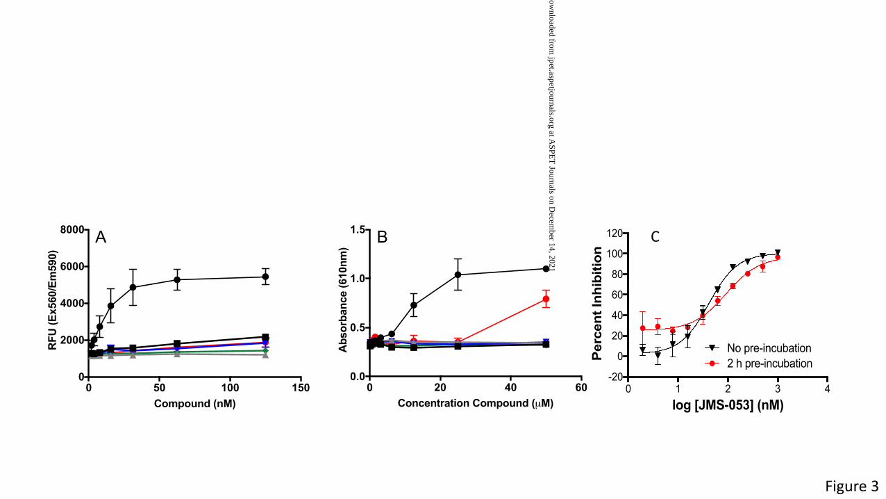
Figure 3
0 20 40 600.0
0.5
1.0
1.5
Concentration Compound (µM)
Abs
orba
nce
(610
nm)
0 50 100 1500
2000
4000
6000
8000
Compound (nM)
RFU
(Ex5
60/E
m59
0)
A B c
0 1 2 3 4-20
0
20
40
60
80
100
120
log [JMS-053] (nM)
Per
cen
t In
hib
itio
n
No pre-incubation2 h pre-incubation
C
This article has not been copyedited and formatted. The final version may differ from this version.JPET Fast Forward. Published on October 10, 2019 as DOI: 10.1124/jpet.119.262188
at ASPE
T Journals on D
ecember 14, 2021
jpet.aspetjournals.orgD
ownloaded from

15.5 16.0 16.5 17.0 17.5 18.0 18.5 19.0 19.5 20.0 20.5 21.0 21.5 22.0 22.5 23.0 23.5 24.0 24.5 25.0 25.5 26.0 26.5 27.0Time (min)
0
10
20
30
40
50
60
70
80
90
1000
10
20
30
40
50
60
70
80
90
100683.71
578.23464.72434.09 447.91
757.14594.91992.69
759.64
578.30
594.86433.98 464.68 992.71448.15
759.71645.05
756.77
644.85
92FC*EAPGSCVAVHCVAGLGR110
507.48
507.46
92FC*EAPGSC*VAVHC*VAGLGR110
Red
uctio
n by
DTT
No
Red
uctio
n 10 20 30 40 50MARMNRPAPV EVSYKHMRFL ITHNPTNATL STFIEDLKKY GATTVVRVCE
60 70 80 90 100VTYDKTPLEK DGITVVDWPF DDGAPPPGKV VEDWLSLVKA KFCEAPGSCV
110 120 130 140 150AVHCVAGLGR APVLVALALI ESGMKYEDAI QFIRQKRRGA INSKQLTYLE
160 170KYRPKQRLRF KDPHTHKTRC CVM
PTP4A3 Sequence Coverage
14.5 15.0 15.5 16.0 16.5 17.0 17.5 18.0 18.5 19.0 19.5 20.0 20.50
20
40
60
80
100
0
20
40
60
80
100
0
20
40
60
80
100
0
20
40
60
80
100 507.49
507.46
683.71
645.05
644.97
VC*EVTYDK
VC*EVTYDK
FC*EAPGSC*VAVHC*VAGLGR
FC*EAPGSCVAVHCVAGLGR
NL: 6.01E6Base Peak m/z=506.96-507.96
Reduction with DTT
NL: 2.70E7Base Peak m/z=644.55-645.55
No Reduction
NL: 1.97E8Base Peak m/z=683.21-684.21
Reduction with DTT
NL: 3.77E6Base Peak m/z=506.96-507.96
No Reduction
Rela
tive
abun
dace
150 200 250 300 350 400 450 500 550 600 650 700 750 800 850 9000
5
10
15
20
25
30
35
40
45
50
55
60
65
70
75
80
85
90
95
100
Rel
ativ
eAb
unda
nce
754.41
260.09457.87
625.42389.20914.49425.23
488.30526.35 867.50
b2
+
b3
+
y3+
y7
2+
b4
+y4
+
y5
+
y6
+
b7
+y7
+
y2+
V C* E V T Y D K
262.26
m/z
/ / / / / /b2
+ b3
+ b4
+ b7
+
y7
+/
y6
+ y5
+ y4
+ y3+ y2
+
Time (min)
A
B C
150 200 250 300 350 400 450 500 550 600 650 700 750 800 850 9000
5
10
15
20
25
30
35
40
45
50
55
60
65
70
75
80
85
90
95
100
RelativeAbundance
754.41
260.09457.87
625.42389.20914.49425.23
488.30526.35 867.50
b2+
b3+
y3+
y72+
b4+y4+
y5+
y6+
b7+
y7+
y2+
V C* E V T Y D K
262.26
m/z
b2+ b3
+ b4+ b7
+
y7+ y6
+ y5+ y4
+ y3+ y2
+
15.5 16.0 16.5 17.0 17.5 18.0 18.5 19.0 19.5 20.0 20.5 21.0 21.5 22.0 22.5 23.0 23.5 24.0 24.5 25.0 25.5 26.0 26.5 27.0
Time (min)
0
10
20
30
40
50
60
70
80
90
100
0
10
20
30
40
50
60
70
80
90
100683.71
578.23464.72434.09 447.91
757.14594.91992.69
759.64
578.30
594.86433.98 464.68 992.71448.15
759.71645.05
756.77
644.85
92FC*EAPGSCVAVHCVAGLGR110
507.48
507.46
92FC*EAPGSC*VAVHC*VAGLGR110
Red
uctio
n by
DTT
No
Red
uctio
n
14.5 15.0 15.5 16.0 16.5 17.0 17.5 18.0 18.5 19.0 19.5 20.0 20.50
20
40
60
80
100
0
20
40
60
80
100
0
20
40
60
80
100
0
20
40
60
80
100507.49
507.46
683.71
645.05
644.97
VC*EVTYDK
VC*EVTYDK
FC*EAPGSC*VAVHC*VAGLGR
FC*EAPGSCVAVHCVAGLGR
Normalized Level: 6.01E6Reduction with DTT
Normalized Level: 2.70E7No Reduction
Normalized Level: 1.97E8Reduction with DTT
Normalized Level : 3.77E6No Reduction
Rel
ativ
e ab
unda
ce
Time (min)
Figure 4
This article has not been copyedited and formatted. The final version may differ from this version.JPET Fast Forward. Published on October 10, 2019 as DOI: 10.1124/jpet.119.262188
at ASPE
T Journals on D
ecember 14, 2021
jpet.aspetjournals.orgD
ownloaded from

Figure 5
ba
Medium +V
ehicl
e
JMS-05
3
NRT-870
-059
EJR-86
6-075
JMS-03
8
DA3003
-10
500
1000
1500
2000
RLU
*
Vehicl
e
5 µM JM
S-038
250 n
M
500 n
M1 µ
M5 µ
M0
5
10
15
# co
loni
es
NRT-870-59
C
A B
* * *NS
0.00 0.02 0.04 0.06Insulin-like Growth Factor-2 mRNA Binding Proteins IGF2BPs/IMPs/VICKZs
Type I hemidesmosome assemblyFormation of the cornified envelope
Inactivation of CDC42 and RAC1Laminin interactions
WNT5A-dependent internalization of FZD2, FZD5 and ROR2Anchoring fibril formation
KeratinizationHS-GAG biosynthesis
Sema4D induced cell migration and growth-cone collapseDiseases associated with O-glycosylation of proteins
XAV939 inhibits tankyrase, stabilizing AXINSema4D in semaphorin signaling
Entities p-value
This article has not been copyedited and formatted. The final version may differ from this version.JPET Fast Forward. Published on October 10, 2019 as DOI: 10.1124/jpet.119.262188
at ASPE
T Journals on D
ecember 14, 2021
jpet.aspetjournals.orgD
ownloaded from