Embed Size (px)
Citation preview
IBSA
FO
UN
DA
TIO
N P
APE
RS
News for hypothyroidism
IBSA
FO
UN
DA
TIO
N P
APE
RS

IBSA FOUNDATION PAPERS 2

News for hypothyroidism
II Forum of Endocrinology27-29 September 2013, Gubbio

1st reprint, May 20151st edition, August 2014© copyright 2014 by Percorsi Editoriali of Carocci Publisher, Rome
Printed in May 2015 by Eurolit, Rome
Cover by Falcinelli&Co. / Stefano VittoriGraphic design by Ulderico Iorillo
Reproduction prohibited under the law (Article 171 of the Law of 22 April 1941 no. 633)
Without proper authorization, you may not reproduce this volume even partially, by any means, including photocopying, even for internal or educational purpose.
We would like to thank Dr. Loredana Bianchi for the editorial support during the preparation of the manuscript.

7 PRESENTATION Silvia Misiti, Giuseppe Zizzo
9 INTRODUCTION Domenico Salvatore
SESSION 1 THE THYROID, THE HORMONES, AND THE METABOLISM: NEW ELEMENTS FOR THE CLINIC
13 THE PLASMATIC CONTROL OF T3 AND T4 LEVELS IN HUMANS Domenico Salvatore
18 GENETIC DEIODINASE POLYMORPHISMS AND CLINICAL IMPLICATIONS
Robin Peeters
23 THE INDIVIDUALLY TAILORED DOSE OF LT4: A SIMPLE ANSWER TO MANY THERAPEUTIC PROBLEMS
Camilla Virili, Cecilia Verga Falzacappa, Marco Centanni
30 WHAT ATA-ETA GUIDELINES INDICATE ON T3/T4 TREATMENT Luca Persani
SESSION 2 NEW HORIZONS FOR HYPOTHYROIDISM
37 THYROID HORMONE IN NORMAL AND STEM CELL Michelina Plateroti
Index

42 WHAT IS THE RATIONALE FOR A COMBINED THERAPY WITH HORMONES T3 AND T4? Salvatore Benvenga
51 CLINICAL STUDIES AND BENEFITS OF THE THERAPY WITH T3 Francesco Saverio Celi
55 A ROLE FOR T3 IN THE “LOW T3 SYNDROME”? Antonio Mancini, Chantal Di Segni, Sebastiano Raimondo, Giuseppe Maria Corbo, Alfredo Pontecorvi
62 T3 SULFATE AS A POTENTIAL PHARMACOLOGICAL AGENT IN THE TREATMENT OF HYPOTHYROIDISM
Ferruccio Santini
66 FUTURE TREATMENT FOR HYPOTHYROID PATIENTS Antonio Bianco, Sabina Casula
75 CONCLUSIONS

7
The aim of the II Forum of Endocrinology organized by IBSA Foundation held in Gub-bio on 27-29 September 2013 was to analyze, in the context of the therapy for hypothyroi-dism, the data coming from basic and clinical research with an eye to the future. During the forum, which was attended by leading experts in the field, several topics have been treated and lively debated:• the inadequacy of TSH as the sole marker of thyroid function and the need to identify
other tissue markers. Currently, TSH remains the marker used in the daily clinical practice, proving not very sensitive in a small niche of patients;
• the difficulty of matching the “well being” with the feeling of “being well” of the pa-tient, not being possible to be guided only by patient preference in the adoption of a specific therapeutic strategy;
• another aspect of the debate has been the need to follow a diagnostic iter to highlight a possible malabsorption, in case the therapeutic target is not reached after admini-stration of the theoretical dose of levothyroxine;
• the potentiality of research in the study of polymorphisms of genes involved in the metabolism of thyroid hormones and the possible correlation between exon sequen-ces and TSH and FT4 values in hundreds of patients, in the clinical practice;
• the correlation between peripheral metabolism of the thyroid hormone and basal metabolism. Having more information in this topic it could help, among other thin-gs, to understand, for example, the reason why there are different metabolic responses to the same stimulus, as may be the cold, in both sexes.We are confident that the forum offered interesting insights and can be a useful study
to achieve the goal desired by all: a timely and correct diagnosis and an always more personalized and effective therapy in the future of the hypothyroid patient.
PresentationSilvia MisitiHead of IBSA Foundation for Scientific Research
Giuseppe ZizzoSecretary of IBSA Foundation for Scientific Research

9
The Forum held by the IBSA Foundation in Gubbio has drawn together a number of internationally acknowledged experts to discuss current therapeutic approaches to hypothyroidism. The treatment of this condition affecting about 10% of the popula-tion living in Western countries is still lively debated by clinicians and basic resear-chers who strive to identify the ideal treatment for this complex condition.
It has been unmistakably shown that substitution therapy based on levothyroxine alone – the main hormone secreted by the thyroid gland that has been regarded as the cornerstone of the therapy for years – is not able to ensure well-being in a subgroup of patients. Moreover, the administration of thyroxine alone causes an excessive rise in plasma concentration of T4 in a group of patients with a limited ability to convert T4 to T3. In the light of these results, T3 might be used in combination therapy with T4. There is a long way to go, though, as various steps and goals need to be achieved before such a potent hormone can be safely used in clinical practice. How is T3 to be used? When? And for which kind of patients?
Answering these questions was one of several challenges faced by the Forum, which aimed to identify the best treatment for each patient affected by hypothyroidism, a common pathology characterized by simple etiology and diagnosis, as well as by a complex therapeutic treatment.
IntroductionDomenico SalvatoreThe University of Naples “Federico II”


11
SESSION 1
THE THYROID, THE HORMONES, AND THE METABOLISM:NEW ELEMENTS FOR THE CLINIC


13
The homeostasis of thyroid hormones in plasma and in tissues is a rather complex sub-ject. Indeed, we know that the thyroid hormone is not able to enter into target cells in a passive manner but needs transporters. A cell that does not express the carrier is not permissive to the action of thyroid hormone. Entered into the cell, the hormone becomes the target of a family of enzymes, the selenium-deiodinase, that, in some way, can completely change its biological activity.
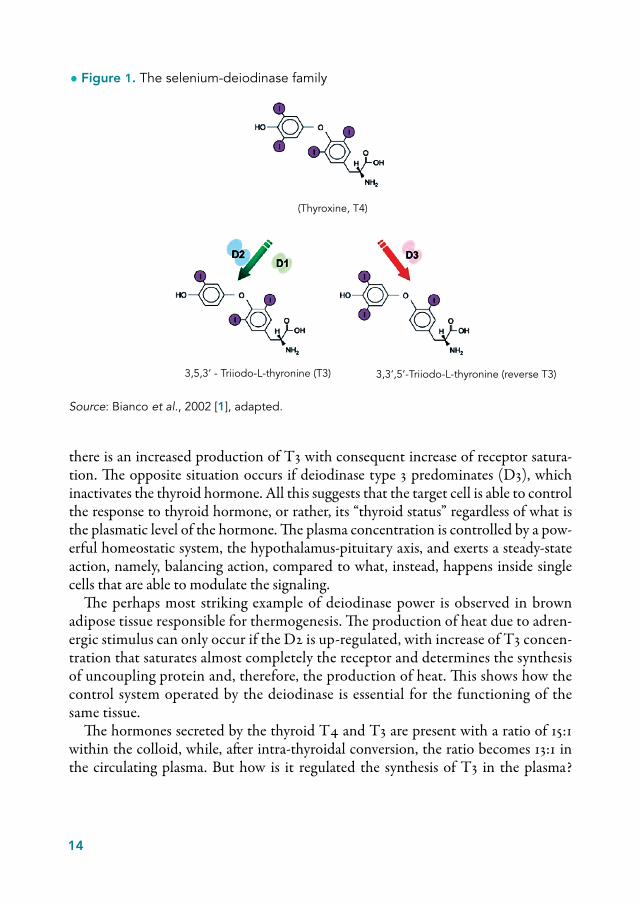
The deiodinase are enzymes that convert thyroxine, the main thyroid product, in a molecule metabolically active, T3, or in an inactive product, the rT3 (• Figure 1). The fate of T4, therefore, varies depending on the pathway of activation or degradation in which is directed, so not necessarily the pre-hormone exerts its hormone activity, but it can also be turned off and, as such, be unable to reach the nuclear targets.
The structure of deiodinase is complex. These enzymes have a membrane domain whose function is not yet fully known, and it is probably necessary so that the mole-cules dimerize and become enzymatically active. The most surprising thing, though, is the presence in these molecules of an atom of selenium, a trace element that, in order to be incorporated into the protein, requires a considerable amount of time and energy by the cell. However, this process is essential because the selenium is localized in the catalytic site of the enzyme, site that T4 must reach in order to be metabolized. Selenium is preserved phylogenetically in all species and is common to all desiodinas-es. Also, in laboratory there was a significant reduction in the deiodination activity following the replacement of this oligo element with a sulfur atom in the catalytic site.
Now imagine that there is a cell deiodinase-free: in this cell T3, produced in small part by the thyroid, enters and performs its function without any control at intracel-lular level. If, instead, in the cell there is predominance of one enzyme on the other, we obtain different effects. In particular, when the deiodinase type 2 (D2) predominates
The plasmatic control of T3 and T4 levels in humans Domenico Salvatore
Department of Clinical Medicine and Surgery, University of Naples Federico II
14
there is an increased production of T3 with consequent increase of receptor satura-tion. The opposite situation occurs if deiodinase type 3 predominates (D3), which inactivates the thyroid hormone. All this suggests that the target cell is able to control the response to thyroid hormone, or rather, its “thyroid status” regardless of what is the plasmatic level of the hormone. The plasma concentration is controlled by a pow-erful homeostatic system, the hypothalamus-pituitary axis, and exerts a steady-state action, namely, balancing action, compared to what, instead, happens inside single cells that are able to modulate the signaling.
The perhaps most striking example of deiodinase power is observed in brown adipose tissue responsible for thermogenesis. The production of heat due to adren-ergic stimulus can only occur if the D2 is up-regulated, with increase of T3 concen-tration that saturates almost completely the receptor and determines the synthesis of uncoupling protein and, therefore, the production of heat. This shows how the control system operated by the deiodinase is essential for the functioning of the same tissue.
The hormones secreted by the thyroid T4 and T3 are present with a ratio of 15:1 within the colloid, while, after intra-thyroidal conversion, the ratio becomes 13:1 in the circulating plasma. But how is it regulated the synthesis of T3 in the plasma?
• Figure 1. The selenium-deiodinase family
Source: Bianco et al., 2002 [1], adapted.
(Thyroxine, T4)
3,5,3’ - Triiodo-L-thyronine (T3) 3,3’,5’-Triiodo-L-thyronine (reverse T3)

15
Understanding is crucial when you consider that in patients with thyroidectomy the T4 administered is converted in T3 ensuring the proportion of hormone needed to tissues. The enzymes responsible for the control of the synthesis of T3 in the periphery are substantially D1 and D2, with some differences. In order to determine the role of D1 compared to D2 it was used propyl-thiouracil (PTU), which selectively blocks the D1. It was found that in patients treated with PTU in euthyroid condition the T3 concentration is only reduced by 20-30%, compared to a reduction of about 50% under conditions of hyperthyroidism. This indicates that D1 predominates in hyper-thyroidism, and this is the reason why the PTU is very effective in reducing the levels of thyroid hormone in hyperthyroid patients.
To recap: about 20% of the hormone synthesis of T3 is guaranteed by the thyroid, but there is also an extra-thyroid production of T3 regulated by the activity of D1 and D2. These two activating enzymes, however, have a different pattern of expres-sion in the various tissues of the body, as well as a different degree of affinity for the T4 (• Table 1).
Another important aspect concerns the homeostatic role of deiodinase enzymes, well described from the regulatory mechanism of D2: in hypothyroidism there is an increased activity of this enzyme, with consequent increase of the conversion of T4 to T3; an opposite regulation occurs in conditions of hyperthyroidism.
It emerges, at this point, a new important question: the exogenous T4 is itself able to ensure proper concentration of T3 both at plasma and tissue level? At this regard, a study by the Catania group [2] on more than 1,800 patients without thyroid has demonstrated how, in these subjects, T3 levels were on average lower compared with control patients. The research shows that in patients treated with T4 theT3 levels, even if within the normal range, are slightly lower than the norm. Furthermore, it
• Table 1. The two activation pathways of T4 in T3
D2 D1
Km(T4) 10-9M 10-6M
Tissues Muscle, skin Liver, kidney
Cellular location End. Retic. Plasma membrane
Enzyme half-life 1 h 12 hPTU effect 0 Inhibit
Hypothyroid, iodine deficiency ↑ ↓Hyperthyroid ↓ ↑
Source: data from Bianco et al., 2001 [1].
16
was found that in a small percentage of patients (approximately 15%) T3 does not normalize perfectly, even though TSH values are normal. In these cases, in order to normalize the T3 levels, it is necessary to increase the T4 dosage so much to reduce the TSH to values not more physiological. But what is the role of deiodinase en-zymes in this context? To respond it is crucial to distinguish the plasma control of the hormone levels from the tissue one. In this regard an important information is provided to us from an animal model deprived of D2, in which, despite normal levels of T3 in plasma, the concentration of T3 in some tissues such as the brain, is reduced. The same occurs in mice in which both deiodinase activators are genetically deleted: the plasma T3 is normal, but TSH and T4 levels are high. In this case, the thyroid provides all the T3 necessary ensure a normal plasma concentration of T3.
From the described animal models it can be deduced that in patients treated with T4, the plasma values of T3 in the normal range does not necessarily reflect a euthy-roid condition at tissue level.
Although there are still some aspects to be clarified, it can be concluded that, prob-ably, through treatment with T4 alone you can get the normalization of T3 concen-tration in plasma but not in tissue.
In humans, the thyroid hormone is essential for proper development of the organ-ism in intrauterine and neonatal life as well as in the adulthood one. A demonstration of the importance of deiodinase enzymes for life comes again from the animal world. Metamorphosis is an event regulated by thyroid hormone: in the absence of deiodi-nation activity, this process is altered. Moreover, the thyroid hormone is essential for the proper maturation of the cochlea, a structure of the inner ear [3]: in the first days of life there is a peak of D2 which leads to an increase in T3 concentration of almost 100 times. This is necessary so that the ear can develop in a proper way, so much that mice deprived of the D2 activity are deaf.
What happens, though, if the balance guaranteed by deiodinase enzymes is lost? That’s what occurs in consumptive hypothyroidism, a frequent paraneoplastic syn-drome in children, characterized by a pathological increased expression of D3. An article appeared in the New England Journal of Medicine [4] described the case of a child suffering by hepatic hemangioma who presented a severe hypothyroidism. The hemangioma is a highly vascularized tumor that may have a highly variable clinical presentation: typically begins with a phase of expansive growth for a period of about a year, to which follows a phase of regression. The growth phase is the most dangerous from a metabolic point of view, since cancer cells express very high amount of D3, re-sulting in massive degradation of T3 and T4 normally produced by the thyroid. The activation of the hypothalamic-pituitary-thyroid feedback in these children is not sufficient to buffer the hepatic hormone degradation and to ensure euthyroidism, and, therefore, occurs a particularly severe hypothyroidism, which in some cases, can lead to death. Several studies have shown that consumptive hypothyroidism can also affect adulthood and may be associated also with other types of cancer.

17
In conclusion, we can say that the deiodinase type 1 is involved in the plasmatic ho-meostasis of T3 in the same way as the deiodinase type 2. The latter is also crucial for the intracellular homeostasis of T3 and for the initiation of the feedback operated by T4 on TSH secretion. The deiodinase type 3 plays a key role during intrauterine life and in different pathophysiological mechanisms, such as pregnancy.
Finally, deiodinase enzymes as homeostatic circuit presiding the control of plasma T3, meet the functional requirements of the different tissues and are able, to some extent, to protect tissues from modest changes in plasmatic T3.
References
[1] Bianco AC, Salvatore D, Gereben B, Berry MJ, Larsen PR. Biochemistry, cellular and molecular biology, and physiological roles of the iodothyronine selenodeiodinases. Endocr Rev 2002 Feb;23(1):38-89.
[2] Gullo D, Latina A, Frasca F, Le Moli R, Pellegriti G, Vigneri R. Levothyroxine monother-apy cannot guarantee euthyroidism in all athyreotic patients. PLoS One 2011;6(8):e22552.
[3] Campos-Barros A, Amma LL, Faris JS, Shailam R, Kelley MW, Forrest D. Type 2 io-dothyronine deiodinase expression in the cochlea before the onset of hearing. Proc Natl Acad Sci USA 2000 Feb 1;97(3):1287-92.
[4] Huang SA, Tu HM, Harney JW, Venihaki M, Butte AJ, Kozakewich HP et al. Severe hypothyroidism caused by type 3 iodothyronine deiodinase in infantile hemangiomas. N Engl J Med 2000 Jul 20;343(3):185-9.

18
Genetic plays an essential role in control of thyroid hormone’s set-point. The impor-tance of hereditariness in the regulation of TSH and FT4 is well described in the study of Hansen et al. [1], in which there is a significant correlation between TSH and FT4 values in dizygotic twins, and, much more, in monozygotic twins. Complicated calcu-lations have showed that in the last ones about 60% of total TSH variability is related to genetic factors.
In one another work [2], a group of healthy voluntaries was monitored during the year through monthly measurings of TSH and FT4: for every subject hormones levels fluctuated around a particular set-point, that was different from set-point of the other subjects. The normal range of TSH is calculated on the basis of variability of values in general population, but as we can see in • Figure 1, this may not be optimal for each individual. So, it may be that a TSH value in the normal range can be still too high for a particular subject. The same holds true for FT4.
The • Figure 2 [3] shows the relation between TSH and FT4; as we can see, small changes in FT4 levels correspond to logatithmic alterations of TSH: high values of FT4 are associated to low values of TSH and vice versa. In general, there is a good correlation between FT4 and TSH, but a lot of values are scattered. For example, if we consider the two subjects marked in • Figure 2, we can see that both of them have had a similar decrease in FT4, leading to a similar increase in TSH; but, while the one with FT4 under the normal range is now considered hypothyroid, for the other one the plasmatic concentration of the hormone is still normal, and he or she is therefore considered subclinically hypothyroid. This demonstrates the importance of individual variation of hormonal set-point.
Another study [4] shows the possible contribution of genetic factors in determining the clinical set-point: in patients with syndrome of resistance to thyroid hormones cau-
Genetic deiodinase polymorphisms and clinical implicationsRobin PeetersAssociate Professor in Clinical Endocrinology, Erasmus Medical Center, Erasmus University, Rotterdam

19
sed by mutations in the THR-β gene, the reduced pituitary’s sensitivity to the negative feedback of thyroid hormones causes high levels of FT4 and FT3; there is still a log-li-near relationship between TSH and FT4, but the set-point has shifted.
About 10 years ago, we analyzed polymorphisms of different genes involved in thyroid pathway [5], including genes for deiodinases, and we showed the existence of two particular polymorphisms in a region of the gene for type 1 deiodinase. We know that this enzyme is responsible for plasmatic FT3 production and for clearance of rT3 (reverse T3). So we speculated that a defective deiodinase would, somehow or other, alter hormone’s homeostasis. In 156 healthy voluntaries, asymptomatic carriers of the polymorphism had lower levels of T3, while rT3 was high.
Moreover, because of the enormous increase in technological possibilities associa-ted to genetic studies in the last 10 years, we have switched from analyzing single gene variants to whole genomes, through the so called GWAS (Genoma Wide Association Studies). In these kind of studies we analyze about 2.5 million polymorphisms in the genome at the same time.
In one of these works [6] we identified a lot of functional variants associated to TSH and FT4 and we saw that the most significant variant for FT4 was the variant in type 1 deiodinase, that we had already analyzed 10 years before (• Figure 3). By using these large consortia we have identified 23 independent polymorphisms associated to TSH, and these contributed not only to TSH variation within the normal range, but also to TSH values outside the reference range. However, these findings explained only 5.64% of the trait variation in TSH, and 2.30% of total variation in FT4.
Of course, the most important question concerns consequences of these findings in clinical practice. There are three reasons why it is important to study this kind of polymorphisms. First of all, we are able to obtain novel insights into mechanisms in-
• Figure 1. Inter- and intra-individual ranges of thyroid hormones
Source: Andersen et al., 2002 [2].

20
volved in the development of the diseases of interest, also for identification of new drug targets. A study from Butler et al. (2010) [7] analyzed functional effects of a particular polymorphism in the type 2 deiodinase (Thr92Ala), showing that subjects who with the rare variant of this polymorphism had an altered sensitivity of the pituitary in the regulation of hypothalamic-pituitary-thyroid axis. When these subjects were injected with TRH they showed a different response in increase of T3 in comparison with heal-thy subjects, also suggesting that this polymorphism is indeed relevant for the control of the hypothalamic-pituitary-thyroid axis and for the regulation of thyroid hormone’s set-point.
These studies of polymorphisms associated with the HPT-axis set-point may also help for diagnosis: if we are able to predict disease risk, consequently it may help us to personalize medicine, and to decide whether or not we should treat patients. But, actually, we can explain only a little percentage of TSH variations so far, and so we can’t yet develop reliable tests for that particular patients. In other words, we are not already able to predict individual risk of disease.
At last, these studies could help us to predict the response to treatment. Panicker et al. (2009) [8] analyzed relationship between different polymorphisms, including polymorphisms of type 1, 2 and 3 deiodinase, and well-being in hypothyroid patients on thyroxine therapy. In particular we know that type 2 deiodinase is the major respon-sible for local production of T3, so it represents the most important resource of active
• Figure 2. Log-linear relationship between TSH and FT4
Source: Spencer et al., 1990 [3], adapted.

21
hormone in the brain. So, if we speculate that hypothyroid patients depend almost completely on local conversion of T4 to T3 by DIO2, less functional variants of this enzyme would be associated to a reduced pool of T3 into the brain, and, consequently, to decreased well-being of patient. The authors demonstrated that this variant was in-deed associated with well-being in patient on LT4 therapy, Moreover, the authors de-monstrated that patients with this DIO2 polymorphism may have more benefit from T4+T3 combination therapy.
However, there are two important remarks here: first of all, a lot of tests were per-formed, and if we apply strict multiple testing corrections, the results are not consi-dered significant. This remark is also made by the authors in this paper. Second, it’s a small study, so we need confirmation from independent studies showing the same results.
In conclusion, although the current evidence suggests that variants in deiodinase genes may explain why some patients continue to have complaints under LT4 therapy, we have not yet sufficient knowledge to predict response to treatment for the indivi-dual patient.
Source: Porcu et al., 2013 [6], adapted.
• Figure 3. GWAS meta-analysis for serum FT4
N=17.52015 cohorts
22
References
[1] Hansen PS, Brix TH, Sørensen TI, Kyvik KO, Hegedüs L. Major genetic influence on the regulation of the pituitary-thyroid axis: a study of healthy Danish twins. J Clin Endocrinol Metab 2004 Mar; 89(3):1181-7.
[2] Andersen S, Pedersen KM, Bruun NH, Laurberg P. Narrow individual variations in serum T(4) and T(3) in normal subjects: a clue to the understanding of subclinical thyroid disease. J Clin Endocrinol Metab 2002 Mar; 87(3):1068-72.
[3] Spencer CA, LoPresti JS, Patel A, Guttler RB, Eigen A, Shen D et al. Applications of a new chemiluminometric thyrotropin assay to subnormal measurement. J Clin Endocrinol Metab 1990;70(2):453-60.
[4] Ercan-Fang S, Schwartz HL, Mariash CN, Oppenheimer JH. Quantitative assessment of pituitary resistance to thyroid hormone from plots of the logarithm of thyrotropin versus serum free thyroxine index. J Clin Endocrinol Metab 2000 Jun;85(6):2299-303.
[5] Peeters RP, Van Toor H, Klootwijk W, De Rijke YB, Kuiper GGJM, Uitterlinden AG, Visser TJ. Polymorphisms in thyroid hormone pathway genes are associated with plasma TSH and iodothyronine levels in healthy subjects. J Clin Endocrinol Metab 2003;88(6):2880-8.
[6] Porcu E, Medici M, Pistis G, Volpato CB, Wilson SG, Cappola AR et al. A meta-analysis of thyroid-related traits reveals novel loci and gender-specific differences in the regulation of thyroid function. PLoS Genet 2013;9(2):e1003266.
[7] Butler PW, Smith SM, Linderman JD, Brychta RJ, Alberobello AT, Dubaz OM et al. The Thr92Ala 5’ type 2 deiodinase gene polymorphism is associated with a delayed triio-dothyronine secretion in response to the thyrotropin-releasing hormone-stimulation test: a pharmacogenomic study. Thyroid 2010 Dec;20(12):1407-12.
[8] Panicker V, Saravanan P, Vaidya B, Evans J, Hattersley AT, Frayling TM, Dayan CM. Common variation in the DIO2 gene predicts baseline psychological well-being and respon-se to combination thyroxine plus triiodothyronine therapy in hypothyroid patients. J Clin Endocrinol Metab 2009;94(5):1623-9.

23
Replacement therapy with sodium levothyroxine involves the use of a hormone with a narrow therapeutic index, usually framed in a treatment of long/very long period. In the past, on empirical grounds, there has been a substantial anarchy of doses and routes of administration, with significant side effects due to a chronic hyper-or hypo-treat-ment.
On the basis of these considerations it is evident the importance of using an individ-ualized dose of thyroxine: first because the minimum effective dose of T4 should be administered, but also to avoid continuous oscillations of the dosage and consequent loss of compliance by the patient. Moreover, customizing the dose allows suspecting possible malabsorption, identifying gastrointestinal disorders. Further advantage of this therapeutic modality regards the ability to obtain substantial savings in terms of pharmaco-economics, avoiding lengthy and costly monitoring. There are however several variables to consider to find an optimal and predictable daily dose of thyroxine (• Figure 1).
The hormonal need changes in the absence of the thyroid gland and several stud-ies have shown that the dose of thyroxine must be incremented after thyroidectomy. Crucial element when the thyroid gland is still in situ is a correct assumption of the therapy, therefore, an effective compliance by the patient. The dose of LT4 should be calculated according to the BMI (Body Mass Index) or the amount of lean body mass of the subject [1]. The administration also can occur at any time of the day, de-nying the belief that levothyroxine must necessarily be taken in the morning [2, 3]. The opportunity to change and to “tailor” the time of assumption guarantees better compliance by the patient and the possibility of a greater intestinal absorption of the hormone. Studies from Salvatore Benvenga have determined that the initial rate of absorption of thyroxine occurs in the first 60-90 minutes after assumption. The in-
The individually tailored dose of LT4: a simple answer to many therapeutic problemsCamilla Virili, Cecilia Verga Falzacappa, Marco Centanni
UOC of Endocrinology, Sapienza University of Rome and St. Maria Goretti Hospital, Latina

24
terval of time necessary to avoid interference with any type of foods and beverages, including coffee, must therefore be of at least 1 hour [4].
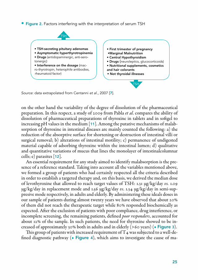
To properly assess the effect of therapy with thyroxine it is also crucial having a good marker of adequacy. The reliability of TSH as a marker of the thyroid homeo-stasis is widely accepted, although there are doubts both quantitative, relative to the reference range [5], and qualitative, about the influence that the tissue-specific meta-bolism of the hormone exerts on the effectiveness of oral thyroxine [6]. To compro-mise the reliability of TSH, however, contribute other factors, often not considered (• Figure 2), which interfere with the dosage of this marker [7]. In particular, some substances contained in cosmetics that falsely lower TSH, affecting the understan-ding of the real thyroid situation. Therefore, the need to re-evaluate the effectiveness of this marker as isolated predictor of the adequacy of thyroxine therapy, associating it always to the dosage of iodothyronines.
To obtain individually tailored dose, it is also necessary to exclude the factors that can cause an increase in the daily intake of the hormone: a) changes in anthropome-tric characteristics of the patient (weight, gestational status); b) different biochemical properties of the pharmaceutical preparation of LT4 (bioequivalence); c) nutritional interference [8]; d) concomitant medications (poly-treatment).
An increased need for levothyroxine dose than the theoretical dose, which persists despite the exclusion of all these conditions, suggests the presence of a gastrointesti-nal malabsorption of the hormone. In general, it was found that can exist an increased need for T4 in the course of gastric diseases (active infections and chronic gastritis from Helicobacter pylori, atrophic chronic gastritis and gastrointestinal resections) or intestinal diseases (coeliac disease, lactose intolerance, etc.) [8-10].
Among the putative mechanisms of increased requirements which is observed in the course of gastric pathologies we remember essentially the possible consequences of increased gastric pH: on one hand, the changes in the ionization state of the thyroxi-ne molecule that may impact on the ability to cross the membrane of enterocytes,
• Figure 1. Variables to be considered in the calculation of the individually tailored dose
Interpretation of the TSH values in light of its variables and its
interferents
Biochemical nature and absorption of oral
thyroxine
The therapeutic efficacy of thyroxin therapy depends from the interlinking of two main parameters
25
on the other hand the variability of the degree of dissolution of the pharmaceutical preparation. In this respect, a study of 2009 from Pabla et al. compares the ability of dissolution of pharmaceutical preparations of thyroxine in tablets and in softgel to increasing pH values in the medium [11]. Among the putative mechanisms of malab-sorption of thyroxine in intestinal diseases are mainly counted the following: a) the reduction of the absorptive surface for shortening or destruction of intestinal villi or surgical removal; b) alterations of intestinal motility; c) permanence of undigested material capable of adsorbing thyroxine within the intestinal lumen; d) qualitative and quantitative variations of mucus that lines the monolayer of intestinalcolumnar cells; e) parasites [12].
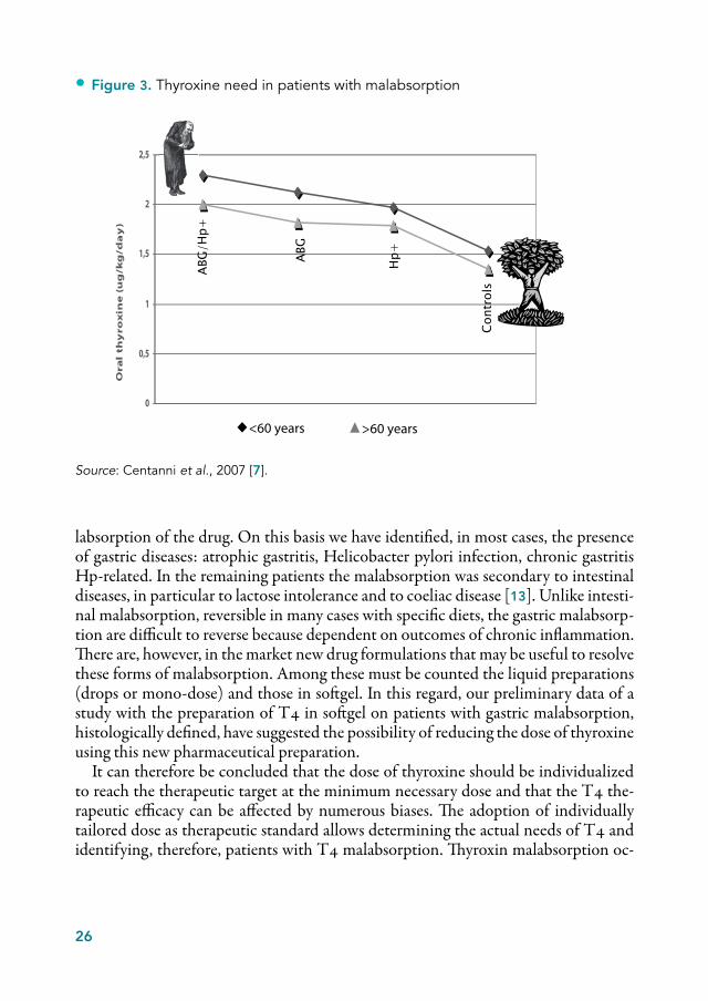
An essential requirement for any study aimed to identify malabsorption is the pre-sence of a reference standard. Taking into account all the variables mentioned above, we formed a group of patients who had certainly respected all the criteria described in order to establish a targeted therapy and, on this basis, we derived the median dose of levothyroxine that allowed to reach target values of TSH: 1.31 µg/kg/day vs. 1.09 µg/kg/day in replacement mode and 1.56 µg/kg/day vs. 1.34 µg/kg/day in semi-sup-presive mode respectively, in adults and elderly. By administering these ideals doses in our sample of patients during almost twenty years we have observed that about 20% of them did not reach the therapeutic target while 80% responded biochemically as expected. After the exclusion of patients with poor compliance, drug interference, or incomplete screening, the remaining patients, defined poor responders, accounted for about 12% of the sample. In such patients, the need for thyroxine showed to be in-creased of approximately 30% both in adults and in elderly (>60 years) (• Figure 3).
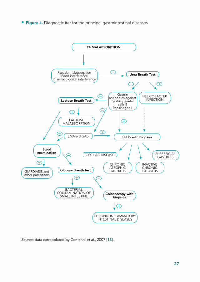
This group of patients with increased requirement of T4 was subjected to a well-de-fined diagnostic pathway (• Figure 4), which aims to investigate the cause of ma-
• Figure 2. Factors interfering with the interpretation of serum TSH
Source: data extrapolated from Centanni et al., 2007 [7].
• TSH-secreting pituitary adenomas • Asymptomatic hyperthyrotropinemia • Drugs (antidopaminergic, anti-sero-tonergic) • Interference on the dosage (mac-ro-thyrotropin, heterophile antibodies, rheumatoid factor)
• First trimester of pregnancy •Marginal Malnutrition • Central Hypothyroidism • Drugs (neuroleptics, glucocorticoids) • Nutritional supplements, cosmetics and hair colorants • Not thyroidal illnesses
TSH
TSH
26
labsorption of the drug. On this basis we have identified, in most cases, the presence of gastric diseases: atrophic gastritis, Helicobacter pylori infection, chronic gastritis Hp-related. In the remaining patients the malabsorption was secondary to intestinal diseases, in particular to lactose intolerance and to coeliac disease [13]. Unlike intesti-nal malabsorption, reversible in many cases with specific diets, the gastric malabsorp-tion are difficult to reverse because dependent on outcomes of chronic inflammation. There are, however, in the market new drug formulations that may be useful to resolve these forms of malabsorption. Among these must be counted the liquid preparations (drops or mono-dose) and those in softgel. In this regard, our preliminary data of a study with the preparation of T4 in softgel on patients with gastric malabsorption, histologically defined, have suggested the possibility of reducing the dose of thyroxine using this new pharmaceutical preparation.
It can therefore be concluded that the dose of thyroxine should be individualized to reach the therapeutic target at the minimum necessary dose and that the T4 the-rapeutic efficacy can be affected by numerous biases. The adoption of individually tailored dose as therapeutic standard allows determining the actual needs of T4 and identifying, therefore, patients with T4 malabsorption. Thyroxin malabsorption oc-
0
0,5
1
1,5
2
2,5
Ora
l th
yro
xin
e (
ug
/kg
/da
y)
<60 years
ABG
/Hp+
Hp+
Con
trol
s
ABG
>60 years
• Figure 3. Thyroxine need in patients with malabsorption
Source: Centanni et al., 2007 [7].
27
Source: data extrapolated by Centanni et al., 2007 [13].
• Figure 4. Diagnostic iter for the principal gastrointestinal diseases
T4 MALABSORPTION
Pseudo-malabsorption Food interference
Pharmacological interferenceUrea Breath Test
Lactose Breath Test
Gastrin antibodies against
gastric parietal cells B
Pepsinogen I
HELICOBACTER INFECTION
LACTOSE MALABSORPTION
EMA e tTGAb EGDS with biopsies
Stool examination
COELIAC DISEASE
CHRONIC ATROPHIC GASTRITIS
INACTIVE CHRONIC GASTRITIS
SUPERFICIAL GASTRITIS
GIARDIASIS and other parasitisms
BACTERIAL CONTAMINATION OF
SMALL INTESTINE
Glucose Breath test
Colonoscopy with biopsies
CHRONIC INFLAMMATORY INTESTINAL DISEASES

28
curs in a patient treated out of 6 and the cause recognizes a gastric pathogenesis in two thirds of patients. Knowledge of the thyroxine pharmacokinetics enables the develop-ment of more absorbable preparations and potentially able to optimize the thyroxine therapy, adapting it to the characteristics and the needs of the individual patient.
References
[1] Santini F, Pinchera A, Marsili A, Ceccarini G, Castagna MG, Valeriano R et al. Lean body mass is a major determinant of levothyroxine dosage in the treatment of thyroid dis-eases. J Clin Endocrinol Metab 2005 Jan;90(1):124-7.
[2] Bolk N, Visser TJ, Kalsbeek A, van Domburg RT, Berghout A. Effects of evening vs morning thyroxine ingestion on serum thyroid hormone profiles in hypothyroid patients. Clin Endocrinol (Oxf ) 2007 Jan;66(1):43-8.
[3] Bach-Huynh TG, Nayak B, Loh J, Soldin S, Jonklaas J. Timing of levothyroxine ad-ministration affects serum thyrotropin concentration. J Clin Endocrinol Metab 2009 Oct;94(10):3905-12.
[4] Benvenga S, Bartolone L, Squadrito S, Lo Giudice F, Trimarchi F. Delayed intestinal absorption of levothyroxine. Thyroid 1995 Aug;5(4):249-53.
[5] Wartofsky L, Dickey RA. The evidence for a narrower thyrotropin reference range is com-pelling. J Clin Endocrinol Metab 2005 Sep;90(9):5483-8.
[6] Alevizaki M, Mantzou E, Cimponeriu AT, Alevizaki CC, Koutras DA. TSH may not be a good marker for adequate thyroid hormone replacement therapy. Wien Klin Wochenschr 2005 Sep;117(18):636-40.
[7] Centanni M, Franchi A, Santaguida MG, Virili C, Nardo S, Gargano L. [Oral thyroxine treatment: towards an individually tailored dose]. Recenti Progr Med 2007;98(9):445-51.
[8] Liwanpo L, Hershman JM. Conditions and drugs interfering with thyroxine absorption. Best Pract Res Clin Endocrinol Metab 2009 Dec;23(6):781-92.
[9] Virili C, Bassotti G, Santaguida MG, Iuorio R, Del Duca SC, Mercuri V et al. Atypical celiac disease as cause of increased need for thyroxine: a systematic study. J Clin Endo-crinol Metab 2012;97(3):E419-E422.
[10] Centanni M, Gargano L, Canettieri G, Viceconti N, Franchi A, Delle Fave G, Anni-
29
bale B. Thyroxine dose in multinodular goiter, Helicobacter pylori infection and chronic gastritis. New Engl J Med 2006;354(17):1787-95.
[11] Pabla D, Akhlaghi F, Zia H. A comparative pH-dissolution profile study of selected com-mercial levothyroxine products using inductively coupled plasma mass spectrometry. Eur J Pharm Biopharm 2009;72(1):105-10.
[12] Hirtz J. The gastrointestinal absorption of drugs in man. A review of current concepts and methods of investigation. Br J Clin Pharmacol 1985;19(Suppl 2):77S-83S.
[13] Centanni M, Santaguida MG, Gargano L. Malabsorption of T4: new insights on oral thyroxine treatment. Hot Thyroidol 2007 June; 69:1-4 (www.hotthyroidology.com).

30
Replacement therapy with levothyroxine is, at the present, the preferential therapeutic approach for hypothyroidism. The guidelines of the ATA-AACE (American Thyroid Association-American Association of Clinical Endocrinologists) [1] and those of ETA (European Thyroid Association) [2] strongly recommend monotherapy with T4, since there is not yet enough evidence in favor of combined treatment T3+T4.
It is known, however, that 5-10% of patients treated with levothyroxine, while achieving euthyroidism in plasma, continues to complain of symptoms of hormonal deficiency. The explanations for this phenomenon may be different. For example, the awareness of being affected by a chronic disease could in some way, affect the fee-ling of well-being reported by the patient; in addition, several conditions related to thyroid disease or condition of the replacement, may compromise the achievement of a state of well-being. We know, in this regard, that often the autoimmune thyroid disease is associated with other diseases (diabetes mellitus, Addison’s disease, early menopause, etc.) in the so-called polyglandular autoimmune syndromes (PGAS). In these cases it is necessary to take into account that the patients complaints attributed to hypothyroidism may actually have a different origin from the thyroid.
At the base of discomfort reported by the patient, theoretically euthyroid, there could be an inadequacy of the therapy itself: several studies reviewed in the guidelines have shown that monotherapy with LT4 may be insufficient to ensure a proper plasma relationship of FT4/FT3 and, in contrast, a combination therapy T3+T4 has proved capable, in some cases, to lower this ratio to more physiological values (• Table 1). An inadequate response to treatment with levothyroxine may also depend on individual variables, such as the existence of deiodinase polymorphisms [2].
If, then, for that famous 5-10% of patients in therapy, symptomatic despite normal hormone levels in plasma, we exclude interfering factors previously described, there may be a rationale for the use of combination therapy and get more clinical benefits.
What ATA-ETA guidelines indicate on T3/T4 treatment Luca Persani Department of Clinical Sciences and of Community, University of Milan; Division of General and Endocrine-Metabolic Medicine, St. Luca Hospital, Auxologic Italian Institute IRCCS, Milan

31
In this regard, the study of Panicker et al. [3] analyzed the correlation between diodi-nase polymorphisms and feeling of well-being reported by the patient, demonstrating that a particular polymorphism for D2, present in approximately 16% of subjects tre-ated with levothyroxine, was associated with a worse feeling of sickness and a better response to combined treatment.
The ETA guidelines [2] emphasize the concept, stating that these data on polymor-phisms involved in the thyroid hormone pathway, especially for genes of transporters and desiodinase, could play a role in order to justify the use combined T3+T4 the-rapy. Several crossover studies show that, generally, patients tend to prefer combina-tion therapy to monotherapy with LT4 [2] (• Table 2). The work of Appelhof et al. [4] (• Figure 1) confirms this trend: the subgroups of patients undergoing therapy T3+T4 in various combinations defined themselves more satisfied compared to pa-tients with levothyroxine monotherapy. Various aspects make, however, questionable this study: short duration, as well as a condition of thyrotoxicosis associated with the combination therapy, both at laboratory (suppressed TSH) and clinical level, with a significant increase in heart rate and decrease of body weight. It is therefore necessary to clarify whether the feeling of satisfaction reported by patients is, in fact, the result of an overtreatment and not more of a replacement therapy aimed at restoring the euthyroid state.
The limits of the comparison studies between the two treatment modalities are different: patient samples of small size, lack of homogeneity of the hypothyroid state of the populations in exam (post-thyroidectomy, primary hypothyroidism...); use of various associations of T3 and T4.
ETA guidelines suggest that the combination therapy may constitute an experi-mental approach in hypothyroid patients that, despite the normalization of bioche-
• Table 1. Ratio of plasmatic concentrations of FT4/FT3 in basic conditions and after treatment with T4 or T4+T3
Author Baseline T4 monotherapy
T4+T3 combination
Saravanan et al. J Clin Endocrinol Metab 2005;90:805-12 5.5 5.5 3.9
Walsh et al. J Clin Endocrinol Metab 2003;88:4543-50 4.5 4.2 3.3
Sawka et al. J Clin Endocrinol Metab 2003;88:4551-5 3.9 4.0 2.2
Escobar-Morreale et al. Ann Intern Med 2005;142:412-24 4.1 4.1 3.4
Siegmund et al. Clin Endocrinol 2004;60:750-7 4.3 4.6 4.0
Mean FT4:FT3 ratio 4.3 4.5 3.4
Source: Wiersinga et al., 2012 [2], adapted.
32
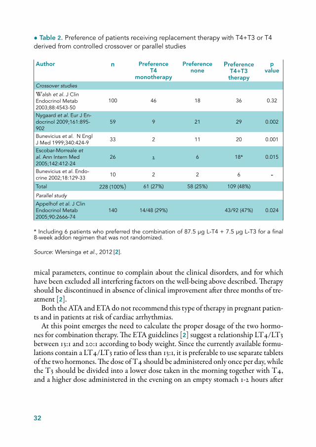
mical parameters, continue to complain about the clinical disorders, and for which have been excluded all interfering factors on the well-being above described. Therapy should be discontinued in absence of clinical improvement after three months of tre-atment [2].
Both the ATA and ETA do not recommend this type of therapy in pregnant patien-ts and in patients at risk of cardiac arrhythmias.
At this point emerges the need to calculate the proper dosage of the two hormo-nes for combination therapy. The ETA guidelines [2] suggest a relationship LT4/LT3 between 13:1 and 20:1 according to body weight. Since the currently available formu-lations contain a LT4/LT3 ratio of less than 13:1, it is preferable to use separate tablets of the two hormones. The dose of T4 should be administered only once per day, while the T3 should be divided into a lower dose taken in the morning together with T4, and a higher dose administered in the evening on an empty stomach 1-2 hours after
• Table 2. Preference of patients receiving replacement therapy with T4+T3 or T4 derived from controlled crossover or parallel studies
Author n PreferenceT4
monotherapy
Preferencenone
PreferenceT4+T3
therapy
p value
Crossover studies
Walsh et al. J Clin Endocrinol Metab 2003;88:4543-50
100 46 18 36 0.32
Nygaard et al. Eur J En-docrinol 2009;161:895-902
59 9 21 29 0.002
Bunevicius et al. N Engl J Med 1999;340:424-9
33 2 11 20 0.001
Escobar-Morreale et al. Ann Intern Med 2005;142:412-24
26 2 6 18* 0.015
Bunevicius et al. Endo-crine 2002;18:129-33
10 2 2 6 -Total 228 (100%) 61 (27%) 58 (25%) 109 (48%)
Parallel study
Appelhof et al. J Clin Endocrinol Metab 2005;90:2666-74
140 14/48 (29%) 43/92 (47%) 0.024
* Including 6 patients who preferred the combination of 87.5 μg L-T4 + 7.5 μg L-T3 for a final 8-week addon regimen that was not randomized.
Source: Wlersinga et al., 2012 [2].

33
dinner, before bedtime. This mode of administration is based on the results of a study appeared on JCEM in 2008 [5]: it is known that TSH has a circadian rhythm with an acrophase with between 2 and 3 a.m., while the FT4 will remain virtually stable during the day. This study has shown that even the FT3 has an acrophase that occurs about half an hour after the peak of TSH.
The ETA still recommends to monitor the combination therapy with thyroid fun-ction test on blood samples taken before administration of the drugs, with the aim of restoring normal plasma values of TSH, FT3 and FT4, but also an adequate ratio T4/T3. If there is need of dose adjustment of the two hormones, it is preferable to start from T3, under the supervision of an experienced endocrinologist.
The ETA suggestions for future research include prospective studies in patien-ts initiated on monotherapy with T4, comparing the characteristics at baseline between those who declare themselves satisfied with the treatment or not; trial on large numbers of patients to identify the most appropriate dose to reproduce the normal FT4/FT3 ratio in the blood; randomized clinical trials comparing the two therapeutic modes in hypothyroid subjects recognized to be carriers of polymor-phisms of deiodinase and transporters; studies with slow-release T3 preparations (e.g. T3 sulfate), prospective studies to evaluate the long-term effects and the safety of combined therapy [2].
• Figure 1. Percentage distribution of the subjective preferences of the patients in the different regimens used in the in parallel study of Appelhof et al.
Source: Appelhof et al. 2005 [4].
29%
41%
52%
60
40
20
%
LT4N = 48
LT4/LT310:1
N = 46
LT4/LT35:1
N = 46
Treatment group
34
References
[1] Garber JR, Cobin RH, Gharib H, Hennessey JV, Klein I, Mechanick JI et al.; Ame-rican Association Of Clinical Endocrinologists And American Thyroid Association Taskforce On Hypothyroidism In Adults. Clinical practice guidelines for hypothyroidism in adults: cosponsored by the American Association of Clinical Endocrinologists and the American Thyroid Association. Thyroid 2012 Dec;22(12):1200-35.
[2] Wiersinga WM, Duntas L, Fadeyevet V, Nygaard B, Vanderpump MPJ. 2012 ETA Guidelines: the use of L-T4 + L-T3 in the treatment of hypothyroidism. Eur Thyroid J 2012;1: 55-71.
[3] Panicker V, Saravanan P, Vaidya B, Evans J, Hattersley AT, Frayling TM, Dayan CM. Common variation in the DIO2 gene predicts baseline psychological well-being and re-sponse to combination thyroxine plus triiodothyronine therapy in hypothyroid patients. J Clin Endocrinol Metab 2009 May;94(5):1623-9.
[4] Appelhof BC, Fliers E, Wekking EM, Schene AH, Huyser J, Tijssen JG et al. Combined therapy with levothyroxine and liothyronine in two ratios, compared with levothyroxine monotherapy in primary hypothyroidism: a double-blind, randomized, controlled clinical trial. J Clin Endocrinol Metab 2005 May;90(5):2666-74.
[5] Russell W, Harrison RF, Smith N, Darzy K, Shalet S, Weetman AP, Ross RJ. Free tri-iodothyronine has a distinct circadian rhythm that is delayed but parallels thyrotropin lev-els. J Clin Endocrinol Metab 2008 Jun;93(6):2300-6.

35
SESSION 2
NEW HORIZONS FOR HYPOTHYROIDISM


37
This paper provides an overview on current knowledge regarding the stem cell, with particular reference to the potential future therapeutic applications; at the same time it will describe the ways in which thyroid hormones influence the so-called “stem cell”.
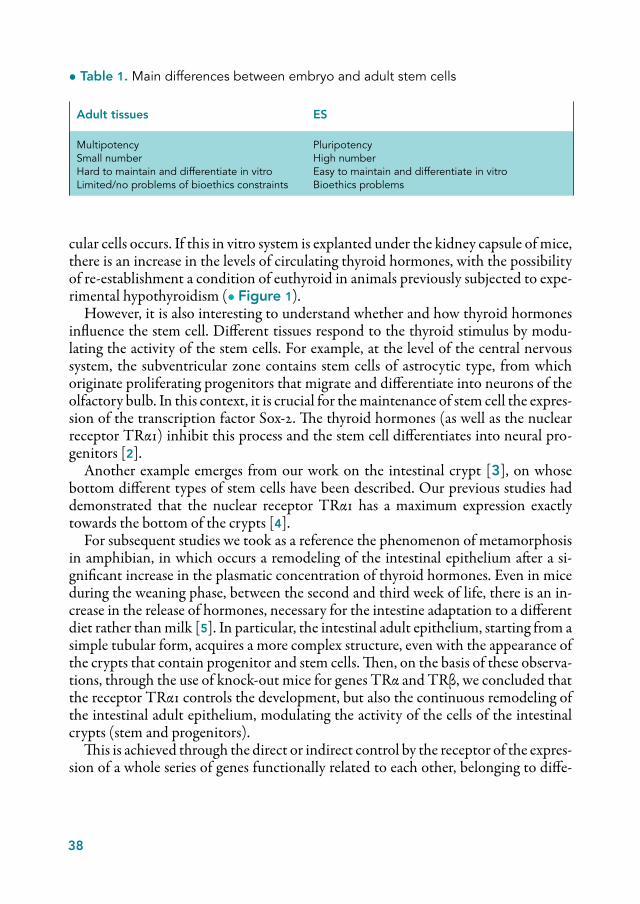
Two properties define the stem cell: the ability to self-renew, i.e. to generate a cell identical to itself and the ability to generate differentiated cells. There are two types of stem cells. Embryonic cells can be recovered in large quantities from the blastocyst and once cultured, they maintain the ability to self-renovate. They are defined pluri-potent, as they are capable of giving rise, if properly stimulated, to any type of diffe-rentiated cell. Cells are easy to handle in vitro, but their use requires a number of bio-ethical issues. The situation is different for somatic stem cells who, instead, are present in tissues in relatively small quantities and are characterized by multipotency, i.e. they have a limited differentiation capacity, relative to the tissue of origin (• Table 1).
Precondition for the stem cell to be that is the presence of the so-called “niche”, a collection of cells and proteins that surround the stem cell itself and form a conducive environment so that it may divide symmetrically or asymmetrically, depending on the case and in function of the stimuli to which is subjected.
The current interest in stem cells comes in part from the desire to give answers to questions related to their physiology, within fundamental research. On the other hand, stem cells represent an attractive target in the scenario of regenerative medicine because they have the potential to differentiate into any tissue of the body.
In this regard, it is interesting the work done in the laboratory of Professor Costa-gliola in Brussels [1], who attempted to recreate the thyroid follicles from undifferen-tiated stem cellular. For the development of the thyroid, it is essential the co-expres-sion of two transcription factors: NKX 2.1 and Pax8. If in vitro stem cells are properly stimulated so that express both the above factors, the differentiation in thyroid folli-
The thyroid hormone in normal and stem cell Michelina Plateroti
Centre de Génétique et de Physiologie Moléculaire et Cellulaire, Université Claude Bernard Lyon 1
38
cular cells occurs. If this in vitro system is explanted under the kidney capsule of mice, there is an increase in the levels of circulating thyroid hormones, with the possibility of re-establishment a condition of euthyroid in animals previously subjected to expe-rimental hypothyroidism (• Figure 1).
However, it is also interesting to understand whether and how thyroid hormones influence the stem cell. Different tissues respond to the thyroid stimulus by modu-lating the activity of the stem cells. For example, at the level of the central nervous system, the subventricular zone contains stem cells of astrocytic type, from which originate proliferating progenitors that migrate and differentiate into neurons of the olfactory bulb. In this context, it is crucial for the maintenance of stem cell the expres-sion of the transcription factor Sox-2. The thyroid hormones (as well as the nuclear receptor TRα1) inhibit this process and the stem cell differentiates into neural pro-genitors [2].
Another example emerges from our work on the intestinal crypt [3], on whose bottom different types of stem cells have been described. Our previous studies had demonstrated that the nuclear receptor TRα1 has a maximum expression exactly towards the bottom of the crypts [4].
For subsequent studies we took as a reference the phenomenon of metamorphosis in amphibian, in which occurs a remodeling of the intestinal epithelium after a si-gnificant increase in the plasmatic concentration of thyroid hormones. Even in mice during the weaning phase, between the second and third week of life, there is an in-crease in the release of hormones, necessary for the intestine adaptation to a different diet rather than milk [5]. In particular, the intestinal adult epithelium, starting from a simple tubular form, acquires a more complex structure, even with the appearance of the crypts that contain progenitor and stem cells. Then, on the basis of these observa-tions, through the use of knock-out mice for genes TRα and TRβ, we concluded that the receptor TRα1 controls the development, but also the continuous remodeling of the intestinal adult epithelium, modulating the activity of the cells of the intestinal crypts (stem and progenitors).
This is achieved through the direct or indirect control by the receptor of the expres-sion of a whole series of genes functionally related to each other, belonging to diffe-
• Table 1. Main differences between embryo and adult stem cells
Adult tissues ES
MultipotencySmall numberHard to maintain and differentiate in vitroLimited/no problems of bioethics constraints
PluripotencyHigh numberEasy to maintain and differentiate in vitroBioethics problems

39
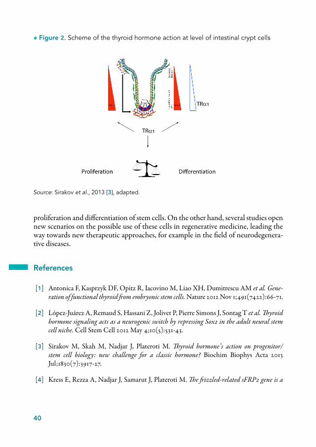
rent pathways of signaliling of the cell (Wint, Notch, c-Fos) involved in the regulation of proliferation and differentiation of the intestinal crypts cells (• Figure 2).
Subsequently, through other studies we have made it clear that the action of thyroid hormone involves in particular the stem cells of the crypts and not just the progeni-tors, and that TRα1 receptor expression is also essential for the regeneration of the epithelium subsequent to intestinal damage; in knock-out mice for the gene TRα1, in fact, the intestinal epithelium was incapable of dealing with the damage caused by massive doses of radiation [6].
The receptor TRα1 also has an important action in vitro on the growth of orga-noids cell, inducing a progressive increase in complexity through a direct monitoring of the activity of stem cells. In fact, in absence of receptor activity there is a conside-rable delay in this development process [7].
Our future goals are aimed at a better understanding of the described phenomena, possibly recreating the same conditions in animals of different genotype. It will also be interesting to evaluate the effects, on the contrary, of an over-repression of TRα1 receptor on the same processes.
In conclusion, there are several demonstrations, both in vivo and in vitro, of the important role of thyroid hormone and TRα1 receptor in controlling the activity of
Source: Antonica et al., 2012 [1], adapted.
Dox rhTSHNkx2.1-Pax8
Day 0 Day 4 Day 7 Day 22 cells
Kidney subcapsulartransplantation
4 weeks 4 weeksHypothyroidism Analysis[131-l]
i.p. injection
Functional thyroidProduction of TH
Rescue of an induced hypothyroidism
• Figure 1. Experimental protocol to evaluate the efficiency of thyroid follicles obtained from embryo stem cells
40
proliferation and differentiation of stem cells. On the other hand, several studies open new scenarios on the possible use of these cells in regenerative medicine, leading the way towards new therapeutic approaches, for example in the field of neurodegenera-tive diseases.
References
[1] Antonica F, Kasprzyk DF, Opitz R, Iacovino M, Liao XH, Dumitrescu AM et al. Gene-ration of functional thyroid from embryonic stem cells. Nature 2012 Nov 1;491(7422):66-71.
[2] López-Juárez A, Remaud S, Hassani Z, Jolivet P, Pierre Simons J, Sontag T et al. Thyroid hormone signaling acts as a neurogenic switch by repressing Sox2 in the adult neural stem cell niche. Cell Stem Cell 2012 May 4;10(5):531-43.
[3] Sirakov M, Skah M, Nadjar J, Plateroti M. Thyroid hormone’s action on progenitor/stem cell biology: new challenge for a classic hormone? Biochim Biophys Acta 2013 Jul;1830(7):3917-27.
[4] Kress E, Rezza A, Nadjar J, Samarut J, Plateroti M. The frizzled-related sFRP2 gene is a
• Figure 2. Scheme of the thyroid hormone action at level of intestinal crypt cells
Source: Sirakov et al., 2013 [3], adapted.
41
target of thyroid hormone receptor alpha1 and activates beta-catenin signaling in mouse intestine. J Biol Chem 2009;284:1234-41.
[5] Sirakov M, Plateroti M. The thyroid hormones and their nuclear receptors in the gut: from developmental biology to cancer. Biochim Biophys Acta 2011 Aug;1812(8):938-46.
[6] Kress E, Rezza A, Nadjar J, Samarut J, Plateroti M. The thyroid hormone receptor-alpha (TRalpha) gene encoding TRalpha1 controls deoxyribonucleic acid damage-induced tissue repair. Mol Endocrinol 2008 Jan;22(1):47-55.
[7] Sato T, Vries RG, Snippert HJ, van de Wetering M, Barker N, Stange DE et al. Single Lgr5 stem cells build crypt-villus structures in vitro without a mesenchymal niche. Nature 2009 May 14;459(7244):262-5.
42
The issue of combination therapy T3+T4 is quite controversial, as shown by recent reviews [1-3]. The important work of Gullo et al. [4] shows that the only therapy with levothyroxine is not able to guarantee euthyroidism in all patients without thyroid: in fact, approximately 15% of these patients supplemented with LT4 alone (but about 17% in the age group over 70 years) had a FT3 lower than in euthyroid subjects (used as normal controls). In addition, values of FT3/FT4 ratio relatively lower of the con-trol group were present in approximately 30% of patients without thyroid treated with single LT4.
In a study conducted in Leiden, the Netherlands, [5] were enrolled about 600 pa-tients 85 years old, 85% of which were euthyroid. Patients were followed for 4 years, monitoring the values of FT3, FT4 and TSH. Already in the process of recruitment was observed a direct and significant correlation between serum levels of type FT3 and well-being reported from the patient. In particular, to values relatively higher of hormone corresponded a better quality of life, lower incidence of depression and a better global cognitive ability. Similar results were observed during follow-up. In ad-dition, during follow-up, the correlation with mortality could be assessed. It was seen that the FT3 serum levels were inversely correlated with mortality: the lower was the serum FT3, the higher the mortality from all causes. Even the FT3/FT4 relationship proved to be predictive of a better outcome, being it 0.25 in the fourth year survivors vs. 0.23 in died patients. An exactly opposite correlation was observed with FT4 levels at enrollment and during follow-up.
Although monotherapy with LT4 is considered the recommended one for hypo-thyroidism [3], about 10% of hypothyroid patients continue to complain of residual hypothyroidism disorders despite adequate dosage of LT4 [1]. The important study of Panicker et al. [6] reveal that only the group of patients with a particular poly-
What is the rationale for a combined therapy with hormones T3 and T4?Salvatore Benvenga
Department of Clinical and Experimental Medicine, University of Messina, Master of Endocrinology of Infancy, Adolescence and Women, University of Messina; Interdepartmental Program in Molecular Endocrinology Clinical & Endocrine Health of Women, Policlinico G. Martino, Messina
43
morphism of deiodinase type 2 (i.e. carriers of the polymorphism genotype CC of rs225014) benefited from combined therapy LT3+LT4. These carriers, already at the start (pre- treatment) had the worst values of psychological well-being than the carri-ers of the other two polymorphic variants (genotype TT and genotype TC).
In the study by De Jong et al. [7] data emerge that should make us reflect: in the period between 2005 and 2011 there has been an increase of percentage of cases in thyroid replacement therapy among the Dutch population. In particular, there has been a 53% increase of patients in monotherapy with levothyroxine, but surprising data are related to the 67% increase of the cases treated with T3+T4 combination therapy and 36% with T3 monotherapy. Another trend that emerges from this study regards the increase of the prescriptions, for all the above types of treatment, made by general practitioners, compared to a relative decrease of prescriptions by specialists.
A situation predisposing to the fact that different hypothyroid patients do not re-spond to the same dose of LT4, becoming euthyroid, and that residuals hypothyroid disorders may persist (variable from patient to patient) is that, given the systemic action of thyroid hormones, the clinical picture of hypothyroidism is also systemic. Furthermore, the thyroid hormonal signal is extremely complex and not compara-ble with other hormonal signals. Indeed, in addition to the fact that there are three deiodinase enzymes in charge of activation/inactivation of the thyroid hormone sig-nal, there are numerous plasmatic transport proteins for thyroid hormones, there are many transporters on the plasma membrane to facilitate the entry/exit of thyroid hormones and numerous are also the isoforms of nuclear receptors of thyroid hor-mones (and different peripheral tissues have a peculiar distribution of these isoforms). And yet, over the main action (the one mediated by nuclear receptors, and known as genomic action), there is one that is not mediated by nuclear receptors (the so-called non-genomic action), with signaling pathways common between T4 and T3 and oth-er routes specific for one or the other hormone. The field of the combined therapy T3+T4 is rather controversial and reflects the complexity of the thyroid hormone pathway in the body. The situation is made more complicated by the existence of a wide range of nuclear receptors for thyroid hormones which have different degrees of expression in different tissues as well as a variable affinity for the hormones them-selves. This explains why a certain amount of T3 in the different cells of the organism does not involve the same degree of hyperthyroidism.
To illustrate the concept of non-correspondence between plasma levels and tissue concentrations of thyroid hormone comes in handy a study on polymorphisms of trans-porters [8], which shows that, in spite of the reduction in intracellular hormone uptake favored by some polymorphic variants compared to the wild type transporter, there was no significant change in circulating hormone concentrations. The work of Liao et al. [9], carried out on different types of knock-out, even triple, for the main carrier of T3, the MCT-8, and for deiodinase (D1, D2), has emphasized that in three types of knock-out mice (MCT-8, and MCT8/D2 MCT8/D1/D2) there was an increase in circulating

44
T3. However, in the same three genotypes of knock-out mice, the tissue concentration of T3 was systematically increased in the liver, but systematically reduced in the cerebral cortex. In practice, as demonstrated also by the expression of some liver genes and some brain genes, there was a condition of hyperthyroidism in the liver, but, in contrast, a con-dition of hypothyroidism in the cerebral cortex. Thus, the biochemical hyperthyroidism (increased circulating concentrations of FT3 and/or FT4) does not necessarily mean universal tissue hyperthyroidism. Even the Italian study of Sampaolo et al. [10] shows how poorly conclusive is the concentration of circulating thyroid hormones: the plasma levels assessed in a group of patients suffering from Alzheimer’s disease were similar to those observed in the controls, on the contrary, the concentration of T3 in the liquor was about three times lower than in healthy subjects.
In this respect, precursors have been the fundamental studies of the Madrid group Escobar-Morreale et al. on mice made hypothyroid and then variously supplemented in order to make them euthyroid [11-13]. While the increase of the plasmatic T3 con-centration (following the increase in the dose of T3 infused) results in an equivalent increase in the intra-tissue concentration of T3 for various tissues (even for the ovaries, the adrenal cortex and the lungs, the intra-tissue increase is higher than the plasmatic one), for other tissues (pituitary, cerebellum and, especially, for cerebral cortex and brown adipose tissue), the increase of intra-tissue concentration of T3 is much lower than the expected one based on the increase of plasmatic concentration. In the cere-bral cortex, the infusion of T3 (and not the one of T4) is the one that does increase the T3 tissue concentration in a dose-dependent manner. And again, the same posol-ogy combination of T4+T3 does not result in concentrations of T4 and T3 which are exactly identical in the various tissues; and, in effect, a tissue concentration of both T3 and T4 fully corresponding to the normal range of a given tissue is achieved with a posology combination of T4+T3 that is different from the one which provides a tissue concentration of both T4 and T3 fully corresponding to the normal range of another tissue. The final practical conclusion of these studies of Escobar-Morreale et al. is that the normalization of the intracellular content of both T4 and, especially, T3, in all tissues of hypothyroid rats, requires the combined administration of T4 and T3.
Returning to the human species, regarding the various parameters of neuropsycho-logical wellness, investigated by the works that have addressed the possible beneficial effect of combination therapy compared with monotherapy with T4, it seems that there is a certain benefit of the combination T4+T3 in depression.
In psychiatric literature, in fact, there is evidence of how the administration of T3, as monotherapy or as an additive to antidepressant therapy or as an accelerator in initial as-sociation with drugs, is effective in improving the outcome of patients being treated with antidepressants. In most cases, T3 was administered at a dose between 5 and 50 g/day. Other studies have evidence of the greater effectiveness of T3 compared to T4 for this purpose [14], as well as of a better response in females than males [15, 16].
However, according to U.S. guidelines, there is insufficient evidence to support the

45
use of thyroid hormones in the treatment of depression in euthyroid patients [3].This mention of the “antidepressant” use of T3 leads to recall a particular mode of
action of thyroid hormones, a mode which is seldom or never mentioned in the liter-ature and textbooks/treatises of endocrinology. The studies of Dratman and Gordon [17] show, in a very convincing way, how thyroid hormones work as neurotransmit-ters. In particular, the structure of the central nervous system richer deiodinase type 2 (D2) is the locus coeruleus, localized in the sub-ependymal area. This site has a high concentration of tyrosine-hydroxylase, an enzyme that converts tyrosine into norep-inephrine. Incidentally, it is not superfluous to recall that even T3 and T4 originate from tyrosine; however, unlike the norepinephrine, are iodine molecule. It is impor-tant to emphasize the role played as powerful activator of D2 by norepinephrine and, on the other hand, the ability of thyroid hormones to stimulate strongly the expres-sion of the adrenergic receptors in the brain. So, although the reasons are still un-known, nature has made sure that the metabolic pathways of T3 and norepinephrine are closely related to each other, that the two neurotransmitters travel along the same axons and interface post-synaptically to transmit signals.
An interesting aspect that emerges from the literature on the neuro-transmissal role of the thyroid hormones T3 and T4 is that they have even different effects. In the clas-sical field of genomic action of thyroid hormones is well known, however, that T3 and T4 act in the same direction, although the T3 is more potent than T4. Caria et al. [18] evaluated the effects of the two hormones separately on the frequency of neuronal dis-charge induced by norepinephrine in cultured hippocampus. When the noradrenergic stimulation was preceded by the infusion of T4, it was noted a depressive effect on the frequency of neuronal discharge, on the contrary, when the noradrenergic stimulation was preceded by the infusion of T3, we obtained a significant increase of neuronal dis-charge (• Figure 1). Other works have also demonstrated the different effect of the two hormones on glutamatergic [19] and GABAergic neurotransmission, both phasic and tonic [20]. Taking into consideration the above background (thyroid hormones as neurotransmitters), found that there was no electrophysiological studies on the motor cerebral cortex in hypothyroid patients (and we just said that the motor cortex is one of the main targets of noradrenergic neurons located in the locus coeruleus) and using the non-invasive transcranial magnetic stimulation we wanted to test parameters of human motor cortex excitability in adult patients with acquired hypothyroidism before and af-ter replacement therapy with LT4 [21]. Trans-cranial magnetic stimulation is a non-in-vasive, painless, safe and accurate technique to induce electrical potentials in brain through electromagnetic induction. The electrical impulse generated in brain (in our case specifically, the motor cortex) is registered with motor-evoked potential in a distal skeletal contra-lateral muscle (in our study the left motor cortex was stimulated and it was recorded in muscle called the first dorsal interosseous of the right index finger).
We carefully selected, from the of neurological and neuropsychiatric point of view, 11 patients with primary overt hypothyroidism, which were evaluated before replace-

46
Spik
es/s 8
4
0
1 min
4TEN
NE
NE
T3
EN3TEN
EN4T
NE
1 min
1 min
Spik
es/s 2
3
1
0
Spik
es/s 2
3
1
0
Spik
es/s 4
6
2
0
Spik
es/s 4
6
2
0
(A)
(B)
(C)
• Figure 1. Non-genomic action of thyroid hormones: effects on excitability of hippocampal neurons
Source: Carla et al., 2009 [18], adapted.
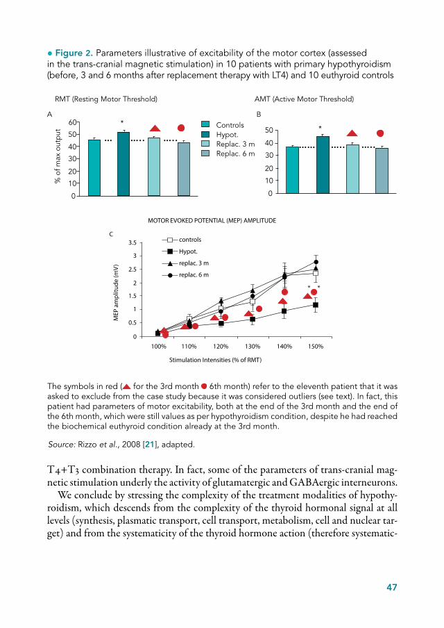
ment with levothyroxine monotherapy and at the end of the 3rd and 6th month post- therapy. As controls, were recruited as many healthy subjects, matched for various features. In the patients group, all reached the euthyroid condition (normalization of TSH and FT4) already at the end of the 3rd month of therapy. The results, summa-rized in • Figure 2, for three illustrative parameters, show normalization of parame-ters both at the end of the 3rd month and at the end of the 6th month of therapy with levothyroxine.
However, it must be said that, in the evaluation phase of the work, it was asked to exclude from the case studies a patient who, despite having reached the biochemical euthyroid, still had abnormal values (as per overt hypothyroidism) of the various parameters of trans-cranial magnetic stimulation. In retrospect, this subject must be interpreted as a patient in which, despite the normalization of circulating thyroid hormones, the biological action of the hormones on the nervous districts under-lying those parameters was still insufficient. Likely in this patient those parameters would have changed in euthyroid sense if the T4 monotherapy had been switched to
47
T4+T3 combination therapy. In fact, some of the parameters of trans-cranial mag-netic stimulation underly the activity of glutamatergic and GABAergic interneurons.
We conclude by stressing the complexity of the treatment modalities of hypothy-roidism, which descends from the complexity of the thyroid hormonal signal at all levels (synthesis, plasmatic transport, cell transport, metabolism, cell and nuclear tar-get) and from the systematicity of the thyroid hormone action (therefore systematic-
60
50
40
30
20
10
0
% o
f max
out
put
AControlsHypot.Replac. 3 mReplac. 6 m
*50
40
30
20
10
0
B
*
RMT (Resting Motor Threshold) AMT (Active Motor Threshold)
• Figure 2. Parameters illustrative of excitability of the motor cortex (assessed in the trans- cranial magnetic stimulation) in 10 patients with primary hypothyroidism (before, 3 and 6 months after replacement therapy with LT4) and 10 euthyroid controls
Source: Rizzo et al., 2008 [21], adapted.
The symbols in red ( for the 3rd month 6th month) refer to the eleventh patient that it was asked to exclude from the case study because it was considered outliers (see text). In fact, this patient had parameters of motor excitability, both at the end of the 3rd month and the end of the 6th month, which were still values as per hypothyroidism condition, despite he had reached the biochemical euthyroid condition already at the 3rd month.
MOTOR EVOKED POTENTIAL (MEP) AMPLITUDE
0
0.5
1
1.5
2
2.5
3
3.5
100% 110% 120% 130% 140% 150%
Stimulation Intensities (% of RMT)
controls
Hypot.
replac. 3 m
replac. 6 m
MEP
am
plitu
de (m
V)
* *
C
48
ity of the clinical manifestations of hypothyroidism). It is therefore readily apparent that the universe of hypothyroid patients cannot be released by 100% of hypothyroid-ism disorders only with the use of a single hormone (indeed, pro hormone, namely the T4), and not even with administration of the same in a single daily dose. It arises, therefore, the need to be able to identify those patients that may take greater benefit from the combination therapy of T4+T3.
References
[1] Wiersinga WM, Duntas L, Fadeyev V, Nygaard B, Vanderpump MPJ. 2012 ETA Guidelines: The use of L-T4 + L-T3 in the treatment of hypothyroidism. Eur Thyroid J 2012;1:55-71.
[2] Biondi B, Wartofsky L. Combination treatment with T4 and T3: toward personalized re-placement therapy in hypothyroidism? J Clin Endocrinol Metab 2012 Jul;97(7):2256-71.
[3] Garber JR, Cobin RH, Gharib H, Hennessey JV, Klein I, Mechanick JI et al.; American Association of Clinical Endocrinologists and American Thyroid Association Taskforce on Hypothyroidism in Adults. Clinical practice guidelines for hypothyroidism in adults: cosponsored by the American Association of Clinical Endocrinologists and the American Thyroid Association. Endocr Pract 2012 Nov-Dec;18(6):988-1028.
[4] Gullo D, Latina A, Frasca F, Le Moli R, Pellegriti G, Vigneri R. Levothyroxine monother-apy cannot guarantee euthyrodism in all athyreotic patients. PlosOne 2011;6(8):e22552.
[5] Gussekloo J, van Exel E, de Craen AJ, Meinders AE, Frölich M, Westendorp RG. Thy-roid status, disability and cognitive function, and survival in old age. JAMA 2004 Dec 1;292(21):2591-9.
[6] Panicker V, Saravanan P, Vaidya B, Evans J, Hattersley AT, Frayling TM, Dayan CM. Common variation in the DIO2 gene predicts baseline psychological well-being and re-sponse to combination thyroxine plus triiodothyronine therapy in hypothyroid patients. JCEM 2009;94:1623-9.
[7] De Jong NW, Baljet GM. Use of T4, T4 +T3, and T3 in the Dutch population in the period 2005-2011. Eur Thyr J 2012;1:135-6.
[8] van der Deure WM, Peeters RP, Visser TJ. Molecular aspects of thyroid hormone trans-porters, including MCT8, MCT10, and OATPs, and the effects of genetic variation in these transporters. J Mol Endocrinol 2010;44:1-11.

49
[9] Liao XH, Di Cosmo C, Dumitrescu AM, Hernandez A, Van Sande J, St Germain DL et al. Distinct roles of deiodinases on the phenotype of Mct8 defect: comparison of eight differ-ent mouse genotypes. Endocrinology 2011;152:1180-91.
[10] Sampaolo S, Campos-Barros A, Mazziotti G, Carlomagno S, Sannino V, Amato G et al. Increased cerebrospinal fluid levels of 3,3’,5’-triiodothyronine in patients with Alzheimer’s disease. J Clin Endocrinol Metab 2005 Jan;90(1):198-202.
[11] Escobar-Morreale HF, Obregón MJ, del Rey FE, de Escobar GM. Replacement therapy for hypothyroidism with thyroxine alone does not ensure euthyroidism in all tissues, as stud-ied in thyroidectomized rats. J Clin Invest 1995 Dec;96(6):2828-38.
[12] Escobar-Morreale HF, del Rey FE, Obregón MJ, de Escobar GM. Only the combined treatment with thyroxine and triiodothyronine ensures euthyroidism in all tissues of the thyroidectomized rat. Endocrinology 1996 Jun;137(6):2490-502.
[13] Escobar-Morreale HF, Obregón MJ, del Rey FE, de Escobar GM. Tissue-specific patterns of changes in 3,5,3’-triiodo-L-thyronine concentrations in thyroidectomized rats infused with increasing doses of the hormone. Which are the regulatory mechanisms? Biochimie 1999 May;81(5):453-62.
[14] Joffe RT, Singer W. A comparison of triiodothyronine and thyroxine in the potentiation of tricyclic antidepressants. Psychiatry Res 1990 Jun;32(3):241-51.
[15] Agid O, Lerer B. Algorithm-based treatment of major depression in an outpatient clinic: clinical correlates of response to a specific serotonin reuptake inhibitor and to triiodothyro-nine augmentation. Int J Neuropsychopharmacol 2003 Mar;6(1):41-9.
[16] Altshuler LL, Bauer M, Frye MA, Gitlin MJ, Mintz J, Szuba MP et al. Does thyroid sup-plementation accelerate tricyclic antidepressant response? A review and meta-analysis of the literature. Am J Psychiatry 2001 Oct;158(10):1617-22.
[17] Dratman MB, Gordon JT. Thyroid hormones as neurotransmitters. Thyroid 1996 Dec;6(6):639-47.
[18] Caria MA, Dratman MB, Kow LM, Mameli O, Pavlides C. Thyroid hormone action: nongenomic modulation of neuronal excitability in the hippocampus. J Neuroendocrinol 2009;21:98-107.
[19] Losi G, Garzon G, Puia G. Nongenomic regulation of glutamatergic neurotransmission in hippocampus by thyroid hormones. Neuroscience 2008;151:155-63.

50
[20] Puia G, Losi G. Thyroid hormones modulate GABA(A) receptor-mediated currents in hip-pocampal neurons. Neuropharmacology 2011 Jun;60(7-8):1254-61.
[21] Rizzo V, Crupi D, Bagnato S, Quartarone A, Benvenga S, Bartolone L et al. Neural response to transcranial magnetic stimulation in adult hypothyroidism and effect of replace-ment treatment. J Neurol Sci 2008 Mar 15;266(1-2):38-43.

51
In physiological conditions, the total amount of T3 in the body is given by the algeb-raic sum of the hormone produced and secreted by the thyroid, the T3 coming from intra-thyroidal and peripheral conversion of T4, and, finally, by the catabolism and excretion of the active hormone. In conditions of replacement therapy, namely in the absence of an endogenous T3 production, the available quantity of the hormone is only guaranteed by the mechanism of peripheral conversion of T4.
The current standard of care of hypothyroidism is the monotherapy with levothy-roxine, of which we know the pharmacokinetic mechanisms, methods of administra-tion and therapeutic targets.
With regard to liothyronine (the synthetic formulation of T3), the pharmacokinet-ic and pharmacodynamic mechanisms are less clear because the data currently availa-ble have been obtained through studies with obsolete methods and non- physiologi-cal situations.
In order to characterize and understand properly the pharmacokinetics of therapy with T3 and its metabolic effects, in our studies we have tried to eliminate some bias which compromised the results of previous works enrolling, in the first place, well standardized groups of patients.
In a double-blind study (2011) [1, 2], we enrolled 14 hypothyroid volunteers, 12 of whom had undergone a total thyroidectomy, one had undergone radioiodine therapy for Basedow’ disease, and one had a diagnosis of Hashimoto’s thyroiditis. Patients with residual glandular >1 cm3 were subjected to study with scintigraphic uptake of radioiodine, to rule out a residual endogenous hormone production.
A prerequisite to be able to participate in the study was that each patient reaches the euthyroid state at pituitary level, by treatment with levothyroxine or with liothy-ronine, lasting at least one month. At the end of the adjustment period of therapy the
Clinical studies and benefits of the therapy with T3 Francesco Saverio Celi
Diabetes, Endocrinology and Obesity Branch, National Institute of Diabetes, Digestive and Kidney Diseases, National Institutes of Health, Bethesda, Maryland, U.S.A.

52
dose of levothyroxine was equal to 1.6 g/kg vs. a dose approximately three times less of liothyronine. Both drugs were taken with three daily doses.
Between the two groups of patients TSH values at baseline were fully overlap-ping, as well as the response to TRH test [3]. Even during 24 hours TSH did not show significant differences. Our patients were thus perfectly identical and so we had achieved our target, i.e. the euthyroid condition at pituitary level. Moreover, we ob-served a significant difference in the levels of plasmatic T3 between patients treated with levothyroxine and those treated liothyronine. In fact, while in patients treated with liothyronine we found average levels of the hormone within the normal range in patients treated only with levothyroxine the T3 was maintained on average at the lower limits of the standard (• Table 1). Our interest however, rather than to the hor-monal levels in plasma, was focused on the study of the metabolic effects of hormone action in the two cases under consideration. In fact, we found significant differences between the two groups of patients: in patients treated with liothyronine there had been a significant reduction in the amount of total cholesterol and LDL, and, in a marginal way, even in HDL (• Table 2). We have observed, moreover, a increase in serum levels of SHBG and a reduction in the average weight of about 2 kg, in absence of dietary modifications. No difference between the two groups was observed within the cardiovascular parameters (systolic blood pressure, diastolic blood pressure, heart rate and echo-cardiographic parameters), the basal and glucose metabolism.
In another study that is not yet complete and from which we present preliminary data we enrolled patients in hypothyroidism after suspension of replacement therapy with levothyroxine, waiting to be subjected to nuclear medicine procedures post-thy-roidectomy for thyroid cancer. In the 30 days prior to the suspension of treatment was performed a shift from levothyroxine to liothyronine in suppressive mode and in
• Table 1. Values of thyroid hormones and TSH in terms of drug-equivalence between levothyroxine (LT4) and liothyronine (LT3)
LT4 LT3 p
TSH (mU/L)± SD0.4-4.0
1.30.79
1.40.79
0.674
T3 (ng/dL)± SD90-215
92.919.0
172.088.2
0.003
fT4 (ng/dL)± SD0.8-1.5
1.570.3
< 0.3 < 0.0001
Source: Celi et al., 2011 [1] adapted.

53
divided doses. The therapeutic target, of course, was represented by the suppression of TSH. We therefore analyzed the pharmacokinetics of the last dose with sequen-tial doses of T3 and TSH. The latter, immediately before discontinuation of therapy, not surprisingly resulted suppressed, while the average T3 at baseline was located at the higher limits of the normal range compared to the mean values measured during treatment with levothyroxine. A preliminary data analysis indicates that the apparent half-life of the drug appeared around 27 hours, with a half-life of distribution of 3-4 hours and half-life of elimination of approximately two days. The administration frac-tionated in three daily doses, also assured normal plasmatic T3 levels in a constant and prolonged manner; in contrast, giving the entire dose in a single time or in two times would have favored the onset of peaks and important oscillations of the hormone concentration in circulation.
These data indicate that the treatment with liothyronine in the correct dosage per weight and per initial dose of levothyroxine in multiple administrations, it ensures an adequate TSH suppression with T3 levels within normal limits. Discontinuation of the therapy ensures TSH levels adequate to radio-metabolic treatment for a period of about two weeks, with minimal symptoms of hypothyroid. The study showed, in addition, that the pharmacokinetics of liothyronine comprises two components: dis-tribution and elimination. An administration in single or double dose of the drug would result in prolonged periods of over-and under-dosing. These results are empir-ical data on which to set up long-term controlled studies regarding combined levo-thyroxine-liothyronine therapy, which identify clinically valid endpoint in order to evaluate the metabolic effects of this treatment modality.
• Table 2. Lipidic metabolism under conditions of drug-equivalence of levothyroxine (LT4) and liothyronine (LT3)
LT4 LT3 p
Total cholesterol(mg/dL)± SD
195.925.9
173.927.7
0.002
HDL (mg/dL)± SD
63.015.0
57.511.7
0.067
LDL (mg/dL)± SD
122.625.2
106.227.7
0.002
Triglycerides(mg/dL)± SD
78.230.8
78.828.6
0.937
Source: Celi et al., 2011 [1] adapted.

54
References
[1] Celi FS, Zemskova M, Linderman JD, Smith S, Drinkard B, Sachdev V et al. Metabolic effects of liothyronine therapy in hypothyroidism: a randomized, double-blind, crossover tri-al of liothyronine versus levothyroxine. J Clin Endocrinol Metab 2011 Nov;96(11):3466-74.
[2] Yavuz S, Linderman JD, Smith S, Zhao X, Pucino F, Celi FS. The dynamic pituitary response to escalating-dose TRH stimulation test in hypothyroid patients treated with liothyronine or levothyroxine replacement therapy. J Clin Endocrinol Metab 2013 May;98(5):E862-6.
[3] Celi FS, Zemskova M, Linderman JD, Babar NI, Skarulis MC, Csako G et al. The pharmacodynamic equivalence of levothyroxine and liothyronine: a randomized, dou-ble blind, cross-over study in thyroidectomized patients. Clin Endocrinol (Oxf ) 2010 May;72(5):709-15.

55
In literature, the topic “low T3 syndrome” is controversial for at least three reasons: the complexity of the pathophysiology, and, consequently, of the treatment; the crit-icality of the clinical conditions of the patients that involves the intervention of nu-merous confounding factors (comorbidities, medications, etc.), the discordance of treatment studies currently in the literature.
For nearly forty years we know that, in course of fasting or of acute or chronic dis-eases, may occur alterations of the thyroid homeostasis, a condition that in literature does not present an univocal name: we speak, in most cases of “low T3 syndrome” or “euthyroid sick syndrome”, but the most suitable definition is probably that of “Not thyroidal Illness syndrome” (NTIS), as to emphasize that the patient is normal from the point of view of thyroid function and that it is a secondary response of the gland to a condition of systemic disease.
The reduction in the plasma concentration of T3, in fact, is only one of the phe-nomena that occur in the context of a clinical systemic picture much more complex. Specifically, the deficit of the active hormone, resulting from the inhibition of dei-odination of T4, is a very early event in the involvement of the hypothalamic-pituit-ary-thyroid axis in response to situations of acute or chronic stress. With the worsening or the prolongation of the underlying disease, in fact, there is a progressive extension of the functional block to the whole axis, which results, in most serious cases, in a re-duction even of the circulating levels of T4 associated with values of TSH inappropri-ately normal. This last data has been interpreted, by some, as an index of hypothalamic alteration, hypothesis supported by the concomitant alteration of other hormones such as GH or cortisol. Furthermore, a normal thyroid function is restored not only in response to TRH infusion, but also by co-administration of other hypothalamic peptides such as, for example, the secretagogues GH.
A role for T3 in the “low T3 syndrome”?Antonio Mancini, Chantal Di Segni, Sebastiano Raimondo,Giuseppe Maria Corbo*, Alfredo Pontecorvi
Department of Medical Sciences, Department of Endocrinology, Catholic University, Rome*Department of Medical Sciences, Department of Pneumology, Catholic University, Rome

56
Other authors [1] have attempted to justify the normality of TSH in these patients as a result of normal deiodination activities at central level but not at peripheral level. All the mechanisms downstream of hormone secretion, implicated in the transmis-sion of the thyroid signal were analyzed in order to better understand the physio-pathology of the low T3 syndrome. In literature there are many conflicting studies on plasmatic transport proteins, on receptors of thyroid hormones, on intracellular transporters, which do not allow drawing a univocal picture of this clinical condition.
Among the mechanisms involved in the pathogenesis of the syndrome, it has re-cently been revealed a significant increase of Nuclear Factor-kβ (NFkβ) in response to the massive release of cytokines such as Interleukin-6 (IL-6) and Tumor Necrosis Factor-α (TNF-α) [2]. Various studies have shown that the NFkβ, mitigating the ex-pression of deiodinase type 1, is able to reduce the circulating levels of T3 in both rats and humans.
To complicate the picture of the syndrome, which makes more difficult to attempt understanding the pathogenic mechanisms, is the complexity of the condition of the patient with “low T3 syndrome”, which comes from the underlying pathology, acute or chronic, from the state of malnutrition that often accompanies it, from the wide range of drugs that can interfere with the endocrine axis at various levels. Often, these patients take glucocorticoids, β-blockers or anti-arrhythmics, all drugs that block deiodinase enzymes. Other drugs, such as heparin, alter the hormone binding to plas-ma proteins and still others, such as catecholamines and dopamine, interfere with the release of TSH at pituitary level.
The clinical conditions that frequently accompany the low T3 syndrome are un-doubtedly heart and lung diseases. Followed by major surgery interventions, sepsis, trauma. At the moment, we try desperately to understand if thyroid changes are only a mechanism of adaptation to the underlying disease or, conversely, a pathogenic fac-tor also implicated in the progression and aggravation of the disease. It is important to give an answer to this question, especially in view of the therapeutic approach: if it were a mere form of adaptation does not require any intervention, otherwise, it would be necessary a substitution treatment. In this context, it is important to iden-tify valid indexes of tissue hypothyroidism, able to reflect the thyroid situation at peripheral level, and that allow clarifying dubious situations from the clinical point of view. Coenzyme Q10, an important component of the mitochondrial respiratory chain and a powerful antioxidant, would seem suitable for this purpose [3, 4]: since the eighties we know, in fact, that the levels of this molecule correlate inversely with circulating values of T3 and T4, in a highly significant manner, to the point to “pick up” even subclinical conditions.
Another important biochemical marker of real hypothyroidism at tissue level is represented by oxidative stress, in particular, the hormone deficiency is associated with an increase in oxidizing and a parallel reduction of antioxidants; the replace-ment therapy reverses this condition. In the clinical practice, 20-30% of patients

57
with dilated cardiomyopathy have a low T3 syndrome, with a higher incidence in decompensated forms. There is a significant correlation between low plasma levels of T3 and mortality from heart disease, and it has also been shown an increase in morbidity and mortality due to heart conditions in the form of subclinical hypothy-roidism.
The opportunity to use T3 in cardiac pathology is supported by biological and clinical evidence, we know that thyroid hormones modulate myocardial contractility, increasing, thus, the performance of both systolic and diastolic; inducing coronary vasodilation also they promote myocardial perfusion.
In a pioneering study of Hamilton et al. [5], patients with dilated cardiomyopathy unresponsive to other inotropic agents were treated with intravenous infusion of T3, with amazing effects : an increase in cardiac output, reduction of vascular resistance, reduction of blood pressure and pulmonary capillary wedge pressure (• Figure 1).
In another study of the Pisan physiology school [6], the infusion of T3 in patients with heart disease resulted in a significant improvement in both systolic and diastolic parameters (• Table 1).
Despite this evidence, however, it is impossible to find a unique perspective with which to look at this complex syndrome in the literature.
De Groot, an eminent expert in low T3 syndrome, confirms the beneficial effects of T3 administration from the cardiovascular point of view, in different categories of patients, especially cardiological patients, without however in fact, significant changes in the prognosis [7].
On the other hand, alarming studies demonstrate even an increase in mortality in patients with renal insufficiency; in addition, the fear of cardiologists and endocrinol-ogists about the fact that the administration of T3, modifying the tissue request of oxygen, may, in patients at risk, precipitate an ischemic event limits its use in clinical practice.
In the field of pneumology, are present in the literature numerous discordant stud-ies regarding the association between low T3 syndrome and COPD (chronic obstruc-tive pulmonary disease). According to some authors, in course of this systemic disease would not occur any change in the thyroid function, while others have shown that there is a reduction in circulating hormone proportional to the hypoxia, associated or not to TSH alteration. In a isolated study, it was even observed an increase of T3 in these patients [8].
In our study on 32 patients with COPD [9] (• Table 2) we put in evidence, in fact, alterations in circulating hormone levels compared to healthy controls: the av-erage TSH, even within the normal range was maintained at high limits, the average T3 was lower than normal, while the T4 stood at the lower limits of the reference range. In addition, we have demonstrated in these subjects a significant reduction in antioxidant defenses. Surprisingly, there was a very significant correlation between low levels of circulating T3, then hypothyroidism, and increased oxidative stress, a
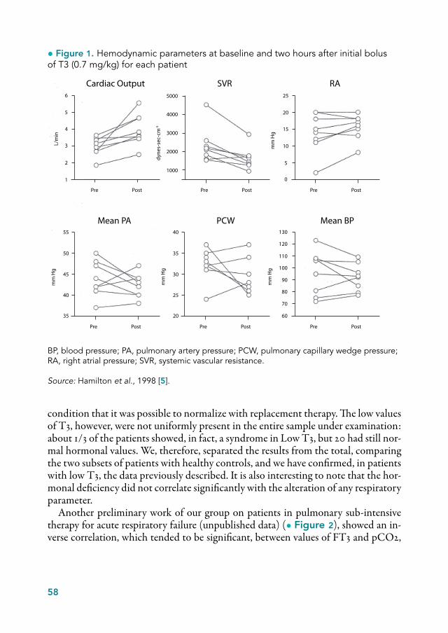
58
condition that it was possible to normalize with replacement therapy. The low values of T3, however, were not uniformly present in the entire sample under examination: about 1/3 of the patients showed, in fact, a syndrome in Low T3, but 20 had still nor-mal hormonal values. We, therefore, separated the results from the total, comparing the two subsets of patients with healthy controls, and we have confirmed, in patients with low T3, the data previously described. It is also interesting to note that the hor-monal deficiency did not correlate significantly with the alteration of any respiratory parameter.
Another preliminary work of our group on patients in pulmonary sub-intensive therapy for acute respiratory failure (unpublished data) (• Figure 2), showed an in-verse correlation, which tended to be significant, between values of FT3 and pCO2,
1
2
3
4
5
6
Pre Post
L/m
in
Cardiac Output SVR RA
1000
2000
3000
4000
5000
dyne
s-se
c-cm
-5
Pre Post Pre Post
0
5
10
15
20
25
Pre Post
35
40
45
50
55
mm
Hg
mm
Hg
mm
Hg
mm
Hg
Pre Post
20
25
30
35
40
Pre Post
60
70
80
90
100
110
120
130
Mean PA PCW Mean BP
• Figure 1. Hemodynamic parameters at baseline and two hours after initial bolus of T3 (0.7 mg/kg) for each patient
BP, blood pressure; PA, pulmonary artery pressure; PCW, pulmonary capillary wedge pressure; RA, right atrial pressure; SVR, systemic vascular resistance.
Source: Hamilton et al., 1998 [5].
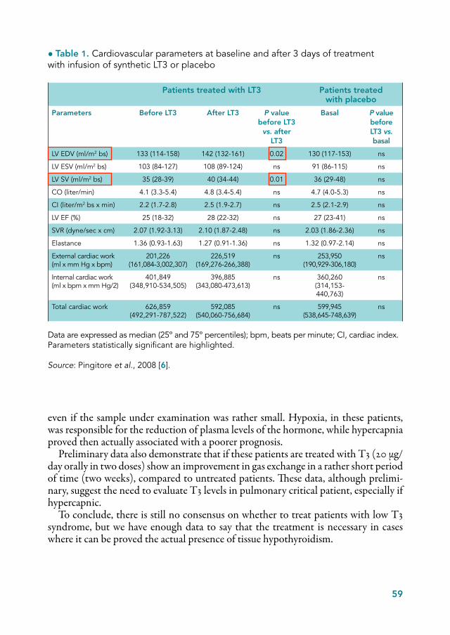
59
even if the sample under examination was rather small. Hypoxia, in these patients, was responsible for the reduction of plasma levels of the hormone, while hypercapnia proved then actually associated with a poorer prognosis.
Preliminary data also demonstrate that if these patients are treated with T3 (20 µg/day orally in two doses) show an improvement in gas exchange in a rather short period of time (two weeks), compared to untreated patients. These data, although prelimi-nary, suggest the need to evaluate T3 levels in pulmonary critical patient, especially if hypercapnic.
To conclude, there is still no consensus on whether to treat patients with low T3 syndrome, but we have enough data to say that the treatment is necessary in cases where it can be proved the actual presence of tissue hypothyroidism.
• Table 1. Cardiovascular parameters at baseline and after 3 days of treatment with infusion of synthetic LT3 or placebo
Patients treated with LT3 Patients treated with placebo
Parameters Before LT3 After LT3 P value before LT3
vs. after LT3
Basal P value beforeLT3 vs. basal
LV EDV (ml/m2 bs) 133 (114-158) 142 (132-161) 0.02 130 (117-153) ns
LV ESV (ml/m2 bs) 103 (84-127) 108 (89-124) ns 91 (86-115) ns
LV SV (ml/m2 bs) 35 (28-39) 40 (34-44) 0.01 36 (29-48) ns
CO (liter/min) 4.1 (3.3-5.4) 4.8 (3.4-5.4) ns 4.7 (4.0-5.3) ns
CI (liter/m2 bs x min) 2.2 (1.7-2.8) 2.5 (1.9-2.7) ns 2.5 (2.1-2.9) ns
LV EF (%) 25 (18-32) 28 (22-32) ns 27 (23-41) ns
SVR (dyne/sec x cm) 2.07 (1.92-3.13) 2.10 (1.87-2.48) ns 2.03 (1.86-2.36) ns
Elastance 1.36 (0.93-1.63) 1.27 (0.91-1.36) ns 1.32 (0.97-2.14) ns
External cardiac work (ml x mm Hg x bpm)
201,226 (161,084-3,002,307)
226,519 (169,276-266,388)
ns 253,950 (190,929-306,180)
ns
Internal cardiac work (ml x bpm x mm Hg/2)
401,849 (348,910-534,505)
396,885 (343,080-473,613)
ns 360,260 (314,153-440,763)
ns
Total cardiac work 626,859 (492,291-787,522)
592,085(540,060-756,684)
ns 599,945 (538,645-748,639)
ns
Data are expressed as median (25º and 75º percentiles); bpm, beats per minute; CI, cardiac index. Parameters statistically significant are highlighted.
Source: Pingitore et al., 2008 [6].
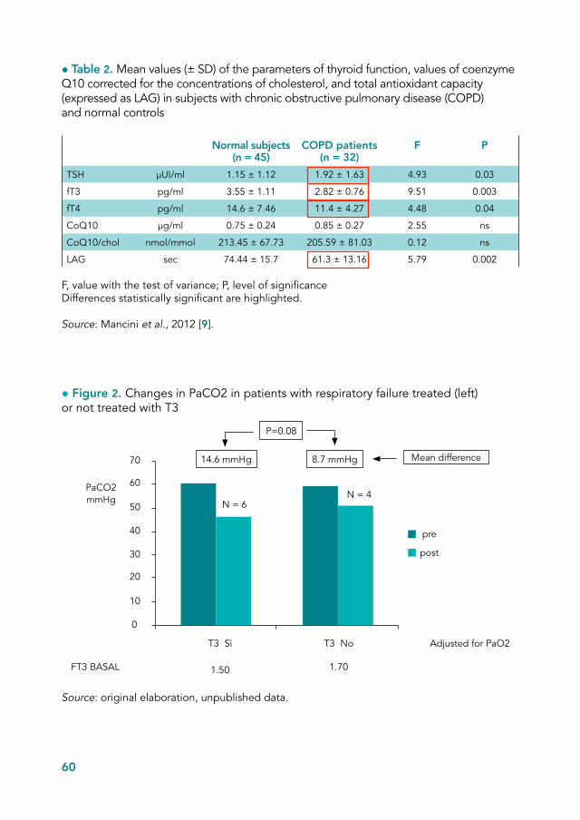
60
• Table 2. Mean values (± SD) of the parameters of thyroid function, values of coenzyme Q10 corrected for the concentrations of cholesterol, and total antioxidant capacity (expressed as LAG) in subjects with chronic obstructive pulmonary disease (COPD) and normal controls
Normal subjects (n = 45)
COPD patients (n = 32)
F P
TSH μUI/ml 1.15 ± 1.12 1.92 ± 1.63 4.93 0.03
fT3 pg/ml 3.55 ± 1.11 2.82 ± 0.76 9.51 0.003
fT4 pg/ml 14.6 ± 7.46 11.4 ± 4.27 4.48 0.04
CoQ10 μg/ml 0.75 ± 0.24 0.85 ± 0.27 2.55 ns
CoQ10/chol nmol/mmol 213.45 ± 67.73 205.59 ± 81.03 0.12 ns
LAG sec 74.44 ± 15.7 61.3 ± 13.16 5.79 0.002
F, value with the test of variance; P, level of significanceDifferences statistically significant are highlighted.
Source: Mancini et al., 2012 [9].
• Figure 2. Changes in PaCO2 in patients with respiratory failure treated (left) or not treated with T3
Source: original elaboration, unpublished data.
70
60
50
40
30
20
10
0
T3 Sì T3 No
1.50 1.70FT3 BASAL
PaCO2mmHg
Adjusted for PaO2
P=0.08
8.7 mmHg14.6 mmHg Mean difference
pre
post
N = 6N = 4

61
References
[1] Dentice M, Salvatore D. Deiodinases: the balance of thyroid hormone. Local impact of thyroid hormone inactivation. J Endocrinol 2011;209:273-82.
[2] Nagaya T, Fujieda M, Otsuka G, Yang JP, Okamoto T, Seo H. A potential role of activat-ed NF-kappa B in the pathogenesis of euthyroid sick syndrome. J Clin Invest 2000;106:393-402.
[3] Mancini A, De Marinis L, Calabrò F, Sciuto R, Oradei A, Lippa S et al. Evaluation of metabolic status in amiodarone-induced thyroid disorders: plasma Coenzyme Q10 deter-mination. J Endocrinol Invest 1989;12:511-6.
[4] Bianchi G, Solaroli E, Zaccheroni V, Grossi G, Bargossi AM, Melchionda N, Marchesini G. Oxidative stress and anti-oxidant metabolites in patients with hyperthyroidism: effect of treatment. Horm Metab Res 1990;31:620-4.
[5] Hamilton MA, Stevenson LW, Fonarow GC, Steimle A, Goldhaber JI, Child JS et al. Safety and hemodynamic effects of intravenous triiodothyronine in advanced congestive heart failure. Am J Cardiol 1998;81:443-7.
[6] Pingitore A, Galli E, Barison A, Iervasi A, Scarlattini M, Nucci D et al. Acute effects of Triiodothyronine (T3) replacemnet therapy in patients with chronic heart failure and low-T3 syndrome: a randomized, placebo-controlled study. J Clin Endocrinol Metab 2008;93:1351-8.
[7] De Groot LJ. Non-thyroidal illness syndrome is a manifestation of hypothalamic-pituitary dysfunction, and in view of current evidence, should be treated with appropriate replace-ment therapies. Crit Care Clin 2006;22:57-86.
[8] Okutan O, Kartaloglu Z, Onde ME, Bozkanat E, Kunter E. Pulmonary function tests and thyroid hormone concentrations in patients with chronic obstructive pulmonary dis-ease. Med Princ Pract 2004;13:126-8.
[9] Mancini A, Corbo GM, Gaballo A, Raimondo S, Di Segni C, Gigliotti P et al. Relation-ship between plasma antioxidants and thyroid hormones in chronic obstructive pulmonary disease. Exp Clin Endocrinol Diabetes 2012;120:623-8.

62
The sulfo-conjugation path of thyroid hormones has been known for many years. Al-ready in 1957 Roche et al. [1] had identified sulfo-conjugates hormones in the blood and in the bile. These observations led to assume, rightly, that sulfo-conjugation was a way to facilitate the biliary excretion of T3. Today we have, without doubt, a wider knowledge about the biological significance of this metabolic pathway.
The sulfo-conjugation of T3 is the addition of a sulfate group at the level of a hy-droxyl of the outer ring of the hormone molecule (• Figure 1).
This process is mediated by enzymes localized at the level of various tissues such as liver, placenta, kidney, central nervous system, and cause a significant impact on the fate of the molecule. The sulfo-conjugation of T3 transforms the hormone from lipophilic molecule to molecule soluble in water. Furthermore, the T3 sulfate is inert from a functional point of view as unable to bind the nuclear receptor, it has a high affinity for the plasmatic carrier proteins and is easily hydrolyzed in acid environment (• Table 1).
To understand the metabolic consequences of sulfo-conjugation, we can take as a reference a work that dates back to the eighties [2]. We know that in the liver deiodi-nase type 1 converts T4 to T3 and that the latter, on the contrary, constitutes a poor substrate for the enzyme itself. In the study by Otten, incubating a sample of hepat-ocytes with T3 and adding increasing amounts of sulfate, occurred an acceleration of the degradation of the hormone with the appearance of free iodine. The explanation of this phenomenon was obtained by adding propyl-thiouracil (PTU) to the culture liquid of the same cells: the PTU is an inhibitor of D1 and its presence inhibited the formation of free iodine while it was associated to the appearance of T3 sulfate. The authors could thus conclude that the sulfate promotes the production of T3 sulfate and that the latter, unlike the active hormone, is rapidly deiodazed from liver D1.
T3 sulfate as a potential pharmacological agent in the treatment of hypothyroidismFerruccio Santini
Endocrinology Unit, University Hospital of Pisa

63
Based on these studies, we now know that the process of sulfo-conjugation is not only a mechanism to facilitate the biliary excretion of T3 but is a way that accelerates its degradation.
In contrast, the sulfo-conjugation guarantees protection from the action of deiodi-nase type 3 (D3), the enzyme that degrades the active T3 and that is found in many tissues such as placenta and skin [3].
This has implications in practical terms, if we measure the serum levels of T3 sul-fate we notice significant differences in various clinical conditions. In particular, low amounts of the hormone can be detected in euthyroid subject, while circulating levels are elevated in hyperthyroid patient (for increase of the precursor, that is T3), but also in hypothyroid patient, in non-thyroidal illnesses, in the fetus and newborn, all conditions in which it is inhibited the D1 [4, 5].
To confirm these observations, when to hyperthyroid patients was administered sodium ipodate, a contrast agent that inhibits the D1, it was observed a reduction of plasmatic levels of T3 accompanied by an increase of T3 sulfate and rT3.
The presence of very high concentrations of the metabolite in the fetal and neona-tal life has led to the hypothesis that the T3 sulfate, after desulphation, can represent to some extent a source of active T3. In this regard, it has been demonstrated the pres-ence in the liver of an enzyme system capable of desulfatingT3 sulfate and, therefore, to reactivate it [6, 7].
Furthermore, by administering T3 sulfate intra-peritoneally in hypothyroid rats it was possible to restore a smooth curve of growth and normalize other parameters re-
I
OHO CH CH COOH
I
I NH 2
I
I
OHO3SO CH2 CH COOH
I NH2
Sulfotransferase (liver, brain, kidney)
2
• Figure 1. Sulfo-conjugation of T3
64
lated to the thyroid status of the animals [8]. In particular, following a single injection of T3 sulfate it was obtained a normalization of serum levels of T3, which remained constant for about 8 hours.
The results of preliminary studies in humans [9] indicate that the T3 sulfate can be absorbed after oral administration in hypothyroid patients, in addition, after adminis-tration of T3 sulfate it is observed a proportional increase in T3 serum concentrations which remains stable for at least 48 hours, unlike what is observed after administra-tion of the active hormone. After T3 administration it occurs in fact a rapid absorp-tion peak followed by an equally fast descent that ends in a matter of hours. In theory, therefore, the T3 sulfate may represent a slow release formulation able to improve the satisfaction of those hypothyroid patients who continue to complain about hormonal deficit disorders, despite the normalization of serum parameters obtained with the administration L- thyroxine alone.
References
[1] Roche J, Michel R, Michel O, Etling N. Biliary excretion of a sulfoconjugate of 3,5,3’-tri-iodo-L-thyronine (T-3) after administration of this hormone to the rat. C R Hebd Seances Acad Sci 1957 Sep 23;245(13):1089-91.
[2] Otten MH, Mol JA, Visser TJ. Sulfation preceding deiodination of iodothyronines in rat hepatocytes. Science 1983 Jul 1;221(4605):81-3.
[3] Santini F, Hurd RE, Chopra IJ. A study of metabolism of deaminated and sulfoconju-gated iodothyronines by rat placental iodothyronine 5-monodeiodinase. Endocrinology 1992;131:1689-94.
• Table 1. Properties of T3 sulfate
• Hydrophilic molecule
• Negligible affinity for the nuclear receptor of T3
• High affinity for plasmatic carrier proteins
• Susceptible to acid hydrolysis
• Rapid deiodination by deiodinase type 1 (D1)
• Resistant to deiodination by deiodinase type 3 (D3)

65
[4] Chopra IJ, Wu SY, Teco GNC, Santini F. A radioammunoassay for measurement of 3,5,3’-triiodothyronine sulfate: studies in thyroidal and nonthyroidal diseases, pregnancy, and neonatal life. J Clin Endocrinol Metab 1992;75:189-94.
[5] Santini F, Chiovato L, Ghirri P, Lapi P, Mammoli C, Montanelli L et al. Serum iodothy-ronines in the human fetus and the newborn: evidence for an important role of placenta in fetal thyroid hormone homeostasis. J Clin Endocrinol Metab 1999;84:493-8.
[6] Kung MP, Spaulding SW, Roth JA. Desulfation of 3,5,3’-triiodothyronine sulfate by micro-somes from human and rat tissues. Endocrinology 1988;122:1195-200.
[7] Santini F, Chopra IJ, Wu SY, Solomon DH, Chua Teco GN. Metabolism of 3,5,3’-tri-iodothyronine sulfate by tissues of the fetal rat: a consideration of the role of desulfation of 3,5,3’-triiodothyronine sulfate as a source of T3. Pediatr Res 1992 Jun;31(6):541-4.
[8] Santini F, Hurd RE, Lee B, Chopra IJ. Thyromimetic effects of 3,5,3’-triiodothyronine sul-fate in hypothyroid rats. Endocrinology 1993;133(l):105-10.
[9] Santini F, Giannetti M, Ricco I, Querci G, Saponati G, Bokor D et al. Steady state serum t3 concentrations for 48 hours following the oral administration of a single dose of T3 sul-fate. 95th Annual Meeting of the Endocrine Society, San Francsisco June 15-18 2013, FP34-1.

66
The hypothalamus-pituitary-thyroid axis (HPT) preserves thyroid hormone’s home-ostasis producing T3 and T4 and controlling concentrations of the two hormones through a sensitive feedback mechanism. Hypothalamic TRH neurons and TSH producing cells can sense at the same time both T4 and T3, because of the presence of type 2 deiodinase that convert T4 to T3. So, the HPT senses at the same time T3 produced from local conversion of T4 in hypothalamus and in pituitary, and the T3 in the circulation (• Figure 1).
We know that most of the biological effect of thyroid hormones, such as cognition, metabolism, growth and development, are mediated by the interaction of T3 with nuclear receptors.
Every day the organism produces about 30 µg of T3, but only 5 µg comes from the thyroid; the rest comes from peripheral conversion from T4 by type 1 and type 2 deiodinases (D1 e D2). These enzymes show different expression patterns in different tissues. For example, D1 is expressed above all in kidneys and liver, while D2 is ex-pressed in brain, skin, skeletal muscle and brown adipose tissue. T4 that comes into the tissue is transformed to T3 in a compartment that is near the cell nucleus, stays in the cell for a few hours (“equilibration time” with the plasma is about 3-4 hours for D2 and approximately 30 minutes for D1), and exits to the plasma. So, every day, about 20 µg of T3 are produced from conversion of T4 by D2, while D1, that has low affinity for T4, produces about 5 µg/day of T3. Ultimately T3, which is a good substrate for type 3 deiodinase, is then inactivated to T2 (• Figure 1).
Studies by Andersen measuring TSH, FT3 and FT4 over a long period of time indicated that serum levels of T3 are stable [1].
To test how well the organism defends serum T3, a few decades ago Larsen evaluated rats kept on low iodine diet. While their serum levels of T4 dropped and serum TSH
Future treatment for hypothyroid patientsAntonio Bianco, Sabina Casula
Division of Endocrinology, Diabetes & Metabolism, Department of Medicine, University of Miami Miller School of Medicine, Miami
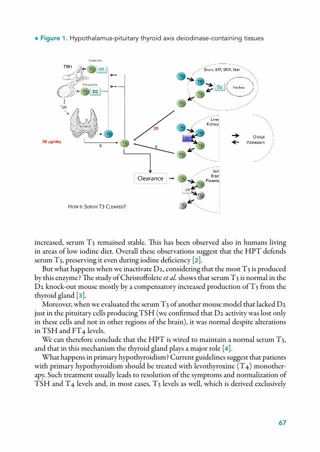
67
increased, serum T3 remained stable. This has been observed also in humans living in areas of low iodine diet. Overall these observations suggest that the HPT defends serum T3, preserving it even during iodine deficiency [2].
But what happens when we inactivate D2, considering that the most T3 is produced by this enzyme? The study of Christoffolete et al. shows that serum T3 is normal in the D2 knock-out mouse mostly by a compensatory increased production of T3 from the thyroid gland [3].
Moreover, when we evaluated the serum T3 of another mouse model that lacked D2 just in the pituitary cells producing TSH (we confirmed that D2 activity was lost only in these cells and not in other regions of the brain), it was normal despite alterations in TSH and FT4 levels.
We can therefore conclude that the HPT is wired to maintain a normal serum T3, and that in this mechanism the thyroid gland plays a major role [4].
What happens in primary hypothyroidism? Current guidelines suggest that patients with primary hypothyroidism should be treated with levothyroxine (T4) monother-apy. Such treatment usually leads to resolution of the symptoms and normalization of TSH and T4 levels and, in most cases, T3 levels as well, which is derived exclusively
• Figure 1. Hypothalamus-pituitary thyroid axis deiodinase-containing tissues

68
from peripheral activation of T4 mediated by deiodinases. However, about 10-15% of hypothyroid patients on levothyroxine monotherapy, despite being biochemically eu-thyroid based on a normal TSH, remain symptomatic. One of the hypotheses there-fore is that the small amount of T3 that the thyroid gland produces has some key role in achieving that sense of wellbeing that some hypothyroid patients on levothyroxine monotherapy seem to struggle to obtain.
A study of Gullo et al. showed that euthyroidism cannot be restored in all post-sur-gical hypothyroid patients through monotherapy with levothyroxine (• Figure 2). Stratifying these data by serum TSH, they compared euthyroid and hypothyroid pa-tients on monotherapy that had the same level of TSH and observed that for every level of TSH the athyrotic patients had a relatively lower serum T3 and a higher serum T4. Consequently post-surgical hypothyroid patients maintain a normal se-rum TSH because of an excess replacement of T4 that compensates for a relative deficiency of serum T3. Therefore serum TSH, in these cases, is not a reliable indica-tor of euthyroid state since even if TSH is normal T3 may be low. This could affect
• Figure 2. Levothyroxine monotherapy cannot guarantee euthyroidism in all athyreotic patients
Source: Gullo et al., 2011 [5], adapted.

69
especially those organs/tissues that do not have D2 and that therefore depend solely on serum T3 [5].
Another bias for the comprehension of thyroid status regards the techniques of measuring T3. Jonklaas et al. compared serum T3 levels of patients after thyroidec-tomy as measured by immunoassay or by mass spectrometry. Immunoassays overesti-mate serum T3 values in comparison with mass spectrometry when values are about 100 ng/dl or lower, concluding that immunoassays underestimate the number of pa-tients with low serum T3 [6].
In the study of Geffner et al., PTU, an inhibitor of type 1 deiodinase was given to hypothyroid patients on monotherapy with levothyroxine. They observed that while FT4 did not change, a relatively small decrease in serum T3 caused serum TSH to double. We could then speculate that the fall in serum T3, albeit small, results in pitu-itary hypothyroidism. Therefore, we should expect that other tissues that depend on serum T3 can become hypothyroid as well [7].
We know that there is an equilibrium between tissue T3 and plasma T3 and that not all cells depend exclusively on plasma T3. In the brain T3 plays a major role in cognitive function. D2 is responsible for more than half of the T3 present in the mu-rine brain. Accordingly, D2KO animals have half as much brain T3 content as their wild type siblings greatly supporting the idea that any interference in the D2 pathway could affect brain function and/or result in intellectual or cognitive symptoms [8].
Is there a clinical syndrome that involves D2 deficiency? If so, could this explain the efficacy of combined therapy for some patients?
The group of Celi discovered a particular polymorphism (Thr92Ala DIO2) that was associated to a specific metabolic phenotype: increased BMI and glucose intoler-ance. D2 KO mice are obese and have intolerance to glucose as well. Later, many other studies confirmed this association and tried to explain how this polymorphism could produce a similar phenotype, since the association is not sufficient to confirm that the polymorphism is really influencing D2 activity [9].
Studies by Panicker et al. showed that hypothyroid patients carrying the polymor-phism in the DIO2 gene had a better neurocognitive outcome with combined therapy (T3 and T4) compared to standard monotherapy. From these data we could speculate that in these patients D2 doesn’t produce sufficient quantities of T3. However this hy-pothesis needs to be confirmed and does not necessarily preclude the possibility that there is another rational behind [10].
When we give combination therapy, what happen to T3? To answer that question we performed a systematic review and meta-analysis in 10 randomized controlled tri-als, 7 of which were crossover. In particular we evaluated and compared the post treat-ment levels of TSH, free T4, and total T3 (unpublished data). Despite similar TSH level, our analysis revealed a significant difference in the serum level of free T4 and total T3 in patients on monotherapy compared with patients on combined therapy. In particular patients on levothyroxine monotherapy had a higher plasmatic level of
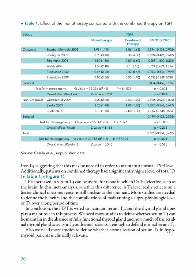
70
free T4 suggesting that this may be needed in order to maintain a normal TSH level. Additionally, patients on combined therapy had a significantly higher level of total T3 (• Table 1, • Figure 3).
This increased in serum T3 can be useful for tissue in which D2 is defective, such as the brain. In this meta-analysis, whether this difference in T3 level really reflects on a better clinical outcome remains still unclear at the moment. More studies are needed to define the benefits and the complications of maintaining a supra-physiologic level of T3 over a long period of time.
In conclusion, the HPT is wired to maintain serum T3, and the thyroid gland does play a major role in this process. We need more studies to define whether serum T3 can be maintain in the absence of fully functional thyroid gland and how much of the resid-ual thyroid gland activity in hypothyroid patients is enough to defend normal serum T3.
Also we need more studies to define whether normalization of serum T3 in hypo-thyroid patients is clinically relevant.
• Table 1. Effect of the monotherapy compared with the combined therapy on TSH
Study TSH
Monotherapy Combined Therapy
SMD* (95%CI)
Crossover Escobar-Morreale 2005 1.95 (1.44)† 2.56 (1.65) 0.394 (-0.139, 0.928)
Rodriguez 2005 2.90 (3.40) 3.30 (4.00) 0.108 (-0.426, 0.642)
Siegmund 2004 1.50 (1.30) 0.50 (0.60) -0.988 (-1.600, -0.376)
Walsh 2003 1.50 (2.10) 3.1 (2.10) 0.763 (0.489, 1.036)
Bunevicius 2002 0.45 (0.48) 0.47 (0.46) 0.043 (-0.834, 0.919)
Bunevicius 2009 0.80 (2.50) 0.50 (1.10) -0.155 (-0.639, 0.328)
Subtotal 0.054 (-0.469, 0.576)
Test for Heterogeneity Q-value = 32.336 (df =5) I2 = 84.537 p < 0.001
Overall effect (Random) Z-value = 0.201 p = 0.841
Non-Crossover Valizadeh M 2009 2.00 (0.80) 2.50 (1.20) 0.490 (-0.023, 1.004)
Sawka 2003 1.70 (1.70) 1.80 (1.80) 0.057 (-0.563, 0.677)
Clyde 2003 2.10 (1.70) 2.00 (1.80) -0.057 (-0.648, 0.534)
Subtotal 0.199 (-0.129, 0.528)
Test for Heterogeneity Q-value = 2.158 (df = 2) I2 = 7.327 p = 0.340
Overall effect (Fixed) Z-value = 1.188 p = 0.235
Total 0.101 (-0.261, 0.462)
Test for Heterogeneity Q-value = 34.788 (df = 8) I2 = 77.004 p < 0.001
Overall effect (Random) Z-value = 0.546 p = 0.585
Source: Casula et al., unpublished data.
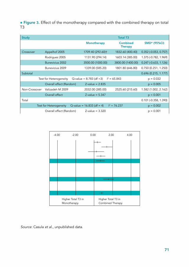
71
• Figure 3. Effect of the monotherapy compared with the combined therapy on total T3
Study Total T3
Monotherapy Combined Therapy
SMD* (95%CI)
Crossover Appelhof 2005 1709.40 (292.60)† 1832.60 (400.40) 0.352 (-0.053, 0.757)
Rodriguez 2005 1131.90 (294.14) 1603.14 (385.00) 1.375 (-0.782, 1.969)
Bunevicius 2002 3500.00 (1000.00) 3800.00 (1400.00) 0.247 (-0.633, 1.126)
Bunevicius 2009 1339.00 (585.20) 1801.80 (646.80) 0.750 (0.251, 1.250)
Subtotal 0.696 (0.215, 1.177)
Test for Heterogeneity Q-value = 8.783 (df =3) I2 = 65.843 p = 0.032
Overall effect (Random) Z-value = 2.835 p = 0.005
Non-Crossover Valizadeh M 2009 2032.00 (385.00) 2525.60 (215.60) 1.582 (1.002, 2.162)
Overall effect Z-value = 5.347 p < 0.001
Total 0.101 (-0.358, 1.390)
Test for Heterogeneity Q-value = 16.833 (df = 4) I2 = 76.237 p = 0.002
Overall effect (Random) Z-value = 3.320 p = 0.001
-4.00 -2.00 0.00 2.00 4.00
Higher Total T3 inMonotherapy
Higher Total T3 inCombined Therapy
Source: Casula et al., unpublished data.

72
Ultimately, it will be important to confirm the clinical role of D2 pathway and specifically to prove that a deficient D2 activity could really explain some residual symptom in hypothyroid patients that have normal serum TSH on monotherapy with levothyroxine.
References
[1] Andersen S, Pedersen KM, Bruun NH, Laurberg P. Narrow individual variations in serum T(4) and T(3) in normal subjects: a clue to the understanding of subclinical thyroid disease. J Clin Endocrinol Metab 2002;87:1068-72.
[2] Riesco G, Taurog A, Larsen R, Krulich L. Acute and chronic responses to iodine deficiency in rats. Endocrinology 1977 Feb;100(2):303-13.
[3] Christoffolete MA, Ribeiro R, Singru P, Fekete C, da Silva WS, Gordon DF et al. Atypi-cal expression of type 2 iodothyronine deiodinase in thyrotrophs explains the thyroxine-medi-ated pituitary thyrotropin feedback mechanism. Endocrinology 2006 Apr;147(4):1735-43.
[4] Fonseca TL, Correa-Medina M, Campos MP, Wittmann G, Werneck-de-Castro JP, Ar-rojo e Drigo R et al. Coordination of hypothalamic and pituitary T3 production regulates TSH expression. J Clin Invest 2013 Apr 1;123(4):1492-500.
[5] Gullo D, Latina A, Frasca F, Le Moli R, Pellegriti G, Vigneri R. Levothyroxine monother-apy cannot guarantee euthyroidism in all athyreotic patients. PLoS One 2011:6(8):e22552.
[6] Jonklaas J, Davidson B, Bhagat S, Soldin SJ. Triiodothyronine levels in athyreotic individ-uals during levothyroxine therapy. JAMA 2008 Feb 20;299(7):769-77.
[7] Geffner DL, Azukizawa M, Hershman JM. Propylthiouracil blocks extrathyroidal con-version of thyroxine to triiodothyronine and augments thyrotropin secretion in man. J Clin Invest 1975 Feb;55(2):224-9.
[8] Galton VA, Wood ET, St Germain EA, Withrow CA, Aldrich G, St Germain GM et al. Thyroid hormone homeostasis and action in the type 2 deiodinase-deficient rodent brain during development. Endocrinology 2007 Jul;148(7):3080-8.
[9] Mentuccia D, Proietti-Pannunzi L, Tanner K, Bacci V, Pollin TI, Poehlman ET et al. Association between a novel variant of the human type 2 deiodinase gene Thr92Ala and in-sulin resistance: evidence of interaction with the Trp64Arg variant of the beta-3-adrenergic receptor. Diabetes 2002;51:880-3.

73
[10] Panicker V, Saravanan P, Vaidya B, Evans J, Hattersley AT, Frayling TM, Dayan CM. Common variation in the DIO2 gene predicts baseline psychological well-being and re-sponse to combination thyroxine plus triiodothyronine therapy in hypothyroid patients. J Clin Endocrinol Metab 2009 May;94(5):1623-9.


75
During the discussion in this second Forum of Endocrinology, organized by IBSA, emerged several topics that have been an opportunity for confrontation, but also for debate amongst the participants.
In particular, a hot topic was the one related to the inadequacy of TSH as the sole marker of thyroid function and the need for other tissue markers. The question is somehow tricky, especially considering that the thyroid tissue condition depends only in part on the plasma concentration of T3, coming also from the expression of mem-brane transporters, the interaction with the selenium-deiodinases and the expression and the activity of nuclear receptors. TSH, then, for how much is a good pituitary marker, does not accurately reflect what is going on in tissues other than the pituitary. The circulating T3 may be helpful in some case but, also, it cannot be considered an optimal marker, for the reasons previously expressed.
On the other hand, it is not the case to generalize: TSH currently remains a “ro-bust” marker in daily clinical practice, resulting less sensitive in a small niche of pa-tients. And it is for the latter that is necessary to identify other markers. Identify a “optimal” tissue marker is a difficult task, especially if one considers that theoretically there should be a specific marker for each individual tissue.
About the reliability of TSH, there is currently a debate about the need and the possibility to perform the simultaneous dosing of the free hormonal fractions, or at least of one of the two hormones. While it is true that the so-called TSH SLR reflex is an expedient to limit health care expenditure, it is inadequate in different circum-stances; as we have already seen the value of TSH may be distorted by many interfer-ing agents, along with these there are other bias related to the dosage, and, therefore, to the kits used in the laboratory, as well as to the individual variations of this markers, including the Circadian ones. TSH reflex, then, it may be a good economic compro-
Conclusions

76
mise to reduce costs, reserving the dosage of at least one of the two hormones (better FT4 according to American guidelines) for the most suspicious cases (autoimmun-ity, familiarity, perspective of a pregnancy, etc.), as well as for the suspicious forms of central hypothyroidism.
Another aspect that emerged from the discussion is related to the ambiguity that is associated with the definition of well-being of the patient. It is, in fact, a param-eter obviously subjective and we do not currently have adequate tools and markers to evaluate it. Not necessarily well-being coincides with the feeling of “being-well” of the patient and, in addition, we cannot be guided solely by patient preference in adopting a specific therapeutic strategy. In this regard, several studies of correlation between therapy and “well-being” were revisited in light of the fact that, in many cases, the well-being of patients resulted from a slight hormone overdose, and there-fore from a state of mild hyperthyroidism. This can be also the reason for which, often, hyperthyroid patients report feeling worse after having restored the euthyroid state through therapy. However, if the ultimate goal of treatment was just to make the patient feel good, a slight increase in the hormone dosage could have a rational, monitoring, then, strictly the patient, also in view of the increased cardiovascular risk to which he would be inevitably exposed. So the debate is still open, but if, on one hand, what the patient refers has its relevance, on the other hand we have to take into account the long term complications that a condition not more physiological might have in different organs and tissues.
Among the conditions that interfere with the sense of well-being of the patient in therapy with levothyroxine and normal TSH levels, you can also count the vitamin D deficit, which may be accompanied by fatigue, muscle fatigue and depression. This forum also discussed the opportunity of dosing vitamin D in these subjects, and, in the case of deficit, to establish an adequate replacement therapy.
Another element of debate was that relating to the need to perform a diagnostic process to highlight a possible malabsorption in case the therapeutic target is not reached following the administration of the theoretical dose of levothyroxine. The justified objection to this practice comes from the significant physiological indi-vidual variability of drug metabolism: absorption, speed degradation, clearance etc. Furthermore, each clinical parameter, by definition, can be misleading, so it could be sufficient to repeat the dosage and possibly minimally adjust the dose, rather than trigger a whole series of investigations also expensive and invasive, to be reserved, perhaps, to cases with more justified clinical suspicion. It should be clear, however, that the screening for malabsorption, in fact, provides initial tests both little invasive and expensive, such as the urea breath test, which, considering the high incidence of Helicobacter pylori infection in the general population, provide, in any case, clinically useful information, regardless the potential malabsorption. The more invasive in-vestigations, such as endoscopy, may be reserved to the later stages of the diagnostic process.

77
From the discussion emerged also another problem : if it is true that the dose in the elderly should be reduced by 20% compared to the standard, how to resolve the con-flict with the WHO guidelines which, instead, suggest increasing the dose in these patients in relation to the natural reduction of drug absorption with the increasing age? In fact, this discrepancy does not seem to exist, as in the elderly the reduction of the absorption is associated with a concomitant slowing down of T4 metabolism, which makes sure that the permanence time of the hormone in circulation is sufficient to ensure circulatory homeostasis.
An additional problem of the treatment of hypothyroidism in the elderly derives by the reduced ability of the body after a certain age to adapt to hormonal deficiency and to ensure tissue homeostasis of the thyroid hormone. Numerous studies also showed a physiological increase in the levels of TSH, “normal” in individuals of advanced age. The treatment of hypothyroidism, especially in the subclinical forms, may therefore be counterproductive because of the potential negative effects that even a slight excess of the hormone may cause in the elderly.
During the forum, it was also discussed at length the potential of research in the study of various polymorphisms of genes involved in the metabolism of thyroid hor-mones and the possible correlation between the exon sequences and values of TSH and FT4 in hundreds of patients, with the clinical practice.
An important and striking point, then, is the association between polymorphisms of some genes and T3 in serum: studies done in a small numbers of patients appear to demonstrate the existence of a significant correlation, but further studies are needed to demonstrate the real meaning.
In clinical practice, it is not uncommon to observe patients who present randomly elevated values of TSH, totally asymptomatic, with hormone free fractions normal, in absence of a family history of thyroid disease or autoimmunity. These cases could be an expression of a polymorphism at pituitary level as to explain the isolated in-crease of TSH? In part, maybe it is so, but at present there are no sufficient data avail-able to confirm this. Another issue related to this subgroup of patients concerns the therapeutic approach. In general they have to be considered subclinical hypothyroid, but there is no consensus regarding the treatment, considering that some of them do not benefit from hormone treatment. Therefore, it would be more appropriate to carefully evaluate each case and decide in relation to the individual patient whether to treat.
They emerged also a number of ideas for the future of the research on the periph-eral metabolism of thyroid hormone. It would be, for example, interesting to see what would happen if the gene for the deiodinase type 2 was inactivated not only in the thyroid but also in other organs, such as the hypothalamus. The issue is very complex. It has been demonstrated a conspicuous presence of D2 in tanyciytes, specialized cells of glial origin that act as stem cells, but it has not yet been possible to create tanycytes deprived of D2.
78
Another stimulating topic concerns the correlation between the peripheral me-tabolism of thyroid hormone and the basal metabolism. Surely the identification of the response of D2 gene to cyclic AMP is one of the first molecular link identified be-tween thyroid metabolism and adrenergic signal, but the metabolic determinants of this crosstalk are still unknown. Having more information about it could help, among other things, to understand, for example, the reason why there are different metabolic responses to the same stimulus, as may be the cold, in both sexes.
There is also need of studies to analyze the ways in which different drugs may in-terfere with thyroxine therapy in hypothyroidism. Often hypothyroid patients take many therapies that we know interfere with the metabolism of levothyroxine, such as absorption, binding to carrier proteins etc. We know still very little, on the contrary, on the possibility of these drugs to interfere with the activity of deiodinase; some, such as amiodarone and β-blockers have been shown effects in that regard, but the currently available data are somehow inadequate.
With regard to the possible clinical use of T3 sulfate, it was exposed the doubt about reintroducing a substance in the body that is physiologically eliminated. In-deed, administering T3 sulfate, there is no claim to mimic a physiological condition, and its metabolic effects have to be yet fully defined.
In conclusion, there are still many questions concerning the future of the hypothy-roid patient and many of the suggestions received are certainly still to be explored. The importance of therapy customization (individually tailored dose) on individual hypo-thyroid patients is the message that must enter into the clinical practice in the daily contact with a disease which has a strong social significance such as hypothyroidism.


The analysis of data from both basic and clinical research
and the future perspectives in therapy for hypothyroidism
are the topics discussed and lively debated during
the II Forum of Endocrinology organized by the Fondazione IBSA.
The Forum, which was attended by prominent international
experts, provides interesting insights and is an useful contribution
to achieve the goal desired by all: a timely and correct diagnosis
and a more personalized and effective therapy in the future
of the hypothyroid patient.
Italia
n ve
rsio
n re
gist
ered
by
aif
a 1
0/01
/201
4
IBSA
FO
UN
DA
TIO
N P
APE
RS


![0) · 2016. 7. 8. · x\hsp[`th`]hy`klwlukpunvu svjh[pvu ;opz^psshhlj[Äuhs lhkpunz ... pj /\tpjhjpk)sluk-sv^ly luohujly t3 t3 t3 t3 t3 t3 t3 t3 t3 t3 t3 t3 t3 t3 t3 t3 t3 t3 t3 t3](https://img.pdfslide.us/doc/110x75/60d98d4a31005a4c8d3c5fa4/0-2016-7-8-xhspthhyklwlukpunvu-svjhpvu-opzpsshhljuhs-lhkpunz-.jpg)