Embed Size (px)
Citation preview
New York
Neck Injury
Medical Treatment Guidelines
Third Edition, September 15, 2014
New York State Workers’ Compensation Board New York Neck Injury Medical Treatment Guidelines
Third Edition, September 15, 2014 i
TABLE OF CONTENTS
GENERAL GUIDELINE PRINCIPLES ............................................. 1
MEDICAL CARE ............................................................................................... 1
RENDERING OF MEDICAL SERVICES ........................................................... 1
POSITIVE PATIENT RESPONSE ..................................................................... 1
RE-EVALUATE TREATMENT ........................................................................... 1
EDUCATION ..................................................................................................... 2
DIAGNOSTIC TIME FRAMES........................................................................... 2
TREATMENT TIME FRAMES ........................................................................... 2
DELAYED RECOVERY .................................................................................... 2
ACTIVE INTERVENTIONS ............................................................................... 3
ACTIVE THERAPEUTIC EXERCISE PROGRAM ............................................. 3
DIAGNOSTIC IMAGING AND TESTING PROCEDURES ................................ 3
SURGICAL INTERVENTIONS .......................................................................... 4
PRE-AUTHORIZATION .................................................................................... 4
PERSONALITY/PSYCHOLOGICAL/PSYCHOSOCIAL EVALUATIONS .......... 4
PERSONALITY/PSYCHOLOGICAL/PSYCHOSOCIAL INTERVENTION ......... 5
FUNCTIONAL CAPACITY EVALUATION (FCE) .............................................. 5
RETURN TO WORK ......................................................................................... 6
JOB SITE EVALUATION ................................................................................... 6
GUIDELINE RECOMMENDATIONS AND MEDICAL EVIDENCE .................... 7
EXPERIMENTAL/INVESTIGATIONAL TREATMENT ....................................... 7
INJURED WORKERS AS PATIENTS ............................................................... 7
SCOPE OF PRACTICE ..................................................................................... 7
INTRODUCTION ............................................................................ 8
HISTORY TAKING AND PHYSICAL EXAMINATION ....................................... 8
History of Present Injury ................................................................................. 8
Past History ...................................................................................................... 9
Physical Examination ...................................................................................... 9
Relationship to Work ..................................................................................... 10
New York State Workers’ Compensation Board New York Neck Injury Medical Treatment Guidelines
Third Edition, September 15, 2014 ii
Spinal Cord Evaluation .................................................................................. 10
Soft Tissue Injury Evaluation ........................................................................ 12
Red Flags ........................................................................................................ 13
IMAGING ......................................................................................................... 13
LABORATORY TESTS ................................................................................... 14
FOLLOW-UP DIAGNOSTIC IMAGING AND TESTING PROCEDURES ........ 14
DIAGNOSTIC STUDIES ............................................................... 16
IMAGING STUDIES ........................................................................................ 16
Magnetic Resonance Imaging (MRI) ............................................................ 16
Computed Axial Tomography (CT) ................................................................ 17
Myelography ................................................................................................... 17
CT Myelogram ................................................................................................ 18
Lineal Tomography ........................................................................................ 18
Bone Scan (Radioisotope Bone Scanning) .................................................... 18
Other Radioisotope Scanning ........................................................................ 18
Dynamic [Digital] Fluoroscopy...................................................................... 18
OTHER TESTS ............................................................................................... 19
Electrodiagnostic Testing (EDX) ................................................................... 19
Injections – Diagnostic .................................................................................. 20
Provocation Discography ............................................................................... 20
Thermography ................................................................................................ 20
THERAPEUTIC PROCEDURES: NON-OPERATIVE ................... 20
ACUPUNCTURE ............................................................................................. 21
BIOFEEDBACK ............................................................................................... 22
INJECTIONS: THERAPEUTIC ........................................................................ 23
Therapeutic Spinal Injections-Introduction ................................................. 23
Trigger Point Injections and Dry Needling Treatment ................................. 32
Prolotherapy ................................................................................................... 34
Platelet Rich Plasma (PRP) ............................................................................ 34
Epiduroscopy and Epidural Lysis of Adhesions ............................................ 34
RADIOFREQUENCY ABLATION, NEUROTOMY, FACET RHIZOTOMY ...... 34
New York State Workers’ Compensation Board New York Neck Injury Medical Treatment Guidelines
Third Edition, September 15, 2014 iii
MEDICATION .................................................................................................. 36
Acetaminophen .............................................................................................. 36
Anti-Depressants ........................................................................................... 37
Anti-Seizure Drugs ......................................................................................... 38
Compound Medications ................................................................................. 39
Narcotics ........................................................................................................ 39
Nonsteroidal Anti-Inflammatory Drugs (NSAIDs) ....................................... 40
Skeletal Muscle Relaxants ............................................................................. 42
Systemic Glucocorticosteroids (aka “Steroids”) ............................................ 44
Topical Drug Delivery .................................................................................... 44
Tramadol ........................................................................................................ 46
Vitamins ......................................................................................................... 46
SPINAL CORD PROGRAMS .......................................................................... 46
ORTHOTICS ................................................................................................... 47
Cervical Collars .............................................................................................. 47
Posture Appliances......................................................................................... 47
Cervicothoracic Orthosis................................................................................ 48
Halo Devices ................................................................................................... 48
Other Orthoses, Devices and Equipment ...................................................... 48
RESTRICTION OF ACTIVITIES ...................................................................... 48
RETURN TO WORK ....................................................................................... 48
Establishment of Activity Level Restrictions................................................. 49
Compliance with Activity Restrictions .......................................................... 49
THERAPY: ACTIVE ........................................................................................ 49
Activities of Daily Living (ADL) ..................................................................... 50
Aquatic Therapy ............................................................................................. 50
Functional Activities ...................................................................................... 50
Functional Electrical Stimulation .................................................................. 50
Neuromuscular Re-education ........................................................................ 51
Spinal Stabilization ........................................................................................ 51
Therapeutic Exercise ...................................................................................... 51
New York State Workers’ Compensation Board New York Neck Injury Medical Treatment Guidelines
Third Edition, September 15, 2014 iv
THERAPY: PASSIVE ...................................................................................... 52
Electrical Nerve Block .................................................................................... 52
Electrical Stimulation (Physician or Therapist Applied) .............................. 53
Iontophoresis ................................................................................................. 53
Manipulation .................................................................................................. 53
Manipulation of the Spine under General Anesthesia (MUA) ...................... 54
Manipulation under Joint Anesthesia (MUJA) ............................................. 54
Massage (Manual or Mechanical) .................................................................. 54
Mobilization (Joint) ....................................................................................... 55
Mobilization (Soft Tissue) .............................................................................. 56
Short-Wave Diathermy .................................................................................. 56
Superficial Heat and Cold Therapy (Excluding Infrared Therapy) .............. 57
Traction .......................................................................................................... 57
Traction: Mechanical ..................................................................................... 57
Transcutaneous Neurostimulator (TCNS/ Electroanalgesic Nerve Block) .. 58
Transcutaneous Electrical Nerve Stimulation (TENS) ................................. 58
Ultrasound (Including Phonophoresis) ......................................................... 58
THERAPY: ONGOING MAINTENANCE CARE .............................................. 59
THERAPEUTIC PROCEDURES: OPERATIVE ............................ 60
ACUTE FRACTURES AND DISLOCATIONS ................................................. 61
Halo Immobilization ...................................................................................... 61
Anterior or Posterior Decompression with Fusion ....................................... 61
DISC HERNIATION AND OTHER CERVICAL CONDITIONS ........................ 63
Specific Indications ........................................................................................ 64
Surgical Procedures ....................................................................................... 65
ELECTRICAL BONE GROWTH STIMULATORS ........................................... 69
ARTIFICIAL CERVICAL DISC REPLACEMENT ............................................. 70
PERCUTANEOUS RADIOFREQUENCY DISC DECOMPRESSION ............. 72
EPIDUROSCOPY AND EPIDURAL LYSIS OF ADHESIONS ......................... 72
INTRAOPERATIVE MONITORING ................................................................. 72
IMPLANTABLE SPINAL CORD STIMULATORS (SCS) ................................. 72
New York State Workers’ Compensation Board New York Neck Injury Medical Treatment Guidelines
Third Edition, September 15, 2014 v
New York State Workers’ Compensation Board New York Neck Injury Medical Treatment Guidelines
Third Edition, September 15, 2014 1
GENERAL GUIDELINE PRINCIPLES
The principles summarized in this section are key to the intended application of the New York State Medical Treatment Guidelines (MTG).
Medical Care
MEDICAL CARE
Medical care and treatment required as a result of a work-related injury should be focused on restoring functional ability required to meet the patient’s daily and work activities and return to work, while striving to restore the patient’s health to its pre-injury status in so far as is feasible.
RENDERING OF MEDICAL SERVICES
Any medical provider rendering services to a workers compensation patient must utilize the Treatment Guidelines as provided for with respect to all work-related injuries and/or illnesses.
POSITIVE PATIENT RESPONSE
Positive results are defined primarily as functional gains which can be objectively measured. Objective functional gains include, but are not limited to, positional tolerances, range of motion, strength, endurance, activities of daily living (ADL), cognition, psychological behavior, and efficiency/velocity measures which can be quantified. Subjective reports of pain and function should be considered and given relative weight when the pain has anatomic and physiologic correlation.
RE-EVALUATE TREATMENT
If a given treatment or modality is not producing positive results, the provider should either modify or discontinue the treatment regime. The provider should evaluate the efficacy of the treatment or modality 2 to 3 weeks after the initial visit and 3 to 4 weeks thereafter. Recognizing that treatment failure is at times attributable to an incorrect diagnosis should prompt the clinician to reconsider the diagnosis in the event of an unexpected poor response to an otherwise rational intervention.
New York State Workers’ Compensation Board New York Neck Injury Medical Treatment Guidelines
Third Edition, September 15, 2014 2
Education
EDUCATION
Education of the patient and family, as well as the employer, insurer, policy makers and the community should be a primary emphasis in the treatment of work-related injury or illness. Practitioners should develop and implement effective educational strategies and skills. An education-based paradigm should always start with communication providing reassuring information to the patient. No treatment plan is complete without addressing issues of individual and/or group patient education as a means of facilitating self-management of symptoms and prevention of future injury.
Time Frames
DIAGNOSTIC TIME FRAMES
Diagnostic time frames for conducting diagnostic testing commence on the date of injury. Clinical judgment may substantiate the need to accelerate or decelerate the time frames discussed in this document.
TREATMENT TIME FRAMES
Treatment time frames for specific interventions commence once treatments have been initiated, not on the date of injury. Obviously, duration may be impacted by disease process and severity, patient compliance, as well as availability of services. Clinical judgment may substantiate the need to accelerate or decelerate the time frames discussed in this document.
DELAYED RECOVERY
For those patients who are failing to make expected progress 6-12 weeks after an injury, reexamination in order to confirm the accuracy of the diagnosis and re-evaluation of the treatment program should be performed. Assessment for potential barriers to recovery (yellow flags/psychological issues) should be ongoing throughout the care of the patient. However, at 6-12 weeks, alternate treatment programs, including formal psychological or psychosocial evaluation, should be considered. Referrals to mental health providers (i.e.: psychology/psychiatry) for the evaluation and management of delayed recovery do not indicate or require the establishment of a psychiatric or psychological condition. The evaluation and management of delayed recovery does not require the establishment of a psychiatric or psychological claim.
New York State Workers’ Compensation Board New York Neck Injury Medical Treatment Guidelines
Third Edition, September 15, 2014 3
Treatment Approaches
ACTIVE INTERVENTIONS
Active interventions emphasizing patient responsibility, such as therapeutic exercise and/or functional treatment, are generally emphasized over passive modalities, especially as treatment progresses. Generally, passive and palliative interventions are viewed as a means to facilitate progress in an active rehabilitation program with concomitant attainment of objective functional gains.
ACTIVE THERAPEUTIC EXERCISE PROGRAM
Active therapeutic exercise program goals should incorporate patient strength, endurance, flexibility, range of motion, sensory integration, coordination, and education as clinically indicated. This includes functional application in vocational or community settings.
DIAGNOSTIC IMAGING AND TESTING PROCEDURES
Clinical information obtained by history taking and physical examination should be the basis for selection and interpretation of imaging procedure results. All diagnostic procedures have variable specificity and sensitivity for various diagnoses.
When a diagnostic procedure, in conjunction with clinical information, provides sufficient information to establish an accurate diagnosis, a second diagnostic procedure will be redundant if it is performed only for diagnostic purposes. At the same time, a subsequent diagnostic procedure (that may be a repeat of the same procedure, when the rehabilitation physician, radiologist or surgeon documents the study was of inadequate quality to make a diagnosis) can be a complementary diagnostic procedure if the first or preceding procedures, in conjunction with clinical information, cannot provide an accurate diagnosis, and is permissible under the MTG.
It is recognized that repeat imaging studies and other tests may be warranted by the clinical course and to follow the progress of treatment in some cases. It may be of value to repeat diagnostic procedures (e.g. imaging studies) during the course of care to reassess or stage the pathology when there is progression of symptoms or findings, prior to surgical interventions and therapeutic injections when warranted, and post-operatively to follow the healing process. Regarding CT examinations, it must be recognized that repeat procedures result in an increase in cumulative radiation dose and associated risks.
New York State Workers’ Compensation Board New York Neck Injury Medical Treatment Guidelines
Third Edition, September 15, 2014 4
SURGICAL INTERVENTIONS
Contemplation of surgery should be within the context of expected functional outcome. The concept of "cure" with respect to surgical treatment by itself is generally a misnomer. All operative interventions must be based upon positive correlation of clinical findings, clinical course and imaging and other diagnostic tests. A comprehensive assimilation of these factors must lead to a specific diagnosis with positive identification of pathologic condition(s). For surgery to be performed to treat severe pain, there must be clear correlation between the pain symptoms and objective evidence of its cause. In all cases, shared decision making with the patient is advised. The patient should be given the opportunity to understand the pros and cons of surgery, potential for rehabilitation as an alternative where applicable, evidence-based outcomes, and specific surgical experience.
PRE-AUTHORIZATION
All diagnostic imaging, testing procedures, non-surgical and surgical therapeutic procedures within the criteria of the Medical Treatment Guidelines and based on a correct application of the Medical Treatment Guidelines are considered authorized, with the exception of the following procedures: Lumbar Fusion, Artificial Disc Replacements, Vertebroplasty, Kyphoplasty, Electrical Bone Growth Stimulators, Spinal Cord Stimulators, Intrathecal Drug Delivery (Pain Pumps), Osteochondral Autograft, Autologous Chondrocyte Implantation, Meniscal Allograft Transplantation and Knee Arthroplasty (Total or Partial Knee Joint Replacement). These are not included on the list of pre-authorized procedures. Providers who want to perform one of these procedures must request pre-authorization from the carrier before performing the procedure.
Second or subsequent procedures (the repeat performance of a surgical procedure due to failure of, or incomplete success from the same surgical procedure performed earlier, if the Medical Treatment Guidelines do not specifically address multiple procedures) also require pre-authorization.
PERSONALITY/PSYCHOLOGICAL/PSYCHOSOCIAL EVALUATIONS
In select patients, diagnostic testing procedures may be useful when there is a discrepancy between diagnosis, signs, symptoms, clinical concerns or functional recovery. Psychological testing should provide differentiation between pre-existing depression versus injury-caused depression, as well as post-traumatic stress disorder, and other psychosocial issues that may include work or non-work-related issues when such conditions are identified in the patient.
New York State Workers’ Compensation Board New York Neck Injury Medical Treatment Guidelines
Third Edition, September 15, 2014 5
For those patients who fail to make expected progress 6-12 weeks after an injury and whose subjective symptoms do not correlate with objective signs and tests, reexamination in order to confirm the accuracy of the diagnosis should be made. Formal psychological or psychosocial evaluation may be considered.
A professional fluent in the primary language of the patient is strongly preferred. When such a provider is not available, services of a professional language interpreter must be provided.
Frequency: One time visit for evaluation. If psychometric testing is indicated by findings in the initial evaluation, time for such testing should not exceed an additional two hours of professional time.
PERSONALITY/PSYCHOLOGICAL/PSYCHOSOCIAL INTERVENTION
Following psychosocial evaluation, when intervention is recommended, such intervention should be implemented as soon as possible. This can be used alone or in conjunction with other treatment modalities.
Time to produce effect: 2 to 8 weeks.
Optimum duration: 6 weeks to 3 months.
Maximum duration: 3 to 6 months. Counseling is not intended to delay but to enhance functional recovery. For select patients, longer supervision may be required, and if further counseling is indicated, documentation of the nature of the psychological factors, as well as projecting a realistic functional prognosis, should be provided by the authorized treating practitioner every 4 to 6 weeks during treatment.
Return to Work
FUNCTIONAL CAPACITY EVALUATION (FCE)
Functional capacity evaluation is a comprehensive or more restricted evaluation of the various aspects of function as they relate to the patient’s ability to return to work. Areas such as endurance, lifting (dynamic and static), postural tolerance, specific range-of-motion, coordination and strength, worker habits, employability, as well as psychosocial, cognitive, and sensory perceptual aspects of competitive employment may be evaluated. Components of this evaluation may include: (a) musculoskeletal screen; (b) cardiovascular profile/aerobic capacity; (c) coordination; (d) lift/carrying analysis; (e) job-specific activity tolerance; (f) maximum voluntary effort; (g) pain assessment/psychological
New York State Workers’ Compensation Board New York Neck Injury Medical Treatment Guidelines
Third Edition, September 15, 2014 6
screening; (h) non-material and material handling activities; (i) cognitive; (j) visual; and (k) sensory perceptual factors.
In most cases, the question of whether a patient can return to work can be answered without an FCE.
When an FCE is being used to determine return to a specific job site, the treating physician is responsible for understanding and considering the job duties. FCEs cannot be used in isolation to determine work restrictions. The authorized treating physician must interpret the FCE in light of the individual patient's presentation and medical and personal perceptions. FCEs should not be used as the sole criteria to diagnose malingering.
An FCE may be considered at time of MMI, following reasonable prior attempts to return to full duty throughout course of treatment, when the treating physician is unable to make a clear determination on work status on case closure.
RETURN TO WORK
For purposes of these guidelines, return to work is defined as any work or duty that the patient is able to perform safely. It may not be the patient’s regular work. Ascertaining a return to work status is part of medical care, and should be included in the treatment and rehabilitation plan. It is normally addressed at every outpatient visit. A description of the patient’s status and task limitations is part of any treatment plan and should provide the basis for restriction of work activities when warranted. Early return to work should be a prime goal in treating occupational injuries. The emphasis within these guidelines is to move patients along a continuum of care and return to work, since the prognosis of returning an injured worker to work drops progressively the longer the worker has been out of work.
JOB SITE EVALUATION
The treating physician may communicate with the employer or the employer’s designee, either in person or by telephone, to obtain information regarding the demands of the patient’s pre-injury job, including a description of the exertional demands of the job, the need for repetitive activities, load lifting, static or awkward postures, or any other factors that would pose a risk of re-injury or impedance of convalescence. When returning to work at the patient’s previous job task/setting is not feasible, given the clinically determined restrictions on the patient’s activities, inquiry should also be made about modified duty work settings, and a similar set of questions should be posed by the physician about work activities/demands in modified duty jobs.
New York State Workers’ Compensation Board New York Neck Injury Medical Treatment Guidelines
Third Edition, September 15, 2014 7
Ideally, the physician would gain the most information from an on-site inspection of the job settings and activities; but it is recognized that this may not be feasible in most cases. If job videos/CDs/DVDs are available from the employer, these can contribute valuable information.
Frequency: 1 or 2 calls
1st call: Patient is in a functional state where the patient can perform some work.
2nd call: Patient has advanced to state where the patient is capable of enhanced functional demands in a work environment.
The physician shall document the conversation.
Other
GUIDELINE RECOMMENDATIONS AND MEDICAL EVIDENCE
The Workers’ Compensation Board and its Medical Advisory Committee have not independently evaluated or vetted the scientific medical literature used in support of the guidelines, but have relied on the methodology used by the developers of various guidelines utilized and referenced in these Guidelines.
EXPERIMENTAL/INVESTIGATIONAL TREATMENT
Medical treatment that is experimental/investigational and not approved for any purpose, application or indication by the FDA is not permitted under these Guidelines.
INJURED WORKERS AS PATIENTS
In these Guidelines, injured workers are referred to as patients recognizing that in certain circumstances there is no doctor-patient relationship.
SCOPE OF PRACTICE
These Guidelines do not address scope of practice or change the scope of practice.
New York State Workers’ Compensation Board New York Neck Injury Medical Treatment Guidelines
Third Edition, September 15, 2014 8
INTRODUCTION
HISTORY TAKING AND PHYSICAL EXAMINATION
History taking and physical examination establish the foundation/basis for and dictate subsequent stages of diagnostic and therapeutic procedures. When findings of clinical evaluations and those of other diagnostic procedures are not consistent with each other, the objective clinical findings should have greater weight. The medical records should reasonably document the following:
History of Present Injury
A detailed history, taken in temporal proximity to the time of injury, should primarily guide evaluation and treatment. The history should include:
B.1.a.i Mechanism of Injury
This includes details of symptom onset and progression. The mechanism of injury should include a detailed description of the incident and the position of the body before, during, and at the end of the incident. Inclusion of work body postures, frequency during the workday and lifting/push/pull requirements should be included in the absence of a known specific incident.
B.1.a.ii Location of pain, nature of symptoms, and alleviating/ exacerbating factors (e.g. sleep positions). Of particular importance is whether raising the arm over the head alleviates radicular-type symptoms. The history should include both the primary and secondary complaints (e.g., primary neck pain, secondary arm pain, headaches, and shoulder girdle complaints).
B.1.a.iii The use of an accepted pain assessment tool, (e.g. the Visual Analog Scale [VAS]) is highly recommended, especially during the first two weeks following injury, to assure that all work-related symptoms, including pain, are being addressed.
B.1.a.iv Presence and distribution of upper and/or lower extremity numbness, paresthesias, or weakness, especially if precipitated or worsened by coughing or sneezing.
B.1.a.v Alteration in bowel, bladder or sexual function.
New York State Workers’ Compensation Board New York Neck Injury Medical Treatment Guidelines
Third Edition, September 15, 2014 9
B.1.a.vi Prior occupational and non-occupational injuries to the same area including specific prior treatment, history of specific prior motor vehicle accidents, chronic or recurrent symptoms, and any functional limitations.
B.1.a.vii History of emotional and/or psychological reactions to the current injury/illness.
B.1.a.viii Ability to perform job duties and activities of daily living.
Past History
B.1.b.i Comprehensive past medical history.
B.1.b.ii Review of systems includes symptoms of rheumatologic, neurologic, endocrine, neoplastic, infectious, and other systemic diseases.
B.1.b.iii Smoking history.
B.1.b.iv Vocational and recreational pursuits.
B.1.b.v History of depression, anxiety, or other psychiatric illness.
Physical Examination
Should include accepted tests and exam techniques applicable to the area being examined, including:
B.1.c.i Visual inspection, including posture.
B.1.c.ii Cervical range of motion, quality of motion, and presence of muscle spasm. Motion evaluation of specific joints may be indicated. Range of motion should not be checked in acute trauma cases until fracture and instability have been ruled out on clinical examination, with or without radiographic evaluation.
B.1.c.iii Examination of thoracic spine.
B.1.c.iv Palpation of spinous processes, facets, and muscles noting myofascial tightness, tenderness, and trigger points.
B.1.c.v Motor and sensory examination of the upper muscle groups with specific nerve root focus, as well as sensation to light touch, pin prick, temperature, position and vibration. More than 2 cm difference in the circumferential
New York State Workers’ Compensation Board New York Neck Injury Medical Treatment Guidelines
Third Edition, September 15, 2014 10
measurements of the two upper extremities may indicate chronic muscle wasting.
B.1.c.vi Deep tendon reflexes. Asymmetry may indicate pathology. Inverted reflexes (e.g. arm flexion or triceps tap) may indicate nerve root or spinal cord pathology at the tested level. Pathologic reflexes include wrist, clonus, grasp reflex, and Hoffman’s sign.
Relationship to Work
This includes a statement of the probability that the illness or injury is work-related. If further information is necessary to determine work-relatedness, the physician should clearly state what additional diagnostic studies or job information is required.
Spinal Cord Evaluation
In cases where the mechanism of injury, history, or clinical presentation suggests a possible severe injury, additional evaluation is indicated. A full neurological examination for possible spinal cord injury may include:
B.1.e.i Sharp and light touch, deep pressure, temperature, and proprioceptive sensory function;
B.1.e.ii Strength testing;
B.1.e.iii Anal sphincter tone and/or perianal sensation;
B.1.e.iv Presence of pathological reflexes of the upper and lower extremities; or
B.1.e.v Evidence of an Incomplete Spinal Cord Injury Syndrome:
Anterior Cord Syndrome is characterized by the loss of motor function and perception of pain and temperature below the level of the lesion with preservation of touch, vibration, and proprioception. This is typically seen after a significant compressive or flexion injury. Emergent CT or MRI is necessary to look for a possible reversible compressive lesion requiring immediate surgical intervention. The prognosis for recovery is the worst of the incomplete syndromes.
Brown-Sequard Syndrome is characterized by ipsilateral motor weakness and proprioceptive disturbance with contralateral alteration in pain and temperature perception
New York State Workers’ Compensation Board New York Neck Injury Medical Treatment Guidelines
Third Edition, September 15, 2014 11
below the level of the lesion. This is usually seen in cases of penetrating trauma or lateral mass fracture. Surgery is not specifically required, although debridement of the open wound may be.
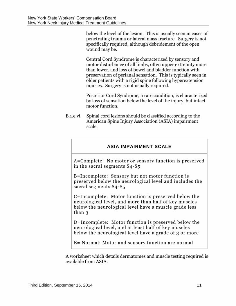
Central Cord Syndrome is characterized by sensory and motor disturbance of all limbs, often upper extremity more than lower, and loss of bowel and bladder function with preservation of perianal sensation. This is typically seen in older patients with a rigid spine following hyperextension injuries. Surgery is not usually required.
Posterior Cord Syndrome, a rare condition, is characterized by loss of sensation below the level of the injury, but intact motor function.
B.1.e.vi Spinal cord lesions should be classified according to the American Spine Injury Association (ASIA) impairment scale.
ASIA IMPAIRMENT SCALE
A=Complete: No motor or sensory function is preserved in the sacral segments S4-S5
B=Incomplete: Sensory but not motor function is preserved below the neurological level and includes the sacral segments S4-S5
C=Incomplete: Motor function is preserved below the neurological level, and more than half of key muscles below the neurological level have a muscle grade less than 3
D=Incomplete: Motor function is preserved below the neurological level, and at least half of key muscles below the neurological level have a grade of 3 or more
E= Normal: Motor and sensory function are normal
A worksheet which details dermatomes and muscle testing required is available from ASIA.
New York State Workers’ Compensation Board New York Neck Injury Medical Treatment Guidelines
Third Edition, September 15, 2014 12
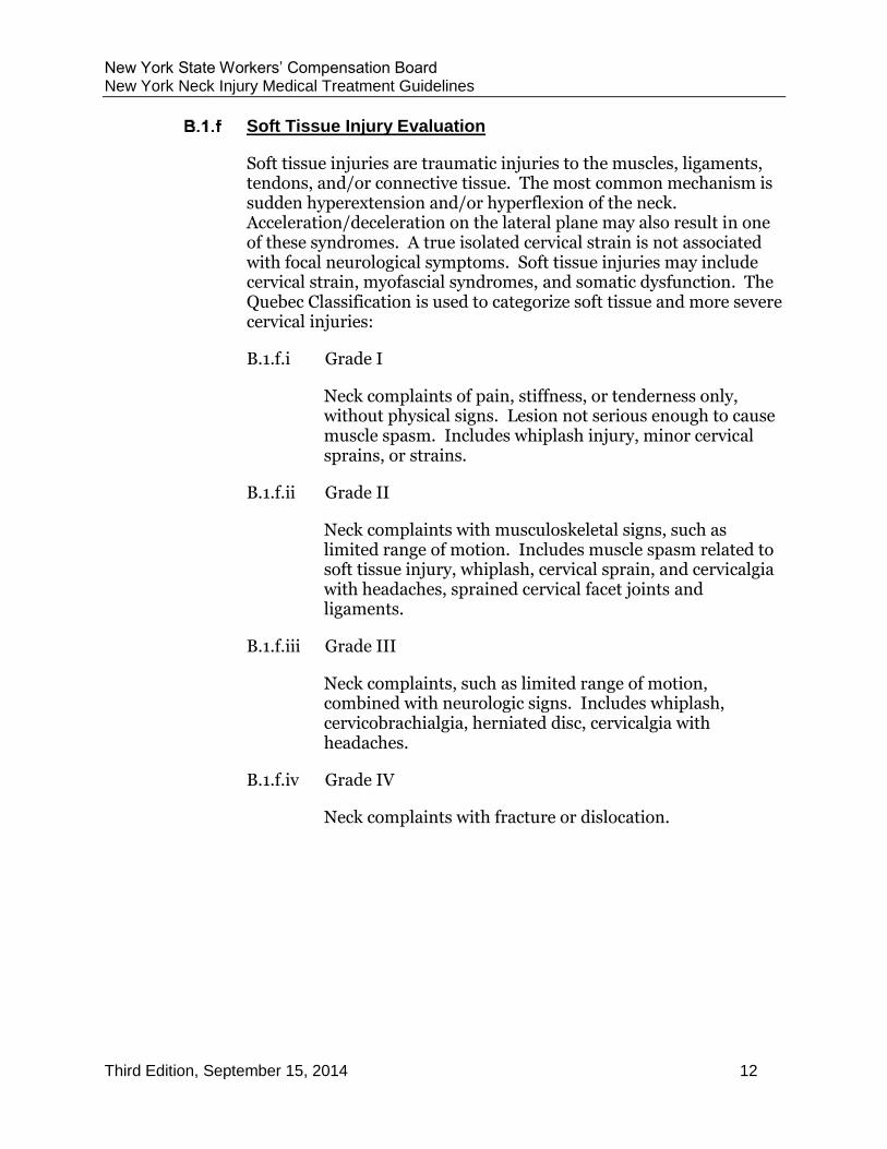
Soft Tissue Injury Evaluation
Soft tissue injuries are traumatic injuries to the muscles, ligaments, tendons, and/or connective tissue. The most common mechanism is sudden hyperextension and/or hyperflexion of the neck. Acceleration/deceleration on the lateral plane may also result in one of these syndromes. A true isolated cervical strain is not associated with focal neurological symptoms. Soft tissue injuries may include cervical strain, myofascial syndromes, and somatic dysfunction. The Quebec Classification is used to categorize soft tissue and more severe cervical injuries:
B.1.f.i Grade I
Neck complaints of pain, stiffness, or tenderness only, without physical signs. Lesion not serious enough to cause muscle spasm. Includes whiplash injury, minor cervical sprains, or strains.
B.1.f.ii Grade II
Neck complaints with musculoskeletal signs, such as limited range of motion. Includes muscle spasm related to soft tissue injury, whiplash, cervical sprain, and cervicalgia with headaches, sprained cervical facet joints and ligaments.
B.1.f.iii Grade III
Neck complaints, such as limited range of motion, combined with neurologic signs. Includes whiplash, cervicobrachialgia, herniated disc, cervicalgia with headaches.
B.1.f.iv Grade IV
Neck complaints with fracture or dislocation.
New York State Workers’ Compensation Board New York Neck Injury Medical Treatment Guidelines
Third Edition, September 15, 2014 13
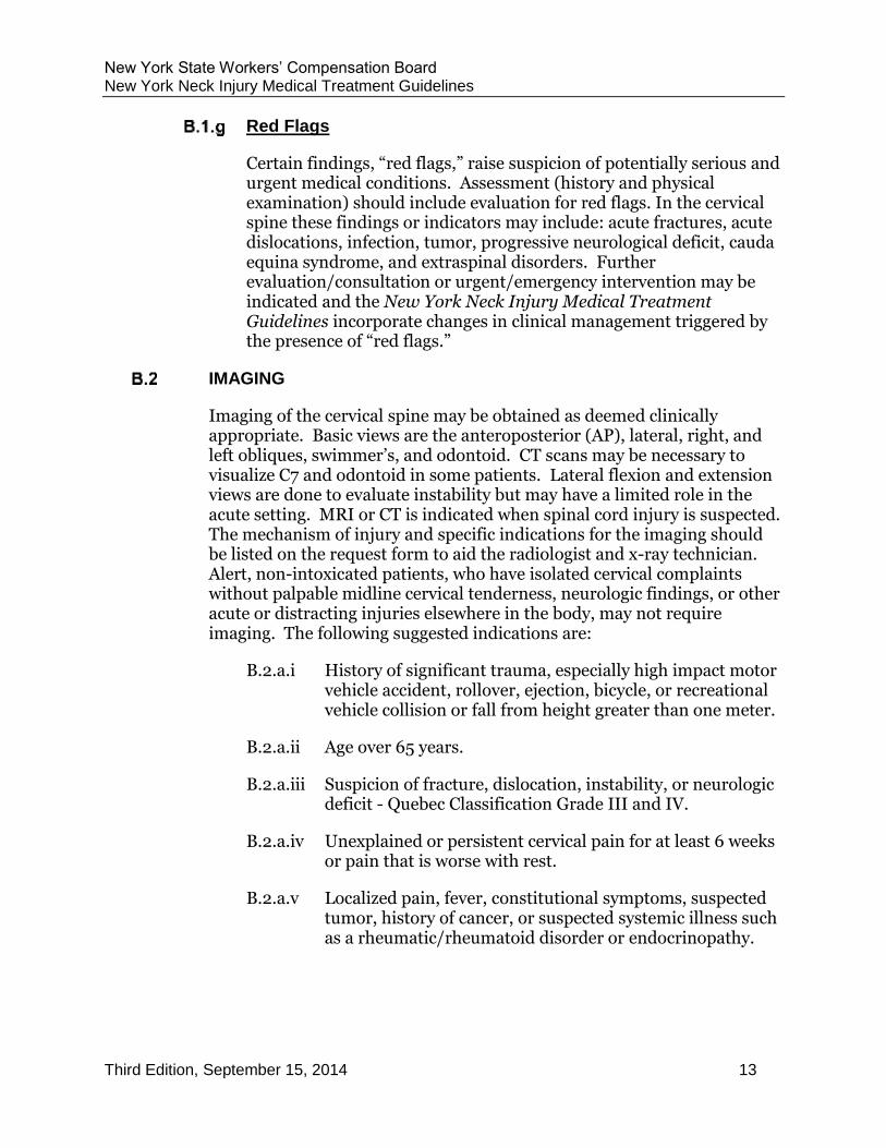
Red Flags
Certain findings, “red flags,” raise suspicion of potentially serious and urgent medical conditions. Assessment (history and physical examination) should include evaluation for red flags. In the cervical spine these findings or indicators may include: acute fractures, acute dislocations, infection, tumor, progressive neurological deficit, cauda equina syndrome, and extraspinal disorders. Further evaluation/consultation or urgent/emergency intervention may be indicated and the New York Neck Injury Medical Treatment Guidelines incorporate changes in clinical management triggered by the presence of “red flags.”
IMAGING
Imaging of the cervical spine may be obtained as deemed clinically appropriate. Basic views are the anteroposterior (AP), lateral, right, and left obliques, swimmer’s, and odontoid. CT scans may be necessary to visualize C7 and odontoid in some patients. Lateral flexion and extension views are done to evaluate instability but may have a limited role in the acute setting. MRI or CT is indicated when spinal cord injury is suspected. The mechanism of injury and specific indications for the imaging should be listed on the request form to aid the radiologist and x-ray technician. Alert, non-intoxicated patients, who have isolated cervical complaints without palpable midline cervical tenderness, neurologic findings, or other acute or distracting injuries elsewhere in the body, may not require imaging. The following suggested indications are:
B.2.a.i History of significant trauma, especially high impact motor vehicle accident, rollover, ejection, bicycle, or recreational vehicle collision or fall from height greater than one meter.
B.2.a.ii Age over 65 years.
B.2.a.iii Suspicion of fracture, dislocation, instability, or neurologic deficit - Quebec Classification Grade III and IV.
B.2.a.iv Unexplained or persistent cervical pain for at least 6 weeks or pain that is worse with rest.
B.2.a.v Localized pain, fever, constitutional symptoms, suspected tumor, history of cancer, or suspected systemic illness such as a rheumatic/rheumatoid disorder or endocrinopathy.
New York State Workers’ Compensation Board New York Neck Injury Medical Treatment Guidelines
Third Edition, September 15, 2014 14
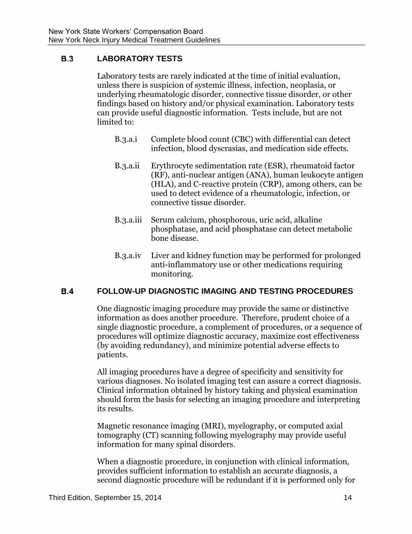
LABORATORY TESTS
Laboratory tests are rarely indicated at the time of initial evaluation, unless there is suspicion of systemic illness, infection, neoplasia, or underlying rheumatologic disorder, connective tissue disorder, or other findings based on history and/or physical examination. Laboratory tests can provide useful diagnostic information. Tests include, but are not limited to:
B.3.a.i Complete blood count (CBC) with differential can detect infection, blood dyscrasias, and medication side effects.
B.3.a.ii Erythrocyte sedimentation rate (ESR), rheumatoid factor (RF), anti-nuclear antigen (ANA), human leukocyte antigen (HLA), and C-reactive protein (CRP), among others, can be used to detect evidence of a rheumatologic, infection, or connective tissue disorder.
B.3.a.iii Serum calcium, phosphorous, uric acid, alkaline phosphatase, and acid phosphatase can detect metabolic bone disease.
B.3.a.iv Liver and kidney function may be performed for prolonged anti-inflammatory use or other medications requiring monitoring.
FOLLOW-UP DIAGNOSTIC IMAGING AND TESTING PROCEDURES
One diagnostic imaging procedure may provide the same or distinctive information as does another procedure. Therefore, prudent choice of a single diagnostic procedure, a complement of procedures, or a sequence of procedures will optimize diagnostic accuracy, maximize cost effectiveness (by avoiding redundancy), and minimize potential adverse effects to patients.
All imaging procedures have a degree of specificity and sensitivity for various diagnoses. No isolated imaging test can assure a correct diagnosis. Clinical information obtained by history taking and physical examination should form the basis for selecting an imaging procedure and interpreting its results.
Magnetic resonance imaging (MRI), myelography, or computed axial tomography (CT) scanning following myelography may provide useful information for many spinal disorders.
When a diagnostic procedure, in conjunction with clinical information, provides sufficient information to establish an accurate diagnosis, a second diagnostic procedure will be redundant if it is performed only for
New York State Workers’ Compensation Board New York Neck Injury Medical Treatment Guidelines
Third Edition, September 15, 2014 15
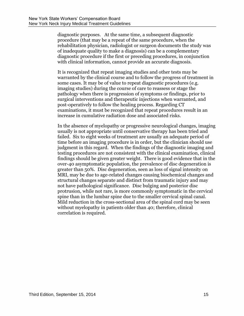
diagnostic purposes. At the same time, a subsequent diagnostic procedure (that may be a repeat of the same procedure, when the rehabilitation physician, radiologist or surgeon documents the study was of inadequate quality to make a diagnosis) can be a complementary diagnostic procedure if the first or preceding procedures, in conjunction with clinical information, cannot provide an accurate diagnosis.
It is recognized that repeat imaging studies and other tests may be warranted by the clinical course and to follow the progress of treatment in some cases. It may be of value to repeat diagnostic procedures (e.g. imaging studies) during the course of care to reassess or stage the pathology when there is progression of symptoms or findings, prior to surgical interventions and therapeutic injections when warranted, and post-operatively to follow the healing process. Regarding CT examinations, it must be recognized that repeat procedures result in an increase in cumulative radiation dose and associated risks.
In the absence of myelopathy or progressive neurological changes, imaging usually is not appropriate until conservative therapy has been tried and failed. Six to eight weeks of treatment are usually an adequate period of time before an imaging procedure is in order, but the clinician should use judgment in this regard. When the findings of the diagnostic imaging and testing procedures are not consistent with the clinical examination, clinical findings should be given greater weight. There is good evidence that in the over-40 asymptomatic population, the prevalence of disc degeneration is greater than 50%. Disc degeneration, seen as loss of signal intensity on MRI, may be due to age-related changes causing biochemical changes and structural changes separate and distinct from traumatic injury and may not have pathological significance. Disc bulging and posterior disc protrusion, while not rare, is more commonly symptomatic in the cervical spine than in the lumbar spine due to the smaller cervical spinal canal. Mild reduction in the cross-sectional area of the spinal cord may be seen without myelopathy in patients older than 40; therefore, clinical correlation is required.
New York State Workers’ Compensation Board New York Neck Injury Medical Treatment Guidelines
Third Edition, September 15, 2014 16
DIAGNOSTIC STUDIES
The studies below are listed in frequency of use, not importance.
IMAGING STUDIES
Magnetic Resonance Imaging (MRI)
MRI is useful in suspected nerve root compression, in myelopathy to evaluate the spinal cord and/or differentiate or rule out masses, infections such as epidural abscesses or disc space infection, bone marrow involvement by metastatic disease, and/or suspected disc herniation or cord contusion following severe neck injury. MRI should be performed immediately if there is a question of infection or metastatic disease with cord compression. MRI is contraindicated in patients with certain implanted devices.
In general, the high field, conventional, MRI provides better resolution. A lower field scan with lower magnetic intensity may be indicated when a patient cannot fit into a high field scanner or is too claustrophobic despite sedation.
Inadequate resolution on the first scan may require a second MRI using a different technique. A subsequent diagnostic MRI may be a repeat of the same procedure when the rehabilitation physician, radiologist or surgeon documents that the study was of inadequate quality to make a diagnosis. All questions in this regard should be discussed with the MRI center and/or radiologist.
Ferrous material/metallic objects present in the tissues is a contraindication for the performance of an MRI.
Specialized MRI Scans
C.1.a.i MRI with 3-dimensional reconstruction:
On rare occasions, MRI with 3-dimensional reconstruction views may be used as a pre-surgical diagnostic procedure to obtain accurate information of characteristics, location, and spatial relationships among soft tissue and bony structures.
C.1.a.ii Dynamic-kinetic MRI of the spine:
Dynamic-kinetic MRI of the spine uses an MRI unit configured with a top-front open design which enables upright, weight-bearing patient positioning in a variety of postures not obtainable with the recumbent images derived from conventional, closed unit MRI systems. Imaging can
New York State Workers’ Compensation Board New York Neck Injury Medical Treatment Guidelines
Third Edition, September 15, 2014 17
be obtained in flexion, extension, and rotation of the spine, as well as in erect positioning. There is a theoretical advantage to imaging sequences obtained under more physiologic conditions than in the supine position. There is currently ongoing research to establish whether the theoretical advantages of positional and kinetic MRI result in improved sensitivity and specificity in detecting spine pathology. Currently it remains investigational, and is not recommended until the correlation with clinical syndromes is firmly established.
Computed Axial Tomography (CT)
Computed Axial Tomography (CT) provides excellent visualization of bone and is used to further evaluate bony masses and suspected fractures not clearly identified on radiographic evaluation. It may sometimes be done as a complement to MRI scanning to better delineate bony osteophyte formation in the neural foramen. CT is usually utilized for suspected cervical spine fracture in a patient with negative plain films, or to further delineate a cervical fracture. CT scanning is also quite useful for congenital anomalies at the skull base and at the C1-2 levels. Plain CT scanning is poor for the C6-7 or C7-T1 levels because of shoulder artifact. Instrument-scatter reduction software provides better resolution when metallic artifact is of concern. When ferrous/ metallic materials are present in the tissues, CT should be ordered rather than an MRI. CT examinations, it should be remembered, deliver a considerable radiation dose and carry with them associated radiation-related risks.
Myelography
Myelography is the injection of radiopaque material into the spinal subarachnoid space, with x-rays then taken to define anatomy. It may be used as a pre-surgical diagnostic procedure to obtain accurate information of characteristics, location, and spatial relationships among soft tissue and bony structures. Myelography is an invasive procedure with complications including nausea, vomiting, headache, convulsion, arachnoiditis, CSF leakage, allergic reactions, bleeding, and infection. Myelography, therefore, should only be considered when CT and MRI are unavailable, for morbidly obese patients or for those who have undergone multiple operations, and when other tests prove non-diagnostic in the surgical candidate. The use of small needles and a less toxic, water-soluble, nonionic contrast is recommended.
New York State Workers’ Compensation Board New York Neck Injury Medical Treatment Guidelines
Third Edition, September 15, 2014 18
CT Myelogram
CT Myelogram provides more detailed information about relationships between neural elements and surrounding anatomy and is appropriate in patients with multiple prior operations or tumorous conditions only for pre-surgical testing.
Lineal Tomography
Lineal Tomography is infrequently used, yet may be helpful in the evaluation of bone surfaces, bony fusion, or pseudoarthrosis.
Bone Scan (Radioisotope Bone Scanning)
Bone scanning is more sensitive but less specific than MRI. 99M Technetium diphosphonate uptake reflects osteoblastic activity and may be useful in diagnosing metastatic/primary bone tumors, stress fractures, osteomyelitis, and inflammatory lesions, but cannot distinguish between these entities. In the cervical spine, the usual indication is to evaluate for neoplastic conditions. Chiefly indicated with persistent symptoms with otherwise normal diagnostic tests or to differentiate old vs. new lesions. Other indications include occult fracture or infection.
Other Radioisotope Scanning
Indium and gallium scans are usually used to help diagnose lesions seen on other diagnostic imaging studies. 67Gallium citrate scans are used to localize tumor, infection, and abscesses.
Dynamic [Digital] Fluoroscopy
Dynamic [Digital] Fluoroscopy of the cervical spine measures the motion of intervertebral segments using a videofluoroscopy unit to capture images as the subject performs cervical flexion and extension, storing the anatomic motion of the spine in a computer. Dynamic Fluoroscopy may be used in designated trauma centers to evaluate the cervical spine. Its superiority over MRI has not been established. If performed, full visualization of the cervical spine (C1 - T1).
New York State Workers’ Compensation Board New York Neck Injury Medical Treatment Guidelines
Third Edition, September 15, 2014 19
OTHER TESTS
The following diagnostic procedures are listed in alphabetical order, not by importance.
Electrodiagnostic Testing (EDX)
EDX include needle EMG (Electromyogram), peripheral nerve conduction velocity studies (NCV) and motor and sensory evoked potentials. Needle EMG can substantiate the diagnosis of radiculopathy or spinal stenosis in patients with neck pain and/or radiculopathy problems. Needle EMG can help determine if radiculopathy is acute or chronic. NCV are done in addition to needle EMG to rule out other potential causes for the symptoms (co-morbidity or alternate diagnosis involving peripheral nerves) and to confirm radiculopathy. It is recommended and preferred that EDX in the out-patient setting be performed and interpreted by physicians board-certified in Neurology or Physical Medicine and Rehabilitation.
In general, electrodiagnostic studies are complementary to imaging procedures such as CT, MRI, and/or myelography. Whereas X-ray, CT and MRI reflect structural changes, electrodiagnostic studies reflect neurologic functional status.
If significant radiating arm symptoms are present for greater than 4-6 weeks after the onset of injury and no obvious level of nerve root dysfunction is evident on examination, electrodiagnostic studies may be indicated. Electrodiagnostic studies may also be useful to determine the extent of injury in patients with an established level of injury.
C.2.a.i Portable Automated Electrodiagnostic Device (also known as Surface EMG).
Surface EMG is not appropriate for diagnostic evaluation of neck pain or neck injuries under any circumstances and is not recommended.
C.2.a.ii Somatosensory Evoked Potential (SSEP)
Somatosensory Evoked Potential (SSEP) is useful for the evaluation of myelopathy and is increasingly used intra-operatively. It is not recommended to identify radiculopathy.
New York State Workers’ Compensation Board New York Neck Injury Medical Treatment Guidelines
Third Edition, September 15, 2014 20
C.2.a.iii Current Perception Threshold Evaluation (CPT)
Current Perception Threshold Evaluation (CPT) may be useful as a screening tool, but its diagnostic efficacy in the evaluation of cervical spine pain has not been determined. Therefore, CPT is not recommended as a diagnostic tool.
Injections – Diagnostic
Atlanto-axial/atlanto-occipital.
Not Recommended.
Provocation Discography
Not Recommended. Improvement in surgical outcomes has not been shown to follow the use of discography, and there is evidence that performing discography on normal discs is associated with an enhanced risk of degenerative changes in those discs in later years.
Thermography
Not Recommended.
THERAPEUTIC PROCEDURES: NON-OPERATIVE
Before initiation of any therapeutic procedure, the authorized treating provider, employer, and insurer must consider these important issues in the care of the patient.
First, patients undergoing therapeutic procedure(s) should be released or returned to modified or restricted duty during their rehabilitation at the earliest appropriate time.
Second, cessation and/or review of treatment modalities should be undertaken when no further significant subjective or objective improvement in the patient’s condition is noted. If patients are not responding within the recommended duration periods, alternative treatment interventions, further diagnostic studies or consultations should be pursued.
Third, providers should provide and document education to the patient. No treatment plan is complete without addressing issues of individual and/or group patient education as a means of facilitating self-management of symptoms.
Lastly, for those patients who fail to make expected progress 6-12 weeks after an injury and whose subjective symptoms do not correlate with objective signs and tests, reexamination in order to confirm the accuracy of the diagnosis should be made. Formal psychological or psychosocial evaluation may be considered.
New York State Workers’ Compensation Board New York Neck Injury Medical Treatment Guidelines
Third Edition, September 15, 2014 21
Home therapy is an important component of therapy and may include active and passive therapeutic procedures as well as other modalities to assist in alleviating pain, swelling, and abnormal muscle tone.
The following procedures are listed in alphabetical order:
ACUPUNCTURE
Acupuncture is a procedure used for the relief of pain and inflammation, and there is some scientific evidence to support its use. The exact mode of action is only partially understood. Western medicine studies suggest that acupuncture stimulates the nervous system at the level of the brain, promotes deep relaxation, and affects the release of neurotransmitters. Acupuncture is commonly used as an alternative or in addition to traditional Western pharmaceuticals. While it is commonly used when pain medication is reduced or not tolerated, it may be used as an adjunct to physical rehabilitation and/or surgical intervention to hasten the return of functional activity. Moxibustion and other complementary integrative medicine techniques are often combined with acupuncture, but have no demonstrated efficacy. No additional reimbursement should be provided for acupuncture combined with moxibustion or other similar adjunctive procedures. Acupuncture must be performed by a professional who is authorized under the Workers’ Compensation Laws and duly certified in New York State to provide acupuncture services.
Acupuncture (with or without electrical stimulation) is the insertion and removal of filiform needles to stimulate acupoints (acupuncture points), with or without the use of electrical current (micro-amperage or milli-amperage) on the needles at the acupuncture site. Needles may be inserted, manipulated and retained for a period of time. Acupuncture can be used to reduce pain, reduce inflammation, increase blood flow, increase range of motion, decrease the side effect of medication-induced nausea, promote relaxation in an anxious patient, and reduce muscle spasm. Indications include joint pain, joint stiffness, soft tissue pain and inflammation, paresthesia, post-surgical pain relief, muscle spasm, and scar tissue pain.
Time to produce effect: 3 to 6 treatments.
Frequency: 1 to 3 times per week.
Optimum duration: 1 month.
Maximum duration: 10 treatments.
Total Time Frames for Acupuncture and Acupuncture with Electrical Stimulation: Time frames are not meant to be applied to each of the above
New York State Workers’ Compensation Board New York Neck Injury Medical Treatment Guidelines
Third Edition, September 15, 2014 22
sections separately. The time frames are to be applied to all acupuncture treatments regardless of the type or combination of therapies being provided.
Acupuncture treatments may extend longer if objective functional gains can be documented or when symptomatic benefits facilitate progression in the patient’s treatment program. Treatment beyond 10 treatments must be documented with respect to need and ability to facilitate positive symptomatic or functional gains.
BIOFEEDBACK
Biofeedback is a form of behavioral medicine that helps patients learn self-awareness and self-regulation skills for the purpose of gaining greater control of their physiology, such as muscle activity, brain waves, and measures of autonomic nervous system activity. Electronic instrumentation is used to monitor the targeted physiology and then displayed or fed back to the patient visually, auditorially, or tactilely, with coaching by a biofeedback specialist. Biofeedback is provided by clinicians certified in biofeedback and/or who have documented specialized education, advanced training, or direct or supervised experience qualifying them to provide the specialized treatment needed (e.g., surface EMG, EEG, or other).
Treatment is individualized to the patient’s work-related diagnosis and needs. Home practice of skills is required for mastery and may be facilitated by the use of home training tapes. The ultimate goal of biofeedback treatment is to normalize the physiology to the pre-injury status to the extent possible, and involves transfer of learned skills to the workplace and daily life. Candidates for biofeedback therapy or training must be motivated to learn and practice biofeedback and self-regulation techniques.
Indications for biofeedback include individuals who are suffering from musculoskeletal injury in which muscle dysfunction or other physiological indicators of excessive or prolonged stress response affects and/or delays recovery. Other applications include training to improve self-management of emotional stress/pain responses such as anxiety, depression, anger, sleep disturbance, and other central and autonomic nervous system imbalances. Biofeedback is often used in conjunction with other treatment modalities.
A.1.a.i Biofeedback is not appropriate for individuals suffering from acute neck pain or acute injury.
New York State Workers’ Compensation Board New York Neck Injury Medical Treatment Guidelines
Third Edition, September 15, 2014 23
A.1.a.ii Biofeedback is recommended for select patients with non-acute neck pain, as a component of an interdisciplinary approach. Please consult the New York Non-Acute Pain Medical Treatment Guidelines for further recommendations.
INJECTIONS: THERAPEUTIC
Therapeutic Spinal Injections-Introduction
Description:
Therapeutic spinal injections may be used after initial conservative treatments, such as physical and occupational therapy, medication, manual therapy, exercise, or acupuncture have been undertaken.
Therapeutic injections should be used only after imaging studies and diagnostic injections have established pathology.
Injections are invasive procedures that can cause catastrophic complications; thus clinical indications and contraindications should be closely adhered to.
The purpose of spinal injections is to facilitate active therapy by providing short-term relief through reduction of pain and inflammation.
All patients should continue appropriate exercise with functionally directed rehabilitation.
Active treatment, which patients should have had prior to injections, will frequently require a repeat of the sessions previously ordered.
Injections, by themselves, are not likely to provide long-term relief. Rather, active rehabilitation with modified work achieves long-term relief by increasing active range of motion, strength, and stability.
Injections should not be repeated if the first injection does not provide:
o Improvement in function
o Temporary and sustained pain relief as measured by accepted pain scales, i.e., 50% pain reduction on Visual Analog Scale
New York State Workers’ Compensation Board New York Neck Injury Medical Treatment Guidelines
Third Edition, September 15, 2014 24
and/or
o Reduction in the use of prescribed analgesic medication.
Medical management should be continued or adjusted based upon patient assessment and response.
Special Considerations:
For all injections (excluding trigger point and occipital nerve blocks) multi-planar fluoroscopy during procedures is required to document technique and needle placement.
All injections (excluding trigger point) must be performed by a physician experienced in the procedure. Trigger point injections may be performed by a physician or a Nurse Practitioner/Physician Assistant experienced in the procedure.
Permanent images are required to verify needle placement.
The subspecialty disciplines of the physicians performing injections may be varied, including, but not limited to: anesthesiology, radiology, surgery, or physiatry.
The practitioner should have completed fellowship training in pain medicine with interventional training or its equivalent. The practitioner must also be knowledgeable in radiation safety.
Complications:
General complications of spinal injections may include:
transient neurapraxia
local pain
nerve injury
infection
headache
vasovagal effects
New York State Workers’ Compensation Board New York Neck Injury Medical Treatment Guidelines
Third Edition, September 15, 2014 25
Epidural hematoma, permanent neurologic damage, dural perforation and CSF leakage, and/or spinal meningeal abscess may also occur.
More serious complications are rare but can include spinal cord damage, quadriplegia, permanent ataxia, and death.
With steroid injections, there may be a dose-dependent suppression of the hypothalamic-pituitary-adrenal axis lasting between one and three months.
Contraindications:
Absolute contraindications to therapeutic injections include:
bacterial infection – systemic or localized to region of injection
bleeding diatheses
hematological conditions
possible pregnancy
Relative contraindications to diagnostic injections may include:
allergy to contrast
poorly controlled Diabetes Mellitus
hypertension
Drugs affecting coagulation, such as aspirin, NSAIDs, anti-platelets or anticoagulants require restriction from use.
Decisions regarding the number of restricted days before a procedure should be made in consultation with the prescribing physician and other specialists as indicated.
D.3.a.i Cervical Epidural/Interlaminar Steroid Injections (ESI)
Description:
Cervical ESI are injections of corticosteroid into the epidural space.
The purpose of ESI is to reduce pain and inflammation, restoring range of motion and thereby facilitating progress in more active programs.
New York State Workers’ Compensation Board New York Neck Injury Medical Treatment Guidelines
Third Edition, September 15, 2014 26
As with all treatments, it is important to insure that patients have realistic expectations regarding treatment outcomes.
Diabetics who are candidates for ESI should be counseled that a blood glucose increase may be apparent post-intervention, but effects should not last longer than approximately two days.
Needle Placement: Cervical ESIs must be fluoroscopically guided to verify needle placement. Permanent images are required to verify needle placement.
Contrast epidurograms allow one to verify the flow of medication into the epidural space. One epidurogram is recommended per series of ESI injections as clinically indicated.
Recommendations:
Cervical ESIs are useful in patients with symptoms of cervical radicular pain syndromes.
Cervical ESIs are not effective for cervical axial pain or non-radicular pain syndromes and they are not recommended for these indications.
Cervical transforaminal injections are not recommended.
Use of anesthetics is generally not recommended for cervical ESI.
Maximum Frequency:
Three injections may be done in one 12-month period (per spinal region) depending on patient response (improved function and pain reduction). No more than one level per treatment session.
It is recommended that each injection be scheduled separately and effects of each injection be evaluated, depending upon patient response (improved function and pain reduction) rather than scheduling a “Series of Three.”
New York State Workers’ Compensation Board New York Neck Injury Medical Treatment Guidelines
Third Edition, September 15, 2014 27
If the first injection does not provide a response with temporary and sustained pain relief (at least 2 weeks) substantiated by accepted pain scales (i.e. 50% pain reduction as measured by tools such as VAS) and improvement in function, repeat injections are not recommended.
A positive result (functional improvement) should include measurable improvement in physical activity goals, and a return to baseline function or to work duties.
Patients should be reassessed after each injection for:
Improvement in function
Temporary and sustained pain relief as measured by accepted pain scales, i.e., 50% pain reduction on Visual Analog Scale
and/or
Reduction in the use of prescribed analgesic medication.
Medical management should be continued or adjusted based upon patient assessment and response.
Discontinuation:
Resolution of symptoms, decrease in symptoms to a tolerable level or absence of response.
Epidural glucocorticosteroid injections are not recommended for acute or non-acute neck pain in the absence of significant radicular symptoms.
They are not recommended as treatment for any non-acute axial neck pain without a radicular component.
D.3.a.ii Cervical Diagnostic and Therapeutic Medial Nerve Branch Blocks/ Facet (Zygapophyseal) Joint Injections
Recommendations:
1) Facet joint (injection into the intra-articular facet joint space) or medial branch block injections (blocking
New York State Workers’ Compensation Board New York Neck Injury Medical Treatment Guidelines
Third Edition, September 15, 2014 28
medial nerve innervation of the facet joint) may be indicated for acute neck pain when there is continuing axial neck pain after an injury (for example, status post whiplash injury) that has not responded to conservative management. For acute pain, these injections involve a combination of an anesthetic and a steroid.
Recommended frequency: Three injections total for acute pain per 12-month period.
2) Diagnostic medial branch block injections (anesthetic only) or diagnostic facet joint injections (anesthetic only) are not recommended for acute neck pain.
3) Medial branch block injections are recommended for a select group of patients with non-acute neck pain in order to determine whether specific interventions targeting the facet joint (by blocking medial nerve innervation to the facet joint) should be performed.
Medial branch block injections are recommended for:
Patients with pain suspected to be largely facet in origin based on exam findings (i.e.: non-radicular pain aggravated by extension-facet loading)
or
Patients who have facet findings with referred pain to the axial thoracic or occipital area
and
Documented evidence (i.e., imaging study) of facet disease (facet arthropathy/hypertrophy at the targeted level(s)
and
Who have completed a documented course of conservative management as defined in the Neck Injury Medical Treatment Guideline, including but not limited to medication, modalities, and active exercises.
In these patients, medial branch block injections may aid in identifying pain generators, therapeutically
New York State Workers’ Compensation Board New York Neck Injury Medical Treatment Guidelines
Third Edition, September 15, 2014 29
reduce pain and may be useful in facilitating progress in a rehabilitation program.
Patients should be reassessed after each injection for a documented 50% improvement in pain as measured by accepted pain scales and evidence of functional improvement for at least 4-6 weeks.
4) These injections must be fluoroscopically guided.
Description:
Cervical medial nerve branch blocks may consist of a diagnostic and/or a therapeutic component.
The diagnostic component consists of an anesthetic and the therapeutic component, a corticosteroid.
For non-acute pain, the diagnostic component (anesthetic only) may be used individually or may be combined with a steroid into a single diagnostic/therapeutic injection.
A medial nerve branch block is indicated for the diagnosis of pain that is suspected of arising from the facet joint.
Facet joint injections are not to be used as diagnostic tools for the purpose of determining the need for radiofrequency ablation.
A history and physical examination should document the rationale for the suspected diagnosis.
Positive Diagnostic Medial Nerve Branch Block Response
A positive response to the diagnostic component of a medial nerve branch block consists of an initial temporary improvement, which may be as short as 1-4 hours, and includes a reduction in pain (50% decrease as measured by accepted pain scales), and improvement in function for the duration of the local anesthetic.
If a patient has a positive response to a diagnostic medial branch block injection (whether or not steroids are used), a repeat medial branch block injection should be performed to confirm the diagnosis.
New York State Workers’ Compensation Board New York Neck Injury Medical Treatment Guidelines
Third Edition, September 15, 2014 30
This repeat comparative medial branch block injection should be performed on a different date to confirm the level of involvement.
If there is a positive response to the repeat diagnostic medial branch block injection, the patient should be evaluated to determine the need for more definitive treatment such as radiofrequency ablation.
When administering a diagnostic injection, consideration should be given to combining the anesthetic agent with steroid to allow for the potential of extended pain relief.
If there is not a positive response to the first diagnostic injection, the diagnosis should be re-evaluated.
If the first injection does not provide a positive response, repeat diagnostic injections are not recommended.
Positive Therapeutic Response (either Medial Branch Block Injection or Facet Joint Injection)
Therapeutically, steroid may be added to provide longer benefit. The goal of the prolonged therapeutic benefit is to decrease pain and increase function with the ability to participate in an active rehabilitation program (which the patient was unable to do prior to the injection).
Patients should be reassessed after each therapeutic injection for a documented 50% improvement in pain as measured by accepted pain scales and evidence of functional improvement.
A positive result (functional improvement) should include measurable improvement in physical activity goals including return to baseline or work activities.
Pain should be measured by accepted pain scales, pre-procedure, immediately post-procedure and at identified intervals after the procedure.
If the first therapeutic injection does not provide sustained pain relief substantiated by accepted pain scales (i.e., 50% documented pain reduction as measured by accepted pain tools) and improvement in
New York State Workers’ Compensation Board New York Neck Injury Medical Treatment Guidelines
Third Edition, September 15, 2014 31
function for at least 4-to-6 weeks, repeat steroid injections are not recommended.
A positive response to a therapeutic injection is not determinative of the need for radiofrequency ablation.
Time to produce effect: up to 30 minutes for local anesthetic; corticosteroid up to 72 hours.
Recommended frequency: 2- 3 injections for each applicable joint may be done in one 12-month period, not to exceed 3 joint levels (4 medial branch nerves) per session, depending upon patient’s documented response (i.e., improved functional gain and pain reduction). Maximum 3 sessions/year.
D.3.a.iii Intradiscal Steroid Therapy
Intradiscal Steroid Therapy consists of injection of a steroid preparation into the intervertebral disc under fluoroscopic guidance at the time of discography. There is good evidence that it is not effective in the treatment of suspected discogenic neck pain. There is no support for its use in the cervical spine and its use is not recommended.
D.3.a.iv Occipital Nerve Block
Description:
Occipital nerve blocks are injections used both diagnostically and therapeutically in the treatment of occipital neuralgia. The greater occipital nerve is the target.
Recommendations:
Diagnosis and treatment of occipital neuralgia/cephalgia. Peripheral block of the greater occipital nerve may be appropriate as initial treatment. It may be indicated in patients unresponsive to peripheral nerve block or those patients in need of additional diagnostic information.
Complications:
Bleeding, infection, neural injury. Post procedural ataxia is common and usually lasts 30 minutes post procedure. Because the occipital artery runs with the
New York State Workers’ Compensation Board New York Neck Injury Medical Treatment Guidelines
Third Edition, September 15, 2014 32
occipital nerve, inadvertent intravascular injection is a risk of this procedure and may lead to systemic toxicity and/or seizures.
Time to Produce Effect: Approximately 30 minutes for local anesthetic; 48 to 72 hours for corticosteroid.
Optimal Duration: 1 to 3 sessions.
Maximum Duration: Continue up to 3 injections if progressive symptomatic and functional improvement can be documented.
Trigger Point Injections and Dry Needling Treatment
Description:
Myofascial trigger points are localized hyperirritable palpable nodules in extremely sensitive bands of taut skeletal muscle fibers. These nodules are painful on compression and give rise to local pain and pain referred to distant structures.
Trigger point treatment consists only of dry needling or injection of local anesthetic into myofascial trigger points.
Trigger point injection is not the equivalent of acupuncture. Please refer to the acupuncture section in each Medical Treatment Guideline.
There is no evidence that injection of medications improves the results of trigger point injections. Needling alone may account for some of the therapeutic response.
As with all treatments, it is important to insure that patients have realistic expectations regarding treatment outcomes.
Recommendations:
Trigger point injections are not recommended for treatment of acute neck pain.
Trigger point injections may be reasonable secondary or tertiary options for non-acute pain that is not resolving with more conservative means (e.g., NSAIDs, progressive aerobic exercises, other exercises) within a 6-week time frame.
New York State Workers’ Compensation Board New York Neck Injury Medical Treatment Guidelines
Third Edition, September 15, 2014 33
Trigger point injections should be utilized primarily for facilitating functional progress.
Trigger point injections may be used to relieve myofascial pain and facilitate active therapy and stretching of the affected areas.
The use of therapeutic injections without participation in an active therapy program or in the context of maintaining employment is not recommended.
Patients should be reassessed two weeks after each injection for:
o Improvement in function
o Temporary and sustained pain relief as measured by accepted pain scales, i.e., 50% pain reduction on Visual Analog Scale
and/or
o Reduction in the use of prescribed analgesic medication.
Medical management should be continued or adjusted based upon patient assessment and response.
Functional improvement should last for 3 months.
A positive result (functional improvement) should include measurable improvement in physical activity goals, and a return to baseline function or to work duties.
The use of ultrasound or other imaging studies for trigger point injections is not recommended.
Frequency:
Not more than 4 injections per session, not to exceed 4 sessions per 12- month period.
If there is a partial demonstrated improvement after the first set of injections, a second set may be reasonable.
It is recommended to allow at least 3 to 4 weeks between these injections.
New York State Workers’ Compensation Board New York Neck Injury Medical Treatment Guidelines
Third Edition, September 15, 2014 34
If there are no subjective and objective improvements at that point, further injections are not recommended.
Repeated injections should be linked to subjective and objective improvement.
Prolotherapy
Also known as sclerotherapy, consists of a series of injections of hypertonic dextrose, with or without glycerine and phenol, into the ligamentous structures of the neck. There is no evidence that prolotherapy is effective in cervical pain. The injections are invasive, may be painful to the patient, and are not generally accepted or widely used. Therefore, the use of prolotherapy for cervical pain is not recommended.
Platelet Rich Plasma (PRP)
Not recommended.
Epiduroscopy and Epidural Lysis of Adhesions
Not recommended in the cervical spine secondary to the potential for dural puncture, hematoma, and spinal cord injury.
RADIOFREQUENCY ABLATION, NEUROTOMY, FACET RHIZOTOMY
Description:
A procedure designed to denervate the facet joint by ablating the corresponding sensory medial branches. Continuous percutaneous radiofrequency is the method generally used.
Radiofrequency medial branch neurotomy is recommended as the procedure of choice over alcohol, phenol, other injectable agents, or cryoablation.
Fluoroscopic guidance is required for precise positioning of the probe.
Permanent images should be recorded to verify placement of the device.
Recommendations:
For patients with proven facet joint pain in whom two diagnostic medial nerve branch blocks have been therapeutically successful, the use of radiofrequency ablation/neurotomy/facet rhizotomy may be indicated.
New York State Workers’ Compensation Board New York Neck Injury Medical Treatment Guidelines
Third Edition, September 15, 2014 35
This procedure is not recommended for involvement of more than 3 facet joints (4 medial branch nerves).
All patients should have a successful response to a diagnostic medial nerve branch block and a separate comparative block.
To be a positive diagnostic block, the patient should report a reduction of pain of 50% or greater from baseline for the length of time appropriate for the local anesthetic used, correlated with functional improvement.
The patient should also identify activities of daily living (which may include measurements of range of motion) that are impeded by their pain. The physician should observe and document functional improvement in the identified activities in the clinical setting.
Post-Procedure Therapy:
Active therapy
Implementation of a gentle reconditioning program within the first post-procedure week is recommended, barring complications.
Instruction and participation in a long-term home-based program of range of motion, cervical, scapular, and thoracic strengthening, postural or neuromuscular re-education, endurance, and stability exercises should be accomplished over a period of four to ten visits post-procedure.
Requirements for repeat radiofrequency neurotomy (or additional level radiofrequency neurotomies):
In some cases pain may recur.
Successful rhizotomy usually provides from six to nine months of relief.
Before a repeat radiofrequency neurotomy is done, a confirmatory medial branch injection may be performed if the patient’s pain pattern presents differently than in the initial evaluation.
Maximum Frequency:
Twice a year as indicated by improvement in pain and function.
New York State Workers’ Compensation Board New York Neck Injury Medical Treatment Guidelines
Third Edition, September 15, 2014 36
MEDICATION
Medication use in the treatment of cervical injuries is appropriate for controlling acute and non-acute pain and inflammation. Use of medications will vary widely due to the spectrum of injuries.
All drugs should be used according to patient needs. A thorough medication history, including use of alternative and over-the-counter medications, should be performed at the time of the initial visit and updated periodically. Treatment for pain control is initially accomplished with acetaminophen and/or NSAIDs. The patient should be educated regarding the interaction with prescription and over-the-counter medications as well as the contents of over-the-counter herbal products.
Please consult the New York Non-Acute Pain Medical Treatment Guidelines for additional recommendations on the use of medications in non-acute pain.
The following medications are listed in alphabetical order.
Acetaminophen
Acetaminophen is an effective analgesic with antipyretic but not anti-inflammatory activity. Acetaminophen is generally well tolerated, causes little or no gastrointestinal irritation, and is not associated with ulcer formation. Acetaminophen has been associated with liver toxicity when the recommended daily dose is exceeded or in patients who chronically use alcohol. Patients may not realize that many over-the-counter preparations may contain acetaminophen. In general, the total daily dose of acetaminophen should not exceed 3 grams per 24-hour period from all sources, including narcotic-acetaminophen combination preparations. Patients who consume three or more alcoholic drinks per day are at greater risk for liver toxicity, and consideration should be given to the use of other analgesics or limiting the acetaminophen dose to 2 grams per 24-hour period from all sources. Monitoring liver function via blood testing for use beyond 10 days is advisable.
Recommendations:
D.5.a.i Acetaminophen is a reasonable alternative to NSAIDs, although evidence suggests it is modestly less efficacious.
D.5.a.ii Acetaminophen is recommended for treatment of neck pain with or without radicular symptoms, particularly for those with contraindications for NSAIDs.
New York State Workers’ Compensation Board New York Neck Injury Medical Treatment Guidelines
Third Edition, September 15, 2014 37
Optimum Duration: 7 to 10 days.
Maximum Duration: Chronic use as indicated on a case-by-case basis. Please consult the New York Non-Acute Pain Medical Treatment Guidelines for further recommendations.
Anti-Depressants
Recommendations:
D.5.b.i Tricyclic anti-depressants (TCAs) are recommended for the treatment of non-acute neck pain that is not fully treated with NSAIDs and an exercise program. This intervention may be helpful where there is nocturnal sleep disruption and mild dysthymia.
Frequency and Duration: Generally prescribed at a very low dose at night and gradually increased (e.g., amitriptyline 25 mg qhs, increase by 25 mg each week until a sub-maximal or maximal dose is achieved, sufficient effects are achieved, or adverse effects occur. Most practitioners use lower doses (e.g. amitriptyline 25-75 mg/day to avoid the adverse effects and necessity of blood level monitoring), as there is no evidence of increased pain relief at higher doses. Imipramine is less sedating, thus if there is carryover daytime sedation, this may be a better option.
Discontinuation: Resolution of the pain, intolerance, or development of adverse effects.
There is limited evidence that tricyclic anti-depressants (TCAs) result in modest reductions in pain ratings in the treatment of radicular pain compared with placebo.
D.5.b.ii The selective serotonin reuptake inhibitors (e.g., paroxetine, as well as bupropion and trazodone) are not recommended for treatment of non-acute neck pain. (They may be nevertheless recommended for treatment of depression as noted previously.) There is strong evidence that treatment with these medications is not of benefit, thus their use is not recommended for the management of non-acute neck pain without depression.
New York State Workers’ Compensation Board New York Neck Injury Medical Treatment Guidelines
Third Edition, September 15, 2014 38
D.5.b.iii There is no quality evidence supporting the efficacy of anti-depressants in the treatment of acute neck pain. Absent other indicators of a need for such treatment, this intervention is not recommended for the management of acute neck pain.
Anti-Seizure Drugs
Recommendations:
Topiramate
D.5.c.i Topiramate is recommended for limited use in select patients with non-acute neck pain, where there has been failure of multiple other modalities including trials of different NSAIDs, aerobic exercise, specific stretching exercise, strengthening exercise, tricyclic anti-depressants, distractants, and manipulation.
Frequency/Dose: This medication is initiated by gradually increasing the dose. It has been initiated with a beginning dose of 50 mg and increasing by 50 mg a week. The most appropriate steady dose is unclear, but appears to be 300 mg. Patients should be carefully monitored for the development of adverse events.
Discontinuation: Resolution, development of adverse effects, or failure to adhere to a functional restoration program. Careful monitoring of employed patients is indicated due in part to elevated risks for central nervous system (CNS) sedating adverse effects.
Topiramate is not recommended for neuropathic pain, including peripheral neuropathy.
Carbamazepine
D.5.c.ii Carbamazepine is recommended as a potential adjunct for non-acute radicular or neuropathic pain after attempting other treatments (e.g., other medications, aerobic exercise, other exercise, manipulation). While there is not quality evidence for treatment of non-acute radicular neck pain, this may be tried if other medications have failed. Oxcarbazepine and lamotrigine may be useful agents to try if the results from carbamazepine are insufficient pain relief.
New York State Workers’ Compensation Board New York Neck Injury Medical Treatment Guidelines
Third Edition, September 15, 2014 39
Frequency/Duration: Frequency and dosing are based on the medication prescribed.
Discontinuation: Resolution of neck pain, lack of efficacy, or development of side effects that necessitate discontinuation. Careful monitoring of employed patients is indicated due to elevated risks for CNS sedating adverse effects.
Gabapentin and Pregabalin
D.5.c.iii Gabapentin is recommended for peri-operative management of pain to reduce need for opioids, particularly in those with side effects from opioids.
Discontinuation: Resolution or intolerance. Careful monitoring of employed patients is indicated due in part to elevated risks for CNS-sedating adverse effects.
Gabapentin may be considered for the treatment of severe neurogenic claudication from spinal stenosis or chronic radicular pain syndromes with limited walking distance.
Discontinuation: Resolution or intolerance. Careful monitoring of employed patients is indicated due in part to elevated risks for CNS-sedating adverse effects.
Gabapentin is not recommended for axial or non-neuropathic pain.
Compound Medications
Topical, oral and/or systemic compound medications are not recommended.
Narcotics
Narcotics should be primarily reserved for the treatment of severe neck pain. In mild-to-moderate cases of pain, narcotic medication should be used cautiously on a case-by-case basis. Adverse effects include respiratory depression, the development of physical and psychological dependence, and impaired alertness. These medications have physically addictive properties and withdrawal symptoms may follow abrupt discontinuation.
New York State Workers’ Compensation Board New York Neck Injury Medical Treatment Guidelines
Third Edition, September 15, 2014 40
Narcotic medications should be prescribed with strict time, quantity, and duration guidelines, and with definitive cessation parameters. Pain is subjective in nature and should be evaluated using a scale to rate effectiveness of the narcotic prescribed.
Optimum Duration: 3 to 7 days.
Maximum Duration: 2 weeks. Use beyond two weeks is acceptable in appropriate cases.
Any use beyond the maximum should be documented and justified based on the diagnosis and/or invasive procedures.
Please consult the New York Non-Acute Pain Medical Treatment Guidelines for detailed recommendations regarding the use of opioids.
Nonsteroidal Anti-Inflammatory Drugs (NSAIDs)
NSAIDs are useful for pain and inflammation. In mild cases, they may be the only drugs required for analgesia. There are several classes of NSAIDs and the response of the individual patient to a specific medication is unpredictable. For this reason a range of orally administered NSAIDs may be tried in each case with the most effective preparation being continued. Patients should be closely monitored for adverse reactions.
The US Food and Drug Administration advises that all NSAIDs may cause an increased risk of serious cardiovascular thrombotic events, myocardial infarction, and stroke, which can be fatal. Prescribers should be aware of the most updated information on this topic. Some NSAIDs may have more favorable cardiovascular risk factors than others.
Administration of proton pump inhibitors, histamine 2 blockers, or misoprostol, a prostaglandin analog, along with these NSAIDs may reduce the risk of duodenal and gastric ulceration associated with NSAID use but do not impact possible cardiovascular complications. Due to the cross reactivity between aspirin and NSAIDs, NSAIDs should not be used in aspirin-sensitive patients, and should be used with caution in all asthma patients. NSAIDs are associated with abnormal renal function, including renal failure, as well as abnormal liver function. Certain NSAIDs may have interactions with various other medications. Individuals may have adverse events not listed above. Intervals for metabolic screening are dependent upon the patient’s age, general health status and should be within parameters listed for each specific medication. Complete blood count (CBC), liver
New York State Workers’ Compensation Board New York Neck Injury Medical Treatment Guidelines
Third Edition, September 15, 2014 41
and renal function should be monitored in patients on chronic NSAIDs.
In patients receiving low-dose aspirin for primary or secondary cardiovascular disease prevention, to minimize the potential for the NSAID to counteract the beneficial effects of aspirin, aspirin should be taken two hours before or at least eight hours after the NSAID.
Chronic use of NSAIDs is generally not recommended. Chronic NSAIDs may be used cautiously in selected cases with regular monitoring.
D.5.f.i Non-selective Nonsteroidal Anti-Inflammatory Drugs: Non-selective NSAIDs are generally recommended as first-line medications.
Serious gastrointestinal toxicity, such as bleeding, perforation, and ulceration can occur at any time, with or without warning symptoms in patients treated with traditional NSAIDs. Patients at particularly high risk for GI bleeding include those with a history of prior GI bleed, diabetes, alcohol use, smoking, corticosteroid or anticoagulant use, patients older than 65 or those who have a longer duration of therapy.
Physicians should inform patients about the signs and/or symptoms of serious gastrointestinal toxicity and what steps to take if they occur.
Anaphylactic reactions may occur in patients taking NSAIDs.
NSAIDs may interfere with platelet function.
Fluid retention and edema, and renal toxicity in those with underlying reduction of renal function have been observed in some patients taking NSAIDs.
D.5.f.ii Selective Cyclo-oxygenase-2 (COX-2) Inhibitors: COX-2 inhibitors should not be first-line for low risk patients who will be using an NSAID short-term but are indicated in select patients for whom traditional NSAIDs are not tolerated. Patients who receive COX-2 inhibitors should take the lowest effective dose for the shortest time necessary to control symptoms.
New York State Workers’ Compensation Board New York Neck Injury Medical Treatment Guidelines
Third Edition, September 15, 2014 42
The major advantages of selective COX-2 inhibitors over traditional NSAIDs are that they have less GI toxicity and no platelet effects.
Serious upper GI adverse events can occur even in asymptomatic patients who are taking COX-2 inhibitors. Patients at a high risk include those with a history of prior GI bleed, diabetes, alcohol use, smoking, corticosteroid or anticoagulant use, patients older than 65 or those who have a longer duration of therapy.
COX-2 inhibitors can worsen renal function in patients with renal insufficiency; thus, renal function may need monitoring.
Selective COX-2 inhibitors should be used with great caution in patients with ischemic heart disease and/or stroke and avoided in patients with risk factors for coronary heart disease. In these patients it appears to be safest to use acetaminophen, aspirin or non-selective NSAIDs as first-line therapy.
Celecoxib is contraindicated in sulfonamide allergic patients.
Skeletal Muscle Relaxants
Recommendations:
D.5.g.i Muscle relaxants are not recommended for mild to moderate acute neck pain due to problems with adverse effects, nor are they recommended for chronic use in subacute or non-acute neck pain (other than acute exacerbations).
D.5.g.ii Muscle relaxants are recommended as a second-line treatment for selected cases of moderate to severe acute neck pain.
For most cases, these agents are not recommended as other medication, progressive walking, and other exercises will be sufficient to control the symptoms. Generally, it is recommended that these agents be prescribed nocturnally initially and not during workdays or when patients plan to operate motor vehicles. Caution should be used in prescribing skeletal muscle relaxants for those with a history of depression, personality disorder, substance addiction and/or abuse, including alcohol or tobacco. If a
New York State Workers’ Compensation Board New York Neck Injury Medical Treatment Guidelines
Third Edition, September 15, 2014 43
muscle relaxant is felt to be necessary in patients with those problems, cyclobenzaprine should be the drug tried since its chemical structure resembles a tricyclic antidepressant, and since addiction and abuse of this drug typically do not occur.
Frequency/Duration: This initial dose should be in the evening. Daytime use is acceptable in circumstances where there are minimal CNS-sedating effects and little concern about sedation compromising function or safety. There is no evidence of benefit from higher doses of medication (e.g., cyclobenzaprine 10 mg over 5 mg). If significant daytime somnolence results, then the medication may need to be discontinued, particularly if it interferes with performance of the aerobic exercise and other components of the rehabilitation plan. Another option is to decrease a dose of cyclobenzaprine by 50% to as little as 2.5 mg. It is not recommended that the first dose be taken prior to starting a work shift, or operating a motor vehicle or machinery.
Discontinuation: Resolution of the pain, non-tolerance, significant sedating effects that carry over into the daytime, or other adverse effects.
D.5.g.iii Muscle relaxants are recommended as second- or third-line agents for moderate to severe radicular pain syndromes or post-surgical pain thought to be musculoskeletal in nature. Other agents may be more efficacious for relieving radicular pain.
Generally, muscle relaxants should be prescribed nocturnally initially and not during workdays or when patients plan on operating motor vehicles.
Frequency/Duration: The initial dose should be in the evening. Daytime use is acceptable in circumstances where there are minimal CNS sedating effects. If significant daytime somnolence results, then the medication may need to be discontinued, particularly if it interferes with the performance of aerobic exercise and other components of the rehabilitation plan.
Optimum Duration: 1 week.
Maximum Duration: 2 weeks (or longer if used only at night).
New York State Workers’ Compensation Board New York Neck Injury Medical Treatment Guidelines
Third Edition, September 15, 2014 44
Discontinuation: Resolution of the pain, non-tolerance, significant sedating effects that carry over into the daytime, or other adverse effects.
Systemic Glucocorticosteroids (aka “Steroids”)
Recommendations:
D.5.h.i Glucocorticosteroids are recommended for treatment of acute severe radicular pain syndromes for purposes of obtaining a short-term reduction in pain.
Frequency/Duration: It is unclear whether parenteral administration or oral administration is more efficacious. In the absence of evidence, it is suggested that oral administration is preferable due to lower invasiveness and costs. It is recommended that only one course (5 to 14 days) of oral medication (i.e.: tapering dose of methylprednisolone) be prescribed for a given episode of radicular pain. If additional treatment is needed, epidural steroid injections are preferable since they better target the medication to the affected tissue.
D.5.h.ii Oral steroids are not recommended for axial pain.
D.5.h.iii Glucocorticosteroids are not recommended for acute or non-acute neck pain without radicular pain or mild to moderate radiculopathy.
D.5.h.iv Intravenous steroids: The risks of permanent neurological damage from acute spinal cord compression generally outweigh the risks of pharmacologic side effects of steroids in an emergency situation. However, intravenous steroids are not recommended in settings other than acute neurological emergencies and should be confined to use only in the hospital setting. The dose and duration of the intravenous steroids should be determined in consultation with spinal cord experts.
Topical Drug Delivery
Topical drug delivery (e.g., capsaicin, lidocaine, topical NSAIDs and topical salicylates and nonsalicylates) may be an acceptable form of treatment in selected patients. A topical agent should be prescribed with strict instructions for application and maximum number of applications per day to obtain the desired benefit and avoid potential toxicity. For most patients, the effects of long-term use are unknown
New York State Workers’ Compensation Board New York Neck Injury Medical Treatment Guidelines
Third Edition, September 15, 2014 45
and thus may be better used episodically. These agents may be used in those patients who prefer topical treatments over oral medications. Localized skin reactions may occur, depending on the medication agent used.
Physicians should consider that topical medication can result in toxic blood levels.
D.5.i.i Capsaicin offers a safe and effective alternative to systemic NSAIDs, although its use is limited by local stinging or burning sensation that typically disappears with regular use. Patients should be advised to apply the cream on the affected area with a plastic glove or cotton applicator to avoid inadvertent contact with eyes and mucous membranes.
Optimal Duration: 1-2 weeks to determine effectiveness.
Discontinuation: Resolution of pain, or development of adverse effects that necessitate discontinuation.
Long-term use of capsaicin is not recommended.
D.5.i.ii Topical lidocaine is only indicated when there is documentation of a diagnosis of neuropathic pain. In this instance, a trial for a period of not greater than four weeks can be considered, with the need for documentation of functional gains as criteria for additional use.
D.5.i.iii Topical NSAIDs (e.g., diclofenac gel) may achieve tissue levels that are potentially therapeutic. Overall the low level of systemic absorption can be advantageous, allowing the topical use of these medications when systemic administration is relatively contraindicated (such as patients with hypertension, cardiac failure, peptic ulcer disease or renal insufficiency).
D.5.i.iv Topical salicylates or nonsalicylates (e.g., methyl salicylate) overall do not appear to be more effective than topical NSAIDs. May be used for a short-term course especially in patients with chronic conditions in whom systemic medication is relatively contraindicated or as an adjuvant to systemic medication.
New York State Workers’ Compensation Board New York Neck Injury Medical Treatment Guidelines
Third Edition, September 15, 2014 46
Tramadol
D.5.j.i Tramadol is useful in relief of pain and has been shown to provide pain relief equivalent to that of commonly prescribed NSAIDs. Although Tramadol may cause impaired alertness, it is generally well tolerated, does not cause gastrointestinal ulceration, or exacerbate hypertension or congestive heart failure. Tramadol should be used cautiously in patients who have a history of seizures or who are taking medication that may lower the seizure threshold, such as monoamine oxidase (MAO) inhibitors, SSRIs, and tricyclic anti-depressants. This medication has physically addictive properties and withdrawal symptoms may follow abrupt discontinuation. It is not recommended for those with prior opioid addiction.
Maximum Duration: 2 weeks. Use beyond 2 weeks is acceptable in appropriate cases.
This medication has physically addictive properties and withdrawal symptoms may follow abrupt discontinuation. It is not recommended for those with prior opioid addiction.
Vitamins
D.5.k.i The use of vitamins in the absence of documented deficiencies or other nutritional deficit states for acute, non-acute, or post-operative neck pain patients, and for patients with radiculopathy, is not recommended.
SPINAL CORD PROGRAMS
Spinal cord systems of care provide coordinated, case-managed, and integrated service for people with spinal cord dysfunction, whether due to trauma or disease. The system includes an inpatient component in an organization licensed as a hospital and an outpatient component. Each component endorses the active participation and choice on the part of the persons served throughout the entire program. The spinal cord system of care also provides or formally links with key components of care that address the lifelong needs of the persons served.
This can include a highly structured program involving a team approach or can involve any of the components thereof. The interdisciplinary team should, at a minimum, be comprised of a qualified medical director who is
New York State Workers’ Compensation Board New York Neck Injury Medical Treatment Guidelines
Third Edition, September 15, 2014 47
board certified and trained in rehabilitation, a case manager, occupational therapy, physical therapy, psychologist, rehabilitation RN, physician and therapeutic recreation specialist. As appropriate, the team may also include a rehabilitation counselor, respiratory therapist, social worker, or speech-language pathologist.
Timeframe durations for any spinal cord program should be determined based upon the extent of the patient’s injury and the discretion of the rehabilitation physician in charge.
ORTHOTICS
Primary principles and objectives of the application of cervical orthosis include: (a) control of the position through the use of control forces; (b) application of corrective forces to abnormal curvatures; (c) aid in spinal stability when soft tissues or osteoligamentous structures cannot sufficiently perform their role as spinal stabilizers; and (d) restrict spinal segment movement after acute trauma or surgical procedure. In cases of traumatic cervical injury, the most important objective is the protection of the spinal cord and nerve root.
Cervical Collars
D.7.a.i Soft Collars are well-tolerated by most patients but may not significantly restrict motion in any plane and are associated with delayed recovery. There is no evidence that their use promotes recovery from cervical sprain. In acute strain/sprain type injuries, use of cervical collars may prolong disability, limit early mobilization, promote psychological dependence, and limit self-activity. There is some evidence that patients encouraged to continue usual activity have less neck stiffness and headache than patients placed in cervical collars following motor vehicle crashes. Their use, therefore, is not recommended.
D.7.a.ii Rigid Collars, such as a Philadelphia Orthosis, are useful post-operative or in emergency situations. These collars restrict flexion and extension motion, and to a lesser degree lateral bending and rotation. Duration of wear post-surgery is dependent upon the physician and degree of cervical healing, but is generally not used beyond 8 weeks.
Posture Appliances
Posture Appliances such as the Miami brace restrict flexion and extension motion to about the same degree as a Philadelphia collar,
New York State Workers’ Compensation Board New York Neck Injury Medical Treatment Guidelines
Third Edition, September 15, 2014 48
and to a greater degree, lateral bending and rotation. Not recommended in sprain or strain injuries.
Cervicothoracic Orthosis
Cervicothoracic Orthoses such as Yale and sternal occipital mandibular immobilization (SOMI) type braces restrict flexion and extension motion to a fuller degree than the Philadelphia collar and to a better degree lateral bending and rotation. Not recommended in sprain or strain type injuries.
Halo Devices
Halo Devices are used in the treatment of cervical fracture, dislocation, and instability at the discretion of the treating surgeon. Refer to Halo Immobilization in the Therapeutic Procedures: Operative section of this Guideline.
Other Orthoses, Devices and Equipment
Special orthoses or equipment may have a role in the rehabilitation of a cervical injury such as those injuries to a cervical nerve root resulting in upper extremity weakness or a spinal cord injury with some degree of paraparesis or tetraparesis. Use of such devices would be in a structured rehabilitation setting as part of a comprehensive rehabilitation program.
RESTRICTION OF ACTIVITIES
There is some evidence to support the continuation of normal daily activities as the recommended treatment for acute and chronic neck injuries without neurologic symptoms. Complete work cessation should be avoided, if possible, since it often further aggravates the pain presentation. Modified return to work is almost always more efficacious and rarely contraindicated in the vast majority of patients with cervical spine injuries.
RETURN TO WORK
Communication is essential between the patient, employer, and provider to determine appropriate restrictions and return to work dates. It is the responsibility of the physician to provide clear concise restrictions, and it is the employer’s responsibility to determine if temporary duties can be provided within the restrictions.
New York State Workers’ Compensation Board New York Neck Injury Medical Treatment Guidelines
Third Edition, September 15, 2014 49
Establishment of Activity Level Restrictions
For cervical spine injuries, the following should be addressed when describing the patient’s activity level:
D.9.a.i Total body position including upper trunk, especially rotation and flexion. To include duration and frequency.
D.9.a.ii Upper extremity requirements including reaching above the shoulder, repetitive motions, pushing, pulling, and lifting or carrying requirements. Duration and frequency should be included.
D.9.a.iii Sitting duration and frequency with regard to posture, work height(s), and movements of the head and neck.
D.9.a.iv Visual field requirements in respect to limitations in head and neck movements and tolerance to looking upward and downward.
D.9.a.v Use of adaptive devices or equipment for proper office ergonomics or to enhance capacities can be included.
Compliance with Activity Restrictions
In some cases, compliance with restriction of activity levels may require a complete jobsite evaluation, a functional capacity evaluation (FCE), or other special testing.
THERAPY: ACTIVE
The following active therapies are based on the philosophy that therapeutic exercise and/or activity are beneficial for restoring flexibility, strength, endurance, function, range of motion, and can alleviate discomfort. Active therapy requires an internal effort by the individual to complete a specific exercise or task. This form of therapy requires supervision from a therapist or medical provider such as verbal, visual, and/or tactile instruction(s). At times, the provider may help stabilize the patient or guide the movement pattern but the energy required to complete the task is predominately executed by the patient.
Patients should be instructed to continue active therapies at home as an extension of the treatment process in order to maintain improvement levels. Follow-up visits to reinforce and monitor progress and proper technique are recommended. Home exercise can include exercise with or without mechanical assistance or resistance and functional activities with assistive devices.
New York State Workers’ Compensation Board New York Neck Injury Medical Treatment Guidelines
Third Edition, September 15, 2014 50
The following active therapies are listed in alphabetical order:
Activities of Daily Living (ADL)
Activities of Daily Living involve instruction, active-assisted training, and/or adaptation of activities or equipment to improve a person's capacity in normal daily activities such as self-care, work re-integration training, homemaking, and driving.
Time to Produce Effect: 4 to 5 treatments.
Frequency: 3 to 5 times per week.
Optimum Duration: 4 to 6 weeks.
Maximum Duration: 6 weeks.
Aquatic Therapy
Not recommended.
Functional Activities
Functional Activities are interventions which involve the use of therapeutic activities to enhance mobility, body mechanics, employability, coordination, balance, and sensory motor integration.
Time to Produce Effect: 4 to 5 treatments.
Frequency: 3 to 5 times per week.
Optimum Duration: 4 to 6 weeks.
Maximum Duration: 6 weeks.
Functional Electrical Stimulation
Functional Electrical Stimulation is the application of electrical current to elicit involuntary or assisted contractions of atrophied and/or impaired muscles. Indications include muscle atrophy, weakness, and sluggish muscle contraction secondary to pain, injury, neuromuscular dysfunction or where the potential for atrophy exists. May be an appropriate treatment in conjunction with an active exercise program.
Time to Produce Effect: 2 to 6 treatments.
Frequency: 3 times per week.
New York State Workers’ Compensation Board New York Neck Injury Medical Treatment Guidelines
Third Edition, September 15, 2014 51
Optimum Duration: 8 weeks.
Maximum Duration: 8 weeks.
Neuromuscular Re-education
Neuromuscular Re-education is the skilled application of exercise with manual, mechanical, or electrical facilitation to enhance strength, movement patterns, neuromuscular response, proprioception, kinesthetic sense, and coordination; education of movement, balance, and posture. Indications include the need to promote neuromuscular responses through carefully timed proprioceptive stimuli, to elicit and improve motor activity in patterns similar to normal neurologically developed sequences, and improve neuromotor response with independent control.
Time to Produce Effect: 2 to 6 treatments.
Frequency: 3 times per week.
Optimum Duration: 4 to 8 weeks.
Maximum Duration: 8 weeks.
Spinal Stabilization
The goal of this therapeutic program is to strengthen the spine in its neutral and anatomic position. The stabilization is dynamic which allows whole body movements while maintaining a stabilized spine. It is the ability to move and function normally through postures and activities without creating undue vertebral stress.
Time to Produce Effect: 4 to 8 treatments.
Frequency: 3 to 5 times per week.
Optimum Duration: 4 to 8 weeks.
Maximum Duration: 8 weeks.
Therapeutic Exercise
Therapeutic exercise, with or without mechanical assistance or resistance, may include isoinertial, isotonic, isometric and isokinetic types of exercises. Indications include the need for cardiovascular fitness, reduced edema, improved muscle strength, improved
New York State Workers’ Compensation Board New York Neck Injury Medical Treatment Guidelines
Third Edition, September 15, 2014 52
connective tissue strength and integrity, increased bone density, promotion of circulation to enhance soft tissue healing, improvement of muscle recruitment, improved proprioception and coordination, increased range of motion, and promotion of normal movement patterns. Therapeutic exercise can also include complementary/ alternative exercise movement therapy (with oversight of a physician or appropriate healthcare professional).
Time to Produce Effect: 2 to 6 treatments.
Frequency: 3 to 5 times per week.
Optimum Duration: 4 to 8 weeks.
Maximum Duration: 8 weeks.
THERAPY: PASSIVE
Therapy: Passive includes those treatment modalities that do not require energy expenditure on the part of the patient. They are principally effective during the early phases of treatment and are directed at controlling symptoms such as pain, inflammation and swelling. If employed, they should be used adjunctively with active therapies such as postural stabilization and exercise programs to help control swelling, pain, and inflammation during the active rehabilitation process. Passive therapies may be used intermittently as a therapist deems appropriate or regularly if there are specific goals with objectively measured functional improvements during treatment.
On occasion, specific diagnoses and post-surgical conditions may warrant durations of treatment beyond those listed as "maximum.” Factors such as exacerbation of symptoms, re-injury, interrupted continuity of care and co-morbidities may also extend durations of care. Specific goals with objectively measured functional improvement during treatment must be cited to justify extended durations of care. It is recommended that, if no functional gain is observed after the number of treatments under “time to produce effect” has been completed, alternative treatment interventions, further diagnostic studies or further consultations should be pursued.
The following passive therapies are listed in alphabetical order:
Electrical Nerve Block
Electrical Nerve Block is not recommended.
New York State Workers’ Compensation Board New York Neck Injury Medical Treatment Guidelines
Third Edition, September 15, 2014 53
Electrical Stimulation (Physician or Therapist Applied)
Electrical Stimulation (like other passive modalities) is not recommended as a stand-alone treatment, but may be a component of a comprehensive treatment plan.
Frequency: 2-3 x week for a maximum of up to two months.
Iontophoresis
Not recommended.
Manipulation
Manipulative treatment (not therapy) is defined as the therapeutic application of manually guided forces by an operator to improve physiologic function and/or support homeostasis that has been altered by the injury or occupational disease, and has associated clinical significance.
Contraindications to manipulation may include joint instability, fractures, severe osteoporosis, infection, metastatic cancer, active inflammatory arthritides, and signs of progressive neurologic deficits, myelopathy, vertebrobasilar insufficiency, or carotid artery disease. Relative contraindications include stenosis, spondylosis, and disc herniation.
D.11.d.i Manipulation is recommended for treatment of acute and sub-acute neck pain when tied to objective measures of improvement.
Time to produce effect for all types of manipulative treatment: 1 to 6 treatments.
Frequency: Up to 3 times per week for the first 4 weeks as indicated by the severity of involvement and the desired effect, then up to 2 treatments per week for the next 4 weeks with re-evaluation for evidence of functional improvement or need for further workup. Continuance of treatment will depend upon functional improvement.
Optimum Duration: 8 to 12 weeks.
Maximum Duration: 3 months. Extended durations of care beyond what is considered “maximum” may be necessary in cases of re-injury, interrupted continuity
New York State Workers’ Compensation Board New York Neck Injury Medical Treatment Guidelines
Third Edition, September 15, 2014 54
of care, exacerbation of symptoms, and in those patients with comorbidities.
D.11.d.ii A maintenance program of spinal manipulation (by a physician (MD/DO), chiropractor or physical therapist) may be indicated in certain situations, after the determination of MMI, when tied to maintenance of functional status. (See Section D.12, Therapy: Ongoing Maintenance Care.)
D.11.d.iii There is no evidence that prophylactic treatment is effective, either for primary prevention (before the first episode of pain) or for secondary prevention (after recovery from an episode of neck pain) and prophylactic treatment is not recommended.
Manipulation of the Spine under General Anesthesia (MUA)
Not recommended.
Manipulation under Joint Anesthesia (MUJA)
Not recommended.
Massage (Manual or Mechanical)
Massage (Manual or Mechanical) consists of manipulation of soft tissue with broad-ranging relaxation and circulatory benefits. This may include stimulation of acupuncture points and acupuncture channels (acupressure), application of suction cups and techniques that include pressing, lifting, rubbing, pinching of soft tissues by or with the practitioner's hands. Indications include edema (peripheral or hard and non-pliable edema), muscle spasm, adhesions, the need to improve peripheral circulation and range of motion, or to increase muscle relaxation and flexibility prior to exercise.
As with all passive therapies, massage must be accompanied by exercise and patient education. Objective benefit (functional improvement along with symptom reduction) must be demonstrated in order for further treatment to continue.
D.11.g.i Massage is recommended for select use in non-acute neck pain as an adjunct to more efficacious treatments consisting primarily of a graded aerobic and strengthening exercise program.
Time to Produce Effect: Immediate.
New York State Workers’ Compensation Board New York Neck Injury Medical Treatment Guidelines
Third Edition, September 15, 2014 55
Frequency: 1 to 2 times per week.
Optimum Duration: 6 weeks.
Maximum Duration: 2 months.
Discontinuation: Resolution, intolerance, lack of benefit, or non-compliance with aerobic and strengthening exercises.
D.11.g.ii Massage is recommended as a treatment for acute neck pain and chronic radicular syndromes in which neck pain is a substantial symptom component.
Time to Produce Effect: Immediate.
Frequency: 1 to 2 times per week.
Optimum Duration: 6 weeks.
Maximum Duration: 2 months.
Discontinuation: Resolution, intolerance or lack of benefit.
D.11.g.iii Massage is recommended for patients with non-acute neck pain without underlying serious pathology, such as fracture, tumor, or infection.
Time to Produce Effect: Immediate.
Frequency: 1 to 2 times per week.
Optimum Duration: 6 weeks.
Maximum Duration: 2 months.
Discontinuation: Resolution, intolerance or lack of benefit.
D.11.g.iv Mechanical devices for administering massage are not recommended.
Mobilization (Joint)
Mobilization consists of passive movement involving oscillatory motions to the vertebral segment(s). The passive mobility is
New York State Workers’ Compensation Board New York Neck Injury Medical Treatment Guidelines
Third Edition, September 15, 2014 56
performed in a graded manner (I, II, III, IV, or V), which depicts the speed and depth of joint motion during the maneuver. It may include skilled manual joint tissue stretching. Indications include the need to improve joint play, segmental alignment, improve intracapsular arthrokinematics, or reduce pain associated with tissue impingement. Mobilization should be accompanied by active therapy. For Level V mobilization, contraindications include joint instability, fractures, severe osteoporosis, infection, metastatic cancer, active inflammatory arthritides, and signs of progressive neurologic deficits, myelopathy, vertebrobasilar insufficiency, or carotid artery disease. Relative contraindications include stenosis, spondylosis, and disc herniation.
Time to Produce Effect: 6 to 9 treatments.
Frequency: Up to 3 times per week.
Optimum Duration: 4 to 6 weeks.
Maximum Duration: 6 weeks.
Mobilization (Soft Tissue)
Mobilization of soft tissue is the skilled application of muscle energy, strain/counter strain, manual trigger point release, and other manual therapy techniques designed to improve or normalize movement patterns through the reduction of soft tissue pain and restrictions. These can be interactive with the patient participating or can be with the patient relaxing and letting the practitioner move the body tissues. Indications include muscle spasm around a joint, trigger points, adhesions, and neural compression. Mobilization should be accompanied by active therapy.
Time to Produce Effect: 4 to 9 treatments.
Frequency: Up to 3 times per week.
Optimum Duration: 4 to 6 weeks.
Maximum Duration: 6 weeks.
Short-Wave Diathermy
Not recommended.
New York State Workers’ Compensation Board New York Neck Injury Medical Treatment Guidelines
Third Edition, September 15, 2014 57
Superficial Heat and Cold Therapy (Excluding Infrared Therapy)
Superficial heat and cold are thermal agents applied in various manners that lower or raise the body tissue temperature for the reduction of pain, inflammation, and/or effusion resulting from injury or induced by exercise. It includes application of heat just above the surface of the skin at acupuncture points. Indications include acute pain, edema and hemorrhage, need to increase pain threshold, reduce muscle spasm, and promote stretching/flexibility. May be used in conjunction with other active therapy and may be self-administered by the patient.
Time to Produce Effect: Immediate.
Frequency: 2 to 5 times per week.
Optimum Duration: 3 weeks as primary or intermittently as an adjunct to other therapeutic procedures up to 2 months.
Maximum Duration: 2 months.
Traction
Manual traction is an integral part of manual manipulation or joint mobilization. Indications include decreased joint space, muscle spasm around joints, and the need for increased synovial nutrition and response. Manual traction is contraindicated in patients with tumor, infection, fracture, or fracture dislocation.
Time to Produce Effect: 1 to 3 sessions.
Frequency: 2 to 3 times per week.
Optimum Duration: 30 days.
Maximum Duration: 1 month.
Traction: Mechanical
Mechanical traction is most commonly used for patients with radicular findings. It is sometimes used to treat symptoms from decreased joint space and muscle spasm around the joints. If successful it should be shifted to home traction. Traction modalities are contraindicated in patients with tumor, infections, fracture, or fracture dislocation. Non-oscillating inversion traction methods are contraindicated in patients with glaucoma or hypertension. A home cervical traction unit may be purchased if therapy proves effective.
New York State Workers’ Compensation Board New York Neck Injury Medical Treatment Guidelines
Third Edition, September 15, 2014 58
Time to Produce Effect: 1 to 3 sessions up to 30 minutes. If response is negative after 3 treatments, discontinue this modality.
Frequency: 2 to 3 times per week. A home cervical traction unit may be purchased if therapy proves effective.
Optimum Duration: 4 weeks.
Maximum Duration: 4 weeks.
Transcutaneous Neurostimulator (TCNS/ Electroanalgesic Nerve Block)
TCNS is not recommended.
Transcutaneous Electrical Nerve Stimulation (TENS)
Transcutaneous Electrical Nerve Stimulation (TENS) treatment should include at least one instructional session for proper application and use. Indications include muscle spasm, atrophy and control of concomitant pain in the office setting. Minimal TENS unit parameters should include pulse rate, pulse width and amplitude modulation. Consistent, measurable, functional improvement must be documented and a determination made of the likelihood of chronicity prior to the provision of a home unit. TENS treatment should be used in conjunction with active physical therapy.
Time to Produce Effect: Immediate.
Frequency: Variable.
Optimum Duration: 3 sessions.
Maximum Duration: 3 sessions. Purchase or provide with home unit if effective.
Ultrasound (Including Phonophoresis)
Ultrasound (including Phonophoresis) uses sonic generators to deliver acoustic energy for therapeutic thermal and/or non-thermal soft tissue effects. Indications include scar tissue, adhesions, collagen fiber and muscle spasm, and the need to extend muscle tissue or accelerate the soft tissue healing. Ultrasound with electrical stimulation is concurrent delivery of electrical energy that involves dispersive electrode placement. Indications include muscle spasm, scar tissue, pain modulation, and muscle facilitation.
New York State Workers’ Compensation Board New York Neck Injury Medical Treatment Guidelines
Third Edition, September 15, 2014 59
Phonophoresis is the transfer of medication through the use of sonic generators to the target tissue to control inflammation and pain. These topical medications include, but are not limited to, steroidal anti-inflammatories and anesthetics.
Time to Produce Effect: 6 to 15 treatments.
Frequency: 3 times per week.
Optimum Duration: 4 to 8 weeks.
Maximum Duration: 8 weeks.
THERAPY: ONGOING MAINTENANCE CARE
A maintenance program of physical therapy, occupational therapy or spinal manipulation (by a physician (MD/DO), chiropractor or physical therapist) may be indicated in certain situations, after the determination of MMI, when tied to maintenance of functional status.
Although the current body of scientific evidence as reviewed does not support the routine use of this intervention, maintenance therapy modalities may be indicated in certain situations in order to maintain functional status, without which an objective deterioration of function has been previously observed and documented in the medical record.
Specific objective goals should be identified and measured in order to support the need for ongoing maintenance care.
Progressively longer trials of therapeutic withdrawal are to be attempted to ascertain whether therapeutic goals can be maintained in the absence of clinical interventions.
Within a year and annually thereafter, a trial without maintenance treatment should be instituted.
The care of chronic neck symptoms should include an ongoing patient self-management program performed by the patient regularly and a self-directed pain management program initiated as indicated:
o An ongoing clinically appropriate self-management plan, typically independent, home-based and self-directed, developed jointly by the provider and patient, should be implemented to encourage physical activity and/or work activities despite residual pain, with the goal of preserving function.
New York State Workers’ Compensation Board New York Neck Injury Medical Treatment Guidelines
Third Edition, September 15, 2014 60
o In addition to the self-management plan, a self-directed pain management plan should be developed which can be initiated by the patient in the event that symptoms worsen and function decreases.
If deterioration of ability to maintain function is documented, reinstatement of ongoing maintenance may be acceptable.
Frequency: Maximum up to 10 visits/year, after the determination of MMI, according to objectively documented maintenance of functional status. No variance from the maximum frequency is permitted.
Ongoing Maintenance Care is a component of the Functional Maintenance Care recommendations detailed in the New York Non-Acute Pain Medical Treatment Guidelines. Please consult the New York Non-Acute Pain Medical Treatment Guidelines for additional information.
THERAPEUTIC PROCEDURES: OPERATIVE
All operative interventions should be based on a positive correlation with clinical findings, the natural history of the disease, the clinical course, and diagnostic tests. A comprehensive assimilation of these factors should have led to a specific diagnosis with positive identification of the pathologic condition(s). It is imperative for the clinician to rule out non-physiologic modifiers of pain presentation, or non-operative conditions mimicking radiculopathy or instability (peripheral compressive neuropathy, chronic soft tissue injuries, and psychological conditions), prior to consideration of elective surgical intervention. Early intervention may be required in acute incapacitating pain or in the presence of progressive neurological deficits. Patients who are not candidates for or refuse surgical treatment should be treated with non-operative therapy as indicated.
If a non-operative treatment approach is initially recommended, surgery may be indicated after the failure of conservative management. The patient must continue to exhibit the designated objective findings, subjective symptoms and (where applicable) imaging findings.
Operative treatment is indicated when the natural history of surgically treated lesions is better than the natural history for non-operatively treated lesions. All patients being considered for surgical intervention should first undergo a comprehensive neuromusculoskeletal examination to identify mechanical pain generators that may respond to non-surgical techniques, or may be refractory to surgical intervention.
In situations requiring the possible need for re-surgery, a second opinion may be necessary. Psychological evaluation is strongly encouraged when surgery is being
New York State Workers’ Compensation Board New York Neck Injury Medical Treatment Guidelines
Third Edition, September 15, 2014 61
performed for isolated axial pain to determine if the patient will likely benefit from the treatment.
Interdisciplinary interventions should be strongly considered post-operatively in patients not making functional progress within expected time frames. Return to work activity restrictions should be specific. Most cervical non-fusion surgical patients can return to a limited level of duty between 3 to 6 weeks. Full activity is generally achieved between 6 weeks to 6 months, depending on the procedure and healing of the individual.
ACUTE FRACTURES AND DISLOCATIONS
Decisions regarding the need for surgery in acute traumatic injury will depend on the specific injury type and possibility of long-term neurologic damage. Acute disc herniations may occur in the presence of traumatic injury.
Halo Immobilization
E.1.a.i Description: Intervention that restricts flexion-extension motion. Halo vest will provide significant but not complete rotational control and is the most effective device for treating unstable injuries to the cervical spine.
E.1.a.ii Complications: May include pin infection, pin loosening, and palsy of the sixth cranial nerve.
E.1.a.iii Surgical Indications: Cervical fractures requiring the need for nearly complete restriction of rotational control, and to prevent graft dislodgment, spine mal-alignment, or pseudarthrosis. Decision for use of halo is at the discretion of the surgeon based upon the patients’ specific injury. Not indicated for unstable skull fractures or if skin overlying pin sites is traumatized.
E.1.a.iv Operative Treatment: Placement of the pins and apparatus.
E.1.a.v Post-Operative Therapy: Traction may be required for re-alignment and or fracture reduction (amount to be determined by surgeon), active and/or passive therapy, pin care.
Anterior or Posterior Decompression with Fusion
E.1.b.i Description: To provide relief of pressure on the cervical spinal cord and nerve roots, and alignment and stabilization of the spine. May involve the use of bone
New York State Workers’ Compensation Board New York Neck Injury Medical Treatment Guidelines
Third Edition, September 15, 2014 62
grafts, sometimes combined with metal devices, to produce a rigid connection between two or more adjacent vertebrae.
E.1.b.ii Complications: Instrumentation failure such as screw loosening, plate failure, or dislodgement (more common in posterior instrumentation), bone graft donor site pain, in-hospital mortality, deep wound infection, superficial infection, graft extrusion, cerebral spinal fluid (CSF) leak, laryngeal nerve damage (anterior approach), and iatrogenic kyphosis.
E.1.b.iii Surgical Indications: When a significant or progressive neurological deficit exists in the presence of spinal canal compromise. Whether early decompression and reduction of neural structures enhances neurological recovery continues to be debated. Currently, a reasonable approach would be to treat non-progressive neurological deficits on a semi-urgent basis, when the patient's systemic condition is medically stable.
E.1.b.iv Operative Treatment: Both anterior and posterior surgical decompression of the cervical spine are widely accepted. The approach is guided by location of the compressive pathology as well as the presence of other concomitant injuries. Posterior stabilization and fusion alone may be indicated for patients who have been realigned with traction and do not have significant canal compromise. The anterior approach is acceptable if there is disc and/or vertebral body anteriorly compromising the canal. The posterior approach may be indicated in radiculopathy in the absence of myelopathy and with evidence of pseudarthrosis on radiographs, or if the compression pathology is arising posteriorly.
The number of levels involved in the fracture pattern determines the choice between the use of wire techniques versus spinal plates. In injuries treated with an anterior decompression procedure, anterior bone grafting alone does not provide immediate internal fixation and an anterior cervical plate is significantly beneficial. Patients who undergo surgery for significant fracture dislocations of the spine (three-level injury) with canal compromise are best managed with anterior cervical decompression, fusion, and plating but in some cases posterior stabilization and fusion are also considered.
New York State Workers’ Compensation Board New York Neck Injury Medical Treatment Guidelines
Third Edition, September 15, 2014 63
E.1.b.v Post-Operative Treatment: Cervical bracing may be appropriate (usually 6-12 weeks with fusion). Home programs with instruction in ADLs, sitting, posture, and a daily walking program should be an early part of the rehabilitation process. Referral to a formal rehabilitation program, with emphasis on cervical, scapular, and thoracic strengthening, and restoration of range of motion, is appropriate once the fusion is solid and without complication. New techniques in cervical fusion with instrumentation may permit more rapid referral to a rehabilitation program, and the decision regarding timing should be left to the surgeon. Active treatment, which patients should have had prior to surgery, will frequently require a repeat of the sessions previously ordered. The goals of the therapy program should include instruction in a long-term home-based exercise program.
DISC HERNIATION AND OTHER CERVICAL CONDITIONS
Operative treatment is indicated only when the natural history of an operatively treatable problem is better than the natural history of the problem without operative treatment. All patients being considered for surgical intervention should undergo a comprehensive neuromuscular examination to identify pain generators that may respond to nonsurgical techniques or may be refractory to surgical intervention. Timely decision making for operative intervention is critical to avoid deconditioning, and increased disability of the cervical spine.
General Recommendations: There is some evidence to suggest that recovery from cervical radiculopathy in patients without clinical signs of spinal cord compression at one year is similar with one-level fusion, physical therapy, or rigid cervical collar use. For patients with whiplash injury (Quebec Classification Grade Levels I or II), there is no evidence of any beneficial effect of operative treatment.
If cervical fusion is being considered, it is recommended that the patient refrain from smoking for at least six weeks prior to surgery and during the time of healing. Because smokers have a higher risk of non-union and higher post-operative costs, it is recommended that insurers cover a smoking cessation program peri-operatively.
General Indications for Surgery: Operative intervention should be considered and a consultation obtained when improvement of symptoms has plateaued and the residual symptoms of pain and functional disability are unacceptable at the end of six weeks of treatment, or at the end of longer duration of non-operative intervention for debilitated patients with complex problems. Choice of hardware instrumentation is based on
New York State Workers’ Compensation Board New York Neck Injury Medical Treatment Guidelines
Third Edition, September 15, 2014 64
anatomy, the patient’s pathology, and surgeon’s experience and preference.
Specific Indications
Specific Indications include:
E.2.a.i For patients with myelopathy immediate surgical evaluation and treatment is indicated.
E.2.a.ii For patients with cervical radiculopathy:
Early intervention may be required for acute incapacitating pain or in the presence of progressive neurological deficits.
Persistent or recurrent arm pain with functional limitations, unresponsive to conservative treatment after six weeks; or progressive functional neurological deficit; or static neurological deficit associated with significant radicular pain; and confirmatory imaging studies consistent with clinical findings.
E.2.a.iii For patients with persistent non-radicular cervical pain: While cervical fusion is appropriate treatment for neck pain due to degeneration with radiculopathy, there is no evidence that cervical fusion for neck pain alone produces results superior to conservative care. In the absence of a radiculopathy, it is recommended that a decisive commitment to surgical or nonsurgical interventions not be made within 4 to 5 months following injury. The effectiveness of cervical vertebral fusion for non-radicular pain has not been established. Therefore, it should not be routinely recommended. In patients with non-radicular cervical pain for whom fusion is being considered, required pre-operative indications include all of the following criteria.
In general, if the program of non-operative treatment fails, operative treatment is indicated when:
Improvement of the symptoms has plateaued, and the residual symptoms of pain and functional disability are unacceptable at the end of 6 to 12 weeks of active treatment, or at the end of longer duration of non-operative programs for debilitated patients with complex problems; and/or
New York State Workers’ Compensation Board New York Neck Injury Medical Treatment Guidelines
Third Edition, September 15, 2014 65
Frequent recurrences of symptoms cause serious functional limitations even if a non-operative active treatment program provides satisfactory relief of symptoms, and restoration of function on each recurrence.
Mere passage of time with poorly guided treatment is not considered an active treatment program.
All pain generators are adequately defined and treated; and
All physical medicine and manual therapy interventions are completed; and
X-ray, MRI, or CT/discography demonstrating disc pathology or spinal instability; and
Spine pathology limited to two levels; and
Psychosocial evaluation for confounding issues addressed.
For any potential surgery, it is recommended that the patient refrain from smoking for at least six weeks prior to surgery and during the period of healing. Because smokers have a higher risk of non-union and higher post-operative costs, it is recommended that insurers cover a smoking cessation program peri-operatively.
Surgical Procedures
Surgical Procedures include:
E.2.b.i Cervical Discectomy with or without Fusion:
Description: Procedure to relieve pressure on one or more nerve roots or spinal cord. It may be performed with or without the use of a microscope.
Complications: May include strut graft dislodgment (multi-level decompression), infection, hemorrhage, CSF leak, hematoma, catastrophic spinal cord injury causing varying degrees of paralysis, pseudarthrosis, in-hospital mortality, non-union of fusion, donor site pain (autograft only). Anterior approach: permanent or transient dysphonia,
New York State Workers’ Compensation Board New York Neck Injury Medical Treatment Guidelines
Third Edition, September 15, 2014 66
permanent or transitory dysphagia, denervation, esophageal perforation, and airway obstruction.
Surgical Indications: Radiculopathy from ruptured disc or spondylosis, spinal instability, or patients with non-radicular neck pain meeting fusion criteria. There is no evidence that discectomy with fusion versus discectomy without fusion has superior long-term results. Discectomy alone is generally considered in patients with pure radicular symptoms from their herniated disc and who have sufficiently large foramena that disc space collapse is unlikely to further compromise the nerve root. Failure rates increase with disease at more than two levels.
Operative Treatment: Cervical plating may be used to prevent graft dislodgment especially for multi-level disease.
Post-Operative Therapy: Cervical bracing may be appropriate (usually 6 - 12 weeks with fusion). Home programs with instruction in ADLs, sitting, posture, and a daily walking program should be an early part of the rehabilitation process. Referral to a formal rehabilitation program, with emphasis on cervical, scapular, and thoracic strengthening and restoration of ROM is appropriate, once fusion is solid and without complication. New techniques in cervical fusion with instrumentation may permit more rapid referral to a rehabilitation program, and the decision regarding timing should be left to the surgeon. Active treatment, which patients should have had prior to surgery, will frequently require a repeat of the sessions previously ordered. The goals of the therapy program should include instruction in a long-term home-based exercise program.
E.2.b.ii Cervical Corpectomy:
Description: Removal of a portion or the entire vertebral body from the front of the spine. May also include removal of the adjacent discs. Usually involves fusion.
Complications: May include strut graft dislodgment (multi-level decompression), infection, hemorrhage, CSF leak, hematoma, catastrophic spinal cord injury causing varying degrees of paralysis, pseudarthrosis, in-hospital mortality, non-union of fusion, donor site pain (autograft only). Anterior approach: permanent or transient dysphonia, permanent or transitory dysphagia, denervation, esophageal perforation, and airway obstruction.
New York State Workers’ Compensation Board New York Neck Injury Medical Treatment Guidelines
Third Edition, September 15, 2014 67
Surgical Indications: Single or two-level spinal stenosis, spondylolisthesis, or severe kyphosis, with cord compression.
Operative Treatment: Neural decompression, fusion with instrumentation, or halo vest placement to maintain cervical position. Hemicorpectomy may be done when only a portion of the vertebral body needs to be resected. Allografts may be used for single bone graft fusion; however, autografts are generally preferable for multi-level fusions unless a large strut graft is required.
Post-Operative Therapy: Dependent upon number of vertebral bodies involved, healing time may be longer than discectomy. Halo vest care has traditionally been required, but new techniques in cervical fusion with instrumentation may permit more rapid mobilization. Home programs with instruction in ADLs, sitting, posture, and a daily walking program should be an early part of the rehabilitation process. Referral to a formal rehabilitation program with emphasis on cervical, scapular, and thoracic strengthening is appropriate for most patients once the cervical spine is deemed stable and without complication. Newer surgical techniques may permit earlier referral to a rehabilitation program, and the decision regarding timing should be left to the surgeon. The goals of the therapy program should include instruction in a long-term home-based exercise program.
E.2.b.iii Cervical Laminectomy with or without Foraminotomy or Fusion:
Description: Surgical removal of the posterior portion of a vertebra in order to gain access to the spinal cord or nerve roots.
Complications: May include perineural fibrosis, kyphosis in fractures without fusion or with failed fusion, nerve injury, post surgical instability (with foraminotomies), CSF leak, infection, non-union of fusion, donor site pain (autograft only).
Surgical Indications: Neural compression.
Operative Treatment: Laminotomy, partial discectomy, and nerve root decompression.
New York State Workers’ Compensation Board New York Neck Injury Medical Treatment Guidelines
Third Edition, September 15, 2014 68
Post-Operative Therapy: Cervical bracing may be appropriate (usually 6 to 12 weeks with fusion), although newer surgical techniques may not require prolonged immobilization. Home programs with instruction in ADLs, sitting, posture, and a daily walking program should be an early part of the rehabilitation process. Referral to a formal rehabilitation program with emphasis on cervical, scapular, and thoracic strengthening and restoration of range of motion is appropriate for most patients once the cervical spine is deemed stable and without complication. Newer surgical techniques may permit earlier referral to a rehabilitation program, and the decision regarding timing should be left to the surgeon. The goals of the therapy program should include instruction in a long-term home-based exercise program.
E.2.b.iv Cervical Laminoplasty:
Description: Technique that increases anterior or posterior dimensions of the spinal canal while leaving posterior elements partially intact. It may be performed with or without the use of a microscope.
Complications: Loss of cervical motion, especially extension.
Surgical Indications: Multi-level disease: cervical spinal stenosis or spondylitic myelopathy. Not indicated in cervical kyphosis.
Operative Treatment: Posterior approach, with or without instrumentation.
Post-Operative Therapy: May include 4 to 12 weeks of cervical bracing. Home programs with instruction in ADLs, sitting, posture, and daily walking program should be an early part of the rehabilitation process. Referral to a formal rehabilitation program with emphasis on cervical, scapular, and thoracic strengthening and restoration of range of motion is appropriate once the cervical spine is stable and without complication. Active treatment, which patients should have had prior to surgery, will frequently require a repeat of the sessions previously ordered. The goals of the therapy program should include instruction in a long-term, home-based exercise program.
New York State Workers’ Compensation Board New York Neck Injury Medical Treatment Guidelines
Third Edition, September 15, 2014 69
E.2.b.v Percutaneous Discectomy:
Description: An invasive operative procedure to accomplish partial removal of the disc through a needle which allows aspiration of a portion of the disc trocar under imaging control.
Complications: Include, but are not limited to, injuries to the nerve or vessel, infection, and hematoma.
Surgical Indications: Percutaneous discectomy is indicated only in cases of suspected septic discitis in order to obtain diagnostic tissue. The procedure is not recommended for contained disc herniations or bulges with associated radiculopathy due to lack of evidence to support long-term improvement.
Operative Treatment: Partial discectomy.
ELECTRICAL BONE GROWTH STIMULATORS
Electrical Bone Growth Stimulators are not included on the list of pre-authorized procedures. Providers who want to perform one of these procedures must request pre-authorization from the carrier before performing the procedure.
Recommendations
E.3.a.i Non-invasive Electrical Bone Growth Stimulators as an adjunct to spinal fusion surgery for those at high risk for pseudoarthrosis, including one or more of the following fusion failure risk factors:
1) One or more previous failed spinal fusion(s)
2) Grade II or worse spondylolisthesis
3) Fusion to be performed at more than one level
4) Presence of other risk factors that may contribute to non-healing:
Current smoking
Diabetes
Renal disease
New York State Workers’ Compensation Board New York Neck Injury Medical Treatment Guidelines
Third Edition, September 15, 2014 70
Other metabolic diseases where bone healing is likely to be compromised (e.g.: significant osteoporosis)
Active alcoholism
Morbid obesity BMI >40
E.3.a.ii Non-invasive Electrical Bone Growth Stimulators may be considered as treatment for individuals with failed spinal fusion. Failed spinal fusion is defined as a spinal fusion that has not healed at a minimum of 6 months after the original surgery, as evidenced by serial x-rays over a course of 3 months during the latter portion of the 6 month period.
ARTIFICIAL CERVICAL DISC REPLACEMENT
Artificial Disc Replacement is not included on the list of pre-authorized procedures. Providers who want to perform one of these procedures must request pre-authorization from the carrier before performing the procedure.
Artificial Cervical Disc Replacement involves the insertion of a prosthetic device into the cervical intervertebral space with the goal of permitting physiologic motion at the treated cervical segment. If cervical disc replacement is to be used, the most current FDA guidelines must be followed. The following criteria must be met:
1) Skeletally mature patient without renal failure, severe diabetes, osteoporosis, severe spondylosis, severe facet pathology, cervical instability, localized fracture, or localized or systemic infections.
and
2) Single-level disc degeneration of C3 to C7 confirmed by imaging studies such as CT or MRI, with one of the following diagnoses:
Herniated disc; or
Osteophyte formation; or
Loss of disc height
and
3) The patient must present with symptoms, which must correspond with the planned level of disc replacement:
New York State Workers’ Compensation Board New York Neck Injury Medical Treatment Guidelines
Third Edition, September 15, 2014 71
Intractable radiculopathy (nerve root compression) and/or myelopathy (functional disturbance or pathological change in the spinal cord) causing radicular pain in the upper extremity; or
Functional and/or neurological deficit.
and
4) Six weeks of non-operative alternative treatments have failed. These treatments may include physical therapy, medications, braces, chiropractic care, bed rest, spinal injections or exercise programs. Documentation of treatments and failure to improve is required.
The disc must be approved by the U.S. Food and Drug Administration (FDA). All other artificial disc systems are considered experimental and investigational.
All other indications, including multilevel degenerative disc disease, are considered experimental and investigational.
Artificial disc replacement is NOT recommended under the following conditions, since safety and effectiveness of the replacement discs has not been established for patients with:
Previous surgical intervention at the involved level;
Prior or proposed fusion at an adjacent cervical level;
More than one cervical level requiring artificial disc replacement;
Clinically compromised vertebral bodies at the affected level due to current or past trauma (including but not limited to the radiographic appearance of fracture callus, malunion or nonunion).
Active systemic infection or infection at the operating site;
Allergy to titanium, polyurethane, or ethylene oxide residues;
Osteoporosis defined as a DEXA bone mineral density T-score equal to or worse than -2.5;
Moderate to advanced spondylosis characterized by bridging osteophytes, marked reduction or absence of motion, or collapse of the intervertebral disc space of greater than 50% of its normal height;
New York State Workers’ Compensation Board New York Neck Injury Medical Treatment Guidelines
Third Edition, September 15, 2014 72
Marked cervical instability on radiographs (e.g., radiographic signs of subluxation greater than 3.5 mm or angulation of the disc space more than 11 degrees greater than adjacent segments);
Significant cervical anatomical deformity or compromised vertebral bodies at the index level (e.g., ankylosing spondylitis, rheumatoid arthritis, or compromise due to current or past trauma);
Significant kyphotic deformity or significant reversal of lordosis; or
Symptoms necessitating surgical treatment at more than one cervical level.
PERCUTANEOUS RADIOFREQUENCY DISC DECOMPRESSION
Percutaneous Radiofrequency Disc Decompression of the cervical spine is an investigational procedure. There have been no randomized clinical trials of this procedure at this time. It is not recommended.
EPIDUROSCOPY AND EPIDURAL LYSIS OF ADHESIONS
Refer to Therapeutic Injections.
INTRAOPERATIVE MONITORING
Intraoperative Monitoring is a common intraoperative electrodiagnostic technique that may include somatosensory evoked potentials (SSEP), motor evoked potentials (MEP), or pedicle screw monitoring. The monitoring procedure may be used to evaluate spinal cord integrity and screw placement during the operative procedure. The use of intraoperative monitoring can be anticipated to become more common as percutaneous spinal procedures gain greater acceptance.
IMPLANTABLE SPINAL CORD STIMULATORS (SCS)
Please consult the New York Non-Acute Pain Medical Treatment Guidelines for information about Spinal Cord Stimulators. Spinal Cord Stimulators are not included on the list of pre-authorized procedures. Providers who want to perform one of these procedures must request pre-authorization from the carrier before performing the procedure.
New York State Workers’ Compensation Board New York Neck Injury Medical Treatment Guidelines
Third Edition, September 15, 2014 73
Sources:
This Treatment Guideline is adopted, with modification, from the State of Colorado’s Cervical Spine Injury Medical Treatment Guideline.
New York State Workers’ Compensation Board New York Neck Injury Medical Treatment Guidelines
Third Edition, September 15, 2014 74
INDEX
Acetaminophen, 36
Active Interventions, 3
Active Therapeutic Exercise Program, 3
Active Therapy, 49, 56, 57
Activities of Daily Living. See ADL
Acupuncture, 21, 22, 54, 57
Acute Fractures And Dislocations, 13, 61
ADL, 9, 50, 62, 66, 67, 68
Allografts, 67
Anterior Decompression, 62
Anti-Depressants, 37, 38, 46
Anti-Seizure Drugs, 38
Aquatic Therapy, 50
Artificial Cervical Disc Replacement, 70
Asia Impairment Scale, 11
Autografts, 67
Biofeedback, 22, 23
Bone Scan, 18
Carbamazepine, 38
Cervical Collars, 47
Cervical Conditions, 63
Cervical Corpectomy, 66
Cervical Discectomy, 65
Cervical Epidural/Interlaminar Steroid Injections (ESI), 25
Cervical Laminectomy, 67
Cervical Laminoplasty, 68
Cervicothoracic Orthosis, 48
Compliance with Activity Restrictions, 49
Computed Axial Tomography. See CT
COX-2 Inhibitors. See Selective Cyclo-Oxygenase-2 (COX-2) Inhibitors
CT, 3, 10, 13, 14, 15, 17, 18, 19, 65, 70
CT Myelogram, 18
Decompression with Fusion, 61
Delayed Recovery, 2, 47
Diagnostic Imaging And Testing Procedures, 3, 14
Diagnostic Injections, 27
Diagnostic Studies, 10, 16, 20, 52
Diagnostic Time Frames, 2
Disc Herniation, 16, 53, 56, 61, 63, 69
Discectomy, 66, 67, 69
Discography, 20, 31, 65
Dry Needling Treatment, 32
Dynamic [Digital] Fluoroscopy, 18
Education, 2
Electrical Bone Growth Stimulators, 4, 69, 70
Electrical Nerve Block, 52
Electrical Stimulation, 21, 53, 58
Electrodiagnostic Testing, 19
Electromyogram. See EMG
EMG, 19, 22
Epiduroscopy And Epidural Lysis Of Adhesions, 34, 72
Establishment of Activity Level Restrictions, 49
Experimental/Investigational Treatment, 7
Facet Joint Injections, 27
Facet Rhizotomy, 34
FCE, 49
Follow-Up Diagnostic Imaging And Testing Procedures, 14
Functional Activities, 50
Functional Capacity Evaluation. See FCE
Functional Capacity Evaluation (FCE), 5
Functional Electrical Stimulation, 50
Gabapentin, 39
Guideline Recommendations And Medical Evidence, 7
Halo Devices, 48
Halo Immobilization, 48, 61, 67
New York State Workers’ Compensation Board New York Neck Injury Medical Treatment Guidelines
Third Edition, September 15, 2014 75
Heat and Cold Therapy, 57
History of Present Injury, 8
History Taking, 8
History Taking And Physical Examination, 3
Imaging, 13, 14, 15, 16, 17, 19, 60, 69
Imaging Studies, 15, 16, 18, 64, 70
Imaging Studies: CT, MRI, myelography, bone scan, video/fluoroscopy, SPECT, ultrasound, 3
Injections, Therapeutic, 23
Injections-Diagnostic. See Diagnostic Injections
Injured Workers As Patients, 7
Intradiscal Steroid Therapy, 31
Intraoperative Monitoring, 72
Iontophoresis, 53
Job Site Evaluation, 6
Laboratory Tests, 14
Lineal Tomography, 18
Magnetic Resonance Imaging. See MRI
Manipulation, 53
Manipulation of the Spine under Anesthesia (MUA), 54
Manipulation under Joint Anesthesia. See MUJA
Massage, 54, 55
Massage (Manual or Mechanical), 54
Medial Branch Block Injections, 27
Medical Care, 1, 6
Medication, 14, 21, 36
Mobilization (Joint), 55
Mobilization (Soft Tissue), 56
MRI, 10, 13, 14, 15, 16, 17, 18, 19, 65, 70
MUJA, 54
Myelography, 14, 17
Narcotics, 39
Neuromuscular Re-education, 51
Non-Selective Nonsteroidal Anti-Inflammatory Drugs, 41
Nonsteroidal Anti-Inflammatory Agents (NAIDs), 40
NSAIDs, 36
Occipital Nerve Block, 31
Occupational Therapy, 59
Ongoing Maintenance Care, 59
Orthoses, 48
Orthotics, 47
Passive Therapy, 61
Percutaneous, 68, 69
Percutaneous Radiofrequency Disc Decompression, 72
Personality/Psychological/Psychosocial Evaluations, 4
Personality/Psychological/Psychosocial Intervention, 5
Phonophoresis, 58
Physical Examination, 8, 9, 13, 14
Physical Therapy, 59
Platelet Rich Plasma (PRP), 34
Positive Patient Response, 1
Posture Appliances, 47
Posterior Decompression, 61
Post-Operative Therapy, 67
Pre-Authorization, 4
Pregabalin, 39
Prolotherapy, 34
Provocation Discography, 20
Quebec Classification, 12, 13
Radio Frequency (RF) Medial Branch Neurotomy, 34
Radioisotope Bone Scanning, 18
Red Flags, 13
Re-evaluate Treatment, 1
Restriction Of Activities, 48
Return To Work, 6, 48
Rigid Collars, 47
Scope of Practice, 7
Selective Cyclo-Oxygenase-2 (COX-2) Inhibitors, 41
Short-Wave Diathermy, 56
New York State Workers’ Compensation Board New York Neck Injury Medical Treatment Guidelines
Third Edition, September 15, 2014 76
Skeletal Muscle Relaxants, 42
Soft Collars, 47
Soft Tissue Injury Evaluation, 12
Spinal Cord Programs, 46
Spinal Cord Stimulators, 72
Spinal Manipulation, 54, 59
Spinal Stabilization, 51
Steroids, 44
Surgical Interventions, 3, 4, 15
Surgical Procedures, 65
Systemic Glucocorticosteroids. See
Steroids
TENS, 58
Therapeutic Exercise, 51
Therapeutic Procedures: Non-Operative, 20
Therapeutic Procedures: Operativ, 60
Therapeutic Spinal Injections, 23
Therapy: Active, 49
Therapy: Ongoing Maintenance Care, 59
Therapy: Passive, 52
Thermography, 20
Topical Drug Delivery, 39, 44, 46
Topiramate, 38
Traction, 57, 61, 62
Traction: Mechanical, 57
Transcutaneous Electrical Nerve Stimulation. See TENS
Treatment Time Frames, 2
Trigger Point Injections, 32
Ultrasound, 58
Vitamins, 46
X-ray: cervical, anteroposterior, lateral, right, right and left, obliques, swimmer’s and odontoid, lateral flexion and extension, 13, 17, 19, 65, 70