Embed Size (px)
Citation preview

Published in: Nuclear Medicine & Biology (2004), vol. 31, iss. 4, pp 459-468 Status: Postprint (Author’s version)
New radioiodinated carboxylic and hydroxamic matrix metalloproteinase inhibitor tracers as potential tumor imaging agents
Ruth Oltenfreitera, Ludovicus Staelensa, Annabelle Lejeuneb, Filip Dumonta, Francis Frankenneb, Jean-Michel Foidartb, Guido Slegersa
aDepartment of Radiopharmacy, Ghent University, Harelbekestraat 72, 9000 Ghent, Belgium bLaboratory of Tumor and Developmental Biology, University of Liège, Sart-Tilman, Liège, Belgium
Abstract
Several studies have demonstrated a positive correlation between tumor progression and expression of extracellular proteinases such as matrix metalloproteinases (MMPs). MMP-2 and MMP-9 have become attractive targets for cancer research because of their increased expression in human malignant tumor tissues of various organs, providing a target for medical imaging techniques. Radioiodinated carboxylic and hydroxamic MMP inhibitors 2-(4'-[123I]iodo-biphenyl-4-sulfonylamino)-3-(1H-indol-3-yl)-propionic acid (9) and 2-(4'-[123I]iodo-biphenyl-4-sulfonylamino)-3-(1H-indol-3-yl)-propionamide (11) were synthesized by electrophilic aromatic substitution of the tributylstannyl derivatives and resulted in radiochemical yields of 60% ± 5% (n = 3) and 70% ± 5% (n = 6), respectively. In vitro zymography and enzyme assays showed high inhibition capacities of the inhibitors on gelatinases. In vivo biodistribution showed no long-term accumulation in organs and the possibility to accumulate in the tumor. These results warrant further studies of radioiodinated carboxylic and hydroxamic MMP inhibitor tracers as potential SPECT tumor imaging agents.
Keywords: Radiolabeled MMP inhibitors; Iodine-123; In vitro assay; In vivo biodistribution; SPECT; Tumor imaging
1. INTRODUCTION
Cancer is one of the most feared diseases in the modern society. Approximately 50% of cancer patients die because the initial tumor becomes metastatic [1]. Several studies have demonstrated a positive correlation between tumor progression and expression of extracellular proteinases such as matrix metalloproteinases (MMPs) [2-5]. In addition, high MMP levels were associated with poor prognosis in cancer patients. MMPs are proteolytic enzymes and their basic mechanism of action—degradation of extracellular matrix components (ECM)—regulates various cell behaviours with relevance for cancer biology [6]. These include cancer cell growth migration, invasion, and the regulation of tumor angiogenesis [5-9] . The MMP family is now known to include at least 20 enzymes and is categorized into several classes based on substrate specificity and domain structure [10]. Among the subfamilies of MMPs, gelatinases (MMP-2 and MMP-9) have become attractive targets for research on cancer and development of anticancer drugs [11-14] because of their increased expressions and activities in human malignant tumor tissues of various organs such as breast, colon, and lung [15-23]. This overexpression of MMPs in tumors provides a target for medical imaging techniques such as single photon emission computed tomography (SPECT) imaging of tumors [8]. Gelatinase inhibitor analogues, labelled with the gamma-emitting radionuclide iodine-123, may enable non-invasive monitoring of cancer MMP levels, diagnosis of primary and secondary tumors, and tumor response to MMP inhibitor therapy using SPECT [24,25].
There has been a great interest in the design and development of MMP inhibitors as therapeutic agents [9,26,27] that are orally active and that may restore the balance of MMP regulation in pathological processes [10]. The majority of MMP inhibitors contain effective chelating groups (hydroxamic acid, carboxylic acid), which interact with the active-site zinc. To improve selectivity, MMP inhibitors are substituted with side chains that interact with specific sub-sites within the active site of the MMP and that determine the selectivity and binding capacities for a particular subtype [8]. The S1' subsite (important pocket of MMPs) for the gelatinases is relatively deeper than for other subtypes and is targeted to obtain highly selective inhibitors. Therefore a sulfonamide group is incorporated in the inhibitor to improve the enzyme-inhibitor binding, not only by forming hydrogen bonds to the enzyme but also by properly directing the hydrophobic substituent to the S1' subsite and enabling it to plunge in deeply [28]. The substances 2-(4'-iodo-biphenyl-4-sulfonylamino)-3-(1H-indol-3-yl)-propionic acid, 2-(4'-iodo-biphenyl-4-sulfonylamino)-3-(1H-indol-3-yl)-propionamide and their iodine-123 analogues are developed from structure-activity data of compounds [10,29] with low nanomolar IC50 potencies
Published in: Nuclear Medicine & Biology (2004), vol. 31, iss. 4, pp 459-468 Status: Postprint (Author’s version)
for gelatinases. Compounds have been tested in vitro for their MMP inhibitory activity. This study reports on primary biodistribution of iodine-123 labelled analogues in Naval Medical Research Institute (NMRI) mice to check their suitability for future SPECT studies.
2. METHODS AND MATERIALS
2.1. General
The biphenyl sulfonamide derivatives are synthesized via a modified method from the literature [10,30]. All commercially available reagents were purchased from Sigma-Aldrich (St. Louis, MO, USA) unless otherwise specified and were used without further purification. Dichloromethane, ethylacetate, hexane, and methanol were purchased from Lab-Scan analytical sciences (Dublin, Ireland). Ethanol was purchased from VWR International (Leuven, Belgium). [123I] Sodium iodide (in 0.05 mol/L NaOH) was purchased from Bristol-Myers Squibb Pharma (Brussels, Belgium). The fluorescent substrate, Mca-Pro-Leu-Gly-Leu-Dpa-Ala-Arg-NH2, was purchased from Bachem AG (Bubendorf, Switzerland). Solutions containing intermediate products were dried over anhydrous magnesium sulfate (MgSO4). All moisture-sensitive reactions were performed under nitrogen atmosphere. Analytical thin layer chromatography (TLC) was carried out using F254 precoated silica gel plates (Polygram Sil G/UV254 Machery-Nagel, Duzen, Germany). Purifications of intermediate or final products were performed by column chromatography using silica gel (silica, 50-200 µm, Sigma-Aldrich, St. Louis, MO, USA) or preparative TLC using precoated silica gel plates (Sil G-200 UV254 Machery-Nagel). Melting points are uncorrected and were measured on an Electrothermal IA9100 digital melting point apparatus. 1H-NMR spectra were recorded on a 300 MHz NMR spectrometer (Varian Mercury 300, Palo Alto, CA, USA) using CD3COCD3, CDCl3or DMSO-d6 as solvent and tetramethylsilane (TMS) as an internal standard. Chemical shift data for the proton resonances were reported in parts per million (δ) relative to the internal standard TMS (δ 0.0). Abbreviations used are: s = singulet, d = doublet, t = triplet, m = multiplet. If necessary, assignment of the signals was confirmed by COSY. Mass spectra and exact masses were obtained using a time of flight (Q-Tof-2, Mi-cromass, Manchester, UK) mass spectrometer equipped with a standard electrospray ionization (ESI) interface (KUL, Rega Institute, Belgium). Radiochromatography was performed using an Alltech Alltima C18 column (250 × 4.6 mm, 5µm) and a Waters 510 pump. The effluent was monitored with an UV-VIS detector at λ = 254 nm (PU 4110 UV/VIS, Philips) and a NaI detector (Bicron Frisktech™, probe 1 x 1 inch).
2.2. Synthesis of standards and precursors
2.2.1. 4'-Bromo-biphenyl-4-sulfonic acid (1)
Chlorosulfonic acid (2.999 g, 25.714 mmol) was added dropwise to a stirred solution of 4'-bromo-biphenyl (5 g, 21.428 mmol) in chloroform (50 mL). During the addition a white solid precipitated. The mixture was stirred for 4 hours at room temperature (RT), and the precipitate was collected by filtration and washed with cold chloroform. The white solid was dried at 40°C in an oven. The yield of 1 was up to 98% and no purification was needed, mp 140-142°C. 1H-NMR (CD3COCD3): δ 7.95 (d, 2H, ArH), 7.85 (d, 2H, ArH), 7.69 (s, 4H, ArH), 2.0 (s, 1H, -SO3H). Exact mass (ESI-MS) [M-H]- calculated for C12H10SO3Br 310.9378-312.9378, found 310.9381-312.9381.
2.2.2. 4'-Bromo-biphenyl-4-sulfonyl chloride (2)
1 (6 g, 19.23 mmol) was brought into a system under nitrogen equipped with a reflux cooler and a gas trap. The sulfonic acid derivative was diluted with thionylchloride (25 mL) and a catalytic amount of anhydrous N,N-dimethylfor-mamide (DMF) was added dropwise. The reaction mixture was refluxed for 4 hours at 80°C. The cooled mixture was concentrated in vacuo and toluene was used to remove residual thionyl chloride. This washing procedure was repeated twice and resulted in a yellow solid. The residue was analyzed with TLC (hexane/ethylacetate: 90/10) and two spots were observed, the starting material 1 (Rf 0.1) and one newly formed product 2 (Rf 0.5) (yield 90%). mp 122-124°C. 1H-NMR (CDCl3): δ 8.1 (d, 2H, ArH), 7.8 (d, 2H, ArH), 7.65 (d, 2H, ArH), 7.5 (d, 2H, ArH).
2.2.3. 2-(4'-Bromo-biphenyl-4-sulfonylamino)-3-(1H-indol-3-yl)-propionic acid methyl ester (3)
A mixture of D-Tryptophan methyl ester (5 g, 19.63 mmol) and the sulfonyl chloride derivative 2 (6.48 g, 19.63 mmol) in aqueous tetrahydrofuran (1:1, 200 mL) was treated dropwise with 2 equivalents of triethylamine (1.97 g, 39.26 mmol). The reaction mixture was stirred at RT for 1 hour, followed by the addition of ethylacetate (300 mL) and aqueous HC1 (1 mol/L, 300 mL), respectively. The organic phase was separated, dried over anhydrous
Published in: Nuclear Medicine & Biology (2004), vol. 31, iss. 4, pp 459-468 Status: Postprint (Author’s version)
magnesium sulfate and concentrated in vacuo. The residue was analyzed with TLC (hexane/ethylacetate: 60/40) and two spots were observed, the starting material 2 (Rf 0.63) and one newly formed product 3 (Rf 0.22). The mixture was purified by column chromatography (silica, 200 × 30 mm) with hexane/ethylacetate (75/25) as eluent, and resulted in a white solid (yield 85%). mp 190-192°C. 1H-NMR (CDC13): δ 11.0 (s, 1H, non aryl indoylH), 8.05 (s, 1H, -SO2NH-), 7.78 (d, 2H, ArH), 7.6 (d, 2H, ArH), 7.55 (d, 2H, ArH), 7.45 (d, 2H, ArH), 7.3 (d, 1H, aryl indoylH), 7.15 (t, 1H, aryl indoylH), 7.05 (d and t, 2H, aryl indoylH), 5.2 (d, 1H, non aryl indoylH), 4.3 (m, 1H, non aryl indoylH), 3.5 (s, 3H, -COOCH3), 3.25 (m, 2H, non aryl indoylH). Exact mass (ESI-MS) [M-H]- calculatedforC24H21SO4N2Br511.0405-513.0405, found 511.0330-513.0330.
2.2.4. 2-(4'-Bromo-biphenyl-4-sulfonylamino)-3-(1H-indol-3-yl)-propionic acid (4)
The methyl ester 3 (10 g, 19.53 mmol) was suspended in p-dioxane/water (1:1, 600 mL) followed by the addition of lithium hydroxide monohydrate (8.5 g, 119 mmol). The reaction mixture was stirred at RT for 24 hours and diluted with water. The suspension was acidified with HC1 (1 mol/L) and the product extracted with ethylacetate. The organic phase was separated, dried over anhydrous magnesium sulfate and concentrated in vacuo to result in a slightly yellow solid. TLC evaluation (hexane/ethylacetate: 60/40) showed one new spot 4 (Rf 0.1). The crude product (yield 95%) was used for further synthesis, mp 172-174°C. :H-NMR (DMSO-d6): δ 12.75 (s, 1H, COOH), 10.8 (s, 1H, non aryl indoylH), 8.2 (s, 1H, -SO2NH-), 7.72 (d, 2H, ArH), 7.62 (d, 2H, ArH), 7.55 (d, 2H, ArH), 7.45 (d, 2H, ArH), 7.38 (d, 1H, aryl indoylH), 7.32 (d, 1H, aryl indoylH), 7.08 (s, 1H, non aryl indoylH), 6.98 (t, 1H, aryl indoylH), 6.9 (t, 1H, aryl indoylH), 4 (m, 1H, non aryl indoylH), 3.1 (m, 2H, non aryl indoylH). Exact mass (ESI-MS) [M-H]- calculated for C23H19SO4N2Br 497.0171-499.0171, found 497.0172-499.0172.
2.2.5. 2-(4'-Bromo-biphenyl-4-sulfonylamino)-3-(1H-indol-3-yl) -N-trityloxy-propionamide (5)
The carboxylic acid 4 (4.98 g, 10 mmol), N-(3-dimeth-ylaminopropyl)-N-ethylcarbodiimide hydrochloride (EDC) (2.49 g, 13 mmol) and 1-hydroxy-1H-benzotriazole hydrate (HOBt) (1.76 g, 13 mmol) were brought into a two-neck, round-bottom flask, provided with a reflux cooler and a gas trap. The system was brought under nitrogen atmosphere and anhydrous tetrahydrofuran (150 mL) and N-methylmor-pholine (NMM) (1.517 g, 15 mmol) were added through a septum with a needle. After stirring at RT for 20 minutes O-trityl hydroxylamine (2.75 g, 15 mmol) was added. After stirring for another 16 hours at RT, the mixture was diluted with diethylether and washed with 0.1 mol/L NaHCO3, 0.1 mol/L HC1 and water respectively. The extract was dried over anhydrous magnesium sulfate and concentrated in vacuo. TLC analysis (hexane/ethylacetate: 60/40) of the mixture showed a newly formed product 5 (Rf 0.35). Purification was conducted on a silica column (silica, 200 × 30 mm) starting with pure hexane and gradually changing to hexane/ethylacetate (70/30). The pure fractions were evaporated and a yellow solid was obtained (50% yield). mp 126-128°C. 1H-NMR (DMSO-d6): δ 10.6 (s, 2H, CO-NH-0, non aryl indoylH), 8 (d, 1H, -SO2NH-), 7.72-7.34 (2 × dd, 8H, ArH), 7.25 (m, 15H, tritylH), 7.20 (d, 1H, aryl indoylH), 7.14 (d, 1H, aryl indoylH), 6.95-6.77 (2 t and s, 3H, 2 aryl indoylH and 1 non aryl indoylH), 3.9 (m, 1H, non aryl indoylH), 3.3 (m, 2H, non aryl indoylH). Exact mass (ESI-MS) [M-H]- calculated for C42H34SO4N3Br 754.1375-756.1375, found 754.1385-756.1381.
2.2.6. 2-(4'-Bromo-biphenyl-4-sulfonylamino)-3-(1H-indol-3-yl)-propionamide (6)
To a solution of 5 (0.6 mmol) in dichloromethane (4 mL) saturated with water was added a solution of 50% trifluoro acetic acid (TFA) in dichloromethane (2 mL) so as to maintain a deep yellow color. The solution was stirred for 1 hour at RT, diluted with dichloromethane, washed with 0.5 mol/L NaHCO3, and dried over anhydrous magnesium sulfate. The mixture was evaluated with TLC (ethylacetate/ hexane: 80/20) and showed a new formed product 6 (Rf 0.37). The TLC was sprayed with a 5% solution of iron (III) chloride in hydrochloric acid (0.5 mol/L) to give a red spot showing the formation of hydroxamic acids. Purification was conducted on a silica column (silica, 200 × 30 mm) starting with hexane/ethylacetate (55/45) and gradually changing to hexane/ethylacetate (35/65). A yellow oily substance was obtained (30% yield). :H-NMR (DMSO-d6): δ 10.8 (s, 2H, CO-NH-O-, non aryl indoyl), 8.85 (s, 1H, -SO2NH-), 7.72-7.34 (2 d and s, 8H, ArH), 7.25 (d, 1H, aryl indoylH), 7.2 (d, 1H, aryl indoylH), 7.0 (s , 1H, non aryl indoylH), 6.8 (2 t, 2H, aryl indoylH), 4.0 (m, 1H, non aryl indoylH), 3.0-2.8 (m, 2H, non aryl indoylH), 1.9 (s, 1H, CO-NH-OH). Exact mass (ESI-MS) [M-H]- calculated for C23H20SO4N3Br 512.0280-514.0280, found 512.0282-514.0282.
2.2.7. Synthesis of iodine analogues
All steps in the synthesis of the iodine molecules were similar to those described for bromine analogues with more or less the same yields. 4'-Iodo-biphenyl was purchased from Daniels Fine Chemicals Ltd. (Edmonton, AL,
Published in: Nuclear Medicine & Biology (2004), vol. 31, iss. 4, pp 459-468 Status: Postprint (Author’s version)
Canada).
2.2.7.1. 2-(4'-Iodo-biphenyl-4-sulfonylamino)-3-(1H-indol-3-yl)-propionic acid (4').
TLC evaluation of the yellow solid (hexane/ethylacetate: 60/40) showed one spot 4' (Rf 0.1, yield 85%). mp 207-208°C. :H-NMR (DMSO-d6): δ 12.7 (s, 1H, COOH), 10.7 (s, 1H, non aryl indoylH), 8.2 (s, 1H, -SO2NH-), 7.83-7.43 (2 d and s, 8H, ArH), 7.4 (d, 1H, aryl indoylH), 7.21 (d, 1H, aryl indoylH), 7.02 (s, 1H, non aryl indoylH), 6.98 (t, 1H, aryl indoylH), 6.86 (t, 1H, aryl indoylH), 3.2 (m, 1H, non aryl indoylH), 3 (m, 2H, non aryl indoylH), Exact mass (ESI-MS) [M-H]- calculated for C23H19SO4N2I 545.0033, found 545.0016.
2.2.7.2. 2-(4'-Iodo-biphenyl-4-sulfonylamino)-3-(lH-indol-3-yl)-propionamide (6').
TLC evaluation of the yellow oily substance (ethylacetate/hexane: 80/20) showed one spot 6' (Rf 0.37, 30% yield). 1H-NMR (DMSO-d6): δ 10.6 (s, 2H, CO-NH-0-, non aryl indoylH), 8.0 (s, 1H, -SO2NH-), 7.72-7.34 (2 d and s, 8H, ArH), 7.25 (d, 1H, aryl indoylH), 7.2 (d, 1H, aryl indoylH), 7.0 (s, 1H, non aryl indoylH), 6.8 (2 t, 2H, aryl indoylH), 3.8 (m, 1H, non aryl indoylH), 3.0-2.8 (m, 2H, non aryl indoylH), 1-9 (s, 1H, CO-NH-OH)- Exact mass (ESI-MS) [M-H]- calculated for C23H20SO4N3I 560.0142, found 560.0156.
2.2.8. 2-(4'-Tributylstannyl-biphenyl-4-sulfonylamino)-3-(1H-indol-3-yl)-propionic acid (7)
4 (165 mg, 0.33 mmol) was flushed with nitrogen for 2 hours and then dissolved in dry toluene (5 mL). Hexabutylditin (570 mg, 0.98 mmol) and a catalytic amount of tetrakistriphenylphosfinepalladium were added and the mixture was refluxed under nitrogen for 15 hours. The reaction mixture was diluted with ethylacetate and washed with water. The organic phase was dried over anhydrous magnesium sulfate and concentrated in vacuo. The mixture was purified on preparative TLC using dichloromethane/methanol/glacial acetic acid (90/8/2) as eluent to give a white oily substance 7 (Rf 0.65, yield 40%). 1H-NMR (DMSO-d6): δ 12.75 (s, 1H, COOH), 10.8 (s, 1H, non aryl indoylH), 8.2 (s, 1H, -SO2NH-), 7.72 (d, 2H, ArH), 7.62 (d, 2H, ArH), 7.55 (d, 2H, ArH), 7.45 (d, 2H, ArH), 7.38 (d, 1H, aryl indoylH), 7.32 (d, 1H, aryl indoylH), 7.08 (s, 1H, non aryl indoylH), 6.98 (t, 1H, aryl indoylH), 6.9 (t, 1H, aryl indoylH), 4 (m, 1H, non aryl indoylH), 3.1 (m, 2H, non aryl indoylH), 1.6-0.8 (m, 27H, (Bu)3SnH). Exact mass (ESI-MS) [M-H]-calculated for C35H46SO4N2Sn 709.2121, found 709.2128.
2.2.9. 2-(4'-Tributylstannyl-biphenyl-4-sulfonylamino)-3-(1H-indol-3-yl) -N-trityloxy-propionamide (8)
5 (150 mg, 0.2 mmol) was flushed with nitrogen for 2 hours and then dissolved in dry toluene (5 mL). Hexabu-tylditin (322.4 mg, 0.6 mmol) and a catalytic amount of tetrakistriphenylphosfinepalladium were added and the mixture was refluxed under nitrogen for 15 hours. The reaction mixture was diluted with ethylacetate and washed with water. The organic phase was dried over anhydrous magnesium sulfate and concentrated in vacuo. Purification was conducted on a silica column (silica, 200 × 30 mm) starting with hexane/ethylacetate (90/10) and gradually changing to hexane/ethylacetate (75/25). TLC evaluation with hexane/ ethylacetate (60/40) showed one spot 8 (Rf 0.51). A yellowish oil was obtained (yield 40%). 1H-NMR (DMSO-d6): δ 10.6 (s, 2H, CO-NH-O, non aryl indoylH), 8 (d, 1H, -SO2NH-), 7.72-7.34 (2 × dd, 8H, ArH), 7.25 (m, 15H, tritylH), 7.20 (d, 1H, aryl indoylH), 7.14 (d, 1H, aryl indoylH), 7.0-6.8 (21 and s, 3H, 2 aryl indoylH and 1 non aryl indoylH), 3.9 (m, 1H, non aryl indoylH), 2.2 (m, 2H, non aryl indoylH), 1.6-0.8 (m, 27H, (Bu)3 SnH). Exact mass (ESI-MS) [M-H]- calculated for C54H61SO4N3Sn 966.3326, found 966.3343.
2.3. Radiosynthesis of iodine-123 labelled carboxylic and hydroxamic MMP inhibitor
2.3.1. 2-(4'-[123I] Iodo-biphenyl-4-sulfonylamino)-3-(1H-indol-3-yl)-propionic acid (9)
1 (500 µg, 0.71 µmol) was dissolved in ethanol (130 µL). N.c.a. [123I]NaI in sodium hydroxide solution (15 µL 0.05 mol/L), chloramine T (1 µL, 0.1 mol/L solution) and glacial acetic acid (5 µL) were added respectively. The mixture was stirred and left for 5 minutes at RT. Afterwards sodium metabisulfite (1 µL, 0.2 mol/L solution) was added to quench the reaction. The mixture was purified by HPLC on an Alltech Alltima C18 column (250 × 4.6 mm, 5µm) with ethanol/phosphate buffer (0.05 mol/L, pH 2) (50/50) as mobile phase at a flow rate of 1 mL/min. The radiolabelled product 9 was collected (Rt 20 min) and analyzed with the same HPLC system. The radiochemical yield was 60% ± 5% (n = 3), the radiochemical purity > 98% and the specific activity > 50 Ci/µmol.
Published in: Nuclear Medicine & Biology (2004), vol. 31, iss. 4, pp 459-468 Status: Postprint (Author’s version)
2.3.2. 2-(4'-[123I]Iodo-biphenyl-4-sulfonylamino)-3-(1H-indol-3-yl)-propionamide (11)
8 (300 µg, 0.31 µmol) was dissolved in ethanol (130 µL). N.c.a. [123I]NaI in sodium hydroxide solution (15 µL 0.05 mol/L), chloramine T (1 µL, 0.1 mol/L solution), and glacial acetic acid (5 µL) were added respectively. The mixture was stirred and left for 5 minutes at RT. Afterwards sodium metabisulfite (1 µL, 0.2 mol/L solution) was added to quench the reaction. The mixture containing radiolabelled product 10 was analyzed by HPLC on an Alltech Alltima C18 column (250 × 4.6 mm, 5µm) with ethanol/ phosphate buffer (0.05 mol/L, pH 6) (70/30) as mobile phase at a flow rate of 1 mL/min (Rt 26 min). In a one-pot reaction 30 µL trifluoroacetic acid was added to compound 10 and stirred for 1 hour to obtain compound 11. This second mixture was purified with the same HPLC conditions as described for product 10 using an ethanol/phosphate buffer (0.05 mol/L, pH 6) (50/50) as mobile phase. The radiolabelled product 11 was collected (Rt 26 minutes) and analyzed with the same HPLC system. Compound 10 was eluated much later than 26 minutes. The radiochemical yield was 70% ± 5% (n = 6), the radiochemical purity > 98% and the specific activity > 58 Ci/µmol.
2.4. Zymography and enzyme assays to determine inhibition capacities of the compounds on gelatinases
2.4.1. Zymography
Analysis of gelatinolytic activities was performed by gelatin zymography as previously described [31]. Briefly, samples of 10 µl conditioned medium (HT1080) were mixed with equal volumes of sample buffer (62.5 mmol/L pH 6.8 Tris-HCl, 2% SDS, 10% glycerol, and 0.1% bromo-phenol blue) and directly submitted to electrophoresis on 10% acrylamide gels containing 0.1% gelatin. Gels were run at 10 mA, washed with 2% Triton X-100 for 1 hour, and incubated in activation buffer (50 mmol/L pH 7.4 Tris-HCl, 200 mmol/L NaCl, 5 mmol/L CaCl2, and 0.02% NaN3) or different concentrations of inhibitor for 16 hours at 37 °C. After staining with Coomassie Brilliant Blue R-250, the gelatinolytic activities were detected as clear bands against a blue background [32].
2.4.2. In vitro enzyme assays
For enzyme assays, the recombinant catalytic domain of MT1-MMP (cMTl), MT3-MMP (cMT3), and pro-MMP-2 were produced in Liège. Pro-MMP-9 was purchased from R&D Systems. Activation of pro-MMP-2 and pro-MMP-9 was achieved by incubating the enzyme with 1 mmol/L APMA (4-aminophenylmercuric acetate) at 25°C for 2 hours for pro-MMP-2 and at 37°C for 16 hours for pro-MMP-9. The solutions were aliquoted and frozen at -20°C until needed.
For inhibition assays, all kinetic Tris-HCl measurements were performed in 50 mmol/L pH 7.5 Tris/HCl, 10 mmol/L CaCl2, 0.05% Brij 35. For each enzyme ([MMP-2] = 0.02 nmol/L, [cMT1] = 0.1 nmol/L, [cMT3] = 0.1 nmol/L, [MMP-9] = 0.25 nmol/L), the initial rate of cleavage of the fluorescent substrate Mca-Pro-Leu-Gly-Leu-Dpa-Ala-Arg-NH2 (5 µmol/L for MMP-2 and cMT3 and 3 µmol/L for cMT1 and MMP-9) was measured over 15-20 minutes in the absence and the presence of the inhibitor at different concentrations (0.5-500 nmol/L). The percentages of inhibition (% of inhibition) at the different inhibitor concentrations were calculated. IC50 values were obtained by fitting the data to equation 1.
% of inhibition = a/(1 + ([I]/IC50)S) + back (1)
Where a is the experimental range of the percentage of inhibition, [I] is the inhibitor concentration, IC50 is the concentration of inhibitor that gives 50% of inhibition, s is the value of the slope at the inflection point of the curve, and back is the background of the experiment [10]. The program GraFit 3.09 (Erithacus Software Ltd., Horley, UK) was used to carry out nonlinear least-squares fitting of the data. Measurements of fluorescence were made on a Spectra Max Gemini-XS spectrofluorimeter (Molecular Devices, Sunnyvale, California, USA) with λexc = 328 nm and λem= 393 nm. The Spectra Max Gemini-XS was controlled by the SOFT Max® PRO 3.1 software (Molecular Devices, Sunnyvale, USA). Replicate measurements (n = 3) were averaged to obtain tabulated values.
2.5. Biodistributions in NMRI mice
All animals were treated according to the regulations of the Belgian law and the local Ethical Committee.
Published in: Nuclear Medicine & Biology (2004), vol. 31, iss. 4, pp 459-468 Status: Postprint (Author’s version)
2.5.1. In vivo evaluation of compounds 9, 11
Approximately 37 kBq (1 µCi) of tracer 9 or tracer 11, dissolved in ethanol/water (200 µL, 5/95), was injected in the tail vein of white mice (NMRI, 20-25 g) of either sex. At 20 and 40 seconds, 1, 1.5, 2, 3, 5, 10, 20, and 40 minutes and 1, 2, 3, 6, 9, 15, 24, and 48 hours post injection, animals (n = 3) were sacrificed by decapitation after halothane anaesthesia. Blood was taken, organs were excised, and excretion was collected. All tissues were weighed and counted for radioactivity with a single-channel gamma-ray spectrometer equipped with a 3 × 3" NaI (Tl) detector. The concentration of radioactivity was expressed as a percentage of the injected dose/g of tissue and decay corrected. The excretion results were expressed as a percentage of the injected dose.
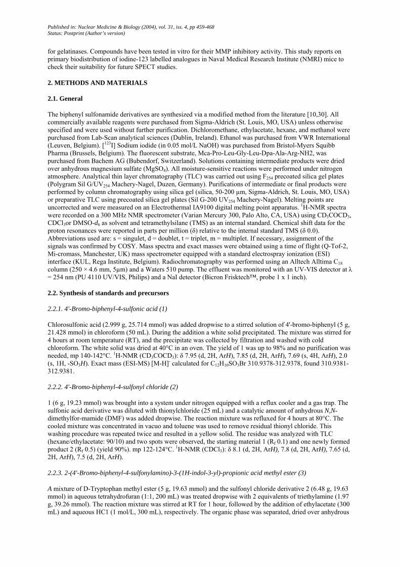
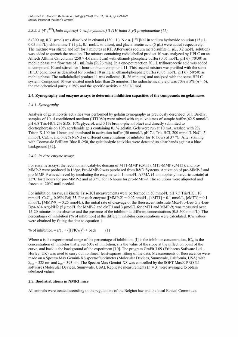
Scheme 1: Synthesis of standards and precursors.
Published in: Nuclear Medicine & Biology (2004), vol. 31, iss. 4, pp 459-468 Status: Postprint (Author’s version)
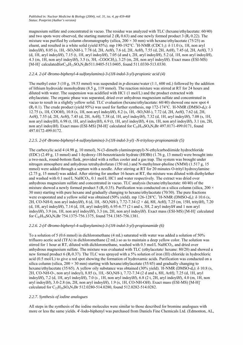
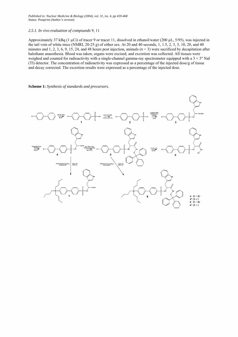
Scheme 2: Radiosynthesis of I-123 radiotracers.
3. Results and discussion
The aim of this study was to assess the biological activity and usefulness of different gelatinase inhibitors in vitro and in vivo. To be an effective inhibitor of MMPs, the molecule requires a functional group (such as carboxylic acid, hydroxamic acid, or sulfhydryl) capable of chelating the active-site zinc (II) ion [28]. To improve selectivity these inhibitors are substituted with side chains which interact with specific subsites and undergo effective van der Waals interactions [33]. Structure-activity data of compounds [10,29] with low nanomolar IC50 potencies for gelatinases were studied and combined to obtain a higher inhibitory activity.
Standards and precursors were synthesized as shown in Scheme 1. The chemical yields of synthesized compounds were moderate to excellent and are detailed in the Methods and materials section. In the literature, different methods of hydroxamic acid synthesis have been described [10,29,34-36]. Some of these pathways were attempted but gave no or insufficient yields. The radio-iodination was conducted by electrophilic aromatic

Published in: Nuclear Medicine & Biology (2004), vol. 31, iss. 4, pp 459-468 Status: Postprint (Author’s version)
substitution of the tributylstannyl derivatives (Scheme 2). Mixtures were purified by HPLC on C18 column at a flow rate of 1 mL/min. The radiochemical yield for synthesis of radioiodinated compound 9 was 60% ± 5% (n = 3), radiochemical purity of the collected fraction >98% and the specific activity > 50 Ci/µmol. The radiochemical yield for synthesis of radioiodinated compound 11 was 70% ± 5% (n = 6), the radiochemical purity of the collected fraction was >98%, and the specific activity >58 Ci/µmol.
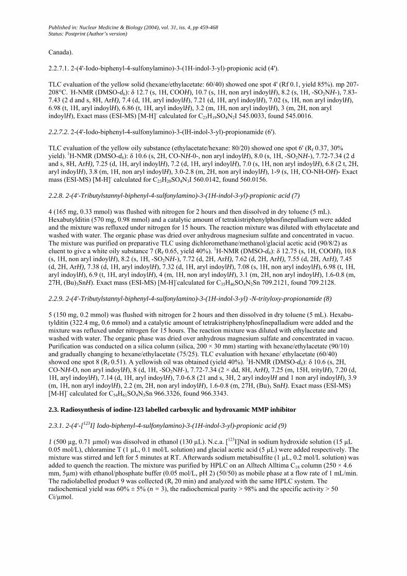
Fig. 1: Zymogram of compounds (4, 6, 4', 6') in two different concentrations (10-7mol/L; 10-9 mol/L) next to a control without inhibitor. The control was run to exclude the impact of DMSO on inhibition of gelatinases.
Table 1: IC50 values of (4, 4', 6, 6') for MMP-2, MMP-9, cMT1, and cMT3 are expressed in nmol/L and averaged (n = 3), standard deviations are reported*
MMP-2 MMP-9 cMT1-MMP CMT3-MMP Br-COOH (4) 7.3 ± 0.6 239.7 ± 15.7 437.0 ± 22.6 252.3 ± 12.2 I-COOH (4') 9.3 ± 1.5 201.0 ± 58.6 859.0 ± 31.1 678.7 ± 45.3
Br-CONHOH (6) 0.5 ± 0.1 4.9 ± 3.1 25.0 ± 6.2 7.0 ± 4.6 I-CONHOH (6') 0.6 ± 0.05 2.4 ± 1.4 21.7 ± 6.4 7.3 ± 0.6
* Kinetic measurements were performed in Tris-HCl 50 mmol/L pH 7.5, CaCl2 10 mmol/L Brij 35 0.05%. Details are described in the Methods and materials setion.
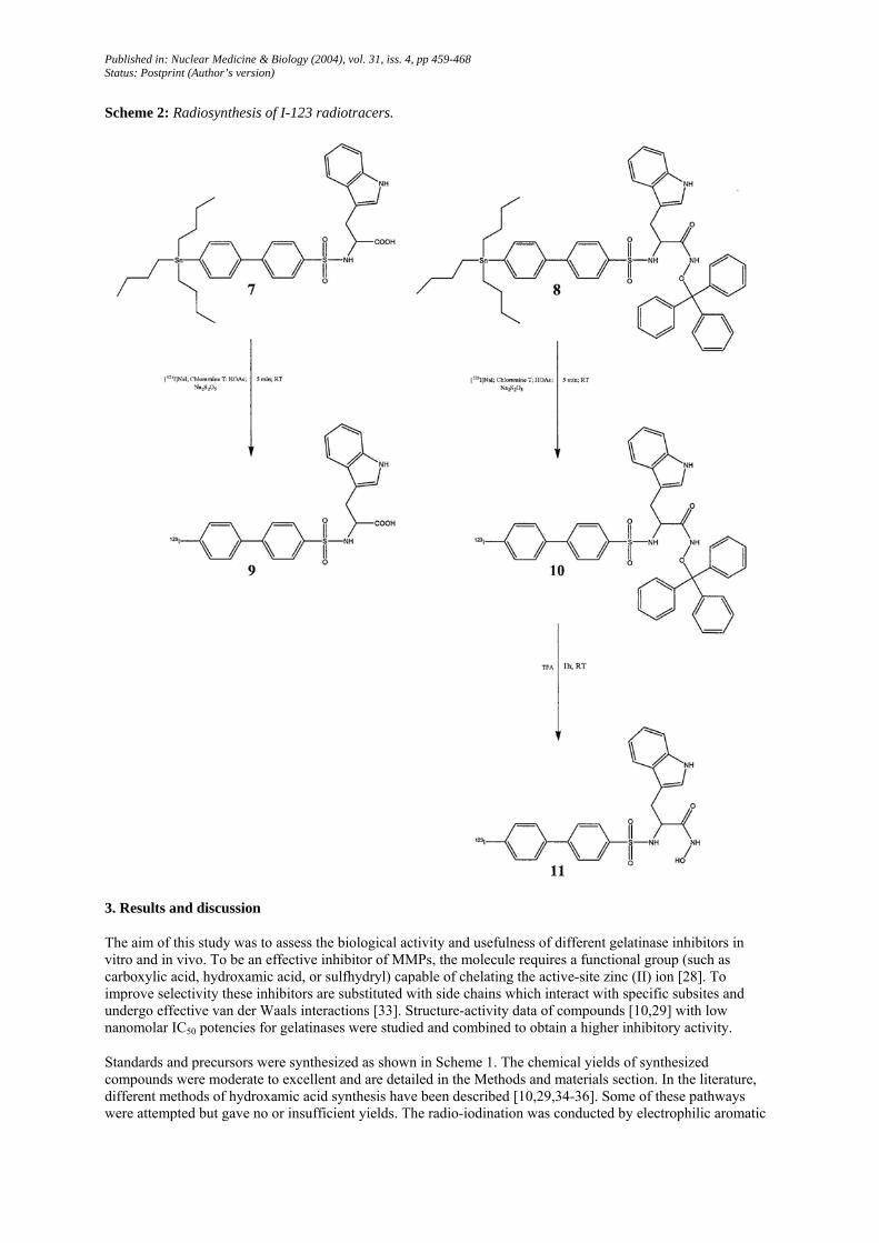
Table 2: Tissue concentrations of radioactivity at various time points after intravenous administration of 2-(4'-[123I]iodo-biphenyl-4-sulfonylamino)-3-(1H/-indol-3-yl)-propionic acid (9)*
Time (min) Time (h)
0.33 0.66 1 1.5 2 3 5 10 20 40 1 2 3 6 9 15 Blood 15.02 ±
0.00 27.63 ± 13.92
15.86 ± 7.46
9.12 ± 5.56
4.54 ± 2.07
2.46 ± 0.66
3.63 ± 2.15
2.18 ± 0.25
2.20 ± 0.14
1.99 ± 0.61
1.94 ± 0.35
1.79 ± 0.22
1.25 ± 0.21
0.73 ± 0.12
0.47 ± 0.18
0.00 ±0.00
Brain 2.14 ± 0.80
0.88 ± 0.38
1.43 ± 1.30
1.17 ± 1.10
0.48 ± 0.38
2.01 ± 2.49
1.16 ± 1.61
0.23 ± 0.14
0.39 ± 0.14
0.48 ± 0.11
0.42 ± 0.09
0.49 ± 0.15
0.43 ± 0.04
0.23 ± 0.03
0.23 ± 0.11
0.00 ±0.00
Heart 16.55 ± 7.17
8.29 ± 4.23
5.34 ± 1.87
4.73 ± 0.24
2.03 ± 0.68
2.38 ± 1.04
1.85 ± 0.72
1.11 ± 0.28
1.15 ± 0.29
1.00 ± 0.08
0.97 ± 0.22
0.88 ± 0.06
1.16 ± 0.73
0.45 ± 0.05
0.36 ± 0.16
0.00 ±0.00
Published in: Nuclear Medicine & Biology (2004), vol. 31, iss. 4, pp 459-468 Status: Postprint (Author’s version)
Lungs 13.15 ± 6.57
8.97 ± 3.70
7.13 ± 1.93
5.79 ± 2.13
3.54 ± 1.29
3.13 ± 2.75
5.21 ± 4.44
1.96 ± 0.29
1.97 ± 0.15
2.09 ± 0.26
1.98 ± 0.87
1.44 ± 0.14
1.04 ± 0.36
0.66 ± 0.09
0.51 ± 0.17
0.00 ±0.00
Stomach 0.98 ± 1.19
1.01 ± 0.15
1.21 ± 0.50
1.77 ± 0.67
1.09 ± 0.41
1.97 ± 0.39
4.30 ± 3.57
4.62 ± 2.00
6.26 ± 6.76
3.86 ± 1.76
11.88 ± 9.61
5.57 ± 5.57
2.40 ± 0.77
4.62 ± 3.18
2.51 ± 1.73
0.15 ±0.12
Spleen 15.10 ± 22.88
3.78 ± 1.38
4.14 ± 0.21
3.17 ± 0.63
2.14 ± 0.47
2.91 ± 1.03
2.24 ± 1.04
1.50 ± 0.24
1.34 ± 0.32
1.37 ± 0.28
1.45 ± 0.31
1.28 ± 0.30
1.45 ± 1.18
0.44 ± 0.01
0.30 ± 0.17
0.00 ±0.00
Liver 18.01 ± 5.30
41.31 ± 16.87
53.82 ± 9.35
53.52 ± 24.23
71.52 ± 14.72
56.08 ± 23.71
52.56 ± 20.47
74.25 ± 23.47
70.02 ± 13.89
48.25 ± 13.78
44.40 ± 19.15
32.97 ± 19.95
9.36 ± 5.99
3.75 ± 0.17
2.55 ± 0.89
0.69 ±0.47
Kidneys 11.51 ± 7.18
20.79 ± 6.73
19.56 ± 3.87
15.32 ± 7.01
15.14 ± 6.66
10.30 ±3.38
10.43 ±6.54
7.41 ± 6.16
9.58 ± 5.94
5.82 ± 2.93
6.10 ± 2.76
5.25 ± 1.38
5.16 ± 2.25
2.46 ± 0.13
1.74 ± 0.54
0.21 ±0.20
Small intestine
2.30 ± 1.85
1.89 ± 0.59
1.66 ± 0.46
1.68 ± 0.44
1.50 ± 0.24
1.72 ± 0.60
1.80 ± 0.74
4.42 ± 0.56
8.57 ± 3.25
16.74 ± 2.24
25.23 ± 6.61
27.81 ± 10.81
19.23 ± 18.09
5.98 ± 2.78
2.51 ± 1.56
0.33 ±0.17
Large intestine
1.68 ± 1.66
1.01 ± 0.15
0.99 ± 0.10
0.90 ± 0.23
0.71 ± 0.19
1.73 ± 0.91
1.46 ± 1.55
0.59 ± 0.20
0.72 ± 0.12
7.69 ± 12.08
0.96 ± 0.07
9.90 ± 15.43
48.50 ± 22.90
67.53 ± 5.67
28.79 ± 10.00
1.11 ±0.69
Bladder 6.82 ± 3.59
6.48 ± 5.82
1.81 ± 0.17
7.32 ± 9.19
1.73 ± 0.43
4.90 ± 4.03
5.11 ± 4.21
3.72 ± 3.53
4.55 ± 2.91
3.17 ± 3.29
7.59 ± 8.38
2.57 ± 1.67
3.25 ± 0.48
3.93 ± 3.02
2.71 ± 2.20
0.00 ±0.00
Fat 3.06 ± 1.79
4.07 ± 1.50
3.02 ± 0.41
4.99 ± 3.18
2.00 ± 0.30
2.23 ± 0.13
1.67 ± 0.18
8.13 ± 11.49
2.67 ± 1.57
2.65 ± 0.62
1.42 ± 1.62
1.78 ± 1.22
1.34 ± 0.55
0.48 ± 0.03
0.33 ± 0.03
0.01 ±0.02
Excretion**
0.00 ± 0.00
0.00 ± 0.00
0.00 ± 0.00
0.00 ± 0.00
0.00 ± 0.00
0.00 ± 0.00
0.00 ± 0.00
0.41 ± 0.45
0.55 ± 0.26
0.94 ± 0.89
2.80 ± 2.32
1.67 ± 0.78
5.34 ± 3.02
7.89 ± 9.13
59.49 ± 18.42
94.92± 2.53
* Animals were injected intravenously with 37 kBq [1231] 2-(4'-iodo-biphenyl-4-sulfonylamino)-3-(1H-indol-3-yl)-propionic acid (9) and sacrificed at designated times. Units are expressed as % injected dose/g of tissue (n = 3) corrected for background radiation and averaged. Below are reported the standard deviations. ** The results of the excretion are expressed as % of the injected dose.
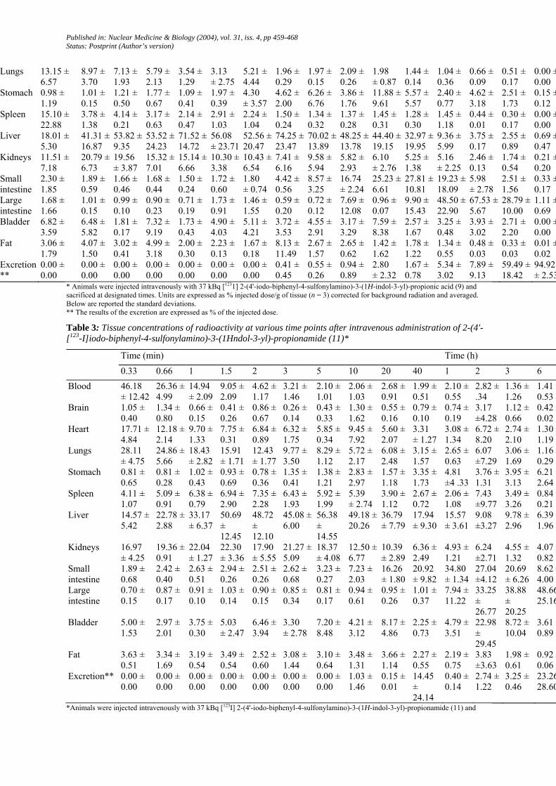
Table 3: Tissue concentrations of radioactivity at various time points after intravenous administration of 2-(4'-[123-I]iodo-biphenyl-4-sulfonylamino)-3-(1Hndol-3-yl)-propionamide (11)*
Time (min) Time (h)
0.33 0.66 1 1.5 2 3 5 10 20 40 1 2 3 6 Blood 46.18
± 12.42 26.36 ± 4.99
14.94 ± 2.09
9.05 ± 2.09
4.62 ± 1.17
3.21 ± 1.46
2.10 ± 1.01
2.06 ± 1.03
2.68 ± 0.91
1.99 ± 0.51
2.10 ± 0.55
2.82 ± .34
1.36 ± 1.26
1.41 ±0.53
Brain 1.05 ± 0.40
1.34 ± 0.80
0.66 ± 0.15
0.41 ± 0.26
0.86 ± 0.67
0.26 ± 0.14
0.43 ± 0.33
1.30 ± 1.62
0.55 ± 0.16
0.79 ± 0.10
0.74 ± 0.19
3.17 ±4.28
1.12 ± 0.66
0.42 ±0.02
Heart 17.71 ± 4.84
12.18 ± 2.14
9.70 ± 1.33
7.75 ± 0.31
6.84 ± 0.89
6.32 ± 1.75
5.85 ± 0.34
9.45 ± 7.92
5.60 ± 2.07
3.31 ± 1.27
3.08 ± 1.34
6.72 ± 8.20
2.74 ± 2.10
1.30 ±1.19
Lungs 28.11 ± 4.75
24.86 ± 5.66
18.43 ± 2.82
15.91 ± 1.71
12.43 ± 1.77
9.77 ± 3.50
8.29 ± 1.12
5.72 ± 2.17
6.08 ± 2.48
3.15 ± 1.57
2.65 ± 0.63
6.07 ±7.29
3.06 ± 1.69
1.16 ±0.29
Stomach 0.81 ± 0.65
0.81 ± 0.28
1.02 ± 0.43
0.93 ± 0.69
0.78 ± 0.36
1.35 ± 0.41
1.38 ± 1.21
2.83 ± 2.97
1.57 ± 1.18
3.35 ± 1.73
4.81 ±4 .33
3.76 ± 1.31
3.95 ± 3.13
6.21 ±2.64
Spleen 4.11 ± 1.07
5.09 ± 0.91
6.38 ± 0.79
6.94 ± 2.90
7.35 ± 2.28
6.43 ± 1.93
5.92 ± 1.99
5.39 ± 2.74
3.90 ± 1.12
2.67 ± 0.72
2.06 ± 1.08
7.43 ±9.77
3.49 ± 3.26
0.84 ±0.21
Liver 14.57 ± 5.42
22.78 ± 2.88
33.17 ± 6.37
50.69 ± 12.45
48.72 ± 12.10
45.08 ± 6.00
56.38 ± 14.55
49.18 ± 20.26
36.79 ± 7.79
17.94 ± 9.30
15.57 ± 3.61
9.08 ±3.27
9.78 ± 2.96
6.39 ±1.96
Kidneys 16.97 ± 4.25
19.36 ± 0.91
22.04 ± 1.27
22.30 ± 3.36
17.90 ± 5.55
21.27 ± 5.09
18.37 ± 4.08
12.50 ± 6.77
10.39 ± 2.89
6.36 ± 2.49
4.93 ± 1.21
6.24 ±2.71
4.55 ± 1.32
4.07 ±0.82
Small intestine
1.89 ± 0.68
2.42 ± 0.40
2.63 ± 0.51
2.94 ± 0.26
2.51 ± 0.26
2.62 ± 0.68
3.23 ± 0.27
7.23 ± 2.03
16.26 ± 1.80
20.92 ± 9.82
34.80 ± 1.34
27.04 ±4.12
20.69 ± 6.26
8.62 ±4.00
Large intestine
0.70 ± 0.15
0.87 ± 0.17
0.91 ± 0.10
1.03 ± 0.14
0.90 ± 0.15
0.85 ± 0.34
0.81 ± 0.17
0.94 ± 0.61
0.95 ± 0.26
1.01 ± 0.37
7.94 ± 11.22
33.25 ± 26.77
38.88 ± 20.25
48.6625.16
Bladder 5.00 ± 1.53
2.97 ± 2.01
3.75 ± 0.30
5.03 ± 2.47
6.46 ± 3.94
3.30 ± 2.78
7.20 ± 8.48
4.21 ± 3.12
8.17 ± 4.86
2.25 ± 0.73
4.79 ± 3.51
22.98 ± 29.45
8.72 ± 10.04
3.61 ±0.89
Fat 3.63 ± 0.51
3.34 ± 1.69
3.19 ± 0.54
3.49 ± 0.54
2.52 ± 0.60
3.08 ± 1.44
3.10 ± 0.64
3.48 ± 1.31
3.66 ± 1.14
2.27 ± 0.55
2.19 ± 0.75
3.83 ±3.63
1.98 ± 0.61
0.92 ±0.06
Excretion** 0.00 ± 0.00
0.00 ± 0.00
0.00 ± 0.00
0.00 ± 0.00
0.00 ± 0.00
0.00 ± 0.00
0.00 ± 0.00
1.03 ± 1.46
0.15 ± 0.01
14.45 ± 24.14
0.40 ± 0.14
2.74 ± 1.22
3.25 ± 0.46
23.2628.60
*Animals were injected intravenously with 37 kBq [123I] 2-(4'-iodo-biphenyl-4-sulfonylamino)-3-(1H-indol-3-yl)-propionamide (11) and
Published in: Nuclear Medicine & Biology (2004), vol. 31, iss. 4, pp 459-468 Status: Postprint (Author’s version) sacrificed at designated times. Units are expressed as % injected dose/g of tissue (n = 3) corrected for background radiation and averaged. Below are reported the standard deviations. ** The results of the excretion are expressed as % of the injected dose.
Zymography was used to obtain a preliminary estimation of the inhibition capacities of the inhibitors on gelatinases (MMP-2 and MMP-9). The control (1% DMSO in incubation buffer) shows two clear bands (proMMP-2 72 kDa, proMMP-9 92 kDa) against a blue background of undegraded gelatin whereas the strips incubated in inhibitor (4, 4', 6, 6') concentrations (10-7, 10-9 mol/L) show no or small bands (Fig. 1).
In vitro enzyme assays using quenched fluorescent peptide substrates were used to determine IC50values of the different compounds for MMP-2, MMP-9, cMT1, and cMT3. The results show for both hydroxamic acid compounds (6, 6') a high inhibition activity and a rather lower selectivity for gelatinases. In opposite, the carboxylic acids (4, 4') show a much higher selectivity for MMP-2 but a lower activity, though still exploitable (Table 1).
To further evaluate the pharmacokinetics (dehalogena-tion, metabolization, excretion, etc.) of tracers 9 and 11, biodistribution studies were performed in NMRI mice. Approximately 37 kBq (1 µCi) of tracer was injected in the tail vein of white mice (NMRI, 20-25 g) and the concentration of radioactivity in various tissues as a function of time was evaluated, as shown in Table 2 (9) and Table 3 (11). Among the organs the heart, lung, stomach, liver, and kidney showed no long-term accumulation of the tracers (9 hours: <3% ID/g). Up to 1.41% ID/g of 11 was accumulated in the blood until 6 hours and approximately the same percentage of 9 up to 3 hours, giving the possibility to accumulate in the tumor in a later phase of this study. There was also an uptake in fat tissue (3 hours: 1.34% ID/g for 9 and 1.98% ID/g for 11). The radioactivity was cleared from the body after 15 hours, and no dehalogenation was observed.
4. CONCLUSION
Radioiodinated carboxylic and hydroxamic MMP inhibitors 2-(4'-[123I]iodo-biphenyl-4-sulfonylamino)-3-(1H-indol-3-yl)-propionic acid (9) and 2-(4'-[123I]iodo-biphenyl-4-sulfonylamino)-3-(1H-indol-3-yl)-propionamide (11), their standards and precursors were synthesized. Preliminary findings of in vitro zymography and enzyme assays showed high inhibition capacities of the inhibitors on gelatinases. In vivo biodistribution showed no long-term accumulation in organs and the possibility to accumulate in the tumor. These data suggest that they may be potential useful agents for non-invasive monitoring of cancer MMP levels in vivo, diagnosis of primary and secondary tumors and tumor response to MMP inhibitor therapy using SPECT. These results also warrant further evaluation of tumor uptake in tumor-bearing athymic mice and metabolite studies of the radioiodinated MMP inhibitors.
Acknowledgments
The authors thank D. Delapierre of the Laboratory of Tumor and Developmental Biology, University of Liège, Sart-Tilman, for her support and great help with the determination of the in vitro MMP inhibitory activity of all synthesized compounds, and Pharm. U. Hillaert of the laboratory for Medicinal Chemistry, University of Ghent, for his help in acquiring and interpreting NMR spectra.
References
[1] Zucker S, Cao J. Imaging metalloproteinase activity in vivo. Nat Med 2001;7:655-6.
[2] Chambers AF, Matrisian LM. Changing views of the role of matrix metalloproteinases in metastasis. J Natl Cancer Inst 1997;89:1260-70.
[3] Nagase H, Woessner JF. Matrix metalloproteinases. J Biol Chem 1999;274:21491-4.
[4] Nelson AR, Fingleton B, Rothenberg ML, Matrisian LM. Matrix metalloproteinases: biologic activity and clinical implications. J Clin Oncol 2000;18:1135-49.
[5] Noël A, Albert V, Bajou K, Bisson C, Devy L, Frankenne F, Maquoi E, Masson V, Sounni NE, Foidart JM. New functions of stromal proteases and their inhibitors in tumor progression. Surg Oncol Clin North Am 2001;10:417-32.
[6] Egeblad M, Werb Z. New functions for the matrix metalloproteinases in cancer progression. Nat Rev 2002;2:161-74.
[7] Coussens LM, Werb Z. Matrix metalloproteinases and the development of cancer. Chem Biol 1996;3:895-904.
Published in: Nuclear Medicine & Biology (2004), vol. 31, iss. 4, pp 459-468 Status: Postprint (Author’s version) [8] Fei X, Zheng QH, Liu X, Wang JQ, Sun HB, Mock BH, Stone KL, Miller KD, Sledge GW, Hutchins GD. Synthesis of radiolabeled biphenylsulfonamide matrix metalloproteinase inhibitors as new potential PET cancer imaging agents. Bioorg Med Chem Lett 2003; 13: 2217-22.
[9] Vihinen P, Kähäri VM. Matrix metalloproteinases in cancer: prognostic markers and therapeutic targets. Int J Cancer 2002;99:157-66.
[10] O'Brien PM, Ortwine DF, Pavlovsky AG, Picard JA, Sliskovic DR, Roth BD, Dyer LL, Johnson RD, Man CF, Hallak H. Structure-activity relationships and pharmacokinetic analysis for a series of potent, systemically available biphenylsulfonamide matrix metalloproteinase inhibitors. J Med Chem 2000;43:156-66.
[11] Bremer C, Bredow S, Mahmood U, Weissleder R, Tung CH. Optical imaging of matrix metalloproteinase-2 activity in tumors: feasibility study in a mouse model. Radiology 2001;221:523-9.
[12] Curran S, Murray GI. Matrix metalloproteinases in tumour invasion and metastasis. J Pathol 1999;189:300-8.
[13] Fang J, Shing, Wiederschain D. Matrix metalloproteinase-2 is required for the switch to the angiogenic phenotype in a tumor model. Proc Natl Acad Sci USA 2000;97:3884-9.
[14] Furumoto S, Iwata R, Ido T. Design and synthesis of fluorine-18 labelled matrix metalloproteinase inhibitors for cancer imaging. J Label Comp Radiopharmacol 2002;45:975-86.
[15] Brown PD, Bloxidge RE, Anderson E, Howell A. Expression of activated gelatinase in human invasive breast carcinoma. Clin Exp Metast 1993;11:183-90.
[16] Cai M, Onoda K, Takao M, Kyoko IY, Shimpo H, Yoshida T, Yada I. Degradation of Tenascin-C and activity of matrix metalloproteinase-2 are associated with tumor recurrece in early stage non-small cell lung cancer. Clin Cancer Res 2002;8:1152-6.
[17] Davies B, Miles DW, Happerfield LC, Naylor MS, Bobrow LG, Rubens RD, Balkwill FR. Activity of type IV collagenases in benign and malignant breast desease. Br J Cancer 1993;67:1126-31.
[18] Furumoto S, Takashima K, Kubota K, Ido T, Iwata R, Fukuda H. Tumor detection using F-18-labelled matrix metalloproteinase-2 inhibitor. Nucl Med Biol 2003;30:119-25.
[19] Kossakowska AE, Huchcroft SA, Urbanski SJ. Comparative analysis of the expression patterns of metalloproteinases and their inhibitors in breast neoplasia, sporadic colorectal neoplasia, pulmonary carcinomas and malignant, non-Hodgkin's lymphomas in humans. Br J Cancer 1996;73:1401-8.
[20] Lee KS, Rha SY, Kim SJ. Seuential activation and production of matrix metalloproteinase-2 during breast cancer progression. Clin Exp Metastasis 1996;14:512-9.
[21] Liabakk NB, Talbot I, Smith RA, Wilkinson K, Balkwill K. Matrix metalloproteinase 2 (MMP-2) and matrix metalloproteinase 9 (MMP-9) type IV collagenaes in colorectal cancer. Cancer Res 1996; 56:190-6.
[22] Passlick B, Sienel W, Seen-Hibler R, Wöckel W, Thetter O, Mut-schler W, Pantel K. Overexpression of matrix metalloproteinase 2 predicts unfavorable outcome in early-stage non-small cell lung cancer. Clin Cancer Res 2000;6:3944-8.
[23] Ylisirniö S, Höyhtyä M, Turpeeniemi-Hujanen T. Serum metallopro-teinasese -2, -9 and tissue inhibitors of metalloproteinases -1, -2 in lung cancer—TIMP-1 as a prognostic marker. Anticancer Res 2000; 20:1311-6.
[24] Kuhnast B, Bodenstein C, Wester HE, Weber W. Carbon-11-labelling of an N-Sulfanylamino acid derivative: a potential tracer for MMP-2 and MMP-9 imaging. J Label Compd Radiopharm 2003;46:539-53.
[25] Zheng QH, Fei X, Liu X, Wang JQ, Sun HB, Mock BM, Stone KL, Martinez TD, Miller KD, Sledge GW, Hutchins GD. Synthesis and preliminary biological evaluation of MMP inhibitor radiotracers [11C]methyl-halo-CGS 27023A analogs, new potential PET breast cancer imaging agents. Nucl Med Biol 2002;29:761-70.
[26] Hagmann WK, Lark MW, Becker JW. Inhibition of matrix metalloproteinases. In: Bristol JA, editor. Annual reports in medical chemistry. New York: Academic Press, 1996. p. 231-40.
[27] Summers JB, Davidsen SK. Matrix metalloproteinase inhibitors and cancer. In: Bristol JA, editor. Annual reports in medical chemistry. New York: Academic Press, 1998. p. 131-40.
[28] Kiyama R, Tamura Y, Watanabe F, Tsuzuki H, Ohtani M, Yodo M. Homology modeling of gelatinase catalytic domains and docking simulations of novel sulfonamide inhibitors. J Med Chem 1999;42: 1723-38.
[29] Tamura Y, Watanabe F, Nakatani T, Yasui K, Fuji M, Komurasaki T, Tsuzuki H, Maekawa R, Yoshioka T, Kawada K, Sugita K, Ohtani M. Higly selective and orally active inhibitors of type IV collagenase (MMP-9 and MMP-2): N-sulfonylamino acid derivatives. J Med Chem 1998;41:640-9.
Published in: Nuclear Medicine & Biology (2004), vol. 31, iss. 4, pp 459-468 Status: Postprint (Author’s version) [30] Hanessian S, Moitessier N, Gauchet C, Viau M. N-Aryl sulfonyl homocysteine hydroxamate inhibitors of matrix metalloproteinases: further probing of the S1; S1', S2' pockets. J Med Chem 2001;44: 3066-73.
[31] Maquoi E, Frankenne F, Baramova E, Munaut C, Sounni NE, Remacle A, Noël A, Murphy G, Foidart JM. Membrane type 1 matrix metalloproteinase-associated degradation of tissue inhibitor of metalloproteinase 2 in human tumor cell lines. J Biol Chem 2000;275: 11368-78.
[32] Hawkes SP, Li H, Taniguchi GT. Zymography and reverse zymog-raphy for detecting MMPs and TIMPs. In: Clark I, editor. Methods in Molecular Biology. Totowa, NJ: Humana Press, 2001. p. 399-410.
[33] Whittaker M, Flooyd CD, Brown P, Gearing AJH. Design and therapeutic application of matrix metalloproteinase inhibitors. Chem Rev 1999;99:2735-76.
[34] Massa S, Artico M, Corelli F, Mai A, Di Santo R, Cortes S, Marongiu ME, Pani A, La Colla P. Synthesis and antimicrobial and cytotoxic activities of pyrrole-containing analogues of trichostatin A. J Med Chem 1990;33:2845-9.
[35] Natchus MG, Bookland RG, De B, Almstead NG, Pikul S, Janusz MJ, Heitmeyer SA, Hookfin EB, Hsieh LC, Dowty ME, Dietsch CR, Patel VS, Garver SM, Gu F, Pokross ME, Mieling GE, Baker TR, Foltz DJ, Peng SX, Bornes DM, Strojnowski MJ, Taiwo YO. Development of new hydroxamic matrix metalloproteinase inhibitors derived from functionalized 4-aminoprolines. J Med Chem 2000;43:4948-63.
[36] Scozzafava A, Supuran CT. Protease inhibitors: Synthesis of potent bacterial collagenase and matrix metalloproteinase inhibitors incorporating N-4-nitrobenzylsulfonylglycine hydroxamate moieties. J Med Chem 2000;43:1858-65.