Embed Size (px)
Citation preview

Seediscussions,stats,andauthorprofilesforthispublicationat:https://www.researchgate.net/publication/40444413
NewInterpretationofthePalateofPterosaurs
ArticleinTheAnatomicalRecordAdvancesinIntegrativeAnatomyandEvolutionaryBiology·February2010
DOI:10.1002/ar.21053·Source:PubMed
CITATIONS
9
READS
124
4authors,including:
Someoftheauthorsofthispublicationarealsoworkingontheserelatedprojects:
Paleo-geographicpatternsv/sclimatechangeinSouthAmericaandtheAntarcticPeninsuladuringthe
latestCretaceous:apossibleexplanationfortheoriginoftheAustralbiota?Viewproject
EarlyCretaceousichthyosaursfromtheeasternborderoftheTyndallGlacier,TorresdelPaineNational
ParkSouthernChileViewproject
EdinaProndvai
GhentUniversity
33PUBLICATIONS226CITATIONS
SEEPROFILE
Eberhard"Dino"Frey
StateMuseumofNaturalHistoryKarlsruhe
152PUBLICATIONS1,979CITATIONS
SEEPROFILE
AllcontentfollowingthispagewasuploadedbyEberhard"Dino"Freyon19March2015.
Theuserhasrequestedenhancementofthedownloadedfile.Allin-textreferencesunderlinedinblueareaddedtotheoriginaldocument
andarelinkedtopublicationsonResearchGate,lettingyouaccessandreadthemimmediately.

THE ANATOMICAL RECORD 293:243–258 (2010)
New Interpretation of the Palate ofPterosaurs
ATTILA O†SI,1* EDINA PRONDVAI,2 EBERHARD FREY,3
AND BURKHARDT POHL4
1Hungarian Academy of Sciences—Hungarian Natural History Museum,Research Group for Paleontology, Budapest, Hungary
2Department of Paleontology, Eotvos Lorand University, Budapest, Hungary3Staatliches Museum fur Naturkunde Karlsruhe,
Karlsruhe, Germany4Wyoming Dinosaur Center, Thermopolis, Wyoming
ABSTRACTOn the basis of a new, three-dimensionally preserved specimen of the
Early Jurassic pterosaur Dorygnathus banthensis we present a reinter-pretation of the pterosaur palate. The hard palate is formed by the exten-sive palatal plate of the maxilla and not by the palatine as has beengenerally reconstructed. This palatal plate of the maxilla emarginates thechoana rostrally and rostrolaterally as in other archosaurs and lepido-saurs. The longitudinally elongate and dorsoventrally flat palatine in Dor-ygnathus is an isolated bone caudal to the palatal plate of the maxillaand morphologically and topographically it resembles that of crocodiliansand birds, respectively. The palatine separates the choana laterally fromthe suborbital fenestra demonstrating the homologous nature of the (pri-
Additional Supporting Information may be found in theonline version of this article.
Abbreviations used: Institutional abbreviations: CA = CarnegieMuseum Pittsburgh, USA; CD = Desiree Collection of RainerAlexander von Blittersdorff, Rio de Janeiro; IGO = Museo MarioSanchez Roig, Instituto de Geologıa y Paleontologıa, La Habana,Cuba; IVPP = Institute of Vertebrate Palaeontology andPalaeoantropology, Beijing, China; KUVP = Museum of NaturalHistory, University of Kansas; NHM = Natural History Museum,London, England; PTH = Philosophische-TheologischeHochschule, Eichstatt; SAO = Naturmuseum, St. Gallen; SMNS =Staatliches Museum fur Naturkunde, Stuttgart, Germany.Anatomical Abbreviations: acav = accessory cavities of theantorbital fossa; amp = apertura maxillo–premaxillaris; aof =antorbital fossa; aofe = antorbital fenestra; asec = articularsurface of ectopterygoid; asj = articular surface for jugal; aslac =articular surface of lacrimal; asmx = articular surface for maxilla;asnas = articular surface of nasal; aspm = articular surface forpremaxilla; asprf = articular surface of prefrontal; aspt =articular surface of pterygoid; bo = basioccipital; bp =basipterygoid; bs = basisphenoid; bw = bony wall separating thecaviconchal and postvestibular recesses from the nasal cavityproper; camxd = cavity for the maxillary diverticula; capmd =cavity for the premaxillary diverticula; ch = choana; cppm =caudal process of the premaxilla; cr = cecal recess; dpmx =dorsally projecting caudomedial edge of the palatal plate of themaxilla; ec = ectopterygoid; fi = foramen incisivum; fm = foramenmagnum; fr = frontal; in = internal nares (choana); iof =infraorbital fenestra; iov = infraorbital vacuity; ipv =interpterygoid vacuity; itv = infratemporal vacuity; j = jugal;jpmx = jugal process of the maxilla; lac = lacrimal; lmch = lateralmargin of choana; lppt = lateral process of pterygoid; ltf = lowertemporal fenestra; lwmx = lateral wall of maxilla; ma = mandible;
mampm = margin of apertura maxillo-premaxillaris; maofe =margin of antorbital fenestra; mas = muscle attachment surface;mppt = medial process of the pterygoid; msofe = medial margin ofsuborbital fenestra; mx = maxilla; nar = naris; nas = nasal; ncpr =nasal cavity proper; npmx = nasal process of premaxilla; oaof-nc =opening between antorbital fossa and the nasal cavity; oc =occipital condyle; ocavre = opening for caviconchal recess; pecf =pterygoectopterygoid fenestra; pf = pneumatic foramen; pl =palatine; plplmx = palatal plate of maxilla; plpmx = palatalprocess of maxilla; pm = premaxilla; pmc = premaxillary cavity;pmfe = premaxilla–maxilla fenestra (sensu Langer 2004); ppf =ppld = pplf and ppv = postpalatine- or posterior palatine fenestra;prppl = prefrontal process of palatine; ps = parasphenoid; pt =pterygoid; ptf = posterior pterygoid fenestra; ptppl = pterygoidprocess of the palatine; q = quadrate; qc = quadrate condyle; qj =quadratojugal; rppt = rostral process of the pterygoid; sa =swollen alveolus; sept = sagittal septum; smxv = vomeromaxillarysuture; sofe = suborbital fenestra; sri = sagittal ridge; stf =subtemporal fenestra; t = teeth; v = vomer; x = transverse bone;z = pterygo–jugal vacuity; 6. = sixth upper tooth; 6.a = sixthalveolus; 11.a = eleventh alveolus
Grant sponsor: Hungarian Scientific Research Fund; Grantnumber: OTKA PD73021 (Paleo Contribution No. 93)
*Correspondence to: Attila O†si, Hungarian Academy of Scien-ces—Hungarian Natural History Museum, Research Group forPaleontology, Ludovika ter 2, Budapest H-1083, Hungary. Fax:+36-1-3382728. E-mail: [email protected]
Received 11 June 2009; Accepted 31 August 2009
DOI 10.1002/ar.21053Published online 2 December 2009 in Wiley InterScience (www.interscience.wiley.com).
VVC 2009 WILEY-LISS, INC.

mary) choana in all archosaurs and lepidosaurs. Our study indicates thatin basal pterosaurs the pterygo–ectopterygoid fenestra existed caudal tothe suborbital fenestra, which became confluent with the adductor cham-ber in pterodactyloids thereby increasing the relative size of the adductorchamber and hence the mass of the jaw adductors. The choana in basalpterosaurs was relatively small compared with the interpterygoid vacuity.With increasing rostroventral inclination of the quadrates in morederived pterosaurs, the interpterygoid vacuity was reduced considerably,whereas the choana increased in size. This exceptional Dorygnathus spec-imen also shows a hitherto unknown pair of fenestrae situated at the pal-atal contact of the premaxilla–maxilla and might represent the aperturefor the vomeronasal organ. Anat Rec, 293:243–258, 2010. VVC 2009 Wiley-Liss, Inc.
Keywords: Jurassic pterosaur Dorygnathus; palate; extantphylogenetic bracket; choana
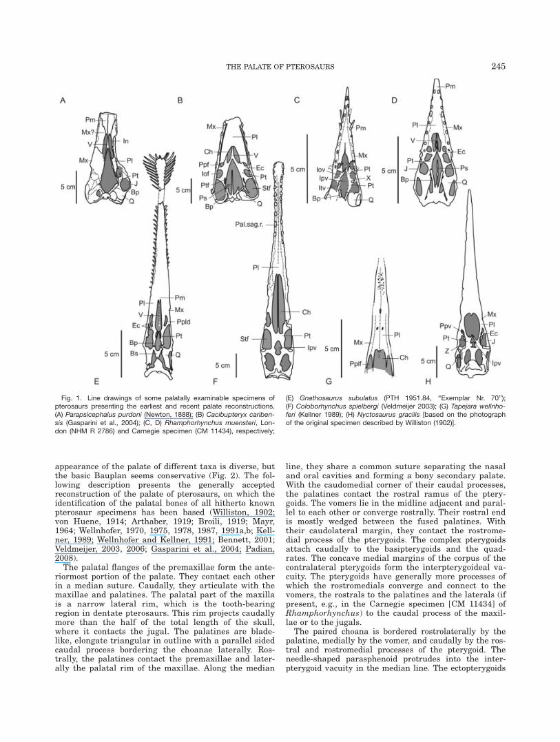
The reconstruction of the palatal region of the ptero-saurian skull has always been problematic, becausemost pterosaur cranial materials are laterally exposedand often compressed or severely crushed. In addition,the complete fusion of cranial elements in most adultpterosaurs prevents the identification of sutures thusthe accurate distinction of cranial elements. There are afew exceptional specimens with skulls that are eitherthree-dimensionally preserved [e.g., Parapsicephaluspurdoni, Newton (1888); the Carnegie-specimen (CM11434) of Rhamphorhynchus muensteri (Wellnhofer,1975); some excellent specimens of Dsungaripterus weii(IVPP 64043), Young (1964); the holotype of Tapejarawellnhoferi, Kellner (1989); Cacibupteryx caribensis,Gasparini et al. (2004); Nyctosaurus gracilis (Williston,1902); and several ornithocheirids from the Santana For-mation, e.g., Tropeognathus (Ornithocheirus) mesembri-nus, Wellnhofer (1987); Anhanguera santanae (Witmeret al., 2003); Coloborhynchus araripensis, Coloborhyn-chus spielbergi (Veldmeijer, 2003)] or palatally exposed[Gnathosaurus subulatus (Wellnhofer, 1970, ‘‘ExemplarNr. 70’’); Rhamphorhynchus ‘‘gemmingi’’ NHM R 2786(Woodward, 1902)] (see Fig. 1 and Supporting Informa-tion). The currently accepted palatal reconstructions ofthe pterosaurian skull have been based on these speci-mens, apparently combined with speculative interpreta-tions of fragmentary, compressed, or isolated material(e.g., Arthaber, 1919; Wellnhofer, 1978; Bennett, 2007).
An exceptionally well-preserved, disarticulated butassociated, and yet undescribed skeletally immaturespecimen of Dorygnathus banthensis, however, chal-lenges these previous palatal reconstructions. The iso-lated and three-dimensionally preserved cranialelements of this specimen provide a unique insight intothe finer morphology and structure of some palatalbones of Dorygnathus. On the basis of the archosaurianaffinities of the group (Sereno, 1991, Benton, 1999; Honeand Benton, 2007), we adopted an extant phylogeneticbracket (‘‘EPB,’’ Witmer, 1995a) approach to identifysome of the isolated cranial elements and to reconstructthe palate of Dorygnathus. Comparison of the newlyreconstructed palate of Dorygnathus with those of otherpterosaur taxa helped to clarify some hitherto unde-scribed or misinterpreted bony elements and fenestrae
in these taxa, and opened the way to propose possibleevolutionary changes in the construction of the ptero-saur palate.
THE CURRENT CONCEPT OF PALATERECONSTRUCTION AND ITS PROBLEMS
The earliest delineation of the complete palate of apterosaur was given by Marsh (1884) who depicted theskull of a Pteranodon longiceps in ventral, dorsal, andlateral views. Unfortunately, neither the specimen onwhich his reconstruction was based nor the bones form-ing the palate were identified. Subsequent early authors,such as Newton (1888), Seeley (1901), Woodward (1902),Williston (1902), von Huene (1914), Broili (1919), andArthaber (1919), were more specific about the assign-ment of bones in their palatal reconstructions. However,there are some significant differences with respect totheir interpretations of the palatal bones (see SupportingInformation for an overview). The reason for this is thedifficulty to identify homologue bones in different taxa,especially if the morphological patterns do not show asimple topographic equivalence between taxa (Coates,1993). After the work of these earliest authors, the pala-tal reconstruction of pterosaurs became progressivelystandardized and most of the later work on the ptero-saurian palate followed this generalized interpretation ofthe palatal bones (Wellnhofer, 1970, 1975; Kellner, 1989;Witmer, 1997; Bennett, 2001, 2007; Gasparini et al.,2004; Padian, 2008). This concept is also apparent inWellnhofer’s review (1978), which illustrates the palateof several different pterosaurs known at that time. Themost reliable reconstructions based on excellent speci-mens in Wellnhofer’s review are that of R. muensteri,Gnathosaurus subulatus, and N. gracilis (Supporting In-formation). The palatal restorations of Scaphognathuscrassirostris and Campylognathoides liasicus were basedon their holotype specimens Nr. 1304 and CM 11424,respectively, where limited insight into the palate con-struction via the naris, and the antorbital, orbital, andlower temporal fenestrae is possible. The palate recon-struction of Pteranodon ingens was based on threeincomplete specimens (KUVP 976, 2212, and YPM 1177).According to the Figs. 3 and 6 in Wellnhofer (1978), the
244 O†SI ET AL.
appearance of the palate of different taxa is diverse, butthe basic Bauplan seems conservative (Fig. 2). The fol-lowing description presents the generally acceptedreconstruction of the palate of pterosaurs, on which theidentification of the palatal bones of all hitherto knownpterosaur specimens has been based (Williston, 1902;von Huene, 1914; Arthaber, 1919; Broili, 1919; Mayr,1964; Wellnhofer, 1970, 1975, 1978, 1987, 1991a,b; Kell-ner, 1989; Wellnhofer and Kellner, 1991; Bennett, 2001;Veldmeijer, 2003, 2006; Gasparini et al., 2004; Padian,2008).
The palatal flanges of the premaxillae form the ante-riormost portion of the palate. They contact each otherin a median suture. Caudally, they articulate with themaxillae and palatines. The palatal part of the maxillais a narrow lateral rim, which is the tooth-bearingregion in dentate pterosaurs. This rim projects caudallymore than the half of the total length of the skull,where it contacts the jugal. The palatines are blade-like, elongate triangular in outline with a parallel sidedcaudal process bordering the choanae laterally. Ros-trally, the palatines contact the premaxillae and later-ally the palatal rim of the maxillae. Along the median
line, they share a common suture separating the nasaland oral cavities and forming a bony secondary palate.With the caudomedial corner of their caudal processes,the palatines contact the rostral ramus of the ptery-goids. The vomers lie in the midline adjacent and paral-lel to each other or converge rostrally. Their rostral endis mostly wedged between the fused palatines. Withtheir caudolateral margin, they contact the rostrome-dial process of the pterygoids. The complex pterygoidsattach caudally to the basipterygoids and the quad-rates. The concave medial margins of the corpus of thecontralateral pterygoids form the interpterygoideal va-cuity. The pterygoids have generally more processes ofwhich the rostromedials converge and connect to thevomers, the rostrals to the palatines and the laterals (ifpresent, e.g., in the Carnegie specimen [CM 11434] ofRhamphorhynchus) to the caudal process of the maxil-lae or to the jugals.
The paired choana is bordered rostrolaterally by thepalatine, medially by the vomer, and caudally by the ros-tral and rostromedial processes of the pterygoid. Theneedle-shaped parasphenoid protrudes into the inter-pterygoid vacuity in the median line. The ectopterygoids
Fig. 1. Line drawings of some palatally examinable specimens ofpterosaurs presenting the earliest and recent palate reconstructions.(A) Parapsicephalus purdoni (Newton, 1888); (B) Cacibupteryx cariben-sis (Gasparini et al., 2004); (C, D) Rhamphorhynchus muensteri, Lon-don (NHM R 2786) and Carnegie specimen (CM 11434), respectively;
(E) Gnathosaurus subulatus (PTH 1951.84, ‘‘Exemplar Nr. 70’’);(F) Coloborhynchus spielbergi (Veldmeijer 2003); (G) Tapejara wellnho-feri (Kellner 1989); (H) Nyctosaurus gracilis [based on the photographof the original specimen described by Williston (1902)].
THE PALATE OF PTEROSAURS 245
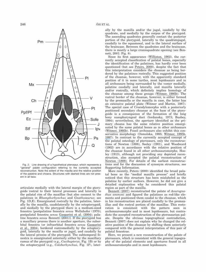
articulate medially with the lateral margin of the ptery-goids rostral to their lateral processes and laterally tothe palatal rim of the maxillae (but also connect to thepalatines in Rhamphorhynchus and Gnathosaurus; seeFig. 1D,E). Emarginated rostrally by the palatine, later-ally by the maxilla, caudolaterally by the ectopterygoid,and medially by the pterygoid there is a medium-sizedfenestra [postpalatine fenestra sensu Wellnhofer (1970);postpalatal fenestra sensu Gasparini et al. (2004); pala-tine fenestra sensu Bennett (2001)]. If the pterygoid hasa maxillary process there is another aperture, the subor-bital fenestra (or infraorbital fenestra sensu Gaspariniet al., 2004), bordered rostromedially by the ectoptery-goid, laterally by the maxilla or jugal, and caudally bythe lateral process of the pterygoid. The subtemporal fe-nestra is emarginated anteriorly either by the maxillaryramus of the pterygoid (e.g., Cacibupteryx, Fig. 1B) or bythe ectopterygoid (e.g., Coloborhynchus, Fig. 1F), later-
ally by the maxilla and/or the jugal, caudally by thequadrate, and medially by the corpus of the pterygoid.The ascending quadrates generally contact the posteriorportion of the pterygoid, laterally to the quadratojugal,caudally to the squamosal, and to the lateral surface ofthe braincase. Between the quadrates and the braincase,there is mostly a large cranioquadrate opening (see Ben-nett, 2001: Fig. 8).
Since its first appearance (Williston, 1902), the cur-rently accepted classification of palatal bones, especiallythe identification of the palatines, has hardly ever beenquestioned (but see Peters, 2000), despite the fact thatthis interpretation considers the choanae as being bor-dered by the palatines rostrally. This suggested positionof the choanae, however, with the apparently standardposition of it in some turtles, most lepidosaurs and inall archosaurs being surrounded by the vomer medially,palatine caudally and laterally, and maxilla laterallyand/or rostrally, which definitely implies homology ofthe choanae among these groups (Witmer, 1995b). Therostral border of the choanae, however, is either formedby the premaxilla or the maxilla in case, the latter hasan extensive palatal plate (Witmer and Martin, 1987).The special case of Crocodylomorpha with a posteriorlypositioned secondary choanae at the base of the ptery-goids is a consequence of the formation of the longbony nasopharyngeal duct (Iordansky, 1973; Busbey,1994); nevertheless, the aperture identified as the pri-mary choana has the same relative position emargi-nated by the same palatal bones as in other archosaurs(Witmer, 1995b). Fossil archosaurs also exhibit this con-servative morphology (Osmolska, 1985; Witmer, 1995b,1997). In contrast to the currently accepted concept ofthe palatal osteology of pterosaurs, only the reconstruc-tions of Newton (1888), Seeley (1901), and Woodward(1902) are in accordance with the relative position ofthe choanae found in all other archosauromorphs. Hoo-ley (1913), although not providing any depicted recon-struction, also accepted the palatal reconstruction ofNewton (1888). For details of the earliest reconstruc-tions and for the discussion of synonym structures, seeSupporting Information.
More recently, Peters (2000) identified the broad pala-tal bone as the ‘‘medial maxilla process’’ and brieflynoticed that this structure has been mislabeled as thepalatine by earlier authors. However, he did not give adetailed explanation why he considered this palatalregion as part of the maxilla.
Bennett (2007) reconstructed the palate of Anurogna-thus ammoni and figured the palatines as rod-like ele-ments and positioned them caudal to the choanae, whichin his reconstruction are placed caudally to the premax-illae and the rostral portion of the maxillae. This resto-ration is consistent with the pattern seen inarchosauromorphs and in most lepidosaurs, but contra-dicts the accepted reconstruction of the pterosaurian pal-ate. Despite the obvious topographical contradiction,Bennett (2007) does not explain why he changed the rel-ative position of the choanae by shifting them anteriorlycompared with the general interpretation of this pair ofpalatal fenestrae.
Here, we present a new reconstruction of the palate ofpterosaurs, which is consistent with the general topogra-phy of the palatal elements and apertures found in allarchosauromorphs and in most lepidosaurs.
Fig. 2. Line drawing of a hypothetical pterosaur, which represents a‘‘general’’ palate configuration referring to the currently acceptedreconstruction. Note the extent of the maxilla and the relative positionof the palatine and choana. Structures with dashed lines are not pres-ent in all taxa.
246 O†SI ET AL.
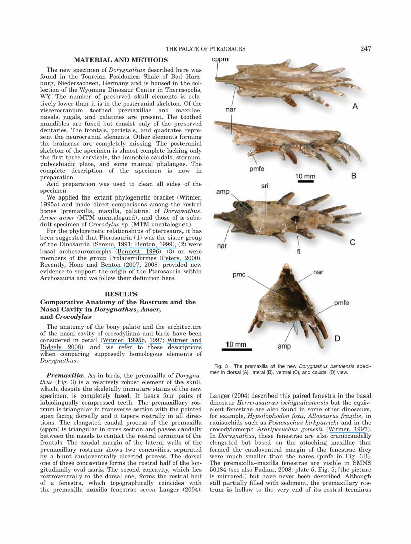
MATERIAL AND METHODS
The new specimen of Dorygnathus described here wasfound in the Toarcian Posidonien Shale of Bad Harz-burg, Niedersachsen, Germany and is housed in the col-lection of the Wyoming Dinosaur Center in Thermopolis,WY. The number of preserved skull elements is rela-tively lower than it is in the postcranial skeleton. Of theviscerocranium toothed premaxillae and maxillae,nasals, jugals, and palatines are present. The toothedmandibles are fused but consist only of the preserveddentaries. The frontals, parietals, and quadrates repre-sent the neurocranial elements. Other elements formingthe braincase are completely missing. The postcranialskeleton of the specimen is almost complete lacking onlythe first three cervicals, the immobile caudals, sternum,puboishiadic plate, and some manual phalanges. Thecomplete description of the specimen is now inpreparation.
Acid preparation was used to clean all sides of thespecimen.
We applied the extant phylogenetic bracket (Witmer,1995a) and made direct comparisons among the rostralbones (premaxilla, maxilla, palatine) of Dorygnathus,Anser anser (MTM uncatalogued), and those of a suba-dult specimen of Crocodylus sp. (MTM uncatalogued).
For the phylogenetic relationships of pterosaurs, it hasbeen suggested that Pterosauria (1) was the sister groupof the Dinosauria (Sereno, 1991; Benton, 1999), (2) werebasal archosauromorphs (Bennett, 1996), (3) or weremembers of the group Prolacertiformes (Peters, 2000).Recently, Hone and Benton (2007, 2008) provided newevidence to support the origin of the Pterosauria withinArchosauria and we follow their definition here.
RESULTSComparative Anatomy of the Rostrum and theNasal Cavity in Dorygnathus, Anser,and Crocodylus
The anatomy of the bony palate and the architectureof the nasal cavity of crocodylians and birds have beenconsidered in detail (Witmer, 1995b, 1997; Witmer andRidgely, 2008), and we refer to these descriptionswhen comparing supposedly homologous elements ofDorygnathus.
Premaxilla. As in birds, the premaxilla of Dorygna-thus (Fig. 3) is a relatively robust element of the skull,which, despite the skeletally immature status of the newspecimen, is completely fused. It bears four pairs oflabiolingually compressed teeth. The premaxillary ros-trum is triangular in transverse section with the pointedapex facing dorsally and it tapers rostrally in all direc-tions. The elongated caudal process of the premaxilla(cppm) is triangular in cross section and passes caudallybetween the nasals to contact the rostral terminus of thefrontals. The caudal margin of the lateral walls of thepremaxillary rostrum shows two concavities, separatedby a blunt caudoventrally directed process. The dorsalone of these concavities forms the rostral half of the lon-gitudinally oval naris. The second concavity, which liesrostroventrally to the dorsal one, forms the rostral halfof a fenestra, which topographically coincides withthe premaxilla–maxilla fenestrae sensu Langer (2004).
Langer (2004) described this paired fenestra in the basaldinosaur Herrerasaurus ischigualastensis but the equiv-alent fenestrae are also found in some other dinosaurs,for example, Hypsilophodon foxii, Allosaurus fragilis, inrauisuchids such as Postosuchus kirkpatricki and in thecrocodylomorph Araripesuchus gomesii (Witmer, 1997).In Dorygnathus, these fenestrae are also craniocaudallyelongated but based on the attaching maxillae thatformed the caudoventral margin of the fenestrae theywere much smaller than the nares (pmfe in Fig. 3B).The premaxilla–maxilla fenestrae are visible in SMNS50184 (see also Padian, 2008: plate 5, Fig. 5; [the pictureis mirrored]) but have never been described. Althoughstill partially filled with sediment, the premaxillary ros-trum is hollow to the very end of its rostral terminus
Fig. 3. The premaxilla of the new Dorygnathus banthensis speci-men in dorsal (A), lateral (B), ventral (C), and caudal (D) view.
THE PALATE OF PTEROSAURS 247
(Fig. 3D). This rostral cavity communicates with thenasal cavity proper, and thus it was most probablyinvaded by air sacs forming the rostralmost segment ofthe premaxillary diverticula of the pneumatized antorbi-tal sinus similar to that of birds (Witmer, 1995b:Fig. 11). On the palatal surface of the fused premaxillaethere is a tear-shaped, large foramen (5.6 mm � 4.0 mm)situated in the midline at the level of the third toothpair, which probably corresponds to the Foramen incisi-vum (Fig. 3C, fi). Rostral to this foramen, there is also apair of smaller formarina that most probably representsnutritive foramina similar to those on the lateral surfaceof the premaxillary rostrum. Caudal to the Foramenincisivum, a low ridge emerges in the midline extendingover the caudal portion of the ventral surface of the pre-maxillary rostrum. The corresponding ridge on the pre-maxillary rostrum of Coloborhynchus spielbergi has beenimproperly referred to as ‘‘palatinal sagittal ridge’’ byVeldmeijer (2003; see later). Because the alveolar rim ofDorygnathus is slightly swollen, there is a concave areabetween the tooth row and the sagittal ridge on bothsides. The caudal margin of the ventral surface of thepremaxillary rostrum forms the rostral margin of apaired fenestra. Despite being depicted in the holotypeof T. wellnhoferi by Kellner (1989), this pair of fenestraehas never been described either there or in any otherpterosaurs. Rostromedially, these fenestrae are sepa-rated by the narrow caudomedial process of the premax-illae and medially probably by the vomers. Caudolaterally,these fenestrae are bordered by the palatal plates of themaxillae (see later). Based on the shape of the emarginat-ing portion of the premaxilla and maxilla, these subspheri-cal fenestrae have a maximum length of about 5 mm anda width of 7 mm.
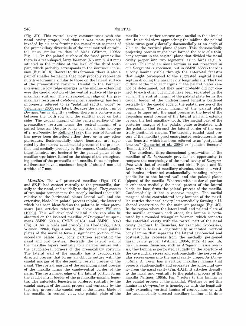
Maxilla. The well-preserved maxillae (Figs. 4E–Gand 5E,F) had contact rostrally to the premaxilla, dor-sally to the nasal, and caudally to the jugal. They consistof two major components: the steeply inclined (about 70�relative to the horizontal plane) lateral wall and theextensive, blade-like palatal process (plplm), the latter ofwhich has been identified as the palatine in other ptero-saurs [see articles referred to above after Williston(1902)]. This well-developed palatal plate can also beobserved on the isolated maxillae of Dorygnathus speci-mens SMNS 50914, SMNS 50184, and SMNS 18969(Fig. 6). As in birds and crocodylians (Iordansky, 1973,Witmer, 1995b, Figs. 4 and 5), the contralateral palatalplates of the maxillae form a significant portion of thesecondary palate (i.e., bony partition separating thenasal and oral cavities). Rostrally, the lateral wall ofthe maxillae tapers ventrally to a narrow suture withthe caudolateral corners of the premaxillary rostrum.The lateral wall of the maxilla has a caudodorsallydirected process that forms an oblique suture with thecaudal margin of the descending rostral process of thenasal. The rostral margin of the ascending nasal processof the maxilla forms the caudoventral border of thenaris. The rostralmost edge of the lateral portion formsthe caudoventral border of the premaxilla–maxilla fenes-tra. The antorbital fenestra is bordered rostrally by thecaudal margin of the nasal process and ventrally by thetapering, process-like caudal end of the lateral blade ofthe maxilla. In ventral view, the palatal plate of the
maxilla has a rather concave area medial to the alveolarrim. In caudal view, approaching the midline the palatalplate turns gently dorsally dorsomedially at an angle of70 � to the vertical plane (dpmx). This dorsomediallyprojecting process might have formed the base of a thin,bony septum in the sagittal plane that divided the nasalcavity proper into two segments, as in birds (e.g., A.anser). This median nasal septum is not preserved inour Dorygnathus specimen, but in SMNS 55886 there isa bony lamina visible through the antorbital fenestrathat might correspond to the suggested sagittal nasalseptum dividing the nasal cavity longitudinally. The trueoutline of the medial margins of the palatal plates can-not be determined, but they most probably did not con-nect to each other but might have been separated by thevomer. The rostral margin of the palatal plate forms thecaudal border of the undetermined fenestra borderedrostrally by the caudal edge of the palatal portion of thepremaxilla. The caudal margin of the palatal platebegins to taper into the jugal process at the level of theascending nasal process of the lateral wall and extendsbeyond the last maxillary tooth. The medial part of theposterior margin of the palatal plate articulated withthe palatine that formed the lateral border of the cen-trally positioned choana. The tapering caudal jugal pro-cess of the maxilla (jpmx) emarginated rostrolaterally anoval fenestra, which has been referred to as ‘‘postpalatalfenestra’’ (Gasparini et al., 2004) or ‘‘palatine fenestra’’(Bennett, 2001).
The excellent, three-dimensional preservation of themaxillae of D. banthensis provides an opportunity tocompare the morphology of the nasal cavity of Dorygna-thus with that of crocodilians and birds (Figs. 4 and 5).Level with the third maxillary alveolus, there is a verti-cal lamina orientated caudomedially standing subper-pendicular to the lateral wall and the palatal plates(plpmx) of the maxilla. Whereas with its dorsal portionit enhances medially the nasal process of the lateralblade, its base firms the palatal process of the maxilla.Caudomedially, it has a concave margin. The medialmargins of the contralateral bony laminae of the maxil-lae restrict the nasal cavity lateromedially forming a U-shaped constriction for the main air passage (Fig. 4G).In the region where the lateral wall and palatal plate ofthe maxilla approach each other, this lamina is perfo-rated by a rounded triangular foramen, which connectsthe antorbital cavity with the rostral part of the nasalcavity (oaof-nc). In Eusuchia, for example, Crocodylus,the maxilla bears a longitudinally orientated, verticalbony lamina that separates the lateral caviconchal andpostvestibular recesses from the medially positionednasal cavity proper (Witmer, 1995b; Figs. 4I and 5A,bw). In some Eusuchia, such as Alligator mississippien-sis, this lamina is perforated caudally by the aperture ofthe caviconchal recess and rostromedially the postvestib-ular recess opens into the nasal cavity proper. As Doryg-nathus, A. anser has a vertical maxillary lamina thatprojects caudomedially and separates the antorbital cav-ity from the nasal cavity (Fig. 4D,H). It attaches dorsallyto the nasal and ventrally to the palatal process of themaxilla (Witmer, 1995b: Fig. 7 refers to this lamina asthe palatal process of the maxilla). Whether or not thislamina in Dorygnathus is homologous with the longitudi-nally extending vertical lamina of crocodylians or withthe caudomedially directed maxillary lamina of birds is
248 O†SI ET AL.

Fig. 4. Comparison of the rostral bony elements of some archo-saurs. (A) Crocodylus sp. (MTM uncatalogued) left maxilla and palatinein ventral and (B) in lateral view. (C) Anser anser (MTM uncatalogued)left face of the rostrum (premaxilla, maxilla, nasal, palatine) in ventral
and (D) in lateral view. (E) Dorygnathus banthensis left maxilla and pal-atine in ventral and (F) in lateral view. (G) Dorygnathus banthensis ros-trum in caudal view. (H) Anser anser rostrum in caudal view.(I) Crocodylus sp. rostrum in caudal view.
THE PALATE OF PTEROSAURS 249
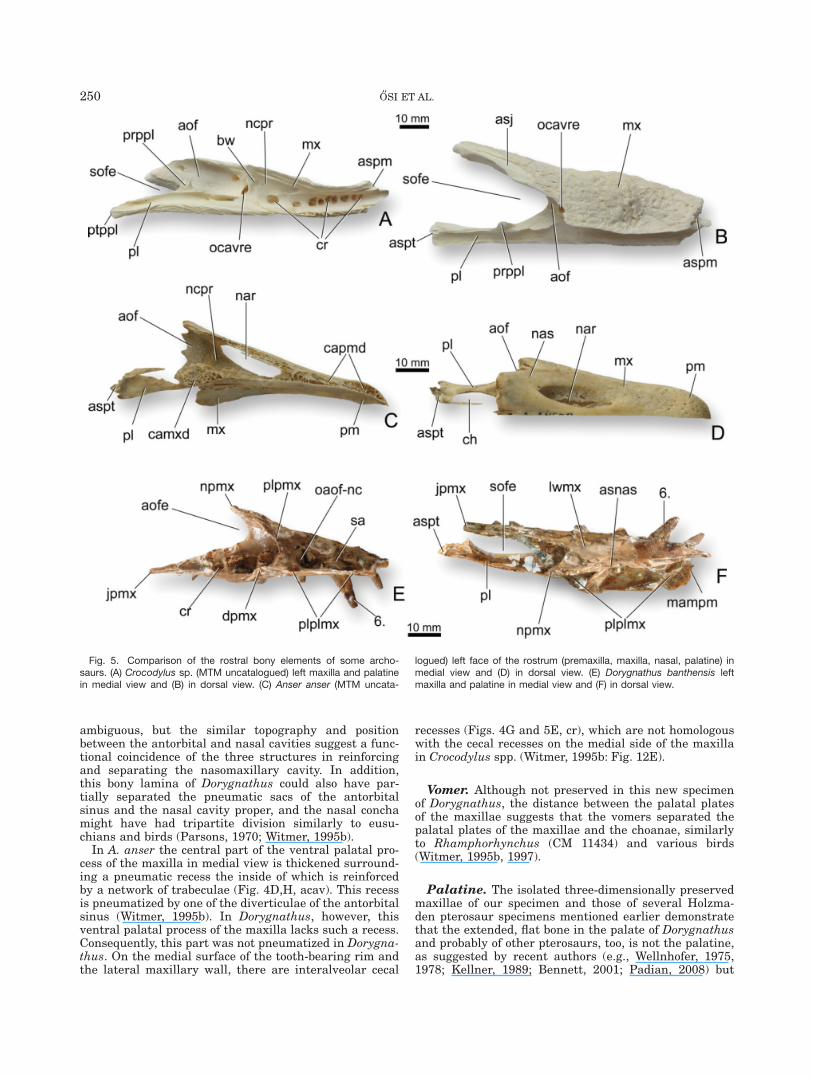
ambiguous, but the similar topography and positionbetween the antorbital and nasal cavities suggest a func-tional coincidence of the three structures in reinforcingand separating the nasomaxillary cavity. In addition,this bony lamina of Dorygnathus could also have par-tially separated the pneumatic sacs of the antorbitalsinus and the nasal cavity proper, and the nasal conchamight have had tripartite division similarly to eusu-chians and birds (Parsons, 1970; Witmer, 1995b).
In A. anser the central part of the ventral palatal pro-cess of the maxilla in medial view is thickened surround-ing a pneumatic recess the inside of which is reinforcedby a network of trabeculae (Fig. 4D,H, acav). This recessis pneumatized by one of the diverticulae of the antorbitalsinus (Witmer, 1995b). In Dorygnathus, however, thisventral palatal process of the maxilla lacks such a recess.Consequently, this part was not pneumatized in Dorygna-thus. On the medial surface of the tooth-bearing rim andthe lateral maxillary wall, there are interalveolar cecal
recesses (Figs. 4G and 5E, cr), which are not homologouswith the cecal recesses on the medial side of the maxillain Crocodylus spp. (Witmer, 1995b: Fig. 12E).
Vomer. Although not preserved in this new specimenof Dorygnathus, the distance between the palatal platesof the maxillae suggests that the vomers separated thepalatal plates of the maxillae and the choanae, similarlyto Rhamphorhynchus (CM 11434) and various birds(Witmer, 1995b, 1997).
Palatine. The isolated three-dimensionally preservedmaxillae of our specimen and those of several Holzma-den pterosaur specimens mentioned earlier demonstratethat the extended, flat bone in the palate of Dorygnathusand probably of other pterosaurs, too, is not the palatine,as suggested by recent authors (e.g., Wellnhofer, 1975,1978; Kellner, 1989; Bennett, 2001; Padian, 2008) but
Fig. 5. Comparison of the rostral bony elements of some archo-saurs. (A) Crocodylus sp. (MTM uncatalogued) left maxilla and palatinein medial view and (B) in dorsal view. (C) Anser anser (MTM uncata-
logued) left face of the rostrum (premaxilla, maxilla, nasal, palatine) inmedial view and (D) in dorsal view. (E) Dorygnathus banthensis leftmaxilla and palatine in medial view and (F) in dorsal view.
250 O†SI ET AL.

the medially extended palatal plate of the maxilla asdescribed by Newton (1888), Seeley (1901), Woodward(1902) and Peters (2000).
In extant crocodylians (e.g., Crocodylus sp., MTM unca-talogued), the palatine is an elongate, flat bone that crani-ally expands into a lateromedially wide maxillary processthat forms a scarf joint with the palatal plate of the max-illa. Caudally, the palatine is not as wide as cranially butits dorsoventral thickness increases. Its lateral, slightlyconcave, smooth margin borders the suborbital fenestramedially (Figs. 4A,I and 5A,B). The palatine of birds (e.g.,A. anser, MTM uncatalogued, Figs. 4C,D,H and 5C,D) isalso longitudinally elongated as in crocodilians. Its crani-ally directed maxillary process is dorsoventrally com-pressed. Caudally, the corpus of the palatine is twistedand laterally compressed standing at an angle of about60� to the maxillary process. The twisted, flat medial faceof the palatine forms the lateral wall of the choana. On
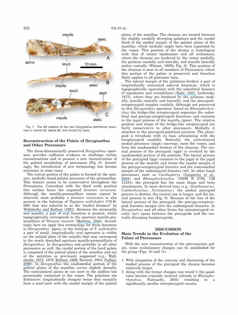
the basis of the comparisons with the palatine of extantcrocodylians and birds, we identified two long and flatbones as the palatines in Dorygnathus (Figs. 4E, 5F, 6,and 7). These bones have a lateromedially wide and dorso-ventrally very thin anterior termination that could wellhave formed a scarf joint with the posterior margin of thepalatal process of the maxilla as in crocodiles. Posteriorly,the lateral margin of these bones comes closer to the sagit-tal plane and forms the concave medial border of the ‘‘pal-atine fenestra’’ (sensu Bennett, 2001), whereas the medialmargin remains almost straight. Caudal to this fenestrathe bone expands lateromedially and becomes thickeneddorsoventrally, as well. Although it retains its dorsoven-tral thickness up to the caudal end of the bone, laterome-dially it constricts again. This terminal portion of thepalatines is likely to form the articular surface for thepterygo–ectopterygoid complex (Figs. 4E,F, 6, and 7,asec).
Fig. 6. Dorygnathus specimens from Holzmaden. (A) Isolated palatine in SMNS 50914; (B) disarticu-lated cranial elements of SMNS 50184. Note that the left palatal plate of the maxilla is separated from themaxilla; (C) right maxilla and jugal of SMNS 18969. Note that the palatal plate of the maxilla is compactedonto the lateral wall of the maxilla; (D) premaxilla and left maxilla of SMNS 18969.
THE PALATE OF PTEROSAURS 251
Reconstruction of the Palate of Dorygnathusand Other Pterosaurs
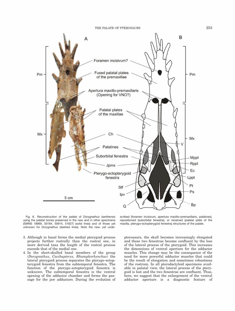
The three-dimensionally preserved Dorygnathus speci-men provides sufficient evidence to challenge earlierreconstructions and to present a new reconstruction ofthe palatal morphology of pterosaurs (Fig. 8). Accord-ingly, the introduction of new terminology has becomenecessary in some cases.
The rostral portion of the palate is formed by the mas-sive, medially fused palatal processes of the premaxillae.This feature seems to be conservative throughout thePterosauria. Coincident with the third tooth positionthis surface bears the unpaired foramen incisivum.Although the maxillo-premaxillary suture cannot beobserved, most probably a foramen incisivium is alsopresent in the holotype of Tapejara wellnhoferi (CD-R-080) that was referred to as the ‘‘medial foramen’’ byWellnhofer and Kellner (1991). Between the premaxillaand maxilla, a pair of oval fenestrae is present, whichtopographically corresponds to the apertura maxillo-pre-maxillaris of Varanus monitor (Mertens, 1942). Accord-ingly, here we apply this terminology for these fenestraein Dorygnathus. Again, in the holotype of T. wellnhoferia pair of small, longitudinally oval apertures is visibleon the palatal plate of the maxilla that may correspondto the newly described apertura maxillo-premaxillaris ofDorygnathus. In Dorygnathus and probably in all otherpterosaurs as well, the caudal portion of the hard palateis composed of the palatal plates of the maxillae and notof the palatines as previously suggested (e.g., Well-nhofer, 1975, 1978; Kellner, 1989; Bennett, 2001; Padian,2008). In Dorygnathus the caudomedial portion of thepalatal plates of the maxillae curves slightly dorsally.The contralateral plates do not meet in the midline butpresumably connected to the vomer. The palatines aredistinctive, longitudinally elongate bones that craniallyform a scarf joint with the caudal margin of the palatal
plates of the maxillae. The choanae are located betweenthe slightly caudally diverging palatines and the caudalthird of the medial margin of the palatal plates of themaxillae, which medially might have been separated bythe vomer. This position of the choana is homologouswith that of extant lepidosaurs and all archosaurs,where the choanae are bordered by the vomer medially,the palatine caudally and laterally, and maxilla laterallyand/or rostrally (Witmer, 1995b; Fig. 9). This position ofthe choanae is seen in all members of Pterosauria wherethis portion of the palate is preserved and thereforelikely applies to all pterosaur taxa.
The lateral margin of the palatines borders a pair oflongitudinally orientated suboval fenestrae, which istopographically equivalent with the suborbital fenestraof squamates and crocodylians (Bahl, 1937; Iordansky,1973), where they are bordered by the palatine medi-ally, maxilla rostrally and laterally, and the pterygoid–ectopterygoid complex caudally. Although not preservedin this Dorygnathus specimen, based on Rhamphorhyn-chus the bridge-like ectopterygoid separates the subor-bital and pterygo-ectopterygoid fenestrae and connectsto the jugal process of the maxilla (jpmx). The relativeposition and shape of the bridge-like ectopterygoid arefairly conservative in other pterosaurs. Medially, itattaches to the pterygoid-palatinal junction. The ptery-goid is triradiate with its base articulating with thebasipterygoid caudally. Rostrally, the contralateralmedial processes (mppt) converge, meet the vomer, andform the caudomedial borders of the choanae. The cra-nial process of the pterygoid (appt) is overlaid by thecaudomedial portion of the palatine. The lateral processof the pterygoid (lppt) connects to the jugal or the jugalprocess of the maxilla and forms the caudal margin ofthe pterygo-ectopterygoid fenestra and the rostromedialmargin of the subtemporal fenestra (stf). In other basalpterosaurs such as Cacibupteryx (Gasparini et al.,2004) and Rhamphorhynchus (NHM R 2786, CM11434), the pterygoid has the same shape and bonyattachments. In more derived taxa (e.g., Gnathosaurus,Coloborhynchus, Nyctosaurus), the medial pterygoidprocess is shorter, the cranial one is longer, and the lat-eral process is lost (Fig. 9). Because of the loss of thelateral process of the pterygoid, the pterygo-ectoptery-goid fenestra merges into the subtemporal fenestra. InDorygnathus and all other forms the interpterygoid va-cuity (ipv) opens between the pterygoids and the ros-trally diverging basipterygoids.
DISCUSSIONMain Trends in the Evolution of thePalate of Pterosaurs
With the new reconstruction of the pterosaurian pal-ate, some evolutionary changes can be established forthe group (Figs. 10 and 11):
1. With elongation of the rostrum and shortening of themedial process of the pterygoid the choana becomesrelatively larger.
2. Along with the former changes (see trend 1) the quad-rates become cranially inclined (already in Rhampho-rhynchus; Fastnacht, 2005) resulting in asignificantly smaller interpterygoid vacuity.
Fig. 7. The left palatine of the new Dorygnathus banthensis speci-men in ventral (A), lateral (B), and dorsal (C) views.
252 O†SI ET AL.
3. Although in basal forms the medial pterygoid processprojects further rostrally than the rostral one, inmore derived taxa the length of the rostral processexceeds that of the medial one.
4. In the short-skulled basal members of the group(Dorygnathus, Cacibupteryx, Rhamphorhynchus) thelateral pterygoid process separates the pterygo–ectop-terygoid fenestra from the subtemporal fenestra. Thefunction of the pterygo–ectopterygoid fenestra isunknown. The subtemporal fenestra is the ventralopening of the adductor chamber and forms the pas-sage for the jaw adductors. During the evolution of
pterosaurs, the skull becomes increasingly elongatedand these two fenestrae become confluent by the lossof the lateral process of the pterygoid. This increasesthe dimensions of ventral aperture for the adductormuscles. This change may be the consequence of theneed for more powerful adductor muscles that couldbe the result of elongation and sometimes robustnessof the rostrum. In all pterodactyloid specimens avail-able in palatal view, the lateral process of the ptery-goid is lost and the two fenestrae are confluent. Thus,here, we suggest that the enlargement of the ventraladductor aperture is a diagnostic feature of
Fig. 8. Reconstruction of the palate of Dorygnathus banthensisusing the palatal bones preserved in the new and in other specimens(SMNS 18969, 50184, 50914, 51827) (solid lines) and of those yetunknown for Dorygnathus (dashed lines). Note the new, yet unde-
scribed (foramen incisivum, apertura maxillo–premaxillaris, palatines),repositioned (suborbital fenestra), or renamed (palatal plate of themaxilla, pterygo–ectopterygoid fenestra) structures of the palate.
THE PALATE OF PTEROSAURS 253
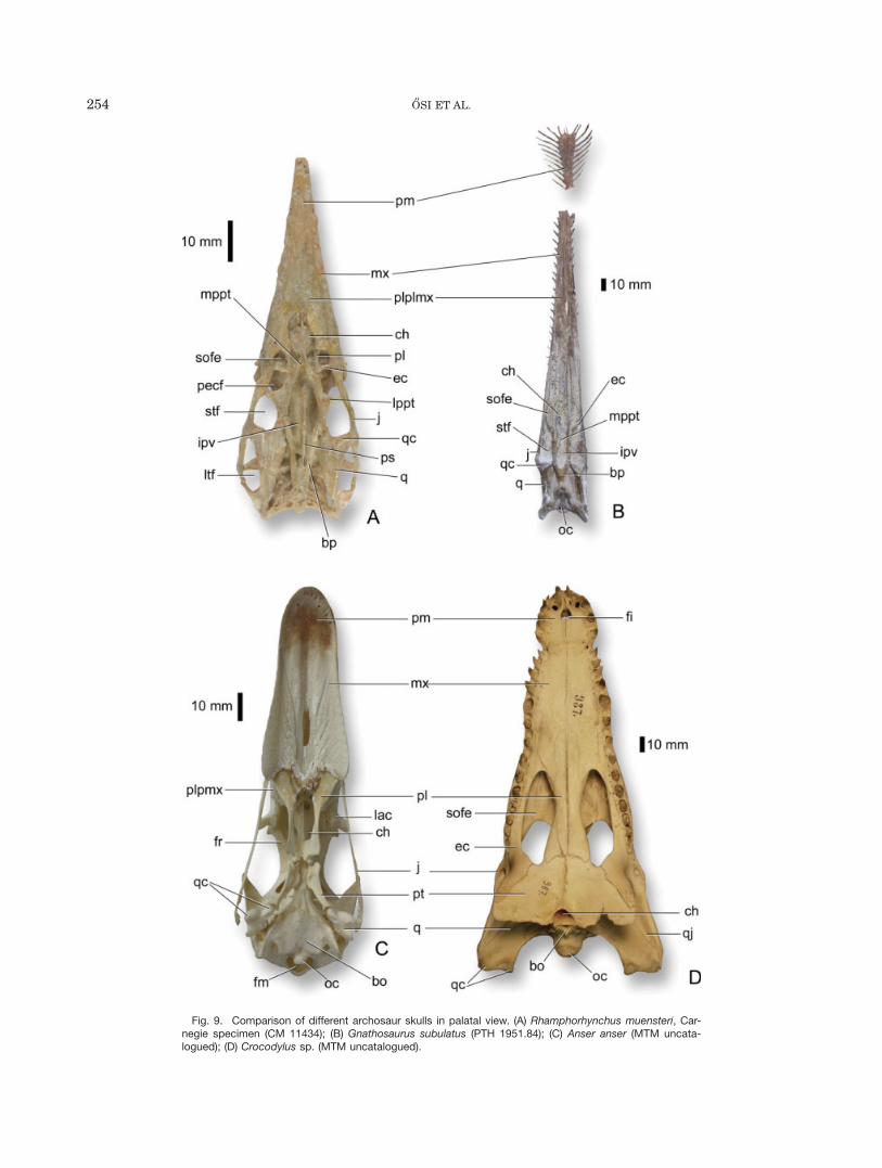
Fig. 9. Comparison of different archosaur skulls in palatal view. (A) Rhamphorhynchus muensteri, Car-negie specimen (CM 11434); (B) Gnathosaurus subulatus (PTH 1951.84); (C) Anser anser (MTM uncata-logued); (D) Crocodylus sp. (MTM uncatalogued).
254 O†SI ET AL.
Pterodactyloidea and a mechanical prerequisite forthe evolution of a long rostrum.
Vomeronasal (Jacobson’s) Organ in Pterosaurs?
The identity as well as the function of the newly rec-ognized palatal fenestrae between the premaxilla andmaxilla (Figs. 3C and 8, amp) is ambiguous. In crocodili-ans and birds, no fenestrae can be observed in topo-graphically equivalent regions (Iordansky, 1973, Witmer,1995b). However, in various squamates (e.g., snakes or
lizards), the incisive foramen or another small fenestrais present between the premaxilla and maxilla, formingthe passage for the vomeronasal organ (Jacobson’sorgan) into the oral cavity (Parsons, 1959). The vomero-nasal organ is a chemoreceptor enclosed within a carti-laginous or partly bony capsule and thus separated fromthe main olfactory epithelium (Keverne, 1999; Hillenius,2000). In mammals, it is mainly used to detect intraspe-cific pheromones; however, in some animals, such assnakes, it also mediates the trailing of prey and fooddetection on an olfactory basis (Halpern, 1987; Døvingand Trotier, 1998).
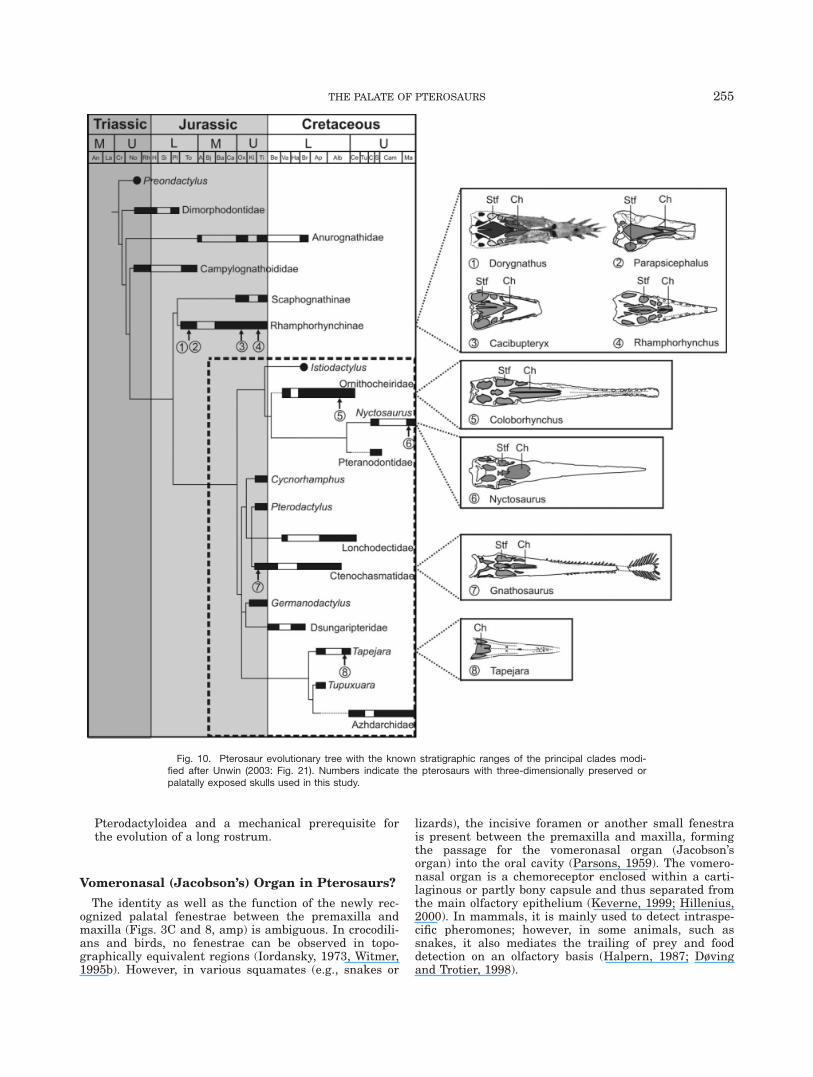
Fig. 10. Pterosaur evolutionary tree with the known stratigraphic ranges of the principal clades modi-fied after Unwin (2003: Fig. 21). Numbers indicate the pterosaurs with three-dimensionally preserved orpalatally exposed skulls used in this study.
THE PALATE OF PTEROSAURS 255
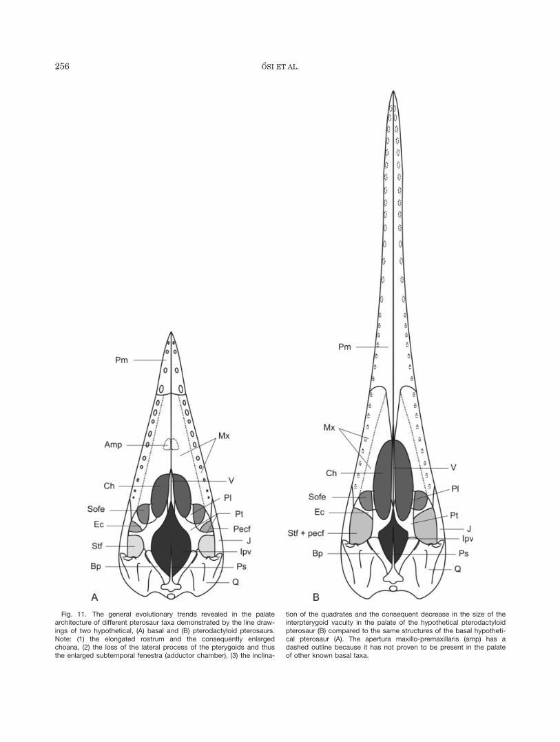
Fig. 11. The general evolutionary trends revealed in the palatearchitecture of different pterosaur taxa demonstrated by the line draw-ings of two hypothetical, (A) basal and (B) pterodactyloid pterosaurs.Note: (1) the elongated rostrum and the consequently enlargedchoana, (2) the loss of the lateral process of the pterygoids and thusthe enlarged subtemporal fenestra (adductor chamber), (3) the inclina-
tion of the quadrates and the consequent decrease in the size of theinterpterygoid vacuity in the palate of the hypothetical pterodactyloidpterosaur (B) compared to the same structures of the basal hypotheti-cal pterosaur (A). The apertura maxillo-premaxillaris (amp) has adashed outline because it has not proven to be present in the palateof other known basal taxa.
256 O†SI ET AL.
In extant crocodylians and birds, there is also evidencefor the presence of the vomeronasal organ at least in earlyontogenetic stages (e.g., Meek, 1893; Parsons, 1970). Par-sons (1959: 178) describes that in Crocodylus embryos,the vomeronasal organ is still present and has an aper-ture opening ventrally. In later ontogenetic stages, how-ever, there is no aperture for the vomeronasal organ inthe secondary palate. Parsons (1959: 181) concluded that‘‘crocodilians and the ancestors of birds (and presumablytheir fossil relatives including dinosaurs and others) lostJacobson’s organ completely’’. Gauthier et al. (1988)argued that the absence of the vomeronasal organ in arch-osaurs is a secondary development. However, using EPBmethod, Senter (2002) investigated phytosaur skull mor-phology and suggested that all extinct archosaurs lackedthe vomeronasal organ system. According to Hillenius(2000), the vomeronasal organ has a close morphologicalassociation with the septomaxilla in squamates, minorassociation in some urodeles, Sphenodon, and dasypodids,but in most tetrapods its presence correlates with thepresence of the vomer. Similar to other archosaurs, thereis no evidence for a septomaxilla in pterosaurs. Thus, ifthis organ was present—a unique case among archo-saurs—it should have been associated with the dorsal sur-face of the vomer, and the receptors of the duct of theorgan should have been positioned in the nasal cavitywith a direct exit into the oral cavity through the maxillo-premaxillary aperture.
Another option for the function of the maxillo-premax-illary aperture would be the lightening of the skull.However, the very thin rostral margin of the maxilla, itsrostral position being in the vicinity of the largest ros-tral teeth, and its relatively small size (especially inTapejara) do not support this hypothesis. There is notrace of a maxillo-premaxillary aperture in ornithocheir-ids, Dsungaripterus, Pteranodon, Nyctosaurus, and prob-ably in other large taxa, either. This indicates that theskull could increase in size and weight without increas-ing or even possessing this aperture.
The potential of these fenestrae for belonging to thevomeronasal organ system cannot be excluded nor sup-ported by any physical evidence. Thus, the function ofthis pair of apertures remains unclear.
CONCLUSIONS
The three-dimensionally preserved specimen of theEarly Jurassic basal pterosaur D. banthensis providesnew insight into the morphology of the pterosaurian pala-tal bones and helps to clarify some aspects of the nasalcavity system. The foregoing comparative study demon-strates that the generally accepted reconstructions of thepalate of pterosaurs, according to which the hard palate ismainly formed by the palatines, are incorrect. The maxillaof the new specimen shows that the hard palate comprisesthe palatal plate of the maxilla and not the palatine. Thepalatal plate of the maxilla forms the rostral and rostrolat-eral margin of the choanae similarly to that of most tur-tles, lepidosaurs, and all other archosaurs. The newspecimen of Dorygnathus also provides evidence for thepresence of a distinct palatine bone that shows morpholog-ical and topographical similarities with that of crocodiliansand birds, respectively. Being attached to the caudal por-tion of the palatal plate of the maxilla, it borders the choa-nae laterally thus revealing the homologous nature of the
(primary) choana in pterosaurs, all other archosaurs, andlepidosaurs. The fenestra lateral to the palatines, medialto the jugal process of the maxilla, and rostral to the ectop-terygoid referred to as the ‘‘postpalatal’’ or ‘‘palatinal fe-nestra’’ is topographically equivalent with the suborbitalfenestra of crocodilians and lizards. In Dorygnathus andsome other basal pterosaurs (Cacibupteryx, Rhampho-rhynchus), an additional fenestra, the pterygo–ectoptery-goid fenestra, is present between the suborbital and thesubtemporal fenestrae (adductor chamber). This fenestrais bordered rostrally by the ectopterygoid and caudally bythe lateral process of the pterygoid.
Similar to birds, the antorbital cavity of Dorygnathuswas at least partially separated from the nasal cavityproper by the palatal processes of the maxilla but, incontrast to birds, the palatal plate of the maxilla wasnot pneumatized.
The new interpretation of the palate also reveals someevolutionary changes of the palatal construction withinPterosauria. One of which is that the lateral pterygoid pro-cess is completely reduced in advanced forms, probably inall Pterodactyloidea. Consequently, the pterygo–ectoptery-goid fenestra merged with the subtemporal fenestraincreasing considerably the relative size of the adductorchamber. This would have allowed for more developedmandibular adductor muscles resulting in an increase ofocclusion power. A second evolutionary trend is the changeof the size of the choana relative to the interpterygoid va-cuity. In basal forms, a pair of small choanae was presentrostral to a huge interpterygoid vacuity. In more derivedmembers of the group the interpteygoid vacuity becamestrongly reduced along with the anterior inclination of thequadrates, whereas the choanae increased in size. Thisexceptionally well-preserved specimen of Dorygnathus fur-thermore reveals a hitherto unknown pair of fenestraecaudal to the incisive foramen, which opens at the palatalcontact of the premaxilla–maxilla and might have servedas the opening for the vomeronasal organ. However, evi-dences on this issue neither pro nor contra can be lined up.
ACKNOWLEDGMENTS
The authors thank M. Witton and the anonymousreviewer for their useful comments that highly improvedthe standards of this manuscript; V. Griener (StaatlichesMuseum fur Naturkunde, Karlsruhe) for taking photosof the specimen; M. Lamanna (Carnegie Museum of Nat-ural History, Pittsburgh), Bill Mueller (Texas Tech Uni-versity, Texas), O. Rauhut (Bayerische Staatsammlungfur Palaontologie und Geologie, Munchen), L. Steel (TheNatural History Museum, London), R. Elgin (StaatlichesMuseum fur Naturkunde, Karlsruhe), W. F. Simpson(The Field Museum, Chicago), and Z. B. de Gasparini(Museo de La Plata, La Plata) for providing pictures ofthe most significant pterosaur specimens; D. Peters foruseful comments; O. Rieppel and P. P. Gallina for infor-mation about the availability of the specimens; K.Padian for providing access to his publication; and T.Antal for helping to prepare the skull of Anser anser.
LITERATURE CITED
Arthaber G. 1919. Studien uber Flugsaurier auf Grund der Bearbei-tung des Wiener Exemplares von Dorygnathus banthensis Theod.sp. Denkschr Akad Wiss Wien math-nat Kl 97:391–464.
THE PALATE OF PTEROSAURS 257
Bahl KN. 1937. Skull of Varanus monitor (Linn.). Rec Ind Mus39:133–174.
Bennett SC. 1996. The phylogenetic position of the Pterosauriawithin the Archosauromorpha. Zool J Linn Soc 118:261–308.
Bennett SC. 2001. The osteology and functional morphology of theLate Cretaceous pterosaur Pteranodon. I. General description ofosteology. Palaeontographica A 260:1–112.
Bennett SC. 2007. A second specimen of the pterosaur Anurogna-thus ammoni. Palaontol Z 81:376–398.
Benton MJ. 1999. Scleromochlus taylori and the origin of dinosaursand pterosaurs. Philos Trans R Soc Lond B Biol Sci 354:1423–1446.
Broili F. 1919. Ctenochasma gracile Oppel. Sond Geog Jahresheften29/30:299–309.
Busbey AB. 1994. The structural consequences of skull flattening incrocodilians. In: Thomason JJ, editor. Functional morphology invertebrate paleontology. New York: Cambridge University Press.p 173–192.
Coates MI. 1993. Ancestors and homology the origin of the tetrapodlimb. A Biotheor 41:411–424.
Døving KB, Trotier D. 1998. Structure and function of the vomero-nasal organ. J Exp Biol 201:2913–2925.
Fastnacht M. 2005. Jaw mechanics of the pterosaur skull construc-tion and the evolution of toothlessness. PhD Thesis. Johannes Gu-tenberg-Universitat, Mainz.
Gasparini Z, Fernandez M, De La Fuente M. 2004. A new pterosaurfrom the Jurassic of Cuba. Palaeontology 47:919–927.
Gauthier JA, Kluge AG, Rowe T. 1988. Amniote phylogeny and theimportance of fossils. Cladistics 4:105–209.
Halpern M. 1987. The organization and function of the vomeronasalsystem. Ann Rev Neurosci 10:325–362.
Hillenius WJ. 2000. Septomaxilla of non-mammalian synapsids:soft-tissue correlates and a new functional interpretation. J Mor-phol 245:29–50.
Hone DWE, Benton MJ. 2007. An evaluation of the phylogeneticrelationships of the pterosaurs to the archosauromorph reptiles.J Syst Palaeontol 5:465–469.
Hone DWE, Benton MJ. 2008. Contrasting supertree and total-evidencemethods: the origin of the pterosaurs. Zitteliana B 28:35–60.
Hooley RW. 1913. On the skeleton of Ornithodesmus latidens; anornithosaur from the Wealden Shales of Atherfield, Isle of Wight.Quart J Geol Soc 69:372–422.
Iordansky NN. 1973. The skull of the Crocodilia. In: Gans C, Par-sons S, editors. Biology of the reptilia, Vol. 4. New York: Aca-demic Press. p 201–262.
Kellner AWA. 1989. A new edentate pterosaur of the Lower Creta-ceous from the Araripe Basin, Northeast Brasil. An Acad BrasCien 61:439–446.
Keverne EB. 1999. The vomeronasal organ. Science 286:716–720.Langer MC. 2004. Basal Saurischia. In: Weishampel DB, Dodson P,Osmolska H, editors. The Dinosauria. 2nd ed. Los Angeles: Uni-versity of California Press. p 25–46.
Marsh OC. 1884. Principal characters of American Cretaceous Pter-odactyls. I. The skull of Pteranodon. Am J Sci 21:423–427.
Mayr FX. 1964. Die naturwissenschaftlichen Sammlungen der Phi-losophisch-Theologischen Hochschule Eichstatt. Festschrift 400Jahre Collegium Willibaldinum Eichstatt. p 302–334.
Meek A. 1893. On the occurrence of a Jacobson’s organ, with notes onthe development of the nasal cavity, the lachrymal duct, and the Har-derian gland in Crocodilus porosus. J Anat Physiol 27:151–160.
Mertens R. 1942. Die Familie der Warane (Varanidae). Zweiter Teil:Der Schadel. Abh Senckenbergischen Naturforschenden Ges465:117–234.
Newton ET. 1888. On the skull, brain and auditory organ of a newspecies of Pterosaurian (Scaphognathus purdoni), from the UpperLias near Whitby, Yorkshire. Philos Trans R Soc Lond B Biol Sci179:503–537.
Osmolska H. 1985. Antorbital fenestra of archosaurs and its suggestedfunction. In: Duncker HR, Fleisher G, editors. Vertebrate morphology.New York: Gustav Fischer Verlag. p 159–162.
Padian K. 2008. The Early Jurassic pterosaur Dorygnathus ban-thensis (Theodori, 1830). Special Papers Paleontol 80:1–64.
Parsons TS. 1959. Studies on the comparative embryology of thereptilian nose. Bull Mus Comp Zool 120:101–277.
Parsons TS. 1970. The nose and Jacobson’s organ. In: Gans C, Par-sons S, editors. Biology of the reptilia, Vol. 2. New York: Aca-demic Press. p 99–191.
Peters D. 2000. A re-examination of four prolacertiforms with impli-cations for pterosaur phylogenies. Riv Italiana Paleontol Strat106:293–336.
Seeley HG. 1901. Dragons of the Air: an account of extinct flyingreptiles. New York: Appleton & Co.; London: Methuen & Co.
Senter P. 2002. Lack of a pheromonal sence in phytosaurs and otherarchosaurs, and its implications for reproductive communication.Paleobiology 28:544–550.
Sereno P. 1991. Basal archosaurs: phylogenetic relationships andfunctional implications. J Vertebrate Paleontol Mem 2:1–53.
Unwin DM. 2003. On the phylogeny and evolutionary history ofpterosaurs. In: Buffetaut E, Mazin J-M, Evolution and Palaeobiol-ogy of Pterosaurs. Geol Soc Spec Publ, 217:139–190.
Veldmeijer AJ. 2003. Description of Coloborynchus spielbergi sp.nov. (Pterodactyloidea) from the Albian Lower Cretaceous of Bra-zil. Scri Geol 125:35–139.
Veldmeijer AJ. 2006. Toothed pterosaurs from the Santana Formation(Cretaceous; Aptain-Albian) of northeastern Brazil. A reappraisal on thebasis of newly describedmaterial. PhD Thesis. Utrecht University. p 269.
von Huene F. 1914. Beitrage zur Kenntnis des Schadels einigerFlugsaurier. Geol Palaontol Abh Neue Folge 13:57–65.
Wellnhofer P. 1970. Die Pterodactyloidea (Pterosauria) der Ober-jura-Plattenkalke Suddeutschlands. Abh Bayerischen Akad WissNeue Folge 141:1–133.
Wellnhofer P. 1975. Die Rhamphorhynchoidea (Pterosauria) derOberjura-Plattenkalke Suddeutschlands. Allgemeine Skelettmor-phologie. Palaeontographica A 148:1–33.
Wellnhofer P. 1978. Handbuch der Palaoherpetologie. Teil 19. Ptero-sauria. Stuttgart: Gustav Fischer Verlag.
Wellnhofer P. 1987. New crested Pterosaurs from the Lower Creta-ceous of Brasil. Mitt Bayerischen Staatsammlung Palaontol HistGeol 27:175–186.
Wellnhofer P. 1991a. The illustrated encyclopedia of pterosaurs.London: Salamander Books Ltd.
Wellnhofer P. 1991b. Weitere Pterosaurierfunde aus der Santana-Formation (Apt) der Chapada do Araripe, Brasilien. Palaeontog-raphica A 215:43–101.
Wellnhofer P, Kellner AWA. 1991. The skull of Tapejara wellnhoferiKellner (Reptilia, Pterosauria) from the Lower Cretaceous SantanaFormation of the Araripe Basin, Northeastern Brazil. Mitt Bayeri-schen Staatsammlung Palaontol Hist Geol 31:89–106.
Williston SW. 1902. On the skull of Nyctodactylus, an Upper Creta-ceous pterodactyl. J Geol 10:520–531.
Witmer LM. 1995a. The extant phylogenetic bracket and the impor-tance of reconstructing soft tissues in fossils. In: Thomason JJ,editor. Functional morphology in vertebrate paleontology. NewYork: Cambridge University Press. p 19–33.
Witmer LM. 1995b. Homology of facial structures in extant Archo-saurs (birds and crocodilians), with special reference to paranasalpneumaticity and nasal conchae. J Morphol 225:269–327.
Witmer LM. 1997. The evolution of the antorbital cavity of archosaurs: astudy in soft-tissue reconstruction in the fossil record with an analysisof the function of pneumaticity. Mem Soc Vertebrate Paleontol 17:1–73.
Witmer LM, Chatterjee S, Franzosa J, Rowe T. 2003. Neuroanatomyof flying reptiles and implications for flight, posture and behavior.Nature 425:950–953.
Witmer LM, Martin LD. 1987. The primitive features of the avianpalate, with special reference to Mesozoic birds. In: Mourer-Chau-vire C, editor. L’Evolution des Oiseaux d’Apres le Temoignage desFossiles ed. Lyon: Universite Claude-Bernard. p 21–40.
Witmer LM, Ridgely RC. 2008. The paranasal air sinuses of predatoryand armored dinosaurs Archosauria: Theropoda & Ankylosauria andtheir contribution to cephalic structures. Anat Rec 291:1362–1388.
Woodward AS. 1902. On two skulls of Ornithosaurian Rhampho-rhynchus. Ann Mag Nat Hist 9:1.
Young CC. 1964. On a New Pterosaurian from Sinkiang, China.Vertebr Pal 8:221–225 (Part translated into English).
258 O†SI ET AL.
View publication statsView publication stats


![PTEROSAURS FLIGHT IN THE AGE OF DINOSAURS · Pterosaurs iPad – FINAL– March 25, 2014 - 6 [watercolor Duria Antiquior, by Henry de la Beche] Duria Antiquior, A More Ancient Dorset,](https://img.pdfslide.us/doc/110x75/5f0435527e708231d40cd917/pterosaurs-flight-in-the-age-of-dinosaurs-pterosaurs-ipad-a-finala-march-25.jpg)