Embed Size (px)
Citation preview
New Insights on the Control and Function of OctopusSuckers
Hosain Bagheri, Anna Hu, Sheldon Cummings, Cayla Roy, Rachel Casleton,Ashley Wan, Nicole Erjavic, Spring Berman, Matthew M. Peet, Daniel M. Aukes,Ximin He, Stephen C. Pratt, Rebecca E. Fisher, and Hamid Marvi*
1. Introduction
The ability to bend, deform, configure, and adapt to surround-ings makes soft robots an ideal choice for navigating
unpredictable, unstructured, and confinedenvironments.[1–8] However, despite recentadvances in soft robotic technologies, thechallenge of controlling high-dimensionalappendages has prevented the develop-ment of versatile soft robots. Biology servesas a rich source of design inspiration forsoft robots, which have been based on suchdiverse organisms as octopuses,[9] caterpil-lars,[10] snakes,[11,12] fish,[13] worms,[14–16]
and humans.[17] Through the integrationof biology, engineering, and physics, thisstudy aims to investigate octopus controlarchitecture in addition to sucker attach-ment and detachment mechanisms.
The octopus has a soft body and arms(Figure 1), and can achieve a diverse arrayof locomotor modes such as swimming,jetting, crawling, and even bipedal walk-ing.[18] In addition, octopuses are capableof complex tasks such as carrying andmanipulating objects and squeezing
through tight spaces. Previous studies[19–23] have shown thatoctopuses use a stereotypical motion for grasping objects.Impressively, the same pattern has been observed in amputatedoctopus arms,[20,21] implying the presence of a distributed
H. Bagheri, A. Hu, Prof. S. Berman, Prof. M. M. Peet, Prof. H. MarviSchool for Engineering of Matter Transport and EnergyArizona State University501 E Tyler Mall, Tempe 85287, AZ, USAE-mail: [email protected]
S. CummingsSchool of Biological and Health System EngineeringArizona State University501 E Tyler Mall, Tempe 85287, AZ, USA
C. Roy, R. Casleton, A. WanSchool of Molecular SciencesArizona State University551 E University Dr, Tempe 85281, AZ, USA
The ORCID identification number(s) for the author(s) of this articlecan be found under https://doi.org/10.1002/aisy.201900154.
© 2020 The Authors. Published by WILEY-VCH Verlag GmbH& Co. KGaA,Weinheim. This is an open access article under the terms of the CreativeCommons Attribution License, which permits use, distribution andreproduction in any medium, provided the original work is properly cited.
DOI: 10.1002/aisy.201900154
N. Erjavic, Prof. S. C. Pratt, Prof. R. E. FisherSchool of Life SciencesArizona State University427 E Tyler Mall, Tempe 85281, AZ, USA
Prof. D. M. AukesThe Polytechnic SchoolFulton Schools of EngineeringArizona State University6075 S. Innovation Way West, Mesa 85212, AZ, USA
Prof. X. HeHenry Samueli School of Engineering and Applied ScienceUniversity of California, Los Angeles7400 Boelter Hall, Los Angeles 90095, CA, USA
Prof. R. E. FisherDepartment of Basic Medical SciencesUniversity of Arizona College of Medicine-Phoenix475 N 5th St., Phoenix 85004, AZ, USA
Octopuses utilize their suckers for a myriad of functions such as chemo- andmechanosensing, exploring and manipulating objects, anchoring the body duringcrawling, and navigating through narrow passages. The sucker attachmentmechanism grants the octopus the ability to perform many of these tasks.The goal of this study is to analyze sucker function and control through theassessment of pull-off forces under different conditions. Sucker pull-off forcesare measured in Octopus bimaculoides (three females, seven males), when thearm is intact, amputated, and amputated with the suckers punctured. Greatersucker pull-off forces are observed for amputated arms, plausibly indicating thatthe brain and/or the interbrachial commissure are responsible for triggeringearly sucker detachment in the intact animal. In addition, after piercing andcompromising the sucker cavity, pull-off force significantly decreases, indicatingthat the primary mechanism for sucker attachment is suction, and is lessdependent on adhesion. These results provide new insights into the control andfunction of octopus suckers that can be integrated into the design and devel-opment of soft robot arms for aquatic applications.
FULL PAPERwww.advintellsyst.com
Adv. Intell. Syst. 2020, 1900154 1900154 (1 of 8) © 2020 The Authors. Published by WILEY-VCH Verlag GmbH & Co. KGaA, Weinheim
control architecture. The adaptability, limitless degrees of free-dom, and distributed control of octopus arms make them anideal candidate for inspiration in the areas of soft robot-ics[2,9,24–33] and control of high-dimensional systems.[4,34–41]
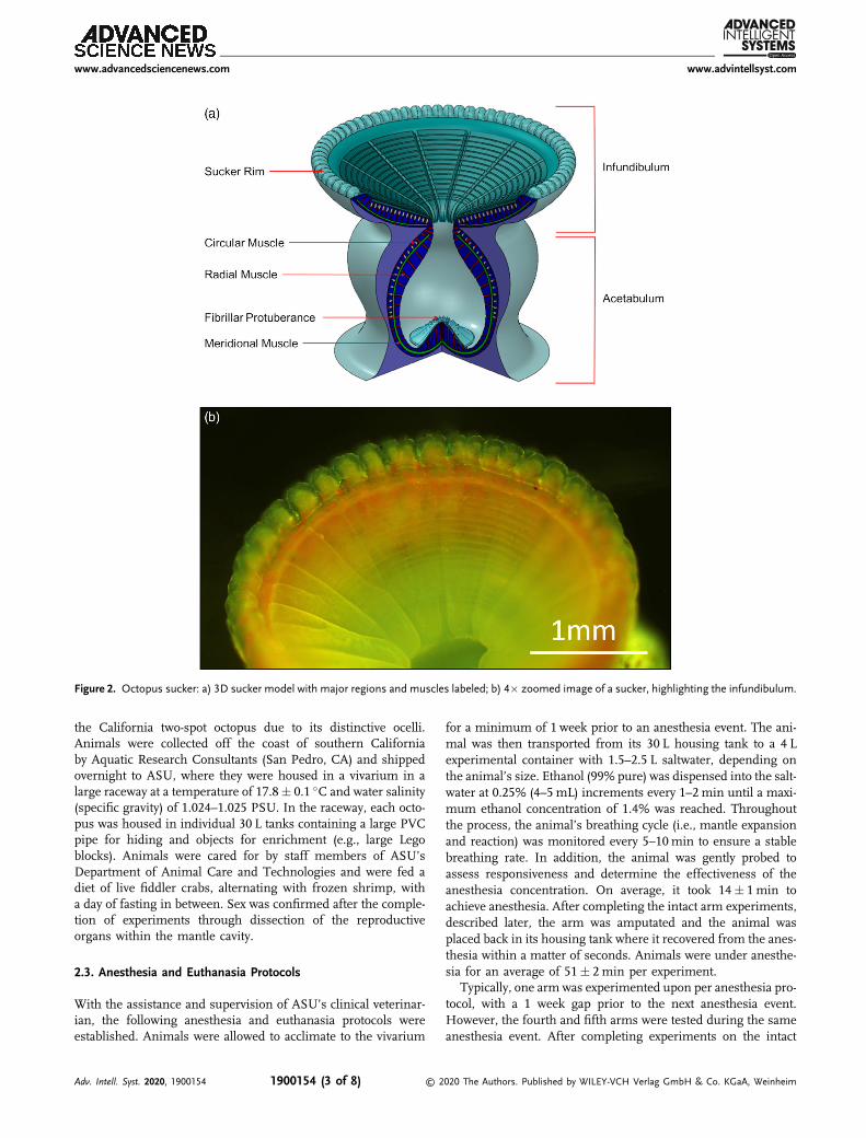
The octopus features an extensive nervous system, includingthe brain, an axial nerve cord in each arm, and the interbrachialcommissure, a ring-like structure connecting all of the axialnerve cords.[42,43] Within the arms, the central axial nerve cordis accompanied by four peripheral intramuscular nerve cordsand nerves that are associated with each sucker.[42,44–47] Thisnetwork of nerve fibers and ganglia is embedded in layers ofmuscle, including transverse, longitudinal, oblique, and circularfibers,[48] granting the animal the ability to elongate, shorten,bend, and twist anywhere along the arm with high precision.Extraordinarily, the suckers display similar capabilities. Themeridional, circular, and radial muscles[49,50] controlled by thesucker ganglia achieve a wide array of motor and proprioceptivefunctions. Octopuses also use their suckers for sensing andexploring the environment.[49]
An octopus sucker consists of two regions, the infundibulumand acetabulum (Figure 2). The infundibulum is the exposed,pliable, denticled face of the sucker that is circumscribed onits rim by a ridge.[51–53] The acetabulum is the more rigid, ellip-soidal cavity of the sucker, consisting of a domed roof featuring afibrillar surface and smooth surrounding walls.[50,53] Throughoutthe years, many investigators have postulated about the attach-ment mechanism of octopus suckers.[49,52,54,55] The proposalby Tramacere et al.[50,53,56] is the most thorough to date.According to these authors, the attachment process begins withthe infundibulum pressing and conforming to the surface, andthe rim sealing the sucker and preventing water leakage. The ace-tabulum then begins contracting radially, decreasing the innerwater pressure. The meridional muscles then contract untilthe acetabulum’s fibrillar protuberance adheres to the orificesidewalls, forming a toroidal water cavity. Lastly, the acetabu-lum’s radial and meridional muscles relax as the sucker config-uration remains passively fixed due to the friction between theprotuberance and side walls and the pressure differential createdby the toroidal water cavity. During detachment, the contractionof circular muscles in the infundibulum and acetabulum rup-tures the seal between the sucker rim and the surface, andthe contact between the fibrillar protuberance and the orifice
sidewalls. This causes the internal sucker pressure to equal thatof the surrounding environment.
While sucker attachment and detachment mechanisms havebeen described in previous studies,[44,49,50,52,53,55–60] a number ofquestions remain. In particular, this study was designed to assesswhether pull-off force is impacted when there is no communi-cation with the brain or interbrachial commissure, and thedegree to which suction versus adhesion contributes to suckerattachment. To analyze these conditions, the pull-off force wasmeasured in intact arms, amputated arms, and amputated armswith punctured suckers. The intact versus amputated arm experi-ments may provide insights into how the sucker attachmentmechanism is influenced by centralized versus distributedneural pathways. Unpunctured versus punctured suckers wereanalyzed in amputated arms to determine the degree to whichthe attachment mechanism is dependent on suction versus adhe-sion. In addition, all eight arms were tested to determine whetherthere are any functional differences based on arm identity (i.e., inanterior vs. posterior, or right vs. left arms). The obtained resultscan potentially shed light on the control and function of octopussuckers.
2. Experimental Section
2.1. Ethics Statement
Although Institutional Animal Care and Use Committee (IACUC)approval is currently not required for cephalopod research in theUnited States, the husbandry, anesthesia, and experimental pro-tocols used in this study were developed under the supervisionof Arizona State University (ASU) veterinary staff, referencingthe EU Directive 2010/63/EU and published guidelines for ceph-alopod use.[61–68] The ASU IACUC reviewed a description of theproposed work and concluded that IACUC approval or waiver wasnot necessary. However, all protocols were developed in close col-laboration with the ASU veterinary staff.
2.2. Animal Husbandry
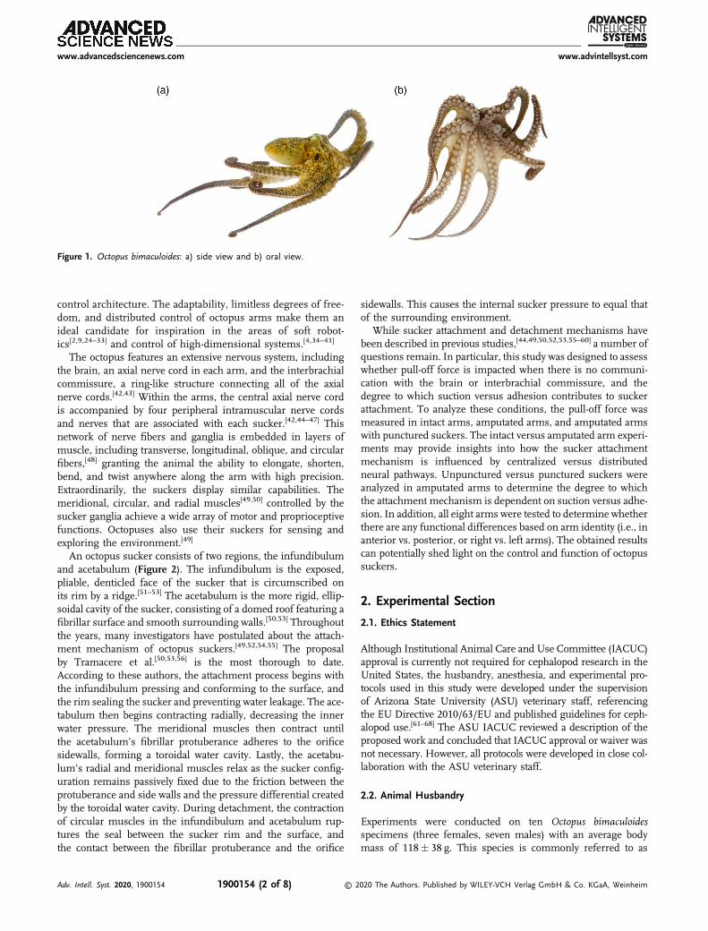
Experiments were conducted on ten Octopus bimaculoidesspecimens (three females, seven males) with an average bodymass of 118� 38 g. This species is commonly referred to as
Figure 1. Octopus bimaculoides: a) side view and b) oral view.
www.advancedsciencenews.com www.advintellsyst.com
Adv. Intell. Syst. 2020, 1900154 1900154 (2 of 8) © 2020 The Authors. Published by WILEY-VCH Verlag GmbH & Co. KGaA, Weinheim
the California two-spot octopus due to its distinctive ocelli.Animals were collected off the coast of southern Californiaby Aquatic Research Consultants (San Pedro, CA) and shippedovernight to ASU, where they were housed in a vivarium in alarge raceway at a temperature of 17.8� 0.1 �C and water salinity(specific gravity) of 1.024–1.025 PSU. In the raceway, each octo-pus was housed in individual 30 L tanks containing a large PVCpipe for hiding and objects for enrichment (e.g., large Legoblocks). Animals were cared for by staff members of ASU’sDepartment of Animal Care and Technologies and were fed adiet of live fiddler crabs, alternating with frozen shrimp, witha day of fasting in between. Sex was confirmed after the comple-tion of experiments through dissection of the reproductiveorgans within the mantle cavity.
2.3. Anesthesia and Euthanasia Protocols
With the assistance and supervision of ASU’s clinical veterinar-ian, the following anesthesia and euthanasia protocols wereestablished. Animals were allowed to acclimate to the vivarium
for a minimum of 1 week prior to an anesthesia event. The ani-mal was then transported from its 30 L housing tank to a 4 Lexperimental container with 1.5–2.5 L saltwater, depending onthe animal’s size. Ethanol (99% pure) was dispensed into the salt-water at 0.25% (4–5mL) increments every 1–2min until a maxi-mum ethanol concentration of 1.4% was reached. Throughoutthe process, the animal’s breathing cycle (i.e., mantle expansionand reaction) was monitored every 5–10min to ensure a stablebreathing rate. In addition, the animal was gently probed toassess responsiveness and determine the effectiveness of theanesthesia concentration. On average, it took 14� 1min toachieve anesthesia. After completing the intact arm experiments,described later, the arm was amputated and the animal wasplaced back in its housing tank where it recovered from the anes-thesia within a matter of seconds. Animals were under anesthe-sia for an average of 51� 2min per experiment.
Typically, one arm was experimented upon per anesthesia pro-tocol, with a 1 week gap prior to the next anesthesia event.However, the fourth and fifth arms were tested during the sameanesthesia event. After completing experiments on the intact
Figure 2. Octopus sucker: a) 3D sucker model with major regions and muscles labeled; b) 4� zoomed image of a sucker, highlighting the infundibulum.
www.advancedsciencenews.com www.advintellsyst.com
Adv. Intell. Syst. 2020, 1900154 1900154 (3 of 8) © 2020 The Authors. Published by WILEY-VCH Verlag GmbH & Co. KGaA, Weinheim
fourth and fifth arms, both arms were amputated and the animalwas euthanized. To achieve euthanasia, the ethanol concentra-tion was increased to 10% in the experimental container, untilthe animal’s breathing ceased. The remaining three arms werethen amputated and preserved along with the mantle in 10%neutral buffered formalin for future anatomical studies.
2.4. Experimental Preparation
Once the animal was anesthetized, such that no active responsewas observed, the arm of interest was isolated. It should be notedthat the order of the selected left (L) and right (R) arms varied perspecimen to ensure data could be collected for all arm identities(i.e., L1–L4 and R1–R4). Arms were selected for study with con-sideration of the animal’s mobility and ability to feed postampu-tation; this was achieved by alternating between anterior andposterior arms, and left and right sides of the animal over thecourse of the experiments.
The distal portion of the selected arm was anchored down toa small rectangular acrylic piece using superglue (Loctite®
Super Glue ULTRA Liquid Control). As the arm was gently heldand extended from the anchored distal end, a longer rectangularacrylic piece was positioned above the specimen, allowing thesuckers to adhere to its surface. With the arm elongated andadhered to the acrylic, the skin on the aboral side of the armwas removed using fine scissors. A thin line of supergluewas applied to another long rectangular acrylic piece, whichwas then situated under the arm, facing the deskinned aboralsurface. With the arm now positioned between the two longrectangular acrylic pieces, it was held outside the saltwater untilthe aboral side adhered to the acrylic (�1–2min). Once the armwas adhered, the acrylic piece on the sucker side was graduallyremoved by disengaging the suckers. While arm stiffness doesplay an important role in the attachment and detachment ofoctopus suckers, further arm restraints were avoided to mini-mize arm and sucker muscle damage while maintaining thearm in a position that would enable replicable engagementof the suckers with the indenter. The potential effects of tissuestiffness variance were minimized through consistency inthe experimental preparation and performing tests on themost proximal region of the arm in animals of similar size(p¼ 0.801) (for more details on the statistical analysis, seeSection 2.7).
A total of nine proximal suckers were selected and labeledusing permanent markers on the acrylic piece next to the suckersof interest. Three neighboring suckers were designated for theintact arm, amputated arm, and amputated arm with puncturedsucker experiments. Using double-sided tape (3M™ Ultra HighTemperature Adhesive Transfer Tape 9082), the acrylic piecewith the adhered arm was secured to the bottom of the experi-mental container with the suckers facing upward. At this stagethe arm was ready for experimentation.
2.5. Experimental Setup
The test setup (Figure S1, Supporting Information) consisted of a12mm diameter borosilicate indenter with a radius of curvatureof 37.22mm attached to the end of a 102mm long oxide alloy
steel screw, which was screwed into a 0.5 N (0.25mN resolution)uniaxial load cell (Transducer Techniques® GSO-50) mountedonto a motorized linear translation stage (Thorlabs Z825V) witha 25mm traveling distance (29 nm resolution). A 5 megapixelcamera (EPL 170) was used for recording side view observations.Through a series of preliminary parameterization studies, opti-mal parameters were established for the indenter pull-off experi-ments (Appendix S1, Supporting Information).
2.6. Experimental Procedure
The 4 L experimental container holding the animal and adheredarm of interest was placed under the experimental setup. Theindenter was lowered into the anesthetic (ethanol/saltwater) bathand zeroed out once centered above the sucker of interest. Thesucker was approached by the indenter (velocity: 0.2 mm s�1,acceleration: 0.2 mm s�2) and pressed upon until a preloadof 7mN was reached. The intender remained in contact for20 s and was then retracted (velocity: 0.4mm s�1, acceleration:0.2mm s�2) until sucker disengaged. One test per sucker wasperformed with the indenter cleaned (using an alcohol wipe)prior to each test.
Experiments were conducted on proximal suckers in 1) theintact arm, 2) the amputated arm, and 3) the amputated arm withthe sucker sidewalls punctured. First, three designated intactarm suckers were tested for pull-off force. Once the intactarm sucker tests were complete, the arm was amputated witha scalpel and the animal was placed in its holding tank for anes-thesia recovery, while the amputated arm was kept in the sameanesthetic bath for the remaining experiments. The unpuncturedand punctured suckers designated for amputated arm tests wererandomized to control for the effect of time elapsed, postampu-tation. The suckers selected for the amputated arm with punc-tured sucker test were pierced using a 0.6mm needle with a0.3mm tip diameter. Inserting the needle into the sucker orifice,four piercings were made along the acetabulum sidewalls, radi-ally spaced.
Once all pull-off force tests were completed for the selectedarm, the arm length and selected sucker rim diameters weremeasured using a metric ruler and digital caliper, respectively.The arm was then preserved in 10% neutral buffered formalinfor future anatomical studies.
2.7. Data Analysis
After experimentation, data anomalies were discarded in thefollowing manner. The coefficient of variation (i.e., ratio ofstandard deviation to mean) was calculated for each set of threeindependent sucker readings (per arm, per condition). A histo-gram of all coefficients of variation (CV) was then plotted, reveal-ing a bimodal distribution with a global maximum at 1 and asecondary peak at 1.8, separated by a relative minimum at 1.5(Figure S3, Supporting Information). As the higher mode mayreflect anomalously deviant measures, all sets of three-suckerreadings with CV above 1.5 were removed from the analysis.To test for factors affecting pull-off force, a linear mixed modelwas fit to the data, using IBM® SPSS® Statistics 25. The depen-dent variable was pull-off force, log-transformed to achieve
www.advancedsciencenews.com www.advintellsyst.com
Adv. Intell. Syst. 2020, 1900154 1900154 (4 of 8) © 2020 The Authors. Published by WILEY-VCH Verlag GmbH & Co. KGaA, Weinheim
normally distributed residuals. The independent fixed variableswere condition (intact arm with unpunctured, amputated armwith unpunctured, or amputated arm with punctured suckers),right/left arm, anterior/posterior arm, and sex. Octopus and arm(nested within octopus) were included as random variables, toaccount for repeated measurements on each arm and each ani-mal. Sucker diameter and octopus body mass were includedas covariates. Pairwise comparisons were performed for thedifferent experimental conditions. To avoid alpha inflation inthese comparisons, the sequential Bonferroni correction wasused. Normality was tested by normal probability plots ofresiduals. Residual versus fitted values were plotted to confirmconstant variance and linearity. The results are reported aslog-transformed values. Estimated means are reported with theirstandard errors.
3. Results
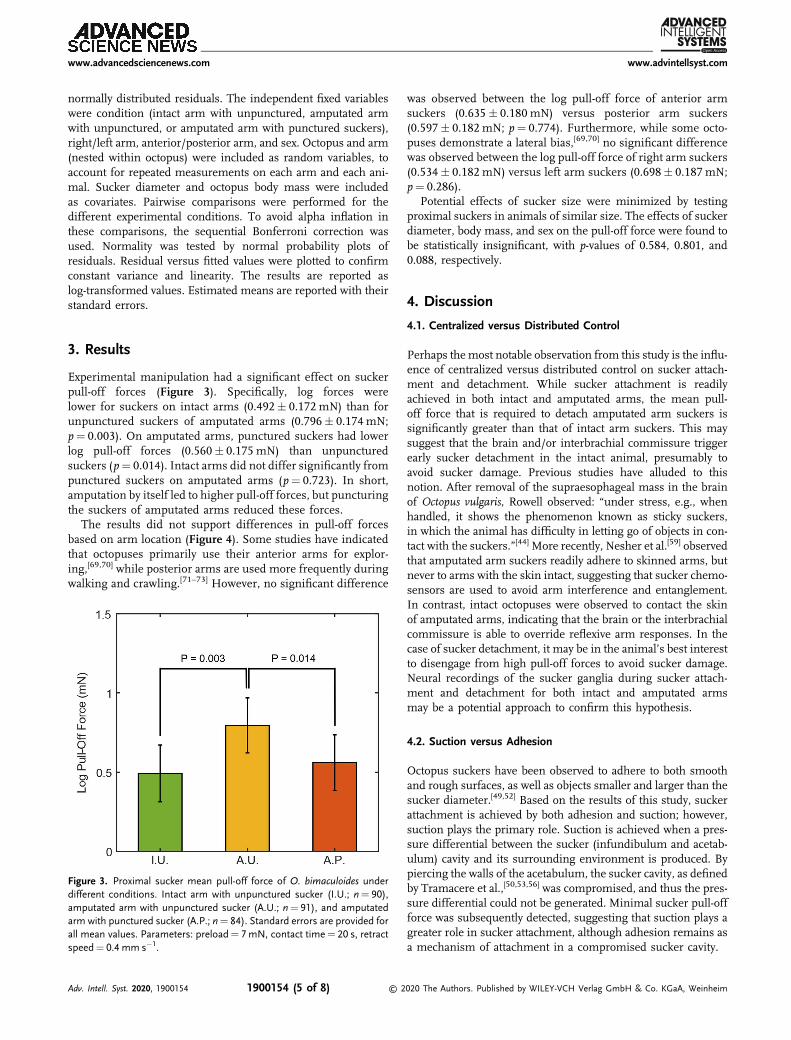
Experimental manipulation had a significant effect on suckerpull-off forces (Figure 3). Specifically, log forces werelower for suckers on intact arms (0.492� 0.172 mN) than forunpunctured suckers of amputated arms (0.796� 0.174 mN;p¼ 0.003). On amputated arms, punctured suckers had lowerlog pull-off forces (0.560� 0.175 mN) than unpuncturedsuckers (p¼ 0.014). Intact arms did not differ significantly frompunctured suckers on amputated arms (p¼ 0.723). In short,amputation by itself led to higher pull-off forces, but puncturingthe suckers of amputated arms reduced these forces.
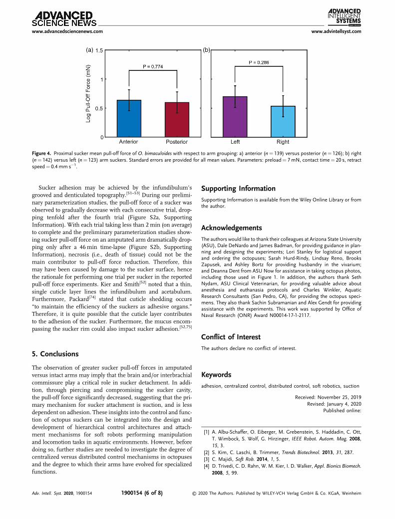
The results did not support differences in pull-off forcesbased on arm location (Figure 4). Some studies have indicatedthat octopuses primarily use their anterior arms for explor-ing,[69,70] while posterior arms are used more frequently duringwalking and crawling.[71–73] However, no significant difference
was observed between the log pull-off force of anterior armsuckers (0.635� 0.180 mN) versus posterior arm suckers(0.597� 0.182 mN; p¼ 0.774). Furthermore, while some octo-puses demonstrate a lateral bias,[69,70] no significant differencewas observed between the log pull-off force of right arm suckers(0.534� 0.182 mN) versus left arm suckers (0.698� 0.187 mN;p¼ 0.286).
Potential effects of sucker size were minimized by testingproximal suckers in animals of similar size. The effects of suckerdiameter, body mass, and sex on the pull-off force were found tobe statistically insignificant, with p-values of 0.584, 0.801, and0.088, respectively.
4. Discussion
4.1. Centralized versus Distributed Control
Perhaps the most notable observation from this study is the influ-ence of centralized versus distributed control on sucker attach-ment and detachment. While sucker attachment is readilyachieved in both intact and amputated arms, the mean pull-off force that is required to detach amputated arm suckers issignificantly greater than that of intact arm suckers. This maysuggest that the brain and/or interbrachial commissure triggerearly sucker detachment in the intact animal, presumably toavoid sucker damage. Previous studies have alluded to thisnotion. After removal of the supraesophageal mass in the brainof Octopus vulgaris, Rowell observed: “under stress, e.g., whenhandled, it shows the phenomenon known as sticky suckers,in which the animal has difficulty in letting go of objects in con-tact with the suckers.”[44] More recently, Nesher et al.[59] observedthat amputated arm suckers readily adhere to skinned arms, butnever to arms with the skin intact, suggesting that sucker chemo-sensors are used to avoid arm interference and entanglement.In contrast, intact octopuses were observed to contact the skinof amputated arms, indicating that the brain or the interbrachialcommissure is able to override reflexive arm responses. In thecase of sucker detachment, it may be in the animal’s best interestto disengage from high pull-off forces to avoid sucker damage.Neural recordings of the sucker ganglia during sucker attach-ment and detachment for both intact and amputated armsmay be a potential approach to confirm this hypothesis.
4.2. Suction versus Adhesion
Octopus suckers have been observed to adhere to both smoothand rough surfaces, as well as objects smaller and larger than thesucker diameter.[49,52] Based on the results of this study, suckerattachment is achieved by both adhesion and suction; however,suction plays the primary role. Suction is achieved when a pres-sure differential between the sucker (infundibulum and acetab-ulum) cavity and its surrounding environment is produced. Bypiercing the walls of the acetabulum, the sucker cavity, as definedby Tramacere et al.,[50,53,56] was compromised, and thus the pres-sure differential could not be generated. Minimal sucker pull-offforce was subsequently detected, suggesting that suction plays agreater role in sucker attachment, although adhesion remains asa mechanism of attachment in a compromised sucker cavity.
Figure 3. Proximal sucker mean pull-off force of O. bimaculoides underdifferent conditions. Intact arm with unpunctured sucker (I.U.; n¼ 90),amputated arm with unpunctured sucker (A.U.; n¼ 91), and amputatedarm with punctured sucker (A.P.; n¼ 84). Standard errors are provided forall mean values. Parameters: preload¼ 7mN, contact time¼ 20 s, retractspeed¼ 0.4 mm s�1.
www.advancedsciencenews.com www.advintellsyst.com
Adv. Intell. Syst. 2020, 1900154 1900154 (5 of 8) © 2020 The Authors. Published by WILEY-VCH Verlag GmbH & Co. KGaA, Weinheim
Sucker adhesion may be achieved by the infundibulum’sgrooved and denticulated topography.[51–53] During our prelimi-nary parameterization studies, the pull-off force of a sucker wasobserved to gradually decrease with each consecutive trial, drop-ping tenfold after the fourth trial (Figure S2a, SupportingInformation). With each trial taking less than 2min (on average)to complete and the preliminary parameterization studies show-ing sucker pull-off force on an amputated arm dramatically drop-ping only after a 46min time-lapse (Figure S2b, SupportingInformation), necrosis (i.e., death of tissue) could not be themain contributor to pull-off force reduction. Therefore, thismay have been caused by damage to the sucker surface, hencethe rationale for performing one trial per sucker in the reportedpull-off force experiments. Kier and Smith[52] noted that a thin,single cuticle layer lines the infundibulum and acetabulum.Furthermore, Packard[74] stated that cuticle shedding occurs“to maintain the efficiency of the suckers as adhesive organs.”Therefore, it is quite possible that the cuticle layer contributesto the adhesion of the sucker. Furthermore, the mucus encom-passing the sucker rim could also impact sucker adhesion.[52,75]
5. Conclusions
The observation of greater sucker pull-off forces in amputatedversus intact arms may imply that the brain and/or interbrachialcommissure play a critical role in sucker detachment. In addi-tion, through piercing and compromising the sucker cavity,the pull-off force significantly decreased, suggesting that the pri-mary mechanism for sucker attachment is suction, and is lessdependent on adhesion. These insights into the control and func-tion of octopus suckers can be integrated into the design anddevelopment of hierarchical control architectures and attach-ment mechanisms for soft robots performing manipulationand locomotion tasks in aquatic environments. However, beforedoing so, further studies are needed to investigate the degree ofcentralized versus distributed control mechanisms in octopusesand the degree to which their arms have evolved for specializedfunctions.
Supporting InformationSupporting Information is available from the Wiley Online Library or fromthe author.
AcknowledgementsThe authors would like to thank their colleagues at Arizona State University(ASU), Dale DeNardo and James Badman, for providing guidance in plan-ning and designing the experiments; Lori Stanley for logistical supportand ordering the octopuses; Sarah Hurd-Rindy, Lindsay Reno, BrooksZapusek, and Ashley Bortz for providing husbandry in the vivarium;and Deanna Dent from ASU Now for assistance in taking octopus photos,including those used in Figure 1. In addition, the authors thank SethNydam, ASU Clinical Veterinarian, for providing valuable advice aboutanesthesia and euthanasia protocols and Charles Winkler, AquaticResearch Consultants (San Pedro, CA), for providing the octopus speci-mens. They also thank Sachin Subramanian and Alex Gendt for providingassistance with the experiments. This work was supported by Office ofNaval Research (ONR) Award N00014-17-1-2117.
Conflict of InterestThe authors declare no conflict of interest.
Keywordsadhesion, centralized control, distributed control, soft robotics, suction
Received: November 25, 2019Revised: January 4, 2020
Published online:
[1] A. Albu-Schaffer, O. Eiberger, M. Grebenstein, S. Haddadin, C. Ott,T. Wimbock, S. Wolf, G. Hirzinger, IEEE Robot. Autom. Mag. 2008,15, 3.
[2] S. Kim, C. Laschi, B. Trimmer, Trends Biotechnol. 2013, 31, 287.[3] C. Majidi, Soft Rob. 2014, 1, 5.[4] D. Trivedi, C. D. Rahn, W. M. Kier, I. D. Walker, Appl. Bionics Biomech.
2008, 5, 99.
Figure 4. Proximal sucker mean pull-off force of O. bimaculoides with respect to arm grouping: a) anterior (n¼ 139) versus posterior (n¼ 126); b) right(n¼ 142) versus left (n¼ 123) arm suckers. Standard errors are provided for all mean values. Parameters: preload¼ 7 mN, contact time¼ 20 s, retractspeed¼ 0.4 mm s�1.
www.advancedsciencenews.com www.advintellsyst.com
Adv. Intell. Syst. 2020, 1900154 1900154 (6 of 8) © 2020 The Authors. Published by WILEY-VCH Verlag GmbH & Co. KGaA, Weinheim
[5] M. Cianchetti, T. Ranzani, G. Gerboni, T. Nanayakkara, K. Althoefer,P. Dasgupta, A. Menciassi, Soft Rob. 2014, 1, 122.
[6] R. Pfeifer, M. Lungarella, F. Iida, Commun. ACM 2012, 55, 76.[7] D. M. Vogt, K. P. Becker, B. T. Phillips, M. A. Graule, R. D. Rotjan,
T. M. Shank, E. E. Cordes, R. J. Wood, D. F. Gruber, PLoS One 2018,13, e0200386.
[8] I. S. Khalil, A. F. Tabak, M. A. Seif, A. Klingner, M. Sitti, PLoS One2018, 13, e0206456.
[9] C. Laschi, M. Cianchetti, B. Mazzolai, L. Margheri, M. Follador,P. Dario, Adv. Rob. 2012, 26, 709.
[10] H.-T. Lin, G. Leisk, B. Trimmer, Bioinspiration Biomimetics 2011, 6,026007.
[11] M. Luo, W. Tao, F. Chen, T. Khuu, S. Ozel, C. Onal, in IEEE Int. Conf.on Technologies for Practical Robot Applications, IEEE, New York 2014,pp. 1–6.
[12] C. Onal, D. Rus, Bioinspiration Biomimetics 2013, 8, 026003.[13] J. Ayers, N. Rulkov, D. Brady, M. Hunt, A. Westphal, Abs. Soc.
Neurosci. 2008, 376, 21.[14] K. Daltorio, A. Boxerbaum, A. Horchler, K. Shaw, H. Chiel, R. Quinn,
Bioinspiration Biomimetics 2013, 8, 035003.[15] S. Seok, C. Onal, K.-J. Cho, R. Wood, D. Rus, S. Kim, IEEE/ASME
Trans. Mechatronics 2013, 18, 1485.[16] H. Yuk, D. Kim, H. Lee, S. Jo, J. Shin, Bioinspiration Biomimetics 2011,
6, 046002.[17] R. Pfeifer, H. Marques, F. Iida, in Proc. of the Twenty-Third Int. Joint
Conf. on Artificial Intelligence, AAAI Press, London, England, UK 2013,pp. 5–11.
[18] C. L. Huffard, J. Exp. Biol. 2006, 209, 3697.[19] Y. Gutfreund, T. Flash, Y. Yarom, G. Fiorito, I. Segev, B. Hochner,
J. Neurosci. 1996, 16, 7297.[20] Y. Gutfreund, T. Flash, G. Fiorito, B. Hochner, J. Neurosci. 1998, 18,
5976.[21] G. Sumbre, Y. Gutfreund, G. Fiorito, T. Flash, B. Hochner, Science
2001, 293, 1845.[22] G. Sumbre, G. Fiorito, T. Flash, B. Hochner, Nature 2005,
433, 595.[23] Y. Yekutieli, R. Sagiv-Zohar, R. Aharonov, Y. Engel, B. Hochner,
T. Flash, J. Neurophysiol. 2005, 94, 1443.[24] M. Calisti, M. Giorelli, G. Levy, B. Mazzolai, B. Hochner, C. Laschi,
P. Dario, Bioinspiration Biomimetics 2011, 6, 036002.[25] M. Cianchetti, A. Arienti, M. Follador, B. Mazzolai, P. Dario, C. Laschi,
Mater. Sci. Eng. C 2011, 31, 1230.[26] B. Mazzolai, L. Margheri, M. Cianchetti, P. Dario, C. Laschi,
Bioinspiration Biomimetics 2012, 7, 025005.[27] F. Renda, M. Giorelli, M. Calisti, M. Cianchetti, C. Laschi, IEEE Trans.
Rob. 2014, 30, 1109.[28] M. Sfakiotakis, A. Kazakidi, A. Chatzidaki, T. Evdaimon, D. P. Tsakiris,
in 2014 IEEE/RSJ Int. Conf. on Intelligent Robots and Systems (IROS2014), IEEE, New York 2014, pp. 302–308.
[29] M. Cianchetti, M. Calisti, L. Margheri, M. Kuba, C. Laschi,Bioinspiration Biomimetics 2015, 10, 035003.
[30] M. Sfakiotakis, A. Kazakidi, D. Tsakiris, Bioinspiration Biomimetics2015, 10, 035005.
[31] J. Fras, Y. Noh, M. Macias, H. Wurdemann, K. Althoefer, in 2018IEEE Int. Conf. on Robotics and Automation (ICRA), IEEE, New York2018, pp. 1583–1588.
[32] R. F. Shepherd, F. Ilievski, W. Choi, S. A. Morin, A. A. Stokes,A. D. Mazzeo, X. Chen, M. Wang, G. M. Whitesides, Proc. Natl.Acad. Sci. 2011, 108, 20400.
[33] S. Li, D. M. Vogt, D. Rus, R. J. Wood, in Proc. of the National Academyof Sciences, IEEE, New York 2017, p. 201713450.
[34] W.-H. Zhu, Y.-G. Xi, Z.-J. Zhang, Z. Bien, J. De Schutter, IEEE Trans.Rob. Autom. 1997, 13, 411.
[35] S. Vijayakumar, S. Schaal, in Proc. of the ICRA’00 IEEE Int.Conf. on Robotics and Automation, IEEE, New York 2000,pp. 1894–1899.
[36] J. Kuwabara, K. Nakajima, R. Kang, D. T. Branson, E. Guglielmino,D. G. Caldwell, R. Pfeifer, in Proc. of the 2012 Int. Joint Conf. onNeural Networks (IJCNN), IEEE, New York 2012, pp. 1–8.
[37] D. Rus, M. T. Tolley, Nature 2015, 521, 467.[38] P. Polygerinos, N. Correll, S. A. Morin, B. Mosadegh, C. D. Onal,
K. Petersen, M. Cianchetti, M. T. Tolley, R. F. Shepherd, Adv. Eng.Mater. 2017, 19, 1700016.
[39] B. Hu, L. Wang, Y. Zhao, Z. Fu, Ind. Rob. Int. J. 2009, 36, 551.[40] F. Tramacere, L. Beccai, F. Mattioli, E. Sinibaldi, B. Mazzolai, in Proc.
of the 2012 IEEE Int. Conf. on Robotics and Automation (ICRA), IEEE,New York 2012, pp. 3846–3851.
[41] M. Follador, F. Tramacere, B. Mazzolai, Bioinspiration Biomimetics2014, 9, 046002.
[42] J. Young, The Anatomy of the Nervous Systems of “Octopus Vulgaris”,Clarendon Press, Oxford, England, UK 1971.
[43] J. Young, in Proc. of the Zoological Society of London, John Wiley andSons Oxford, England, UK 1963, pp. 229–254.
[44] C. F. Rowell, J. Exp. Biol. 1966, 44, 589.[45] P. Graziadei, H. Gagne, Tissue Cell 1976, 8, 229.[46] P. Graziadei, The Anatomy of the Nervous System of Octopus Vulgaris
(Ed: J. Z. Young), Clarendon Press, Oxford 1971, pp. 44–61.[47] Y. Gutfreund, H. Matzner, T. Flash, B. Hochner, Biol. Bull. 2006,
211, 212.[48] W. M. Kier, Front. Cell Dev. Biol. 2016, 4, 10.[49] W. M. Kier, A. M. Smith, Biol. Bull. 1990, 178, 126.[50] F. Tramacere, L. Beccai, M. Kuba, A. Gozzi, A. Bifone, B. Mazzolai,
PLoS One 2013, 8, e65074.[51] R. Villanueva, A. Guerra, Bull. Mar. Sci. 1991, 49, 288.[52] W. M. Kier, A. M. Smith, Integr. Comp. Biol. 2002, 42, 1146.[53] F. Tramacere, E. Appel, B. Mazzolai, S. N. Gorb, Beilstein
J. Nanotechnol. 2014, 5, 561.[54] P. Graziadei, H. Gagne, J. Morphol. 1976, 150, 639.[55] A. Smith, J. Exp. Biol. 1996, 199, 949.[56] F. Tramacere, N. M. Pugno, M. J. Kuba, B. Mazzolai, Interface Focus
2015, 5, 20140050.[57] G. H. Parker, J. Exp. Zool. 1921, 33, 390.[58] A. M. Smith, J. Exp. Biol. 1991, 157, 257.[59] N. Nesher, G. Levy, F. W. Grasso, B. Hochner, Curr. Biol. 2014, 24,
1271.[60] F. Tramacere, A. Kovalev, T. Kleinteich, S. N. Gorb, B. Mazzolai,
J. R. Soc. Interface 2014, 11, 20130816.[61] P. L. Andrews, A.-S. Darmaillacq, N. Dennison, I. G. Gleadall,
P. Hawkins, J. B. Messenger, D. Osorio, V. J. Smith, J. A. Smith,J. Exp. Mar. Biol. Ecol. 2013, 447, 46.
[62] I. G. Gleadall, J. Exp. Mar. Biol. Ecol. 2013, 447, 23.[63] J. S. Alupay, S. P. Hadjisolomou, R. J. Crook, Neurosci. Lett. 2014,
558, 137.[64] G. Polese, W. Winlow, A. Di Cosmo, J. Aquat. Anim. Health 2014,
26, 285.[65] G. Fiorito, A. Affuso, D. B. Anderson, J. Basil, L. Bonnaud, G. Botta,
A. Cole, L. D’Angelo, P. De Girolamo, N. Dennison, L. Dickel,Invertebr. Neurosci. 2014, 14, 13.
[66] G. Fiorito, A. Affuso, J. Basil, A. Cole, P. De Girolamo, L. D’angelo,L. Dickel, C. Gestal, F. Grasso, M. Kuba, F. Mark, Lab. Anim. 2015,49, 1.
[67] V. M. Lopes, E. Sampaio, K. Roumbedakis, N. K. Tanaka, L. Carulla,G. Gambús, T. Woo, C. P. Martins, V. Penicaud, C. Gibbings, J. Eberle,Invertebr. Neurosci. 2017, 17, 8.
[68] H. M. Butler-Struben, S. M. Brophy, N. A. Johnson, R. J. Crook, Front.Physiol. 2018, 9, 109.
www.advancedsciencenews.com www.advintellsyst.com
Adv. Intell. Syst. 2020, 1900154 1900154 (7 of 8) © 2020 The Authors. Published by WILEY-VCH Verlag GmbH & Co. KGaA, Weinheim
[69] R. A. Byrne, M. J. Kuba, D. V. Meisel, U. Griebel, J. A. Mather, J. Comp.Psychol. 2006, 120, 198.
[70] R. A. Byrne, M. J. Kuba, D. V. Meisel, U. Griebel, J. A. Mather, Behav.Brain Res. 2006, 172, 195.
[71] J. A. Mather, J. Comp. Psychol. 1998, 112, 306.
[72] G. Levy, T. Flash, B. Hochner, Curr. Biol. 2015, 25, 1195.[73] C. L. Huffard, F. Boneka, R. J. Full, Science 2005, 307, 1927.[74] A. Packard, Nature 1961, 190, 736.[75] G. Accogli, G. Scillitani, D. Mentino, S. Desantis, Eur. J. Histochem.
2017, 61, 3.
www.advancedsciencenews.com www.advintellsyst.com
Adv. Intell. Syst. 2020, 1900154 1900154 (8 of 8) © 2020 The Authors. Published by WILEY-VCH Verlag GmbH & Co. KGaA, Weinheim