Embed Size (px)
Citation preview

NEW INSIGHTS INTO THE MAPK FUNCTION IN MEIOTIC PROGRESSION
AND THE REGULATION OF OSMOSTRESS-INDUCED APOPTOSIS
IN XENOPUS OOCYTES

Institut de Neurociències
Unidad de Bioquímica de Medicina
New insights into the MAPK function in meiotic progression
and the regulation of osmostress-induced apoptosis in Xenopus oocytes
Memoria de tesis doctoral presentada por Jicheng Yue para optar al grado de Doctor en Neurociencias de la Universidad Autónoma de Barcelona.
Trabajo realizado en la Unidad de Bioquímica de Medicina del Departamento de Bioquímica y Biología Molecular y el Instituto de Neurociencias de la Universidad Autónoma de Barcelona, bajo la dirección del Dr. José Manuel López Blanco.
Doctorando Director de tesis
Jicheng Yue Dr. José Manuel López Blanco
Barcelona, 26 de septiembre de 2014

ACKNOWLEDGEMENTS
First and foremost, my deepest gratitude is to my supervisor, Dr. José Manuel López Blanco, for
improving my knowledge in the area and helping me to understand my research area better. His
attentive works significantly improved my research perspectives and capabilities. I am grateful for
his carefully correcting grammar and notation in my writings and for circumspectly reading and
commenting on this manuscript.
I owe my special gratitude to Nabil for his elaborate technical teaching. He paid a lot of attention on
explaining technical tips to me. I am also deeply grateful to Dani, he helped me a lot when I came to
the laboratory.
I would like to thank Roberto Pinto, Montserrat Carrascal, and Joaquín Abian, from laboratory of
proteomics at Universitat Autònoma de Barcelona for mass spectrometry analysis support. I am
grateful to Dr. José Lizcano and José Bayascas for their advice and concern. I am also thankful to
the secretarial staffs and members of the institute who maintained all the machines in the lab so
efficiently.
Last but not least, I would like to thank my parents. Nothing could come true without their constant
support and patience.
I appreciate the China Scholarship Council for the continuous financial support for my PhD study
and daily life, providing me with the opportunity to complete my PhD thesis at the Universitat
Autònoma de Barcelona, in Spain.

INDEX 1. SUMMARY ................................................................................................................................... 1
2. ABBREVIATIONS ....................................................................................................................... 3
3. INTRODUCTION ......................................................................................................................... 5
3.1 Mitogen activated protein kinase signaling pathway ........................................................ 5
3.1.1 The ERK cascade ...................................................................................................... 7
3.1.2 The JNK cascade ....................................................................................................... 7
3.1.3 The p38 cascade ........................................................................................................ 9
3.1.4 Molecular scaffold proteins assemble MAPK signaling components ..................... 10
3.1.5 Phosphatases are MAPK activity regulators ........................................................... 11
3.2 Xenopus oocyte is an excellent system for ootidogenesis and apoptosis ........................ 12
3.3 Xenopus oocyte maturation involves complicate regulations ......................................... 14
3.3.1 Potential progesterone receptor ............................................................................... 15
3.3.2 Early-stage response to progesterone ...................................................................... 16
3.3.3 Polyadenylation-dependent protein synthesis is required in meiotic resumption .... 16
3.3.4 MPF is one crucial factor for meiotic initiation....................................................... 18
3.3.5 ERK signaling pathway is one classic mediator in oocyte maturation .................... 20
3.3.6 Involvement of JNK and p38 MAPK in meiotic resumption is not clear................ 21
3.4 Osmotic shock stimulates Xenopus oocyte apoptosis ..................................................... 22
3.4.1 Apoptosis, caspases, the extrinsic and the intrinsic apoptotic pathways ................. 22
3.4.2 Mitochondria and mitochondrial proteins release ................................................... 24
3.4.3 Bcl-2 family members are key components in regulating mitochondrial out
membrane permeability ..................................................................................................... 26
3.4.4 JNK activation are implicated in both intrinsic and extrinsic apoptotic signaling
pathways ............................................................................................................................ 31
4. OBJECTIVES .............................................................................................................................. 35
5. MATERIALS AND METHODS ................................................................................................. 36
5.1 Plasmid constructs and in vitro transcription .................................................................. 36
5.1.1 RT-PCR ................................................................................................................... 36
5.1.2 DNA purification ..................................................................................................... 39
5.1.3 DNA digestion ......................................................................................................... 39
5.1.4 DNA ligation and transformation ............................................................................ 39
5.1.5 Plasmid extraction ................................................................................................... 40
5.1.6 Site-directed mutagenesis and DNA sequencing ..................................................... 40
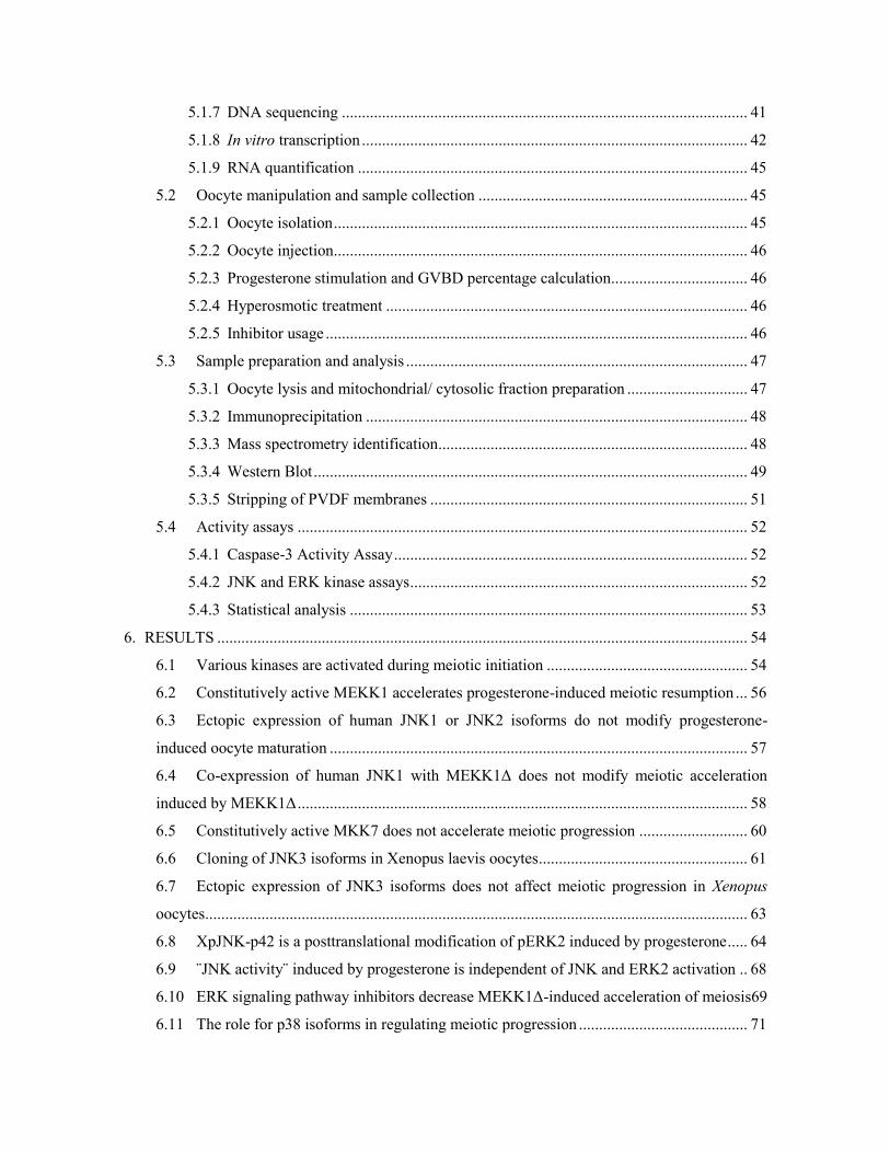
5.1.7 DNA sequencing ..................................................................................................... 41
5.1.8 In vitro transcription ................................................................................................ 42
5.1.9 RNA quantification ................................................................................................. 45
5.2 Oocyte manipulation and sample collection ................................................................... 45
5.2.1 Oocyte isolation ....................................................................................................... 45
5.2.2 Oocyte injection ....................................................................................................... 46
5.2.3 Progesterone stimulation and GVBD percentage calculation.................................. 46
5.2.4 Hyperosmotic treatment .......................................................................................... 46
5.2.5 Inhibitor usage ......................................................................................................... 46
5.3 Sample preparation and analysis ..................................................................................... 47
5.3.1 Oocyte lysis and mitochondrial/ cytosolic fraction preparation .............................. 47
5.3.2 Immunoprecipitation ............................................................................................... 48
5.3.3 Mass spectrometry identification ............................................................................. 48
5.3.4 Western Blot ............................................................................................................ 49
5.3.5 Stripping of PVDF membranes ............................................................................... 51
5.4 Activity assays ................................................................................................................ 52
5.4.1 Caspase-3 Activity Assay ........................................................................................ 52
5.4.2 JNK and ERK kinase assays .................................................................................... 52
5.4.3 Statistical analysis ................................................................................................... 53
6. RESULTS .................................................................................................................................... 54
6.1 Various kinases are activated during meiotic initiation .................................................. 54
6.2 Constitutively active MEKK1 accelerates progesterone-induced meiotic resumption ... 56
6.3 Ectopic expression of human JNK1 or JNK2 isoforms do not modify progesterone-
induced oocyte maturation ........................................................................................................ 57
6.4 Co-expression of human JNK1 with MEKK1Δ does not modify meiotic acceleration
induced by MEKK1Δ ................................................................................................................ 58
6.5 Constitutively active MKK7 does not accelerate meiotic progression ........................... 60
6.6 Cloning of JNK3 isoforms in Xenopus laevis oocytes .................................................... 61
6.7 Ectopic expression of JNK3 isoforms does not affect meiotic progression in Xenopus
oocytes ....................................................................................................................................... 63
6.8 XpJNK-p42 is a posttranslational modification of pERK2 induced by progesterone ..... 64
6.9 ¨JNK activity¨ induced by progesterone is independent of JNK and ERK2 activation .. 68
6.10 ERK signaling pathway inhibitors decrease MEKK1Δ-induced acceleration of meiosis69
6.11 The role for p38 isoforms in regulating meiotic progression .......................................... 71

6.12 Osmostress induces the proteolysis of JNK1-2 by caspase-3 at Asp385 and engages a
positive feedback loop increasing the release of cytochrome c and caspase-3 activation ........ 74
6.13 Cloning and expression of Bcl-2 family members in Xenopus oocytes .......................... 77
6.14 Bid and mono-ubiquitinated Bid are proteolyzed during osmostress-induced apoptosis in
Xenopus oocytes ........................................................................................................................ 79
6.15 Hypeosmotic shock induces Bid proteolysis by initiator caspases and by caspase-3 ..... 83
6.16 Osmostress-induced proteolysis of Bid at Asp52 engages a positive feedback loop
increasing cytochrome c release and caspase-3 activation ........................................................ 87
7. DISCUSSION .............................................................................................................................. 90
7.1 JNK proteins and activities in Xenopus laevis oocytes ................................................... 90
7.2 The role of ERK2 and p38 in Xenopus laevis oocyte maturation ................................... 91
7.3 Bcl-2 family members in osmostress induced apoptosis ................................................. 96
7.4 Bid is mono- and bi-ubiquitinated in Xenopus oocytes, but the function of ubiquitinated
Bid is not clear .......................................................................................................................... 98
7.5 A model for osmostress-induced apoptosis in Xenopus oocytes ................................... 100
8. CONCLUSIONS ........................................................................................................................ 102
9. REFERENCES........................................................................................................................... 103

Summary
1
1. SUMMARY
In the model organism Xenopus laevis, oocytes at stage VI are standing in prophase (G2/M) of
meiosis I indefinitely until proper hormone stimulation. A positive regulation network around the
Mos/MEK/ERK cascade ensures the rapid maturation (GVBD) of the oocytes upon stimulation with
progesterone. However, for the stress associated MAPK families, JNK and p38, their involvement
in meiotic resumption is not so clear. Here we analyze a protein of 42 kDa detected by pJNK
antibodies (XpJNK-p42) that appears around GVBD in progesterone treated oocytes. Ectopic
expression of a constitutively active MEKK1 accelerates oocyte maturation through activation of
the p38 and ERK signaling pathways, but not the JNK cascade. Moreover, four dormant JNK3
transcripts are described in Xenopus oocytes and none of them are activated during progesterone-
induced oocyte maturation. Protein mass spectrometry analysis indicates that XpJNK-p42 is
actually phosphorylated ERK2. Intriguingly, the pJNK antibody only recognizes pERK2 in mature
oocytes but not in oocytes exposed to hyperosmotic shock, suggesting that a posttranslational
modification of pERK2 occurs during meiotic progression. Importantly, neither ERK2
overexpression nor JNK inhibitor SP600125 affects c-Jun phosphorylation detected in mature
oocytes extracts. In conclusion, JNK proteins are not involved in Xenopus oocyte maturation, and
the phosphorylation of c-Jun detected in mature oocytes is independent of JNK and ERK2.
We previously reported that hyperosmotic shock induces apoptosis in Xenopus oocytes through
activation of four independent pathways: p38, JNK, calpains and Smac/DIABLO release. We also
reported that activation of p38β, JNK1-1, and JNK1-2 is clearly pro-apoptotic. However, several
hours after hyperosmotic shock the JNK1-2 isoform disappears, suggesting some type of
degradation during cell death. In addition, our previous studies did not address the role of the Bcl-2
family members in the regulation of cytochrome c release. Here we show that Xenopus pJNK1-2 is
proteolyzed at Asp385 by caspase-3, and the resulting cleaved protein accelerates cytochrome c
release and caspase-3 activation, thus creating a positive feedback loop. We also show that
overexpression of Bcl-xL in Xenopus oocytes protect from osmostress-induced apoptosis. In
oocytes expressing Bid in combination with Bcl-xL, three different types of Bid are detected: non-
ubiquitinated Bid, mono- and bi-ubiquitinated Bid. All Bid types reside both in the cytosol and the
mitochondria. Hyperosmotic shock rapidly induces a slight increase of mono- and bi-ubiquitinated
Bid in the mitochondria. Subsequently, Bid is cleaved at Asp52 at very low levels, probably by an

Summary
2
initiator caspase, generating an N-terminal fragment (nBid) and a highly pro-apoptotic C-terminal
fragment (tBid). When cytochrome c is released and caspase-3 is activated a massive proteolysis of
non-ubiquitinated and mono-ubiquitinated Bid occurs at Asp52, mediated by caspase-3, thus
creating another positive feedback loop. Although some experiments suggest that non-ubiquitinated
Bid is proteolyzed faster and is more pro-apoptotic than wild type Bid, the functional effects of Bid
ubiquitination are not so clear. However, the pro-apoptotic function of Bid is markedly attenuated in
mutant Bid-D52N that is not cleaved by caspases, indicating that Bid proteolysis regulates
osmostress-induced apoptosis. In conclusion, caspase-3 activation induced by hyperosmotic shock
engages two positive feedback loops through the cleavage of JNK1-2 and Bid, thus promoting an
irreversible death of the oocytes.

Abbreviations
3
2. ABBREVIATIONS
AMPK AMP-activated protein kinase
Apaf apoptotic protease activating factor
ASK1 apoptosis signal-regulating kinase 1
Bad Bcl-2-associated agonist of cell death
Bak Bcl-2 antagonist killer 1
Bax Bcl-2-associated X protein
Bcl-2 B-cell CLL/lymphoma 2
Bcl-xL B-cell lymphoma-extra large
Bid Bcl-2 interacting domain death agonist
Bim Bcl-2-interacting mediator of cell death
CDK cyclin-dependent kinase
CPE cytoplasmic polyadenylation element
CPEB CPE-binding protein
cPLA2 cytosolic phospholipase A2
ERK extracellular signal-regulated kinase
GVBD germinal vesicle breakdown
JIP JNK interacting protein
JNK c-Jun N-terminal kinase
MAPK mitogen-activated protein kinase
MAPKK or MAP2K MAPK kinase
MAPKKK or MAP3K MAPK kinase kinase
MBS modified Barth´s Saline
MEKK1 mitogen-activated protein kinase kinase kinase 1
MKK4 MAP kinase kinase 4
MKK6 MAP kinase kinase 6
MKK7 MAP kinase kinase 7
MKP MAPK phosphatase
MLK mixed-lineage protein kinase
MOMP mitochondrial outer membrane permeabilization
Abbreviations
4
OMM outer mitochondrial membrane
PKA cAMP-dependent protein kinase
PLK polo-like kinase
PP2A protein phosphatase 2A
RSK ribosome S6 kinase
SAPK stress-activated MAP kinase
Smac/DIABLO second mitochondria-derived activator of caspases/ direct IAP binding
protein with low pI

Introduction
5
3. INTRODUCTION
3.1 Mitogen activated protein kinase signaling pathway
Mitogen-activated protein k inases (MAPKs), also known as MAP k inases, a re se rine/threonine/
tyrosine specific protein kinases belonging t o the C MGC ( CDK/MAPK/GSK3/CLK) k inase
superfamily. Its closest relative kinase family, the cyclin-dependent kinases (CDKs), is another well
studied kinase group so far (Manning et al. 2002). The first MAPK discovered and characterized in
mammals was ER K1 (MAPK3). Sin ce ERK1 an d E RK2 ( MAPK1) a re both involved in g rowth
factor signaling, the family was termed "mitogen-activated" (Boulton et al. 1991). For quite a long
time, plenty of reports about MAPK referred to ERK proteins.
As an ancient and conserved protein family, five MAPK proteins were characterized in the budding
yeast Saccharomyces cerevisiae (Schaeffer and Weber 1999). These kinases share related structures
and biochemical properties, especially, these MAPKs are activated by dual phosphorylation on a
tripeptide motif (Thr-X-Tyr) located in the kinase activation loop (T-loop) (Davis R. J. 2000a).
Figure 1.Schematic representation of the overall structures of conventional and atypical MAPKs. All MAPKs
contain a Ser/Thr k inase domain f lanked by N - and C -terminal regions o f d ifferent lengths. Di fferent additional
domains are also present in some MAPKs, including a transactivation domain (TAD), a nuclear localization sequence
(NLS), a region conserved in ERK3 and ERK4 (C34), and a domain rich in Ala, His, and Glu (AHQr) (Cargnello and
Roux 2011).

Introduction
6
In mammalian cells, 14 MAPKs divided into 7 groups were cloned and characterized. Besides four
prototypical or conv entional MAPK groups, extracellular r egulated kinase ( ERK1/2), C-Jun N -
terminal kinase (JNK), p38 MAPK and ERK5, which work in a typical three-tiered module, at least
three atypical MAPK types, ERK3/4, ERK7/8, and nemo-like kinase (NLK), which do n ot follow
the classical t hree-tiered, dual -phosphorylation signaling st ructure, have been i dentified (Fig. 1)
(Kholodenko and Birtwistle 2009).
In the conventional MAPK families, the ERK and p38 groups are related to kinases found in the
budding y east and contain t he dual pho sphorylation motifs Thr-Glu-Tyr (TEY) and T hr-Gly-Tyr
(TGY) respectively. The JNK group, also known as stress-activated MAP kinase (SAPK), contains
the dual p hosphorylation motif Thr -Pro-Tyr (TPY) and represents a third M APK sub -family i n
mammals (Davis R. J. 2000a). These mitogen activated protein kinase (MAPK) cascades process a
myriad of signaling pa thways stimulated by ce ll-surface r eceptors in t he so called t hree-tiered
MAPK cascade, comprising a MAPK, a MAPK kinase (MAPKK or MAP2K) and a MAPK kinase
kinase (MAPKKK or M AP3K) (Fig. 2) (Kholodenko and Birtwistle 2009 ). MAPK s ignaling
pathways p lay a pi votal role in regulating various f undamental cellular pr ocesses, i ncluding ce ll
growth, division, migration and differentiation.
Figure 2. MAPK cascades. Illustration of the three-tiered MAPK cascades for ERK, JNK and p38 family
members (Raman et al. 2007).
Introduction
7
3.1.1 The ERK cascade
ERK1 which was found to be phosphorylated on residues Thr and Tyr in response to cell growth
factors is the first MAPK member described in mammalians (Cooper et al. 1982, Ray and Sturgill
1988). In early 1990s, both ERK1 and ERK2 were cloned and characterized (Boulton et al. 1991).
These two ERK isoforms, with molecular weight of 43 and 41 kDa and sharing 83% amino acid
identity, were ubiquitously expressed in all tissues (Boulton et al. 1990). Isoforms derived from
alternative splicing were described for both ERK1 (ERK1b and ERK1c) (Shaul and Seger 2006,
Yung et al. 2000) and ERK2 (ERK2b) (Gonzalez et al. 1992).
MEK1/2 are the upstream kinases (MAP2K) that phosphorylate tyrosine and threonine residues in
the ERK1/2 activation loop, and MAP2Ks are phosphorylated on two serine residues or a serine
residue and a threonine residue in the activation loop by MAP3Ks. Raf isoforms are the best-studied
MAP3Ks that regulate the ERK signaling pathways. Raf proteins are activated through binding the
small G proteins of the Ras family to its N-terminus. Another well-known MAP3K is Mos.
Different to Ras kinases, Mos is more restrictively selected to specific cells and stimuli, such as in
Mos/MEK/ERK cascade of Xenopus oocytes, Mos is activated by progesterone (Raman et al. 2007,
Rapp et al. 2006).
In ERK cascade proteins, the docking motif (D motif) is one crucial motif for its interaction with
ERK binding proteins, including its regulators and substrates. Frequently, motifs in substrates
preferred by ERK reside in the N-terminal and are characterized by a cluster of positively charged
residues with two or more nearby hydrophobic residues. D motifs bind to a conserved C-terminal
common docking (CD) sites identified in ERK1/2 and other MAPKs. A variety of motifs implicated
in MAPK interaction were detailed described (Raman et al. 2007, Sharrocks et al. 2000, Tanoue et
al. 2000).
3.1.2 The JNK cascade
C-Jun N-terminal kinase (JNK) was firstly identified as a p45 microtubule associated protein kinase
in cycloheximide injected rat (Kyriakis and Avruch 1990). Later, it was notice that c-jun, a
component of the AP-1 transcription factor, is regulated by MAPK via phosphorylating on two
serine residues in the N-terminal in response to a variety of mitogens, and, these MAPKs include
pp45 and pp42/44 kinases (Pulverer et al. 1991). The directly binding and phosphorylation was
confirmed in a c-jun binding assay using extracts from UV-irradiated cells, the kinases were then
named JNKs (Hibi et al. 1993).

Introduction
8
Three distinct JNK genes have been isolated in mammalians (Dhanasekaran N and Reddy 1998).
The jnk1 and jnk2 are ubiquitously expressed. In contrast, the jnk3 expression pattern is relatively
restricted to brain, heart, and testis. The three jnk genes express at least ten JNK isoforms by distinct
modification in selective transcription and alternative splicing. Transcripts derived from JNK genes
have two distinct 3´extension, and more transcripts are generated through alternative splicing from
these original transcripts. It is clear that the alternative splicing influences the substrate specificity
of the JNK isoforms by altering the binding motif of JNK to docking sites in other proteins (Gupta
et al. 1996, Kallunki et al. 1994, Sluss et al. 1994).
Figure 3. Schematic il lustration of functional domains identified in M KK4 and M KK7. MAPK and MAPKKK
docking d omains referred a s D and DV D sites i n M KKs permit the f ormation o f s table c omplexes b etween the
components of the MAPK signaling pathway. These interactions contribute to accurate and efficient enzyme-substrate
recognition and are essential for the specific transmission of signals from upstream kinases to the MAPKs. The D sites
in MKK4 and MKK7 consist of a cluster of two to three basic residues, followed by a short spacer of 1–2 residues, and
a hydrophobic-X-hydrophobic sub-motif. Unlike MKK4, MKK7 contains three weak D-sites that interact in a partially
additive, p artially s ynergistic manner to c reate high a ffinity J NK-docking p latform. The specific MAPKKK MKKs
interaction via the DVD site facilitates the phosphorylation of MKK4 and MKK7 at Ser and Thr residues within the S–
X–A–K–T motif, by increasing t he local concentration of the activating MAPKKK and a ltering the structure of the
MKKs. Consensus-matching residues in the putative D and DVD sites of MKK4 and MKK7 are in boldface (Wang
Xin et al. 2007d).

Introduction
9
JNK proteins are activated by concomitant phosphorylation on Thr and Tyr in the TPY motif in the
activation loop by MKK7 and MKK4 (SEK1). Like JNK isoforms, alternative splicing modification
generates six MKK7 isoforms with different N-terminal and C-terminal and three MKK4 isoforms
with distinct N-terminal. These different isoforms of MKK7 and MKK4 are biochemically different
and activated by distinct MAPKKKs through different docking motif preference (Fig. 3) (Tournier
et al. 1999, Wang Xin et al. 2007d). Both MKK7 and MKK4 can activate JNK, and MKK4 can also
activate p38 (Fig. 2). It seems that MKK4 preferentially phosphorylates JNK on Tyr and MM7
preferentially phosphorylates JNK on Thr, indicating that MKK7 and MKK4 are not functionally
exclusive and they may function cooperatively under particular circumstances (Lawler et al. 1998,
Raman et al. 2007).
The MKK7 and MKK4 are also activated by dual phosphorylation on two residues in the activation
loop by up tier kinase. Several MAP3Ks that phosphorylate and activate MKK4 and/or MKK7 have
been isolated. Fourteen out of twenty identified MAP3Ks are proved to activate JNK via
phosphorylating MKK4 or MKK7. Generally, the MAP3K is identified by transfection assay or in
vitro protein kinase assays. It is unclear whether these MAP3Ks are physiological regulators of the
JNK signaling pathway and which stimulus correspondingly activates these specific kinases (Davis
R. J. 2000a). Significant progression has been made toward understanding the function of the
MEKK group through targeted gene disruption techniques in mice. MEKK1 deficient mice have no
gross morphological disabilities except for an eyelid closure defect (Yujiri et al. 2000) while defect
of MEKK3 is lethal for mice (Yang Annie et al. 2000). Studies have demonstrated that
MEKK1functions as a component of the JNK cascade. The functional consequences of
MEKK1disruption in ES cells includes defects in cell migration and increase apoptosis in response
to microtubule destabilizing drugs (Davis Roger J 2000b).
3.1.3 The p38 cascade
The archetypical p38 member, p38α was identified in 1994. It has a significant homology with the
budding yeast Hog1, and shares 50% identity with ERK2. p38 is the second identified MAPK
module in response to stress stimuli (Han J et al. 1994, Rouse et al. 1994). After the isolation of
p38α, another three p38 isoforms, p38β, p38γ and p38δ, were isolated and characterized. While
p38α and p38β are ubiquitously expressed in cell lines and tissues, the expression pattern of p38γ
and p38δ is restricted (Cuadrado and Nebreda 2010, Jiang Yong et al. 1996, Jiang Yong et al. 1997).
Even MKK4 has been shown to activate p38, MKK3 and MKK6 are supposed to be the major
kinases responsible for p38 activation (Derijard et al. 1995, Han Jiahuai et al. 1996, Meier et al.
Introduction
10
1996). MKK3/MKK6 activate(s) p38 by dua l pho sphorylation o n Thr and Tyr i n t he conserved
TGY m otif i n their a ctivation l oop. M KK6 ca n a ctivate all p38 isoforms, w hereas M KK3
preferentially phosphorylates the p38α, p38γ, and p38δ.
MKK3/MKK6 are activated by a great quantity of MAP3Ks, including MEKK1 to -3, MLK2/3 and
ASK1 (Cuadrado and N ebreda 2010 ). I n m ost con texts, MAP 3Ks i nvolved in p38 m odule a re
shared by the JNK module. In the process of investigating the role of p38 module, to eliminate the
intervention between different MAPK modules, many p38 inhibitors were exploited in thousands of
studies. The anti-inflammatory drug SB203580 and its close relative SB202190 specifically target
and inhibit the p38α and p38β as competitive inhibitors of ATP binding (Lee et al. 2000). A novel
inhibitor, B IRB0796, i nhibits all p38 isoforms by b locking an allosteric binding si te as well a s
establishing binding interactions in the ATP pocket (Pargellis et al. 2002, Regan et al. 2002).
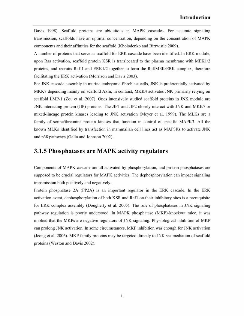
3.1.4 Molecular sca ffold proteins a ssemble M APK sig naling
components
Precise signaling transduction is guaranteed
by pr oper pr otein-protein i nteractions v ia
forming pr otein com plexes. Generally, t he
interacting pr oteins are r ecruited and
assembled by a third p rotein g roup, one
significant protein group i nvolved in t he
complex-assembly i s the scaffold p rotein.
The scaffold protein is described as protein
whose m ain f unction is t o br ing ot her
proteins together. G enerally, t hese pr oteins
have various protein dock ing motifs.
Scaffold protein t ethers signaling
components together and l ocalizes these
proteins t o proper areas of t he cell,
regulates s ignaling transduction pr ecisely
and insulates co rrect s ignaling pr oteins
from com peting pr oteins si multaneously
(Shaw and Filbert 2009 , Whitmarsh an d
Figure 4. Scaffold proteins mediating th e str uctural a nd
functional organization o f the th ree-tier JNK sig naling
module. Model of how a typical scaffold protein supports the
assembly of a t hree-tier JNK signaling module consisting of
a MAPKKK, a MAPKK (MKK4 or MKK7) and a JNK. Such
scaffold proteins may p lay a c atalytic ro le as well a s an
anchoring r ole de pending on t he na ture of t he s caffold
protein and the cellular context (Asaoka and Nishina 2010).
Introduction
11
Davis 1998). Scaffold proteins are ubiquitous in MAPK cascades. For accurate signaling
transmission, scaffolds have an optimal concentration, depending on the concentration of MAPK
components and their affinities for the scaffold (Kholodenko and Birtwistle 2009).
A number of proteins that serve as scaffold for ERK cascade have been identified. In ERK module,
upon Ras activation, scaffold protein KSR is translocated to the plasma membrane with MEK1/2
proteins, and recruits Raf-1 and ERK1/2 together to form the Raf/MEK/ERK complex, therefore
facilitating the ERK activation (Morrison and Davis 2003).
For JNK cascade assembly in murine embryonic fibroblast cells, JNK is preferentially activated by
MKK7 depending mainly on scaffold Axin, in contrast, MKK4 activates JNK primarily relying on
scaffold LMP-1 (Zou et al. 2007). Ones intensively studied scaffold proteins in JNK module are
JNK interacting protein (JIP) proteins. The JIP1 and JIP2 closely interact with JNK and MKK7 or
mixed-lineage protein kinases leading to JNK activation (Meyer et al. 1999). The MLKs are a
family of serine/threonine protein kinases that function in control of specific MAPK3. All the
known MLKs identified by transfection in mammalian cell lines act as MAP3Ks to activate JNK
and p38 pathways (Gallo and Johnson 2002).
3.1.5 Phosphatases are MAPK activity regulators
Components of MAPK cascade are all activated by phosphorylation, and protein phosphatases are
supposed to be crucial regulators for MAPK activities. The dephosphorylation can impact signaling
transmission both positively and negatively.
Protein phosphatase 2A (PP2A) is an important regulator in the ERK cascade. In the ERK
activation event, dephosphorylation of both KSR and Raf1 on their inhibitory sites is a prerequisite
for ERK complex assembly (Dougherty et al. 2005). The role of phosphatases in JNK signaling
pathway regulation is poorly understood. In MAPK phosphatase (MKP)-knockout mice, it was
implied that the MKPs are negative regulators of JNK signaling. Physiological inhibition of MKP
can prolong JNK activation. In some circumstances, MKP inhibition was enough for JNK activation
(Jeong et al. 2006). MKP family proteins may be targeted directly to JNK via mediation of scaffold
proteins (Weston and Davis 2002).

Introduction
12
3.2 Xenopus oocyte is an excellent system for
ootidogenesis and apoptosis
Since the original studies started by John Gurdon and colleagues, oocytes from the South African
clawed frog have been established as an excellent functional expression system. When injected with
messenger RNA, the oocytes were able to translate the mRNA into relative proteins after a period
of i ncubation (Gurdon e t al . 1974 ). T he l arge s ize of the o ocyte (Fig. 5) facilitates t he
microinjection of mRNAs to express proteins in enough quantities for proper biochemical studies.
Most of the mechanisms that r egulate oog enesis and maturation hav e been di scovered using
Xenopus oocytes as a cell model.
The Xenopus laevis oocyte begins as a cell sl ightly bi gger t han a typical so matic cell. A t t he
beginning of meiotic cycle, the oocyte undergoes a round of DNA replication and then enters the
prophase of meiosis I, during this stage the chromatin condenses into chromosome, then homologs
pair and r ecombination occurs. In t he following several months, the oocyte remains in a G2-like
growth stage with a intact nuclear envelope (germinal vesicle envelope), and the volume of oocyte
grows bigger and bigger. Once the oocyte is fully grown, it enters into the so called Dumont Stage
VI, a G2 ar rested st ate (Fig. 6) . T his arrest r etains indefinitely i n a stable environment (18ºC,
12h/12h light/dark for Xenopus laevis) before progesterone induction. The volume of oocyte and its
ability to synthesize protein on dem and make the oocyte an almost ideal s ingle-cell experimental
system (Ferrell 1999).
The process of s tage VI oocyte developing into fertilizable egg i s termed oocyte maturation. The
maturing oocy te undergoes g erminal v esicle breakdown ( GVBD), followed by chr omatin
condensation and microtubule reorganization and f ormation o f t he metaphase spindle and
Figure 5. Stage V I oocytes f rom Xenopus laevis.
The sta ge V I oocyte is trem endously larg e w ith a
volume o f 1 µl containing a bout 25µg p roteins.
About half of the oocyte´s volume consists of some
yolk platelets which are concentrated in the lightly
pigmented v egetal h emisphere, th e y olk w ill
ultimately makes its way into the gut of the tadpole
and s erve as energy r esource. T he p igmented d ark
brown a nimal hemisphere mainly c ontains the
cytoplasm a nd n ucleoplasm (m odified f rom
Xenopus express).

Introduction
13
subsequent completion of the first meiosis I by emitting the first polar body. Then the oocyte enters
meiosis II w ithout the intervening i nterphase and i s ar rested in m etaphase ag ain. T he m aturing
process completes (Fig. 6). In the maturing process, GVBD is easy to score because of a white spot
appearing at the animal pole, a result of rearrangement of cortical pigment granules (Ferrell 1999).
Importantly, in vitro incubation of isolated Xenopus oocytes at stage VI with progesterone resume
the maturation process until meiosis II, facilitating the study of this biological process.
Besides studies o f m eiotic progression, Xenopus oocytes have also been us ed for research in
apoptosis. A ce ll-free sy stem based on Xenopus egg ext racts was used for t he seminal w orks
describing the role of cytochrome c release from the mitochondria in the engagement of apoptosis
(Newmeyer et al . 199 4, K luck et a l 1997 ). M oreover, t he role of se veral m embers of t he Bcl-2
family in regulating cytochrome c r elease was discovered using this system. Xenopus egg extracts
incubated a t r oom t emperature for se veral h ours spontaneously r ecapitulate m any ev ents of
apoptosis, including chromatin condensation, shrinkage, and fragmentation of the nuclei, even from
nuclei added exogenously. This morphological changes required the presence of a dense organelle
fraction enriched in mitochondria and could be blocked by t he addition of baculovirus-expressed
Bcl-2 protein (Newmeyer et al. 1994). Bcl-2 blocked apoptotic activity by preventing cytochrome c
release from mitochondria and thereby blocking activation of caspase-3 like proteases (CPP32) and
downstream apoptotic events (Kluck et a l. 1997, Kuwana e t a l. 1998). More recently i t has been
discovered that the sp ontaneous dea th of t he oocytes at room t emperature i s due to g lucose-6-
phosphate and NADPH depletion, which induces caspase-2 activation (Nutt et al. 2005). Caspase-2
is phosphorylated and inhibited by Calcium calmodulin-dependent kinase II (CaMKII) (Nutt et al.
2005), and protein phosphatase 1 activation by nutrient depletion would induce apoptosis through
dephosphorylation and activation o f c aspase-2 (McCoy et al. 2013 ). B esides t hese s tudies w ith
Xenopus egg extracts, the oocytes have been used as an easily manipulable in vivo system to study
apoptosis induced by different stimuli. For instance, cytochrome c release was detected in isolated
Figure 6. Schematic view of Xenopus oogenesis, maturation, and early embryogenesis (Ferrell 1999).

Introduction
14
intact oocytes 48h later after initiating meiotic maturation by progesterone. It seems that the default
fate of unfertilized oocyte is to die through a mitochondria dependent apoptosis pathway after
meiotic maturation, and it has been described that this apoptosis is regulated by JNK and Cdc2
activation (Du Pasquier et al. 2011). It has also been reported that incubation of Xenopus oocytes
with bacterial neutral sphingomyelinase (bSMase) causes GVBD, whereas microinjection of
bSMase results in apoptosis, indicating that an increase of ceramide production induced by bSMase
can induce meiosis or apoptosis, depending on the location of ceramide (Coll et al. 2007, Strum et
al. 1995). Although in mammalian cells it has been reported that ceramide can initiate apoptosis
through a Rac1-regulated activation of the JNK/p38 cascade or ASK1-regulated p38 and JNK
activation, as well as activation of the endoplasmic reticulum (ER) stress cascade (Brenner et al.
1997, Chen Chia-Ling et al. 2008, Verheij et al. 1996) in Xenopus oocytes it was not determined the
signaling pathway responsible for ceramide-induced apoptosis. Hyperosmotic shock also induces
cytochrome c release and caspase-3 activation in Xenopus oocytes (Bagowski et al. 2002, Martiañez
et al. 2009), and several mechanisms that regulate osmostress-induced apoptosis have been
discovered by our group (see introduction, section 4). Finally, Xenopus oocytes allow greater
manipulation as a system to study the analog or digital responses in single cells for particular
external stimulation or microinjected proteins (Bagowski and Ferrell Jr 2001, Martiáñez et al. 2009).
3.3 Xenopus oocyte maturation involves complicate
regulations
Generally, progesterone is considered the hormone inducing oocyte maturation in vivo in Xenopus
laevis, which is synthesized and released by follicular cells surrounding the oocyte. However,
androgen, rather than progesterone, has been proposed to be the primary hormone produced in
Xenopus ovaries (Lutz et al. 2001). Maturation can also be induced in vitro by insulin and insulin-
like growth factor-1 via an IGF-1 receptor. However, it is unclear whether these hormones have a
role in in vivo maturation (Ferrell 1999, Schmitt and Nebreda 2002). As we will see in this section,
progesterone induces several signaling pathways in Xenopus oocytes, and the translational and
posttranslational control of several genes is of great relevance in the maturation of the oocytes.

Introduction
15
3.3.1 Potential progesterone receptor
In Xenopus oocytes, progesterone can be recognized by a plasma membrane receptor as a ligand.
The receptor of progesterone does not reside in cytoplasm or nucleus, because oocytes mature only
when progesterone is applied outside of the oocytes instead of microinjected into the cytoplasm or
nucleoplasm (Smith and Ecker 1971). Furthermore, oocytes can also mature when incubated with
steroids immobilized on agarose beads or linked to a synthetic polymer. These issues indicate that
the receptor of progesterone resides on the plasma membrane and faces out (Godeau et al. 1978,
Ishikawa et al. 1977). Early responses of oocytes to progesterone involve a modest inhibition of
adenylate cyclase and subsequent modest decrease in cyclic AMP concentration in the oocytes,
without significant alteration of cAMP phosphodiesterase activity. These responses suggest that the
progesterone receptor might be a transmembrane protein coupled to heterotrimeric G proteins which
inhibit adenylate cyclase. However, the functions of the G proteins are not completely understood
(Ferrell 1999, Schmitt and Nebreda 2002).
An intracellular Xenopus progesterone receptor (XPR-1) has been implicated in meiotic maturation
(Bayaa et al. 2000, Tian et al. 2000). Subsequently it was reported to be localized at the oocyte
membrane (Bagowski et al. 2001b), possibly through interaction of its proline-rich motif with the
SH3 domain of the c-Src protooncogene (Boonyaratanakornkit et al. 2001). Addition of a
myristoilation and palmytoilation signal at the amino terminus of XPR-1 increased the amount of
receptor associated to the oocyte plasma membrane and accelerated progesterone-induced oocyte
maturation (Martinez et al. 2006).
A membrane-associated progesterone-binding protein from porcine liver was also purified by
Nehling and his colleagues and its corresponding cDNA was cloned too (Falkenstein et al. 1996).
Sequence analysis revealed that homologous cDNAs are present in human, mice, rat, and yeast
(Ferrell 1999). This membrane progesterone receptor (mPR) is not termed progesterone membrane
receptor component 1 (PGMRC1) and was proved located in membrane with many other
progesterone binding proteins (Lösel Ralf et al. 2004). Even though PGMRC1 was originally
described as steroid binding protein, current evidences support the perception that it may be
involved in steroid metabolism or homeostasis and survival (Lösel Ralf M et al. 2008). Intriguingly,
it is shown to be mediator of progesterone´s antiapoptotic action (Peluso et al. 2008, Peluso et al.
2006). Another distinct family of membrane progestin receptors (mPRs) has been cloned in fish and
many other vertebrate species (Zhu et al. 2003). In Xenopus laevis, a transcript (Xenopus mPR,
XmPR) of the mPRβ ortholog was cloned by RT-PCR. This protein is present on the oocyte plasma
membrane, and microinjection of mRNA encoding XmPR resulted in acceleration of progesterone-
Introduction
16
induced oocyte maturation. Binding studies in mammalian cells expressing XmPR protein
confirmed specific binding of progesterone. These results suggest that XmPR might be a
physiological progesterone receptor involved in meiotic resumption of Xenopus oocytes (Josefsberg
Ben-Yehoshua et al. 2007). In Rana pipiens oocytes it has been reported that progesterone binds to
the ouabain binding site on the N-terminal region of the alpha-subunit of Na/K-ATPase triggering a
cascade of lipid second messengers and meiotic progression (Morrill et al. 2005). The authors
propose that helix-helix interactions between the alpha-subunit of Na/K-ATPase and
phosphatidylethanolamine-N-methyltransferase (PE-NMT) occur in the plasma membrane inducing
the activation of PE-NMT and sphingomyelin synthase within seconds. (Morrill et al. 2010, Morrill
et al. 2005). There are no reports about the binding of progesterone to Xenopus alpha-subunit of
Na/K-ATPase and its role in meiotic progression.
3.3.2 Early-stage response to progesterone
Xenopus oocytes go through many changes including the metabolism of nutrient stockpile, which
may supply energy for the viability and survival of the oocyte. Proteome analysis of maturing
oocytes indicate that the glycolytic metabolites may be critical modulators of oocyte maturation
(Berger and Wilde 2013, Nutt 2012).
One of the key earliest biochemical changes observed in maturing oocytes, as described before, is
the decrease of cAMP concentration after progesterone incubation. The decrease of cAMP occurs
within minutes after progesterone incubation; it lasts a few hours, and is accompanied by inhibition
of the cAMP-dependent protein kinase (PKA), which has been found to be a potent inhibitor of
oocyte maturation. Correspondingly, inhibition of PKA alone is enough to induce meiotic
maturation in Xenopus oocytes (Schmitt and Nebreda 2002).
3.3.3 Polyadenylation-dependent protein synthesis is required in
meiotic resumption
In Xenopus, oocyte maturation is independent of transcription, but it is regulated at the level of
translation and post-translational modifications of proteins. Nearly two decades ago, it was found
that there is a maternal stockpile of mRNAs harboring a cytoplasmic polyadenylation element (CPE)
in the 3´untranslated regions (3´UTR) that may control cytoplasmic polyadenylation and
translational activation. CPE is the binding platform for CPE-binding proteins (CPEB). The binding
Introduction
17
of CPEB to the 3´UTR recruits the cleavage and pol yadenylation specificity factor (CPSF) to the
polyadenylation hex anucleotide (Hex, A AAUAA). Subsequently, the com plex recruits t he
cytoplasmic poly (A) polymerase GLD2 and many other factors including eIF4 translation factors,
which are required to recruit the 40S ribosomal subunit to the 5´end of the mRNA after the CPEB
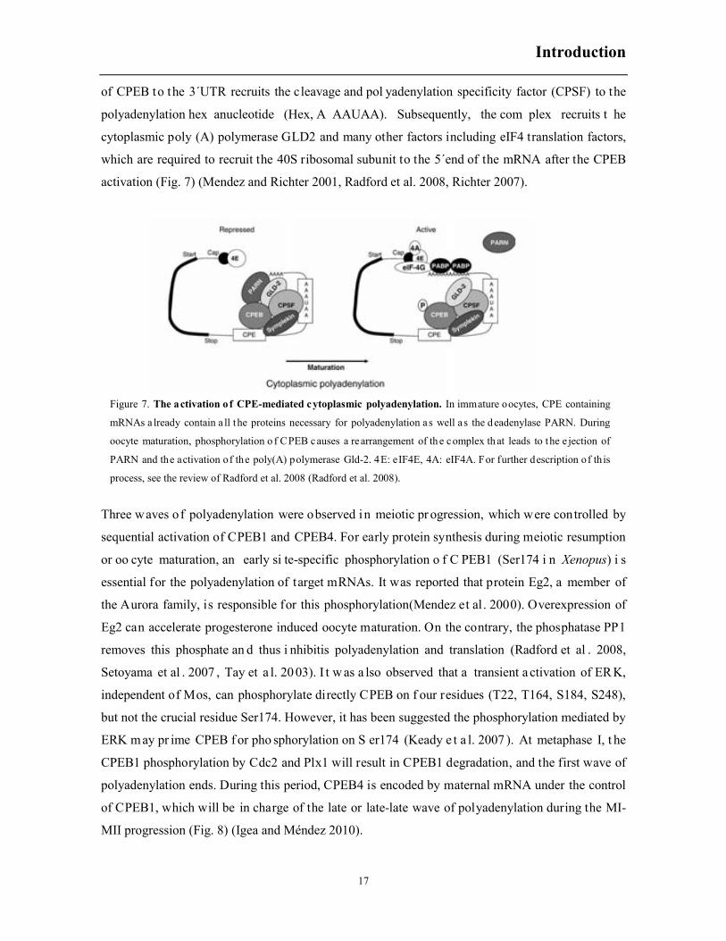
activation (Fig. 7) (Mendez and Richter 2001, Radford et al. 2008, Richter 2007).
Three waves of polyadenylation were observed in meiotic pr ogression, which were controlled by
sequential activation of CPEB1 and CPEB4. For early protein synthesis during meiotic resumption
or oo cyte maturation, an early si te-specific phosphorylation o f C PEB1 (Ser174 i n Xenopus) i s
essential for the polyadenylation of target mRNAs. It was reported that protein Eg2, a member of
the Aurora family, is responsible for this phosphorylation(Mendez et al . 2000). Overexpression of
Eg2 can accelerate progesterone induced oocyte maturation. On the contrary, the phosphatase PP1
removes this phosphate an d thus i nhibitis polyadenylation and translation (Radford et al . 2008,
Setoyama et al . 2007 , Tay et a l. 20 03). I t w as a lso observed that a transient a ctivation of ERK,
independent of Mos, can phosphorylate directly CPEB on f our residues (T22, T164, S184, S248),
but not the crucial residue Ser174. However, it has been suggested the phosphorylation mediated by
ERK may pr ime CPEB f or pho sphorylation on S er174 (Keady e t a l. 2007 ). At metaphase I, t he
CPEB1 phosphorylation by Cdc2 and Plx1 will result in CPEB1 degradation, and the first wave of
polyadenylation ends. During this period, CPEB4 is encoded by maternal mRNA under the control
of CPEB1, which will be in charge of the late or late-late wave of polyadenylation during the MI-
MII progression (Fig. 8) (Igea and Méndez 2010).
Figure 7. The activation of CPE-mediated cytoplasmic polyadenylation. In immature oocytes, CPE containing
mRNAs a lready contain a ll the proteins necessary for polyadenylation as well a s the d eadenylase PARN. During
oocyte maturation, phosphorylation o f CPEB causes a rearrangement of th e complex th at leads to the e jection of
PARN and the activation of the poly(A) polymerase Gld-2. 4E: eIF4E, 4A: eIF4A. For further description of th is
process, see the review of Radford et al. 2008 (Radford et al. 2008).

Introduction
18
3.3.4 MPF is one crucial factor for meiotic initiation
About 30 -60 m in bef ore GVBD, t here is a significant i ncrease of p rotein k inase activity an d
phosphorylated proteins in the oocytes. These proteins can be classified into two groups: 1) the cell
cycle regulators, such as Cdc2, Cdc25, Plx1, and xPlkk1; and 2) the MAPK proteins including Mos,
MEK1, ERK2, Rsk1/2 and so on (Ferrell 1999).
In oocy te maturation v arious signals finally conv erge to activate MPF, w hich is a k ey com plex
formed of a catalytic subunit ( Cdc2/Cdk1) and a regulatory subun it ( Cyclin B). O nce ac tivated,
MPF promotes t he entry i nto M-phase of t he first meiotic di vision. MPF ac tivity f alls during
Figure 8. Schematic diagram s howing th e se quential activities of CPEB 1 a nd CPEB 4 mediating th e th ree
waves of polyadenylation driving meiotic progression (Igea and Méndez 2010).
Figure 9. Changes in Xenopus oocytes in the levels of cyclin B protein, and the cyclinB-Cdc2 kinase and MAP
kinase activities throughout the course of meiotic maturation. For species-specific variations, see the main text.
The que stion m ark in dicates th at early phosphorylation o f M AP k inase h as been seen, but whether o r n ot this
represents a ctive M AP kinase is unknown. G VBD, germinal vesicle breakdown; M1, meiosis I; M2, meiosis II;
NEBD, nuclear envelope breakdown (Abrieu et al. 2001).

Introduction
19
anaphase I , due to par tial deg radation (Dupré et al . 2010 ). I ntensive studies r evealed that the
function of MPF in promoting oocyte maturation is universal but the MPF activation mechanism is
sort of sp ecies-dependent ( Fig. 9) (Abrieu e t a l. 2 001, Schmitt and N ebreda 2 002). In Xenopus
oocytes, there is a small stock of inactive dimer complexes named pre-MPF, which can be activated
by dephosphorylation of Cdc2 on Tyr15 and Thr14 (Frank-Vaillant et al . 1999). The phosphatase
responsible for this dephosphorylation i s most p robably C dc25, which can be regulated by bot h
phosphorylation an d subc ellular l ocalization. Plx1 is a potential ac tivator of C dc25 (Qian et a l.
2001), but is not clear how it is regulated during meiotic progression. The MAPK cascade is another
key regulator of MPF. It has been confirmed that Mos is involved not only in the ERK cascade, but
also i n a mechanism t hat directly ac tivates MPF and i mproves t he stability of cyclin B which is
supposed to be necessary for MPF activation (Dupré et al. 2010, Fan Heng-Yu and Sun 2004). It
has al so been demonstrated t hat ER K2 interacts with hy pophosphorylated Cdc25 bef ore meiotic
induction a nd ph osphorylates C dc25 a t T48, T138, and S205, t hereby i ncreasing C dc25
phosphatase ac tivity dur ing meiotic induction (Wang R uoning et al . 2007b ). Furt hermore, ERK
leads to the ac tivation o f the k inase p90R sk that in t urn p hosphorylates and inhibits the C dc2
inhibitory kinase Myt1 (Ferrell 1999, Schmitt and Nebreda 2002).
Figure 10. Revisiting M PF a ctivation. MPF activation is i nitiated b y th e generation o f a threshold level of active
Cdk1 in response to progesterone. This step depends on the synthesis of Cyclin B and is negatively regulated by PKA.
This starter amount of a ctive Cd k1 p hosphorylates its own re gulatory e nzymes, Cdc25 a nd Myt1, a nd in duces the
accumulation of Mos leading to the activation of the Mos/MAPK pathway. These reactions are under the control of
PKA and are counteracted by PP2A. Therefore, the conversion of inactive pre-MPF into MPF cannot take place unless
PP2A is in hibited. G wl is a ctivated b y th e th reshold a mount of Cd k1 a ctivity. It phosphorylates A RPP19 a t S67,
leading to PP2A inhibition independently of P KA. The MPF auto-amplification loop then becomes independent of
PKA activity and protein synthesis, irreversibly driving the cell into M-phase (Dupré et al. 2013).

Introduction
20
Besides of regulation from other proteins, a MPF auto-amplification loop is also detected in
Xenopus oocytes (Fig. 10). In this positive loop, a threshold level of cdc2 activity brings about
Cdc25 activation and Myt1 inactivation, hence establishing a positive feedback loop. This positive
feedback regulation is rendered via removing the inhibitory effect of PP2A by phosphorylated
ARPP19 (Dupré et al. 2013) (Fig. 10). Cdc2 also phosphorylates the regulatory domain in the N-
terminal of Cdc25 at a specific S/T-P motif, therefore increasing the phosphatase activity of Cdc25
(Izumi and Maller 1993, 1995). In addition, a polo-like kinase (PLK), located downstream of Cdc2,
can also activate Cdc25 by phosphorylating the S/T-P motif. Therefore, PLK seems to be also
involved in this positive feedback loop (Barr et al. 2004, Nakajima and Masukata 2002, Toyoshima‐
Morimoto et al. 2002).
3.3.5 ERK signaling pathway is one classic mediator in oocyte
maturation
In the early 1980s, Mos was first identified in cells transformed by Moloney murine leukemia virus
(v-mos) (Papkoff et al. 1982). Then its cellular homologue (c-mos) was isolated as a proto-
oncogene. Mos is a Ser/Thr kinase and its expression is restricted to germ cells. In Xenopus oocytes,
it accumulates during the meiotic divisions and undergoes selective proteolysis upon fertilization
(Watanabe et al. 1989). There is an abundance of maternal stockpiled mos transcripts in arrested
Xenopus stage VI oocytes. Mos protein appears 2 to 3 h after progesterone stimulation, before MPF
activation (Sagata et al. 1989). The early translation of Mos is not sufficient to its accumulation
because the ubiquitination at Lys34 induces its proteolysis. In Xenopus oocytes, MPF ensures Mos
stability via phosphorylating the residue Ser3 in the N-terminal of Mos (Freeman et al. 1992). In
1993, it was discovered that Mos can activate ERK2 by directly phosphorylating and activating
MEK1. The two amino acids phosphorylated by Mos in MEK are identical to those phosphorylated
by Raf-1 (Pham et al. 1995, Posada et al. 1993). Inside the Mos/MEK/ERK cascade, the ERK2 can
activate Mos in a positive feedback loop. Microinjection of activated MEK2 or ERK is sufficient to
stimulate Mos accumulation implying that ERK2 could contribute to CPEB phosphorylation and
activation (Gotoh et al. 1995, Howard et al. 1999, Keady et al. 2007). More recently it has been
reported that ERK2 phosphorylates Cdc25 at T48, T138, and S205, thereby increasing Cdc25
phosphatase activity during meiotic induction (Wang et al. 2007).
The first found and best known physiological substrate of ERK in the oocyte is a 90-kDa protein
kinase p90RSK (ribosome S6 kinase) (Jones et al. 1988). p90RSK seems to play important role in
Introduction
21
meiotic maturation probably via inhibition of Myt1 (Palmer et al. 1998, Schmitt and Nebreda 2002).
However, the Mos/MEK/ERK/p90RSK signaling pathway is dispensable for the regulation of
microtubule assemble and spindle organization and even the activation of histone H3 kinase during
oocyte maturation (Schmitt et al. 2002, Yu et al. 2007).
3.3.6 Involvement of JNK and p38 MAPK in meiotic resumption
is not clear
Among the three MAPK families, ERK family proteins are mainly activated by mitogens and serum
stimulation while the p38 and JNK families are mainly involved in response to environmental and
genotoxic stresses (Ambrosino and Nebreda 2001). The role of ERK in meiotic initiation has been
documented in detail, however, little is known about the function of p38 and JNK in oocyte
maturation.
In fission yeast, when spc1, a homologue of p38 MAPK, was mutated, a G2 delay was observed,
and then it was confirmed that spc1 is required for mitosis entry in fission yeast (Shieh et al. 1998,
Shiozaki and Russell 1995). The function of p38 activation during G1/S transition seems to depend
on the experimental systems. In Xenopus cell-free extracts, activated p38 induces arrest of M-phase
(Takenaka et al. 1998). It has been proposed that the activation of p38γ isoform in mitosis can
indirectly regulate the activity of the Chk2 kinase, which in turn phosphorylates Cdc25C (Wang
Xiaofei et al. 2000).
In pig oocytes, p38 becomes active around the GVBD and maintains active until metaphase II. A
specific p38 inhibitor SB203580 inhibits phosphorylation of p38 and block FSH induced pig
meiotic resumption (Yamashita et al. 2009). In Xenopus oocyte, it has been reported that
overexpression of a constitutively active MKK6, an upstream kinase of p38 proteins, accelerates
progesterone-induced maturation. Furthermore, co-expression of active MKK6 with Xp38γ induces
oocyte maturation in the absence of progesterone. The same authors also reported that expression of
MKK6 and Xp38γ inactive mutants inhibit progesterone induced maturation (Perdiguero et al.
2003). However, it is not clear whether progesterone can activate MKK6. Moreover, co-expression
of active MKK6 with Xp38γ induced the appearance of a white spot morphologycally different to
the one obtained after progesterone treatment, and 50% of the mature oocytes presented
abnormalities in location of the spindles (Perdiguero et al. 2003).
For JNK cascades, the meiotic regulation function seems more equivocal and less convincing
evidences appear. An increase in JNK activity, measured as GST-Jun phosphorylation, has been

Introduction
22
reported just prior to germinal vesicle breakdown (GVBD) (Bagowski et al. 2001a, Bagowski et al.
2001b). This JNK activity increased by activated Mos or MEK1 proteins (Bagowski et al. 2001b),
but the JNK isoform/s responsible was/were not identified. In addition, a JNK isoform has been
detected by phospho-JNK antibodies at GVBD during Xenopus oocyte maturation and remains
phosphorylated throughout meiosis I and II (Adler et al. 2005, Du Pasquier et al. 2011, Mood et al.
2004). However, this JNK isoform has not been characterized in detail, as well as its role in meiotic
maturation.
3.4 Osmotic shock stimulates Xenopus oocyte apoptosis
We have reported that hyperosmotic stress induces cytochrome c release and caspase-3
activation in Xenopus laevis oocytes (Martiáñez et al. 2009). In recent works, we observed at the
very beginning of osmotic shock induced oocyte apoptosis a rapid Smac/DIABLO releases, calpain
activation, and that stress associated MAPK proteins (JNK/p38) were phosphorylated around 15
min later in 300 mM sorbitol treatment. It was proved that all of these early events played some role
in promoting cytochrome c and caspase-3 activation. Interestingly, caspase-3 activation in turn
enhanced these events and thus positively regulated the apoptotic progression. We will review here
the basic mechanisms that regulate cytochrome c release and caspase-3 activation through the Bcl-2
family members and the JNK signaling pathway.
3.4.1 Apoptosis, caspases, the extrinsic and the intrinsic
apoptotic pathways
Multicellular organisms have evolved a self-demise mechanism to remove infected, damaged and
unwanted cells which was referred as apoptosis (Kerr et al. 1972). Apoptotic signaling pathways
have been widely studied and explosively progressed. The critical event in apoptosis is the
activation of caspases, a group of cysteine protease that are the executioners of apoptosis, can
cleave many cellular contents (Salvesen and Dixit 1997). Caspases comprise two distinct classes,
the initiators (caspase-2, -8, -9 and -10) and the effectors (caspases-3,-6 and -7). Although general
structural features are shared between the initiator and the effector caspases, their activation,
inhibition and release of inhibition are differentially regulated (Riedl and Shi 2004).

Introduction
23
In mammalian cells, the apoptotic response is mediated through either the intrinsic pathway or the
extrinsic pathway (Elmore 2007). The extrinsic pathway (caspase-8 and -10) that is responsible for
elimination of unwanted cells is initiated by t he binding of an ex tracellular d eath ligand to its
cellular surface death receptor (such as FasL b inding to Fas). The l igand-receptor complex would
recruits f urther cytosolic factors, s uch a s FA DD and ca spase-8, f orming an oligomeric death-
Figure 11. Schematic d iagram of t he mammalian c aspases. Except cas pase-11 ( mouse), -12 ( mouse), a nd -13
(bovine), all listed caspases are of human origin. Their phylogenetic relationship (left) appears to correlate with their
function in apoptosis or inflammation. The initiator and effector caspases are labeled in purple and red, respectively.
The position of the first activation cleavage (between the large and small subunits) is h ighlighted with a large arrow
while a dditional sites o f c leavage a re re presented b y medium a nd sm all arrows. In c ontrast to o ther protease
zymogens, removal of the prodomain of a caspase is unnecessary for its catalytic activity. The four surface loops (L1-
L4) that shape the catalytic groove are indicated. The catalytic residue Cys is shown as a red line at the beginning of
loop L2. This diagram is scaled a ccording to th e lengths of c aspases and th e l ocation o f functional segments (Shi
2002).

Introduction
24
inducing signaling complex (DISC) (Peter and Krammer 2003), thus leads to activation of the
caspase-8 which leaves and activates caspase-3. The intrinsic pathway (caspases-9 and -2) that is
used to eliminate cells in response to apoptotic stimuli is mediated by mitochondria. Several
proteins are released from the intermembrane space of mitochondria into the cytoplasm (Wang
Xiaodong 2001). Some of the well-studied proteins include cytochrome c and Smac/DIABLO and
AIF. The most intriguing one of these proteins is cytochrome c. The mechanisms how cytochrome c
activates caspase-9 and caspase-3 would be described in detail in next section.
These two apoptotic signaling pathways, which would finally converge on the activation of the
effector caspase-3, are differentially utilized by specific apoptotic stimuli. Although these two
pathways are distinct, signaling interactions between the two pathways have been detected. In
certain cell types, the extrinsic pathway, initiated by cell surface receptors, such as Fas, can cross
talk with the intrinsic pathway through caspase-8 mediated cleavage of Bid, truncated tBid will
translocate to mitochondria and trigger Cyt C release (Li Honglin et al. 1998, Luo et al. 1998)
3.4.2 Mitochondria and mitochondrial proteins release
Mitochondria are essential organelles because they are not only the energy center in supplying ATP
but also master control room in processing the cellular death program of apoptosis. In the so-called
intrinsic pathway of apoptosis, signals activated by stressor receptors finally converge on regulating
mitochondrial outer membrane permeabilization (MOMP), enabling protein release from the
mitochondrial inter-membrane space. The release of soluble proteins is a key event in initiating
caspase activation in the cytosol (Kroemer et al. 2007, Vaux 2011). One key protein released from
mitochondria is cytochrome c (Cyt c). Cyt c was first identified as an essential component of the
mitochondrial electron transport chain. It was then identified as one of three apoptotic protease
activating factors (Apafs) for caspase activation (Jiang Xuejun and Wang 2000). The role of Cyt c
in apoptosis was systematically established both biochemically and genetically. First, purified Cyt c
can trigger caspase activation in a cell free system using extracts from healthy cells (Liu et al. 1996).
Second, apoptosome activity can be obtained in vitro by reconstituting Cyt c, Apaf-1, and caspase-9
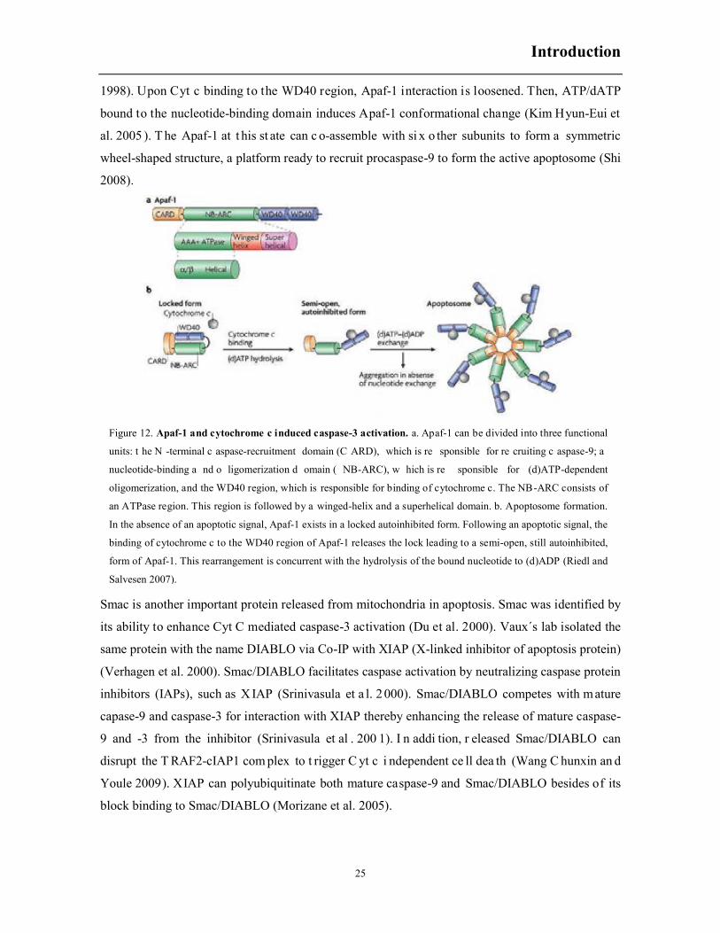
in the presence of ATP/dATP (Li Peng et al. 1997). Apaf-1 is another indispensable component of
the apoptosome besides Cyt c. Crystal structure analysis shows that the inactive Apaf-1 protein may
exist in a compact close form, probably through intramolecular interaction between the N-terminal
CARD domain and the C-terminal WD40 repeats. The compact Apaf-1 is not accessible to
procaspase-9. Deletion of the WD40 repeats results in Apaf-1 constitutively binding and activating
caspase-9, indicating that the inactive Apaf-1 is in an auto-inhibited state (Bao et al. 2005, Hu et al.
Introduction
25
1998). Upon Cyt c binding to the WD40 region, Apaf-1 interaction is loosened. Then, ATP/dATP
bound to the nucleotide-binding domain induces Apaf-1 conformational change (Kim Hyun-Eui et
al. 2005 ). T he Apaf-1 at t his st ate can c o-assemble with si x o ther subunits to form a symmetric
wheel-shaped structure, a platform ready to recruit procaspase-9 to form the active apoptosome (Shi
2008).
Smac is another important protein released from mitochondria in apoptosis. Smac was identified by
its ability to enhance Cyt C mediated caspase-3 activation (Du et al. 2000). Vaux´s lab isolated the
same protein with the name DIABLO via Co-IP with XIAP (X-linked inhibitor of apoptosis protein)
(Verhagen et al. 2000). Smac/DIABLO facilitates caspase activation by neutralizing caspase protein
inhibitors (IAPs), such as X IAP (Srinivasula et a l. 2 000). Smac/DIABLO competes with mature
capase-9 and caspase-3 for interaction with XIAP thereby enhancing the release of mature caspase-
9 and -3 from the inhibitor (Srinivasula et al . 200 1). I n addi tion, r eleased Smac/DIABLO can
disrupt the T RAF2-cIAP1 com plex to t rigger C yt c i ndependent ce ll dea th (Wang C hunxin an d
Youle 2009). XIAP can polyubiquitinate both mature caspase-9 and Smac/DIABLO besides of its
block binding to Smac/DIABLO (Morizane et al. 2005).
Figure 12. Apaf-1 and cytochrome c induced caspase-3 activation. a. Apaf-1 can be divided into three functional
units: t he N -terminal c aspase-recruitment domain (C ARD), which is re sponsible for re cruiting c aspase-9; a
nucleotide-binding a nd o ligomerization d omain ( NB-ARC), w hich is re sponsible for (d)ATP-dependent
oligomerization, and the WD40 region, which is responsible for binding of cytochrome c. The NB-ARC consists of
an ATPase region. This region is followed by a winged-helix and a superhelical domain. b. Apoptosome formation.
In the absence of an apoptotic signal, Apaf-1 exists in a locked autoinhibited form. Following an apoptotic signal, the
binding of cytochrome c to the WD40 region of Apaf-1 releases the lock leading to a semi-open, still autoinhibited,
form of Apaf-1. This rearrangement is concurrent with the hydrolysis of the bound nucleotide to (d)ADP (Riedl and
Salvesen 2007).

Introduction
26
Smac/DIABLO and Cyt C reside in the same nar row space in t he mitochondria. However, some
stimuli seems to induce a differential release of both proteins. It has been reported that TNFα-
induced apoptosis generates jBid, a cleaved Bid induced by JNK activation, which translocates to
mitochondria to trigger Smac/DIABLO but not Cyt c release (Deng Yibin et al. 2003). Consistently,
overexpression of Bid△25, a mimic of jBid, only induces Smac/DIABLO release. Cephalostatin, a
bis-steroidal m arine natural pr oduct, induces selective release of Sm ac/DIABLO and subsequent
apoptosis (Dirsch et al. 2003). It has also been reported that inhibition of mitochondrial fission in
apoptosis prevents C yt c r elease m ore t han Sm ac/DIABLO release (Parone et al . 2006 ). The
mechanism involved in this differential release is not clear, but a pool of cytochrome c is thighly
bound to (Cortese et al. 1998) and oxidative modification of cardiolipin, as a consequence of ROS
production, might be important for mobilization of cytochrome c (Ott et al. 2002).
3.4.3 Bcl-2 fa mily members are key c omponents in regulating
mitochondrial out membrane permeability
Original information about Bcl-2 family proteins came from C. elegans, in which four genes Egl-1,
Ced-3, Ced-4 and Ced-9 involved in phenotype change were classified (Muzio et al. 1996). Shortly
Figure 13. Diagrammatic representation of the mammalian B-cell lymphoma 2 (BCL-2) family (Strasser 2005).
Introduction
27
after, 13 Ced-9 homologues (known as multi-region Bcl-2 proteins) and a number of highly
divergent proteins analogous to Egl-1 (known as BH3-only proteins) were identified in mammalian
cells. The common feature of Bcl-2 members is that they contain one or more Bcl-2 homology (BH)
domain. Within the Bcl-2 family, the evolutionary relationship between multi-region Bcl-2 family
members and BH3-only proteins (other than Bid) is distant. Phylogenetic analysis indicates that the
multi-region proteins Bcl-2, Bcl-xl, Bax and Bid share a common origin, and other BH3-only
proteins evolved later (Billen et al. 2008). Generally, Bcl-2 family proteins are subdivided into three
groups based on their pro- or anti- apoptotic action and the Bcl-2 homology (BH) domains they
possess (Fig. 13). The interactions between the three groups determine MOMP and apoptosis
(Chipuk et al. 2010).
Anti-apoptotic Bcl-2-like proteins (e.g., Bcl-2, Bcl-xL, Bcl-w, and Mcl-1) and proapoptotic Bax-like
proteins (e.g., Bax, Bak, and Bok) consist of four BH domains (Kvansakul et al. 2008). On the other
hand, the proapoptotic BH3-only proteins (e.g., Bid, Bim, Bad, Bmf, Bik, Blk, Noxa, Puma)
possess only a short motif called the BH3 domain. The anti-apoptotic Bcl-2 proteins are generally
located in OMM, but may also be in the cytosol or ER membrane. The Bcl-2-like proteins possess
various membrane insertion domains and have been reported to interact with different lipids when
targeted to membranes (Petros et al. 2004). The proapoptotic Bcl-2 members consist of the BH3-
only proteins and a number of multi-BH3 domain proteins (effectors). BH3-only proteins that only
bind to the anti-apoptotic repertoire are referred to as ‘‘sensitizer’’ or ‘‘derepressor’’, whereas the
ones interacting with both the anti-apoptotic proteins and the effectors are classified as activators,
such as Bid (Bcl-2-interacting domain death agonist), Bim (Bcl-2-interacting mediator of cell death),
Bad (Bc-2 antagonist of cell death) and Noxa (Martinou and Youle 2011). Bid and Bim can interac
with the anti-apoptotic repertoire, as well as with the effectors directly inducing Bak and Bax
oligomerization and MOMP. Therefore, they are referred as ‘‘direct activators’’ (Giam et al. 2008,
Shamas-Din et al. 2011).
Bid, first cloned in 1996 as a novel death agonist that heterodimerises with either BAX or Bcl-2, is
a member of the BH3-only proteins that plays a crucial role in regulating the permeability of the
OMM. Bid contains eight α-helices, the BH3 region located in helix 3 comprises a region of
sequence homology to other Bcl-2 proteins which is required for interaction with both pro-apoptotic
Bax and anti-apoptotic protein Bcl-xL (Wang Kun et al. 1996) (Fig. 14a). Bid also contains a large
unstructured loop (amino acids 42–79) that separates helices 2 and 3. This loop contains a variety of
sites that are subjected to post-translation modifications, regulating Bid localization and apoptotic
function (Kvansakul et al. 2008) (Fig. 14a). In 1998, Bid was identified as a caspase-8 substrate.
The resulted tBid translocates to mitochondria and initiates mitochondrial protein release. The
Introduction
28
cleavage site of caspase-8 in human Bid is Asp60 (Li Honglin et al. 1998) and in X. laevis is Asp52
(Saitoh et al. 2009). Removal of the N-terminal fragment has been suggested to increase the number
of exposed hydrophobic residues thereby facilitating its binding to membranes (McDonnell et al.
1999). It was shown that cardiolipin, a negatively charged lipid specific to mitochondria, mediates
the specific targeting of tBid to MOM (Lutter et al. 2000) and that the membrane protein
MTCH2/MIMP was also a major facilitator of tBid insertion into the MOM (Zaltsman et al. 2010).
MTCH2/MIMP may enhance tBid function by facilitating interactions with cardiolipin rich regions
(Shamas-Din et al. 2011). In addition, caspase-3 can also cleave human Bid at Asp60 (Slee et al.
2000). Similar cleavage sites for non-caspase proteases were also detected in human Bid, such as
Gly70 for calpain, Arg71 for cathepsins and Asp75 for granzyme B. Studies shown that granzyme
B treatment of cells resulted in cleavage of Bid, accumulation of tBid at mitochondria and release of
cytochrome c that was not inhibited by caspase inhibitors, similar effects were observed for
protease calpain. Tissue extracts from normal mice treated with lysosomal extracts released
cytochrome c, however similar treatment in tissue extracts from Bid-/- mice failed to release
cytochrome c, indicating that Bid may also be cleaved by cathepsins (Cirman et al. 2004, Reiners Jr
et al. 2002, Sutton et al. 2003, Sutton et al. 2000). Finally, human Bid cleavage at Leu25 by an
unknown protease is described in a JNK dependent manner to generate a large C-terminal fragment
(jBid) that could accumulate at mitochondria like tBid (Deng Hongbin et al. 2008).
In summary, when Bid was cleaved by proteases, the C-terminal product accumulates at
mitochondria. This implicates the N-terminus of Bid as a negative regulatory sequence that prevents
the mitochondrial localization of Bid, thereby preventing apoptosis. After cleavage by caspase-8,
the N-terminus of Bid (nBid) remains attached to tBid, possibly masking the BH3 domain. However,
it was shown that only tBid inserts while nBid remains in solution (Lovell et al. 2008).The
inhibitory role of the N-terminal fragment was elegantly demonstrated by Tan and colleagues. They
concluded that the N-terminal fragment negatively regulates the exposure of the BH3-domain and
thereby its binding to the OMM (Mandic et al. 2002).
Bid cleavage mediated by caspase-8 can be attenuated when residues in the vicinity of the cleavage
site are phosphorylated. In human Bid phosphorylation of Thr59 (Bid is cleaved after Asp60)
severely inhibited Bid cleavage by caspase-8 (Degli Esposti et al. 2003). Similarly, phosphorylation
of murine Bid (at Ser61 and Ser64) also attenuated its cleavage by caspase-8 (Desagher et al. 2001).
Xenopus Bid is mono- and bi- ubiquitinated in egg extracts, and not degraded by the proteasome
(Saithoh et al. 2009). There are at least three sites important for ubiquitination (K18, K21, K37),
located close to the cleavage site Asp52. However, it is not clear the role of mono- and bi-
uiquitinated Bid in apoptosis (Saitoh et al. 2009).

Introduction
29
The effector pr oteins, B cl-2 ant agonist k iller 1 ( Bak) and Bcl-2-associated x pr otein ( Bax) w ere
originally de scribed to contain on ly B H1-3 motifs; h owever, s tructure-based alignment of g lobal
Bcl-2 family proteins revealed a con served BH4 motif (Kvansakul et a l. 2008). Upon ac tivation,
Bak and Bax homo-oligomerize into proteolipid pores within the OMM to promote MOMP. It was
reported that t he Bak/Bax com plex seems to be i ndispensable in r egulating MOMP. The
proapoptotic function of Bax/Bak was proved by the extreme resistance of bax−/− bak−/− DKO
cells to a variety of apoptotic stimuli, and Cyt C release from the mitochondria was not detected
(Wei et al. 2001).
Figure 14. Regions and residues of Bid and Bax that regulate binding to membranes and apoptotic function.
The human Bid protein (a) and the human Bax protein (b) are depicted. Bid contains 195 amino acids and 8α-helices
whereas Bax contains 192 amino acids and 9α-helices. The n umbers above each α-helix in dicate th e sta rting a nd
ending amino acids for that helix. The regions of Bid and Bax that control recruitment of the proteins to membranes
and f unction, in cluding th e BH regions and d omains believed to in sert into m embranes, are in dicated a bove th e
proteins. Where appropriate, parentheses indicate the amino acids that correspond to a particular region. Individual
amino a cids that regulate b inding to m embranes or a re su bjected to p osttranslational modifications that regulate
binding to membranes are indicated below the proteins, with parentheses indicating the identity and number of each
amino acid (Billen et al. 2008).
Introduction
30
Bak is constitutively inserted in the outer mitochondiral membrane (OMM) by a C-terminal
transmembrane domain. In contrast, Bax is mainly cytosolic with a minor fraction loosely attached
to mitochondria. It has been observed that Bax constantly travels back and forth from the cytoplasm
to mitochondria (Edlich et al. 2011). Several reports suggested that the mitochondrial sub-
population of Bax could be treated as a measure of the stress experienced by the cells. Once
attached to mitochondria, Bax can be retrotranslocated to the cytosol by Bcl-xL or other prosurvival
Bcl-2 proteins which would ensure that Bax does not chronically accumulate at the OMM to reach a
critical level that could promote Cyt c release (Martinou and Youle 2011). Unlike Bid, Bax does not
require a cleavage-activation to enable its pro-apoptotic function. However, the binding of Bax to
mitochondria-bound tBid can drive the translocation of Bax from cytoplasm to the mitochondrial
outer membrane. Early tudies suggested that the recruitment of Bax might be regulated by a
proposed C-terminal TM region, TM sequence release not only potentiates its insert into
membranes, but also opens the hydrophobic binding pocket where it resides. Then studies indicate
that N-terminal conformational changes in Bax is required for its insert into the mitochondria
membrane, the N-terminus of Bax contains regions that both positively and negatively regulate the
targeting of Bax to membranes. The conformational changes at both the N- and C- terminal of Bas
may due to phosphorylation occurring in these regions (Billen et al. 2008).
BH3-only proteins and antiapoptotic Bcl-2 proteins are positive and negative regulators of Bax/Bak
complex respectively. The protein tBid has been originally described as a direct activator of Bax,
inducing a conformational change of the Bax N-terminus, to modulate Bax insertion and
oligomerization in the OMM (Eskes et al. 2000). Many groups have confirmed the contribution of
other BH3-only proteins, Bim and Puma, to this process. However, the mechanism how BH3 only
proteins induce apoptosis is not clear (Lovell et al. 2008). Three different models have been
proposed to explain the regulation of apoptosis by Bcl2 family members (Fig. 15).
In a “membrane-embedded together” model proposed by Andrews and colleagues, it was described
that tBid first inserts in the membrane, then recruits Bax and other antiapoptotic proteins, including
Bcl-xl and Bcl-w. It seems both anti- and pro- apoptotic proteins are activated by recruiting
sensitizer BH3-only proteins (Shamas-Din et al. 2011). This conferred an ambiguous role on the
BH3-only proteins. The tBid, is capable of inducing MOMP in cells. Similar evidence exists for
Bim-meidated Bak/Bax activation, and for PUMA promoting Bak/Bax activation. Bid and Bim
BH3 peptides induce Bak and Bax oligomerization and pore-forming activity with isolated
mitochondria or large unilamellar vesicles (Chipuk and Green 2008, Kuwana et al. 2005, Letai et al.
2002)

Introduction
31
3.4.4 JNK a ctivation a re i mplicated in both intrinsic and
extrinsic apoptotic signaling pathways
.It has been reported that JNK1 is involved in ultraviolet C and γ-radiation-induced apoptosis (Chen
Yi-Rong et al. 1996). In 2000, evidence came up from the studies of embryonic fibroblasts derived
from J NK1-/- & JNK2-/- mice. These MEF cells displayed r emarkable r esistance t o UV i nduced
Figure 15. Overview of the three models of regulation of apoptosis by Bcl-2 family proteins. BH3-S and BH3-
A represent sensitizer and activator BH3-only proteins, respectively. A) The Direct activation model proposes that
activator BH3-only proteins are required for activating Bax and Bak. Anti-apoptotic proteins inhibit the activator
BH3-only proteins but not Bax and Bak to suppress apoptosis. Sensitizer BH3-only proteins displace the activator
BH3-only p roteins from the anti-apoptotic proteins to promote apoptosis. B) The Displacement model postulates
that Bax a nd Ba k a re c onstitutively a ctive in c ells and m ust be se questered b y a nti-apoptotic p roteins for c ell
survival. BH3 -only p roteins only p lay th e se nsitizer ro le and in hibit th eir respective a nti-apoptotic proteins t o
promote apoptosis. Both the direct activation and the displacement model do not define a role for the membrane. C)
Embedded together model highlights the active role of the membrane where Bcl-2 family proteins insert into and
change their conformations th at dictate t heir functions. Cytoplasmic a nti-apoptotic proteins are r ecruited t o
membranes and a re a ctivated b y b oth se nsitizer a nd a ctivator BH3-only proteins a s w ell as B ax/Bak. At the
membrane, an ti-apoptotic proteins inhibit th e a ctivator BH3 -only p roteins a nd Ba x/Bak to p revent MOMP.
Sensitizer BH3 -only p roteins displace th e a ctivator BH3-only p roteins and Ba x/Bak f rom th e a nti-apoptotic
proteins to p romote apoptosis. Activator BH3 -only p roteins re cruit Ba x to th e membrane to in duce M OMP a nd
apoptosis. These in teractions are re versible and a re g overned b y equilibrium c onstants t hat are a ltered b y th e
concentrations and interactions of the proteins with each other and with membranes (Shamas-Din et al. 2011).

Introduction
32
apoptosis (Tournier et al. 2000). The function of JNK3 in apoptosis was also explored through
JNK3-/- mice, in the mutant mice the phosphorylation of c-Jun and the transcriptional activity of the
AP-1 transcription factor complex were markedly reduced, and the reduction in seizure activity and
hippocampal neuron apoptosis was prevented. (Yang Derek D et al. 1997). Furthermore, it has been
reported that activated JNK is involved in both transcription-dependent and transcription-
independent apoptotic mechanisms, which could cooperate through activation of specific
transcription factors and directly modifying apoptosis associated proteins (Davis R. J. 2000a,
Dhanasekaran Danny N and Reddy 2008) (Fig. 16).
Phosphorylated c-Jun by JNK translocates to nucleus and regulates a vast of transcription factors
which result in regulation of a wide range of proteins including increased expression of pro-
apoptotic proteins and decreased expression of anti-apoptotic proteins (Dhanasekaran Danny N and
Reddy 2008, Raman et al. 2007). In addition, JNKs play an essential role in regulating the pro-
and/or anti- apoptotic proteins residing in the mitochondria (Schroeter et al. 2003). It has been
shown that the activation of JNK is indispensable for cytochrome C release, because JNK1-/- &
JNK2-/- MEFs fail to show cytochrome C release in front of various apoptotic stimuli (Tournier et
al. 2000). Similarly, redistribution and conformational changes of Bax/Bak cannot be observed in
JNK-deficient cells (Lei et al. 2002). Some researches show that Bid cleavage induced by JNK
activation may play an essential role in this process (Madesh et al. 2002). For instance, in TNF-α
induced apoptosis of Hela cells, JNK activation has been proven to induce caspase-8 independent
cleavage of Bid to get a 21kDa fragment (jBid) which translocates to mitochondria and selectively
promotes Smac/DABLO release (Deng Yibin et al. 2003). It has been reported that other pro-
apoptotic BH3-only family proteins were also modulated by JNK signaling, such as Bim and Bmf
(Lei and Davis 2003). During UV-induced apoptosis of HEK293K cells, Bim and Bmf are released
from their sequestering proteins because of their phosphorylation by JNK. The released Bim and
Bmf subsequently activate Bax and/or Bak (Lei et al. 2002, Makani et al. 2002, Putcha et al. 2003).
In another way, phosphorylated Bim can neutralize the activity of anti-apoptotic proteins, such as
Bcl2 and Bcl-xL, thereby releasing Bcl-xL and/or Bak from inhibition (Puthalakath and Strasser
2002). Another BH3-only pro-apoptotic member regulated by JNK is Bad. Generally, the pro-
survival kinases, such as Akt-1, PAK-1 and PKA, inhibit the activity of Bad by phosphorylation at
Ser136 or/and Ser112, and phosphorylated Bad is sequestered by 14-3-3 family proteins (Gross et al.
1999). In contrast, JNK specifically phosphorylates Bad at Ser128 thereby sequestering its
interaction with 14-3-3 family proteins (Wang Xiao-Tian et al. 2007c). Furthermore, JNK seems to
release sequestered Bad from 14-3-3 protein by phosphorylating 14-3-3 protein at Ser184
(Sunayama et al. 2005, Tsuruta et al. 2004).

Introduction
33
It has been reported that Bax is also a JNK substrate. The JNK-mediated Bax phosphorylation may
cause Bax activation (Kim Bong-Jo et al. 2006), and Bax has also been reported to be sequestered
by 14-3-3 proteins, like Bad, via interaction with the C-terminal region of 14-3-3 protein (Weston
and Davis 2007). Additionally, JNK may regulate the anti-apoptotic Bcl-2 family members. It has
been shown that JNK can phosphorylates Bcl-2, Bcl-xL and Mcl-1 thereby suppressing their anti-
apoptotic functions (Dhanasekaran Danny N and Reddy 2008, Srivastava et al. 1999, Yamamoto et
Figure 16. Nucleus- and mitochondria- targeted signaling by JNK. JNK can promote apoptosis by two distinct
mechanisms. In the f irst mechanism targeted a t the nuclear events, activated JNK translocates to the nucleus and
transactivates c -Jun a nd o ther target transcription f actors (TF). JN K can p romote apoptosis by in creasing th e
expression of pro-apoptotic genes through the transactivation of c-Jun/AP1-dependent or p53/73 protein-dependent
mechanisms. In pathways directed at mitochondrial apoptotic proteins, activated JNK translocates to mitochondria.
There, JNK can p hosphorylate th e BH3 -only family o f Bc l-2 p roteins to antagonize the anti-apoptotic activity o f
Bcl-2 or Bcl-XL. In addition, JNK can stimulate the release of cytochrome c (Cyt C) from the mitochondrial inner
membrane th rough a Bi d-Bax-dependent mechanism, pr omoting t he f ormation of a poptosomes consisting of
cytochrome c, caspase-9 (Casp 9) and Apaf-1. This complex initiates the activation of caspase-9-dependent caspase
cascade. I n an other m echanism, J NK can p romote the r elease o f S mac/DIABLO (Smac) th at can in hibit t he
TRAF2/IAP1 in hibitory c omplex, th ereby re lieving th e in hibition o n c aspase-8 to initiate c aspase a ctivation. I n
addition, by phosphorylating Bad and its sequestering partner 14-3-3, JNK can promote Bad-mediated neutralization
of the Bcl-2 family of anti-apoptotic proteins. Finally, JNK can phosphorylate Bcl2 for suppressing its anti-apoptotic
activity. These nuclear and mitochondrial events regulated by JNK need not be mutually exclusive (Dhanasekaran
Danny N and Reddy 2008).

Introduction
34
al. 1999). Interestingly, besides the MOMP regulation function via modifying Bcl-2 family
members, another anti-apoptotic function has been reported for JNK1-1 that JNK interacts with
Apaf1 and cytochrome c to inhibit the apoptosome complex (Tran et al. 2007).

Objectives
35
4. OBJECTIVES
The general objectives of this project are to investigate the role of MAPK proteins in meiotic
maturation and the role of JNK1-2 proteolysis and Bcl-2 family members in the regulation of
osmostress-induced apoptosis in Xenopus oocytes. The specific aims of the research project are:
To address whether JNK proteins are key factors for oocyte maturation.
To identify which protein/JNK isoform is phosphorylated and detected by the pJNK antibody
in mature oocytes and the upstream kinase responsible for its phosphorylation.
To identify the cleavage site and the protease responsible for JNK1-2 proteolysis induced by
osmostress and to understand the biological role of JNK1-2 proteolysis during osmostress-
induced apoptosis.
To investigate the function of Bcl-2 family members on hyperosmotic shock-induced apoptosis.
To analyze the posttranslational modifications of Bid induced by hyperosmotic shock and their
effect to Bid function.
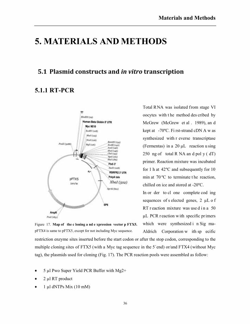
Materials and Methods
36
5. MATERIALS AND METHODS
5.1 Plasmid constructs and in vitro transcription
5.1.1 RT-PCR
Total RNA was isolated f rom stage VI
oocytes with t he method des cribed by
McGrew (McGrew et al . 1989), an d
kept at -70ºC. Fi rst-strand cDN A w as
synthesized with r everse transcriptase
(Fermentas) in a 20 µL reaction u sing
250 ng of total R NA an d pol y ( dT)
primer. Reaction mixture was incubated
for 1 h at 42ºC and subsequently for 10
min at 70 ºC to terminate t he reaction,
chilled on ice and stored at -20ºC.
In or der to cl one complete cod ing
sequences of s elected genes, 2 µL o f
RT r eaction mixture was use d i n a 50
µL PCR r eaction w ith specific pr imers
which were synthesized i n Sig ma-
Aldrich Corporation w ith sp ecific
restriction enzyme sites inserted before the start codon or after the stop codon, corresponding to the
multiple cloning sites of FTX5 (with a Myc tag sequence in the 5´end) or/and FTX4 (without Myc
tag), the plasmids used for cloning (Fig. 17). The PCR reaction pools were assembled as follow:
5 µl Pwo Super Yield PCR Buffer with Mg2+
2 µl RT product
1 µl dNTPs Mix (10 mM)
Figure 17. Map of the c loning a nd e xpression vector p FTX5.
pFTX4 is same to pFTX5, except for not including Myc sequence.

Materials and Methods
37
1.25 µl sense primer (20 µM) *
1.25 µl antisense primer (20 µM) *
39 µl H2O
0.5 µl Pwo Super Yield DNA polymerase (5U/µl)
* For specific primers, see Table 1.
PCR reaction was performed as follow:
- 95ºC, 5 min
- 95ºC, 45 sec
- 60ºC, 45 sec 30x
- 72ºC, 3 min
- 72ºC, 10 min
- 4ºC, hold.
Table 1. Primers for RT-PCR and site-directed mutagenesis
OBJECTIVE ORIENTATION SEQUENCE (5´-3´) RESTRICTION
ENZYME
H. sapiens
JNK2-1
S GCCCCATGGCAGACAGTAAATGACAGT Nco I
AS GCCCTCGAGTTACTGCTGCATCTGTGCTGAAGG Xho I
X. laevis
JNK1-1
S GCCCCGGCACGAAGCAAGCGTGACAGC Nco I
AS GCCCTCGAGTTACTGCTGCACCTGTGCTAAAGG Xho I
X. laevis
JNK1-2
S GCCCCATGGCAAGAAGCAAGCGTGACAACAATTTT Nco I
AS GCCCTCGAGTCATCATCGACAGCAACCCAG Xho I
X. laevis
JNK1-2Δ412
S GCCCCATGGCAAGAAGCAAGCGTGACAACAATTTT Nco I
AS GCCCTCGAGTTAGTCTGTGTCTGAG Xho I
X. laevis
JNK1-2 (D412A)
S TGCCCTCAGACACAGCCAGCAGCCTTGAAAC
AS GTTTCAAGGCTGCTGGCTGTGTCTGAGGGCA
X. laevis
JNK1-2Δ385
S GCCCCATGGCAAGAAGCAAGCGTGACAACAATTTT Nco I
AS GCCCTCGAGTTAATCAGTTACTGCTGCAC Xho I
X. laevis
JNK1-2 (D385A)
S GTGCAGCAGTAACTGCTGGTTCCCAGGCTCA
AS TGAGCCTGGGAACCAGCAGTTACTGCTGCAC
JNK3 sequencing S GTGAATTGTTGCAGTGCAGGC
AS GGGTTTCGCAGGCAGGCGGC
X. laevis
JNK3-1
S GCCCCATGGCCCGTCATTTCTTGTATAACTGCAG Nco I
AS GCCCTCGAGTCACTGCTGCACCTGTG Xho I
X. laevis
JNK3-2
S GCCCCATGGCCCGTCATTTCTTGTATAACTGCAG Nco I
AS GCCCTCGAGTCACCTGCAACAACCGA Xho I
X. laevis S GCCCCATGGCCAAAAGCAAAGTTGACAACCAG Nco I

Materials and Methods
38
JNK3-3 AS GCCCTCGAGTCACCTGCAACAACCGA Xho I
X. laevis
JNK3-4
S GCCCCATGGCCAAAAGCAAAGTTGACAACCAG Nco I
AS GCCCTCGAGTCACTGCTGCACCTGTG Xho I
X. laevis
MKK7
S GCCCCATGGCGGCGTCCTCCCTGGAGCAGAAACTCT Nco I
AS GCCCTCGAGCTACCTGCCGAAAAATGGCAGGTTTTG Xho I
X. laevis
MKK7-DED
S GGGCGCCTTGTGGACGACAAGGCAAAGGAGAGGGATGCAG
GCTGTGCTGC
AS GCAGCACAGCCTGCATCCCTCTCCTTTGCCTTGTCGTCCACA
AGGCGCCC
X. laevis
MKK7 (K146M)
S GGACACGTCATCGCAGTTATGCAAATGCGTCG
AS CGACGCATTTGCATAACTGCGATGACGTGTCC
X. laevis
MKK7-3A
S GGCGCCTTGTGGACGCCAAGGCAAAGGCGATTTCTGCAGGC
TGTGCTGC
AS GCAGCACAGCCTGCAGCCCTCGCCTTTGCCTTGGCGTCCACA
AGGCGCC
X. laevis
caspase-10
S GCCCCATGGATTTCAATTCCATGCTTCTCCGT Nco I
AS GCCCTCGAGTTATGGACTGTTTTCTTGACAAG Xho I
X. laevis
caspase-10 (C385S)
S GTTTTTTATACAAGCGAGCCAAGGGAGATATACAC
AS GTGTATATCTCCCTTGGCTCGCTTGTATAAAAAAC
X. laevis
ERK2a
S GCCGGATCCATGGCAGCGGCAGGAGCTG Bam HI
AS GCCCTCGAGTCAGTACCCTGGCTGGAATCGAGC Xho I
X. laevis
ERK2b
S GCCGGATCCATGGCAGCGGCAGCGGCCT Bam HI
AS GCCCTCGAGTCAGTACCCTGGCTGGAATCGAGC Xho I
X. laevis
ERK2b (T184S)
S CAGACCATGATCACTCTGGCTTTCTCACAG
AS CTGTGAGAAAGCCAGAGTGATCATGGTCTG
X. laevis
ERK2b (T193S)
S CACAGAATATGTAGCCTCTCGCTGGTACAGAGC
AS GCTCTGTACCAGCGAGAGGCTACATATTCTGTG
X. laevis
Bcl-xl
S GCCCCATGGAGGGCAGCAGTAGAGATCTGGTGG Nco I
AS GCCCTCGAGCTATCGGCGCCTCATGTAGCAGAC Xho I
X. laevis
Bak
S GCCCCATGGTCTCTTCCGCATGTGAAGTAGAGTT Nco I
AS GCCCTCGAGTCAGGTCAATCGTTGAAGCAGAAAC Xho I
X. laevis
Bax
S GCCGGATCCCATGGAGCCGGTGGGGATTGT Bam HI
AS GCCCTCGAGTTAGGACATCTTCCAGATGGCAAGA Xho I
X. laevis
Bid
S GCCCCATGGGTCTGAATGTGAAGACGATTCTCG Nco I
AS GCCCTCGAGTCACCTCTGTCTAGCGAGACGCTC Xho I
X. laevis
nBid
S GCCCCATGGGTCTGAATGTGAAGACGATTCTCG Nco I
AS GCCCTCGAGTCAATCAGTTTCCAGATCCCCATC Xho I
X. laevis
tBid
S GCCCCATGGGAAACGTCCAATTCAGATCTACAG Nco I
AS GCCCTCGAGTCACCTCTGTCTAGCGAGACGCTC Xho I
X. laevis
Bid (D52N)
S GGGGATCTGGAAACTAATGGAAACGTCCAATTC
AS GAATTGGACGTTTCCATTAGTTTCCAGATCCCC
X. laevis
Bid (K18/21R)
S TGGAGATCGACATAAGGAATCACAGAGATGAATTCTGGGA
AS TCCCAGAATTCATCTCTGTGATTCCTTATGTCGATCTCCA
X. laevis
Bid (K37R)
S GCGAGGGAAGAGGGGAGGTTAGTGGCACAGATG
AS CATCTGTGCCACTAACCTCCCCTCTTCCCTCGC

Materials and Methods
39
5.1.2 DNA purification
PCR products were purified by agarose gel electrophoresis. To prepare 1% agarose gel, 1 g agarose
was dissolved in 100 ml 1X TAE (40mM Tris, 20mM acetic acid, and 1mM EDTA, pH 8.0).
´´SYBR Safe DNA gel stain´´ (Invitrogen) at a dilution of 1:10.000 was added to the gel to
visualize the DNA band under UV irradiation.
The proper band was cut and recovered through the kit ´´Illustra GFX PCR DNA and Gel Band
Purification Kit´´ of GE Healthcare, and quantified by agarose gel electrophoresis. To indicate the
molecular weight and quantify the DNA, lambda DNA EcoRI/HindIII Marker was used in the
electrophoresis.
5.1.3 DNA digestion
DNA digestion was performed with the corresponding restriction enzymes and buffers (Fermentas)
following the recipe below:
10x buffer 10µl
DNA/FTX vector 5µg
restriction enzyme 2µl
nuclease-free water to 100µl
The mixture was gently mixed by pipetting up and down and spined down briefly to collect all
samples at the bottom of the tubes. Samples were incubated at appropriate temperature (usually
37°C) for 2 h following the manufacturer’s instructions. Finally, DNA products were purified
with ¨Illustra GFX PCR DNA and Gel Band Purification Kit¨.
5.1.4 DNA ligation and transformation
PCR products were ligated to FTX5/FTX4 vectors (Figure 1). Accurate ligation clones were
selected after transformation of E. coli DH5α ultracompetent cells (Inoue et al. 1990).
DNA ligation reaction was assembled in a microcentrifuge tube on ice using a molar ratio of 1:3
(vector to insert for the indicated DNA sizes). The components for a 20 μl reaction follow the recipe
below:
10X T4 DNA Ligase Buffer 2 μl

Materials and Methods
40
Vector DNA (FTX4/5) 50 ng
Insert DNA 50 ng (depending of DNA size)
Nuclease-free water to 19 μl
T4 DNA Ligase (Fermentas) 1 μl
The reaction was gently mixed by pipetting up and down and spined down briefly. Then the mixture
was incubated at 16°C overnight or at room temperature for 10 minutes.
For transformation, 50 μl of competent cells were incubated with 10 μl of DNA ligation mix on ice
for 30 min. Transformed cells were selected on LB plate with Ampicillin (50 µg/ml).
5.1.5 Plasmid extraction
Single colonies from the LB plate were selected and added to 5 ml LB medium containing 50 µg/ml
Ampicillin, and then incubated at 37°C with 200 rpm shaking overnight. Bacterial cells were
harvested by centrifugation at the maximum speed for 1 min. Plasmids were prepared
using ¨GenElute™ Plasmid Miniprep Kit¨ according to the instruction manual. Finally, the plasmids
were eluted with 50μl nuclease-free water. Generally, the plasmids were sequenced as described
below.
5.1.6 Site-directed mutagenesis and DNA sequencing
For construction of interested mutants used in different experiments, PCR-based mutagenesis was
employed to mutate specific amino acids in the coding sequences. Wild-type cDNA cloned in the
FTX5/4 vector was used as template and oligonucleotides for site-directed mutagenesis were
designed based on the QuikChange® Primer Design Program (Agilent Technologies) (Table 1). The
mutagenesis processes were performed according to QuikChange® Site-Directed Mutagenesis Kit
instruction manual. Briefly, we used high fidelity Pfu Turbo DNA polymerase (Stratagene, Ref.
600250) for the PCR reaction, and the following mix reaction was prepared:
5 µl 10x Cloned Pfu reaction buffer
1 µl DNA template (10 ng/µl)
1 µl dNTPs (10 mM)
0.7 µl sense primer (20 µM)*
0.7 µl antisense primer (20 µM)*
40.6 µl H2O
Materials and Methods
41
1 µl Pfu Turbo DNA polymerase (2.5 U/µl)
* For specific primers, see Table 1.
The PCR conditions were:
- 95ºC 30 s
- 95ºC 30 s
- 55ºC 1 min 16 cycles
- 68ºC 5 min
- 4ºC hold
After PCR reaction, the parental DNA was eliminated by adding 1 µl of the restriction enzyme Dpn
I (10 U/µl) directly into the tube and incubating the mix for 2.5 h at 37ºC (without the restriction
enzyme buffer). The PCR product obtained was transformed into E. coli DH5α ultracompetent cells
and individual colonies were selected in agar plates by their resistance to ampiciline. Minipreps
were obtained from single colonies and plasmids were purified using GenElute™ Plasmid Miniprep
Kit and sequenced as described below.
5.1.7 DNA sequencing
All cloned genes and mutations generated were confirmed by DNA sequencing using the “BigDye®
Terminator v3.1 Cycle Sequencing Kit” purchased from Applied Biosystems with the following
reaction set:
1.5 µl 5x Big Dye buffer
1 µl DNA template (400 ng/µl)
1 µl sequencing primer (5µM) (Table 2)
1 µl Big Dye
5.5 µl H20
Table 2. Primers for DNA sequencing
NAME SEQUENCE (5´-3´) Tm(ºC) ORIENTATION
T7 TAATACGACTCACTATAGGG 50.8 Sense
SP6 ATTTAGGTGACACTATAG 42.6 Antisense
FTX5 reverse CCAGGGTGGATTGAGTCCAG 66.9 Antisense

Materials and Methods
42
Parameters for sequencing were seted as follow:
- 96ºC, 6 sec
- 96ºC, 30 sec
- 50ºC, 15 sec 24 cycles
- 60ºC, 4 min
- 4ºC, hold
To precipitate the sequencing product, the 10 µl reaction pool was mix thoroughly with 40 µl
ethanol/sodium acetate precipitation buffer (650 µl ethanol 100%, 30 µl sodium acetate 3M pH 5.2,
130 µl water) and left on desk for 15 min at room temperature. Then the mix was centrifuged at
14.000 rpm for 15 min at 4ºC to get sequencing product pellet. The pellet was washed one time with
70% ethanol and centrifuged at 14.000 rpm for 5 min at 4ºC. Then the pellet was dried at room
temperature for 10 min before sent to Servei de Genòmica i Bioinformàtica of Universidad
Autónoma de Barcelona.
5.1.8 In vitro transcription
In vitro transcriptions of capped RNAs (cRNAs) were performed using mMESSAGE
mMACHINE® T7 Transcription Kit (Ambion®). DNA templates used for transcription were
linearized at the Xba I or Sgr AI site, then about 1 μg linearized DNA was used in a 20 µL reaction
assembly as described in its instruction manual. After incubated at 37ºC for two hours, extra DNA
was removed by incubating with 1 µL TURBO DNase at 37ºC for 15 min and terminated by adding
115 µL H2O and 15 µL Ammonium Acetate stop solution. The RNA was extracted with
phenol/chloroform and precipitated with 1 volume of isopropanol, the resulting mRNA was
resuspended in water to a final concentration of 1 mg/ml, as compared to RNA standards in
denaturing gel electrophoresis as described below.
Table 3. cRNAs prepared for oocyte microinjection cRNAs Source Vector Mutation Notes
MEKK1Δ Mus musculus FTX5 Deletion of aminoacids from 1 to 351
from wild type MEKK1 Constitutively active
MEKK1-KM Mus musculus FTX5 Mutation of aminoacid Lys-432 to Met
of MEKK1Δ Catalytically inactive
MKK7-DED Xenopus laevis FTX4 Mutation of aminoacids Ser-268, Thr-
272, and Ser-274 to Asp, Glu and Asp Constitutively active
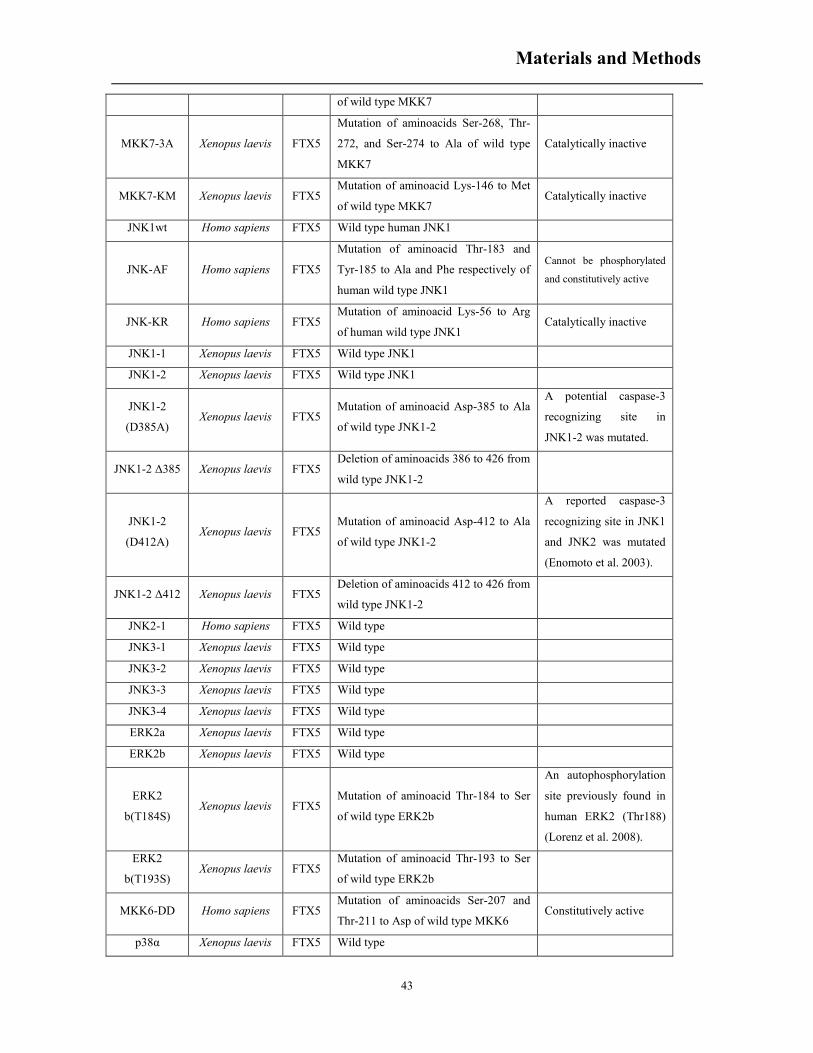
Materials and Methods
43
of wild type MKK7
MKK7-3A Xenopus laevis FTX5
Mutation of aminoacids Ser-268, Thr-
272, and Ser-274 to Ala of wild type
MKK7
Catalytically inactive
MKK7-KM Xenopus laevis FTX5 Mutation of aminoacid Lys-146 to Met
of wild type MKK7 Catalytically inactive
JNK1wt Homo sapiens FTX5 Wild type human JNK1
JNK-AF Homo sapiens FTX5
Mutation of aminoacid Thr-183 and
Tyr-185 to Ala and Phe respectively of
human wild type JNK1
Cannot be phosphorylated
and constitutively active
JNK-KR Homo sapiens FTX5 Mutation of aminoacid Lys-56 to Arg
of human wild type JNK1 Catalytically inactive
JNK1-1 Xenopus laevis FTX5 Wild type JNK1
JNK1-2 Xenopus laevis FTX5 Wild type JNK1
JNK1-2
(D385A) Xenopus laevis FTX5
Mutation of aminoacid Asp-385 to Ala
of wild type JNK1-2
A potential caspase-3
recognizing site in
JNK1-2 was mutated.
JNK1-2 Δ385 Xenopus laevis FTX5 Deletion of aminoacids 386 to 426 from
wild type JNK1-2
JNK1-2
(D412A) Xenopus laevis FTX5
Mutation of aminoacid Asp-412 to Ala
of wild type JNK1-2
A reported caspase-3
recognizing site in JNK1
and JNK2 was mutated
(Enomoto et al. 2003).
JNK1-2 Δ412 Xenopus laevis FTX5 Deletion of aminoacids 412 to 426 from
wild type JNK1-2
JNK2-1 Homo sapiens FTX5 Wild type
JNK3-1 Xenopus laevis FTX5 Wild type
JNK3-2 Xenopus laevis FTX5 Wild type
JNK3-3 Xenopus laevis FTX5 Wild type
JNK3-4 Xenopus laevis FTX5 Wild type
ERK2a Xenopus laevis FTX5 Wild type
ERK2b Xenopus laevis FTX5 Wild type
ERK2
b(T184S) Xenopus laevis FTX5
Mutation of aminoacid Thr-184 to Ser
of wild type ERK2b
An autophosphorylation
site previously found in
human ERK2 (Thr188)
(Lorenz et al. 2008).
ERK2
b(T193S) Xenopus laevis FTX5
Mutation of aminoacid Thr-193 to Ser
of wild type ERK2b
MKK6-DD Homo sapiens FTX5 Mutation of aminoacids Ser-207 and
Thr-211 to Asp of wild type MKK6 Constitutively active
p38α Xenopus laevis FTX5 Wild type
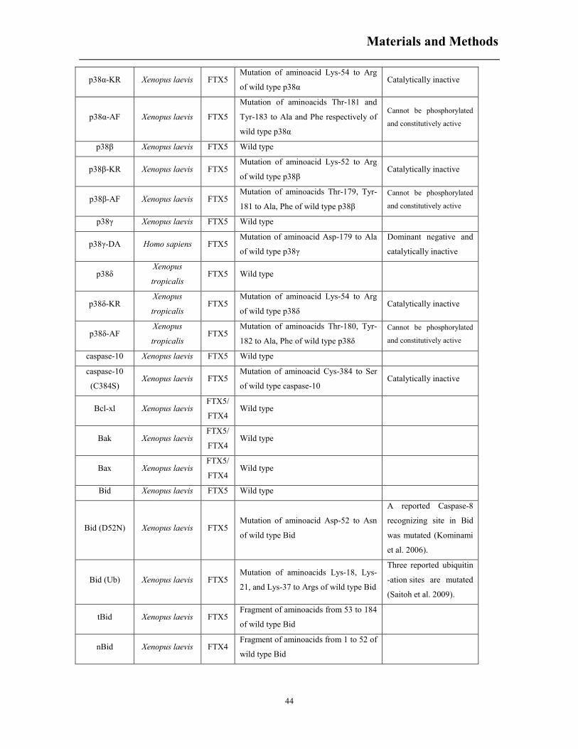
Materials and Methods
44
p38α-KR Xenopus laevis FTX5 Mutation of aminoacid Lys-54 to Arg
of wild type p38α Catalytically inactive
p38α-AF Xenopus laevis FTX5
Mutation of aminoacids Thr-181 and
Tyr-183 to Ala and Phe respectively of
wild type p38α
Cannot be phosphorylated
and constitutively active
p38β Xenopus laevis FTX5 Wild type
p38β-KR Xenopus laevis FTX5 Mutation of aminoacid Lys-52 to Arg
of wild type p38β Catalytically inactive
p38β-AF Xenopus laevis FTX5 Mutation of aminoacids Thr-179, Tyr-
181 to Ala, Phe of wild type p38β Cannot be phosphorylated
and constitutively active
p38γ Xenopus laevis FTX5 Wild type
p38γ-DA Homo sapiens FTX5 Mutation of aminoacid Asp-179 to Ala
of wild type p38γ
Dominant negative and
catalytically inactive
p38δ Xenopus
tropicalis FTX5 Wild type
p38δ-KR Xenopus
tropicalis FTX5
Mutation of aminoacid Lys-54 to Arg
of wild type p38δ Catalytically inactive
p38δ-AF Xenopus
tropicalis FTX5
Mutation of aminoacids Thr-180, Tyr-
182 to Ala, Phe of wild type p38δ Cannot be phosphorylated
and constitutively active
caspase-10 Xenopus laevis FTX5 Wild type
caspase-10
(C384S) Xenopus laevis FTX5
Mutation of aminoacid Cys-384 to Ser
of wild type caspase-10 Catalytically inactive
Bcl-xl Xenopus laevis FTX5/
FTX4 Wild type
Bak Xenopus laevis FTX5/
FTX4 Wild type
Bax Xenopus laevis FTX5/
FTX4 Wild type
Bid Xenopus laevis FTX5 Wild type
Bid (D52N) Xenopus laevis FTX5 Mutation of aminoacid Asp-52 to Asn
of wild type Bid
A reported Caspase-8
recognizing site in Bid
was mutated (Kominami
et al. 2006).
Bid (Ub) Xenopus laevis FTX5 Mutation of aminoacids Lys-18, Lys-
21, and Lys-37 to Args of wild type Bid
Three reported ubiquitin
-ation sites are mutated
(Saitoh et al. 2009).
tBid Xenopus laevis FTX5 Fragment of aminoacids from 53 to 184
of wild type Bid
nBid Xenopus laevis FTX4 Fragment of aminoacids from 1 to 52 of
wild type Bid
Materials and Methods
45
5.1.9 RNA quantification
The cRNAs obtained by in vitro transcription were quantified in denaturing 1% agarose gel
electrophoresis. To prepare the gel, 0.5 g agarose was dissolved in 36 ml water by heating and then
when it was cooled down to about 60ºC, 5 ml 10X MOPS running buffer (0.1 M Sodium Acetate,
0.01 M EDTA, 0.4 M MOPS, pH 7.0) and 9 ml 37% formaldehyde (12.3 M) were added. After
polymerization the gel was assembled in the tank and covered with 1X MOPS running buffer.
0.5 μl RNA sample was diluted with 14.5μl nuclease-free water and then 5 μl denaturing buffer (65
μl 10X MOPS Buffer, 113 μl Formaldehyde, 323 μl Formamide, 0.5 μl Ethidium Bromide Stock
(10mg/ml)) was added. The samples were heated at 85ºC for 10 min and then chilled on ice for 10
min. 10 μl sample with 2 μl loading buffer (50% glycerol, 10 mM EDTA pH 8.0, 0.6% orange G)
were analyzed in the electrophoresis.
5.2 Oocyte manipulation and sample collection
5.2.1 Oocyte isolation
Oocytes were obtained from sexually mature Xenopus laevis females (purchased from Centre
d’Elevage de Xenopes, Montpellier, or from Xenopus Express, Vernassal, France) via a minimally
invasive surgery. Xenopus was anesthetized in 0.02% benzocaine. Then, portions of ovary were
removed through a small incision on the abdomen. The incision was sutured and the animal was
kept in a separate tank until it had fully recovered from the anesthesia. Frog was then returned to a
large tank in which all the frogs were kept to recover at least 4 weeks until the next surgery. The
tissue was dissected into small pieces if the ovaries were healthy enough to use. Oocytes were
defolliculated for 3h at 18ºC with collagenase/dispase (0.8 mg/ml (Sigma), 0.48 mg/ml (Roche)) in
MBS (5 mM HEPES, 88 mM NaCl, 1 mM KCl, 1 mM MgSO4·7H2O, 2.5 mM NaHCO3 , 0.7 mM
CaCl2 , pH 7.8) with gentle agitation. The defolliculated oocytes were then washed thoroughly with
MBS and transferred to a petri dish. Stage VI oocytes in good state were sorted manually and
incubated overnight in MBS at 18ºC. The next day, healthy survivors were selected and transferred
to another petri dish containing fresh MBS. Pools of oocytes were treated with drugs at
concentrations and times indicated or injected with cRNAs and treated as described below.
Materials and Methods
46
5.2.2 Oocyte injection
Stage VI Xenopus oocytes were microinjected near their equator with 50 nL (5 ng) of the
corresponding cRNAs using a Nanoject II™ Automatic Nanoliter Injector (Drummond Scientific
Company). Injected oocytes were incubated 18 h at 18ºC and poor oocytes were eliminated the next
day before proper manipulation. Pools of 20 oocytes were collected at the indicated times before or
after treatment. In some experiments, oocytes were injected with cytochrome c from horse heart (c-
7752, Sigma).
5.2.3 Progesterone stimulation and GVBD percentage
calculation
For stimulating Xenopus oocyte maturation, 3 µg/mL progesterone (from 1000x stock solution
dissolved in ethanol) at final concentration was used in MBS. Mature oocytes were identified by the
white spot at the animal pole. The number of GVBD oocytes was counted and samples were
collected every hour as described. The percentage of mature oocyte was quantified by the number
of mature oocytes/number of total oocytes.
5.2.4 Hyperosmotic treatment
To investigate oocyte response to hyperosmotic shock, oocytes were treated with 300 mM sorbitol
in MBS. Samples were collected at indicated times and samples were prepared as described below.
5.2.5 Inhibitor usage
In order to inhibit specific protein activities, oocytes were either pre-incubated for 1 h or injected
with proper inhibitors before designed manipulation as described below. When oocytes were
preincubated and then treated with progesterone or 300 mM sorbitol, the final concentration of
inhibitor was maintained in the medium. We used a higher concentration of inhibitors compared to
mammalian cells due to specific properties of Xenopus oocytes (presence of vitelline membrane and
the yolk) that reduce the actual concentration of drugs at the cell membrane. In general, IC50 values
are approximately 10 to 20-fold higher when the drugs are applied to the extracellular surface of
Xenopus oocytes (Thomas et al. 2001, Thomas et al. 2004).

Materials and Methods
47
Table 4. Inhibitors used in oocyte manipulation
Inhibitor Target Final concentration Supplier
SP600125 JNK MAPK 25 µM (b), 50 or 100 µM (a) Calbiochem
BIRB796 p38 MAPK 50 or 100 µM (a) Axon Medchem
MDL28170 Calpains 50 µM (a) Sigma
U0126 MEK1 50 µM (a) Calbiochem
FR 180204 ERK MAPK 50 µM (a) Calbiochem
Z-VAD-fmk caspases 50 µM (a) Bachem
Z-DEVD-fmk caspase-3 50 µM (a) Calbiochem
TI-JIP153-163 JNK MAPK 5 or 10 µM (b) Calbiochem
a. pre-incubation with indicated final concentration in MBS or 300mM sorbitol;
b. injected with indicated final concentration.
5.3 Sample preparation and analysis
5.3.1 Oocyte lysis and mitochondrial/ cytosolic fraction
preparation
Fresh oocytes were lysed by pipetting up and down in 200 µl (pools of 20 oocytes) of ice-cold lysis
buffer (0.25 M sucrose, 0.1 M NaCl, 2.5 mM MgCl2, 20 mM HEPES, 1 mM EDTA, 1 mM EGTA,
pH 7.2) containing protease inhibitors (10 µg/ml leupeptin, 1 mM PMSF, 10 µg/ml aprotinin) and
phosphatase inhibitors (50 mM β-glycerolphosphate, 50 mM sodium fluoride, 1 mM sodium
orthovanadate, 5 mM sodium pyrophosphate). Samples were clarified by centrifugation at 14.500
rpm for 5 min and supernatants were collected and processed for western blotting or activity assay
as described below.
For subcellular fractionation, 30 oocytes were lysed in the lysis buffer and the extract obtained was
centrifuged at 1.000 g for 5 min at 4ºC to remove lipids and the yolk. The supernatant was isolated
and centrifuged at 16.000 g for 15 min at 4ºC. The supernatant obtained (cytosolic fraction) was
stored at -20ºC, and the pellet (mitochondrial fraction) was resuspended in 50 ul of lysis buffer and
stored at -20ºC too (Brun et al., 1981).

Materials and Methods
48
5.3.2 Immunoprecipitation
For denaturing IP, 10 oocytes were lysed in 50 µl lysis buffer. Then the 50 µl protein extract was
denatured with 10µl 6x denaturing buffer (375 mM Tris-HCl pH 6.8, 600 mM dithiothreitol, 9%
SDS) at 95ºC for 10 min and then purified through chloroform-methanol precipitation method
(Wessel and Flügge 1984). The pellets were then resuspended in 50 µl of resuspension buffer (100
mM Tris, pH 11.3% SDS, 3 mM DTT) by heating to 65ºC for 5 min and 95ºC for 10 min. 450 µl of
IP cell lysis buffer ( 20 mM Tris (pH 7.5), 150 mM NaCl, 1 mM EDTA, 1 mM EGTA, 1% Triton
X-100, 2.5 mM Sodium pyrophosphate, 1 mM β-glycerophosphate, 1 mM Na3VO4, 1 μg/ml
Leupeptin) was then added to the protein suspension (Campbell and Hardwick 2003). 0.5 µl of the
pJNK antibody was added to 100 µl cell lysate, and the mixture was incubated overnight at 4ºC
with gentle rocking. 10 µl G agarose beeds slurry (washed two times with IP buffer) were added to
the lysate and incubated with gentle rocking for 2 h 4ºC. A 30 s centrifugation was then performed
at max speed at 4ºC. The pellet was washed 3 times with 500 µl of IP cell lysis buffer. The sample
was kept on ice during washes. For immunoblotting analysis, the pellet was resuspended with 6 µl
5× Laemmli sample buffer (50 mM Tris HCl, pH 6.8, SDS 2%, 100 mM dithiothreitol, 10%
glycerol), vortex and centrifuged for 30 s. The sample was incubated at 100ºC for 5 min and
centrifuged for 30 s before was loaded to SDS-PAGE gels for analysis.
In order to get high quality immunoprecipitation product for mass spectrometry analysis, a Pierce®
Crosslink Immunoprecipitation Kit (Thermo Scientific) was applied. 10 µl pJNK antibodies were
incubated with 20 µl protein A/G plus agarose for 1 h in 1X coupling buffer to bind the antibody to
the agarose. The crosslink reaction was performed via incubating antibody-bound agarose with 450
µM DSS in 1X coupling buffer for 1 h. In the IP reaction, 20 µl antibody-crosslinked agarose were
incubated with 200 µl denatured lysate and washed as described above with kit supplied columns.
Finally, samples were eluted with 0.2% TFA elution buffer and concentrated before mass
spectrometry. The antibody-crosslinked agarose can be regenerated and reused in the next
Immunoprecipitation reactions.
5.3.3 Mass spectrometry identification
The pool of several immunoprecipitations (samples eluted in 0.2% TFA) was concentrated and sent
to Laboratori de Proteomica CSIC/UAB, Facultat de Medicina, Universitat Autònoma de Barcelona,
for mass spectrometry analysis. Briefly, the sample was separated on SDS-PAGE electrophoresis
and visualized with a compatible mass spectrometric silver staining (acetic acid was used as a fixer

Materials and Methods
49
instead of glutaraldehyde). The gel band obtained with the target protein was cut and the protein in
the gel slice digested in situ with trypsin. The tryptic extracts were analyzed by LC-MS/MS in data-
dependent mode. The MS system used was an LTQ XL Orbitrap (ThermoFisher) equipped with a
nanoESI ion source. LC-MS/MS spectra were searched using SEQUEST (Proteome Discoverer
v1.4, ThermoFisher). The database used for searching was obtained from Uniprot (organism:
Xenopus). Peptide identifications were filtered at 1% FDR and only proteins identified with two or
more peptides and peptide rank 1 were considered.
5.3.4 Western Blot
Samples collected were denatured in Laemmli sample buffer at 100ºC and subject to 10% or 14%
SDS/PAGE. Generally polyacrylamide gels for electrophoresis were prepared from commercial 40%
acrylamide (37.5 :1 acrylamide:bis, Bio-Rad, Cat. No. 161-0148), except for special gels for
detecting protein shift induced by phosphorylation which were prepared from 30% polyacrylamide
(100 :1 acrylamide:bis, Sigma).
Table 5. Gel preparation buffer for SDS-PAGE
For 100 ml Lower Gel Buffer (4x) For 100 ml Upper Gel Buffer (4x)
18.17 g Tris base
2 ml 20% SDS
Adjust pH to 8.8 with HCl.
6.06 g Tris base
2 ml 20% SDS
Adjust pH to 6.8 with HCl.
Polyacrylamide gels were prepared as described in table 6 depending on the final acrylamide
concentration in the separating gel.
Table 6. Polyacrylamide gel recipes (2 gels)
Lower Gel Upper Gel
8% 10% 14% 3%
4x buffer (ml) 4 4 4 2
40% acrylamide (ml) 3.2 4 5.6 0.6
H2O (ml) 8.8 8 6.4 5.4
PSA (µl) 60 60 60 32
TEMED (µl) 20 20 20 8

Materials and Methods
50
To get a proper separation resolution, electrophoresis was performed in Laemmli Runing Buffer 1X
(Table 7) at 150V for 1 h and, subsequently, proteins were transferred to Immobilon-P membranes
(Millipore) in Transfer Buffer with 10% methanol (Table 7) at 400mA for 2 h.
Table 7. Electrophoresis buffer for SDS-PAGE
For 1L Laemmli Running Buffer (10x) For 1L Transfer Buffer (10x)
30.3 g Tris Base
144.1 g Glycine
50ml 20% SDS
144 g glycine
30.2 g Tris base
To make 1X Transfer Buffer with 10% methanol: Mix 100 ml of 10 x Transfer Buffer, 100 ml of methanol and 800 ml of
dH2O per liter.
Uniformity of samples loading was verified by Ponceau (Sigma) staining of the blots. Membranes
were blocked for 1 h with 5% dried skimmed milk in TBST (50 mM Tris, 150 mM NaCl, 100 mM
KCl, pH 7.4, and 0.1% Tween20), washed several times with TBST, and then incubated overnight
with the specific primary antibody (see Table 8) diluted 1:1000 in TBST containing 5% bovine
serum albumin (BSA).
Table 8. Antibodies used in identification of Xenopus oocyte proteins
Antibody Product
Number Supplier
AMPKα #2532 Cell Signaling Technology
Phospho - AMPKα (Thr172) #2531 Cell Signaling Technology
SAPK/JNK #9252 Cell Signaling Technology
Phospho - SAPK/JNK (Thr183/Tyr185) #9251 Cell Signaling Technology
JNK(FL) SC-571 Santa Cruz Biotechnology
MAPK10 (N-term) AP14264a ABGENGT
p44/42 MAPK (Erk1/2) (137F5) Rabbit mAb #4695 Cell Signaling Technology
Phospho-p44/42 MAPK (Erk1/2)
(Thr202/Tyr204) (D13.14.4E) XP™ Rabbit mAb #4370 Cell Signaling Technology
cleaved caspase-3 (Asp175) #9661 Cell Signaling Technology
Cytochrome C 556433 BD Pharmingen
Phospho-cPLA2 (Ser505) Antibody #2831 Cell Signaling Technology
p38 α/ß (H-147) sc-7149 Santa Cruz Biotechnology

Materials and Methods
51
Phospho-p38 MAP Kinase (Thr180/Tyr182)
antibody #9211 Cell Signaling Technology
Smac Antibody #2409 ProSci Inc
monoclonal anti-β-actin A19789 Sigma-Aldrich
monoclonal anti-Myc M4439 Sigma-Aldrich
monoclonal anti-ATP-synthase α A21350 Invitrogen
Antibody binding was detected next morning by washing the membranes with TBST several times
and incubating for 1 h at room temperature with horseradish peroxidase–coupled secondary
antibody (anti-Rabbit from Cultek, Ref 31460; or anti-Mouse from Bio-Rad, Ref. P0260) diluted
1:3000 with TBST containing 5% dried skimmed milk. Membranes were washed several times with
TBST and signals were detected using Fuji films and enhanced chemiluminescence (ECL) reagents.
Table 9. ECL solutions
Solution A Solution B
5 ml 1 M Tris pH 8.5
250 µl 0.5 M p-Coumaric acid
250 µl 250 mM Luminol
Make up to 50 ml with H2O
5 ml 1 M Tris pH 8.5
32 µl 30% H2O2
Make up to 50 ml with H2O
5.3.5 Stripping of PVDF membranes
In order to remove primary and secondary antibodies of first western blot and reuse the PVDF
membranes in a second analysis, membranes were incubated in ´´Stripping buffer´´ (100 mM 2-
mercaptoetanol, 2% SDS, 62.5 mM Tris-HCl pH 6.7) at 50°C for 30 min with gentle agitation.
Then, the membranes were washed with TBS-T three times and incubated with 5% non-fat milk for
1 h. Subsequently, western blot was performed as described above.

Materials and Methods
52
5.4 Activity assays
5.4.1 Caspase-3 Activity Assay
Caspase-3 activity was measured in terms of DEVDase activity assay in 96 cells opaque plates. 25
µl cytosolic fraction (corresponding to 2.5 oocytes) were assayed with 50 µl 2× Reaction Buffer (20%
Glycerol, 40mM Hepes, 4mM DTT, pH 7.5 ) and 25 µl lysis buffer. Before the reaction assembly,
20 µl 10 mM synthetic peptide Z-DEVD-AMC (Peptide Institute, Inc.) was added to 980µl 2x
Reaction Buffer. Generally, 2 reactions were performed for each sample. Fluorescence at 360 nm
for excitation and at 460 nm for emission was measured after incubation of the samples for 60 min
at 37ºC.
Caspase-3 activity was determined as the concentration of fluorescent AMC formation from Z-
DEVD-AMC substrate, and represented as arbitrary units (AU) of caspase-3 activity, giving value
1 to non-treated oocytes or oocytes injected with H2O.
5.4.2 JNK and ERK kinase assays
Fresh oocytes were lysed by pipetting up and down in 200 µl (pools of 20 oocytes) of ice-cold
kinase buffer (20 mM Hepes pH 7.6, 20 mM MgCl2, 10 mM β-glycerophosphate, 0.5 mM sodium
orthovanadate, 2 mM dithiothreitol) containing protease inhibitors (10 µg/ml leupeptin, 1 mM
PMSF, 10 µg/ml aprotinin). Samples were clarified by centrifugation at 14.500 rpm for 5 min and
supernatants were collected for activity assay (performed the same day that the lysis). JNK activity
was measured using GST-cJun (1-79) (Sigma, C3233) as a substrate, and ERK activity using
myelin basic protein (MBP) (Sigma, M1891). Briefly, 5 µl of oocytes extract (equivalent to 10 µg
of protein) were incubated with 3 µg of substrate (GST-cJun or MBP) in kinase buffer with protease
inhibitors (total volume of reaction 20 µl) containing 50 µM ATP and 2 µCi [γ32P] ATP at 30ºC for
30 min with moderate shaking (800 rpm). The amounts added for a reaction, based on the stocks
prepared in the laboratory, were as follow:
5 µl oocytes extract (2 µg/µl)
3 µl GST-cJun or MBP (1 µg/ µl)
1 µl ATP (1 mM)
0.2 µl [γ32P] ATP (10 µCi/µl)

Materials and Methods
53
10.8 µl kinase buffer with protease inhibitors
The reaction was stopped by adding 5 µl of 5x Laemmli Buffer and heating the samples at 100ºC
for 5 min. The phosphorylated proteins were resolved by SDS-PAGE (15% polyacrylamide gel for
GST-cJun and 12% for MBP) at 150V for 1 h. The gel was washed briefly with Laemmli Running
Buffer and signals were detected by autoradiography by exposure with Fuji films at -80ºC.
5.4.3 Statistical analysis
Most of the experiments described have been repeated at least three times. Due to the variability in
the absolute values obtained using different frogs we decided to present here only a representative
graph or Western blot for each experiment instead of the statistical analysis. Otherwise indicated,
the independent experiments performed showed similar results, leading to the same conclusions.
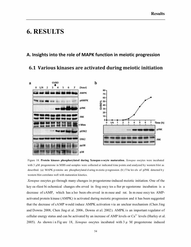
Results
54
6. RESULTS
A. Insights into the role of MAPK function in meiotic progression
6.1 Various kinases are activated during meiotic initiation
Xenopus oocytes go through many changes in progesterone-induced meiotic initiation. One of the
key ea rliest bi ochemical changes obs erved in frog oocy tes a fter pr ogesterone incubation is a
decrease of cAMP, which has a lso been obs erved in m ouse and rat. In m ouse oocy tes AMP-
activated protein kinase (AMPK) is activated during meiotic progression and it has been suggested
that the decrease of cAMP would induce AMPK activation via an unclear mechanism (Chen Jing
and Downs 2008, Chen Jing et al . 2006, Downs et al. 2002). AMPK is an important regulator of
cellular energy status and can be activated by an increase of AMP levels or Ca2+ levels (Hurley et al.
2005). As shown i n Fig ure 18, Xenopus oocytes incubated with 3 µ M progesterone induced
Figure 18. Protein kinases phosphorylated during Xenopus o ocyte maturation. Xenopus oocytes were incubated
with 3 µM progesterone in MBS and samples were collected at indicated time points and analyzed by western blot as
described. (a) MAPK p roteins are phosphorylated d uring m eiotic p rogression. (b ) T he lev els of pJNK detected b y
western blot correlates well with maturation kinetics.
a b
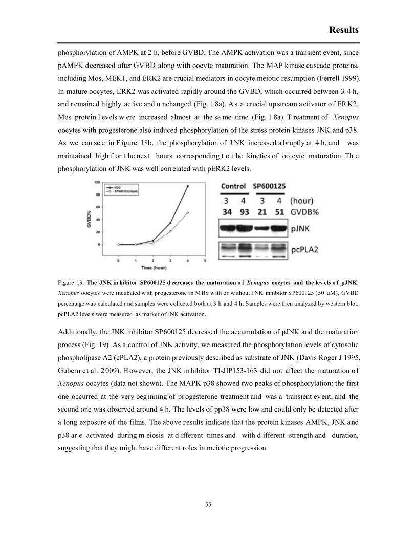
Results
55
phosphorylation of AMPK at 2 h, before GVBD. The AMPK activation was a transient event, since
pAMPK decreased after GVBD along with oocyte maturation. The MAP kinase cascade proteins,
including Mos, MEK1, and ERK2 are crucial mediators in oocyte meiotic resumption (Ferrell 1999).
In mature oocytes, ERK2 was activated rapidly around the GVBD, which occurred between 3-4 h,
and r emained h ighly active and u nchanged (Fig. 1 8a). As a crucial upstream activator o f ERK2,
Mos protein l evels w ere increased almost at the sa me time (Fig. 1 8a). T reatment of Xenopus
oocytes with progesterone also induced phosphorylation of the stress protein kinases JNK and p38.
As we can se e in F igure 18b, the phosphorylation of J NK increased a bruptly at 4 h, and was
maintained high f or t he next hours corresponding t o t he kinetics of oo cyte maturation. Th e
phosphorylation of JNK was well correlated with pERK2 levels.
Additionally, the JNK inhibitor SP600125 decreased the accumulation of pJNK and the maturation
process (Fig. 19). As a control of JNK activity, we measured the phosphorylation levels of cytosolic
phospholipase A2 (cPLA2), a protein previously described as substrate of JNK (Davis Roger J 1995,
Gubern e t al . 2009). H owever, the JNK inhibitor TI-JIP153-163 did not affect the maturation of
Xenopus oocytes (data not shown). The MAPK p38 showed two peaks of phosphorylation: the first
one occurred at the very beg inning of pr ogesterone treatment and was a transient event, and the
second one was observed around 4 h. The levels of pp38 were low and could only be detected after
a long exposure of the films. The above results indicate that the protein kinases AMPK, JNK and
p38 ar e activated during m eiosis at d ifferent times and with d ifferent strength and duration,
suggesting that they might have different roles in meiotic progression.
Figure 19. The JNK in hibitor SP600125 d ecreases the maturation o f Xenopus oocytes and the lev els o f pJNK.
Xenopus oocytes were incubated with progesterone in MBS with or without JNK inhibitor SP600125 (50 µM), GVBD
percentage was calculated and samples were collected both at 3 h and 4 h. Samples were then analyzed by western blot.
pcPLA2 levels were measured as marker of JNK activation.

Results
56
6.2 Constitutively active MEKK1 accelerates progesterone-
induced meiotic resumption
It is well known that the upstream protein kinase MEKK1 activates JNK and p38 (Davis Roger J
2000b, Wagner and Nebreda 2009). In order to investigate the role of the stress protein kinases JNK
and p38 in meiotic progression, we injected a cRNA of a constitutively active mutant of MEKK1
(MEKK1Δ) into Xenopus oocytes t o ac tivate JNK and p38 pr oteins. If JNK a nd p38 s ignaling
pathways play any r ole in m eiotic progression, w e should expect t hat ectopic expression o f
MEKK1Δ in Xenopus oocytes would modify the kinetics of maturation induced by progesterone.
As shown in f igure 20a, expression of MEKK1Δ accelerated m eiotic m aturation compared w ith
water-injected oocytes o r w ith oocy tes expressing a catalytically in active mutant of ME KK1
(MEKK1-KM). Both protein kinases (MEKK1 and MEKK1-KM) were efficiently expressed in the
oocytes (Fig. 20b, myc blot) and MEKK1Δ induced the phosphorylation of JNK, p38 and ERK,
whereas MEKK1-KM did not activate any of these signaling pathways, as expected. Activation of
JNK and p38 by MEKK1Δ was also confirmed by an increase of cPLA2 phosphorylation, which
has been reported to be a substrate for both protein kinases (Davis Roger J 1995, Gubern et al. 2009,
Figure 20. Constitutively a ctive M EKK1 ( MEKK1Δ) accelerates p rogesterone-induced ooc yte m aturation.
Xenopus oocytes were in jected w ith c atalytically in active M EKK1 (M EKK1-KM), c onstitutively a ctive M EKK1
(MEKK1Δ) or water, and trea ted w ith p rogesterone 1 8 h late r. GVBD percentage was calculated a nd s amples w ere
collected ev ery h our. Samples w ere t hen an alyzed b y western b lot. (a) MEKK1Δ speeds up meiotic p rogression. ( b)
MEKK1Δ activates MAPK family proteins without any treatment and progesterone treatment induces XpJNK-p42.
b
a
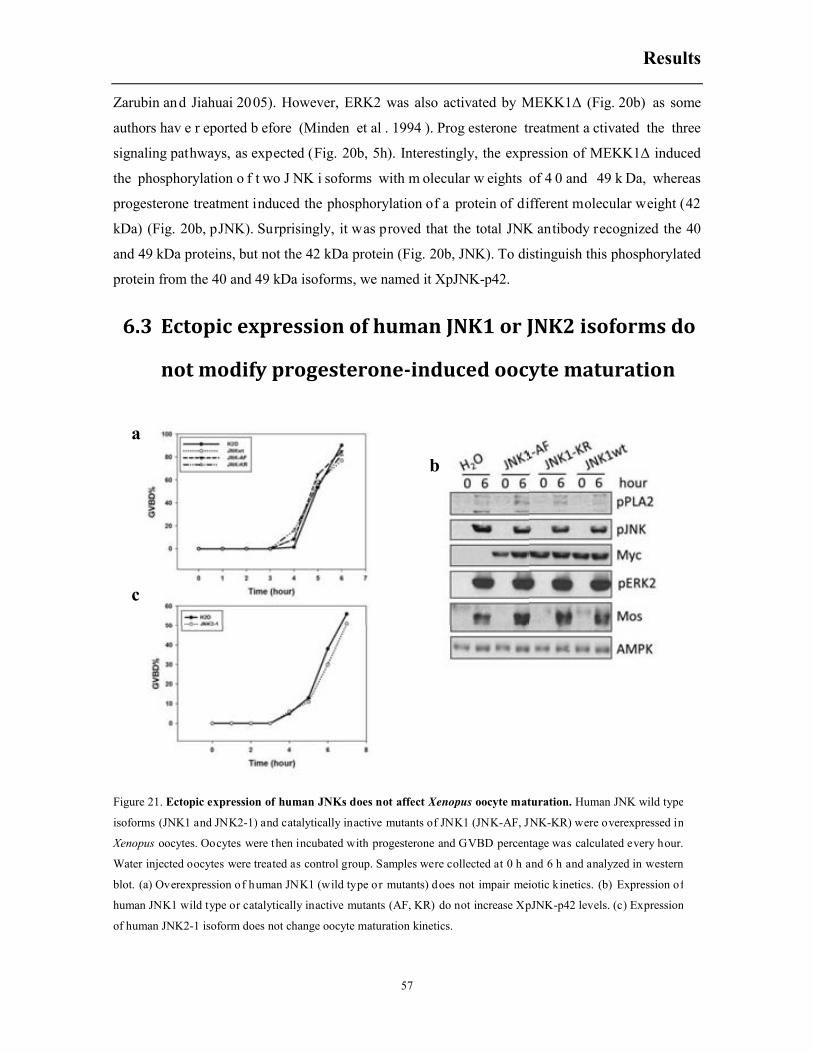
Results
57
Zarubin and Jiahuai 2005). However, ERK2 was also activated by MEKK1Δ (Fig. 20b) as some
authors hav e r eported b efore (Minden et al . 1994 ). Prog esterone treatment a ctivated the three
signaling pathways, as expected (Fig. 20b, 5h). Interestingly, the expression of MEKK1Δ induced
the phosphorylation o f t wo J NK i soforms with m olecular w eights of 4 0 and 49 k Da, whereas
progesterone treatment induced the phosphorylation of a protein of different molecular weight (42
kDa) (Fig. 20b, pJNK). Surprisingly, it was proved that the total JNK antibody recognized the 40
and 49 kDa proteins, but not the 42 kDa protein (Fig. 20b, JNK). To distinguish this phosphorylated
protein from the 40 and 49 kDa isoforms, we named it XpJNK-p42.
6.3 Ectopic expression of human JNK1 or JNK2 isoforms do
not modify progesterone-induced oocyte maturation
a
b
c
Figure 21. Ectopic expression of human JNKs does not affect Xenopus oocyte maturation. Human JNK wild type
isoforms (JNK1 and JNK2-1) and catalytically inactive mutants of JNK1 (JNK-AF, JNK-KR) were overexpressed in
Xenopus oocytes. Oocytes were then incubated with progesterone and GVBD percentage was calculated every hour.
Water injected oocytes were treated as control group. Samples were collected at 0 h and 6 h and analyzed in western
blot. (a) Overexpression of human JNK1 (wild type or mutants) does not impair meiotic kinetics. (b) Expression of
human JNK1 wild type or catalytically inactive mutants (AF, KR) do not increase XpJNK-p42 levels. (c) Expression
of human JNK2-1 isoform does not change oocyte maturation kinetics.
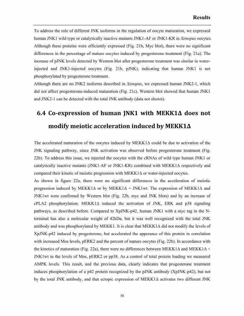
Results
58
To address the role of different JNK isoforms in the regulation of oocyte maturation, we expressed
human JNK1 wild type or catalytically inactive mutants JNK1-AF or JNK1-KR in Xenopus oocytes.
Although these proteins were efficiently expressed (Fig. 21b, Myc blot), there were no significant
differences in the percentage of mature oocytes induced by progesterone treatment (Fig. 21a). The
increase of pJNK levels detected by Western blot after progesterone treatment was similar in water-
injected and JNK1-injected oocytes (Fig. 21b, pJNK), indicating that human JNK1 is not
phosphorylated by progesterone treatment.
Although there are no JNK2 isoforms described in Xenopus, we expressed human JNK2-1, which
did not affect progesterone-induced maturation (Fig. 21c). Western blot showed that human JNK1
and JNK2-1 can be detected with the total JNK antibody (data not shown).
6.4 Co-expression of human JNK1 with MEKK1Δ does not
modify meiotic acceleration induced by MEKK1Δ
The accelerated maturation of the oocytes induced by MEKK1Δ could be due to activation of the
JNK signaling pathway, since JNK activation was observed before progesterone treatment (Fig.
22b). To address this issue, we injected the oocytes with the cRNAs of wild type human JNK1 or
catalytically inactive mutants (JNK1-AF or JNK1-KR) combined with MEKK1Δ respectively and
compared their kinetic of meiotic progression with MEKK1Δ or water-injected oocytes.
As shown in figure 22a, there were no significant differences in the acceleration of meiotic
progression induced by MEKK1Δ or by MEKK1Δ + JNK1wt. The expression of MEKK1Δ and
JNK1wt were confirmed by Western blot (Fig. 22b, myc and JNK blots) and by an increase of
cPLA2 phosphorylation. MEKK1Δ induced the activation of JNK, ERK and p38 signaling
pathways, as described before. Compared to XpJNK-p42, human JNK1 with a myc tag in the N-
terminal has also a molecular weight of 42kDa, but it was well recognized with the total JNK
antibody and was phosphorylated by MEKK1. It is clear that MEKK1Δ did not modify the levels of
XpJNK-p42 induced by progesterone, but accelerated the apperance of this protein in correlation
with increased Mos levels, pERK2 and the percent of mature oocytes (Fig. 22b). In accordance with
the kinetics of maturation (Fig. 22a), there were no differences between MEKK1Δ and MEKK1Δ +
JNK1wt in the levels of Mos, pERK2 or pp38. As a control of total protein loading we measured
AMPK levels. This result, and the previous data, clearly indicates that progesterone treatment
induces phosphorylation of a p42 protein recognized by the pJNK antibody (XpJNK-p42), but not
by the total JNK antibody, and that ectopic expression of MEKK1Δ activates two different JNK
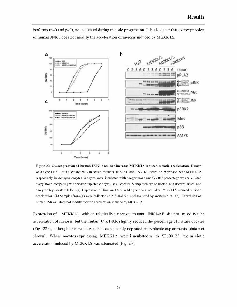
Results
59
isoforms (p40 and p49), not activated during meiotic progression. It is also clear that overexpression
of human JNK1 does not modify the acceleration of meiosis induced by MEKK1Δ.
Expression of MEKK1Δ with ca talytically i nactive mutant JNK1-AF did not m odify t he
acceleration of meiosis, but the mutant JNK1-KR slightly reduced the percentage of mature oocytes
(Fig. 22c), although t his result w as no t co nsistently r epeated in replicate exp eriments (data n ot
shown). When oocytes expr essing MEKK1Δ were i ncubated w ith SP600125, the m eiotic
acceleration induced by MEKK1Δ was attenuated (Fig. 23).
Figure 22. Overexpression of human JNK1 does not increase MEKK1Δ-induced meiotic acceleration. Human
wild t ype J NK1 or it s catalytically in active mutants JNK-AF and J NK-KR were co-expressed with M EKK1Δ
respectively in Xenopus oocytes. O ocytes were incubated with p rogesterone and GVBD percentage was cal culated
every hour comparing w ith w ater injected o ocytes as a control. S amples w ere co llected at d ifferent times and
analyzed b y western b lot. (a) Expression of hum an J NK1wild t ype doe s not alter MEKK1Δ-induced m eiotic
acceleration. (b) Samples from (a) were collected at 2, 3 and 6 h, and analyzed by western blot. (c) Expression of
human JNK-AF does not modify meiotic acceleration induced by MEKK1Δ.
a b
c

Results
60
6.5 Constitutively active MKK7 does not accelerate meiotic
progression
To address more specifically the role of JNK cascade in progesterone-induced meiotic progression,
we m icroinjected a w ild type MKK 7, a c onstitutively active m utant (MKK7-DED), or t wo
catalytically i nactive mutants ( MKK7-KM, M KK7-3A) and treated the oocytes w ith 3 µ M
progesterone.
As shown in Figure 24a, the oocytes injected with wild type or mutants MKK7 matured with the
same kinetics that of water-injected oocytes. The constitutively active mutant MKK7-DED induced
the phosphorylation of t he p40 and p49 J NK i soforms, but di d n ot activate the ERK or the p38
signaling pat hways (Fig. 24b). T he p40 and p49 JNK isoforms activated by MKK7 were also
activated by hy perosmotic shock (Fig. 2 4b), as p reviously r eported (Martiáñez et a l. 2009).
Recently, we have characterized these isoforms as Xenopus JNK1-1 and JNK1-2, which differ in
the C-terminal domain obtained by di fferential spl icing of t he JNK1 gene ( Messaoud NB, These
Dissertation, 20 14). Progesterone t reatment induced t he phosphorylation of XpJNK-p42, clearly
different to t he p40 and p49 J NK i soforms. Furthermore, co-overexpression o f MKK 7-DED and
Xenopus JNK1-1 or JNK1-2 did not increase progesterone-induced oocyte maturation, but on the
contrary decreased it (Fig. 24c).
Figure 23. The JNK inhibitor SP600125 attenuates MEKK1Δ-induced acceleration of m eiosis. Xenopus oocytes
expressing MEKK1Δ were treated with progesterone in the presence or absence of the JNK inhibitor SP600125 (50μM).
GVBD percentage was calculated every hour.
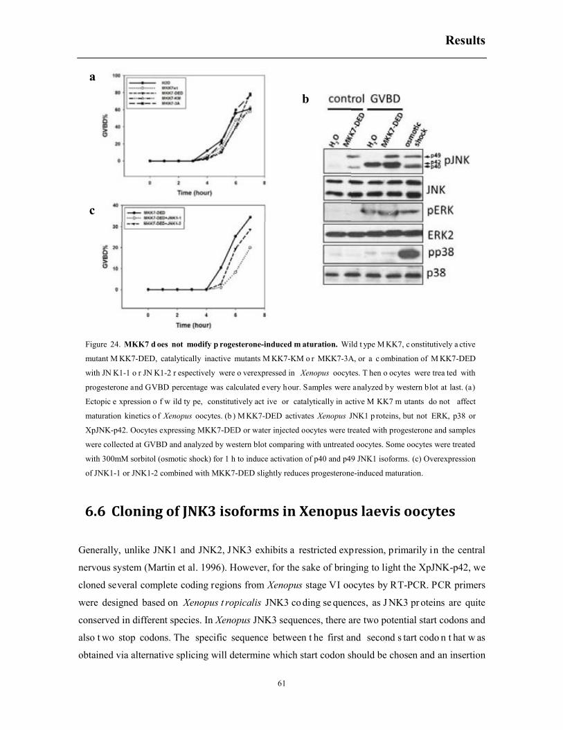
Results
61
6.6 Cloning of JNK3 isoforms in Xenopus laevis oocytes
Generally, unlike JNK1 and JNK2, JNK3 exhibits a restricted expression, primarily in the central
nervous system (Martin et al. 1996). However, for the sake of bringing to light the XpJNK-p42, we
cloned several complete coding regions from Xenopus stage VI oocytes by RT-PCR. PCR primers
were designed based on Xenopus t ropicalis JNK3 co ding se quences, as J NK3 pr oteins are quite
conserved in different species. In Xenopus JNK3 sequences, there are two potential start codons and
also t wo stop codons. The specific sequence between t he first and second s tart codo n t hat w as
obtained via alternative splicing will determine which start codon should be chosen and an insertion
Figure 24. MKK7 d oes not modify p rogesterone-induced m aturation. Wild t ype M KK7, c onstitutively a ctive
mutant M KK7-DED, catalytically inactive mutants M KK7-KM o r MKK7-3A, or a c ombination of M KK7-DED
with JN K1-1 o r JN K1-2 r espectively were o verexpressed in Xenopus oocytes. T hen o ocytes were trea ted with
progesterone and GVBD percentage was calculated every hour. Samples were analyzed by western blot at last. (a)
Ectopic e xpression o f w ild ty pe, constitutively act ive or catalytically in active M KK7 m utants do not affect
maturation kinetics o f Xenopus oocytes. (b ) M KK7-DED activates Xenopus JNK1 p roteins, but not ERK, p38 or
XpJNK-p42. Oocytes expressing MKK7-DED or water injected oocytes were treated with progesterone and samples
were collected at GVBD and analyzed by western blot comparing with untreated oocytes. Some oocytes were treated
with 300mM sorbitol (osmotic shock) for 1 h to induce activation of p40 and p49 JNK1 isoforms. (c) Overexpression
of JNK1-1 or JNK1-2 combined with MKK7-DED slightly reduces progesterone-induced maturation.
a
b
c

Results
62
of a 5-nucleotide before the stop codon which also results from alternative splicing will determine
which stop codon would be selected. In other words, exon al ternative selection indicates the start
and stop of the coding region, giving to different JNK3 isoforms (Fig. 25a).
Sense and antisense primers were designed based on sequence before the first start codon or af ter
the last codon of Xenopus tropicalis JNK3. Ten complete-coding-region clones were sequenced and
four JNK3 i soforms were obt ained, w hich dem onstrates that Xenopus l aevis stage V I oocytes
contain at least four poly A JNK3 transcripts obtained by alternative splicing. Subsequently, these
four JNK3 isoforms were subcloned to vector FTX5, adding a Myc tag to the N-terminal. Sequence
Figure 25. Four JNK3 isoforms were cloned from Xenopus laevis oocytes. In order to clone JNK3 isoforms from
Xenopus laevis, reverse transcription was performed using oligo (dT) as primer. PCR primers were designed based on
conserved sequences in Xenopus tropicalis JNK3 transcripts with the sense and antisense primers residing before start
codon or stop codon respectively a s depicted above. Finally, 10 PCR f ragments were sequenced and four d ifferent
JNK3 isoforms were detected. (a) Schematic representation of Xenopus JNK3 isoforms. (b) JNK3-1 is quite conserved
between human and Xenopus except for a 19aa insertion (square box) in Xenopus isoforms. (c) Four JNK3 isoforms
are reported in humans and four were cloned in Xenopus laevis by RT-PCR. The table indicates the molecular weight
of the predicted proteins obtained.
H. sapiens X. laevis
JNK3-1 422AA 441AA
JNK3-2 464AA 483AA
JNK3-3 426AA 445AA
JNK3-4 277AA /
/ 403AA
a
b
c

Results
63
alignment of JNK3-1 shown that JNK3 proteins are quite conserved between human and Xenopus
laevis, the only specificity of Xenopus proteins is that a distinct 19 amino acids insert was detected
which leads to a higher molecular weight than human proteins (Fig.25b and c).
6.7 Ectopic expression of JNK3 isoforms does not affect
meiotic progression in Xenopus oocytes
To confirm their characteristics and function in response to progesterone and osmotic shock, these
four J NK3 i soforms with myc tag were ectopically expressed i n Xenopus oocytes. 1 8 h after
Figure 26. JNK3 proteins are not involved in meiotic progression. Xenopus JNK3-1, 3-2, 3-3, 3-4 with Myc tag or
JNK3-1, 3-4 without Myc tag were overexpressed in Xenopus oocytes. Then oocytes were treated with progesterone or
300 mM so rbitol (osmotic shock). GVBD percentage was calculated in th e oocytes trea ted with p rogesterone every
hour. Samples were collected at GVBD or 1 h after osmotic shock and analyzed by western blot. (a) Overexpression of
different Xenopus JNK3 iso forms with M yc t ag in th e N -terminal do not modify o ocyte maturation k inetics. ( b)
Xenopus JNK3 i soforms are not phosphorylated dur ing oocyte m aturation, but are phosphorylated by hyperosmotic
shock. (c) Overexpression of JNK3 isoforms 3-1 and 3-4 without Myc tag do not alter meiotic progression. (d) JNK3-1
and J NK3-4 without myc tag a re d etected b y western b lot with total JNK antibody and d o not modify X pJNK-p42
levels in progesterone treated oocytes.
a b
c d

Results
64
microinjection of the corresponding cRNAs, the oocytes were treated either with 3 µg progesterone
or 300 mM sorbitol in MBS. As shown in figure 26a, neither of these four JNK3 isoforms modified
the kinetics of oocyte maturation. Western blot analysis indicated that none of the isoforms were
phosphorylated at GVBD, although the proteins were well expressed (Fig. 26b). In contrast, the four
JNK3 i soforms were phosphorylated in r esponse t o hyperosmotic shock, like Xenopus JNK1
isoforms and can be recognized by western blot with the JNK antibody (Fig. 26b).
Considering that t he Myc tag may impair the molecular st ructure of J NK3 and that J NK3-1 and
JNK3-4 hav e very si milar molecular w eights to JNK1-2 (p49) and X pJNK-p42 respectively, w e
cloned JNK3-1 and JNK3-4 without a Myc tag at the N-terminal. Expression of JNK3-1 or JNK3-4
without My c tag did not af fect oocyte meiotic progression ( Fig. 26c). Moreover, J NK3-1 and
JNK3-4 w ithout t ags did not m odify t he levels of exp ression of X pJNK-p42 i nduced by
progesterone, although bo th i soforms were well detected by w estern bl ot w ith t he total JNK
antibody (Fig. 26d). Since total JNK antibody detected by western blot all the four overexpressed
JNK3 isoforms, it seems that there is no expression, or tiny amounts, of endogenous JNK3 proteins
in cytosolic extracts of stage VI oocytes, although the mRNAs are present.
6.8 XpJNK-p42 is a posttranslational modification of
pERK2 induced by progesterone
To reveal the true feature of XpJNK-p42, crosslinking immunoprecipitation and mass spectrometry
analysis were performed. Denaturing protein immunoprecipitation with pJNK antibody showed that
Figure 27. The affinity of pJNK to XpJNK-p42 is weak. In denaturing immunoprecipitation, 0.5µl pJNK antibodies
were incubated with 100 µl denatured IP lysate overnight, and 20µl sepharose G proteins were added to the lysate the
next day. The mixture was incubated for 2 h with gentle up-and-down shake. Two repeats were performed. F or one
repeat, the sepharose G proteins were then washed 3 times (IP1) and another 5 times (IP2). The sepharose G proteins
were boiled in 20µl 1X Laemmli sample buffer and analyzed by western blot.

Results
65
its a ffinity to X pJNK-p42 w as very w eak (Fig. 27). To co llect eno ugh sa mple for mass
spectrometry analysis, elution of many crosslink immunoprecipitation reactions were pooled, and
concentrated by centrifugal vacuum. The final sample was run in a 10% polyacrylamide-SDS gel
and s tained w ith silver; the band corresponding t o 42k Da was cu t a nd a nalyzed by mass
spectrometry.
Mass sp ectrometry an alysis shown that X pJNK-p42 may be pERK2 (pMAPK1). In MS analysis
report, 18 un ique peptides of mitogen-activated protein k inase1 (ERK2) c overing 46.81% of t he
whole protein were recognized (Table 10).
To p rove that p hosphorylated ER K2 is r ecognized b y pJNK antibody we c loned and expressed
ERK2 in Xenopus oocytes. Using t otal RNA f rom st age VI oocy tes, we cloned by T R-PCR two
different sequences corresponding to ERK2, which we named ERK2a and ERK2b that are different
only i n a f ew amino ac ids. These two isoforms were cloned i nto F TX5 plasmid and in v itro
transcribed i nto cRNAs that were i njected and ex pressed i n Xenopus oocytes. Sinc e the proteins
expressed contain a Myc tag, they have a higher molecular weight than endogenous ERK2.
Either p rogesterone treatment or osm otic sho ck t reatment induced the phosphorylation o f
endogenous ER K2 and e ctopically expr essed ERK2a and ER K2b ( Fig. 2 8a). Intriguingly, ER K2
phosphorylated at GVBD induced by progesterone was also recognized by pJNK antibody but not
ERK2 phosphorylated by osmotic shock. Only the endogenous p49 and p40 JNK1 isoforms were
detected by pJNK antibody in oocytes treated with osmotic shock (Fig. 28a). This implied that the
protein recognized by pJNK antibody is pERK2 and that some posttranslational modification occurs
a b
Figure 28. The XpJNK-p42 i nduced by pr ogesterone is confirmed to b e pERK2. Both wild type ERK2a and
ERK2b or mutants ERK T 184S and T193S of ERK2b were overexpressed in Xenopus oocytes. Subsequently these
oocytes were trea ted with p rogesterone o r 300 mM so rbitol (osmotic sh ock). S amples o f c ontrol oocytes, m ature
oocytes, a nd 3 00 m M so rbitol treated o ocytes (2.5 h ) w ere collected a nd a nalyzed b y western b lot. (a ) p ERK2
induced by progesterone can be recognized by pJNK antibody but pERK2 induced by osmotic shock can not . (b )
Mutations of Thr184 or Thr193 to Ser of wild type ERK2b do not affect its phosphorylation and detection by pJNK
antibody.

Results
66
after progesterone treatment but not after osmotic shock treatment, which allows ERK2 to be
recognized by pJNK antibody.
Potential phosphorylation sites (T184 and T193) in ERK2b around the TEY motif (170-
FGLARVADPDHDHT(184)GFLTEYVAT(193)RWYRAPEIMLNSKGYTK -210) were mutated
to Ser to scan for potential phosphorylation sites that may impair the recognition by pJNK antibody.
Unfortunately, these mutations did not affect the recognition of ectopic ERK2 by pJNK antibody
(Fig. 28b). Therefore, the modification induced by progesterone in ERK2 that allows recognition
by pJNK antibody needs farther studies.

Results
67
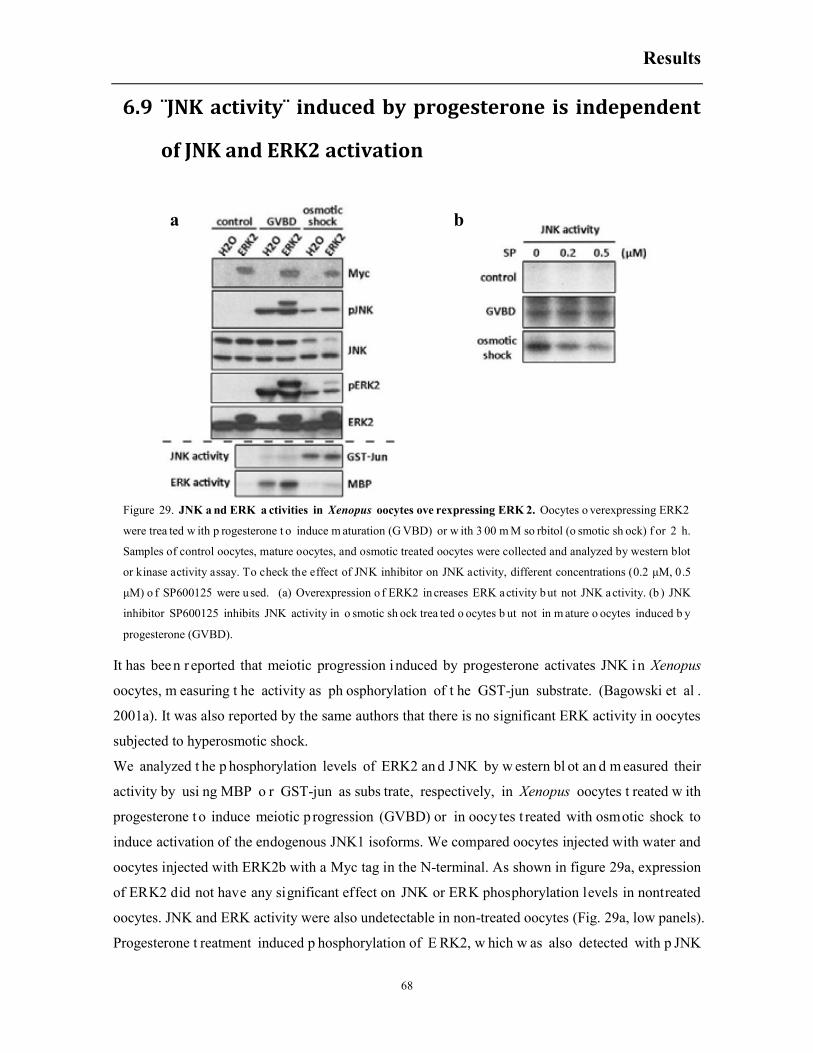
Results
68
6.9 ¨JNK activity¨ induced by progesterone is independent
of JNK and ERK2 activation
It has bee n r eported that meiotic progression i nduced by progesterone activates JNK in Xenopus
oocytes, m easuring t he activity as ph osphorylation of t he GST-jun substrate. (Bagowski et al .
2001a). It was also reported by the same authors that there is no significant ERK activity in oocytes
subjected to hyperosmotic shock.
We analyzed t he p hosphorylation levels of ERK2 an d J NK by w estern bl ot an d m easured their
activity by usi ng MBP o r GST-jun as subs trate, respectively, in Xenopus oocytes t reated w ith
progesterone t o induce meiotic p rogression (GVBD) or in oocytes t reated with osmotic shock to
induce activation of the endogenous JNK1 isoforms. We compared oocytes injected with water and
oocytes injected with ERK2b with a Myc tag in the N-terminal. As shown in figure 29a, expression
of ERK2 did not have any significant effect on JNK or ERK phosphorylation levels in nontreated
oocytes. JNK and ERK activity were also undetectable in non-treated oocytes (Fig. 29a, low panels).
Progesterone t reatment induced p hosphorylation of E RK2, w hich w as also detected with p JNK
Figure 29. JNK a nd ERK a ctivities in Xenopus oocytes ove rexpressing ERK 2. Oocytes o verexpressing ERK2
were trea ted w ith p rogesterone t o induce m aturation (G VBD) or w ith 3 00 m M so rbitol (o smotic sh ock) f or 2 h.
Samples of control oocytes, mature oocytes, and osmotic treated oocytes were collected and analyzed by western blot
or kinase activity assay. To check the effect of JNK inhibitor on JNK activity, different concentrations (0.2 μM, 0.5
μM) o f SP600125 were u sed. (a) Overexpression o f ERK2 in creases ERK activity b ut not JNK activity. (b ) JNK
inhibitor SP600125 inhibits JNK activity in o smotic sh ock trea ted o ocytes b ut not in m ature o ocytes induced b y
progesterone (GVBD).
a b
Results
69
antibodies, and a significant ERK activity, incremented in the oocytes expressing ectopic ERK2, but
very low levels of JNK activity were observed in progesterone treated oocytes with or without
expression of ectopic ERK2. On the contrary, osmotic shock induced phosphorylation of ERK2 and
JNK1 and high levels of JNK activity, which was not incremented by ERK2b ectopic expression
(Fig. 29a, low panel). The level of ERK2 activity induced by hyperosmotic shock was low and
slightly increased by ERK2 ectopic expression. Of note, the p49 JNK1 isoform is not observed in
the oocytes treated with osmotic shock, since p49 is proteolyzed when caspase-3 is activated by
osmostress (see next section). In conclusion, JNK activity induced by progesterone is very low in
Xenopus oocytes and independent of ERK2 activity.
Additionally, as shown in figure 29b, the JNK inhibitor SP600125 clearly inhibited the JNK activity
induced by osmotic shock but not the JNK activity detected in mature oocytes, indicating that the
GST-jun phosphorylation (JNK activity) induced by progesterone is JNK independent.
6.10 ERK signaling pathway inhibitors decrease
MEKK1Δ-induced acceleration of meiosis
ERK signaling pathway is a classic cascade in regulating meiotic resumption. As described above,
the protein kinase MEKK1Δ activates ERK2 without progesterone treatment. We used inhibitors of
the ERK signaling pathway, U0126 or FR180204, to evaluate the contribution of ERK2 to
MEKK1Δ-induced acceleration of meiosis. U0126 is a selective inhibitor of MAP kinase kinases,
MEK1 and MEK2 which prevents the activation of ERK proteins (Favata et al. 1998). FR180204 is
a selective and cell-permeable inhibitor of ERK proteins. The compound inhibits ERK1/2 in an
ATP competitive manner and blocks their activities (Ohori et al. 2005). When oocytes were treated
with the inhibitor U0126 or FR180204 18 h after microinjection of MEKK1Δ, there were no
differences in maturation kinetics compared with MEKK1Δ injected oocytes without inhibitors (Fig.
30a). However, when the oocytes were pre-incubated for 1 h with the corresponding inhibitors
before microinjection and also maintained in the medium after MEKK1Δ injection, there was a
significant decrease in maturation in the oocytes incubated with U0126 and nearly a complete
inhibition of the oocytes incubated with FR180204 (Fig. 30b).

Results
70
MEKK1Δ expression i nduced phos phorylation of E RK2, w hich was completely bl ocked by pre-
incubation with inhibitor U0126 (Fig. 30c). Not surprisingly, ERK2 was phosphorylated even in the
presence of the inhibitor FR180204. These results indicate that a proper ERK function is crucial for
oocyte maturation and that ER K s ignaling pathway ac tivation co ntributes to MEKK1Δ-induced
acceleration of meiosis.
Figure 30. Inhibitors of th e ERK si gnaling p athway in hibitors decrease MEKK1Δ-induced a cceleration o f
meiosis. Xenopus oocytes were treated w ith in hibitors of th e E RK signaling p athway a fter or before MEKK1Δ
injection. T hese o ocytes we re trea ted w ith p rogesterone 18 h a fter the i njection. O ocyte mature p ercentage w as
calculated every one hour and samples of control oocytes and oocytes 4 h or 5 h after progesterone treatment were
collected and analyzed by western blot. (a) Oocytes incubated with inhibitors U0126 or FR180204 ( 50 μM) 18 h
after microinjection o f MEKK1Δ do n ot have d ifferent maturation k inetics to th at of MEKK1Δ injected o ocytes
without in hibitors. (b) Oocytes p re-incubated f or 1 h w ith t he inhibitors U0126 or FR180204 (50 μM) and
maintained i n t he m edium af ter MEKK1Δ injection have r educed m aturation t hat MEKK1Δ injected o ocytes
without in hibitors. (c) W estern b lot shown th at inhibitor U0 126 b locks phosphorylation o f ERK2 induced by
MEKK1Δ, bu t not induced b y p rogesterone trea tment in MEKK1Δ injected oocytes. (d ) U0 126 partially b locks
ERK2 phosphorylation recognized by pERK antibodies and completely b locks ERK2 phosphorylation recognized
by pJNK antibodies in Xenopus oocyte maturing process induced by progesterone.
a b
c
d

Results
71
Interestingly, ERK2 was phosphorylated in the presence of the inhibitor U0126 at 5 h after
progesterone treatment (Fig. 30c), implying that a MEK1/2 independent signaling pathway may be
responsible for ERK2 phosphorylation. In non-injected oocytes, the phosphorylation of ERK2 is
markedly reduced by incubation with U0126, but not completely blocked, pointing also to a
MEK1/2 independent pathway of phosphorylation (Fig. 30d). What is clear is that the levels of
pERK2 (XpJNK-p42) detected with pJNK antibodies correlate quite well with the percent of mature
oocytes (GVBD), whereas this is not true for pERK2 levels detected with pERK antibodies (Fig.
30c and d).
We proposed that pERK antibodies recognizes both early and late phosphorylation of ERK2,
whereas pJNK antibodies recognize only late phosphorylation of ERK2, highly associated to
GVBD. Of note, it is surprising that the inhibitor FR180204 increased the phosphorylation of ERK2
detected by pJNK antibodies at time 0 h (Fig. 30c). We do not have an explanation for the presence
of this band in the western blot. More experiments should be performed to confirm this results and
to discard that this band is not a result of JNK1-2 proteolysis, which could appear in apoptotic
oocytes (see next section).
6.11 The role for p38 isoforms in regulating meiotic
progression
It has been reported that ectopic expression of constitutively active MKK6-DD, an upstream kinase
of p38, accelerates meiotic maturation through activation of 38γ (Perdiguero et al. 2003). Since p38
was activated by MEKK1Δ, we addressed the role of p38γ in the MEKK1Δ-induced acceleration of
meiosis. As shown in figure 29a, expression of a dominant negative p38γ mutant (p38γ-DA)
reduced significantly the meiotic acceleration caused by MEKK1Δ. We also addressed the role of
p38 isoforms in combination with MKK6-DD, a more specific activator of the p38 signaling
pathways that does not activate the JNK or ERK signaling pathways (data not shown). Co-
expression of MKK6-DD with different p38 isoforms accelerated oocyte maturation to various
extents depending on the combination. When p38γ and MKK6-DD were co-overexpressed in
Xenopus oocytes, the oocytes matured spontaneously without progesterone stimulation as reported
previously (Perdiguero et al. 2003). For the other p38 isoforms expressed in combination with
MKK6-DD, we observed a ranking of acceleration of meiosis as follow p38δ>p38β> p38α (Fig.
31b).

Results
72
However, when the wild type p38 isoforms or their mutants, were overexpressed, the maturation of
the oocytes induced by progesterone was not affected (Fig. 32a and 32b). Western Blot indicated
that a ll pr oteins w ere well expr essed (Fig. 32c, My c blot). T he p38 t otal antibodies co uld only
recognize p38α and p38γ isoforms (Fig. 32 c), b ut a ll p p38 i soform ca n be r ecognized by pp38
antibodies as d escribed before i n our l aboratory ( Messaoud N B, Thesis Dissertation, 201 4). As
shown in figure 32c, overexpression of p38α and p38β slightly increased the basal phosphorylation
state at time 0 h. However, progesterone treatment for 8 h did not induce any phosphorylation of the
p38 isoforms (Fig. 32c). These results pose some doubts about the relevance of p38 isoforms in the
regulation of meiotic progression (see discussion).
Figure 31. p38 proteins are involved in MEKK1Δ/MKK6-DD induced meiotic acceleration. Constitutively active
MEKK1Δ or MKK6-DD were o verexpressed i n Xenopus oocytes c ombined w ith dominant ne gative p38γ mutant
(p38γ-DA) or with different p38 wild type isoforms (p38α, β, δ, γ). Then the oocytes were treated with progesterone
and G VBD p ercentage w as calculated e very o ne h our. (a ) C atalytically in active mutant p38γ-DA d ecreases
MEKK1Δ-induced meiotic acceleration. (b) Constitutively active MKK6-DD combined with p38 isoforms accelerates
meiotic progression.
a b

Results
73
B. Ins ights in to the func tion of JNK1-2 a nd Bcl-2 fa mily
members in the regulation of osmostress-induced apoptosis. Previous work i n our laboratory has cha racterized i n det ail t he time-course ev ents during
osmostress-induced apoptosis of Xenopus oocytes and the role of st ress protein kinases, calpains,
caspases, Sm ac/DIABLO and cytochrome c i n t he cell d eath p rogram (Messaoud NB, Thes is
Dissertation, 2014). Four independent pathways early activated by hyperosmotic shock regulate late
cytochrome c release and caspase-3 activation: p38, JNK, calpains and Smac/DIABLO release. We
have proposed t hat osm ostress-induced ap optosis is basically a program of t hree ac ts: 1 ) r apid
release of Sm ac/DIABLO and calpain a ctivation, with progressive JNK/p38 phos phorylation
followed by, 2) m aximum and sustained JNK/p38 ac tivity i n coope ration w ith ca lpains and
a b
c
Figure 32. p38 MAPKs might not be implicated in meiotic progression. cRNAs of wild type p38 isoforms or their
catalytically inactive mutants we re in jected in to Xenopus oocytes and 1 8 h late r these o ocytes were trea ted w ith
progesterone, GVBD percentage was calculated every hour and samples of oocytes at 0h and 8h were collected and
analyzed by western blot. (a) Expression of wild type p38 isoforms or (b) their catalytically inactive mutants do not
affect progesterone induced oocyte maturation. ( c) No phosphorylated p38 proteins are detected after p rogesterone
treatment even though these proteins are efficiently expressed. The total p38 antibody recognizes only p38α and p38γ
isoforms.

Results
74
Smac/DIABLO t o i nduce the release o f cytochrome c and c aspase-3 ac tivation and f inally, 3 )
multiple f eedback l oops engaged by ca spase-3 ac tivation, including ca lpain activation and p38
phosphorylation, that w ould det ermine an irreversible death ( Messaoud N B, Thesis Dissertation,
2014). We hav e also characterized the p38 and JNK i soforms activated by hyperosmotic shock:
p38α, p38β, p38γ, JNK1-1, and JNK1-2, and we have demonstrated a c lear pro-apoptotic effect in
three isoforms: p38β , JNK1-1 and JNK1-2 (Messaoud NB, Thesis Dissertation, 2014). However,
we did not address the role of the Bcl-2 family members in the regulation of cytochrome c release.
Moreover, we realized that several hours after hyperosmotic shock the JNK1-2 isoform disappeared,
suggesting some kinds of proteolysis. Here, we characterized the mechanism of JNK1-2 proteolysis
induced by osmostress and i ts role i n apoptosis. In addition, we st udied the r ole of Bcl-2 f amily
members, especially Bid, in the regulation of osmostress-induced apoptosis.
6.12 Osmostress induces the proteolysis of JNK1-2 by
caspase-3 at Asp385 and engages a positive feedback
loop increasing the release of cytochrome c and
caspase-3 activation
a b
c
Figure 33. JNK1-2 was proteolyzed i n o smotic shock in duced a poptosis. Xenopus oocytes were trea ted in
sorbitol (300 m M/400 m M) and sa mples w ere c ollected a t indicated ti mes and a nalyzed b y w estern b lot. (a)
Proteolysis o f JNK1-2 induced by os motic shock. Western b lots at 3 h and 4 h were q uantified by d ensitometry.
Comparing the protein amount at 4 h to 3 h, the amount of JNK1-2 (p49) decreased about 40% while the amount of
JNK1-1 (p40) increased 40% correspondingly. (b) Quantification of JNK1-2 proteolysis. When oocytes were treated
by 300 m M s orbitol w ith t he presence of pa n c aspase i nhibitor Z -VAD.fmk, th e p roteolysis o f JN K1-2 wa s
inhibited. (c) JNK1-2 proteolysis is protected by Z-VAD.fmk.

Results
75
Hyperosmotic shock induces a rapid activation of two JNK isoforms (JNK1-1 p40, and JNK1-2 p49)
in Xenopus oocytes ( Messaoud NB, Thesis D issertation, 2014). I ncubation of the oocy tes w ith
300mM sorbitol during several hours induced the disappearance of JNK1-2 (p49), and an apparent
increase i n the b and co rresponding to JNK1-1 ( p40) ( Fig. 3 3a). Sev eral W estern b lots were
quantified by densitometry showing that decreases in JNK1-2 were well correlated with increases in
the band corresponding to JNK1-1 (Fig. 33b). The proteolysis of JNK1-2 was inhibited by the broad
caspase i nhibitor Z -VAD.fmk ( Fig. 3 3c), i ndicating t hat caspase-dependent c leavage of JNK1-2
resulted into a JNK1-1 like fragment.
Although there are no previous reports about the proteolysis of Xenopus JNKs by caspases, it has
been reported that human JNK1 and JNK2 are proteolyzed by caspase-3 (Enomoto et al. 2003). To
address the role of caspase-3 in the proteolysis of Xenopus JNK1-2 we microinjected oocytes with
cytochrome c, w hich has been reported t o ac tivate caspase-3 qui ckly (Bhuyan et al . 200 1). As
depicted in f ig. 34, cytochrome c m icroinjection initiated r apid caspase-3 activation and
disappearance of JNK1-2 (p49) with a corresponding increase of the JNK1-1 (p40) band. In contrast,
when the broad caspase i nhibitor Z -VAD.fmk or t he spe cific caspase-3 i nhibitor ( Z-DEVD.fmk)
was present in the medium, cytochrome c microinjection did not induce JNK1-2 proteolysis (Fig.
34). These data clearly indicate that JNK1-2 is a substrate of caspase-3 in vivo.
Caspase substrate cleavage sites scanning via an online software “Cascleave” (Song et a l. 2010)
indicated t hat t here a re two po tential caspase-3 cl eavage sites a t A sp385 and Asp412 at t he C-
terminal of J NK1-2 (Fig. 3 5a). Therefore, w e generated t he mutants JNK1-2D385A and J NK1-
Figure 34. Xenopus JNK1-2 is p roteolyzed
into a J NK1-1 li ke p rotein b y caspase-3.
Xenopus oocytes were in jected w ith M BS o r
cytochrome c ( 0.5μM, f inal c oncentration i n
the o ocyte) then th ese o ocytes w ere trea ted
with o smotic sh ock (3 00 m M so rbitol).
Samples at 1 h, 2 h , 4 h were collected and
analyzed b y western b lot. F or c ytochrome c
injection, so me o ocytes were p reincubated
with p an cas pase inhibitor Z -VAD.fmk o r
caspase-3 sp ecific inhibitor Z -DEVD.fmk f or
1 h , a nd m aintained d uring osmotic sh ock
treatment.
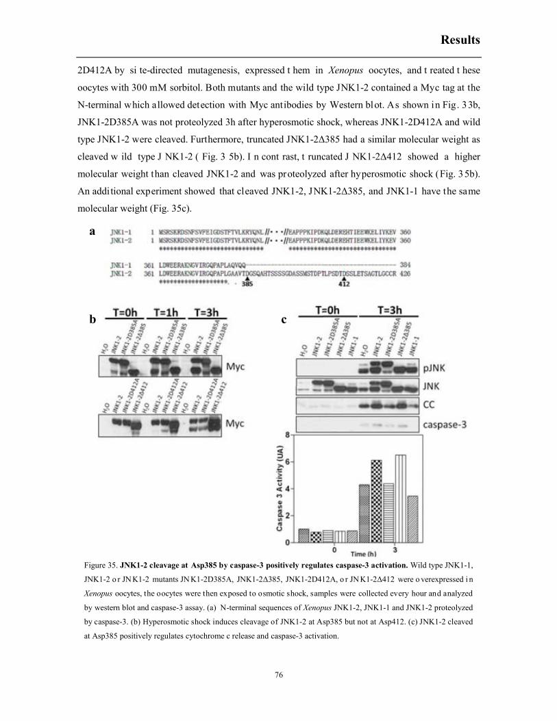
Results
76
2D412A by si te-directed mutagenesis, expressed t hem in Xenopus oocytes, and t reated t hese
oocytes with 300 mM sorbitol. Both mutants and the wild type JNK1-2 contained a Myc tag at the
N-terminal which allowed detection with Myc antibodies by Western blot. As shown in Fig. 33b,
JNK1-2D385A was not proteolyzed 3h after hyperosmotic shock, whereas JNK1-2D412A and wild
type JNK1-2 were cleaved. Furthermore, truncated JNK1-2Δ385 had a similar molecular weight as
cleaved w ild type J NK1-2 ( Fig. 3 5b). I n cont rast, t runcated J NK1-2Δ412 showed a higher
molecular weight than cleaved JNK1-2 and was proteolyzed after hyperosmotic shock (Fig. 35b).
An additional experiment showed that cleaved JNK1-2, JNK1-2Δ385, and JNK1-1 have the same
molecular weight (Fig. 35c).
Figure 35. JNK1-2 cleavage at Asp385 by caspase-3 positively regulates caspase-3 activation. Wild type JNK1-1,
JNK1-2 o r JN K1-2 mutants JN K1-2D385A, JNK1-2Δ385, JNK1-2D412A, o r JN K1-2Δ412 were o verexpressed i n
Xenopus oocytes, the oocytes were then exposed to osmotic shock, samples were collected every hour and analyzed
by western blot and caspase-3 assay. (a) N-terminal sequences of Xenopus JNK1-2, JNK1-1 and JNK1-2 proteolyzed
by caspase-3. (b) Hyperosmotic shock induces cleavage of JNK1-2 at Asp385 but not at Asp412. (c) JNK1-2 cleaved
at Asp385 positively regulates cytochrome c release and caspase-3 activation.
a
b c

Results
77
Next, we addressed the role of JNK1-2 proteolysis in the regulation of osmostress-induced
apoptosis. We expressed JNK1-1, JNK1-2, JNK1-2D385A, or JNK1-2Δ385 in Xenopus oocytes,
treated them with sorbitol 300 mM for 3 h, and measured cytochrome c release and caspase-3
activation compared with water-injected oocytes as a control. Expression of JNK1-2 increased the
release of cytochrome c and caspase-3 activity induced by osmostress compared with JNK1-
2D385A, JNK1-1 or water-injected oocytes (Fig. 35c). This result clearly indicates that cleavage of
JNK1-2 by caspase-3 accelerates the apoptotic program via increasing the release of cytochrome c
and caspase-3 activation, and therefore creates a positive feedback loop. Accordingly, expression of
JNK1-2Δ385, the cleaved product of JNK1-2 by caspase-3, also enhaced the release of cytochrome
c and caspase-3 activity compared with water-injected oocytes (Fig. 35c). Since expression of
JNK1-2Δ385 has a clear apoptotic effect compared with JNK1-1 (Fig. 35c), this imply that the last
amino acids of JNK1-2Δ385 might be important for regulating cytochrome c release and caspase-3
activation in the positive feedback loop engaged after JNK1-2 cleavage (Fig. 35a).
6.13 Cloning and expression of Bcl-2 family members
in Xenopus oocytes
Bcl-2 family members are key proteins in regulating mitochondrial membrane permeability during
apoptosis. One anti-apoptotic protein, Bcl-xL, and three pro-apoptotic proteins (Bak, Bax, and Bid)
were cloned by RT-PCR from Xenopus oocytes (stage VI). Bcl-xL, Bak, and Bid were cloned into
Ftx5 plasmid that contains a Myc tag at the N-terminal, whereas Bax was cloned into Ftx4 plasmid,
without a Myc tag.
As shown in figure 36a, microinjection of Bak, Bax or Bid cRNAs induced rapid caspase-3 activity,
without osmotic shock treatment, and the membrane of the oocytes was broken 16 h later (data not
shown). However, microinjection of Bcl-xL cRNA did not alter oocyte morphology and did not
induce caspase-3 activity, even 18 h after microinjection. Importantly, overexpression of Bcl-xL
blocked caspase-3 activation induced by osmostress (Fig. 36a). Expression of Bcl-2 family
members was monitored by Western blot using Myc antibodies. As shown in Fig. 36a, Bcl-xL was
located in the mitochondria before and 4 h after hyperosmotic shock. When we analyzed expression
of Bak and Bid 3 h after microinjection of the corresponding cRNAs, most of Bak protein was
located in the mitochondria while almost equal amounts of Bid were located both in the
mitochondria and the cytosol (Fig. 36a). However, the amounts of Bak and Bid decreased markedly
at 18 h after microinjection of the corresponding cRNAs (Fig. 36a). The localization of Bax was not

Results
78
monitored in t he present expe riment because i t was not Myc t agged. An unspecific b and w as
detected with the Myc antibodies in all the lanes and is marked with an asterisk (*) in the Western
blot.
When Bcl-xL was expressed in combination with any of these pro-apoptotic proteins, it saved the
oocytes from apoptosis, since there was not caspase-3 activation even 18 h after microinjection (Fig.
36b, lower graphic, time 0 h). In these conditions, Bcl-xL and most Bak protein were located in the
mitochondria; however, unlike described above, a higher amount of Bid protein seems to be present
in the cy tosolic fraction ( Fig. 3 6b, upper p anel), a lthough t his W estern-blot does not allow to
differentiate Bcl-xL and Bid mitochondrial proteins, cause they have similar molecular weight (see
next section to clarify this point). After osmotic shock treatment, the amounts of cytosolic Bak and
Bid decreased. Intriguingly, a short fragment with Myc tag was detected 4 h after osmostress in the
Figure 36. Bcl-2 family proteins in Xenopus oocytes. (a) Bak, Bax or Bid induce caspase-3 activation while Bcl-xL
blocks o smotic shock-induced caspase-3 act ivation. cRNAs o f Bc l-2 family members were in jected in to Xenopus
oocytes, for p ro-apoptotic m embers samples were c ollected 3 h a nd 1 8 h a fter microinjection. F or Bcl-xL, t he
oocytes were exposed to osmotic shock 18 h after microinjection, and samples were collected at 0 h and 4 h after
treatment. S amples were a nalyzed b y western b lot and c aspase-3 a ctivity a ssay. (b) Co-expression o f Bc l-xLwith
Bak, Bax or Bid and subcellular localization in response to osmotic shock. Pro-apoptotic proteins were coexpressed
with Bcl-xL, then oocytes were exposed to osmotic shock and samples were collected at 0 h and 4 h and analyzed by
western blot and caspase-3 activity assay.
a b

Results
79
oocytes m icroinjected with Bcl-xL and B id (Fig. 36b) . In t his experiment w e cannot conc lude
whether this fragment came up as a consequence of Bcl-xL or Bid proteolysis, since both proteins
are Myc tagged. This issue will be addressed in the next section. When we analyzed cytochrome c
release and caspase-3 activation induced by osmostress in all sets of combinations, we observed that
Bcl-xL expression preserved from osmostress-induced apoptosis the oocytes expressing Bak, but not
the oocytes expressing Bid, and with partial protection the oocytes expressing Bak (Fig. 36a, lower
graph, t ime 4 h) . Note that in this experiment, Bcl-xL did not block as described in Fig. 36a, but
markedly reduced osmostress-induced apoptosis (Fig. 36b).
To address properly the distribution of Bax in Xenopus oocytes, we cloned the complete sequence
into Ftx5 plasmid to add a Myc tag at the N-terminal, and the Bax cRNA was microinjected and
expressed in the oocytes. Interestingly, when tiny amount of Bax cRNA were injected to obtain low
levels of p rotein ( Fig. 3 7), oocytes sur vived and ca spase-3 w as not activated ( data not shown).
However, in this condition osmotic shock treatment induced the disappearance of Bax protein in the
cytosol, suggesting that a protease activated by osmotic shock might cleave its N-terminal (Fig. 37).
When Bax was expressed in h igher amounts in combination with Bcl-xL (not Myc tagged), about
half of the Bax protein was located in the mitochondrial fraction and osmotic shock did not induce a
significant proteolysis of Bax located in the cytoplasm or in the mitochondria (Fig. 37). However,
the western bl ot sug gests t hat m aybe small am ounts of cy tosolic Bax are translocated to t he
mitochondria at 2 h after hyperosmotic shock (Fig. 37).
6.14 Bid and mono-ubiquitinated Bid are proteolyzed
during osmostress-induced apoptosis in Xenopus
oocytes
In or der to con firm w hich pr otein, B cl-xL or B id, i s p roteolyzed during os mostress-induced
apoptosis (Fig. 3 6d), t wo di fferent c ombinations of B cl-xL and Bid cRN As w ere injected in
Figure 37. Bax e xpression a nd lo calization in Xenopus oocytes.
Tiny amount of cRNA of FTX5-Bax (upper blot) or combination of
FTX4-Bcl-xL and F TX5-Bax (m iddle and b ottom b lots) w ere
injected in to Xenopus oocytes. Oocytes were exposed to o smotic
shock 18 h after microinjection, and samples were collected at 0 h, 2
h, 4 h and analyzed by western blot.

Results
80
Xenopus oocytes: FTX5-Bcl-xL/FTX4-Bid or FTX4-Bcl-xL/FTX5-Bid. This allowed us to express
high l evels of both pr oteins without any apop totic effect on un treated oocytes and to d etect by
Western blot Bcl-xL or Bid, when the Myc tag is attached at the N-terminal (FTX5 constructs).
As shown in figure 38 non-treated oocytes (T= 0h) expressed Bcl-xL (23 kDa, but 24.5 kDa with
myc tag) in the mitochondria (second lane of the blots), whereas approximately 50% of Bid (21 kDa,
but 22.5 kDa with myc tag) was located in the mitochondria and 50% in the cytosol (third lane of
the blots, only Myc tagged proteins were observed in each combination). Besides the 22.5 kDa Bid
band, another two extra bands (31 kDa and 39 kDa) were clearly detected in the cytosolic fraction,
with l ittle amounts also present in the mitochondria (see additional Western blots on the sides of
Figure 38. Bid is u biquitinated and cleaved during osmotic shock-induced apoptosis. Two protein combinations
(FTX5-Bcl-xL/FTX4-Bid or FTX4-Bcl-xL/FTX5-Bid) were overexpressed in Xenopus oocytes. These oocytes were
then exposed to osmotic shock. Samples were collected at different times and analyzed by western blot. (a) Bid is
mono-and bi-ubiquitinated in Xenopus oocytes an d osmotic sh ock in duces proteolysis of Bid a nd m onoUb.-Bid,
generating N-terminal fragments of Bid that can b e r ecognized by ubi quitin a ntibodies a nd by cleaved caspase-3
antibodies. (b) Sequence alignment of Xenopus Bid with caspase-3 proteins indicates that the potential cleavage site
in Bid is sim ilar to c aspase-3 a ctivation c leavage site. c) Os motic sh ock in duces a slig ht and ra pid in crease o f
monoUb.-Bid and biUb.-Bid in the mitochondrial fraction.
a
b c

Results
81
Figure 38a). An unspecific protein (*) detected with Myc antibodies overlapped with the 39 kDa
Bid protein, but in some Westerns, as shown in the side blots of figure 38a, the background was
lower and the proteins separated better. It has been reported that Bid expressed in Xenopus egg
extracts can be detected as mono and biubiquitinated forms (Saitoh et al. 2009), and since ubiquitin
has an expected molecular weight of 8.5 kDa, this could explain the two extra bands obtained in our
blots. Western blot analysis with ubiquitin antibodies confirmed that these two bands corresponded
to mono- and bi-ubiquitinated Bid (monoUb.-Bid and biUb.-Bid) (Fig. 38a, middle panel). The
bands detected with ubiquitin antibodies corresponded to Bid protein confirmed by the shift in
molecular weight of Bid caused by the Myc tag attached at the N-terminal (compare lanes 2 and 3,
middle panel in Fig. 38a). In the mitochondrial fraction it was detected the bi-Ub.-Bid (39 kDa), but
not the mono-Ub.-Bid (31 kDa) by using the ubiquitin antibodies, although in the Western blot
using Myc antibodies the amounts of mono-Ub.-Bid and biUb.-Bid looked quite similar (Fig. 38a
top and side blots). We speculate that this might be due to some posttranslational modification in
the mitochondrial mono-Ub.-Bid pool which could interfere with its ubiquitin detection by Western
blot. Alternatively, low amounts of mono-ub.-Bid in the mitochondria are not well detected with the
ubiquitin antibody.
We can conclude that in untreated oocytes Bid exists in three different forms: Bid, monoUb.-Bid
and biUb.-Bid. Bid is located in the cytosol and the mitochondria (probably interacting with Bcl-xL),
whereas monoUb.-Bid and biUb.-Bid are mainly located in the cytosol, although some little
amounts of biUb.-Bid and modified Ub.-Bid might be located in the mitochondria (see Fig. 47, Bid
ubiquitination model). After osmotic shock, a 15 kDa band was detected both in the cytosolic and
mitochondrial fractions in the combination FTX4-Bcl-xL/FTX5-Bid but not in the combination
FTX5-Bcl-xL/FTX4-Bid (Fig. 38a, compare lanes 5 and 6 in the upper panels), which means that
this fragment resulted from cleavage of Bid instead of Bcl-xL. Western Blot shown that this 15 kDa
band was ubiquitinated, and the shift in molecular weight induced by Myc tag at the N-terminal
indicated that this was the N-terminal of Bid (Fig. 38a, lanes 5 and 6 middle panel). In consideration
of the molecular weight of ubiquitin (8.5 kDa) and the molecular weight expected for nBid after
caspase cleavage of Bid (6 kDa) (Kominami et al. 2006, Saitoh et al. 2009), this 15 kDa band might
be mono-ubiquitinated N-terminal of Bid (monoUb.-nBid) (expected molecular weight 14.5 kDa,
but 16 kDa with myc tag). Another pair of ubiquitinated bands (22 and 23 kDa) with a molecular
weight shift caused by Myc was detected too, and must correspond to biubiquitinated N-terminal of
Bid (biUb.-nBid). biUb.-nBid was detected by Western blot with ubiquitin antibodies in both the
cytosolic and the mitochondrial fractions, whereas monoUb.-nBid was mainly detected in the
cytosol, with very small amounts in the mitochondria (Fig. 38b, lanes 5 and 6 middel panel). The
Results
82
above results clearly indicate that hyperosmotic shock induces Bid cleavage at a specific site in the
N-terminal. Osmostress induced the proteolysis of Bid and monoUb.-Bid since the amount of both
proteins were reduced in the cytosol (Fig. 38a). Note that the reduction Bid induced by osmostress
was difficult to appreciate with Myc antibodies due to the generation of biUb.-nBid, which has a
similar molecular weight to Bid (22,5 kDa for Bid and 24 kDa for biUb.-nBid), but careful
inspection of the blot at higher resolution clearly indicated the presence of the two bands. Although
Bid and monoUb.-Bid decreased in the cytosol after osmotic shock treatment, the amount of
cytosolic biUb.-Bid was maintained quite constant (Fig. 38a). In addition, monoUb.-nBid and
biUb.-nBid accumulated in the cytosol, whereas only significant amounts of biUb.-nBid
accumulated in the mitochondria. Therefore, it seems that biUb.-nBid came from the ubiquitination
of monoUb.-Bid instead of direct cleavage of biUb.-Bid. In addition, osmotic shock quickly
induced a slightly increase of monoUb.-Bid and biUb.-Bid in the mictochondrial fraction (Fig. 38c.
lane 2 at 30 min after treatment; and Fig. 38a, side blot at 4 h after treatment). All this information
is depicted as a working Bid ubiquitination model (Fig. 47).
In traditional signaling pathways, Bid is cleaved by caspase-8 generating the so called tBid
fragment to regulate the mitochondrial outer membrane permeability. For Xenopus Bid, this
cleavage site has been reported at Asp52 (Kominami et al. 2006). As shown in Figure 38b, Bid
proteolysis at Asp52 exposes three amino acids (-ETD-), similar to activated caspase-3. It has been
reported that cleaved-Caspase-3 antibody from Cell Signaling Technology (Ref. #9661) recognizes
the epitope –ETD, and therefore it can be used for detection of other caspase-9 substrates (Fan Yun
and Bergmann 2009). Since the epitope –ETD at N-terminal fragment of Bid (nBid) is identical to
cleaved caspase-3, it should be detected by the Cell Signaling antibody. As shown in Fig. 38a,
lower panel, besides of the activated caspase-3 (18kDa) extra bands corresponding to monoUb.-
nBid and biUb.-nBid were recognized by caspase-3 antibody, thus confirming the identity of the
Bid fragments. Interestingly, the mitochondrial fraction showed high levels of monoUb.-nBid
detected with the cleaved-caspase 3 antibodies from Cell Signaling, whereas the Western blot with
ubiquitin antibodies only detected small amount of monoUb.-nBid. We speculate, as described
above for the mitochondrial monoUb-Bid, that some modification in the mitochondrial pool of
monoUb.-nBid might exist. Actually, a tiny shift of mitochondrial monoUb.-nBid respect to
cytosolic monoUb.-nBid was observed (data not shown).

Results
83
6.15 Hypeosmotic shock induces Bid proteolysis by
initiator caspases and by caspase-3
Next, w e analyzed the proteolysis of B id i nduced by os mostress in a time-course exp eriment.
Hyperosmotic shock induced weak proteolysis of Bid at 1 h, detected by the appearance of a 15 kDa
band corresponding to monoUb.-Bid as determined in the previous section, which was synchronized
with Smac/DIABLO and c ytochrome c release ( Fig. 39, c ompare My c with S mac/DIABLO and
cytochrome c blots). At 1 h a significant amount of cytochrome c was realeased into the cytosol, but
no caspase-3 activity was detected by Western blot or by enzymatic assay (Fig. 39, lower blot and
graph). A t 3 and 4 h af ter osm ostress caspase-3 ac tivity w as det ected by We stern bl ot and by
enzymatic assay, which was well correlated with a marked proteolysis of Bid (Fig. 39, upper panel
and lower g raph). I ncubation w ith t he specific caspase-3 in hibitor Z -DEVD.fmk cl early r educed
Bid pr oteolysis at 4 h, m easured by quan tification of cy tosolic monoUb.-nBid with t he Myc
antibody or by quantification of monoUb.-nBid and biUb.-nBid cytosolic fragments with the Cell
Signaling cl eaved-caspase-3 ant ibody ( Fig. 3 9, middle panel, com pare samples at 4 h in the
presence or absence of inhibitor). Note that caspase-3 inhibitor markedly reduced, but did not block
completely t he generation of m onoUb.-nBid and biUb.-nBid. T he cleavage of ca spase-3,
supposedly induced by caspase-9, was unchanged in the presence of caspase-3 inhibitor as expected,
but it was near completely blocked with the broad caspase inhibitor Z-VAD.fmk. Bid proteolysis,
Figure 39. Hyperomotic shock induces early a nd la te
proteolysis of Bid Bid b y c aspases. cRNA co mbination
of FTX4-Bcl-xL and FTX5-Bid was injected into Xenopus
oocytes and 18 h later the oocytes were exposed to osmotic
shock i n t he p resence or ab sence o f cas pase i nhibitor,
samples were collected at different times and analyzed by
western blot and caspase-3 activity.

Results
84
measured as monoUb.-nBid fragment detected with Myc antibodies or as monoUb.-nBid and biUb.-
nBid detected with cleaved-Caspase 3 antibody, was completely blocked by Z-VAD.fmk (Fig. 39).
Of not e, Smac/DIABLO a nd cy tochrome c release induced by osm ostress at 4 h w ere slightly
reduced in the presence of ca spase-3 i nhibitor, and more clearly decreased in the presence of Z -
VAD.fmk (Fig. 39 lower blots). We have previously reported that Smac/DIABLO release is a very
early ev ent i n osm ostress-induced apo ptosis and bo th Smac/DIABLO and cy tochrome c r elease
decrease i n the pr esence of Z-VAD.fmk due to i nhibition of se veral pos itive feedback l oops
(Messaoud NB, Thesis Dissertation, 2014). In addi tion, previous results suggest that Z-VAD.fmk
delays osmostress induced apoptosis by inhibition of a caspase distinct to caspase-3 (Messaoud NB,
Thesis Dissertation, 2014). Altogether, the above results suggest that Bid seems to be proteolyzed in
low amounts by an initiator ca spase that might be activated by ea rly S mac/DIABLO release and
low am ounts of cy tochrome c in the cytosol, a nd w hen caspase-3 i s fully ac tive, a m assive
proteolyisis of Bid occurs, which can be clearly detected by Western blot. The proteolysis of Bid
induced by osmostress was accelerated by co-expression of Bax in oocytes expressing Bid and Bcl-
xL (Fig. 40), confirming that changes in the equilibrium between pro- and anti-apoptotic members of
the B cl-2 f amily ca n modify t he kinetics of omostress-induced ap optosis via r egulating
mitochondrial protein release.
We hav e previously r eported that hy perosmotic shock i nduces early ca lpain activation, w hich
contributes to r egulate osmostress-induced a poptosis (Messaoud N B, T hesis Dissertation, 2014).
Calpains have be en reported t o proteolyze B id and Bax a t the N -terminal (Gao and D ou 200 1,
Mandic et al. 2002). In addition, it has been reported that in Fas-induced apoptosis JNK activation
can mediate Bid proteolysis generating a specific jBid fragment by a yet unknown protease (Deng
Yibin e t al . 2003). Therefore, we t reated oocytes with a specific calpain i nhibitor or expressed a
constitutive active MKK7 mutant (MKK7DED) kinase to activate endogenous JNK1-1 and JNK1-2
to address the roles of these signaling pathways in Bid proteolysis induced by osmostress. As shown
in Fig. 41 the calpain inhibitor MDL28170 did not have any significant effect on Bid proteolysis at
Figure 40. Bax overexpression accelerates t he Bid
proteolysis induced by osmotic sh ock. Two c RNA
combinations, F TX4-Bcl-xL + F TX5-Bid o r FTX4-Bcl-
xL + FTX5-Bid + FTX4-Bax, were injected into Xenopus
oocytes and 18 h later oocytes were exposed to osmotic
shock. S amples were co llected at different ti mes and
analyzed by western blot.

Results
85
4 h a fter osmostress, m easured as the amount of monoUb.-Bid f ragment det ected with My c
antibodies, even though the inhibitor decreased the activation of caspase-3 induced by osmostress
(Fig. 41, compare in caspase-3 graph Bcl-xL + Bid with or without MDL28170). Indeed, expression
of MKK7DED induced the phosphorylation of JNK1-1 and JNK1-2 (Fig. 41, time 0 h of Bcl-xL +
Bid + M KK7-DED) but di d not cha nge Bid p roteolysis and c aspase-3 ac tivity i nduced by
osmostress. As a control, Z-VAD.fmk efficiently blocked Bid proteolysis and caspase-3 activation.
The previous results clearly show that B id is mainly pr oteolyzed by ca spase-3 at l ater t imes o f
osmostress and that it might be proteolyzed early at low levels by an initiator caspase. To address
whether the role of cytochrome c release in the proteolysis of Bid is independent of other signaling
patways activated by osmostress, we microinjected cytochrome c ( 0.5µM, final co ncentration) in
Xenopus oocytes expressing Bcl-xL and Bid, and the cleaved products of Bid by Western blot in the
presence or absence of caspase i nhibitors. As ex pected, cytochrome c i nduced a high c aspase-3
activity 1 h af ter m icroinjection w ithout any os motic shock stimulation, and bot h, t he specific
caspase-3 i nhibitor or t he pan -caspase i nhibitor Z -VAD-fmk, co mpletely i nhibited caspase-3
activity, m easured by t he enz ymatic assay (Fig. 42). Western b lot an alysis w ith t he cleaved-
caspase-3 C ell S ignaling antibody showed a m arked p roteolysis of ca spase-3 and Bid in th e
presence or absence of inhibitors, suggesting that the apoptosome formation and activation was not
impaired by the inhibitor Z-VAD.fmk when an excess of cytochrome c exi sted in the cytosol (Fig.
42). Considering the similar aminoacids residues around the cleavage site of both caspase-3 and Bid
Figure 41. Calpain in hibitor o r s ustained
JNK activation does not alter Bid proteolysis
induced by osmostress. cRNA combination of
FTX4-Bcl-xL a nd F TX5-Bid was in jected into
Xenopus oocytes and 18 h later the oocytes were
exposed t o os motic s hock i n t he pr esence o r
absence of pan caspase inhibitor Z-VAD.fmk or
calpain inhibitor MDL28170 and samples were
collected at 4 h an d an alyzed b y western b lot
and cas pase-3 a ctivity. Some o ocytes were
injected w ith H 2O o r Bcl-xL cRNA a nd 18 h
later exposed t o osmotic sh ock f or 4 h a nd
samples analyzed as described above.

Results
86
(Fig. 38b), i t seems probable that caspase-9 mediates Bid cleavage at Asp52 when high levels of
cytochrome c are present in the cytosol.
Potential i nitiator caspases ac tivated dur ing osm ostress that m ight i nduce Bid cl eavage include
caspase-8, caspase-9 and caspase-10. We could not clone caspase-8 in stage VI oocytes, but it has
been reported very low levels, if any, of caspase-8 in Xenopus oocytes at stage VI, and high levels
at stages 15/16 in early embryogenesis. However, caspase-10 is reported to be highly expressed in
stage V I oocytes (Kominami et a l. 2 006). We c loned c aspase-10 f rom st age VI oocy tes and
expressed with a Myc tag at t he N-terminal i n t he presence or absence of the usual combination
Bcl-xL and Bid. Low amounts of caspase-10 were expressed in the oocytes 4 h after cRNA injection,
and hi gh levels o f cytochrome c w ere det ected in the cy tosol (Fig. 43), i ndicating t hat a small
increase of c aspase-10 in the oocyte i nduces apop tosis. The com bined expression o f c aspase-10
with Bcl-xL and Bid induced the release of cytochrome c and t he proteolysis of Bid (Fig. 43), as
expected. Although we do not know whether Bid proteolyisis is mediated directly by caspase-10 or
indirectly by ac tivation of ca spase-9 or ca spase-3, t here are r eports des cribing i n v itro d irect
proteolyisis of B id by Xenopus caspase-10β (Kominami et al . 200 6) or by hum an caspase-10
(Milhas et a l. 2005 ). Fut ure experiments will addr ess w hether ca spase-10 i s activated early by
osmostress and their role in primary Bid cleavage.
Figure 42. High a mount o f cy tochrome c i nduces
Bid c leavage e ven w hen c aspase-3 a ctivity is
inhibited. cRNA co mbination o f F TX4-Bcl-xL and
FTX5-Bid was injected into Xenopus oocytes and 18
h later a second microinjection of cytochrome c was
performed. S amples were c ollected 1 h late r a nd
analyzed b y western b lot an d cas pase-3 a ctivity
assay.
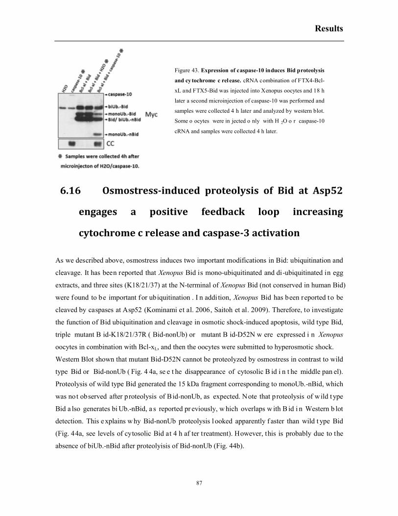
Results
87
6.16 Osmostress-induced proteolysis of Bid at Asp52
engages a positive feedback loop increasing
cytochrome c release and caspase-3 activation
As we described above, osmostress induces two important modifications in Bid: ubiquitination and
cleavage. It has been reported that Xenopus Bid is mono-ubiquitinated and di-ubiquitinated in egg
extracts, and three sites (K18/21/37) at the N-terminal of Xenopus Bid (not conserved in human Bid)
were found to be important for ubiquitination . I n addi tion, Xenopus Bid has been reported to be
cleaved by caspases at Asp52 (Kominami et al. 2006, Saitoh et al. 2009). Therefore, to investigate
the function of Bid ubiquitination and cleavage in osmotic shock-induced apoptosis, wild type Bid,
triple mutant B id-K18/21/37R ( Bid-nonUb) or mutant B id-D52N w ere expressed i n Xenopus
oocytes in combination with Bcl-xL, and then the oocytes were submitted to hyperosmotic shock.
Western Blot shown that mutant Bid-D52N cannot be proteolyzed by osmostress in contrast to wild
type Bid or Bid-nonUb ( Fig. 4 4a, se e t he disappearance of cytosolic B id i n t he middle pan el).
Proteolysis of wild type Bid generated the 15 kDa fragment corresponding to monoUb.-nBid, which
was not observed after proteolysis of Bid-nonUb, as expected. Note that proteolysis of wild t ype
Bid a lso generates bi Ub.-nBid, a s reported pr eviously, w hich overlaps w ith B id i n Western b lot
detection. This e xplains w hy Bid-nonUb proteolysis l ooked apparently f aster than wild t ype Bid
(Fig. 44a, see levels of cytosolic Bid at 4 h af ter treatment). However, this is probably due to the
absence of biUb.-nBid after proteolyisis of Bid-nonUb (Fig. 44b).
Figure 43. Expression of caspase-10 induces Bid proteolysis
and cy tochrome c rel ease. cRNA combination of FTX4-Bcl-
xL and FTX5-Bid was injected into Xenopus oocytes and 18 h
later a second microinjection of caspase-10 was performed and
samples were collected 4 h later and analyzed by western blot.
Some o ocytes were in jected o nly with H 2O o r caspase-10
cRNA and samples were collected 4 h later.

Results
88
In an additional experiment, where osmostress-induced apoptosis was accelerated by the expression
of Bax in combination with wild type Bid or mutant Bid-nonUb, we detected increased levels of
nBid in the oocytes expressing Bid-nonUb. suggesting that Bid-nonUb. might be proteolyzed faster
than wild type Bid (Fig. 45).
Ubiquitination analysis by Western blot indicated that Bid-nonUb presented a clear reduction of the
mono- and bi - ubiquitinated bands b efore treatment and also of the b i-ubiquitinated-N-terminal
fragment (biUb.-nBid) af ter osmotic shock, compared with t he wild type Bid (Fig. 4 4b). Despite
this marked reduction, low levels of ubiquitination were still detected in the Bid-nonUb mutant (Fig.
44b), as previously reported by other authors (Saitoh et al. 2009). Importantly, expression of mutant
Bid-nonUb did not modify cytochrome c release and caspase-3 activation induced by osmostress,
compared with wild type Bid, whereas mutant BidD52N clearly reduced cytochrome c release and
caspase-3 activation induced by osmostress (Fig. 44a and 44c). This result clearly indicates that Bid
Figure 44. Functional effects of B id m odification o n o smostress-induced a poptosis. Wild type Bid o r mutants
combined with FTX4-Bcl-xL or tiny amount of these proteins were expressed in Xenopus oocytes. Then oocytes were
exposed to osmotic shock and samples were collected at different times and analyzed by western b lot or caspase-3
activity assay. (a) Site-directed mutation of Bid at Asp52 (Bid-D52N) blocks its proteolysis in osmotic shock-induced
apoptosis and reduces cytochrome c re lease. (b ) The tri ple mutant Bid-K18/21/37R (Bid-nonUb) p resents marked
reduction of u biquitination b efore a nd a fter hyperosmotic sh ock trea tment. (c ) Ca spase-3 a ctivity in re sponse to
osmostress in oocytes expressing wild type Bid or mutants combined with Bcl-xL. (d) Caspase-3 activity in response
to osmostress in oocytes expressing tiny amount of wild type Bid or mutants.
a c
d
b

Results
89
cleavage by caspases, but not B id ubiquitination, r egulates osmostress-induced apoptosis through
activation of caspase-3 and therefore creating a positive feedback loop.
Expression of t runcated C-terminal Bid (53-184 aa), also known as tBid, induced high caspase-3
activity in oocytes co-expressing Bcl-xL even without hyperosmotic shock treatment (Fig. 44c, time
0 h). On the contrary, expression of nBid (1-52 aa) did not have any effect on osmostress-induced
apoptosis compared with water-injected oocytes (Fig. 44c). A small functional effect in osmostress-
induced apoptosis by the lack of Bid ubiquitination was observed when tiny amounts of wild type
Bid or mutants were expressed in the oocytes. As shown in figure 44d, expression of the mutant
Bid-nonUb i ncreased c aspase-3 ac tivity i nduced by osm ostress compared with w ild t ype Bid,
whereas BidD52N s lightly dec reased caspase-3 ac tivity at 3 or 4 h after osm ostress ( Fig. 4 4d).
However, the levels of Bid expression were so low that we could no detect the proteins by Western
blot to prove equal levels of expression for wild type Bid and their mutants.
Figure 45. Mutant B id-nonUb. is
proteolyzed f aster t han w ild ty pe
Bid. Wild ty pe Bid o r m utants
combined w ith b oth F TX4-Bcl-xL
and F TX5-Bax w ere e xpressed i n
Xenopus oocytes. Then oocytes were
exposed t o os motic s hock and
samples w ere co llected at d ifferent
times and an alyzed b y western.
Water injected o ocytes were tr eated
and analyzed as control.

Discussion
90
7. DISCUSSION
7.1 JNK proteins and activities in Xenopus laevis oocytes
Six JNK transcripts (JNK1-1 and -2, JNK3-1, -2, -3 and -4) were cloned by RT-PCR using total
RNA extracted from Xenopus laevis oocytes (stage VI). Sequence alignment shows that the JNK
sequences are quite conserved. However, Xenopus oocytes express only JNK1-1 (40kDa) and
JNK1-2 (49kDa) proteins detected by Western blot with total JNK antibodies. In mammalian, there
are three JNK genes, the jnk1 and jnk2 are ubiquitously expressed, whereas jnk3 expression is
brain-, heart-, and testis-restricted (Gupta et al. 1996). Xenopus oocytes do not express enough
amounts of JNK3 proteins to be detected by Western blot, and probably the jnk3 transcripts are
maternal stockpiles like many other mRNAs in the oocyte. It was reported that JNKs are necessary
to promote embryonic survival and regulate eye development in vertebrates. During goldfish
development, JNK1 and JNK2 are expressed at every stage from cleavage to hatching larvae,
whereas JNK3 is turned on at the gastrulation stage and expressed at similar levels to that of JNK2
(Huang et al. 2013). Similarly, Xenopus JNK3 isoforms might be expressed after fertilization to
regulate early embryogenesis and differentiation.
In Xenopus laevis, when oocytes are treated with osmotic shock, both JNK1-1 and JNK1-2 are
phosphorylated (Messaoud NB, Thesis Dissertation, 2014). It is also reported that in oocytes treated
with progesterone JNK activity increases rapidly when oocytes mature (Bagowski et al. 2001a). We
addressed the role of JNK activation in meiotic progression by expressing upstream kinases of JNK,
as well as different JNK isoforms.
MKK4 and MKK7 are the only upstream kinases known to directly activate JNK proteins (Asaoka
and Nishina 2010, Moriguchi et al. 1997). Overexpression of a constitutively active mutant MKK7-
DED in Xenopus oocytes can activate JNK1-1 and JNK1-2 isoforms without affecting the
phosphorylation of ERK2 and p38 family members. Here we show that JNK activation induced by
MKK7-DED does not affect the oocyte maturation induced by progesterone, but previous results in
our laboratory demonstrated that MKK7-DED overexpression in combination with JNK1-1 or
JNK1-2 accelerates apoptosis induced by hyperosmotic shock (Messaoud NB, Thesis Dissertation,
2014). In other cellular systems it has also been reported that sustained activation of JNK induces
apoptosis (Ventura et al. 2006).

Discussion
91
In Xenopus there are no JNK2 isoforms reported. We ectopically expressed human JNK2-1 or the
four JNK3 isoforms cloned in our laboratory in Xenopus oocytes. Similar to JNK1, all these
proteins are phosphorylated by osmotic shock and are recognized with total JNK antibodies while
none of them can increase the amount of XpJNK-p42 in mature oocytes.
We confirmed that the protein XpJNK-p42 recognized by pJNK antibody but not total JNK
antibody in GVBD oocyte extracts is phosphorylated ERK2 (pERK2). ERK2 can be phosphorylated
by both osmostress treatment and progesterone incubation, however, phosphorylated ERK2 induced
by osmotic shock can only be recognized by pERK antibodies while activated ERK2 in mature
oocytes is detected by both pJNK and pERK antibodies, which indicates that a posttranslational
modification is induced in ERK2 by progesterone treatment. Given the close relative of JNK to
ERK in the MAPK family and since it was reported that ERK activation may induce c-jun
phosphorylation (Pulverer et al. 1991), we considered the possibility that the JNK activity in mature
oocytes may come from the activated ERK2. However, here we show that overexpression of ERK2
does not affect JNK activity in mature oocytes (Fig. 29). Similar to our result, it has been reported
that MEK1/2 inhibitor U1026 effectively blocked progesterone induced ERK2 activation but had no
apparent effect on JNK activity (Bagowski et al. 2001a). Surprisingly, we also show that the JNK
inhibitor SP600125 does not inhibit c-Jun phosphorylation in mature oocytes extracts (Fig. 29). All
these information imply that the JNK activity (c-Jun phosphorylation) detected in mature oocytes
(GVBD) results from activation of another protein kinase, instead of JNK or ERK2, induced by
progesterone. It has been reported that cdc2 (Cdk1) can phosphorylate c-Jun (Baker et al. 1992) and,
more recently, that Cdk3 can mediate c-Jun phosphorylation at Ser63 and Ser73 in mouse skin
epidermal cells (Cho et al. 2009). More experiments are necessary to identify the protein
responsible for JNK activity in mature oocytes. The inhibitor SP600125 reduces progesterone-
induced maturation of Xenopus oocytes (Fig. 19). Since JNKs are not activated during meiotic
progression, this implies that SP600125 may targets another protein regulating meiosis. It has been
reported that SP600125 inhibits Cdc2 (Cdk1) (Kim JA et al. 2010).
7.2 The role of ERK2 and p38 in Xenopus laevis oocyte
maturation
Besides the Mos/MEK/ERK signal pathway described in oocyte maturation, another extensively
researched cascade that involves ERK activation is Ras⁄Raf⁄MEK/ERK signaling pathway. This
signaling pathway plays a crucial role in almost all cell functions. Upon receptor activation, GTP-
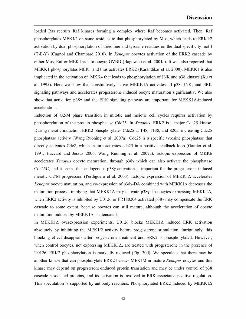
Discussion
92
loaded Ras recruits Raf kinases forming a complex where Raf becomes activated. Then, Raf
phosphorylates MEK1⁄2 on same residues to that phosphorylated by Mos, which leads to ERK1⁄2
activation by dual phosphorylation of threonine and tyrosine residues on the dual-specificity motif
(T-E-Y) (Cagnol and Chambard 2010). In Xenopus oocytes activation of the ERK2 cascade by
either Mos, Raf or MEK leads to oocyte GVBD (Bagowski et al. 2001a). It was also reported that
MEKK1 phosphorylates MEK1 and thus activates ERK2 (Karandikar et al. 2000). MEKK1 is also
implicated in the activation of MKK4 that leads to phosphorylation of JNK and p38 kinases (Xu et
al. 1995). Here we show that constitutively active MEKK1Δ activates all p38, JNK, and ERK
signaling pathways and accelerates progesterone induced oocyte maturation significantly. We also
show that activation p38γ and the ERK signaling pathway are important for MEKK1Δ-induced
acceleration.
Induction of G2/M phase transition in mitotic and meiotic cell cycles requires activation by
phosphorylation of the protein phosphatase Cdc25. In Xenopus, ERK2 is a major Cdc25 kinase.
During meiotic induction, ERK2 phosphorylates Cdc25 at T48, T138, and S205, increasing Cdc25
phosphatase activity (Wang Ruoning et al. 2007a). Cdc25 is a specific tyrosine phosphatase that
directly activates Cdc2, which in turn activates cdc25 in a positive feedback loop (Gautier et al.
1991, Haccard and Jessus 2006, Wang Ruoning et al. 2007a). Ectopic expression of MKK6
accelerates Xenopus oocyte maturation, through p38γ which can also activate the phosphatase
Cdc25C, and it seems that endogenous p38γ activation is important for the progesterone induced
meiotic G2/M progression (Perdiguero et al. 2003). Ectopic expression of MEKK1Δ accelerates
Xenopus oocyte maturation, and co-expression of p38γ-DA combined with MEKK1Δ decreases the
maturation process, implying that MEKK1Δ may activate p38γ. In oocytes expressing MEKK1Δ,
when ERK2 activity is inhibited by U0126 or FR180204 activated p38γ may compensate the ERK
cascade to some extent, because oocytes can still mature, although the acceleration of oocyte
maturation induced by MEKK1Δ is attenuated.
In MEKK1Δ overexpression experiments, U0126 blocks MEKK1Δ induced ERK activation
absolutely by inhibiting the MEK1/2 activity before progesterone stimulation. Intriguingly, this
blocking effect disappears after progesterone treatment and ERK2 is phosphorylated. However,
when control oocytes, not expressing MEKK1Δ, are treated with progesterone in the presence of
U0126, ERK2 phosphorylation is markedly reduced (Fig. 30d). We speculate that there may be
another kinase that can phosphorylate ERK2 besides MEK1/2 in mature Xenopus oocytes and this
kinase may depend on progesterone-induced protein translation and may be under control of p38
cascade associated proteins, and its activation is involved in ERK associated positive regulation.
This speculation is supported by antibody reactions. Phosphorylated ERK2 induced by MEKK1Δ
Discussion
93
ectopic expression or osmotic shock ca nnot be recognized by pJ NK a ntibody but , as w e proved
above, phosphorylated ERK2 in mature oocyte can be detected by both pERK and pJNK antibodies.
In cons ideration of MEK1 kinase activity described above, and the effective inhibition MEK1 by
U0126, t here should be a ME K1 independent p athway f or ER K2 ph osphorylation. In Xenopus
oocytes p rotein synthesis i s absolutely r equired f or ERK ac tivation, and suppression of Mos
translation inhibits MAP kinase activation in both mouse and Xenopus oocytes (Abrieu et al. 2001).
In MEKK1Δ overexpression experiments, the inhibitor U0126 does not inhibit oocyte maturation
when ERK2 i s already phosphorylated (18 h a fter MEKK1Δ cRNA i njection); how ever, the
inhibitor SP 600125 still w orks, indicating t hat SP 600125 inhibits an unk nown ER K2 ups tream
kinase instead of MEK1. A plausible candidate for ERK2 modification beyond the phosphorylation
executed by MEK 1 i s Cdk1/Cdc2. It was r eported that ac tive cyclin-B/Cdc2 kinase can ac tivate
ERK2 with the absence of detectable Mos (Guadagno and Ferrell 1998, Minshull et al. 1994) and
we obse rved t hat SP600125 attenuated the phosphorylation o f ER K2 i n mature o ocytes t hereby
decreasing the percent of mature oocytes (GVBD). Importantly, it has been reported that SP600125
can s uppress C dk1activity thus b locking the pr ogression of hum an G2 c ells into mitosis,
independent of JNK inhibition (Kim JA et al. 2010).
Figure 46. Schematic diagram of MAP Kinase interaction in Xenopus oocytes. JNK and p38 signaling pathways
are mainly involved in cellular stress response, while ERK signaling pathway is crucial for cellular cycle regulation.
However, it is also reported that p38 isoforms are implicated in mitotic and meiotic cell cycle regulation by activating
CDC proteins. Additionally, a cdc2 dependent posttranslational modification of ERK2 may exist.

Discussion
94
Interestingly, in oocytes expressing MEKK1Δ and treated with FR180204, an ERK inhibitor, some
phosphorylated ERK2 can be recognized by pJNK antibody. X-ray crystal structure analysis of the
human ERK2/FR180204 complex revealed that the ATP-binding pocket on ERK formed by
residues Q105, D106, L156, and C166 plays important roles in the ERK2-FR180204 interaction
(Ohori et al. 2005). We speculate that FR180204 binding to phosphorylated ERK2 may induce a
conformational change that allows detection with pJNK antibodies. It has been reported that the
glycine-rich loop of the FR180204-ERK2 complex had a different conformation from those of the
other complexes previously reported (Ohori et al. 2005).
It has been reported that ERK activity is very low in cells transfected with constitutively active
MEKK1-C or in MEKK1-C and MEK1/2 co-transfection experiments (Xu et al. 1995). Similarly,
we found very low level of ERK activity in apoptotic oocytes induced by osmotic shock although
ERK2 phosphorylation signal was quite strong; however, ERK activity was high in mature oocytes.
Our results suggest that an ERK2 posttranslational modification, independent of MEK1/2 activity,
exists in Xenopus oocyte maturation, and this modification is critical for ERK2 activity (using MBP
as a substrate). Alternatively, ERK phosphorylation induced by MEK1/2 would be sufficient to
engage ERK activity, but in apoptotic oocytes (induced by osmotic shock) some modification
would induce ERK inactivation. So more experiments are needed to solve these questions.
Osmostress-induced cleavage of JNK1-2 by caspase-3 promotes cytochrome c release and caspase-
3 activation in a positive feedback loop
Previous work in our laboratory has shown that sustained activation of JNK1-1 and JNK1-2
contributes to hyperosmotic shock-induced apoptosis of Xenopus oocytes, with a higher pro-
apoptotic function for JNK1-2 (Messaoud NB, Thesis Dissertation, 2014). It has been shown that
JNK may have a pro- or anti-apoptotic role in a stimulus and tissue dependent mechanism (Davis
Roger J 2000b, Weston and Davis 2007). It seems that a transient activation of JNK is a struggle for
survival, while sustained activation of JNK induces apoptosis (Ventura et al. 2006). Incubation of
Xenopus oocytes with the JNK inhibitor SP600125 slightly increases caspase-3 activity 1 h after
osmostress, suggesting that early activation of JNK might be anti-apoptotic (Messaoud NB, Thesis
Dissertation, 2014). JNK has been reported to prevent cell death by up-regulating transcription of
survival genes (Lamb et al. 2003). However, in Xenopus oocytes there is no transcriptional control,
which might also explain why JNK activation is sustained, since phosphatases transcriptionally
induced by JNK activation down-regulate JNK phosphorylation in mammalian cells (Owens and
Keyse 2007, Ventura et al. 2006). It has also been reported that transient activation of human JNK
p46 splice variant (similar to Xenopus JNK1-1) delays activation of caspase-9 by direct interaction
with Apaf-1 and cytochrome c in the apoptosome complex (Tran et al. 2007). Sustained activation
Discussion
95
of JNK could induce apoptosis through phosphorylation of Bax sequestration proteins 14-3-3,
which promotes Bax translocation to mitochondria (Tsuruta et al. 2004), phosphorylation of Bad at
Ser128 (Donovan et al. 2002), or modulating the activities of other pro-apoptotic BH3-only proteins
such as Bim and Bmf (Lei and Davis 2003) (see Introduction for a review). In this work, we have
not addressed the posttranslational modifications of the Bcl-2 family members induced by JNK1-1
or JNK1-2 activation after hyperosmotic shock treatment. We do not observe any significant change
in Bax distribution during osmostress-induced apoptosis (Fig. 35). However, these measurements
were performed in oocytes overexpressing Bax combined with Bcl-xL, and small changes in the
distribution of Bax could be difficult to detect in these conditions. It would be interesting to check
how the endogenous levels of Bax respond to hyperosmotic shock. It has also been reported that
JNK activation can induce apoptosis through Bid proteolysis and generation of truncated jBid
(Deng et al. 2003). However, our results show that expression of a constitutively active MKK7
mutant (MKK7-DED) in Xenopus oocytes activates endogenous JNK1-1 and JNK1-2, but neither
increases the proteolysis of Bid induced by osmostress (which generates tBid and nBid mono- or bi-
ubiquitinated) nor produces a different N-terminal fragment as would be expected after generation
of truncated jBid (Fig. 37b). In fact, the protolytic site reported in human Bid that is responsible for
the generation of jBid (Deng et al. 2003) is not conserved in Xenopus. In the present work we also
demonstrate that in osmotic stress-induced apoptosis Xenopus JNK1-2 is cleaved into a JNK1-1 like
protein after caspase-3 activation. There are two potential caspase-3 recognition sites at D385 and
D412. In humans, JNK1 and JNK2 are proteolyzed by caspase-3 at D413 and D410, respectively
(Enomoto et al. 2003), in a sequence identical to Xenopus recognition site D412 (SDTD). In
humans, JNKs do not have an optimal sequence for caspase-3 cleavage (DXXD), but the atypical
cleavage site SDTD is recognized and cleaved by caspase-3 (Enomoto et al. 2003). However, our
data clearly demonstrates that cleavage of Xenopus JNK1-2 occurs only at the consensus
recognition site D385 but not at the atypical cleavage site D412. It might be possible that the
atypical sequence at D412 in Xenopus serves as a binding site for recruitment of caspase-3 but not
for cleavage. Alternatively, the atypical sequence might be masked by some kind of protein
interaction distinct to caspase-3. We have not determined the JNK activity of the phosphorylated
Xenopus JNK1-1 like fragment generated by osmostress. However, in humans, the proteolyzed
fragments of pJNK1 and pJNK2 generated by caspase-3 have similar activities that full length
pJNKs (Enomoto et al. 2003). Importantly, here we show, for the first time, that proteolysis of JNK
has a functional role in apoptosis. Cleavage of JNK1-2 by caspase-3 positively regulates
cytochrome c release and caspase-3 activity induced by osmostress, thus creating a positive
feedback loop.
Discussion
96
It seems that the positive feedback comes from the JNK1-1 like fragment (aminoacids 1-385)
generated by proteolysis of JNK1-2, since overexpression of the JNK1-1 like fragment mimics the
effect of JNK1-2 overexpression accelerating osmostress-induced apoptosis (Fig. 35b). JNK1-2 is
more pro-apoptotic that JNK1-1 (Fig. 35b), the only distinction of these two proteins are the
aminoacids located at the C-terminal (Fig. 35a), which are generated by alternative splicing. We
propose that these different residues could determine different protein interactions. JNK1-1 like
fragment may interact preferentially with Bcl-2 family members, thus inhibiting anti-apoptotic or
activating pro-apoptotic proteins, and therefore increasing the release of cytochrome c from the
mitochondria. Alternatively, the JNK1-1 like fragment increases the activity of the apoptosome
complex, competing with JNK1-1, which has been reported previously to interact with Apaf1 and
cytochrome c to inhibit the apoptosome complex (Tran et al. 2007).
7.3 Bcl-2 family members in osmostress induced apoptosis
Bcl-2 family members are central proteins in regulating mitochondrial permeability in apoptosis.
Anti-apoptotic proteins, such as Bcl-xL, bind and sequester pro-apoptotic proteins, including
activator BH3-only proteins and executor Bax and Bak, to prevent apoptosis. Here we show that
Xenopus oocytes overexpressing the anti-apoptotic protein Bcl-xL present a marked reduction of
cytochrome c release and caspase-3 activation induced by hyperosmotic shock. In some
experiments the inhibition of caspase-3 activity was complete (Fig. 36a). This result indicates that
osmostress-induced apoptosis is mainly regulated by activation of the mitochondrial pathway, and
that the extrinsic pathway is not activated or has a minor role in caspase-3 activation. On the
contrary, ectopic expression of pro-apoptotic Bcl-2 member Bid, that was initially identified as a
protein binding to both pro-apoptotic Bax and anti-apoptotic Bcl-2 (Wang Kun et al. 1996), induces
oocyte apoptosis without any stress stimulation (Fig. 34a). Although it is reported that Bid locates
in the cytosol, and the full activity of Bid is not exerted until proteolytic cleavage by caspase-8 (Li
Honglin et al. 1998), full length Bid can still translocates to mitochondria, and it is sufficient to
induce apoptosis of cultured cells (König et al. 2007, Valentijn and Gilmore 2004). Overexpression
of Bax and Bak also induces apoptosis in Xenopus oocytes, as expected, but co-expression of Bcl-xL
with Bax, Bak or Bid blocks the apoptosis induced by the pro-apoptotic family members. Bcl-xL
expression also reduces cytochrome c release and caspase-3 activitation induced by osmostress in
oocytes co-expressing Bak, but not in the oocytes expressing Bax or Bid. Although this might
seems an artificial system it allowed us to express high levels of both pro- and anti-apoptotic
proteins to study some modifications induced by osmostress that would be difficult to detect
Discussion
97
analyzing the small amounts of endogenous proteins present in the oocytes. In addition, there are
not good commercial antibodies to detect Xenopus Bcl-2 family members, but ectopic expression of
the proteins with a Myc tag at the N-terminal can be easily detected. In Xenopus oocytes,
overexpressed full-length Bid is located in the cytosol and the mitochondria and hyperosmotic
shock induces Xenopus Bid proteolysis in both pools. We also demonstrate that cleavage of Bid
induced by osmostress occurs after residue Asp52, in the conserved caspase-8/9/10 site ETD, and is
markedly reduced in oocytes incubated with the specific caspase-3 inhibitor, and completely
blocked in the presence of Z-VAD.fmk. Therefore, it seems that most of Bid proteolysis is due to
caspase-3 activity, but an additional caspase also participates to a minor extent in the proteolyisis of
Bid. It has been reported that caspase-3 can cleaves human Bid at residue Asp60, and cell extracts
depleted of caspase-3 or extracts of MCF-7 cells that are devoid of caspase-3, due to a deletion in
exon3 of the caspase-3 gene, fails to induce Bid cleavage (Slee et al. 2000). Interestingly, we show
here that in Xenopus oocytes, cytochrome c microinjection in the presence or absence of Z-
VAD.fmk induces the proteolysis of Bid at Asp52, probably through activation of caspase-9. It has
been reported that Z-VAD.fmk does not inhibit caspase-9, moreover, in front of some apoptotic
stimuli it could activate it (Rodríguez-Enfedaque et al. 2012). The cleavage site in Xenopus Bid is
similar to the cleavage activation site in Xenopus caspase-3 (Fig. 38b), and therefore caspase-9
could cleave both proteins. It has also been reported that Xenopus Bid can be proteolyzed by
Xenopus caspase-8 and caspase-10β (Kominami et al. 2006). We have confirmed that caspase-10
expression in Xenopus oocytes induces Bid proteolysis and cytochrome c release. Altogether the
above data indicates that the ETD site present in Xenopus Bid can be recognized by caspase-3,
caspase-8, caspase-9 and caspase-10. Xenopus caspase-8, however, is mainly expressed at stages
15/16 in early embryogenesis with very low levels, if any, in stage VI oocytes (Kominami et al.
2006). During apoptosis, Bid can be cleaved not only by caspases, but also by granzyme B, calpains
and cathepsins (Billen et al. 2008). We have reported previously that calpain inhibitors, but not
cathepsin inhibitors, delay osmostress induced apoptosis in Xenopus oocytes (Messaoud NB, Thesis
Dissertation, 2004). However, we show here that calpain inhibitor MDL28170 does not decrease
Bid proteolysis induced by osmostress, although decreases caspase-3 activation (Fig. 41). The time-
course analysis of Bid-cleaved induced by osmostress combined with the data obtained in the
presence of inhibitors indicate that low amounts of Bid are proteolyzed very early after
hyperosmotic shock by an initiator caspase, whereas most of Bid is cleaved by caspase-3 at later
times. Candidates for early proteolysis of Bid are caspase-10, which is highly expressed in stage VI
oocytes (Kominami et al. 2006), and caspase-9. Future experiments wil address the role of these
caspases in early cleavage of Bid induced by osmostress. The C-terminal product of Bid-cleaved at
Discussion
98
residue Asp52, tBid, is more active than full-length Bid inducing cytochrome c release and caspase-
3 activation. Whereas overexpression of Bcl-xL can block the apoptotic function of wild type Bid in
the oocytes, tiny amount of tBid (1/100 of wild type Bid) is enough to induce apoptosis without
any stress stimulation (Fig. 44c). Therefore, osmostress-induced Bid cleavage mediated by initiator
caspases generates tBid that could migrate to the mitochondria and induce cytochrome c release and
caspase-3 activation, which in turn generates more tBid, thus creating a positive feedback loop.
Indeed, we demonstrate that expression of Bid mutated at the cleavage caspase site ETD (BidD52N)
reduces the release of cytochrome c and caspase-3 activity induced by osmostress compared with
the wild type Bid (Fig. 44c).
7.4 Bid is mono- and bi-ubiquitinated in Xenopus oocytes,
but the function of ubiquitinated Bid is not clear
It has been reported that three Lys residues in the N-terminal region of Xenopus Bid (not conserved
in human) are important for mono- and biubiquitination of Bid, and both mono and biubiquitinated
Bid are not degraded by the proteasome in oocyte extracts (Saitoh et al. 2009). In human Bid the C-
terminal fragment (tBid) is polyubiquitinated and degraded by the proteasome (Breitschopf et al.
2000), and it has been reported that the N-terminal fragment (nBid) is also polyubiquitinated in
Ser/Thr/Cys residues in an unconventional manner and degraded by the proteasome (Tait et al.
2007). Here we show that when Xenopus Bid and Bcl-xL are overexpressed in the oocyte, a
significant portion of Bid is mono- and biubiquitinated in the cytosol. Some small amounts of
mono- and biubiquitinated Bid are present in the mitochondria, which are slighly increased after
osmotic shock treatment. We do not know if this increase is due to migration of cytosolic Bid to the
mitochondria or to ubiquitination of the mitochondrial Bid pool. To address this issue we should
analyze oocytes treated with hyperosmotic shock in the presence or absence of specific
ubiquitination inhibitors. Not only ubiquitinated full-length Bid but also ubiquitinated N-terminal
Bid fragments obtained by proteolyisis of Bid are quite stable. The quantification of these fragments
by Western blot indicates that proteolysis of Bid and monoUb-Bid in the cytosol generate N-
terminal fragments that are ubiquitinated and accumulated in the cytosol and the mitochondria as
monoUb-nBid and biUb-nBid. Non ubiquitinated N-terminal Bid is difficult to be detected after
hyperosmotic shock, even after microinjection of a cRNA coding for N-terminal Bid. It looks like
the non-ubiquitinated N-terminal Bid is not a stable peptide in the oocyte. As described above,
hyperosmotic stress produces a marked increase of monoUb-nBid and biUb-nBid in the cytosolic

Discussion
99
and mitochondrial fractions, detected with the cleaved caspase-3 Cell Signaling antibodies,
although monoUb-nBid (and also monoUb-Bid) was difficult to detect in the mitochondria with
anti-Ub antibodies. We speculate, as described in the Results, that some posttranslational
modification in the mitochondrial pool of monoUb-nBid and monoUb-Bid may mask their detection
with anti-Ub antibodies. It is difficult to know the role of mono- and biubiquitination in Bid, but in
other proteins it has been reported that these modifications can alter the localization in the cell, the
interaction with other proteins, and their function (Kerscher et al. 2006). Human Bid is subject to
autoinhibition in the absence of stimuli, since the N- and C- terminal fragments bind each other
through interactions of the BH3-like region at the N-terminal and the BH3 region at the C-terminal
(Tan et al. 1999). As described above, unconventional ubiquitination and degradation of its N-
terminal fragment is required to unleash the proapoptotic ability of human tBid (Tait et al. 2007).
Removal of the N-terminal of Bid has been suggested to increase the number of exposed
hydrophobic residues in tBid thereby facilitating binding of the protein to membranes (McDonnell
et al. 1999). However, in Xenopus Bid the BH3-like region at the N-terminal fragment is not
conserved. Therefore, mono and bi-ubiquitination of the N-terminal fragment could be a protective
mechanism in Xenopus Bid to avoid its interaction with the mitochondrial membrane to induce
cytochrome c release in the absence of a stressfull stimulus. Alternatively, Bid ubiquitination may
inhibits Bid cleavage mediated by caspases, like phosphorylation of human Bid inhibits Bid
cleavage by caspase-8 or caspase-3 (Degli Esposti et al. 2003, Desagher et al. 2001). However,
when we expresse a triple mutant Bid (K18/21/37R) (Bid-nonUb.) with a marked reduction in
ubiquitination in Xenopus oocytes, and compare its effect with wild type Bid, we do not find a clear
effect on osmostress-induced apoptosis (Fig. 44c and 44d). It is possible that Bid-nonUb. mutant
does not affect the small pool of endogenous Bid ubiquitinated present in the mitochondria.
Therefore, an excess of the Bid-nonUb. mutant expressed in the oocytes does not necessarily have
to affect the function of this mitochondrial pool. To address this issue properly, we should deplete
the endogenous ubiquitinated Bid, which could be achieved by microinjection of specific antibodies
for Xenopus ubiquitinated Bid (not available yet). We only find a functional effect of the mutant
Bid-nonUb. when tiny amounts of wild type or mutant Bid are expressed in the oocytes in the
absence of Bcl-xL (Fig 44d), or when Bax is co-expressed with Bcl-xL and Bid to accelerate
osmostress-induced apoptosis (Fig 40). In the first experiment Bid-nonUb. mutant is slightly more
pro-apoptotic that wild type Bid, and in the second experiment Bid-nonUb. seems to be proteolyzed
faster that wild type Bid. Because these effects are small, and only found under certain conditions,
we conclude that the function of mono- and bi- ubiquitinated Bid on osmostress-induced apoptosis
is not yet clear.

Discussion
100
7.5 A model for osmostress-induced apoptosis in Xenopus
oocytes
Hyperosmotic shock induces rapid calpain activation, Smac/DIABLO release and JNK/p38
activation in Xenopus oocytes (Messaoud NB, Thesis Dissertation, 2014). The stress protein kinases
JNK and p38 are efficient sensors, with high ultrasensitivity, that evaluate stressful situations in the
cells. When oocytes are exposed for several hours to hyperosmotic shock sustained activation of
p38 and JNK in combination with calpain activation and Smac/DIABLO release induce cytochrome
c release and caspase-3 activation (Messaoud NB, Thesis Dissertation, 2014). Here we show that
hyperosmotic shock induces early Bid cleavage at very low levels, probably by activation of an
initiator caspase, generating tBid that would induce cytochrome c release (Fig. 47, top).
Hyperosmotic shock also induces a slight and rapid increase of mono- and bi-ubiquitinated Bid in
the mitochondria (Fig. 47, bottom), although their role in osmostress-induced apoptosis in not clear.
We cannot discard that rapid Bid ubiquitination might delay Bid proteolysis and therefore be a
protective response to hyperosmotic shock. In an early stress situation it is expected that anti-
apoptotic and pro-apoptotic responses occurs synchronically. Oocytes exposed to hyperosmotic
shock must evaluate carefully all the information to assess the strength and duration of the stress, as
well as the damage suffered in the cell machinery, before engaging an irreversible program. Bcl-xL
overexpression protects oocytes from hyperosmotic shock-induced apoptosis, indicating that
cytochrome c release from the mitochondria is crucial for caspase-3 activation. However, when
cytochrome c is released and reaches a threshold level, caspase-3 is activated and engages several
positive feedback loops (Fig. 47, top). For instance, caspase-3 increases calpain activation (Pörn-
Ares et al. 1998, Wang Kevin KW et al. 1998), Smac/DIABLO release and p38 phosphorylation
(Messaoud NB, Thesis Dissertation, 2014), which in turn increase cytochrome c release and
caspase-3 activity. Here we show that caspase-3 also induces JNK1-2 proteolysis, which in turn
positively regulates cytochrome c release and caspase-3 activation. In addition, we demonstrate that
a massive proteolysis of Bid at Asp52, generating t-Bid, is mediated by caspase-3, thus creating
another positive feedback loop. In conclusion, hyperosmotic shock induces the activation of
different pathways that converge on the activation of caspase-3, which engages an irreversible
apoptotic program through activation of multiple positive feedback loops.
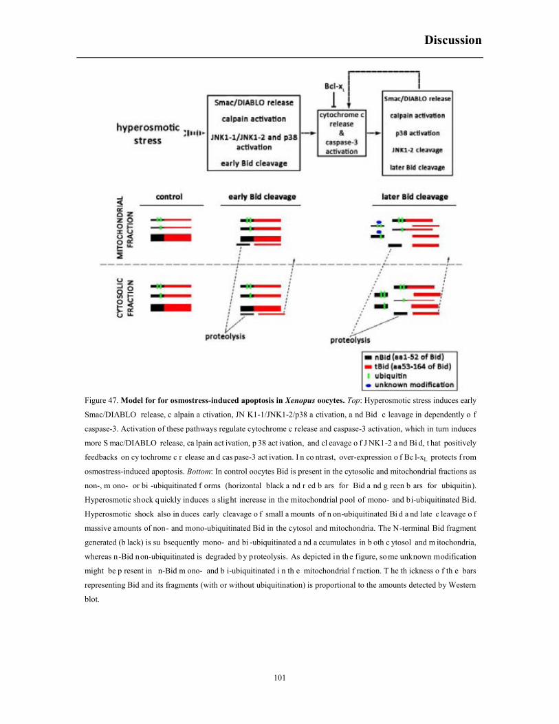
Discussion
101
Figure 47. Model for for osmostress-induced apoptosis in Xenopus oocytes. Top: Hyperosmotic stress induces early
Smac/DIABLO release, c alpain a ctivation, JN K1-1/JNK1-2/p38 a ctivation, a nd Bid c leavage in dependently o f
caspase-3. Activation of these pathways regulate cytochrome c release and caspase-3 activation, which in turn induces
more S mac/DIABLO release, ca lpain act ivation, p 38 act ivation, and cl eavage o f J NK1-2 a nd Bi d, t hat positively
feedbacks on cy tochrome c r elease an d cas pase-3 act ivation. I n co ntrast, over-expression o f Bc l-xL protects f rom
osmostress-induced apoptosis. Bottom: In control oocytes Bid is present in the cytosolic and mitochondrial fractions as
non-, m ono- or bi -ubiquitinated f orms (horizontal black a nd r ed b ars for Bid a nd g reen b ars for ubiquitin).
Hyperosmotic shock quickly induces a slight increase in the mitochondrial pool of mono- and bi-ubiquitinated Bid.
Hyperosmotic shock also in duces early cleavage o f small a mounts of n on-ubiquitinated Bi d a nd late c leavage o f
massive amounts of non- and mono-ubiquitinated Bid in the cytosol and mitochondria. The N-terminal Bid fragment
generated (b lack) is su bsequently mono- and bi -ubiquitinated a nd a ccumulates in b oth c ytosol and m itochondria,
whereas n-Bid non-ubiquitinated is degraded by proteolysis. As depicted in the figure, so me unknown modification
might be p resent in n-Bid m ono- and b i-ubiquitinated i n th e mitochondrial f raction. T he th ickness o f th e bars
representing Bid and its fragments (with or without ubiquitination) is proportional to the amounts detected by Western
blot.

Conclusions
102
8. CONCLUSIONS
1. There are at least six different jnk transcripts in Xenopus laevis oocytes (stage VI), two for jnk1
and four for jnk3; however the oocytes only express significant levels of JNK1-1 and JNK1-2
proteins.
2. Ectopic expression of a constitutively active MEKK1 accelerates progesterone-induced oocyte
maturation through activation of the ERK and p38 signaling pathways.
3. Progesterone treatment does not activate any JNK isoform in mature oocytes.
4. Phosphorylation of c-jun (JNK activity) detected in mature oocytes extracts is independent of
ERK2 and JNK.
5. A protein of 42 kDa detected with pJNK antibodies (XpJNKp42) is a posttranslational
modification of pERK2 induced during meiotic progression.
6. Hyperosmotic shock induces the activation of JNK1-1 and JNK1-2, but JNK1-2 is cleaved at
Asp385 by caspase-3 to generate a JNK1-1 like fragment, which positively regulates cytochrome
c release and caspase-3 activation, creating a positive feedback loop.
7. A pool of Bid mono- and bi-ubiquitinated is located in the cytosol and mitochondria of Xenopus
oocytes. Hyperosmotic shock induces a rapid increase of mono- and bi-ubiquitinated Bid in the
mitochondria. However, the effect of Bid ubiquitination in the regulation of osmostress-induced
apoptosis is not clear.
8. Osmostress induces an early cleavage of small amounts of Bid at Asp52 by an initiator caspase,
and a massive cleavage of Bid at later times in the same residue by caspase-3. The cleavage of
Bid positively regulates cytochrome c release and caspase-3 activation, creating a positive
feedback loop.
In conclusion, JNK proteins are not involved in Xenopus oocyte maturation, and the
phosphorylation of c-Jun detected in mature oocytes is independent of JNK and ERK2. Caspase-3
activation induced by hyperosmotic shock engages two positive feedback loops through the
cleavage of JNK1-2 and Bid, thus promoting an irreversible death of the oocytes.

Refferences
103
9. REFERENCES
Abrieu A, Dorée M, Fisher D. 2001. The interplay between cyclin-B-Cdc2 kinase (MPF) and MAP kinase during maturation of oocytes. Journal of Cell Science 114: 257-267.
Adler V, Qu Y, Smith SJ, Izotova L, Pestka S, Kung H-F, Lin M, Friedman FK, Chie L, Chung D. 2005. Functional interactions of Raf and MEK with Jun-N-terminal kinase (JNK) result in a positive feedback loop on the oncogenic Ras signaling pathway. Biochemistry 44: 10784-10795.
Ambrosino C, Nebreda AR. 2001. Cell cycle regulation by p38 MAP kinases. Biology of the Cell 93: 47-51.
Asaoka Y, Nishina H. 2010. Diverse physiological functions of MKK4 and MKK7 during early embryogenesis. Journal of biochemistry 148: 393-401.
Bagowski CP, Ferrell Jr JE. 2001. Bistability in the JNK cascade. Current Biology 11: 1176-1182.
Bagowski CP, Xiong W, Ferrell JE. 2001a. c-Jun N-terminal kinase activation in Xenopus laevis eggs and embryos a possible non-genomic role for the JNK signaling pathway. Journal of Biological Chemistry 276: 1459-1465.
Bagowski CP, Myers JW, Ferrell JE. 2001b. The Classical Progesterone Receptor Associates with p42 MAPK and Is Involved in Phosphatidylinositol 3-Kinase Signaling inXenopus Oocytes. Journal of Biological Chemistry 276: 37708-37714.
Baker SJ, Kerppola T, Luk D, Vandenberg MT, Marshak D, Curran T, Abate C. 1992. Jun is phosphorylated by several protein kinases at the same sites that are modified in serum-stimulated fibroblasts. Molecular and cellular biology 12: 4694-4705.
Bao Q, Riedl SJ, Shi Y. 2005. Structure of Apaf-1 in the auto-inhibited form: a critical role for ADP. Cell Cycle 4: 1001-1003.
Barr FA, Silljé HH, Nigg EA. 2004. Polo-like kinases and the orchestration of cell division. Nature reviews Molecular cell biology 5: 429-441.
Bayaa M, Booth RA, Sheng Y, Liu XJ. 2000. The classical progesterone receptor mediates Xenopus oocyte maturation through a nongenomic mechanism. Proceedings of the National Academy of Sciences 97: 12607-12612.
Berger L, Wilde A. 2013. Glycolytic Metabolites Are Critical Modulators of Oocyte Maturation and Viability. PloS one 8: e77612.
Bhuyan A, Varshney A, Mathew M. 2001. Resting membrane potential as a marker of apoptosis: studies on Xenopus oocytes microinjected with cytochrome c. Cell death and differentiation 8: 63-69.
Refferences
104
Billen L, Shamas-Din A, Andrews D. 2008. Bid: a Bax-like BH3 protein. Oncogene 27: S93-S104.
Boonyaratanakornkit V, Scott MP, Ribon V, Sherman L, Anderson SM, Maller JL, Miller WT, Edwards DP. 2001. Progesterone receptor contains a proline-rich motif that directly interacts with SH3 domains and activates c-Src family tyrosine kinases. Molecular cell 8: 269-280.
Boulton TG, Yancopoulos GD, Gregory JS, Slaughter C, Moomaw C, Hsu J, Cobb MH. 1990. An insulin-stimulated protein kinase similar to yeast kinases involved in cell cycle control. Science 249: 64-67.
Boulton TG, Nye SH, Robbins DJ, Ip NY, Radzlejewska E, Morgenbesser SD, DePinho RA, Panayotatos N, Cobb MH, Yancopoulos GD. 1991. ERKs: a family of protein-serine/threonine kinases that are activated and tyrosine phosphorylated in response to insulin and NGF. Cell 65: 663-675.
Breitschopf K, Zeiher AM, Dimmeler S. 2000. Ubiquitin-mediated Degradation of the Proapoptotic Active Form of Bid A FUNCTIONAL CONSEQUENCE ON APOPTOSIS INDUCTION. Journal of Biological Chemistry 275: 21648-21652.
Brenner B, Koppenhoefer U, Weinstock C, Linderkamp O, Lang F, Gulbins E. 1997. Fas-or ceramide-induced apoptosis is mediated by a Rac1-regulated activation of Jun N-terminal kinase/p38 kinases and GADD153. Journal of Biological Chemistry 272: 22173-22181.
Cagnol S, Chambard JC. 2010. ERK and cell death: Mechanisms of ERK‐induced cell death–apoptosis, autophagy and senescence. FEBS journal 277: 2-21.
Campbell L, Hardwick KG. 2003. Analysis of Bub3 spindle checkpoint function in Xenopus egg extracts. Journal of cell science 116: 617-628.
Cargnello M, Roux PP. 2011. Activation and function of the MAPKs and their substrates, the MAPK-activated protein kinases. Microbiology and Molecular Biology Reviews 75: 50-83.
Chen C-L, Lin C-F, Chang W-T, Huang W-C, Teng C-F, Lin Y-S. 2008. Ceramide induces p38 MAPK and JNK activation through a mechanism involving a thioredoxin-interacting protein-mediated pathway. Blood 111: 4365-4374.
Chen J, Downs SM. 2008. AMP-activated protein kinase is involved in hormone-induced mouse oocyte meiotic maturation in vitro. Developmental biology 313: 47-57.
Chen J, Hudson E, Chi MM, Chang AS, Moley KH, Hardie DG, Downs SM. 2006. AMPK regulation of mouse oocyte meiotic resumption in vitro. Developmental biology 291: 227-238.
Chen Y-R, Wang X, Templeton D, Davis RJ, Tan T-H. 1996. The role of c-Jun N-terminal kinase (JNK) in apoptosis induced by ultraviolet C and γ radiation duration of JNK activation may determine cell death and proliferation. Journal of Biological Chemistry 271: 31929-31936.
Chipuk JE, Green DR. 2008. How do BCL-2 proteins induce mitochondrial outer membrane permeabilization? Trends in cell biology 18: 157-164.
Chipuk JE, Moldoveanu T, Llambi F, Parsons MJ, Green DR. 2010. The BCL-2 family reunion. Molecular cell 37: 299-310.

Refferences
105
Cho Y-Y, Tang F, Yao K, Lu C, Zhu F, Zheng D, Pugliese A, Bode AM, Dong Z. 2009. Cyclin-dependent kinase-3–mediated c-jun phosphorylation at Ser63 and Ser73 enhances cell transformation. Cancer research 69: 272-281.
Cirman T, Orešić K, Mazovec GD, Turk V, Reed JC, Myers RM, Salvesen GS, Turk B. 2004. Selective disruption of lysosomes in HeLa cells triggers apoptosis mediated by cleavage of Bid by multiple papain-like lysosomal cathepsins. Journal of Biological Chemistry 279: 3578-3587.
Coll O, Morales A, Fernández-Checa JC, Garcia-Ruiz C. 2007. Neutral sphingomyelinase-induced ceramide triggers germinal vesicle breakdown and oxidant-dependent apoptosis in Xenopus laevis oocytes. Journal of lipid research 48: 1924-1935.
Cooper JA, Bowen-Pope DF, Raines E, Ross R, Hunter T. 1982. Similar effects of platelet-derived growth factor and epidermal growth factor on the phosphorylation of tyrosine in cellular proteins. Cell 31: 263-273.
Cortese JD, Voglino AL, Hackenbrock CR. 1998. Multiple conformations of physiological membrane-bound cytochrome c. Biochemistry 37: 6402-6409.
Cuadrado A, Nebreda A. 2010. Mechanisms and functions of p38 MAPK signalling. Biochem. J 429: 403-417.
Davis RJ. 1995. Transcriptional regulation by MAP kinases. Molecular reproduction and development 42: 459-467.
Davis RJ. 2000a. Signal transduction by the JNK group of MAP kinases. Cell 103: 239-252.
Davis RJ. 2000b. Signal transduction by the JNK group of MAP kinases. Pages 13-21. Inflammatory Processes:, Springer.
Degli Esposti M, Ferry G, Masdehors P, Boutin JA, Hickman JA, Dive C. 2003. Post-translational modification of Bid has differential effects on its susceptibility to cleavage by caspase 8 or caspase 3. Journal of Biological Chemistry 278: 15749-15757.
Deng H, Yu F, Chen J, Zhao Y, Xiang J, Lin A. 2008. Phosphorylation of Bad at Thr-201 by JNK1 promotes glycolysis through activation of phosphofructokinase-1. Journal of biological chemistry 283: 20754-20760.
Deng Y, Ren X, Yang L, Lin Y, Wu X. 2003. A JNK-dependent pathway is required for TNFα-induced apoptosis. Cell 115: 61-70.
Derijard B, Raingeaud J, Barrett T, Wu I-H, Han J, Ulevitch RJ, Davis RJ. 1995. Independent human MAP-kinase signal transduction pathways defined by MEK and MKK isoforms. Science 267: 682-685.
Desagher S, Osen-Sand A, Montessuit S, Magnenat E, Vilbois F, Hochmann A, Journot L, Antonsson B, Martinou J-C. 2001. Phosphorylation of bid by casein kinases I and II regulates its cleavage by caspase 8. Molecular cell 8: 601-611.
Dhanasekaran DN, Reddy EP. 2008. JNK signaling in apoptosis. Oncogene 27: 6245-6251.
Dhanasekaran N, Reddy EP. 1998. Signaling by dual specificity kinases. Oncogene 17.

Refferences
106
Dirsch VM, Müller IM, Eichhorst ST, Pettit GR, Kamano Y, Inoue M, Xu J-P, Ichihara Y, Wanner G, Vollmar AM. 2003. Cephalostatin 1 selectively triggers the release of Smac/DIABLO and subsequent apoptosis that is characterized by an increased density of the mitochondrial matrix. Cancer research 63: 8869-8876.
Donovan N, Becker EB, Konishi Y, Bonni A. 2002. JNK phosphorylation and activation of BAD couples the stress-activated signaling pathway to the cell death machinery. Journal of Biological Chemistry 277: 40944-40949.
Dougherty MK, Müller J, Ritt DA, Zhou M, Zhou XZ, Copeland TD, Conrads TP, Veenstra TD, Lu KP, Morrison DK. 2005. Regulation of Raf-1 by direct feedback phosphorylation. Molecular cell 17: 215-224.
Downs SM, Hudson ER, Hardie DG. 2002. A potential role for AMP-activated protein kinase in meiotic induction in mouse oocytes. Developmental biology 245: 200-212.
Du C, Fang M, Li Y, Li L, Wang X. 2000. Smac, a mitochondrial protein that promotes cytochrome c–dependent caspase activation by eliminating IAP inhibition. cell 102: 33-42.
Du Pasquier D, Dupré A, Jessus C. 2011. Unfertilized Xenopus eggs die by Bad-dependent apoptosis under the control of Cdk1 and JNK. PloS one 6: e23672.
Dupré A, Haccard O, Jessus C. 2010. Mos in the oocyte: how to use MAPK independently of growth factors and transcription to control meiotic divisions. Journal of signal transduction 2011.
Dupré A, Buffin E, Roustan C, Nairn AC, Jessus C, Haccard O. 2013. The phosphorylation of ARPP19 by Greatwall renders the auto-amplification of MPF independently of PKA in Xenopus oocytes. Journal of cell science 126: 3916-3926.
Edlich F, Banerjee S, Suzuki M, Cleland MM, Arnoult D, Wang C, Neutzner A, Tjandra N, Youle RJ. 2011. Bcl-xL Retrotranslocates Bax from the Mitochondria into the Cytosol. Cell 145: 104-116.
Elmore S. 2007. Apoptosis: a review of programmed cell death. Toxicologic pathology 35: 495-516.
Enomoto A, Suzuki N, Morita A, Ito M, Liu CQ, Matsumoto Y, Yoshioka K, Shiba T, Hosoi Y. 2003. Caspase-mediated cleavage of JNK during stress-induced apoptosis. Biochemical and biophysical research communications 306: 837-842.
Eskes R, Desagher S, Antonsson B, Martinou J-C. 2000. Bid induces the oligomerization and insertion of Bax into the outer mitochondrial membrane. Molecular and cellular biology 20: 929-935.
Falkenstein E, Meyer C, Eisen C, Scriba PC, Wehling M. 1996. Full-length cDNA sequence of a progesterone membrane-binding protein from porcine vascular smooth muscle cells. Biochemical and biophysical research communications 229: 86-89.
Fan H-Y, Sun Q-Y. 2004. Involvement of mitogen-activated protein kinase cascade during oocyte maturation and fertilization in mammals. Biology of reproduction 70: 535-547.
Fan Y, Bergmann A. 2009. The cleaved-Caspase-3 antibody is a marker of Caspase-9-like DRONC activity in Drosophila. Cell Death & Differentiation 17: 534-539.

Refferences
107
Favata MF, Horiuchi KY, Manos EJ, Daulerio AJ, Stradley DA, Feeser WS, Van Dyk DE, Pitts WJ, Earl RA, Hobbs F. 1998. Identification of a novel inhibitor of mitogen-activated protein kinase kinase. Journal of Biological Chemistry 273: 18623-18632.
Ferrell JE. 1999. Xenopus oocyte maturation: new lessons from a good egg. Bioessays 21: 833-842.
Frank-Vaillant M, Jessus C, Ozon R, Maller JL, Haccard O. 1999. Two distinct mechanisms control the accumulation of cyclin B1 and Mos in Xenopus oocytes in response to progesterone. Molecular biology of the cell 10: 3279-3288.
Freeman RS, Meyer AN, Li J, Donoghue DJ. 1992. Phosphorylation of conserved serine residues does not regulate the ability of mosxe protein kinase to induce oocyte maturation or function as cytostatic factor. The Journal of cell biology 116: 725-735.
Gallo KA, Johnson GL. 2002. Mixed-lineage kinase control of JNK and p38 MAPK pathways. Nature reviews Molecular cell biology 3: 663-672.
Gao G, Dou QP. 2001. N‐terminal cleavage of Bax by calpain generates a potent proapoptotic 18‐
kDa fragment that promotes Bcl‐2‐independent cytochrome C release and apoptotic cell death. Journal of cellular biochemistry 80: 53-72.
Gautier J, Solomon MJ, Booher RN, Bazan JF, Kirschner MW. 1991. cdc25 is a specific tyrosine phosphatase that directly activates p34 cdc2. Cell 67: 197-211.
Giam M, Huang D, Bouillet P. 2008. BH3-only proteins and their roles in programmed cell death. Oncogene 27: S128-S136.
Godeau JF, Schorderet-Slatkine S, Hubert P, Baulieu E-E. 1978. Induction of maturation in Xenopus laevis oocytes by a steroid linked to a polymer. Proceedings of the National Academy of Sciences 75: 2353-2357.
Gonzalez FA, Raden DL, Rigby MR, Davis RJ. 1992. Heterogeneous expression of four MAP kinase isoforms in human tissues. FEBS letters 304: 170-178.
Gotoh Y, Masuyama N, Dell K, Shirakabe K, Nishida E. 1995. Initiation of Xenopus oocyte maturation by activation of the mitogen-activated protein kinase cascade. Journal of Biological Chemistry 270: 25898-25904.
Gross A, McDonnell JM, Korsmeyer SJ. 1999. BCL-2 family members and the mitochondria in apoptosis. Genes & development 13: 1899-1911.
Guadagno TM, Ferrell JE. 1998. Requirement for MAPK activation for normal mitotic progression in Xenopus egg extracts. Science 282: 1312-1315.
Gubern A, Barceló-Torns M, Barneda D, López JM, Masgrau R, Picatoste F, Chalfant CE, Balsinde J, Balboa MA, Claro E. 2009. JNK and ceramide kinase govern the biogenesis of lipid droplets through activation of group IVA phospholipase A2. Journal of Biological Chemistry 284: 32359-32369.
Gupta S, Barrett T, Whitmarsh AJ, Cavanagh J, Sluss HK, Derijard B, Davis RJ. 1996. Selective interaction of JNK protein kinase isoforms with transcription factors. The EMBO Journal 15: 2760.
Refferences
108
Gurdon J, Woodland H, Lingrel J. 1974. The Translation of mammalian globin mRNA injected into fertilized eggs of Xenopus laevis: I. Message stability in development. Developmental biology 39: 125-133.
Haccard O, Jessus C. 2006. Extra View Oocyte Maturation, Mos and Cyclins. Cell Cycle 5: 1152-1159.
Han J, Lee J, Bibbs L, Ulevitch R. 1994. A MAP kinase targeted by endotoxin and hyperosmolarity in mammalian cells. Science 265: 808-811.
Han J, Lee J-D, Jiang Y, Li Z, Feng L, Ulevitch RJ. 1996. Characterization of the structure and function of a novel MAP kinase kinase (MKK6). Journal of Biological Chemistry 271: 2886-2891.
Hibi M, Lin A, Smeal T, Minden A, Karin M. 1993. Identification of an oncoprotein-and UV-responsive protein kinase that binds and potentiates the c-Jun activation domain. Genes & development 7: 2135-2148.
Howard EL, Charlesworth A, Welk J, MacNicol AM. 1999. The Mitogen-Activated Protein Kinase Signaling Pathway Stimulates Mos mRNA Cytoplasmic Polyadenylation duringXenopus Oocyte Maturation. Molecular and cellular biology 19: 1990-1999.
Hu Y, Ding L, Spencer DM, Núñez G. 1998. WD-40 repeat region regulates Apaf-1 self-association and procaspase-9 activation. Journal of Biological Chemistry 273: 33489-33494.
Huang X-Q, Huang Z-X, Li Z-L, Chen X-W, Li X, Tang X-C, Liu F-Y, Liu Y, Chen L, Han W-J. 2013. C-Jun Terminal Kinases Play an Important Role in Regulating Embryonic Survival and Eye Development in Vertebrates. Current molecular medicine 13: 228-237.
Hurley RL, Anderson KA, Franzone JM, Kemp BE, Means AR, Witters LA. 2005. The Ca2+/calmodulin-dependent protein kinase kinases are AMP-activated protein kinase kinases. Journal of Biological Chemistry 280: 29060-29066.
Igea A, Méndez R. 2010. Meiosis requires a translational positive loop where CPEB1 ensues its replacement by CPEB4. The EMBO journal 29: 2182-2193.
Inoue H, Nojima H, Okayama H. 1990. High efficiency transformation of Escherichia coli with plasmids. Gene 96: 23-28.
Ishikawa K, Hanaoka Y, Kondo Y, Imai K. 1977. Primary action of steroid hormone at the surface of amphibian oocyte in the induction of germinal vesicle breakdown. Molecular and cellular endocrinology 9: 91-100.
Izumi T, Maller JL. 1993. Elimination of cdc2 phosphorylation sites in the cdc25 phosphatase blocks initiation of M-phase. Molecular biology of the cell 4: 1337-1350.
—. 1995. Phosphorylation and activation of the Xenopus Cdc25 phosphatase in the absence of Cdc2 and Cdk2 kinase activity. Molecular biology of the cell 6: 215-226.
Jeong DG, Yoon T-S, Kim JH, Shim MY, Jung S-K, Son JH, Ryu SE, Kim SJ. 2006. Crystal structure of the catalytic domain of human MAP kinase phosphatase 5: structural insight into constitutively active phosphatase. Journal of molecular biology 360: 946-955.

Refferences
109
Jiang X, Wang X. 2000. Cytochrome c promotes caspase-9 activation by inducing nucleotide binding to Apaf-1. Journal of Biological Chemistry 275: 31199-31203.
Jiang Y, Chen C, Li Z, Guo W, Gegner JA, Lin S, Han J. 1996. Characterization of the structure and function of a new mitogen-activated protein kinase (p38β). Journal of Biological Chemistry 271: 17920-17926.
Jiang Y, Gram H, Zhao M, New L, Gu J, Feng L, Di Padova F, Ulevitch RJ, Han J. 1997. Characterization of the structure and function of the fourth member of p38 group mitogen-activated protein kinases, p38δ. Journal of Biological Chemistry 272: 30122-30128.
Jones SW, Erikson E, Blenis J, Maller JL, Erikson R. 1988. A Xenopus ribosomal protein S6 kinase has two apparent kinase domains that are each similar to distinct protein kinases. Proceedings of the National Academy of Sciences 85: 3377-3381.
Josefsberg Ben-Yehoshua L, Lewellyn AL, Thomas P, Maller JL. 2007. The role of Xenopus membrane progesterone receptor β in mediating the effect of progesterone on oocyte maturation. Molecular Endocrinology 21: 664-673.
König H-G, Rehm M, Gudorf D, Krajewski S, Gross A, Ward MW, Prehn JH. 2007. Full length Bid is sufficient to induce apoptosis of cultured rat hippocampal neurons. BMC cell biology 8: 7.
Kallunki T, Su B, Tsigelny I, Sluss HK, Dérijard B, Moore G, Davis R, Karin M. 1994. JNK2 contains a specificity-determining region responsible for efficient c-Jun binding and phosphorylation. Genes & Development 8: 2996-3007.
Karandikar M, Xu S, Cobb MH. 2000. MEKK1 binds raf-1 and the ERK2 cascade components. Journal of Biological Chemistry 275: 40120-40127.
Keady BT, Kuo P, Martínez SE, Yuan L, Hake LE. 2007. MAPK interacts with XGef and is required for CPEB activation during meiosis in Xenopus oocytes. Journal of cell science 120: 1093-1103.
Kerr JF, Wyllie AH, Currie AR. 1972. Apoptosis: a basic biological phenomenon with wide-ranging implications in tissue kinetics. British journal of cancer 26: 239.
Kerscher O, Felberbaum R, Hochstrasser M. 2006. Modification of proteins by ubiquitin and ubiquitin-like proteins. Annu. Rev. Cell Dev. Biol. 22: 159-180.
Kholodenko BN, Birtwistle MR. 2009. Four-dimensional dynamics of MAPK information processing systems. Wiley Interdiscip Rev Syst Biol Med 1: 28-44.
Kim B-J, Ryu S-W, Song B-J. 2006. JNK-and p38 kinase-mediated phosphorylation of Bax leads to its activation and mitochondrial translocation and to apoptosis of human hepatoma HepG2 cells. Journal of Biological Chemistry 281: 21256-21265.
Kim H-E, Du F, Fang M, Wang X. 2005. Formation of apoptosome is initiated by cytochrome c-induced dATP hydrolysis and subsequent nucleotide exchange on Apaf-1. Proceedings of the National Academy of Sciences of the United States of America 102: 17545-17550.
Kim J, Lee J, Margolis R, Fotedar R. 2010. SP600125 suppresses Cdk1 and induces endoreplication directly from G2 phase, independent of JNK inhibition. Oncogene 29: 1702-1716.

Refferences
110
Kluck RM, Martin SJ, Hoffman BM, Zhou JS, Green DR, Newmeyer DD. 1997. Cytochrome c activation of CPP32‐like proteolysis plays a critical role in a Xenopus cell‐free apoptosis system. The EMBO journal 16: 4639-4649.
Kominami K, Takagi C, Kurata T, Kitayama A, Nozaki M, Sawasaki T, Kuida K, Endo Y, Manabe N, Ueno N. 2006. The initiator caspase, caspase ‐ 10β, and the BH ‐ 3 ‐ only molecule, Bid,
demonstrate evolutionary conservation in Xenopus of their pro‐apoptotic activities in the extrinsic and intrinsic pathways. Genes to Cells 11: 701-717.
Kroemer G, Galluzzi L, Brenner C. 2007. Mitochondrial membrane permeabilization in cell death. Physiological reviews 87: 99-163.
Kuwana T, Smith JJ, Muzio M, Dixit V, Newmeyer DD, Kornbluth S. 1998. Apoptosis induction by caspase-8 is amplified through the mitochondrial release of cytochrome c. Journal of Biological Chemistry 273: 16589-16594.
Kuwana T, Bouchier-Hayes L, Chipuk JE, Bonzon C, Sullivan BA, Green DR, Newmeyer DD. 2005. BH3 domains of BH3-only proteins differentially regulate Bax-mediated mitochondrial membrane permeabilization both directly and indirectly. Molecular cell 17: 525-535.
Kvansakul M, Yang H, Fairlie W, Czabotar P, Fischer S, Perugini M, Huang D, Colman P. 2008. Vaccinia virus anti-apoptotic F1L is a novel Bcl-2-like domain-swapped dimer that binds a highly selective subset of BH3-containing death ligands. Cell Death & Differentiation 15: 1564-1571.
Kyriakis JM, Avruch J. 1990. pp54 microtubule-associated protein 2 kinase. A novel serine/threonine protein kinase regulated by phosphorylation and stimulated by poly-L-lysine. Journal of Biological Chemistry 265: 17355-17363.
Lösel R, Dorn-Beineke A, Falkenstein E, Wehling M, Feuring M. 2004. Porcine spermatozoa contain more than one membrane progesterone receptor. The international journal of biochemistry & cell biology 36: 1532-1541.
Lösel RM, Besong D, Peluso JJ, Wehling M. 2008. Progesterone receptor membrane component 1—many tasks for a versatile protein. Steroids 73: 929-934.
Lamb JA, Ventura J-J, Hess P, Flavell RA, Davis RJ. 2003. JunD mediates survival signaling by the JNK signal transduction pathway. Molecular cell 11: 1479-1489.
Lawler S, Fleming Y, Goedert M, Cohen P. 1998. Synergistic activation of SAPK1/JNK1 by two MAP kinase kinases in vitro. Current Biology 8: 1387-1391.
Lee JC, Kumar S, Griswold DE, Underwood DC, Votta BJ, Adams JL. 2000. Inhibition of p38 MAP kinase as a therapeutic strategy. Immunopharmacology 47: 185-201.
Lei K, Davis RJ. 2003. JNK phosphorylation of Bim-related members of the Bcl2 family induces Bax-dependent apoptosis. Proceedings of the National Academy of Sciences 100: 2432-2437.
Lei K, Nimnual A, Zong W-X, Kennedy NJ, Flavell RA, Thompson CB, Bar-Sagi D, Davis RJ. 2002. The Bax subfamily of Bcl2-related proteins is essential for apoptotic signal transduction by c-Jun NH2-terminal kinase. Molecular and cellular biology 22: 4929-4942.
Refferences
111
Letai A, Bassik MC, Walensky LD, Sorcinelli MD, Weiler S, Korsmeyer SJ. 2002. Distinct BH3 domains either sensitize or activate mitochondrial apoptosis, serving as prototype cancer therapeutics. Cancer cell 2: 183-192.
Li H, Zhu H, Xu C-j, Yuan J. 1998. Cleavage of BID by caspase 8 mediates the mitochondrial damage in the Fas pathway of apoptosis. Cell 94: 491-501.
Li P, Nijhawan D, Budihardjo I, Srinivasula SM, Ahmad M, Alnemri ES, Wang X. 1997. Cytochrome c and dATP-dependent formation of Apaf-1/caspase-9 complex initiates an apoptotic protease cascade. Cell 91: 479-489.
Liu X, Kim CN, Yang J, Jemmerson R, Wang X. 1996. Induction of apoptotic program in cell-free extracts: requirement for dATP and cytochrome c. Cell 86: 147-157.
Lorenz K, Schmitt JP, Schmitteckert EM, Lohse MJ. 2008. A new type of ERK1/2 autophosphorylation causes cardiac hypertrophy. Nature medicine 15: 75-83.
Lovell JF, Billen LP, Bindner S, Shamas-Din A, Fradin C, Leber B, Andrews DW. 2008. Membrane binding by tBid initiates an ordered series of events culminating in membrane permeabilization by Bax. Cell 135: 1074-1084.
Luo X, Budihardjo I, Zou H, Slaughter C, Wang X. 1998. Bid, a Bcl2 interacting protein, mediates cytochrome c release from mitochondria in response to activation of cell surface death receptors. Cell 94: 481-490.
Lutter M, Fang M, Luo X, Nishijima M, Xie X-s, Wang X. 2000. Cardiolipin provides specificity for targeting of tBid to mitochondria. Nature cell biology 2: 754-761.
Lutz L, Cole L, Gupta M, Kwist K, Auchus R, Hammes S. 2001. Evidence that androgens are the primary steroids produced by Xenopus laevis ovaries and may signal through the classical androgen receptor to promote oocyte maturation. Proceedings of the National Academy of Sciences 98: 13728-13733.
Madesh M, Antonsson B, Srinivasula SM, Alnemri ES, Hajnóczky G. 2002. Rapid kinetics of tBid-induced cytochrome c and Smac/DIABLO release and mitochondrial depolarization. Journal of Biological Chemistry 277: 5651-5659.
Makani S, Gollapudi S, Yel L, Chiplunkar S, Gupta S. 2002. Biochemical and molecular basis of thimerosal-induced apoptosis in T cells: a major role of mitochondrial pathway. Genes and immunity 3: 270-278.
Mandic A, Viktorsson K, Strandberg L, Heiden T, Hansson J, Linder S, Shoshan MC. 2002. Calpain-mediated Bid cleavage and calpain-independent Bak modulation: two separate pathways in cisplatin-induced apoptosis. Molecular and cellular biology 22: 3003-3013.
Manning G, Whyte DB, Martinez R, Hunter T, Sudarsanam S. 2002. The protein kinase complement of the human genome. Science 298: 1912-1934.
Martiáñez T, Francès S, López JM. 2009. Generation of digital responses in stress sensors. Journal of Biological Chemistry 284: 23902-23911.
Refferences
112
Martin JH, Mohit AA, Miller CA. 1996. Developmental expression in the mouse nervous system of the p49 3F12 SAP kinase. Molecular brain research 35: 47-57.
Martinez S, Grandy R, Pasten P, Montecinos H, Montecino M, Olate J, Hinrichs MV. 2006. Plasma membrane destination of the classical Xenopus laevis progesterone receptor accelerates progesterone‐induced oocyte maturation. Journal of cellular biochemistry 99: 853-859.
Martinou J-C, Youle RJ. 2011. Mitochondria in apoptosis: Bcl-2 family members and mitochondrial dynamics. Developmental cell 21: 92-101.
McCoy F, Darbandi R, Chen S-I, Eckard L, Dodd K, Jones K, Baucum AJ, Gibbons JA, Lin S-H, Colbran RJ. 2013. Metabolic regulation of CaMKII protein and caspases in Xenopus laevis egg extracts. Journal of Biological Chemistry 288: 8838-8848.
McDonnell JM, Fushman D, Milliman CL, Korsmeyer SJ, Cowburn D. 1999. Solution structure of the proapoptotic molecule BID: a structural basis for apoptotic agonists and antagonists. Cell 96: 625-634.
McGrew LL, Dworkin-Rastl E, Dworkin MB, Richter JD. 1989. Poly (A) elongation during Xenopus oocyte maturation is required for translational recruitment and is mediated by a short sequence element. Genes & development 3: 803-815.
Meier R, Rouse J, Cuenda A, Nebreda AR, Cohen P. 1996. Cellular Stresses and Cytokines Activate Multiple Mitogen‐Activated‐Protein Kinase Kinase Homologues in PC12 and KB Cells. European Journal of Biochemistry 236: 796-805.
Mendez R, Richter JD. 2001. Translational control by CPEB: a means to the end. Nature Reviews Molecular Cell Biology 2: 521-529.
Mendez R, Hake LE, Andresson T, Littlepage LE, Ruderman JV, Richter JD. 2000. Phosphorylation of CPE binding factor by Eg2 regulates translation of c-mos mRNA. Nature 404: 302-307.
Messaoud NB. Mecanismos de regulación de la muerte del oocito por shock hiperosmótico.Thesis Dissertation. Universitat Autònoma de Barcelona, 2014. Meyer D, Liu A, Margolis B. 1999. Interaction of c-Jun amino-terminal kinase interacting protein-1 with p190 rhoGEF and its localization in differentiated neurons. Journal of Biological Chemistry 274: 35113-35118. Milhas D, Cuvillier O, Therville N, Clavé P, Thomsen M, Levade T, Benoist H, Ségui B. 2005. Caspase-10 triggers Bid cleavage and caspase cascade activation in FasL-induced apoptosis. Journal of Biological Chemistry 280: 19836-19842. Minden A, Lin A, McMahon M, Lange-Carter C, Derijard B, Davis RJ, Johnson GL, Karin M. 1994. Differential activation of ERK and JNK mitogen-activated protein kinases by Raf-1 and MEKK. Science 266: 1719-1723. Minshull J, Sun H, Tonks NK, Murray AW. 1994. A MAP kinase-dependent spindle assembly checkpoint in Xenopus egg extracts. Cell 79: 475-486.

Refferences
113
Mood K, Bong Y-S, Lee H-S, Ishimura A, Daar IO. 2004. Contribution of JNK, Mek, Mos and PI-3K signaling to GVBD in Xenopus oocytes. Cellular signalling 16: 631-642.
Moriguchi T, Toyoshima F, Masuyama N, Hanafusa H, Gotoh Y, Nishida E. 1997. A novel SAPK/JNK kinase, MKK7, stimulated by TNFα and cellular stresses. The EMBO Journal 16: 7045-7053.
Morizane Y, Honda R, Fukami K, Yasuda H. 2005. X-linked inhibitor of apoptosis functions as ubiquitin ligase toward mature caspase-9 and cytosolic Smac/DIABLO. Journal of biochemistry 137: 125-132.
Morrill GA, Kostellow AB, Askari A. 2010. Progesterone modulation of transmembrane helix-helix interactions between the α-subunit of Na/K-ATPase and phospholipid N-methyltransferase in the oocyte plasma membrane. BMC structural biology 10: 12.
Morrill GA, Erlichman J, Gutierrez-Juarez R, Kostellow AB. 2005. The steroid-binding subunit of the Na/K-ATPase as a progesterone receptor on the amphibian oocyte plasma membrane. Steroids 70: 933-945.
Morrison DK, Davis RJ. 2003. Regulation of map kinase signaling modules by scaffold proteins in mammals*. Annual review of cell and developmental biology 19: 91-118.
Muzio M, Chinnaiyan AM, Kischkel FC, O'Rourke K, Shevchenko A, Ni J, Scaffidi C, Bretz JD, Zhang M, Gentz R. 1996. FLICE, a novel FADD-homologous ICE/CED-3–like protease, is recruited to the CD95 (Fas/APO-1) death-inducing signaling complex. Cell 85: 817-827.
Nakajima R, Masukata H. 2002. SpSld3 is required for loading and maintenance of SpCdc45 on chromatin in DNA replication in fission yeast. Molecular biology of the cell 13: 1462-1472.
Newmeyer DD, Farschon DM, Reed JC. 1994. Cell-free apoptosis in Xenopus egg extracts: inhibition by Bcl-2 and requirement for an organelle fraction enriched in mitochondria. Cell 79: 353-364.
Nutt LK. 2012. The Xenopus oocyte: A model for studying the metabolic regulation of cancer cell death. Pages 412-418. Seminars in cell & developmental biology: Elsevier.
Nutt LK, Margolis SS, Jensen M, Herman CE, Dunphy WG, Rathmell JC, Kornbluth S. 2005. Metabolic regulation of oocyte cell death through the CaMKII-mediated phosphorylation of caspase-2. Cell 123: 89-103.
Ohori M, Kinoshita T, Okubo M, Sato K, Yamazaki A, Arakawa H, Nishimura S, Inamura N, Nakajima H, Neya M. 2005. Identification of a selective ERK inhibitor and structural determination of the inhibitor–ERK2 complex. Biochemical and biophysical research communications 336: 357-363.
Ott M, Robertson JD, Gogvadze V, Zhivotovsky B, Orrenius S. 2002. Cytochrome c release from mitochondria proceeds by a two-step process. Proceedings of the National Academy of Sciences 99: 1259-1263.
Owens D, Keyse S. 2007. Differential regulation of MAP kinase signalling by dual-specificity protein phosphatases. Oncogene 26: 3203-3213.

Refferences
114
Pörn-Ares M, Ares M, Orrenius S. 1998. Calcium signalling and the regulation of apoptosis. Toxicology in vitro 12: 539-543.
Palmer A, Gavin AC, Nebreda AR. 1998. A link between MAP kinase and p34cdc2/cyclin B during oocyte maturation: p90rsk phosphorylates and inactivates the p34cdc2 inhibitory kinase Myt1. The EMBO Journal 17: 5037-5047.
Papkoff J, Verma IM, Hunter T. 1982. Detection of a transforming gene product in cells transformed by Moloney murine sarcoma virus. Cell 29: 417-426.
Pargellis C, Tong L, Churchill L, Cirillo PF, Gilmore T, Graham AG, Grob PM, Hickey ER, Moss N, Pav S. 2002. Inhibition of p38 MAP kinase by utilizing a novel allosteric binding site. Nature Structural & Molecular Biology 9: 268-272.
Parone PA, James DI, Da Cruz S, Mattenberger Y, Donzé O, Barja F, Martinou J-C. 2006. Inhibiting the mitochondrial fission machinery does not prevent Bax/Bak-dependent apoptosis. Molecular and cellular biology 26: 7397-7408.
Peluso JJ, Romak J, Liu X. 2008. Progesterone receptor membrane component-1 (PGRMC1) is the mediator of progesterone’s antiapoptotic action in spontaneously immortalized granulosa cells as revealed by PGRMC1 small interfering ribonucleic acid treatment and functional analysis of PGRMC1 mutations. Endocrinology 149: 534-543.
Peluso JJ, Pappalardo A, Losel R, Wehling M. 2006. Progesterone membrane receptor component 1 expression in the immature rat ovary and its role in mediating progesterone’s antiapoptotic action. Endocrinology 147: 3133-3140.
Perdiguero E, Pillaire MJ, Bodart JF, Hennersdorf F, Frödin M, Duesbery NS, Alonso G, Nebreda AR. 2003. Xp38γ/SAPK3 promotes meiotic G2/M transition in Xenopus oocytes and activates Cdc25C. The EMBO journal 22: 5746-5756.
Peter ME, Krammer P. 2003. The CD95 (APO-1/Fas) DISC and beyond. Cell Death & Differentiation 10: 26-35.
Petros AM, Olejniczak ET, Fesik SW. 2004. Structural biology of the Bcl-2 family of proteins. Biochimica et Biophysica Acta (BBA)-Molecular Cell Research 1644: 83-94.
Pham CD, Arlinghaus RB, Zheng C-F, Guan K-L, Singh B. 1995. Characterization of MEK1 phosphorylation by the v-Mos protein. Oncogene 10: 1683-1688.
Posada J, Yew N, Ahn N, Woude GV, Cooper J. 1993. Mos stimulates MAP kinase in Xenopus oocytes and activates a MAP kinase kinase in vitro. Molecular and Cellular Biology 13: 2546-2553.
Pulverer BJ, Kyriakis JM, Avruch J, Nikolakaki E, Woodgett JR. 1991. Phosphorylation of c-jun mediated by MAP kinases. Nature 353: 670-674.
Putcha GV, Le S, Frank S, Besirli CG, Clark K, Chu B, Alix S, Youle RJ, LaMarche A, Maroney AC. 2003. JNK-mediated BIM phosphorylation potentiates BAX-dependent apoptosis. Neuron 38: 899-914.

Refferences
115
Puthalakath H, Strasser A. 2002. Keeping killers on a tight leash: transcriptional and post-translational control of the pro-apoptotic activity of BH3-only proteins. Cell death and differentiation 9: 505-512.
Qian Y-W, Erikson E, Taieb FE, Maller JL. 2001. The polo-like kinase Plx1 is required for activation of the phosphatase Cdc25C and cyclin B-Cdc2 in Xenopus oocytes. Molecular biology of the cell 12: 1791-1799.
Radford HE, Meijer HA, de Moor CH. 2008. Translational control by cytoplasmic polyadenylation in Xenopus oocytes. Biochimica et Biophysica Acta (BBA)-Gene Regulatory Mechanisms 1779: 217-229.
Raman M, Chen W, Cobb M. 2007. Differential regulation and properties of MAPKs. Oncogene 26: 3100-3112.
Rapp UR, Götz R, Albert S. 2006. BuCy RAFs drive cells into MEK addiction. Cancer Cell 9: 9-12.
Ray LB, Sturgill TW. 1988. Insulin-stimulated microtubule-associated protein kinase is phosphorylated on tyrosine and threonine in vivo. Proceedings of the National Academy of Sciences 85: 3753-3757.
Regan J, Breitfelder S, Cirillo P, Gilmore T, Graham AG, Hickey E, Klaus B, Madwed J, Moriak M, Moss N. 2002. Pyrazole urea-based inhibitors of p38 MAP kinase: from lead compound to clinical candidate. Journal of medicinal chemistry 45: 2994-3008.
Reiners Jr J, Caruso J, Mathieu P, Chelladurai B, Yin X-M, Kessel D. 2002. Release of cytochrome c and activation of pro-caspase-9 following lysosomal photodamage involves Bid cleavage. Cell Death & Differentiation 9.
Richter JD. 2007. CPEB: a life in translation. Trends in biochemical sciences 32: 279-285.
Riedl SJ, Shi Y. 2004. Molecular mechanisms of caspase regulation during apoptosis. Nature Reviews Molecular Cell Biology 5: 897-907.
Riedl SJ, Salvesen GS. 2007. The apoptosome: signalling platform of cell death. Nature Reviews Molecular Cell Biology 8: 405-413.
Rodríguez-Enfedaque A, Delmas E, Guillaume A, Gaumer S, Mignotte B, Vayssière J-L, Renaud F. 2012. zVAD-fmk upregulates caspase-9 cleavage and activity in etoposide-induced cell death of mouse embryonic fibroblasts. Biochimica et Biophysica Acta (BBA)-Molecular Cell Research 1823: 1343-1352.
Rouse J, Cohen P, Trigon S, Morange M, Alonso-Llamazares A, Zamanillo D, Hunt T, Nebreda AR. 1994. A novel kinase cascade triggered by stress and heat shock that stimulates MAPKAP kinase-2 and phosphorylation of the small heat shock proteins. Cell 78: 1027-1037.
Sagata N, Watanabe N, Woude GFV, Ikawa Y. 1989. The c-mos proto-oncogene product is a cytostatic factor responsible for meiotic arrest in vertebrate eggs.
Saitoh T, Tsuchiya Y, Kinoshita T, Itoh M, Yamashita S. 2009. Pro-apoptotic activity and mono-/diubiquitylation of Xenopus Bid in egg extracts. Biochemical and biophysical research communications 384: 491-494.

Refferences
116
Salvesen GS, Dixit VM. 1997. Caspases: intracellular signaling by proteolysis. Cell 91: 443-446.
Schaeffer HJ, Weber MJ. 1999. Mitogen-activated protein kinases: specific messages from ubiquitous messengers. Molecular and cellular biology 19: 2435-2444.
Schmitt A, Nebreda AR. 2002. Signalling pathways in oocyte meiotic maturation. Journal of cell science 115: 2457-2459.
Schmitt A, Gutierrez GJ, Lénárt P, Ellenberg J, Nebreda AR. 2002. Histone H3 phosphorylation during Xenopus oocyte maturation: regulation by the MAP kinase/p90Rsk pathway and uncoupling from DNA condensation. FEBS letters 518: 23-28.
Schroeter H, BOYD C, Ahmed R, Spencer J, DUNCAN R, Rice-Evans C, Cadenas E. 2003. c-Jun N-terminal kinase (JNK)-mediated modulation of brain mitochondria function: new target proteins for JNK signalling in mitochondrion-dependent apoptosis. Biochem. J 372: 359-369.
Setoyama D, Yamashita M, Sagata N. 2007. Mechanism of degradation of CPEB during Xenopus oocyte maturation. Proceedings of the National Academy of Sciences 104: 18001-18006.
Shamas-Din A, Brahmbhatt H, Leber B, Andrews DW. 2011. BH3-only proteins: Orchestrators of apoptosis. Biochimica et Biophysica Acta (BBA)-Molecular Cell Research 1813: 508-520.
Sharrocks AD, Yang S-H, Galanis A. 2000. Docking domains and substrate-specificity determination for MAP kinases. Trends in biochemical sciences 25: 448-453.
Shaul YD, Seger R. 2006. ERK1c regulates Golgi fragmentation during mitosis. The Journal of cell biology 172: 885-897.
Shaw AS, Filbert EL. 2009. Scaffold proteins and immune-cell signalling. Nature Reviews Immunology 9: 47-56.
Shi Y. 2002. Mechanisms of caspase activation and inhibition during apoptosis. Molecular cell 9: 459-470.
—. 2008. Chapter Seven Apoptosome Assembly. Methods in enzymology 442: 141-156.
Shieh J-C, Wilkinson MG, Millar JB. 1998. The Win1 mitotic regulator is a component of the fission yeast stress-activated Sty1 MAPK pathway. Molecular biology of the cell 9: 311-322.
Shiozaki K, Russell P. 1995. Cell-cycle control linked to extracellular environment by MAP kinase pathway in fission yeast.
Slee EA, Keogh SA, Martin SJ. 2000. Cleavage of BID during cytotoxic drug and UV radiation-induced apoptosis occurs downstream of the point of Bcl-2 action and is catalysed by caspase-3: a potential feedback loop for amplification of apoptosis-associated mitochondrial cytochrome c release. Cell Death & Differentiation 7.
Sluss HK, Barrett T, Derijard B, Davis RJ. 1994. Signal transduction by tumor necrosis factor mediated by JNK protein kinases. Molecular and Cellular Biology 14: 8376-8384.
Smith LD, Ecker R. 1971. The interaction of steroids with Rana pipiens oocytes in the induction of maturation. Developmental biology 25: 232-247.

Refferences
117
Song J, Tan H, Shen H, Mahmood K, Boyd SE, Webb GI, Akutsu T, Whisstock JC. 2010. Cascleave: towards more accurate prediction of caspase substrate cleavage sites. Bioinformatics 26: 752-760.
Srinivasula SM, Datta P, Fan X-J, Fernandes-Alnemri T, Huang Z, Alnemri ES. 2000. Molecular determinants of the caspase-promoting activity of Smac/DIABLO and its role in the death receptor pathway. Journal of Biological Chemistry 275: 36152-36157.
Srinivasula SM, Hegde R, Saleh A, Datta P, Shiozaki E, Chai J, Lee R-A, Robbins PD, Fernandes-Alnemri T, Shi Y. 2001. A conserved XIAP-interaction motif in caspase-9 and Smac/DIABLO regulates caspase activity and apoptosis. Nature 410: 112-116.
Srivastava RK, Mi Q-S, Hardwick JM, Longo DL. 1999. Deletion of the loop region of Bcl-2 completely blocks paclitaxel-induced apoptosis. Proceedings of the National Academy of Sciences 96: 3775-3780.
Strasser A. 2005. The role of BH3-only proteins in the immune system. Nature Reviews Immunology 5: 189-200.
Strum JC, Swenson KI, Turner JE, Bell RM. 1995. Ceramide triggers meiotic cell cycle progression in Xenopus oocytes. A potential mediator of progesterone-induced maturation. Journal of Biological Chemistry 270: 13541-13547.
Sunayama J, Tsuruta F, Masuyama N, Gotoh Y. 2005. JNK antagonizes Akt-mediated survival signals by phosphorylating 14-3-3. The Journal of cell biology 170: 295-304.
Sutton VR, Wowk ME, Cancilla M, Trapani JA. 2003. Caspase activation by granzyme B is indirect, and caspase autoprocessing requires the release of proapoptotic mitochondrial factors. Immunity 18: 319-329.
Sutton VR, Davis JE, Cancilla M, Johnstone RW, Ruefli AA, Sedelies K, Browne KA, Trapani JA. 2000. Initiation of apoptosis by granzyme B requires direct cleavage of bid, but not direct granzyme B–mediated caspase activation. The Journal of experimental medicine 192: 1403-1414.
Tait SW, de Vries E, Maas C, Keller AM, D'Santos CS, Borst J. 2007. Apoptosis induction by Bid requires unconventional ubiquitination and degradation of its N-terminal fragment. The Journal of cell biology 179: 1453-1466.
Takenaka K, Moriguchi T, Nishida E. 1998. Activation of the protein kinase p38 in the spindle assembly checkpoint and mitotic arrest. Science 280: 599-602.
Tan KO, Tan KML, Victor CY. 1999. A novel BH3-like domain in BID is required for intramolecular interaction and autoinhibition of pro-apoptotic activity. Journal of Biological Chemistry 274: 23687-23690.
Tanoue T, Adachi M, Moriguchi T, Nishida E. 2000. A conserved docking motif in MAP kinases common to substrates, activators and regulators. Nature cell biology 2: 110-116.
Tay J, Hodgman R, Sarkissian M, Richter JD. 2003. Regulated CPEB phosphorylation during meiotic progression suggests a mechanism for temporal control of maternal mRNA translation. Genes & development 17: 1457-1462.

Refferences
118
Thomas D, Wendt-Nordahl G, Röckl K, Ficker E, Brown AM, Kiehn J. 2001. High-affinity blockade of human ether-a-go-go-related gene human cardiac potassium channels by the novel antiarrhythmic drug BRL-32872. Journal of Pharmacology and Experimental Therapeutics 297: 753-761.
Thomas D, Hammerling BC, Wimmer A-B, Wu K, Ficker E, Kuryshev YA, Scherer D, Kiehn J, Katus HA, Schoels W. 2004. Direct block of hERG potassium channels by the protein kinase C inhibitor bisindolylmaleimide I (GF109203X). Cardiovascular research 64: 467-476.
Tian J, Kim S, Heilig E, Ruderman JV. 2000. Identification of XPR-1, a progesterone receptor required for Xenopus oocyte activation. Proceedings of the National Academy of Sciences 97: 14358-14363.
Tournier C, Whitmarsh AJ, Cavanagh J, Barrett T, Davis RJ. 1999. The MKK7 gene encodes a group of c-Jun NH2-terminal kinase kinases. Molecular and cellular biology 19: 1569-1581.
Tournier C, Hess P, Yang DD, Xu J, Turner TK, Nimnual A, Bar-Sagi D, Jones SN, Flavell RA, Davis RJ. 2000. Requirement of JNK for stress-induced activation of the cytochrome c-mediated death pathway. Science 288: 870-874.
Toyoshima‐Morimoto F, Taniguchi E, Nishida E. 2002. Plk1 promotes nuclear translocation of human Cdc25C during prophase. EMBO reports 3: 341-348.
Tran TH, Andreka P, Rodrigues CO, Webster KA, Bishopric NH. 2007. Jun kinase delays caspase-9 activation by interaction with the apoptosome. Journal of Biological Chemistry 282: 20340-20350.
Tsuruta F, Sunayama J, Mori Y, Hattori S, Shimizu S, Tsujimoto Y, Yoshioka K, Masuyama N, Gotoh Y. 2004. JNK promotes Bax translocation to mitochondria through phosphorylation of 14‐3‐3 proteins. The EMBO journal 23: 1889-1899.
Valentijn AJ, Gilmore AP. 2004. Translocation of full-length Bid to mitochondria during anoikis. Journal of Biological Chemistry 279: 32848-32857.
Vaux DL. 2011. Apoptogenic factors released from mitochondria. Biochimica et Biophysica Acta (BBA)-Molecular Cell Research 1813: 546-550.
Ventura J-J, Hübner A, Zhang C, Flavell RA, Shokat KM, Davis RJ. 2006. Chemical genetic analysis of the time course of signal transduction by JNK. Molecular cell 21: 701-710.
Verhagen AM, Ekert PG, Pakusch M, Silke J, Connolly LM, Reid GE, Moritz RL, Simpson RJ, Vaux DL. 2000. Identification of DIABLO, a mammalian protein that promotes apoptosis by binding to and antagonizing IAP proteins. cell 102: 43-53.
Verheij M, Bose R, Lin XH, Yao B, Jarvis WD, Grant S, Birrer MJ, Szabo E, Zon LI, Kyriakis JM. 1996. Requirement for ceramide-initiated SAPK/JNK signalling in stress-induced apoptosis.
Wagner EF, Nebreda ÁR. 2009. Signal integration by JNK and p38 MAPK pathways in cancer development. Nature Reviews Cancer 9: 537-549.
Wang C, Youle RJ. 2009. The role of mitochondria in apoptosis*. Annual review of genetics 43: 95-118.

Refferences
119
Wang K, Yin X-M, Chao DT, Milliman CL, Korsmeyer SJ. 1996. BID: a novel BH3 domain-only death agonist. Genes & development 10: 2859-2869.
Wang KK, Posmantur R, Nadimpalli R, Nath R, Mohan P, Nixon RA, Talanian RV, Keegan M, Herzog L, Allen H. 1998. Caspase-mediated fragmentation of calpain inhibitor protein calpastatin during apoptosis. Archives of biochemistry and biophysics 356: 187-196.
Wang R, He G, Nelman-Gonzalez M, Ashorn CL, Gallick GE, Stukenberg PT, Kirschner MW, Kuang J. 2007a. Regulation of Cdc25C by ERK-MAP Kinases during the G2/M Transition. Cell 128: 1119-1132.
—. 2007b. Regulation of Cdc25C by ERK-MAP Kinases during the G2/M Transition. Cell 128: 1119-1132.
Wang X-T, Pei D-S, Xu J, Guan Q-H, Sun Y-F, Liu X-M, Zhang G-Y. 2007c. Opposing effects of Bad phosphorylation at two distinct sites by Akt1 and JNK1/2 on ischemic brain injury. Cellular signalling 19: 1844-1856.
Wang X. 2001. The expanding role of mitochondria in apoptosis. Genes & development 15: 2922-2933.
Wang X, Destrument A, Tournier C. 2007d. Physiological roles of MKK4 and MKK7: insights from animal models. Biochimica et Biophysica Acta (BBA)-Molecular Cell Research 1773: 1349-1357.
Wang X, McGowan CH, Zhao M, He L, Downey JS, Fearns C, Wang Y, Huang S, Han J. 2000. Involvement of the MKK6-p38γ cascade in γ-radiation-induced cell cycle arrest. Molecular and cellular biology 20: 4543-4552.
Watanabe N, Vande WG, Ikawa Y, Sagata N. 1989. Specific proteolysis of the c-mos proto-oncogene product by calpain on fertilization of Xenopus eggs. Nature 342: 505-511.
Wei MC, Zong W-X, Cheng EH-Y, Lindsten T, Panoutsakopoulou V, Ross AJ, Roth KA, MacGregor GR, Thompson CB, Korsmeyer SJ. 2001. Proapoptotic BAX and BAK: a requisite gateway to mitochondrial dysfunction and death. Science 292: 727-730.
Wessel D, Flügge U-I. 1984. A method for the quantitative recovery of protein in dilute solution in the presence of detergents and lipids. Analytical biochemistry 138: 141-143.
Weston CR, Davis RJ. 2002. The JNK signal transduction pathway. Current opinion in genetics & development 12: 14-21.
—. 2007. The JNK signal transduction pathway. Current opinion in cell biology 19: 142-149.
Whitmarsh AJ, Davis RJ. 1998. Structural organization of MAP-kinase signaling modules by scaffold proteins in yeast and mammals. Trends in biochemical sciences 23: 481-485.
Xu S, Robbins D, Frost J, Dang A, Lange-Carter C, Cobb MH. 1995. MEKK1 phosphorylates MEK1 and MEK2 but does not cause activation of mitogen-activated protein kinase. Proceedings of the National Academy of Sciences 92: 6808-6812.

Refferences
120
Yamamoto K, Ichijo H, Korsmeyer SJ. 1999. BCL-2 is phosphorylated and inactivated by an ASK1/Jun N-terminal protein kinase pathway normally activated at G2/M. Molecular and cellular biology 19: 8469-8478.
Yamashita Y, Hishinuma M, Shimada M. 2009. Journal of Ovarian Research. Journal of ovarian research 2: 20.
Yang A, Walker N, Bronson R, Kaghad M, Oosterwegel M, Bonnin J, Vagner C, Bonnet H, Dikkes P, Sharpe A. 2000. p73-deficient mice have neurological, pheromonal and inflammatory defects but lack spontaneous tumours. Nature 404: 99-103.
Yang DD, Kuan C-Y, Whitmarsh AJ, Rinócn M, Zheng TS, Davis RJ, Rakic P, Flavell RA. 1997. Absence of excitotoxicity-induced apoptosis in the hippocampus of mice lacking the Jnk3 gene. Nature 389: 865-870.
Yu L, Xiong B, Gao W, Wang C, Zhong Z, Huo L, Wang Q, Hou Y, Liu K, Liu XJ. 2007. MEK1/2 regulates microtubule organization, spindle pole tethering and asymmetric division during mouse oocyte meiotic maturation. CELL CYCLE-LANDES BIOSCIENCE- 6: 330.
Yujiri T, Ware M, Widmann C, Oyer R, Russell D, Chan E, Zaitsu Y, Clarke P, Tyler K, Oka Y. 2000. MEK kinase 1 gene disruption alters cell migration and c-Jun NH2-terminal kinase regulation but does not cause a measurable defect in NF-κB activation. Proceedings of the National Academy of Sciences 97: 7272-7277.
Yung Y, Yao Z, Hanoch T, Seger R. 2000. ERK1b, a 46-kDa ERK isoform that is differentially regulated by MEK. Journal of Biological Chemistry 275: 15799-15808.
Zaltsman Y, Shachnai L, Yivgi-Ohana N, Schwarz M, Maryanovich M, Houtkooper RH, Vaz FM, De Leonardis F, Fiermonte G, Palmieri F. 2010. MTCH2/MIMP is a major facilitator of tBID recruitment to mitochondria. Nature cell biology 12: 553-562.
Zarubin T, Jiahuai H. 2005. Activation and signaling of the p38 MAP kinase pathway. Cell research 15: 11-18.
Zhu Y, Bond J, Thomas P. 2003. Identification, classification, and partial characterization of genes in humans and other vertebrates homologous to a fish membrane progestin receptor. Proceedings of the National Academy of Sciences 100: 2237-2242.
Zou H, Li Q, Lin S-C, Wu Z, Han J, Ye Z. 2007. Differential requirement of MKK4 and MKK7 in JNK activation by distinct scaffold proteins. FEBS letters 581: 196-202.