Embed Size (px)
Citation preview
Article
New Gene Evolution in the Bonus-TIF1-cTRIM33 FamilyImpacted the Architecture of the Vertebrate DorsalndashVentralPatterning NetworkRobert G Wisotzkeyy1 Janine C Quijanoy2 Michael J Stinchfield2 and Stuart J Newfeld2
1Department of Biological Sciences California State University East Bay2School of Life Sciences Arizona State UniversityyThese authors contributed equally to this work
Corresponding author E-mail newfeldasuedu
Associate editor John Logsdon
Abstract
Uncovering how a new gene acquires its function and understanding how the function of a new gene influences existinggenetic networks are important topics in evolutionary biology Here we demonstrate nonconservation for the embryonicfunctions of Drosophila Bonus and its newest vertebrate relative TIF1-cTRIM33 We showed previously that TIF1-cTRIM33 functions as an ubiquitin ligase for the Smad4 signal transducer and antagonizes the Bone MorphogeneticProtein (BMP) signaling network underlying vertebrate dorsalndashventral axis formation Here we show that Bonus func-tions as an agonist of the Decapentaplegic (Dpp) signaling network underlying dorsalndashventral axis formation in flies Theabsence of conservation for the roles of Bonus and TIF1-cTRIM33 reveals a shift in the dorsalndashventral patterningnetworks of flies and mice systems that were previously considered wholly conserved The shift occurred when thenew gene TIF1-cTRIM33 replaced the function of the ubiquitin ligase Nedd4L in the lineage leading to vertebratesEvidence of this replacement is our demonstration that Nedd4 performs the function of TIF1-cTRIM33 in flies duringdorsalndashventral axis formation The replacement allowed vertebrate Nedd4L to acquire novel functions as a ubiquitinligase of vertebrate-specific Smad proteins Overall our data reveal that the architecture of the DppBMP dorsalndashventralpatterning network continued to evolve in the vertebrate lineage after separation from flies via the incorporation of newgenes
Key words BonusTIF1TRIM DorsalNF-iB DppBMPTGF-b dorsalndashventral axis embryonic development Drosophila
IntroductionThe classic model for the origin of new genes and their ac-quisition of novel functions is based on gene duplication onecopy of a pair of newly duplicated genes maintains the orig-inal function whereas the other copy accumulates mutationsAccumulation of deleterious mutations leads to a loss offunction whereas accumulation of mutations conferring aselective advantage leads to a novel function and phenotypicevolution (Haldane 1933) The first empirical evidence for thismodel of new gene origination came from studies revealingthat a duplication of the Bar locus effected eye phenotypes inDrosophila (Muller 1936)
Several recent studies have advanced our understanding ofnew gene origins their influence on genetic systems (includ-ing developmental networks) and the resulting effects onphenotypic evolution Regarding origins a study in mouseembryonic stem cells revealing pervasive bidirectional tran-scription suggested to the authors that opposite strand tran-scription could provide a robust source for new genes(Almada et al 2013 Wu and Sharp 2013) Regarding geneticsystems studies of new genes in Drosophila (those present injust a few species) showed that they can quickly becomeessential for viability male fertility and foraging behavior(Chen et al 2010 Chen Ni et al 2012 Chen Spletter et al
2012) Indispensability for fertility and behavior was shown toresult from the incorporation of new genes into existing net-works and the reshaping of those networks to take maximumadvantage of the new genersquos novel function (Chen et al 2013Long et al 2013) Detailed analysis of a new gene required forcentromere integrity in Drosophila revealed that the rapidacquisition of essentiality is facilitated by a surprisingly smallnumber of mutations (Ross et al 2013)
Regarding development two recent studies have ex-panded our understanding of the multifaceted nature ofthe evolution of development a process for which a majorfocus of investigation was evolutionary conservation (eg Hoxgenes) A comparative analysis of the Hippo growth controlpathway in mice and flies revealed that differences in pathwayregulation were achieved via the gain and loss of function bypre-existing genes This report demonstrates that the archi-tecture of a vertebrate signaling pathway is not static butinstead was impacted by the acquisition of new functionsby existing genes after divergence from the lineage leadingto flies (Bossuyt et al 2014) A second study identified anexample of a new gene-driven developmental switch leadingto phenotypic evolution in the nematode genus Pristionchus(Ragsdale et al 2013) Here alternative forms of adult mouthmorphology were found to be dependent upon the dosage of
The Author 2014 Published by Oxford University Press on behalf of the Society for Molecular Biology and Evolution All rights reserved For permissions pleasee-mail journalspermissionsoupcom
Mol Biol Evol 31(9)2309ndash2321 doi101093molbevmsu175 Advance Access publication May 31 2014 2309
at Arizona State U
niversity Libraries on A
ugust 19 2014httpm
beoxfordjournalsorgD
ownloaded from
an often duplicated sulfatase that functions downstream of apheromone sensing pathway This indicated that gene dupli-cation and the subsequent incorporation of the duplicatesinto a developmental network even without neofunctionali-zation can facilitate phenotypic diversity
Developmental networks often employ morphogen gradi-ent systems as a mechanism for regulating gene expression Ina morphogen gradient secreted signaling molecules orches-trate distinct differentiation programs in target cells via con-centration-dependent responses (Wolpert 1969) Among thebest-understood gradients are the two that pattern the dor-salndashventral axis of the Drosophila embryo (Anderson 1998)First activation of the transmembrane receptor Toll on theventral side leads to a maternal ventralizing gradient of thetranscription factor Dorsal with the highest level of Dorsalactivity in the ventral-most region Dorsal activates ventral-specific genes and modulates the expression of two signalingproteins it represses decapentaplegic (dpp) and activatesshort gastrulation (sog) Second extracellular interactions be-tween Dpp and Sog then create a zygotic dorsalizing gradientof Dpp The highest level of Dpp activity is in the dorsal-mostregion Cells interpret the local concentration of Dpp alongthe dorsalndashventral axis via a pair of Smad signal transducersMad and Medea and then adopt one of the five cell fates(OrsquoConnor et al 2006)
The reversal of dorsalndashventral polarity between insect andvertebrate embryos (insect ldquonerve cordsrdquo develop on the ven-tral side) is due to a zygotic ventralizing gradient of BMP thatemploys homologous proteins in same architecture and playsthe same role as the Dpp dorsalizing gradient (Holley et al1995 1996 De Robertis 2008) Recent studies identified a newcomponent of the developmental network supporting theDppBMP gradients that play a conserved role in flies andvertebrates In both networks the activities of the homolo-gous signal transducers Medea and Smad4 are activated bythe homologous deubiquitylases Fat facets (Faf) and USP9X(Dupont et al 2009 Stinchfield et al 2012) In vertebrates anadditional new participant was recently revealed TIF1-gTRIM33 is a Ring class E-3 ubiquitin ligase that functionsopposite USP9X by deactivating Smad4 and antagonizingBMP signaling (Dupont et al 2005 2012) The conservationof roles for MedeaSmad4 and FafUSP9X formally requires anubiquitin ligase for Medea without a ligase there is no needfor the Faf deubiquitylase We wondered which Drosophilaubiquitin ligase performs the functions of TIF1-gTRIM33 inthe Dpp dorsalndashventral signaling network
Bonus is the Drosophila protein most closely related to TIF-gTRIM33 More precisely Bonus is most closely related tofour vertebrate members of a tightly linked subfamily withinthe TIF1TRIM family TIF1-aTRIM24 TIF1-bTRIM28 TIF-gTRIM33 and TIF-dTRIM66 The TIF1TRIM family is alarge and ancient one whose origins predate the diversifica-tion of metazoan animals as shown by the presence of aTRIM37-like protein in a variety of protozoan species TheTIF-gTRIM33 subfamily is among the older subfamilies witha representative in all Bilaterian species (Marin 2012) To con-firm subfamily age our reciprocal Blast searches easily
identified and confirmed a TIF-gTRIM33 family member inNematostalla vectensis
The connection between Bonus and the TIF-gTRIM33subfamily is supported by experimental data Studies ofbonus mutants revealed activities as a nuclear receptor cofac-tor and as an inhibitor of bFTZ-F1-dependent transcriptionduring metamorphosis (Beckstead et al 2001 2005) TIF1-aTRIM24 interacts with retinoic acid and estrogen nuclear re-ceptors via the same LxxLL motif that mediates Bonus andbFTZ-F1 binding (Le Douarin et al 1996) Side-by-side assaysof Bonus and TIF1-bTRIM28 in cultured cells identified acommon role as a nuclear corepressor of c-Myb-dependenttranscription (Nomura et al 2004) A further parallel betweenBonus and TIF1-bTRIM28 is the ability to recruit the chro-modomain protein HP1a to specific sites where together theyalter chromatin configuration and modulate transcription(Nielsen et al 1999 Ito et al 2012)
We tested the hypothesis that Bonus has a conservedfunction as an ubiquitin ligase for Medea that serves as anantagonist of the Dpp dorsalndashventral network We found thatthe role of Bonus is not the same as that of TIF1-gTRIM33and instead the ubiquitin ligase Nedd4 does the job of TIF-gTRIM33 in Dpp signaling Taken together our phylogeneticand developmental genetics data demonstrate that the archi-tecture of the DppBMP dorsalndashventral patterning networkcontinued to evolve in the vertebrate lineage after the sepa-ration from flies via the incorporation of new genes
Results
Birth Order of Vertebrate TIF1TRIM SubfamilyMembers Most Closely Related to Bonus
To better characterize the relationship between Bonus TIF1-gTRIM33 and other vertebrate subfamily members we con-ducted a series of phylogenetic studies First we created a setof six small trees that utilized 11 sequences from humans fliesand nematodes that share multiple structural domains withBonus (see supplementary table S1 Supplementary Materialonline for accession numbers and supplementary table S2Supplementary Material online for domain comparisons) Inour view employing just these sequences has three benefits 1)the alignments contain a large number of informative sites 2)the resulting trees can be easily compared with trees from theTGF-b Wnt and Hippo pathways (eg Newfeld et al 1999Konikoff et al 2010 Wisotzkey et al 2012) and 3) flies andnematodes provide the ability to experimentally evaluate thefunctional consequences of sequence divergence
We then created a set of six larger trees employing 47sequences These included an additional 36 that we identifiedin species that lie taxonomically between nematodes andhumans We added sequences from four additional verte-brate species (mouse chicken zebrafish and pufferfish) aswell as four species from groups that are regarded as verte-bratesrsquo closest relatives (cephalochordates echinoderms uro-chordates and hemichordates) We also included threelophotrocozoan species (two annelids and a sea slug) to fillthe large evolutionary gap between nematodesflies and ver-tebrateschordates The alignments underlying these trees
2310
Wisotzkey et al doi101093molbevmsu175 MBE at A
rizona State University L
ibraries on August 19 2014
httpmbeoxfordjournalsorg
Dow
nloaded from
had fewer informative sites leading to lower resolution butnevertheless revealed important insights on the origin of theBonus-TIF1-gTRIM33 subfamily
Initial alignments with the 11 sequences revealed that thecurrent sequence of human TIF1-dTRIM66 corresponds tothe shorter mouse TIF1-dTRIM66 isoform2 that does notcontain a RING domain Given that the RING domain is acanonical feature of the TIF1TRIM family (Boudinot et al2011) we predicted and then identified a human TIF1-dTRIM66 isoform1 that contains this domain (supplementaryfigs S1ndashS4 Supplementary Material online) We employedthis extended sequence that we named TIF1-dTRIM66 inour analysis
We included vertebrate TRIM71 proteins as they belong toa distinct TIF1TRIM subfamily (SubgroupD Sardiello et al2008) SubgroupD also contains two fly proteins (DmMei-P26and DmBrat) and two nematode proteins (CeNHL-2 andCeNCL-1) The SubgroupD sequences provided an importantsecond subfamily for our trees that aided in interpreting theplacement of nonmodel organism sequences We utilizedthree distinct algorithms (Neighbor Joining MaximumLikelihood and Bayesian) to generate trees and each wasrooted with two carefully chosen outgroups We employedCeBET-2 as an outgroup in half of our trees because it con-tains only two Bromo domains (one of the five types of struc-tural domain found in TRIM family members supplementarytable S2 Supplementary Material online) We utilizedHsTRIM37 as an outgroup in the other half because this pro-tein never clustered with any other human TRIM protein(Sardiello et al 2008) Features common to many trees en-gender the most confidence
All trees (fig 1 and supplementary fig S5 SupplementaryMaterial online) support the previously reported subfamilyof Bonus with four vertebrate TIF1TRIM sequences(SubgroupC TIF1-aTRIM24 TIF1-bTRIM28 TIF1-gTRIM33 and TIF1-dTRIM66 Sardiello et al 2008)Examination of branch topology and length as well as nodeconfidence in our trees supports the hypotheses that humanTIF1-aTRIM24 and TIF1-gTRIM33 resulted from the mostrecent duplication in this cluster These two sequences clustertogether in all trees and always have the shortest branchesEleven of our 13 trees provide strong statistical support forthis cluster with bootstrap or posterior probabilities above90 In addition two trees in Sardiello et al (2008) display100 bootstrap confidence in this cluster (their figs 4C and6B) Taking a step back the data suggest that TIF1-bTRIM28is the progenitor of this pair via an older duplication it islinked to them in seven trees whereas TIF1-dTRIM66 islinked to them in three (two of our trees have TIF1-bTRIM28 and TIF1-dTRIM66 clustered)
Identifying the oldest vertebrate subfamily memberproved more complicated as TIF1-bTRIM28 and TIF1-dTRIM66 show a variety of relationships to Bonus TIF1-dTRIM66 maps closest in eight trees and TIF1-bTRIM28 clos-est in four trees However TIF1-bTRIM28 has the longestbranch in eight trees and TIF1-dTRIM66 the longest infour trees In this regard we noted that the paper reportingthe cloning and characterization of mouse TIF1-dTRIM66
the only paper describing its expression in any species statesthat this gene is testis-specific (Khetchoumian et al 2004)Thus it seems likely that distinct selective pressures on germ-line-exclusive genes led this protein to accumulate amino acidchanges at a rate exceeding those of its somatically expressedsiblings As a result it moved from its expected position toone of the greater divergence and branch length in a subset oftrees Taken together one hypothesis we favor is that TIF1-bTRIM28 is the oldest human family member and the mostsimilar to Bonus TIF1-bTRIM28 gave rise first to TIF1-dTRIM66 and then to the original member of the TIF1-aTRIM24 and TIF1-gTRIM33 pair
Completing the larger picture of this subfamily in verte-brates we noted that TIF1-bTRIM28 was lost in the lineageleading to fish whereas TIF1-aTRIM24 experienced a dupli-cation in pufferfish In contrast to the four subfamily mem-bers present in all vertebrates except fish all nonvertebratespecies contain a single family member Thus the threerounds of duplication that led from TIF1-bTRIM28 toTIF1-gTRIM33 occurred in the common ancestor of all ver-tebrates but after the divergence from their closest nonverte-brate relatives This scenario of frequent duplication andsubsequent lineage-specific deletion is visible throughoutthe TRIM family (Marin 2012) As a result there are 66 mem-bers in humans (Sardiello et al 2008) with a mix of commonand unique family members among the 55 zebrafish and 44pufferfish family members (Boudinot et al 2011)
To identify the newest vertebrate subfamily membereither TIF1-aTRIM24 or TIF1-gTRIM33 we relied on threelines of evidence Each of these supports the hypothesis thatTIF1-gTRIM33 is the newest First TIF1-gTRIM33 has theshortest branch in 9 of 12 trees Second TIF1-gTRIM33 doesnot contain an HP1-interacting PxVxL sequence (fig 1E) Thisinteraction is conserved in Bonus via a similar PxVxI motif(Nielsen et al 1999 Ito et al 2012) Third domain compari-sons indicate that TIF1-gTRIM33 never shows the greatestidentity or similarity to Bonus (supplementary table S3 andfig S6 Supplementary Material online) Taken together thesecriteria support the hypothesis that TIF1-gTRIM33 is themost evolutionarily distant from Bonus and thus thenewest vertebrate subfamily member This designation is con-sistent with reports that TIF1-gTRIM33 functions primarilyas a monoubiquitin ligase a role not frequently associatedwith its siblings (Deshaies and Joazeiro 2009) The best-characterized role of TIF1-gTRIM33 is as an antagonist ofthe BMP signaling network during dorsalndashventral axis forma-tion (Dupont et al 2005 2009 Morsut et al 2010 Agricolaet al 2011)
Notwithstanding our hypothesis that TIF1-bTRIM28 isthe oldest vertebrate member of the subfamily the patternof conservation between Bonus and its four vertebrate rela-tives is uneven None of the vertebrate proteins was the mostidentical or the most similar to Bonus in more than two of thesix structural domains Further in two domains the verte-brate protein with the highest identity to Bonus did not havethe highest similarity (eg greatest identity in the BBC domainis TIF1-bTRIM28 but greatest similarity is TIF1-dTRIM66supplementary table S3 Supplementary Material online)
2311
New Gene Evolution and Development doi101093molbevmsu175 MBE at A
rizona State University L
ibraries on August 19 2014
httpmbeoxfordjournalsorg
Dow
nloaded from
Thus it was conceivable that Bonus as the only subfamilymember in flies was capable of functioning similarly to any ofthe vertebrate proteins including TIF1-gTRIM33 We thentested the hypothesis that Bonus serves as an ubiquitin ligasefor Medea that antagonizes the Dpp signaling network duringdorsalndashventral axis formation in Drosophila
bonus Zygotic Nuclear Requirement for DppResponsiveness
Initial studies of embryonic cuticles revealed that two bonuszygotic mutant genotypes displayed defects in dorsalndashventralpatterning Eighteen percent of bonus487bonus21B and 13
FIG 1 Phylogenetic relationships and structure of Bonus-Tif1-gTRIM33 subfamily members (A) Neighbor Joining (NJ) (B) Maximum Likelihood (ML)and (C) Bayesian trees of ten human (Hs) fly (Dm) and nematode (Ce) TRIM family sequences related to Bonus Accession numbers are insupplementary table S1 Supplementary Material online HsTRIM71 was chosen to anchor the non-Bonus fly and nematode sequences into asingle subfamily based on Sardiello et al (2008) CeBET-2 was chosen as the outgroup because it does not belong to the TRIM family and sharesonly the BROMO of the five structural domains shared by the others The alignment contained 551 informative positions A scale bar showing aminoacid substitutions per site is present Bootstrap values (NJ and ML trees) above 40 and posterior probabilities above 05 (Bayesian tree) are shownBootstrap values above 70 and posterior probabilities above 095 are considered statistically significant Each tree contains the same two distinctsubfamilies Within one subfamily Bonus is either the most distant sequence or between TIF1-bTRIM28 and TIF1-dTRIM61 whereas TIF1-gTRIM33and TIF1-aTRIM24 consistently group together (D) Expanded Bayesian tree containing all 47 sequences in supplementary table S1 SupplementaryMaterial online derived from an alignment with 173 informative positions The same two statistically supported subfamilies are visible The topology ofthe Bonus-TIF1-gTRIM33 subfamily is a slightly different from the other trees The four vertebrate sequences resolve into two clusters with Bonus adistant outlier (E) Schematic comparison of the domain structure of Bonus-TIF1-gTRIM33 subfamily proteins The locations of seven distinct domainsare shown RING orange BBOX black (2) BBox C-terminal blue PxVxL (or the biochemically similar PxVxI in Bonus) red PHD aqua and BROMOgreen
2312
Wisotzkey et al doi101093molbevmsu175 MBE at A
rizona State University L
ibraries on August 19 2014
httpmbeoxfordjournalsorg
Dow
nloaded from
of bonus21BbonusEY1763 cuticles were partially ventralized(see Materials and Methods for descriptions of alleles) Themost severely affected individuals resembled dpphr4 cuticlesand cuticles generated by Medea15 females (fig 2 and supple-mentary table S4 Supplementary Material online) Furtherheterozygosity for bonus21B partially rescued dorsalization de-fects due to hemizygosity for sogyl26 (supplementary table S5and fig S7 Supplementary Material online) This effect is alsoseen with dpphr4 Medea15 and fafB6 (Stinchfield et al 2012)These data suggest that Bonus plays an instructive role indorsalndashventral patterning a role not predicted by analogyto TIF1-gTRIM33 If Bonus were a ubiquitin ligase forMedea then bonus zygotic mutants would be dorsalizeddue to reduced Medea ubiquitylation and hyperactive Dppsignaling
We then adjusted our model for Bonus function by incor-porating a different target In the new hypothesis Bonusserves as a Toll pathway ubiquitin ligase Here the predictionis that loss of Bonus ubiquitin ligase activity will lead to Tollhyperactivation expansion of Dorsal increased repression ofDpp and ventralization of the embryo We examined thishypothesis in bonus21BbonusEY1763 embryos stained withBonus antibody
Dorsal protein shows normal ventral nuclear accumu-lation at stage 5 in bonus mutants (fig 3AndashD) To con-firm this result we examined pMad expression as amarker of Dpp pathway activity In wild type at stage5 pMad is present as a narrow stripe in the dorsal-mostregion reflecting the concentration of Dpp there via ex-tracellular transport by Sog dpphr4 mutants have a pointmutation that interferes with transport and thus displayreduced dorsal pMad accumulation in a background ofdiffuse staining Medea15 maternal mutants show normalpMad due to the loss of signaling downstream of Madphosphorylation bonus zygotic mutants have normalpMad as well (fig 3EndashH) Thus Bonus does not antago-nize either of these pathways
These experiments also showed that at stage 5 Bonus isa ubiquitous nuclear protein Thus we then hypothesizedthat bonusrsquos positive role in dorsalndashventral patterning wasassociated with the regulation of Dpp responsiveness Totest this we analyzed Hindsight expression in stage 10embryos Hindsight is a marker for the amnioserosa thedorsal-most tissue and the one that requires the highestlevel of Dpp signaling (Ray et al 1991) Roughly 17 ofbonus21BbonusEY1763 embryos displayed severely reducedDpp-dependent Hindsight expression in the amnioserosa(fig 3IndashL) consistent with the frequency of cuticle defectsin this genotype Individuals of this genotype containednormal Dpp-independent Hindsight expression in the fore-gut and hindgut (supplementary fig S8 SupplementaryMaterial online) Data from bonus mutants together withBonus nuclear localization after stage 5 and documentedroles in chromatin remodeling (Beckstead et al 2001) areconsistent with a zygotic nuclear requirement for Bonus indorsalndashventral patterning as a modulator of Dppresponsiveness
Bonus and Dorsal Nuclear Translocation IsSynchronous in Multiple Genotypes
The nonconservation of zygotic bonus function in the Dppdorsalndashventral signaling network with that of TIF1-gTRIM33in the BMP dorsalndashventral signaling network did not precludethe possibility that a maternal Bonus function could be con-served Examination of unfertilized eggs showed that Bonus ismaternally loaded and cytoplasmic (supplementary figS9A0ndashA000 Supplementary Material online) As Bonus is ubiq-uitously nuclear by stage 5 (eg fig 3A) we tested the hy-pothesis that Bonus nuclear translocation occurs betweenmitotic cycles 9 and 12 the stages when Dorsal (cycle 9)and pMad (cycle 12) enter the nucleus in response to theirrespective signals (Roth et al 1989 Steward 1989 Shimmiet al 2005) We examined wild type as well as Toll maternal
FIG 2 bonus zygotic mutants phenocopy dpp mutants Cuticles inlateral view with anterior to the left and dorsal up The maternallycontributed allele is listed first The molecular lesion in each mutantis described in the Materials and Methods (A) Wild type The broadwhite denticle belts on the ventral surface (bottom) narrow whiteFilzkorper in the posterior spiracles (upper right corner) and internalhead skeleton (left side) are visible (B) Homozygous dpphr4 ventralizedcuticle with a short curved body dorsally extended denticles herniatedhead and defective Filzkorper (C) Homozygous maternal Medea15 ven-tralized cuticle is similar to dpphr4 (D) bonusEY1763bonus21B transheter-ozygous ventralized cuticle is also similar to dpphr4
2313
New Gene Evolution and Development doi101093molbevmsu175 MBE at A
rizona State University L
ibraries on August 19 2014
httpmbeoxfordjournalsorg
Dow
nloaded from
gain and loss of function embryos to test the hypothesis thatthis pathway influences Bonus nuclear translocation
In wild type at cycle 8 the last cycle of strictly maternalexpression (Pritchard and Schubiger 1996) Bonus and Dorsalare wholly cytoplasmic The maternal to zygotic transitionbegins during cycle 9 and in wild type embryos Bonus be-comes ubiquitously nuclear At the same time Dorsal be-comes nuclear within the ventral-most 40 of nucleiwhereas remaining cytoplasmic elsewhere The maternal-to-zygotic transition is complete during stage 10 and in wild typeembryos Bonus and Dorsal maintain their respective subcel-lular locations through cycle 14 (fig 4AndashG left column withhigh magnification views in supplementary fig S9BndashDSupplementary Material online)
Tollr4 is a loss of function allele with modest haploinsuffi-ciency (Anderson Bokla et al 1985 Anderson Jurgens et al1985) In embryos from Tollr4 heterozygous mothers Bonusand Dorsal are wholly cytoplasmic at stage 8 and then theirnuclear translocation is delayed During cycles 9 and 10 ubiq-uitous cytoplasmic expression of both proteins persists Incycle 11 they begin nuclear translocation with normal spatialparameters During cycles 12 and 13 they become
increasingly nuclear until the wild type pattern appears atcycle 14 a delay of four nuclear cycles (fig 4A0ndashG0 middlecolumn) Toll8 is strong gain of function allele (AndersonBokla et al 1985 Anderson Jurgens et al 1985) In embryosfrom Toll8 heterozygous mothers Bonus and Dorsal arewholly cytoplasmic at stage 8 They then become ubiquitouslynuclear during cycle 9 and maintain this pattern throughcycle 14 (fig 4A00ndashG00 right column) In all genotypes Bonusand Dorsal translocate to the nucleus synchronously
bonus Maternal Cytoplasmic Requirement for DorsalNuclear Translocation
Given this synchrony we then examined Dorsal nuclear lo-calization in embryos without Bonus derived from bonus21B
germline clone bearing females We mated these females toheterozygous bonus21B males Germline clone eggs are mater-nally hemizygous for the bonus mutation Germline cloneembryos can be either zygotically homozygous or zygoticallyheterozygous for bonus21B depending upon the paternallycontributed chromosome bonus21B deletes the normal initi-ator methionine (Beckstead et al 2001) and thus a reduction
FIG 3 bonus zygotic requirement for Dpp responsiveness (AndashD) Stage 5 embryos (nuclear cycle 14) in lateral view displaying Bonus (red) and Dorsal(green) (EndashH) Stage 5 embryos in dorsal view displaying Bonus (red) and pMad (green) with pMad alone as an inset (IndashL) Stage 10 embryos in lateralview with Bonus (red) and Hindsight (green) (A E I) Wild type embryos with ubiquitous nuclear Bonus ventral Dorsal nuclear stripe sharp dorsalpMad nuclear stripe and nuclear Hindsight in amnioserosa cells (B F J) dpphr4 homozygous embryo with normal nuclear Bonus and Dorsal The pMaddorsal stripe is expanded with diffuse pMad throughout the embryo Hindsight is absent (C G K) Embryo from a homozygous Medea15 female withnormal Bonus Dorsal and pMad but no Hindsight (D H L) bonusEY1763bonus21B embryo has normal nuclear Bonus Dorsal and pMad but significantlyreduced Hindsight
2314
Wisotzkey et al doi101093molbevmsu175 MBE at A
rizona State University L
ibraries on August 19 2014
httpmbeoxfordjournalsorg
Dow
nloaded from
FIG 4 Synchronous nuclear translocation of Bonus and Dorsal Embryos in lateral view in mitotic cycles 8ndash14 Mothers were wild type (AndashG) Tollr4 +
(A0ndashG0) and Toll8 + (A00ndashG00) mated to wild type fathers The molecular lesion in each Toll mutant is described in the Materials and Methods Embryosdisplay Bonus (red) Dorsal (green) and the mitotic marker pH3 (blue) for cycles 8 and 9 (wild type and Toll8) or cycles 8ndash12 (Tollr4) For three-colorimages each channel is also shown as an inset The remaining images display only Bonus (red) and Dorsal (green) Left column Wild type (A) Cycle 8pH3 is ubiquitously nuclear with Bonus and Dorsal ubiquitously cytoplasmic (BndashG) Cycles 9ndash14 Bonus is ubiquitously nuclear with Dorsal visible withinthe ventral-most 40 of nuclei while remaining cytoplasmic elsewhere High magnification views of nuclei in cycles 8ndash10 embryos are shown insupplementary figure S9 Supplementary Material online Middle column Tollr4 + (A0ndashC0) Cycles 8ndash10 Bonus and Dorsal are ubiquitously cytoplasmic(D0) Cycle 11 Bonus shows a mixture of ubiquitiously cytoplasmic and weak nuclear expression Dorsal is also mixed with primarily cytoplasmic andweak nuclear expression in the ventral-most 40 (E0) Cycle 12 Increasing ubiquitous nuclear and diminished cytoplasmic Bonus Dorsal showsincreasing nuclear expression in the ventral-most 40 with cytoplasmic expression elsewhere (F0 G0) Cycles 13 and 14 Roughly wild type expression ofBonus and Dorsal Right column Toll8 + (A00) Cycle 8 Bonus and Dorsal are ubiquitously cytoplasmic (B00ndashG00) Cycles 9ndash14 Bonus and Dorsal areubiquitously nuclear
2315
New Gene Evolution and Development doi101093molbevmsu175 MBE at A
rizona State University L
ibraries on August 19 2014
httpmbeoxfordjournalsorg
Dow
nloaded from
of wild type Bonus expression identified the zygotically ho-mozygous bonus21B embryos In this experiment we can dis-tinguish maternal from zygotic requirements for Bonus inembryonic development
At stage 5 wild type arm-lacZ germline clone embryos(a control for the method) and zygotically heterozygousbonus21B germline clone embryos display normal Dorsal ex-pression Zygotically homozygous bonus21B germline cloneembryos show no visible nuclear Dorsal (fig 5AndashD) This re-veals a maternal cytoplasmic requirement for bonus in Dorsalnuclear translocation Without any nuclear Dorsal bonus21B
germline clone embryos should be fully dorsalized due to lossof Dorsal repression of dpp ventrally and the absence ofDorsal activation of Sog Consistent with this prediction ho-mozygous bonus21B germline clone embryos display ventrallyexpanded pMad nuclear accumulation barely above a back-ground of ubiquitous expression (fig 5EndashH) This observationis supported by dpp in situ hybridization studies in theseembryos revealing ventral expansion of Dpp transcriptionThe presence of any pMad nuclear accumulation in embryoswithout visible Dorsal nuclear translocation may be explained
by the presence of a cryptic methionine in the portion ofbonus exon1 that is not deleted in bonus21B (supplementaryfig S10 Supplementary Material online) bonus21B may not bea protein null allowing invisible but functional amounts ofDorsal into the nucleus and the slight dorsalndashventral pMadpolarity seen in homozygous bonus21B germline cloneembryos
The absence of Dorsal nuclear translocation and subse-quent reduction in pMad nuclear accumulation are inconsis-tent with the loss of Hindsight expression in the amnioserosaat stage 10 observed in homozygous bonus21B germline cloneembryos (fig 5IndashL) If bonus has no effect on dorsalndashventralpatterning beyond the maternal cytoplasmic requirement forDorsal nuclear translocation then bonus21B germline cloneembryos should be almost completely dorsalized Dorsalizedembryos display expanded Hindsight To confirm this predic-tion we examined the effect of reduced Dorsal nuclearaccumulation on Hindsight expression in embryos derivedfrom Tollr3Tollr4 females This temperature-sensitivegenotype has severely but not completely compromisedToll signaling (Schneider et al 1991) In embryos from these
FIG 5 bonus maternal requirement for Dorsal nuclear translocation (AndashD) Stage 5 embryos in lateral view displaying Bonus (red) Dorsal (green) andthe DNA dye DAPI (blue) plus individual channels as insets (EndashH) Stage 5 embryos in dorsal view with Bonus (red) pMad (green) and DAPI (blue) plusinsets (IndashL) Stage 10 embryos in lateral view with Bonus (red) Hindsight (green) and DAPI (blue) (A E I) Wild type embryos show ubiquitiously nuclearBonus ventral Dorsal nuclear stripe sharp dorsal pMad nuclear stripe and Hindsight in amnioserosa cells (B F J) arm-lacZ germline clone embryosdisplay normal Bonus Dorsal pMad and Hindsight (C G K) Heterozygous bonus21B germline clone embryos with paternal rescue display normal BonusDorsal pMad and Hindsight (D H L) Homozygous bonus21B germline clone embryos with no paternal rescue were identified via reduction of Bonusexpression These embryos display no visible nuclear Dorsal faint ventrally expanded dorsal pMad stripe with diffuse pMad background and signif-icantly reduced Hindsight in amnioserosa cells
2316
Wisotzkey et al doi101093molbevmsu175 MBE at A
rizona State University L
ibraries on August 19 2014
httpmbeoxfordjournalsorg
Dow
nloaded from
females pMad nuclear accumulation and Hindsight amnio-serosa expression are both ventrally expanded (supplemen-tary fig S11 Supplementary Material online) Homozygousbonus21B germline clone embryos display roughly the sameeffect on pMad as Tollr3Tollr4 (supplementary fig S12Supplementary Material online) but they display the oppositeeffect on Hindsight In homozygous bonus21B germline cloneembryos Hindsight expression is instead consistent with thatseen in bonus zygotic mutant embryos reflecting a distinctzygotic nuclear requirement in Dpp responsiveness
Discussion
Bonus Contributes to Two Embryonic DorsalndashVentralPatterning Networks
From a developmental perspective the data reveal that bonusplays two distinct roles in dorsalndashventral axis formationThere is a maternal cytoplasmic role in Toll signaling requiredfor Dorsal nuclear accumulation and there is a zygotic nuclearrole downstream of pMad required for Dpp responsivenessThe latter fits squarely within the existing paradigm for TIF1TRIM proteins in chromatin remodeling and transcriptionregulation TIF1TRIM proteins including Bonus interact di-rectly via their PxVxLI domains with members of the HP1family to promote the modulation of gene expression (LeDouarin et al 1996 Nielsen et al 1999 Beckstead et al2001) Perhaps the PxVxI motif in Bonus facilitates Dpp re-sponsiveness via interactions with its signal transducers MadMedea or Schnurri
Regarding the maternal role the presence of a near-perfect match in Dorsal to the AF2-AD consensus se-quence (supplementary fig S13 Supplementary Materialonline) suggests that this function may also be mediatedby a TIF1TRIM family paradigmatic mechanism In thiscase Bonus and TIF1TRIM proteins employ their LxxLLmotif to bind the AF2-AD consensus sequence in bFTZ-F1 (Beckstead et al 2001) or nuclear hormone receptors(Le Douarin et al 1996) We determined that there is noAF2-AD sequence in Cactus the protein that sequestersDorsal in the cytoplasm further suggesting that Bonus reg-ulation of Dorsal nuclear translocation may be directPerhaps this interaction is necessary because several reportssuggest that release from Cactus is insufficient for Dorsalnuclear translocation For example the phosphorylation ofDorsal is also required (Whalen and Steward 1993 Drieret al 1999) Thus one potential function for Bonus may beto facilitate Dorsal phosphorylation Alternatively Bonusmay act as a chaperone to help Dorsal efficiently locateits targets during the rapid nuclear cycles 8 9 and 10 Wepropose that the ability of Bonus to translocate to nucleithroughout the embryo rather than only ventrally likeDorsal is due to its ability to respond to lower doses ofToll signaling than Dorsal This scenario is similar to thedifferential responses of spalt (narrow response to highconcentrations) and optomotor blind (wide response tohigh and low concentrations) to the Dpp gradient inthird instar wing imaginal discs (OrsquoConnor et al 2006)
Acquisition of Nedd4 Function by TIF1-gTRIM33during Vertebrate Evolution
From an evolutionary perspective the analyses showed thatvertebratesrsquo closest relatives have only a single Bonus-TIF1-gTRIM33 subfamily sequence A preponderance of the phylo-genetic data suggests that TIF1-bTRIM28 is most similar tothe common ancestor of the four vertebrate subfamily mem-bers Then during the vertebrate whole-genome duplicationTIF1-dTRIM66 was created and diverged significantlySubsequently but prior to the divergence of fish TIF1-bTRIM28 generated TIF1-aTRIM24 and then TIF1-aTRIM24 generated TIF1-gTRIM33 After the divergence offish but before the split between zebrafish and pufferfishthe gene for TIF1-bTRIM28 was lost in the fish lineage
Thus our demonstration that the role of the newest sub-family member TIF1-gTRIM33 as an ubiquitin ligase forSmad4 is not conserved for Bonus and Medea begs the ques-tion who does this job in flies There must be an ubiquitinligase for Medea that complements the role of the conserveddeubiquitylase Faf in Dpp dorsalndashventral signaling We elim-inated the ubiquitin ligase dSmurf (known as Lack in Flybase)even though it is a well-known antagonist of Dpp dorsalndashventral signaling (Podos et al 2001) dSmurf was explicitlyshown to target Mad but not Medea (Liang et al 2003)Another candidate is Highwire (Hiw) an ubiquitin ligase forMedea at the larval neuro-muscular junction (McCabe et al2004) A third candidate is Nedd4 the only fly member of aHECT class E-3 ubiquitin ligase family and homolog of Nedd4Lthat antagonizes Smad activity during TGF-b signaling inmammals (Alarcon et al 2009 Gao et al 2009 Aragon et al2011) To date Nedd4 has not been connected to TGF-bsignaling in Drosophila
We examined the ability of the latter candidates to sup-press the roughly 40 haploinsufficiency associated withdpphr27 (Wharton et al 1993) The logic was that an excessof maternally provided nonubiquitylated Medea will amplifythe weak Dpp signal associated with this allele Maternal ho-mozygosity for two hiw alleles had no effect consistent withreports that hiw mRNA is not visible in unfertilized eggs (Wanet al 2000) Maternal heterozygosity for either nedd4DG05310 ornedd4T119FS (Huet et al 2002 Sakata et al 2004) dominantlysuppressed dpphr27 haploinsufficiency at roughly the samerate as smurf KG07014 (supplementary table S6 SupplementaryMaterial online) This is consistent with reports that nedd4mRNA is maternally loaded (Tadros et al 2007) and suggeststhat Nedd4 is the Medea ubiquitin ligase functioning oppo-site Faf in Dpp dorsalndashventral patterning
One explanation for Bonus nonconservation is that theubiquitylation role for TIF1-gTRIM33 is derived After theamplification of the TIF1TRIM family in the lineage leadingto vertebrates the newest member TIF1-gTRIM33 accumu-lated sufficient mutations to become an effective Ring classubiquitin ligase Supporting this hypothesis expression ofXenopus TIF1-gTRIM33 in fly wings generates vein defectsconsistent with a role as a Medea ubiquitin ligase and thesedefects are suppressed by coexpression of Faf (Dupont et al2009) Then after becoming an ubiquitin ligase TIF1-g
2317
New Gene Evolution and Development doi101093molbevmsu175 MBE at A
rizona State University L
ibraries on August 19 2014
httpmbeoxfordjournalsorg
Dow
nloaded from
TRIM33 did not assume a novel role that would confer aselective advantage as predicted by the classical model ofnew gene evolution by gene duplication Instead TIF1-gTRIM33 assumed the role in dorsalndashventral patterning per-formed by the existing gene Nedd4L as a Smad4 ubiquitinligase Perhaps the selective advantage of this replacement isthat the RING class TIF1-gTRIM33 is more efficient then theHECT class Nedd4L as a Smad4 ubiquitin ligase Thus Nedd4Lbecame free to assume a novel function
Phylogenetic analyses of the TGF-b family (Kahlem andNewfeld 2009) and the Nedd4 family (supplementary figS14 Supplementary Material online) support this view TheTGF-bActivinNodal subfamily that employs Smad2Smad3signal transducers expanded from 4 in flies to 14 during ver-tebrate evolution Alternatively there is just a pair of Nedd4proteins in vertebrates and sequence similarity to DrosophilaNedd4 indicates that Nedd4L is the ancestral form (supple-mentary table S7 Supplementary Material online)Notwithstanding its close relationship to fly Nedd4 Nedd4Lspecifically targets Smad3 and Smad7 two vertebrate-specificSmads (Aragon et al 2011 2012) Evidence for a vertebrateorigin of Smad3 rather than Smad2 and for Smad7 ratherthan Smad6 derives from analyses of conservation and ex-pression in fly wings (supplementary table S8 SupplementaryMaterial online Marquez et al 2001) Thus in the vertebratelineage Nedd4L was freed from the responsibility of ubiqui-tylating Smad4 by TIF1-gTRIM33 at the same time thatduplications were creating new TGF-b pathways Nedd4Lthen adopted the novel function of regulating the verte-brate-specific signal transducers Smad3 and Smad7
Multi-Step Model of New Gene Evolution and theDynamic Nature of Developmental Networks
Taken together the results suggest an expansion of the classicmodel for the origin of new genes and their acquisition ofnovel functions based on gene duplication What is necessaryis the inclusion of a potential intermediate step in the processIn this step new genes resulting from duplication do notimmediately assume a novel function themselves but insteadthey assume the function of an existing gene This substitu-tion then allows the gene whose function was replaced toacquire a novel function
Studies in Drosophila revealed that the rapid incorporationof new genes into existing genetic networks underlies theirrequirement for fertility and foraging behavior (Chen et al2013 Long et al 2013) Thus a prediction is that the incor-poration of new genes into developmental networks under-lies their requirement for viability Our data that the new geneTIF1-gTRIM33 replaced the function of the existing geneNedd4L in the vertebrate dorsalndashventral patterning networkvalidate this prediction The replacement of the Nedd4 HECTdomain ubiquitin ligase by the TIF1-gTRIM33 RING domainligase as an antagonist of the vertebrate BMP gradient is con-sistent with a study of the cave crustacean Asellus aquaticus(Protas et al 2011) That study also showed that developmen-tal phenotypes (in their case eye loss) can be achieved bymultiple underlying genetic mechanisms
As was demonstrated for the Hippo signaling network(Bossuyt et al 2014) our data indicate that the architectureof the vertebrate dorsalndashventral patterning network was notsimply conserved since the divergence from their commonancestor with flies Instead these two networks were im-pacted by the acquisition of new functions by existinggenes or by the incorporation of new genes after divergencefrom each other Thus the vertebrate developmental pro-gram contains highly conserved features such as Hox genesand dynamic features such as Hippo signaling and the dorsalndashventral patterning network
In summary our data extend our understanding ofdevelopmental evolution and the architecture of two devel-opmental networks For developmental evolution the non-conservation of Bonus and TIF1-gTRIM33 functions in theconserved DppBMP dorsalndashventral patterning pathway re-veals that new genes may displace an existing gene allowingthe displaced gene to assume a novel function For develop-mental networks our data reveal that Bonus is a commonsignal transducer in the Toll and Dpp pathways whose func-tion is likely mediated by distinct mechanisms We concludethat the architecture of the DppBMP dorsalndashventral pattern-ing network continued to evolve in the vertebrate lineageafter the separation from arthropods via the incorporation ofnew genes
Materials and Methods
Identification of Human TIF1-dTRIM66 Isoform1
Alignment of human and mouse TIF1-dTPIM66 isoformsshowed that the human initiator methionine is homologousto the methionine at position103 in mouse isoform1 and tothe initiator methionine of mouse isoforms2 and 3 TBLASTNwas employed to query the human RefSeq Genomic databasewith the N-terminal region of mouse isoform1 This identifiedtwo regions in the human genome upstream of the currentTIF1-dTRIM66 start site that displayed significant similaritywhen translated One region is 20-kb distant and the other isimmediately adjacent to the current initiator methionine Thedistantly upstream genomic region contains an in-frame me-thionine that was identified as a potential new start codonHowever there are stop codons in all three frames roughly100ndash120 amino acids downstream of this new initiator me-thionine Further analysis identified potential splice sites de-leting the stop codons and allowing an open-reading framecontaining the new initiator methionine and a RING domainto connect in-frame with the immediately upstream regionThe immediately upstream region then continues in-frameinto the current TIF1-dTRIM66 sequence TBLASTN revealedthat sequences encoding peptides with 98 identity to the50-extension of human TIF1-dTRIM66 are present in the Pantroglodytes chromosome 11 genomic scaffold TBLASTN withthe proposed upstream extension of human Tif1-dTRIM66was utilized to query the human expressed sequencetag (EST) database This analysis identified an EST(TESTI4024751) containing both the donor and acceptorsites confirming their existence However the EST does notdirectly join the two splice sites together This suggests
2318
Wisotzkey et al doi101093molbevmsu175 MBE at A
rizona State University L
ibraries on August 19 2014
httpmbeoxfordjournalsorg
Dow
nloaded from
human TIF1-dTRIM66 isoform3 contains three additionalexons between the start of isoform1 and the current TIF1-dTRIM66 GenBank recently added a new prediction for aHsTIF1-dTRIM66 isoform that contains a nearly identical50-extension (GI 530396083 and Protein XP_0052533271)
Phylogenetics
An alignment of 47 sequences (supplementary table S1Supplementary Material online) was created with default set-tings in the Clustal Omega server at the EMBL-EBI website(wwwebiacukToolsmsaclustalo) and visually inspectedNeighbor Joining and Maximum Likelihood trees were gener-ated in MEGA5 (Tamura et al 2011) For both methodsbootstrap consensus trees are inferred from 1000 replicatesand evolutionary distances computed using the Poisson cor-rection method Bootstrap values above 70 are consideredstatistically significant (Sitnikova 1996) Bayesian trees werecreated in MrBayes 32 (Ronquist et al 2011) The prior aminoacid model was set to Poisson (assumes equal stationary statefrequencies and equal substitution rates mrbayessourcefor-genet) The number of generations was set to 100000 with asample frequency of 100 and burn-in of 025 Posterior prob-abilities above 095 are considered significant Other parame-ters were set to default
Domains and Comparisons
Known structural or functional domains were predicted viaSMART at EMBL (httpsmartembl-heidelbergde) The Evalue for a given region represents the number of sequenceswith a score greater than or equal to the score of the querysequence that can be expected absolutely by chance as cal-culated by Hidden Markov Models Pairwise alignments ofindividual domains were generated with EMBOSS Needle atEMBL-EBI (httpwwwebiacukToolspsa) a program em-ploying the NeedlemanndashWunsch algorithm to produce anoptimal global alignment of two sequences The scorewas determined using the Blosum62 matrix with a gappenalty of 10
Mutants
Fly stocks are as described bonusS048706 and bonus21B
(Beckstead et al 2001) bonusEY1763 (Bellen et al 2004)dpphr4 and dpphr27 (Spencer et al 1982) faf B6 (Fischer-Vizeet al 1992) hiwBG02015 and hiwEP1308 (Wan et al 2000)Medea15 and Medea17 (Hudson et al 1998) nedd4DG05310
(Huet et al 2002) nedd4T119FS (Sakata et al 2004) sogy506
(Ferguson and Anderson 1992) smurf KG07014 (Tyler et al2007) nosGal4VP16-MVD1 (van Doren et al 1998) andUASpGFP-aTub84B (Grieder et al 2000) Tollr3 Tollr4 andToll8 were originally known as Tollr632 Tollrm9rm10 andToll10B respectively (Anderson Bokla et al 1985 AndersonJurgens et al 1985) bonus21B is a small deletion resulting froman imprecise excision of a P-element in the 50-untranslatedregion of exon1 This deletion does not affect the promoter Iteliminates all but the first 12 nt of exon1 including the initiatormethionine as well as the splice acceptor and 324 nt of intron1However low level expression of nearly full-length Bonus is
visible on Westerns and in embryos (Beckstead et al 2001)Within the remaining 12 nt of exon1 is a methionine that ifconnected to the splice donor at the 50-end of exon2 wouldresult in a protein missing only the coding portion of exon1(roughly 5 of the protein) Such a protein would contain allof the functional domains of full-length Bonus bonus487 has aP element insertion in intron1 that acts as a strong loss offunction allele bonusEY1763 has a P element insertion in the50-untranslated region of exon1 that acts as a weak loss offunction allele (Bellen et al 2004) Tollr3 is recessive loss offunction allele with moderate effect and no identifiable mu-tations (Anderson Bokla et al 1985 Anderson Jurgens et al1985 Schneider et al 1991) Tollr4 is a recessive loss of functionallele associated with modest haploinsufficiency with two mis-sense mutations in the extracellular domain (Anderson Boklaet al 1985 Anderson Jurgens et al 1985 Schneider et al 1991)Temperature sensitivity of Tollr3Tollr4 is as described (Wanget al 2005) Toll8 is a dominant gain of function allele with amissense mutation in the extracellular domain (AndersonBokla et al 1985 Anderson Jurgens et al 1985 Erdelyi andSzabad 1989 Schneider et al 1991) Standard blue balancersand germline clone-related strains are described in Flybase(Marygold et al 2013)
Genetics
Toll maternal effect mutations were analyzed in two waysFirst in embryos derived from matings of Tollr4 + or Toll8 +females with wild type males Second egg-lays containingtemperature-sensitive transheterozygous Tollr3Tollr4 femaleswere collected at the permissive temperature (18 C) andmaintained at that temperature through virgin collectionTollr3Tollr4 females were mated to wild type males at therestrictive temperature (25 C) and embryos aged 4 h atthat temperature before fixation Germline clone bearing fe-males were generated employing a FRT82B bonus21B chromo-some (Beckstead et al 2001) FRT82B arm-lacZ was employedas a control for germline clone induction as described (Chouand Perrimon 1996) In brief germline clone bearing femaleswere mated to bonus21B heterozygous males to assay bonus21B
homozygous embryos and paternally rescued heterozygousembryos Germline clone embryos containing a paternalbonus21B chromosome were identified via wild type Bonusantibody staining The only difference from the publishedgermline clone method was that heat shocks were employedon Day 8 (pupal ages 144ndash192 AEL) and Day 9 (168ndash216 AEL)
Embryos
Cuticle preparations antibody staining and RNA in situ hy-bridization were as described (Stinchfield et al 2012) Primaryantibodies were Hindsight (DSHB-1G9) Dorsal (DSHB-7A4)lacZ (DSHB-401A and Organon Teknika) Bonus-GP37(Beckstead et al 2001) pSmad (Epitomics) and pH3(Abcam) Primary antibodies were detected with AlexaFluor 488 546 or 633 goat a-rabbit a-mouse a-sheep ora-guinea pig (Molecular Probes) or with VectaStain Elite(Vector Labs) eGFP and DAPI (Sigma) were visualized di-rectly Labeled Dpp-HI cDNAs were detected with a-DIG
2319
New Gene Evolution and Development doi101093molbevmsu175 MBE at A
rizona State University L
ibraries on August 19 2014
httpmbeoxfordjournalsorg
Dow
nloaded from
according to Takaesu et al (2008) Pixel intensity plots reflect-ing pMad expression were created from single channel imagesin ImageJ A single perpendicular line was drawn at the mid-point of the embryonic AP axis Pixel intensity along the linewas analyzed with Plot Profile and the pixel intensity valueswere imported into Excel and graphed
Supplementary MaterialSupplementary tables S1ndashS8 and figures S1ndashS14 are availableat Molecular Biology and Evolution online (httpwwwmbeoxfordjournalsorg)
Acknowledgments
The authors thank the following Estela Arciniega HugoBellen Ela Serpe Osamu Shimmi Nancy Tran BloomingtonStock Center and Iowa Hybridoma Bank This work was sup-ported by National Institute of Health grants to SJN(HG002516 and NS072128) The authors declare no compet-ing financial interests SJN designed research and wrotepaper RGW JCQ MJS and SJN performed researchRGW JCQ and SJN analyzed data
ReferencesAgricola E Randall R Gaarenstroom T Dupont S Hill CS 2011
Recruitment of TIF1-g to chromatin via its PHD finger-bromodo-main activates its ubiquitin ligase and transcriptional repressor activ-ities Mol Cell 4385ndash96
Alarcon C Zaromytidou A Xi Q Gao S Yu J Fujisawa S Barlas A MillerAN Manova-Todorova K Macias MJ et al 2009 Nuclear CDKs driveSmad activation and turnover in BMP and TGF-b pathways Cell139757ndash769
Almada A Wu X Kriz A Burge C Sharp PA 2013 Promoter direction-ality is controlled by U1 snRNP and polyadenylation signals Nature499360ndash366
Anderson KV 1998 Pinning down positional information dorsalndashventral polarity in the Drosophila embryo Cell 95439ndash442
Anderson KV Bokla L Nusslein-Volhard C 1985 Establishment ofdorsalndashventral polarity in the Drosophila embryo induction ofpolarity by Toll Cell 42791ndash798
Anderson KV Jurgens G Nusslein-Volhard C 1985 Establishment ofdorsalndashventral polarity in the Drosophila embryo genetic studies ofToll Cell 42779ndash789
Aragon E Goerner N Xi Q Gomes T Gao S Massague J Macias M 2012Structural basis for the interactions of Smad7 with WW domains inTGF-b pathways Structure 201726ndash1736
Aragon E Goerner N Zaromytidou A Xi Q Escobedo A Massague JMacias M 2011 A Smad action turnover switch operated by WWdomain readers of a phosphoserine code Genes Dev 251275ndash1288
Beckstead R Ner S Hales K Grigliatti T Baker BS Bellen H 2005 Bonus aDrosophila TIF1 homolog is a chromatin-associated protein that actsas a modifier of position-effect variegation Genetics 169783ndash794
Beckstead R Ortiz J Sanchez C Prokopenko S Chambon P Losson RBellen H 2001 Bonus a Drosophila homolog of TIF1 proteins in-teracts with nuclear receptors and can inhibit FTZ-F1-dependenttranscription Mol Cell 7753ndash765
Bellen H Levis R Liao G He Y Carlson JW Tsang G Evans-Holm MHiesinger PR Schulze KL Rubin GM et al 2004 BDGP gene disrup-tion project transposon insertions associated with 40 ofDrosophila genes Genetics 167761ndash781
Bossuyt W Chen C Chen Q Sudol M McNeill H Pan D Kopp A HalderG 2014 An evolutionary shift in the regulation of the Hippo path-way between mice and flies Oncogene 331218ndash1228
Boudinot P van der Aa L Jouneau L Du Pasquier L Pontarotti P BriolatV Benmansour A Levraud J 2011 Origin and evolution of TRIM
proteins new insights from the complete TRIM repertoire of zebra-fish and pufferfish PLoS One 6e22022
Chen S Krinsky B Long M 2013 New genes as drivers of phenotypicevolution Nat Rev Genet 14645ndash660
Chen S Ni X Krinsky B Zhang Y Vibranovski M White KP Long M2012 Reshaping of global gene expression networks and sex-biasedgene expression by integration of a young gene EMBO J 312798ndash2809
Chen S Spletter M Ni X White KP Luo L Long M 2012 Frequentrecent origination of brain genes shaped the evolution of foragingbehavior in Drosophila Cell Rep 1118ndash132
Chen S Zhang Y Long M 2010 New genes in Drosophila quicklybecome essential Science 3301682ndash1685
Chou T Perrimon N 1996 The autosomal FLP-DFS technique for gen-erating germline mosaics in Drosophila Genetics 1441673ndash1679
De Robertis EM 2008 Evo-devo variations on ancestral themes Cell 132185ndash195
Deshaies R Joazeiro C 2009 Ring domain E3 ubiquitin ligases Ann RevBiochem 78399ndash434
Drier E Huang L Steward R 1999 Nuclear import of the Drosophila Relprotein Dorsal is regulated by phosphorylation Genes Dev 13556ndash568
Dupont S Inui M Newfeld S 2012 Regulation of TGF-b signal trans-duction by mono- and deubiquitylation of Smads FEBS Lett 5861913ndash1920
Dupont S Mamidi A Cordenonsi M Montagner M Zacchigna LAdorno M Martello G Stinchfield MJ Soligo S Morsut L et al2009 FAMUSP9X a deubiquitinating enzyme in TGF-b signalingcontrols Smad4 monoubiquitination Cell 136123ndash135
Dupont S Zacchigna L Cordenonsi M Soligo S Adorno M Rugge MPiccolo S 2005 Germ-layer specification and control of cell growthby Ectodermin a Smad4 ubiquitin ligase Cell 12187ndash99
Erdelyi M Szabad J 1989 Isolation and characterization of dominantfemale sterile mutations of Drosophila I Mutations on the thirdchromosome Genetics 122111ndash127
Ferguson EL Anderson KV 1992 Dpp acts as a morphogen to organizedorsalndashventral pattern in the Drosophila embryo Cell 71451ndash461
Fischer-Vize J Rubin G Lehmann R 1992 The fat facets gene is requiredfor Drosophila eye and embryo development Development 116985ndash1000
Gao S Alarcon C Sapkota G Rahman S Chen P Goerner N Macias MErdjument-Bromage H Tempst P Massague J 2009 Ubiquitin ligaseNedd4L targets activated Smad23 to limit TGF-b signaling Mol Cell36457ndash468
Grieder N de Cuevas M Spradling A 2000 The fusome organizes themicrotubule network during oocyte differentiation in DrosophilaDevelopment 1274253ndash4264
Haldane JBS 1933 Part played by recurrent mutation in evolution AmNat 675ndash19
Holley S Jackson P Sasaim Y Lum B De Robertis EM Hoffmann FMFerguson EL 1995 A conserved system for dorsalndashventral patterningin insects and vertebrates involving sog and chordin Nature 376249ndash253
Holley S Neul J Attisano L Wrana J Sasai Y OrsquoConnor MB De RobertisEM Ferguson EL 1996 The Xenopus dorsalizing factor Noggin ven-tralizes Drosophila embryos by preventing Dpp from activating itsreceptor Cell 86607ndash617
Hudson J Podos S Keith K Simpson S Ferguson EL 1998 TheDrosophila Medea gene is required downstream of Dpp and en-codes a functional homolog of human Smad4 Development 1251407ndash1420
Huet F Lu J Myrick K Baugh L Crosby M Gelbart W 2002 A deletion-generator compound element allows saturation analysis forgenomewide phenotypic annotation Proc Natl Acad Sci U S A999948ndash9953
Ito H Sato K Koganezawa M Ote M Matsumoto K Hama CYamamoto D 2012 Fruitless recruits two antagonistic chromatinfactors to establish single-neuron sexual dimorphism Cell 1491327ndash1338
2320
Wisotzkey et al doi101093molbevmsu175 MBE at A
rizona State University L
ibraries on August 19 2014
httpmbeoxfordjournalsorg
Dow
nloaded from
Kahlem P Newfeld S 2009 Informatics approaches to understandingTGF-b pathway regulation Development 1363729ndash3740
Khetchoumian K Teletin M Mark M Lerouge T Cervino M Oulad-Abdelghani M Chambon P Losson R 2004 TIF1-d a novel HP1-interacting member of the TIF1 family expressed by elongatingspermatids J Biol Chem 27948329ndash48341
Konikoff C Wisotzkey R Stinchfield M Newfeld SJ 2010 Distinct mo-lecular evolutionary mechanisms underlie the functional diversifica-tion of the Wnt and TGF-b pathways J Mol Evol 70303ndash312
Le Douarin B Nielsen A Garnier J Ichinose H Jeanmougin F Losson RChambon P 1996 A possible involvement of TIF1-a and TIF1-b inthe epigenetic control of transcription by nuclear receptors EMBO J156701ndash6715
Liang Y Lin X Liang M Brunicardi F ten Dijke P Chen Z Choi K FengXH 2003 dSmurf selectively degrades Dpp-activated Mad and itsoverexpression disrupts imaginal disc development J Biol Chem 27826307ndash26310
Long M VanKuren N Chen S Vibranovski M 2013 New gene evolutionlittle did we know Annu Rev Genet 47325ndash351
Marin I 2012 Origin and diversification of TRIM ubiquitin ligases PLoSOne 7e50030
Marquez R Singer M Takaesu N Waldrip W Kraytsberg Y Newfeld SJ2001 Transgenic analysis of the Smad family of TGF-b signal trans-ducers suggests new roles and new interactions between familymembers Genetics 1571639ndash1648
Marygold S Leyland P Seal R Goodman J Thurmond J Strelets VWilson R FlyBase Consortium 2013 FlyBase improvements tothe bibliography Nucleic Acids Res 41D751ndashD757
McCabe B Hom S Aberle H Fetter R Marques G Haerry T Wan HOrsquoConnor MB Goodman CS Haghighi AP 2004 Highwire regulatespresynaptic BMP signaling essential for synaptic growth Neuron 41891ndash905
Morsut L Yan K Enzo E Aragona M Soligo SM Wendling O Mark MKhetchoumian K Bressan G Chambon P et al 2010 Negative con-trol of Smad activity by EctoTif1-g patterns the mammalianembryo Development 1372571ndash2578
Muller HJ 1936 Bar duplication Science 83528ndash530Newfeld SJ Wisotzkey R Kumar S 1999 Molecular evolution of a de-
velopmental pathway phylogenetic analyses of TGF-b family li-gands receptors and Smad signal transducers Genetics 152783ndash795
Nielsen A Ortiz J You J Oulad-Abdelghani M Khechumian RGansmuller A Chambon P Losson R 1999 Interaction with HP1family members and histone deacetylation are differentially involvedin transcriptional silencing by members of the TIF1 family EMBO J186385ndash6395
Nomura T Tanikawa J Akimaru H Kanei-Ishii C Ichikawa-Iwata E KhanM Ito H Ishii S 2004 Oncogenic activation of c-Myb correlates witha loss of negative regulation by TIF1-b and Ski J Biol Chem 27916715ndash16726
OrsquoConnor MB Umulis D Othmer H Blair S 2006 Shaping BMP mor-phogen gradients in the Drosophila embryo and pupal wingDevelopment 133183ndash193
Podos S Hanson K Wang Y Ferguson EL 2001 The dSmurf ubiquitinligase restricts BMP signaling spatially and temporally duringDrosophila embryogenesis Dev Cell 1567ndash578
Pritchard D Schubiger G 1996 Activation of transcription in Drosophilaembryos is a gradual process mediated by the nucleocytoplasmicratio Genes Dev 101131ndash1142
Protas M Trontelj P Patel NH 2011 Genetic basis of eye and pigmentloss in the cave crustacean Asellus aquaticus Proc Natl Acad SciU S A 1085702ndash5707
Ragsdale E Muller M Rodelsperger C Sommer RJ 2013 A developmen-tal switch coupled to the evolution of plasticity acts through asulfatase Cell 155922ndash933
Ray R Arora K Nusslein-Volhard C Gelbart W 1991 The control of cellfate along the dorsalndashventral axis of the Drosophila embryoDevelopment 11335ndash54
Ronquist F Teslenko M vanderMark P Ayres D Darling A Hohna SLarget B Liu L Suchard M Huelsenbeck J 2011 MrBayes32 efficient
phylogenetic inference and model choice across a large model spaceSyst Biol 61539ndash542
Ross B Rosin L Thomae A Hiatt M Vermaak D de la Cruz A Imhof AMellone B Malik HS 2013 Stepwise evolution of essentialcentromere function in a Drosophila neogene Science 3401211ndash1214
Roth S Stein D Nusslein-Volhard C 1989 A gradient of nuclear local-ization of Dorsal determines dorsoventral pattern in the Drosophilaembryo Cell 591189ndash1202
Sakata T Sakaguchi H Tsuda L Higashitani A Aigaki T Matsuno KHayashi S 2004 Drosophila Nedd4 regulates endocytosis of Notchand suppresses its ligand-independent activation Curr Biol 142228ndash2236
Sardiello M Cairo S Fontanella B Ballabio A Meroni G 2008 Genomicanalysis of the TRIM family reveals two groups with distinct evolu-tionary properties BMC Evol Biol 8225
Schneider D Hudson K Lin T Anderson KV 1991 Dominant and re-cessive mutations define functional domains of Toll a transmem-brane protein required for dorsalndashventral polarity in the Drosophilaembryo Genes Dev 5797ndash807
Shimmi O Umulis D Othmer H OrsquoConnor MB 2005 Facilitated trans-port of DppScw by SogTsg leads to robust patterning of theDrosophila blastoderm embryo Cell 120873ndash886
Sitnikova T 1996 Bootstrap method of interior-branch test for phylo-genetic trees Mol Biol Evol 13605ndash611
Spencer F Hoffmann FM Gelbart W 1982 Decapentaplegic a genecomplex affecting morphogenesis in Drosophila Cell 28451ndash461
Steward R 1989 Relocalization of the Dorsal protein from the cytoplasmto the nucleus correlates with its function Cell 591179ndash1188
Stinchfield M Takaesu NT Quijano JC Castillo A Tiusanen N ShimmiO Enzo E Dupont S Piccolo S Newfeld SJ 2012 Fat facets deubi-quitylation of MedeaSmad4 modulates interpretation of a Dppmorphogen gradient Development 1392721ndash2729
Tadros W Goldman A Babak T Menzies F Vardy L Orr-Weaver THughes T Westwood J Smibert C Lipshitz H 2007 Smaug is aregulator of maternal mRNA destabilization and its translation isactivated by Pangu Dev Cell 12143ndash155
Takaesu NT Bulanin D Johnson A Orenic T Newfeld SJ 2008 A com-binatorial enhancer recognized by Mad TCF and Brinker firstactivates then represses dpp expression in the posterior spiraclesof Drosophila Dev Biol 313829ndash843
Tamura K Peterson D Peterson N Stecher G Nei M Kumar S 2011MEGA5 molecular evolutionary genetics analysis using maximumlikelihood evolutionary distance and maximum parsimony meth-ods Mol Biol Evol 282731ndash2739
Tyler D Li W Zhuo N Pellock B Baker NE 2007 Genes affecting cellcompetition in Drosophila Genetics 175643ndash657
van Doren M Williamson A Lehmann R 1998 Regulation of zygoticgene expression in Drosophila primordial germ cells Curr Biol 8243ndash246
Wan H DiAntonio A Fetter R Bergstrom K Strauss R Goodman CS2000 Highwire regulates synaptic growth in Drosophila Neuron 26313ndash329
Wang J Tao Y Reim I Gajewski K Frasch M Schulz R 2005 Expressionregulation and requirement of Toll in dorsal vessel formation inDrosophila Mol Cell Biol 254200ndash4210
Whalen A Steward R 1993 Dissociation of DorsalndashCactus complex andphosphorylation of Dorsal protein correlates with nuclear localiza-tion of Dorsal J Cell Biol 123523ndash534
Wharton K Ray R Gelbart W 1993 An activity gradient of dpp isnecessary for the specification of dorsal pattern in the Drosophilaembryo Development 117807ndash822
Wisotzkey R Konikoff C Newfeld SJ 2012 Hippo pathway phylogeneticspredicts monoubiquitylation of Salvador and MerlinNf2 PLoS One7e51599
Wolpert L 1969 Positional information and the spatial pattern of cel-lular differentiation J Theor Biol 251ndash47
Wu X Sharp PA 2013 Divergent transcription a driving force for newgene origination Cell 155990ndash996
2321
New Gene Evolution and Development doi101093molbevmsu175 MBE at A
rizona State University L
ibraries on August 19 2014
httpmbeoxfordjournalsorg
Dow
nloaded from
13 1
Eight Supplemental Tables Table S1 Accession numbers
Symbol GI number Protein number Speciesa Ac LOC101846096 524907846 XP_005109034
1 Aplysia californica
Bf BRAFLDRAFT_240325 260817524
XP_0026036361
Branchiostoma floridae Bf BRAFLDRAFT_85511 26082794
5 XP_0026089241
Branchiostoma floridae Ce NCL-1 17554338 NP_4986841 Caenorhabditis elegans Ce NHL-2 17553200 NP_4980261 Caenorhabditis elegans Ci_zf(bboxring)-1 19308307
7 NP_0011223711
Ciona intestinalis Ci_zf(bboxringphd)-1 11834420
2 NP_0010719231
Ciona intestinalis Ct CAPTEDRAFT_102254 44372503
9 ELU127811 Capitella teleta
Ct CAPTEDRAFT_90967 443695248
ELT961901 Capitella teleta Dm Bonus 24648457 NP_5247242 Drosophila melanogaster Dm BRAT 17136846 NP_4769451 Drosophila melanogaster Dm MeiP26 21355671 NP_6520221 Drosophila melanogaster Dr TIF1-δTRIM66 52848502
4 XP_0051664541
Danio rerio Dr TIF1-αTRIM24 67514537 NP_001002870
2 Danio rerio
Dr TIF1-γTRIM33 347300253
NP_0010028712
Danio rerio Dr TRIM71 32667466
3 XP_6902525 Danio rerio
Gg TIF1-αTRIM24 513159789
XP_4163404 Gallus gallus Gg TIF1-βTRIM28 51323955
0 XP_4280954 Gallus gallus
Gg TIF1-δTRIM66 513186341
XP_0049414491
Gallus gallus Gg TIF1-γTRIM33 51322520
7 XP_4180094 Gallus gallus
Gg TRIM71 167830408
NP_0010323522
Gallus gallus Hr HELRODRAFT_130735 55569546
7 ESN986991 Helobdella robusta
Hs TIF1-αTRIM24 47419911 NP_0569892 Homo sapiens Hs TIF1-βTRIM28 5032179 NP_0057531 Homo sapiens Hs TIF1-δTRIM66 20997709b
7
NP_055633b extended)
Homo sapiens Hs TIF1-γ TRIM33 74027249 NP_0569903 Homo sapiens Hs TRIM37 52487176 NP_001005207
1 Homo sapiens
Hs TRIM71 84993742 NP_0010342001
Homo sapiens Mm TIF1-αTRIM24 94420998 NP_6595423 Mus musculus Mm TIF1-βTRIM28 17029584
0 NP_0357182 Mus musculus
Mm TIF1-δTRIM66 45861756 AAS786761 Mus musculus Mm TIF1-γTRIM33 56404945 Q99PP72 Mus musculus Mm TRIM71 10994830
0 NP_0010359681
Mus musculus Sk LOC100367035 29124261
4 XP_0027412011
Saccoglossus kowalevskii Sk LOC100372618 29123009
7 XP_0027350051
Saccoglossus kowalevskii Sp LOC580275 39035004
0 XP_7854383 Strongylocentrotus purpuratus
Tr LOC101061951 410908365
XP_0039676611
Takifugu rubripes Tr LOC101064561 41091315
1 XP_0039700521
Takifugu rubripes Tr LOC101073124 41089906
2 XP_0039630161
Takifugu rubripes Tr LOC101076050 41091117
2 XP_0039690641
Takifugu rubripes Tr LOC101078035 41091901
3 XP_0039729791
Takifugu rubripes Xt TIF1-αTRIM24 18923024
8 NP_0011214481
Xenopus tropicalis Xt TIF1-βTRIM28 30161644
6 XP_0029376481
Xenopus tropicalis Xt TIF1-δTRIM66 51283299
6 XP_0029429092
Xenopus tropicalis Xt TIF1-γTRIM33 11860117
4 NP_0010730321
Xenopus tropicalis Xt TRIM71 42593626
4 F6QEU41 Xenopus tropicalis
13 2
a Taxonomy and nomenclature Vertebrates Human Hs Mouse Mm Checken Gg Frog Xt
Sebrafish Dr and PufferfishTr Vertebrate closest relatives Cephalochordate Bf Hemichordate
Sk Urochordate Ci and Echinoderm Sp Lophotrocozoans Annelid Ct Leech Hr and Mollusc
Ac Arthropod Dm Nematode Ce In the TRIM family naming is highly variable with the
mouse names utilizing the TIF1 prefix and human names employing TRIM We consistently
apply both names while excluding unique names such as Ectodermin for TIF1-γTRIM33
b The 5 extended HsTIF1-δTRIM66 was employed to generate the alignment underlying all
trees The existing prediction for HsTIF1-δTRIM66 does not contain a RING domain but the
extended isoform we identified from genomic sequence conforms to the RING domain bearing
mouse TIF1-δTRIM66 isoform1 (see Material and Methods and Figs S1-4) Note the most
recent release of Genbank contains a new prediction for a HsTIF1-δTRIM66 isoform that
contains a nearly identical 5rsquo extension (GI 530396083 and Protein XP_0052533271)13
13 3
Table S2 Location and probability of known domains in the TRIM family
members Domain Name Start End E-value Bonus RING 83 130 000121 low complexity 152 167 000121 BBOX 173 220 291E-06 BBOX 235 276 184E-08 BBC 283 409 215E-40 low complexity 448 491 215E-40 low complexity 583 605 215E-40 low complexity 612 627 215E-40 low complexity 650 669 215E-40 low complexity 777 794 215E-40 low complexity 810 824 215E-40 PHD 899 944 818E-10 BROMO 964 1071 717E-18 HsTIF1-βTRIM28 RING 65 120 158E-07 BBOX 148 195 297E-12 BBOX 204 245 111E-11 BBC 252 378 198E-39 low complexity 419 431 198E-39 low complexity 525 551 198E-39 PHD 627 670 216E-09 BROMO 697 801 134E-08 HsTIF1-αTRIM24 RING 76 130 600E-08 BBOX 158 205 627E-11 BBOX 218 259 222E-11 BBC 266 392 555E-43 low complexity 473 491 555E-43 low complexity 499 514 555E-43 low complexity 577 595 555E-43 low complexity 685 708 555E-43 low complexity 758 773 555E-43 PHD 828 871 315E-11 BROMO 901 1006 493E-39 low complexity 1017 1032 200E-17 HsTIF1-γ TRIM33
RING 125 184 261E-08 BBOX 212 259 124E-09 BBOX 271 312 154E-10 BBC 319 445 755E-45 low complexity 526 569 755E-45 low complexity 637 649 755E-45 low complexity 718 760 755E-45 low complexity 807 835 755E-45 low complexity 875 888 755E-45 PHD 889 932 415E-11 BROMO 959 1082 106E-29 Hs TIF1-δTRIM66 RING 33 86 09 BBOX 110 142 116 BBOX 170 211 00238 BBC 218 344 280E-39 PHD 1082 1125 135E-10 BROMO 1153 1259 358E-27
13
13 4
13
Table S3 Domain comparison between Bonus and TRIM proteins
HsTIF1-αTRIM24 HsTIF1-βTRIM28 HsTIF1-δTRIM66 HsTIF1-γTRIM33
RING
Length 54 Id 2154 (44)a Sim 2554 (46)b Gap 1954 (35) Score 940
Length 56 Id 1756 (30) Sim 2356 (41) Gap 0856 (14) Score 785
Length 58 Id 1858 (31) Sim 2158 (36) Gap 1458 (24) Score 525
Length 60 Id 2160 (35) Sim 2660 (43) Gap 1260 (20) Score 890
BBOX
Length 48 Id 2348 (48) Sim 3648 (75) Gap 0048 (00) Score 1510
Length 48 Id 2748 (56) Sim 3848 (79) Gap 0048 (00) Score 1610
Length 48 Id 1048 (21) Sim 0848 (38) Gap 0948 (19) Score 670
Length 48 Id 2348 (48) Sim 3448 (71) Gap 0048 (00) Score 1480
BBOX
Length 42 Id 2442 (57) Sim 3342 (79) Gap 0042 (00) Score 155
Length 42 Id 2342 (55) Sim 3142 (74) Gap 0042 (00) Score 1450
Length 42 Id 2042 (48) Sim 2642 (62) Gap 0042 (00) Score 1190
Length 42 Id 2342 (55) Sim 3242 (76) Gap 0042 (00) Score 1505
BBC
Length 130 Id 29130 (22) Sim 65130 (50) Gap 08130 (06) Score 1130
Length 128 Id 31128 (24) Sim 63128 (49) Gap 02128 (02) Score 1190
Length 127 Id 25127 (20) Sim 63127 (50) Gap 00127 (00) Score 1330
Length 131 Id 30131 (23) Sim 65131 (50) Gap 06131 (05) Score 1030
PHD
Length 46 Id 2646 (57) Sim 3446 (74) Gap 0246 (04) Score 1735
Length 46 Id 1946 (41) Sim 2746 (59) Gap 0246 (04) Score 1155
Length 46 Id 2846 (61) Sim 3546 (76) Gap 0246 (04) Score 1755
Length 46 Id 2646 (57) Sim 3246 (70) Gap 0246 (04) Score 1695
BROMO
Length 108 Id 39108 (36) Sim 58108 (54) Gap 02108 (02) Score 1600
Length 114 Id 27114 (24) Sim 41114 (36) Gap 15114 (13) Score 555
Length 108 Id 40108 (37) Sim 54108 (50) Gap 01108 (01) Score 1650
Length 125 Id 38125 (30) Sim 61125 (49) Gap 18125 (14) Score 1515
ALL
Length 1251 Id 3161251 (25) Sim 4881251 (39) Gap 3191251 (26) Score 1000
Length 1171 Id 2691171 (23) Sim 4071171 (35) Gap 3741171 (32) Score 7250
Length 1448 Id 3111448 (22) Sim 4901448 (34) Gap 4371448 (30) Score 7180
Length 1302 Id 3231302 (24) Sim 5021302 (38) Gap 3441302 (27) Score 9725
a - red indicates the protein with the greatest percent identity to Bonus for that domain b - blue indicates the protein with the greatest percent similarity to Bonus for that domain (identical amino acids plus conservative substitutions as defined biochemically)
13 5
Table S4 Cuticle defects in bonus zygotic mutants
ventralized phenotypes bonus487 bonus21B bonus21B bonusEY1763
wt (expect 100) 82 86
herniated head 04 06
denticles extended 00 02
Filzkorper defects 10 04
partial U-shape 04 02
At least 50 cuticles were examined for each cross
Table S5 bonus zygotic suppression of sogy506
paternal genotype wt dpphr4 + Medea15 bonus21B + fafB6+
wt (expect 50) 62 56 52 56 58
no denticles 30 12 08 16 18
denticles presentrescued 08 32 40 28 24 40 28
At least 50 cuticles were examined for each cross
Table S6 Maternal heterozygosity for nedd4 partially rescues dpphr27 haploinsufficiency
maternal genotype wt Medea17 smurfKG07014+ nedd4DG05310+ nedd4T119FS+ hiwBG02015hiwEP1308
adult expected 58a 01 84 80 78 61b
At least 200 adults were scored for each cross
a dpphr27 haploinsufficiency is consistent with a previous report (Wharton et al 1993)
b This result is not statistically different from wild type
Wharton K Ray R Gelbart W 1993 An activity gradient of dpp is necessary for the
specification of dorsal pattern in the Drosophila embryo Development 117807-822
13 6
Table S7 Domain comparison between fly Nedd4 and two human Nedd4 proteins
HsNedd4 HsNedd4L
C2
Length 108 Id 62108 (57) Sim 77108 (71) Gap 09108 (08) Score 2945
Length 106 Id 63106 (59) Sim 78106 (74) Gap 04106 (04) Score 3125
WW
Length 33 Id 2433 (73) Sim 2533 (76) Gap 0033 (00) Score 1350
Length 33 Id 3333 (100) Sim 3333 (100) Gap 0033 (00) Score 1870
WW
Length 33 Id 1433 (42) Sim 1933 (58) Gap 0033 (00) Score 820
Length 33 Id 3333 (100) Sim 3333 (100) Gap 0033 (00) Score 1890
WW
Length 33 Id 2633 (79) Sim 2933 (88) Gap 0033 (00) Score 1620
Length 33 Id 2633 (79) Sim 2933 (88) Gap 0033 (00) Score 1630
HECT
Length 337 Id 244337 (72) Sim 282337 (84) Gap 00337 (00) Score 13510
Length 337 Id 238337 (71) Sim 283337 (84) Gap 00337 (00) Score 13280
ALL
Length 1043 Id 4881043 (47) Sim 6061043 (58) Gap 1791043 (17) Score 22995
Length 1079 Id 4921079 (46) Sim 6331079 (59) Gap 1761079 (16) Score 23480
a - red as in Table S3
b - blue as in Table S3
13 7
Table S8 Domain comparison between fly Dad and two human Smad proteins
HsSmad6 HsSmad7
MH1
Length 129 Id 35129 (27) Sim 53129 (41) Gap 33129 (26) Score 1055
Length 146 Id 36146 (25) Sim 53146 (36) Gap 55146 (38) Score 895
MH2
Length 174 Id 71174 (41) Sim 86174 (49) Gap 15174 (09) Score 3020
Length 173 Id 66173 (38) Sim 94173 (54) Gap 12173 (07) Score 2830
ALL
Length 622 Id 171622 (28) Sim 227622 (37) Gap 180622 (29) Score 4490
Length 650 Id 138650 (21) Sim 193650 (30) Gap 306650 (47) Score 3980
a - red as in Table S3 and S7
b - blue as in Table S3 and S7
13 8
Fourteen Supplemental Figures
MmTIF1-δ_isoform1 MSPGLPVSIPSQPHCSTDERVEALAPTCSMCGRDLQAEGSRLLPCQHLLCKDCYQ 55 MmTIF1-δ_isoform3 ------------------------------------------------------- MmTIF1-δ_isoform2 ------------------------------------------------------- HsTIF1-δ current ------------------------------------------------------- MmTIF1-δ_isoform1 GFMQELGHATRAYPGKLISCPGCQRVYLTRDVTEHIFLQCFSPVKPTMARNCSECKE 112 MmTIF1-δ_isoform2 -----------------------------------------------MARNCSECKE 10 MmTIF1-δ_isoform3 -----------------------------------------------MARNCSECKE 10 HsTIF1-δ current -----------------------------------------------MARNCSECKE 10
Fig S1 Alignment of human and mouse TIF1-δ TRIM66 5 ends Three experimentally
identified isoforms of mouse TIF1-δTRIM66 are shown isoform1 NP_001164383 isoform2
NP_862901 and isoform3 NP_001164384 (Khetchoumian et al 2004) These are aligned with
the current prediction for human TIF1-δTRIM66 (from the genomic scaffold of chromosome 11
corresponding to 11p154) Note that only mouse isoform1 contains the RING domain (Red)
found in at least one isoform of all TIF1TRIM family members and Bonus Sequences identical
in all TIF1-δTRIM66 isoforms are shown in green
Khetchoumian K Teletin M Mark M Lerouge T Cervintildeo M Oulad-Abdelghani M Chambon
P Losson R 2004 TIF1δ a novel HP1-interacting member of the TIF1 family expressed by
elongating spermatids J Biol Chem 27948329-48341
13 9
A
Score Expect Method Identities Positives Gaps Fre
682 bits(165) 2e-08 Compositional matrix adjust 3365(51) 4465(67) 265(3) Features 19994bp at 5 side of TIF1-δTRIM66 Query 4 GLPVSIPSQPHCSTDERVEALAPTCSMCGRDLQAEGSRLLPCQHLLCKDCYQGFMQELGH 63 GLP++ Q HC ++EA+ TCS+C +DL GS LL CQHLL KDC+QG +QELG Sbjct 8633537 GLPLA--GQKHCPKSGQMEAMVMTCSLCHQDLPGMGSHLLSCQHLLRKDCFQGLIQELGQ 8633364 Query 64 ATRAY 68 +A+ Sbjct 8633363 IAKAH 8633349
B
Score Expect Method Identities Positives Gaps 574 bits(137) 4e-05 Compositional matrix adjust 2535(71) 2835(80) 035(0) Features TIF1-δTRIM66 Query 71 KLISCPGCQRVYLTRDVTEHIFLQCFSPVKPTMAR 105 +LISCPGC+RVYLTRDVTEH FL C +P MAR Sbjct 8613451 ELISCPGCERVYLTRDVTEHFFLHCVPTEQPKMAR 8613347
Fig S2 Identification of the RING domain in human TIF1-δ TRIM66 Results of a TblastN
query employing the N-terminal region of mouse TIF1-δTRIM66 isoform1 against the human
RefSeq Genomic database A) Region of significant similarity is identified roughly 20kb
upstream of the current translation start codon B) Region of significant similarity is identified
immediately upstream and in-frame with the current translation start codon (underlined M)
13 10
A MmTIF1-δ isoform1 -----MSPGLPVSIPSQPHCSTDERVEALAPTCSMCGRDLQAEGSRLLPCQHLLCKDCYQ 55 MmTIF1-δ_isoform3 ------------------------------------------------------------ MmTIF1-δ_isoform2 ------------------------------------------------------------ HsTIF1-δ current ------------------------------------------------------------ HsTIF1-δ_new MAVDMGMSFMGLPLAGQKHCPKSGQMEAMVMTCSLCHQDLPGMGSHLLSCQHLLSKDCFQ 60 MmTIF1-δ_isoform1 GFMQELGHATRAYP---GKLISCPGCQRVYLTRDVTEHIFLQCFSPVKPTMARNCSECKE 112 MmTIF1-δ_isoform2 --------------------------------------------------MARNCSECKE 10 MmTIF1-δ_isoform3 --------------------------------------------------MARNCSECKE 10 HsTIF1-δ current --------------------------------------------------MARNCSECKE 10 HsTIF1-δ_new GLIQELGQIAKAHETVADELISCPGCERVYLTRDVTEHFFLHCVPTEQPKMARNCSECKE 120 B
Score Expect Method Identities Positives Gaps 160 bits(404) 4e-37 Compositional matrix adjust 7678(97) 7678(97) 078(0) Features 20167bp at 5 side of TIF1-δTRIM66 Query 1 MAVDMGMSFMGLPLAGQKHCPKSGQMEAMVMTCSLCHQDLPGMGSHLLSCQHLLSKDCFQ 60 MAVDMGMSFMGLPLAGQKHCPKSGQMEAMVMTCSLCHQDLPGMG HLLSCQHLL KDCFQ Sbjct 4346862 MAVDMGMSFMGLPLAGQKHCPKSGQMEAMVMTCSLCHQDLPGMGCHLLSCQHLLCKDCFQ 4346683 Query 61 GLIQELGQIAKAHETVAD 78 GLIQELGQIAKAHETVAD Sbjct 4346682 GLIQELGQIAKAHETVAD 4346629
Score Expect Method Identities Positives Gaps 793 bits(194) 1e-12 Compositional matrix adjust 3535(100) 3535(100) 035(0) Features TIF1-δTRIM66 Query 79 ELISCPGCERVYLTRDVTEHFFLHCVPTEQPKMAR 113 ELISCPGCERVYLTRDVTEHFFLHCVPTEQPKMAR Sbjct 4326558 ELISCPGCERVYLTRDVTEHFFLHCVPTEQPKMAR 4326454
Fig S3 Alignment of the new and current human 5 ends with mouse TIF1-δ TRIM66 A)
The new open reading frame created by connecting the distant upstream region the immediately
upstream region and the current sequence of human TIF1-δTRIM66 is shown The presence of a
well-defined RING domain in the new sequence that is 60 identical to the RING domain in
mouse TIF1-δTRIM66 isoform1 is indicated (red) Sequences identical in all TIF1-δTRIM66
isoforms are shown in green B) The 5 extension of the human TIF1-δTRIM66 open reading
frame is 98 identical to sequences encoded by two regions of the Pan troglodytes chromosome
11 genomic scaffold (current translation start codon for human TIF1-δTRIM66 is the underlined
M)
13 11
8693567 ATGGCTGTGGACATGGGC M A V D M G ATGAGCTTTATGGGGCTGCCACTAGCTGGCCAGAAACACTGCCCTAAGAGTGGACAGATG M S F M G L P L A G Q K H C P K S G Q M GAGGCCATGGTAATGACCTGCTCATTGTGCCATCAGGACCTGCCAGGTATGGGCTCTCAT E A M V M T C S L C H Q D L P G M G S H CTCCTATCCTGCCAGCATTTGCTCAGTAAGGACTGCTTCCAGGGCTTGATACAGGAGCTA L L S C Q H L L S K D C F Q G L I Q E L GGGCAGATTGCCAAGGCTCATGAGACTGTTGCAGATGGTAAGTACAAAGGCACCACAAGT G Q I A K A H E T V A D 8693332 TCACCTCCCACTACATAACAGGTCTCTTCCTGTTTCATAGGAACTGAATTTTGGGTGTAG TTTGGCACACATGGGTATTGCCATGTATCTTGGTGTTGTGTATTAGAAAGCCTTATGTCC AGGCCTTAGTTATAATCAGCCATGTTGCCCTGACTCTGCTATCTTTCTTTCTTGCACAGA -19 8673452 E GCTCATCTCCTGTCCTGGGTGTGAACGAGTATATCTTACCAGGGATGTAACTGAACATTT L I S C P G C E R V Y L T R D V T E H F TTTTCTGCATTGTGTTCCTACTGAGCAACCCAAGATGGCCAGGGTAAGTCAGGACAAGGA F L H C V P T E Q P K M A R V S Q D K E
Fig S4 Locations of new initiator methionine and splice sites for human TIF1-δ TRIM66
Genomic sequence derived from GRCh37p10 the primary assembly of human chromosome 11
is shown (NC_0000119) Similarity to mouse TIF1-δTRIM66 isoform1 identifies the proposed
new methionine at 8693567 An EST (TESTI4024751) identifies a splice donor site at 8693331
and a splice acceptor site at 8673452 The 5rsquo and 3rsquo ends of the intron are highlighted in yellow
and the intervening 19880 nucleotides are shown as dots between pairs of forward slashes A
potential branch site at -19 from the splice acceptor site is underlined and italicized The
upstream extension of the current human TIF1-δTRIM66 open reading frame is in red with the
corresponding nucleotide sequence underlined The initiator methionine of the current human
TIF1-δTRIM66 open reading frame is in bold
13 12
13
13
13 13
13 14
Fig S5 Evolutionary relationships within the Bonus-Tif1-γ TRIM33 subfamily are
topologically consistent in multiple trees A) Neighbor Joining (NJ) B) Maximum Likelihood
(ML) and C) Bayesian trees of eleven human (Hs) fly (Dm) and nematode (Ce) TRIM family
sequences from Table S1 related to Bonus HsTRIM71 was chosen to anchor the non-Bonus fly
and nematode sequences into a single subfamily HsTRIM37 was chosen as the outgroup based
Sardiello et al (2008) indicating that HsTRIM37 did not cluster with any other human TRIM
proteins The alignment contained 517 informative positions A scale bar showing amino acid
substitutions per site is present Bootstrap values (NJ tree and ML tree) above 40 or posterior
probabilities above 05 (Bayesian tree) are shown Bootstrap values above 70 and posterior
probabilities above 095 are considered statistically significant Each tree contains the same two
distinct subfamilies Within one subfamily Bonus is consistently the most distant sequence
either TIF1-βTRIM28 or TIF1-δTRIM61 are the oldest vertebrate and TIF1-γTRIM33 is the
youngest vertebrate sequence D) Expanded Bayesian tree containing all 47 sequences in Table
S1 derived from an alignment with 154 informative positions The same two statistically
supported subfamilies are visible The topology of the Bonus-TIF1-γTRIM33 subfamily shows a
slightly different topology than the other trees with Bonus midway between the four vertebrate
sequences rather than as an outlier E F) Expanded NJ and ML trees employing CeBET-2 as an
outgroup derived from an alignment with 173 informative positions and displaying all bootstrap
values GH) Expanded NJ and ML trees employing HsTRIM37 as an outgroup derived from an
alignment with 154 informative positions and displaying all bootstrap values Each of the four
large trees contains the same two distinct subfamilies with occasional non-significant outliers
(eg CeNHL-2DmMeiP26 in panel H) In the TIF1-γTRIM33 subfamily Bonus consistently
maps as the most divergent sequence Based on topology and branch lengths either TIF1-
βTRIM28 or TIF1-δTRIM61 is the oldest and TIF1-γTRIM33 the youngest vertebrate
sequence in this subfamily
Sardiello M Cairo S Fontanella B Ballabio A Meroni G 2008 Genomic analysis of the TRIM
family reveals two groups with distinct evolutionary properties BMC Evol Biol 8225
13 15
13 16
13 17
Fig S6 Alignment of Bonus-TIF1-γ TRIM33 subfamily proteins A) Color-coded schematic
of domains and their location in Bonus-TIF1-γTRIM33 subfamily proteins (RING - orange
BBOX - blue BBC - green PHD - crimson and BROMO - purple) B) Alignment of Bonus-
TIF1-γTRIM33 subfamily proteins The background of an amino acid is shaded if the residue is
identical (black) or similar (grey) at that position in at least three sequences The color of an
amino acid indicates the location of a functional domain following the coordinates shown in
Table S2 and the color-scheme above
13 18
Fig S7 bonus zygotic heterozygosity partially suppresses sog hemizygous mutant
phenotypes Cuticles in lateral view with anterior to the left and dorsal up The maternally
contributed allele is listed first A) sogy506 hemizygous dorsalized cuticle containing no denticles
U-shaped body herniated head and Filzkorper defects B) sogy506 hemizygous and dpphr4
heterozygous cuticle with head and Filzkorper defects but partially restored denticles and body
shape C) sogy506 hemizygous and Medea8 heterozygous cuticle shows partial rescue D) sogy506
hemizygous and fat facetsB6 heterozygous cuticle shows partial rescue E) sogy506 hemizygous and
bonus21B heterozygous cuticle shows partial rescue
13 19
Fig S8 Loss of Hindsight amnioserosa but not foregut or hindgut expression in bonus
zygotic mutants Stage 10 embryos in lateral view revealing Dpp-dependent Hindsight
expression in amnioserosa cells Dpp-independent Hindsight expression in the foregut and
hindgut is visible below the plane of focus The latter staining acts as an internal control and is
observed in all genotypes The maternally contributed allele is listed first A) Wild type embryo
B) dpphr4 homozygous ventralized embryo with a few scattered amnioserosa cells C) Ventralized
maternal Medea15 embryo is similar to the dpphr4 embryo with a few scattered amnioserosa cells
D) bonusEY1763bonus21B transheterozygous embryo with a reduced number of amnioserosa cells
13 20
Fig S9 Bonus and Dorsal are maternally loaded and cytoplasmic in eggs then translocate
synchronously into nuclei at cycle 9 A) Unfertilized egg in lateral view from a female
expressing UASeGFP under the control of the maternal driver nosGal4 Low magnification
three-color and single channel images of eGFP (green) Bonus (red) and Dorsal (blue) All
proteins are present and cytoplasmic B-D) Wild type embryos in lateral view with ventral at the
bottom High magnification three color and single channel images of Dorsal (green) Bonus (red)
and DAPI (blue) B) Cycle 8 embryo similar to Fig 4A with uniformly cytoplasmic Dorsal and
Bonus and uniformly nuclear DAPI C) Cycle 9 embryo similar to Fig 4B with cytoplasmic
Dorsal toward the top (dorsal region) and a mix of nuclear and cytoplasmic Dorsal toward the
bottom (ventral region) as well as a uniform mix of nuclear and cytoplasmic Bonus and
uniformly nuclear DAPI D) Cycle 10 embryo similar to Fig 4C with cytoplasmic Dorsal toward
the top (dorsal region) and nuclear Dorsal toward the bottom (ventral region) as well as
uniformly nuclear Bonus and DAPI
13 21
bonus exon1 A nt16418861-16419766 Chromosome 3 sequence NT_0337772 bonus21B deleted from +12 to past exon1 splice donor AAAATGGCTGAACATTTTTTGTACAGAAGAAGCGTCCAACGAGAGCAAACGCCGTGAGACCGTGTGTTTTCGCAGCGAATTACGCCAATAAAACCGCACTAAGAGCGCGCTCGCAGCGCGAATAAAGAATAGCAGCCGCGTATGGACGAAATGCGGCACGAATCGCTGGAAAAATCGACGGCCAGACCGAAAAATAGCAGCGAAATTCGAGCGAACGTCGTGTATAAGTAAAACGAAAGTTGTGTCGCTCTGTGCGAAAGAGAGAGGGAGAACCCAATATTTTTGCAAGCCAGAAGTCGAAGGTGAAATTAAAATGCATTAGCCACCCAATTGAAGAGGAGTCAACTACGAACAAAACTTTAAGAATCAGCGAAAAATCGTTTGTGAACATCCATACAAGCACAAATCGTTGTTTTTGCCTGCTCTCGTGTAGTTCCTGTGTATTGGTGCGCGCGCCCTGTGTGTGTTTGTGCGTGTGCGTGCGTGTAAGCATTGGAATGGATTAACTACCCAACTAATTCCAAAACAATAATACCGCAACATAATCGCAATAGTCATGCCGTTTCAAGGCGACAGCCTTTAGGGAAACATTCGGTCGGTCGGTCAGTCAACCAAAAATCAGTTGCTATAATTAGCACGCCCACGTTTTAATTTCAACGATCGCCAGCAACCGCCTACGCGGTTAAATCGCAGGACTTCGCCAACATGGATATGGATTTGGAGCAGCTAAAGAACGACTTCCTGCCGCTTATCGCCGGGATCAAGCAGGAGCAGCTGGATGCCGTGCCCACGGATGCCCTGCCCCAGATGAGCACACCAAACACGGCCAGCGGTGCGCCCACAACCTCGTCATTGAGCTCCTCTTCGCTGAGTCTGAGCAACCCCTGCGACAGTGCCGAGAAAAG B translated bonus21B deleted from +12 to past exon 1 splice donor K M A E H F L Y R R S V Q R E Q T P D R V F S Q R I T P I K P H E R A R S A N K E Q P R M D E M R H E S L E K S T A R P K N S S E I R A N V V Y K N E S C V A L C E R E R E N P I F L Q A R S R R N N A L A T Q L K R S Q L R T K L E S A K N R L T S I Q A Q I V V F A C S R V V P V Y W C A R P V C V C A C A C V A L E W I N Y P T N S K T I I P Q H N R N S H A V S R R Q P L G K H S V G R S V N Q K S V A I I S T P T F F Q R S P A T A Y A V K S Q D F A N M D M D L E Q L K N D F L P L I A G I K Q E Q L D A V P T D A L P Q M S T P N T A S G A P T T S S L S S S S L S L S N P C D S A E K
Fig S10 Cryptic methionine in bonus21B may yield a nearly full-length protein A) Complete
DNA sequence of bonus exon1 indicating the start of the deletion in bonus21B and the presence of
a start codon (bold) in the remaining transcribed sequence Sequences encoding the Bonus open
reading frame are in red B) Translation of bonus exon1 revealing the cryptic Methionine (bold)
and the 66 amino acids of the Bonus protein (red) that would be missing if the cryptic
Methionine were able to splice into exon2 in frame Previously Table S2 showed that the Ring
domain of Bonus begins at amino acid 83 and thus none of the defined domains are located in the
deleted region of exon1
13 22
Fig S11 Reduction in maternal Toll signaling leads to expanded pMad and Hindsight
expression A) Stage 5 wild type embryo in dorsal view displaying a sharp dorsal nuclear stripe
of pMad expression B) Stage 5 embryo in dorsal view from a Tollr3Tollr4 female mated to a wild
type male The reduction in maternal Toll signaling associated with these loss of function alleles
is reflected in a broad ventrally expanded pMad stripe C) Stage 10 wild type embryo in lateral
view displaying Hindsight in the amnioserosa and a wide lateral ectoderm stripe of pMad D)
Stage 10 embryo in lateral view from a Tollr3Tollr4 female mated to a wild type male The
reduction in maternal Toll signaling is reflected in the ventral expansion of both Hindsight and
pMad
13 23
Fig S12 Bonus21B germline clone and maternal Tollr3Tollr4 similarly effect pMad
expression Pixel intensities along a cross section from top to bottom on each image are shown
from left to right in each graph employing arbitrary units Left Intensities for the green (pMad)
channel from each of the four genotypes shown in Fig 5E-H are graphed The wild type and
bonus21B germline clone embryos with the location of their cross section lines are shown as
insets The legend indicates the color associated with each genotype Right Intensities for the
green (pMad) channel from the two genotypes shown in Fig S11AB are graphed The wild type
and maternal Tollr3Tollr4 embryos with the location of their cross section lines are shown as
insets Note that in both graphs the blue peak (wild type) is considerably narrower and sharper
than the green peak (bonus21B germline clones or maternal Tollr3Tollr4)
13 24
NP_7240521 Dorsal isoform A [Drosophila melanogaster] MFPNQNNGAAPGQGPAVDGQQSLNYNGLPAQQQQQLAQSTKNVRKKPYVKITEQPAGKALRFRYECEGRSAGSIPGVNSTPENKTYPTIEIVGYKGRAVVVVSCVTKDTPYRPHPHNLVGKEGCKKGVCTLEINSETMRAVFSNLGIQCVKKKDIEAALKAREEIRVDPFKTGFSHRFQPSSIDLNSVRLCFQVFMESEQKGRFTSPLPPVVSEPIFDKKAMSDLVICRLCSCSATVFGNTQIILLCEKVAKEDISVRFFEEKNGQSVWEAFGDFQHTDVHKQTAITFKTPRYHTLDITEPAKVFIQLRRPSDGVTSEALPFEYVPMDSDPAHLRRKRQKTGGDPMHLLLQQQQKQQLQNDHQDGRQTNMNCWNTQNIPPIKTEPRDTSPQPFGLSYRAPPELTPSPQPLSPSSNYNHNSTPSPYNMASAVTPTNGQQQLMSPNHPQQQQQQQQYGATDLGSNYNPFAQQVLAQQQQHQQQQQQHQHQHQQQHQQQQQQQQQQQQQSLQFHANPFGNPGGNSWESKFSAAAVAAAAATATGAAPANGNSNNLSNLNNPFTMHNLLTSGGGPGNANNLQWNLTTNHLHNQHTLHQQQQLQQQQQQQYDNTAPTNNNANLNNNNNNNNTAGNQADNNGPTLSNLLSFDSGQLVHINSEDQQILRLNSEDLQISNLSIST
Fig S13 Dorsal contains a candidate Bonus-interacting AF-2 AD domain Bonus physically
interacts with the AF-2 AD domain in βFTz-F1 (Beckstead et al 2001) This domain has the
consensus hhXEhh (h = hydrophobic amino acid X = any amino acid E = glutamic acid) Dorsal
(red residues 211-216) contains a near-perfect match to this consensus with a weakly
hydrophobic proline in the penultimate position
Beckstead R Ortiz J Sanchez C Prokopenko S Chambon P Losson R Bellen HJ 2001 Bonus
a Drosophila homolog of TIF1 proteins interacts with nuclear receptors and can inhibit FTZ-F1-
dependent transcription Mol Cell 7753-765
13 25
Fig S14 Maximum likelihood tree of fly Nedd4 and closely related proteins An unrooted
tree generated from an alignment of the eleven sequences from human (Hs) fly (Dm) and
nematode (Ce) most closely related to Drosophila Nedd4 These are all C2 and WW domain
containing HECT class E-3 ubiquitin ligases (Grau-Boveacute et al 2013) The statistically supported
Nedd4 cluster contains a single member in flies and nematodes with two in humans
Accession numbers (we employed the longest isoform as noted)
CeWWP1-NP_740775 (isoformA)
CeY92H12A2-NP_490865
DmC42797-NP_001188572 (isoformE)
DmLackdSmurf-NP_523779 (isoformA)
DmNedd4-NP_648993 (isoformJ)
DmSu(dx)-NP_722753 (isoformA)
HsItchy-NP_001244066 (isoform1)
HsNEDD4-NP_006145 (isoform1)
HsNEDD4L-NP_001138439 (isoform1)
HsWWP1-NP_008944
HsWWP2-NP_001257383 (isoformFL)
Grau-Boveacute X Sebeacute-Pedroacutes A Ruiz-Trillo I 2013 A genomic survey of HECT ubiquitin ligases
in eukaryotes reveals independent expansions of the HECT system in several lineages Genome
Biol Evol 5833-847
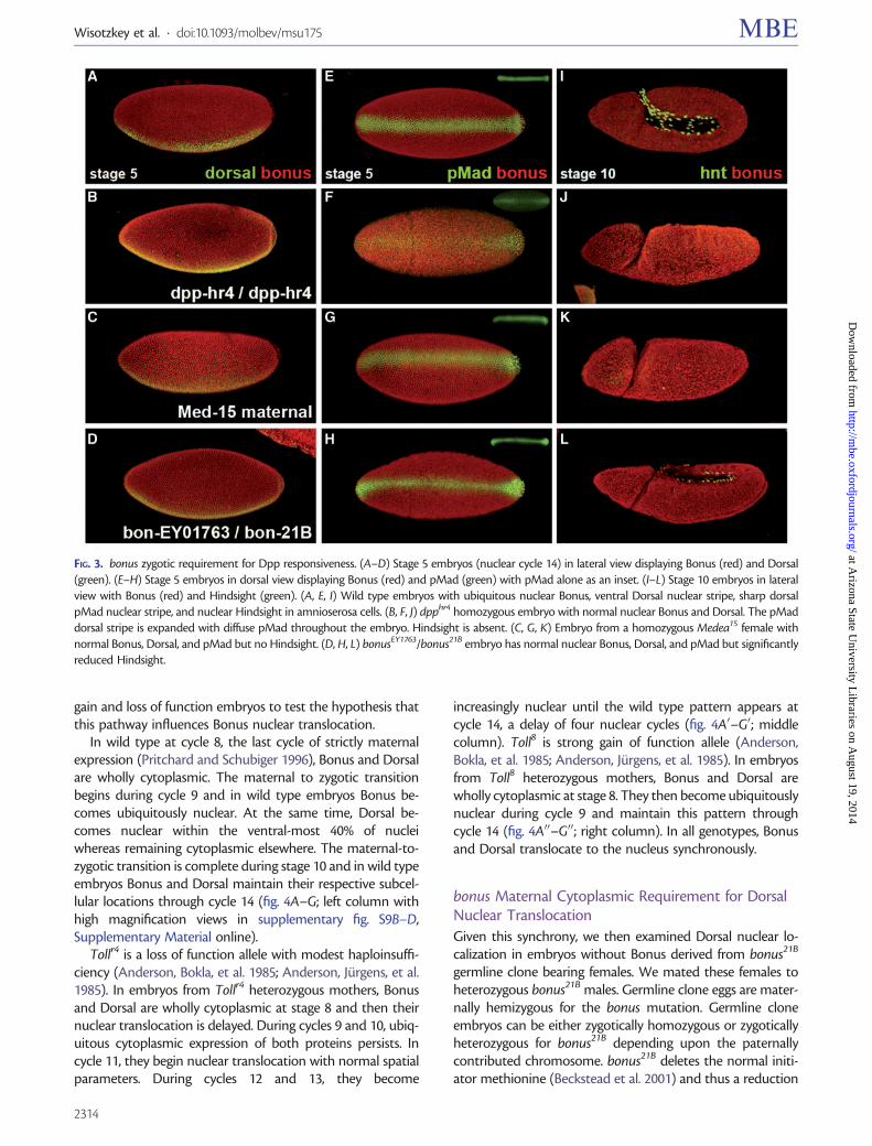
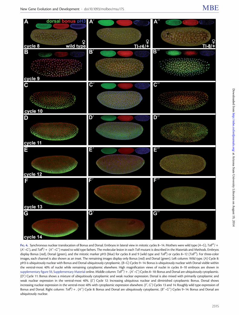
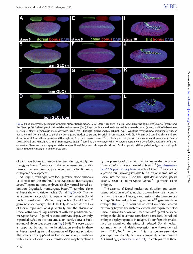
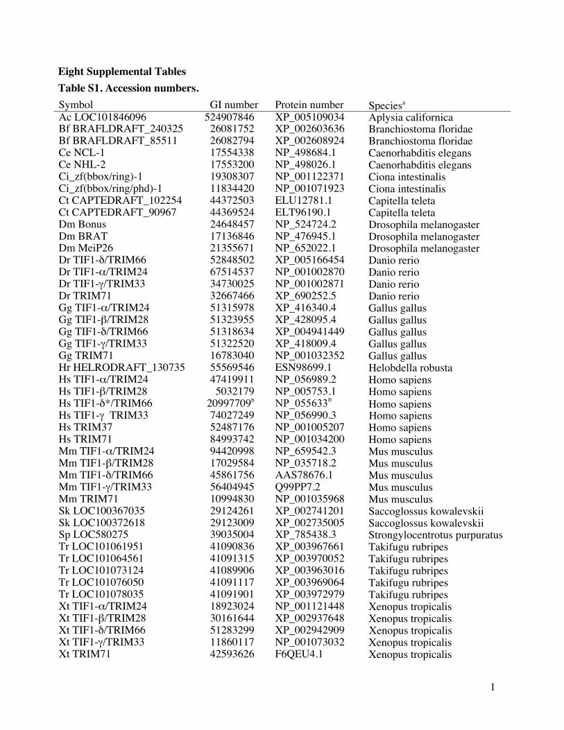
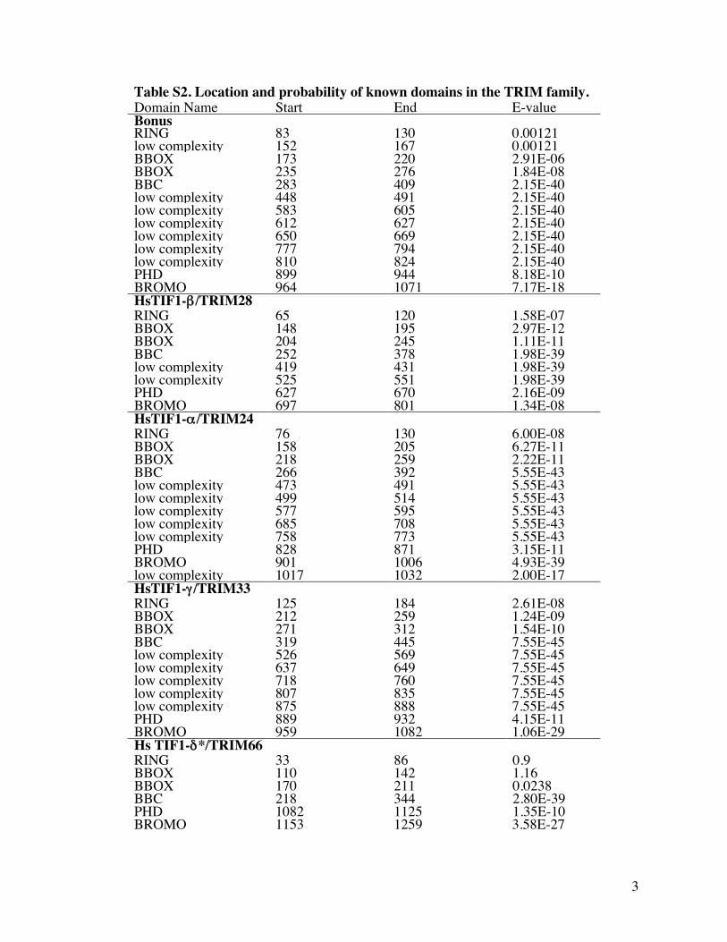
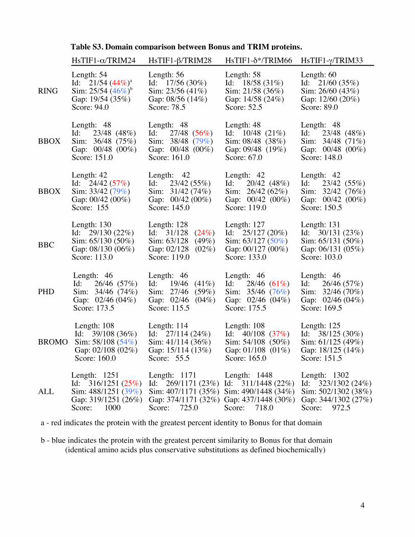
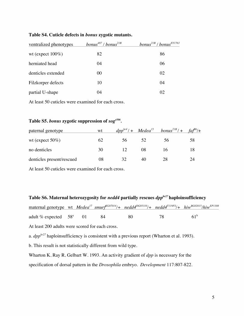
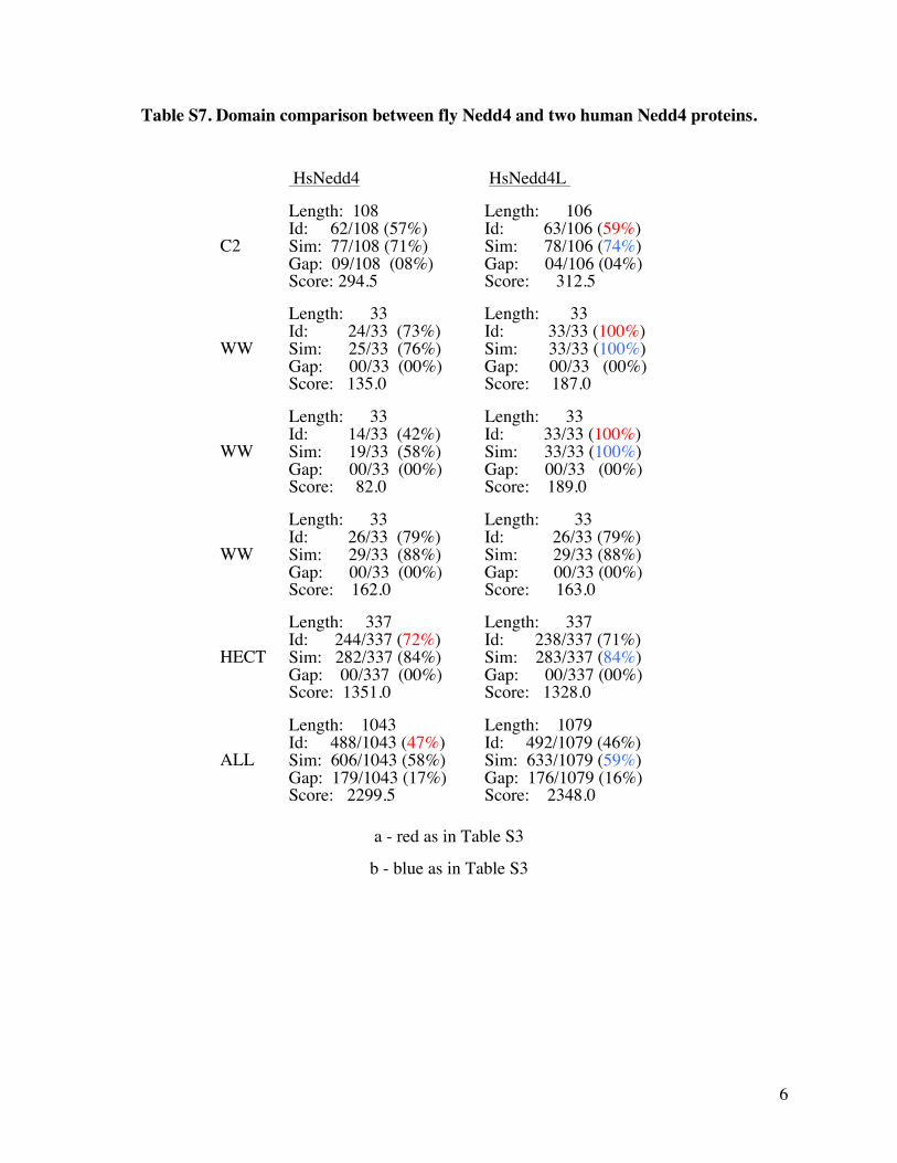
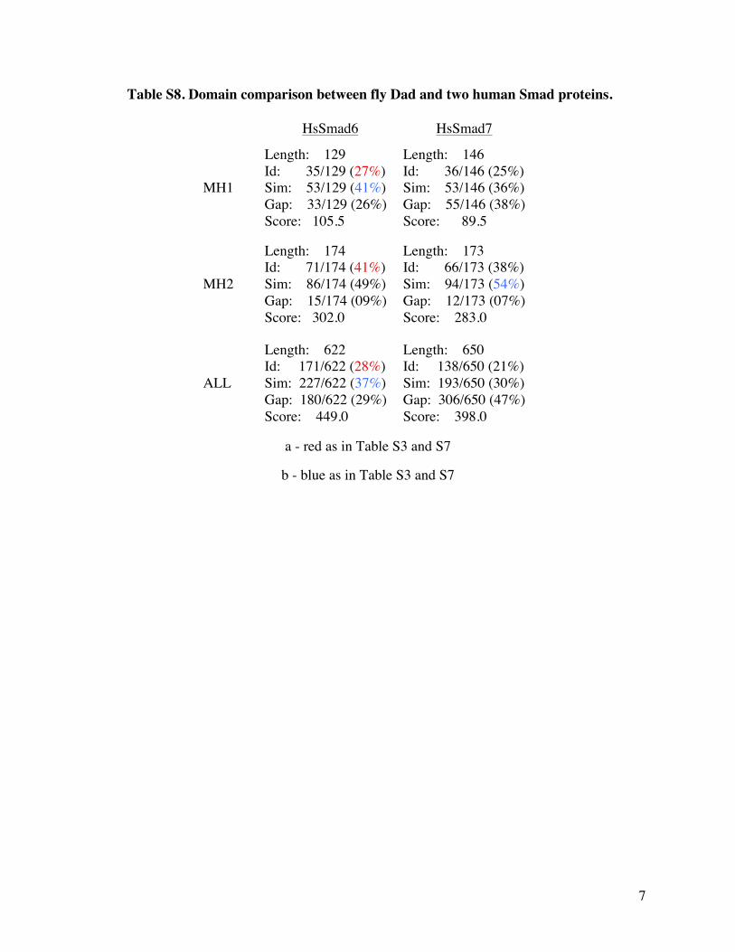
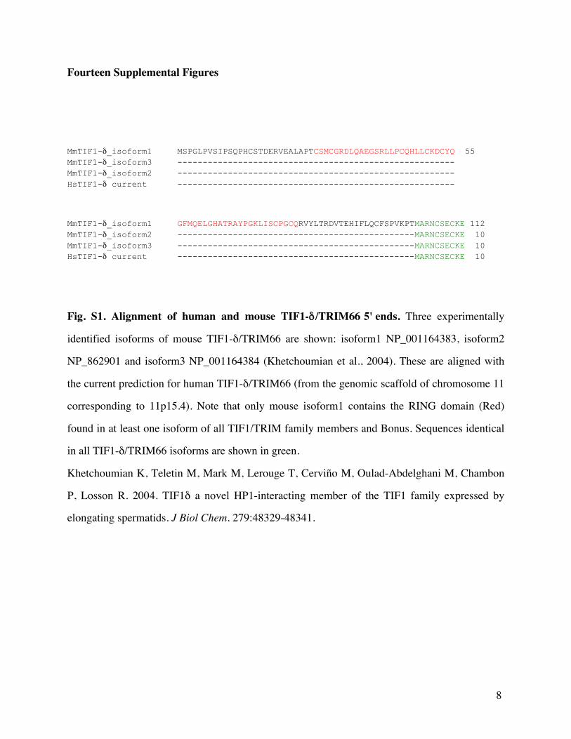
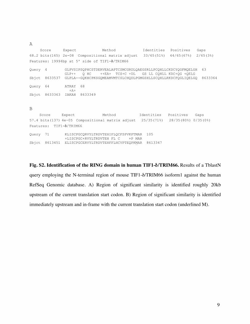
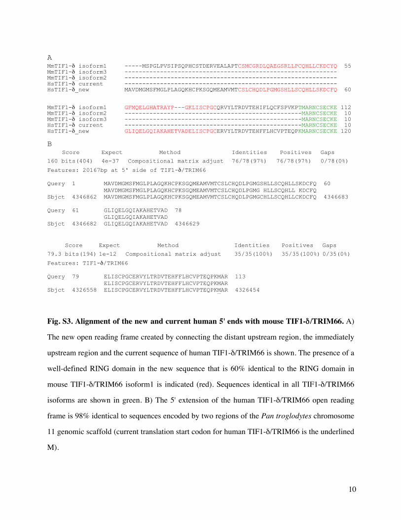
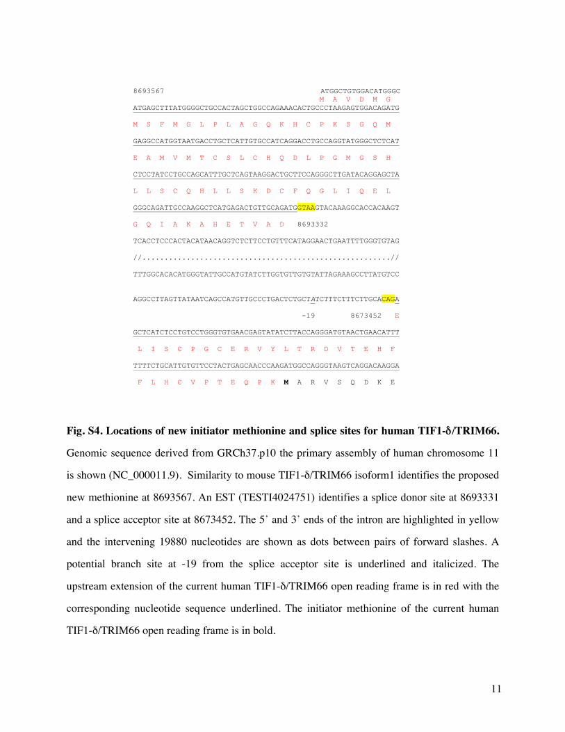
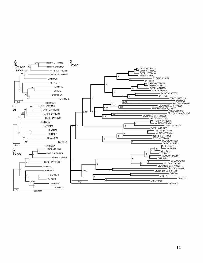
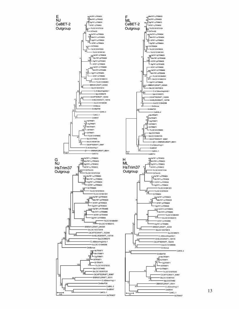
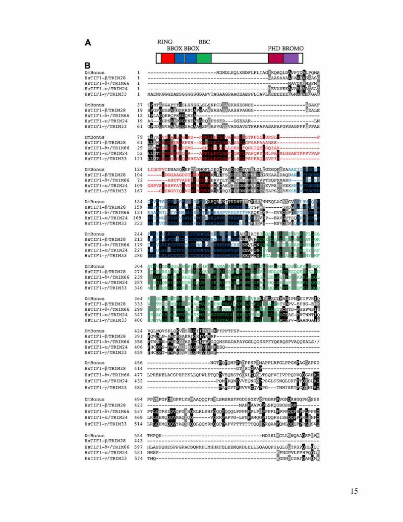
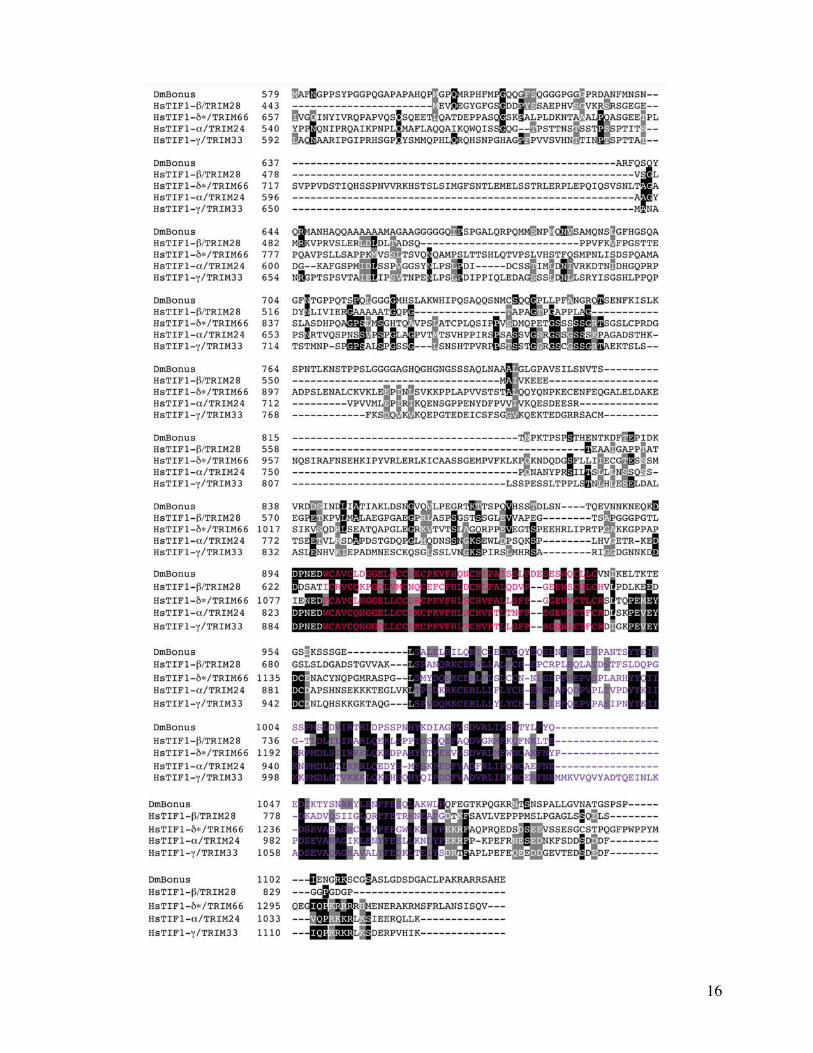
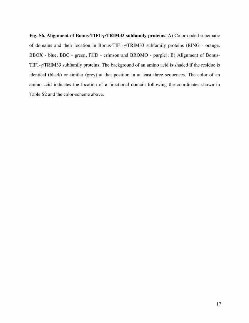
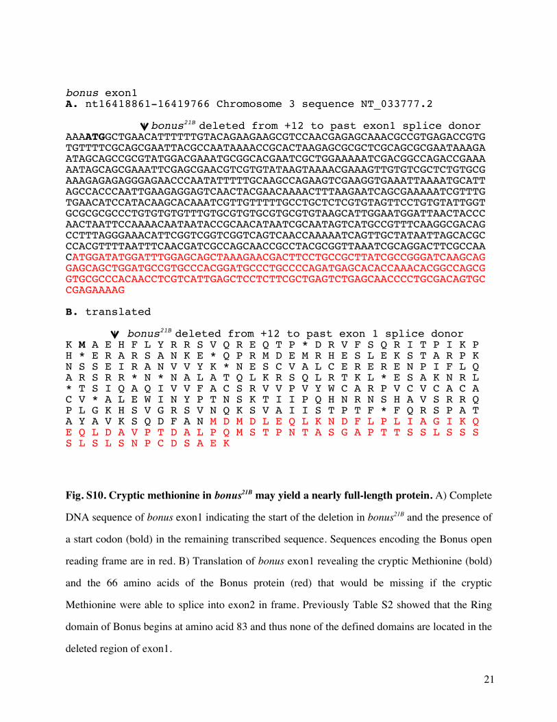
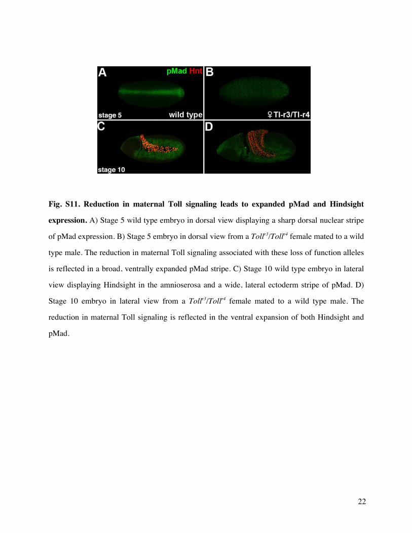
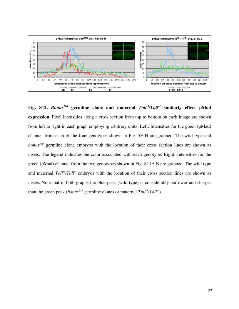
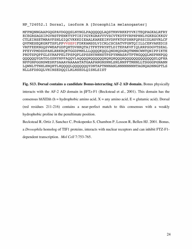
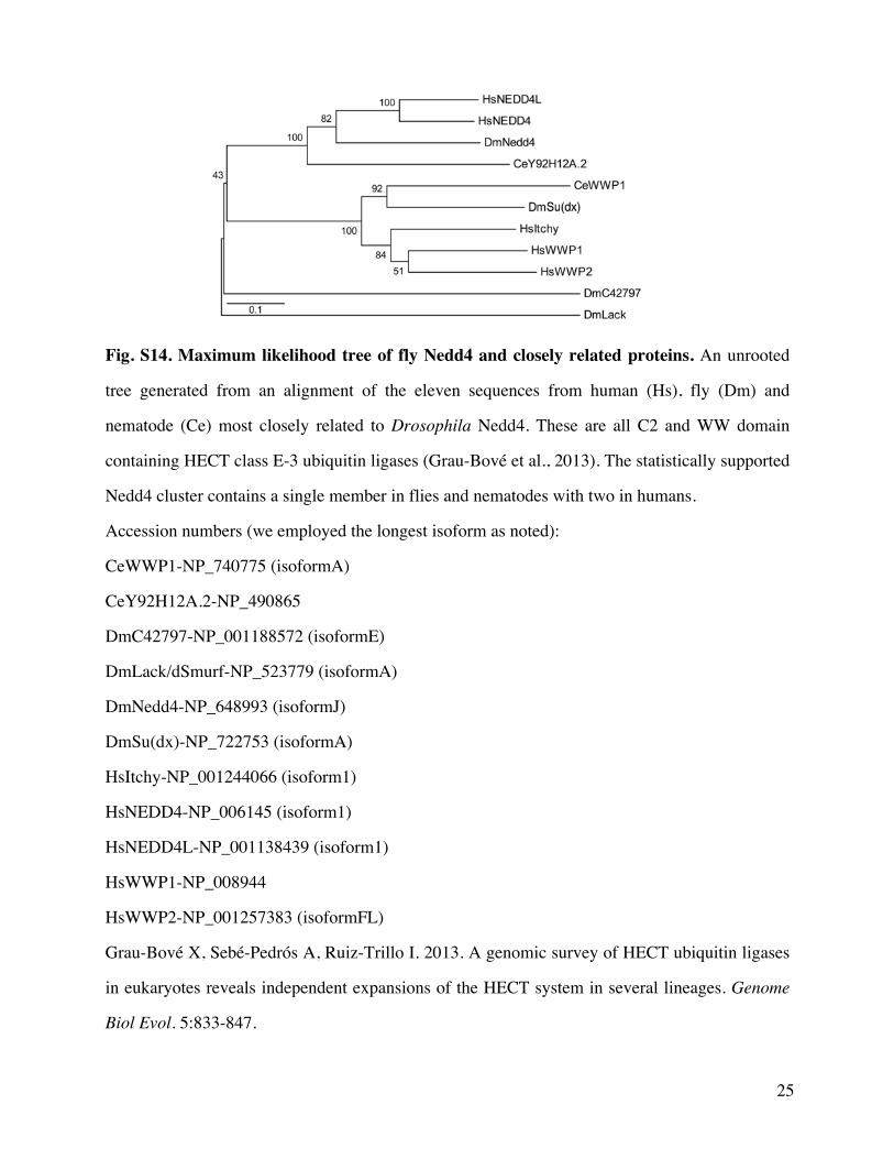