Embed Size (px)
Citation preview
New Dextramer® technology for T-cell epitope profiling, and single cell phenotyping using DNA barcodes, transcriptomic and genetic sequence analysis, on single cells.
Kivin Jacobsen1, Katie Pfeiffer2, Sarah E. B. Taylor2, Danial Riordan2, Josephine Y. Lee2, Charlotte Halgreen1, Liselotte Brix1
1Immudex, Fruebjergvej 3, 2100 Copenhagen, Denmark. 210x genomics, 7068 Koll Center Parkway Pleasanton, CA 94566, USA
Abstract: Identification of disease-specific T-cell epitopes is key to the development of many novel vaccines and immunotherapies. Profiling disease-specific T cells, emerging during a cellular immune response e.g. in tumor development or destruction, is an important aspect of personalized immunotherapy. The epitope diversity of the human population is large, and the technologies for identifying disease-specific epitopes have been inadequate. We have developed a process in which DNA barcoded Dextramer reagents are used to simultaneously screen for hundreds, or potentially thousands, of T-cell epitopes in a few milliliters of blood. Single cell deep phenotyping is quickly emerging, analyzing both cellular proteins, transcriptome and genetics of single cells by sequence analysis. DNA barcoded Dextramer reagents extend these technologies, to include simultaneous analysis of antigen specific T cells, by single cell sequencing. Methods: We present a process in which DNA barcoded Dextramer reagents, dCODE™ Dextramer® can be used to simultaneously screen for hundreds, or potentially thousands, of T-cell epitopes in one small patient-sample. Similar to previously reported academic methods, CITE-seq and REAP-seq, it is now possible to combine DNA barcoded MHCp Dextramer technology, with 10x Genomics Single Cell Immune Profiling Workflow, allowing direct correlation between the TCR specificity of the antigen specific T cell and its cognate T cell receptor sequence.
Conclusion: dCODE™ Dextramer® reagents in large libraries, allows for high-throughput screening of antigen-specific T cells in limited sample material. dCODE™ Dextramer® used in 10x Genomics Chromium Single Cell Immune Profiling Workflow[2], allows for deep T cell phenotyping by MHCp specificity, and TCR clonotype identification. Making a powerful and sensitive assay for T cell profiling, as well as allowing for identification of the cognate T cell receptor se-quence. Ref: [1]: Largescale detection of antigen specific T-cells using peptide MHC-I multimers labeled with DNA barcodes; A Kai Bentzen et. al., Nature, 2016. [2]: 10x Genomics poster no. 229: Simultaneous single cell analysis of multiple analytes resolves T cell populations at high resolution
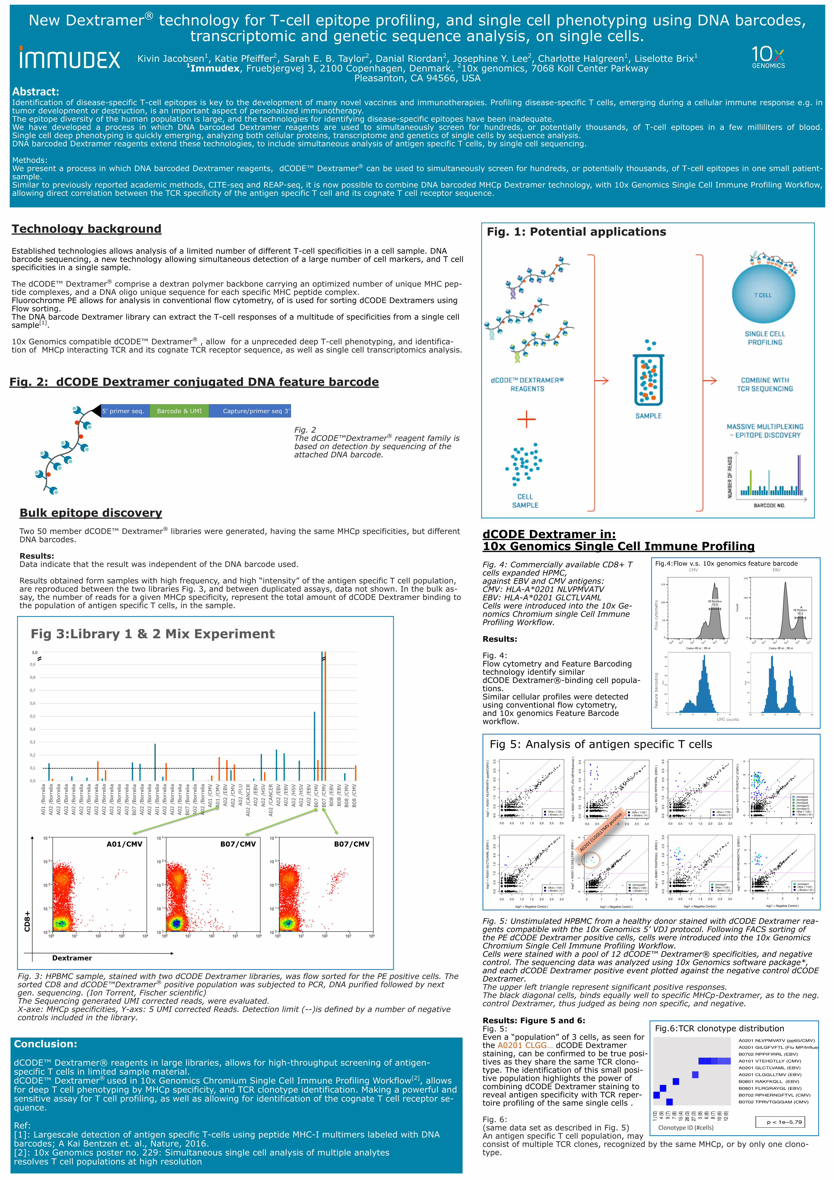
Fig 4
Fig. 2 The dCODE™Dextramer® reagent family is based on detection by sequencing of the attached DNA barcode.
Fig. 2: dCODE Dextramer conjugated DNA feature barcode
Fig. 1: Potential applications Technology background Established technologies allows analysis of a limited number of different T-cell specificities in a cell sample. DNA barcode sequencing, a new technology allowing simultaneous detection of a large number of cell markers, and T cell specificities in a single sample. The dCODE™ Dextramer® comprise a dextran polymer backbone carrying an optimized number of unique MHC pep-tide complexes, and a DNA oligo unique sequence for each specific MHC peptide complex. Fluorochrome PE allows for analysis in conventional flow cytometry, of is used for sorting dCODE Dextramers using Flow sorting. The DNA barcode Dextramer library can extract the T-cell responses of a multitude of specificities from a single cell sample[1]. 10x Genomics compatible dCODE™ Dextramer® , allow for a unpreceded deep T-cell phenotyping, and identifica-tion of MHCp interacting TCR and its cognate TCR receptor sequence, as well as single cell transcriptomics analysis.
Bulk epitope discovery Two 50 member dCODE™ Dextramer® libraries were generated, having the same MHCp specificities, but different DNA barcodes. Results: Data indicate that the result was independent of the DNA barcode used. Results obtained form samples with high frequency, and high “intensity” of the antigen specific T cell population, are reproduced between the two libraries Fig. 3, and between duplicated assays, data not shown. In the bulk as-say, the number of reads for a given MHCp specificity, represent the total amount of dCODE Dextramer binding to the population of antigen specific T cells, in the sample.
Fig. 3: HPBMC sample, stained with two dCODE Dextramer libraries, was flow sorted for the PE positive cells. The sorted CD8 and dCODE™Dextramer® positive population was subjected to PCR, DNA purified followed by next gen. sequencing. (Ion Torrent, Fischer scientific) The Sequencing generated UMI corrected reads, were evaluated. X-axe: MHCp specificities, Y-axs: 5 UMI corrected Reads. Detection limit (--)is defined by a number of negative controls included in the library.
dCODE Dextramer in: 10x Genomics Single Cell Immune Profiling
Results: Fig. 4: Flow cytometry and Feature Barcoding technology identify similar dCODE Dextramer®-binding cell popula-tions. Similar cellular profiles were detected using conventional flow cytometry, and 10x genomics Feature Barcode workflow.
Fig. 4: Commercially available CD8+ T cells expanded HPMC, against EBV and CMV antigens: CMV: HLA-A*0201 NLVPMVATV EBV: HLA-A*0201 GLCTLVAML Cells were introduced into the 10x Ge-nomics Chromium single Cell Immune Profiling Workflow.
Fig. 5: Unstimulated HPBMC from a healthy donor stained with dCODE Dextramer rea-gents compatible with the 10x Genomics 5’ VDJ protocol. Following FACS sorting of the PE dCODE Dextramer positive cells, cells were introduced into the 10x Genomics Chromium Single Cell Immune Profiling Workflow. Cells were stained with a pool of 12 dCODE™ Dextramer® specificities, and negative control. The sequencing data was analyzed using 10x Genomics software package*, and each dCODE Dextramer positive event plotted against the negative control dCODE Dextramer. The upper left triangle represent significant positive responses. The black diagonal cells, binds equally well to specific MHCp-Dextramer, as to the neg. control Dextramer, thus judged as being non specific, and negative.
Results: Figure 5 and 6: Fig. 5: Even a “population” of 3 cells, as seen for the A0201 CLGG… dCODE Dextramer staining, can be confirmed to be true posi-tives as they share the same TCR clono-type. The identification of this small posi-tive population highlights the power of combining dCODE Dextramer staining to reveal antigen specificity with TCR reper-toire profiling of the same single cells . Fig. 6: (same data set as described in Fig. 5) An antigen specific T cell population, may consist of multiple TCR clones, recognized by the same MHCp, or by only one clono-type.