Embed Size (px)
Citation preview
New concepts in white adiposetissue physiology
A.R.G. Proenca3*, R.A.L. Sertie1*, A.C. Oliveira2*, A.B. Campana1, R.O. Caminhotto1,
P. Chimin1 and F.B. Lima1
1Departamento de Fisiologia e Biofısica, Instituto de Ciencias Biomedicas, Universidade de Sao Paulo, Sao Paulo, SP, Brasil2Instituto Superior de Ciencias Biomedicas, Universidade Estadual do Ceara, Fortaleza, CE, Brasil
3Laboratorio de Biotecnologia, Faculdade de Ciencias Aplicadas, Universidade Estadual de Campinas, Limeira, SP, Brasil
Abstract
Numerous studies address the physiology of adipose tissue (AT). The interest surrounding the physiology of AT is primarily the
result of the epidemic outburst of obesity in various contemporary societies. Briefly, the two primary metabolic activities of white
AT include lipogenesis and lipolysis. Throughout the last two decades, a new model of AT physiology has emerged. Although
AT was considered to be primarily an abundant energy source, it is currently considered to be a prolific producer of biologically
active substances, and, consequently, is now recognized as an endocrine organ. In addition to leptin, other biologically active
substances secreted by AT, generally classified as cytokines, include adiponectin, interleukin-6, tumor necrosis factor-alpha,
resistin, vaspin, visfatin, and many others now collectively referred to as adipokines. The secretion of such biologically active
substances by AT indicates its importance as a metabolic regulator. Cell turnover of AT has also recently been investigated in
terms of its biological role in adipogenesis. Consequently, the objective of this review is to provide a comprehensive critical
review of the current literature concerning the metabolic (lipolysis, lipogenesis) and endocrine actions of AT.
Key words: Adipose tissue; Lipogenesis; Lipolysis; Adipogenesis; Endocrine
Introduction
It is well known that adipose tissue (AT) is the largest
energy reservoir of the body (1). Due to a lack of precise
knowledge regarding the functions of AT, for a long time
its metabolic purpose was considered to be passive
participation in the metabolic support system. Throughout
the previous two decades, however, this notion has
changed, and a new functional view of AT has emerged.
AT is now known to be a producer of biologically active
substances and is consequently recognized as an
endocrine organ, thus indicating a role for AT in the
regulation of energy metabolism (1).
Interest in the functional role of AT arose primarily due
to the epidemic surge of obesity in various contemporary
societies. This epidemic of obesity resulted in an increase
in AT research triggered primarily by an influx of AT
modus operandi, thus providing a better understanding of
the role of AT in the body’s metabolic control. Animal and
human studies (both in vivo and in vitro), utilizing several
methodological tools such as cellular and molecular
physiological, pharmacological, and clinical setting
approaches, have been utilized to investigate the func-
tional role of AT. Such studies, in addition to clarifying the
role of individual factors affecting metabolic control such
as diet, exercise, disease, age, and stress, also increased
understanding of the repercussions of an increase or a
decrease in adiposity in the body as a whole.
There are two primary metabolic activities of white
AT (1). They are lipogenesis (fatty acid synthesis and
storage) and lipolysis (mobilization or hydrolysis of
triglycerides). Both lipolysis and lipogenesis are regulated
by the integration of endocrine and neural mechanisms,
which cooperate in order to maintain the relative
constancy of body fat under normal conditions.
The discovery of leptin, a hormone secreted by AT
and related to satiety, significantly altered the perception
of AT in the body’s metabolism (2). Other biologically
active substances, generally known as cytokines, includ-
ing adiponectin, interleukin (IL)-6, tumor necrosis factor
alpha (TNF-a), resistin, vaspin, visfatin, and many others
secreted by AT have been identified (now collectively
Correspondence: A.R.G. Proenca, Laboratorio de Biotecnologia, Faculdade de Ciencias Aplicadas, UNICAMP, R. Pedro Zaccaria,
1300, 13484-350 Limeira, SP, Brasil. E-mail: [email protected]
*These authors contributed equally to this work.
Received May 2, 2013. Accepted November 4, 2013. First published online February 18, 2014.
Brazilian Journal of Medical and Biological Research (2014) 47(3): 192-205, http://dx.doi.org/10.1590/1414-431X20132911
ISSN 1414-431X Review
Braz J Med Biol Res 47(3) 2014 www.bjournal.com.br
termed adipokines), reinforcing the importance of AT as a
metabolic regulator (3). Adipokines have many functions,
including regulation of carbohydrate and lipid metabolism,
regulation of feeding behavior (including hunger and
satiety), and insulin sensitivity. The expression of adipo-
kines is altered by many clinical conditions such as
obesity and diabetes, indicating a role for adipokines in
the metabolic control of such illnesses.
Cell turnover of AT has also been extensively studied.
The fat tissue hyperplasia occurs ultimately due to a
physiological imbalance between adipogenesis and apop-
tosis, which ultimately results in the gain or loss of fat mass.
In pubescent adults, the formation of new fat cells is an
active process, resulting in the growth of AT. Throughout
pubescent adulthood, a certain bodily homeostatic control of
fat mass is maintained. In contrast, during the ages of 50-60,
adipogenesis tends to dominate, suggesting an increase in
adiposity in this age group. The process of adipogenesis
occurs repeatedly throughout human life. In addition, AT
turnover has been investigated, i.e., with the appearance of
newly formed adipocytes, the oldest adipocytes die, are
gradually removed from the tissue, and are replaced by new
adipocytes. Changes in the number of adipocytes occur
through a complex series of events involving proliferation
and differentiation of preadipocytes. It is estimated that
approximately 30% of the total cells present in AT are
immature or less differentiated adipocytes and precursors or
preadipocytes, i.e., cells that are capable of differentiating
intomature adipocytes, assuming their specific physiological
and morphological features.
The primary purpose of this review is to examine well
established concepts in the literature as well as the current
understanding of the metabolic (e.g., lipolysis, lipogenesis,
and cell turnover) and endocrine actions of AT.
Lipogenesis
In adipocytes, triacylglycerols (TAGs), the major
cellular constituents of white AT, are stored in a single
large lipid inclusion or droplet. These droplets occupy 80-
90% of the cell volume and the most central position of
the cytoplasm, displacing the nucleus, cytosol, and
remaining organelles to the periphery of the cell near
the plasma cell membrane. Accumulation of TAGs can
also occur in several other cell types; however, the
occurrence of TAGs in other cell types is indicative of
cellular malfunction and is a potential inductor of lipotoxic
reactions that trigger cellular apoptosis. When such
lipotoxicity occurs in muscle and liver, it leads to steatosis
in addition to the development of insulin resistance,
diabetes, and many other pathological diseases.
For the synthesis of TAGs, three moieties of fatty acids
(FAs) are assembled with a molecule of glycerol. The AT is
well prepared to perform this task; however, it requires
sources of FAs and glycerol. FAs can be obtained through
uptake from external sources [chylomicrons and very
low-density lipoproteins (VLDLs)] or by local synthesis from
acetyl coenzyme-A (CoA). Glycerol is obtained as glycerol-
3-phosphate (G3P) from glycolysis, glyceroneogenesis,
and phosphorylation of glycerol by glycerol kinase activity.
Lipogenic pathways
Sources of FAsFAs can be taken up as free FAs (FFAs), which are
transported in the circulation bound to albumin or can be
obtained by enzymatic hydrolysis of TAGs carried by
lipoproteins such as chylomicrons (released from the
intestinal tract after fat absorption) and, particularly,
VLDLs, and to a lesser extent LDLs. The TAGs contained
in these lipoproteins must first be hydrolyzed by lipoprotein
lipase (LPL) located in the endothelium of the AT capillaries.
Adipocytes express several FA transporters located in
the cell membrane to facilitate and control their transport,
such as protein CD36, FA transporter protein, and
membrane FA-binding protein (FABP). Because FAs are
not soluble in the cytosolic aqueous medium and can
induce toxic effects on membranes, once inside the cells,
FAs bind to specific transporter proteins, known as FABPs
(FABP-4 or aP2). These proteins carry FAs from the cell
membrane to the site of action of the enzyme acyl-CoA
synthase, whereby FAs are esterified with CoA to form
acyl-CoA. Following this, acyl-CoA binds to and is carried
by acyl-CoA-binding protein, to the sites of esterification
with G3P to form TAG in the endoplasmic reticulum (ER).
De novo lipogenesisDe novo lipogenesis is the synthesis of FA from
nonlipid substrates, primarily carbohydrates. De novolipogenesis can occur both in the liver and in AT. The
importance and contribution of the liver and AT varies
between species. Studies indicate that de novo lipogenesis
is less active in human AT than in the liver, on a per gram of
tissue basis (4). Furthermore, controversy exists regarding
differences in the lipogenic capacity of rats and humans.
Although some studies indicate that this pathway is more
active in rats, other studies suggest that such differences
are due to diet composition, whereby when rats and
humans consume diets with similar composition, a
difference in lipogenic capacity is no longer observed (5).
For de novo lipogenesis to occur, acetyl-CoA is
carboxylated by acetyl-CoA carboxylase (ACC) into
malonyl-CoA, while oxaloacetate is reduced to malate
by malate dehydrogenase (MDH). Activation of FA
synthase (FAS) allows malonyl-CoA and acetyl-CoA to
assemble and to elongate the hydrocarbonic chain of FA,
thus forming palmitic acid (16:0). Palmitic acid is then
further elongated by an elongase to form stearic acid
(18:0). Both palmitic acid and stearic acid are desaturated
by stearoyl-CoA desaturase to form palmitoleic (16:1, n7)
and oleic acids (18:1, n9), which are subsequently
esterified with G3P to form TAG.
New concepts in white adipose tissue physiology 193
www.bjournal.com.br Braz J Med Biol Res 47(3) 2014
An important cofactor in FA synthesis is reduced
nicotinamide-adenine dinucleotide phosphate (NADPH),
synthesized in the cytoplasm as a by-product of two
pathways. The first pathway involves the following
reactions: oxaloacetate formed by cleavage of cytosolic
citrate is reduced to MDH by the enzyme NAD-malate
dehydrogenase. This MDH then undergoes oxidative
decarboxylation to form pyruvate and CO2, while gen-
erating NADPH from NADP+ in a reaction catalyzed by
malic enzyme. Reuptake of pyruvate by mitochondria
occurs, whereby pyruvate combines with CO2 to regen-
erate oxaloacetate in a reaction catalyzed by the enzyme
pyruvate carboxylase. The second pathway, or the
pentose synthesis pathway, involves the conversion of
glucose-6-phosphate (G6P) to 6-phosphogluconate by
the action of G6P dehydrogenase (G6PDH).
It is suggested that, in primary white adipocyte culture,
G6PDH mRNA levels change in parallel with FAS and
ACC mRNA levels, indicating that this enzyme may be
involved in the expression of other lipogenic enzymes (6).
Accordingly, overexpression of G6PDH in mouse 3T3-L1
cells promoted the expression of adipogenic and lipogenic
gene markers, including FAS, sterol regulatory-element
binding protein 1c (SREBP-1c), peroxisome proliferator-
activated receptor-gamma (PPAR-c), and aP2 (7).
Production of G3PThe synthesis of TAGs also requires a constant supply
of G3P, as availability of G3P controls the esterification of
FA. Hepatocytes use the glycerol released by AT during
lipolysis for the phosphorylation of glycerol to G3P via
the enzyme glycerol kinase (8). The presence of glycerol
kinase in adipocytes is, however, controversial. In
adipocytes, although some glycerol released during
lipolysis can be directly phosphorylated and reused for
TAG synthesis, the contribution of this pathway to G3P
production is negligible. In contrast, studies have shown
that G3P is generated in adipocytes via an important
metabolic pathway known as glyceroneogenesis, which
has been shown to be the quantitatively predominant
source of G3P (9).
Hence, cytoplasmic G3P is derived from three path-
ways: glycolysis, gluconeogenesis, and glycerol kinase
activity. In the glycolysis pathway, after entry into the cell,
glucose is phosphorylated and ultimately converted via
the glycolytic pathway to dihydroxyacetone phosphate
(DHAP), and glyceraldehyde-3-phosphate. The DHAP is
then further reduced by glycerol phosphate dehydrogen-
ase (GPDH) to form G3P.
Through the glyceroneogenic pathway (which consists
of the initial stages of canonical gluconeogenesis),
precursors other than glycerol or glucose are converted
to G3P, with the main substrates being pyruvate, lactate,
and amino acids. Pyruvate is carboxylated to oxaloace-
tate, which then leaves the mitochondria and is de-
carboxylated by cytoplasmic phosphoenolpyruvate
carboxykinase to form phosphoenolpyruvate. This is the
rate-limiting step of the glyceroneogenic pathway (10).
Phosphoenolpyruvate is then converted to glyceralde-
hyde-3-phosphate, which is reduced to DHAP by glycer-
aldehyde-3-phosphate dehydrogenase and then to G3P
by GPDH.
TAG synthesisIn adipocytes, the biosynthesis of TAG is the result
of esterification of alcoholic residues of G3P by various
enzymes, namely, G3P acyltransferases (GPATs, the
most abundant isoforms being GPAT1 and GPAT2), 1-
acylglycerol-3-phosphate acyltransferase (AGPAT, the
most abundant isoform being AGPAT2), phosphatidic
acid phosphatase, and diacylglycerol acyltransferase
(DGAT, the most abundant isoforms being DGAT1 and
DGAT2). The isoforms of these enzymes are encoded by
different genes (11). All enzymes involved in this pathway
of the biosynthesis of TAG are found in smooth ER. The
importance of these enzymes in the control of TAG
storage in AT has been demonstrated in studies of DGAT-
deficient mice and in studies of human subjects with
congenital lipodystrophy (11).
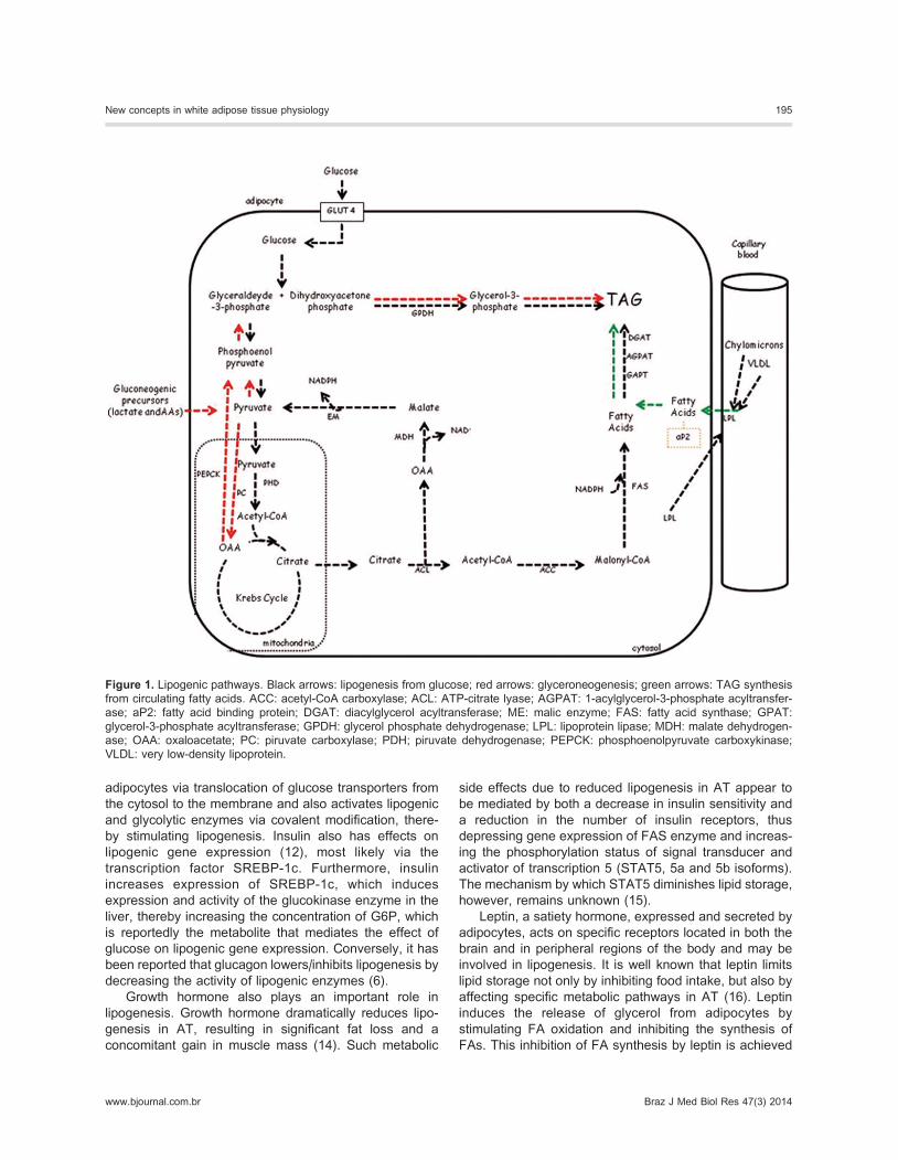
A summary of all pathways involved in lipogenesis is
provided in Figure 1.
Regulation of lipogenesis
NutritionalLipogenesis is highly influenced by factors such as
feeding, fasting, and diet composition. Excessive carbo-
hydrate consumption stimulates lipogenesis in both the
liver and AT, increasing the availability of TAG in the
postabsorptive state. In contrast, a high-fat/low-carbohy-
drate diet and fasting reduces de novo lipogenesis and
lipogenesis, respectively, in AT (12). These changes are
related to the increased or reduced expression and
activity of LPL enzyme (high-carbohydrate/high-fat diets
and fasting, respectively). In addition, it has been reported
that the reduced lipogenic response during fasting is
primarily due to a decreased capacity of white AT to
generate acetyl-CoA from glucose, rather than an inhibi-
tion of lipogenic enzymes involved in FA synthesis.
Blood glucose levels act directly on lipogenic cap-
ability via three distinct mechanisms. First, because
glucose is a substrate for lipogenesis, thus producing
acetyl-CoA by glycolysis, glucose activates FA synthesis
(13). Second, glucose stimulates the lipogenic enzyme
synthesis of ATP-citrate lyase, G6PDH, ACC, malic
enzyme, and FAS (8). Finally, glucose promotes lipogen-
esis by stimulating insulin secretion and inhibiting
glucagon release from the pancreas (6).
HormonalInsulin is one of the most important hormonal factors
that affect lipogenesis. Insulin increases glucose uptake in
194 A.R.G. Proenca et al.
Braz J Med Biol Res 47(3) 2014 www.bjournal.com.br
adipocytes via translocation of glucose transporters from
the cytosol to the membrane and also activates lipogenic
and glycolytic enzymes via covalent modification, there-
by stimulating lipogenesis. Insulin also has effects on
lipogenic gene expression (12), most likely via the
transcription factor SREBP-1c. Furthermore, insulin
increases expression of SREBP-1c, which induces
expression and activity of the glucokinase enzyme in the
liver, thereby increasing the concentration of G6P, which
is reportedly the metabolite that mediates the effect of
glucose on lipogenic gene expression. Conversely, it has
been reported that glucagon lowers/inhibits lipogenesis by
decreasing the activity of lipogenic enzymes (6).
Growth hormone also plays an important role in
lipogenesis. Growth hormone dramatically reduces lipo-
genesis in AT, resulting in significant fat loss and a
concomitant gain in muscle mass (14). Such metabolic
side effects due to reduced lipogenesis in AT appear to
be mediated by both a decrease in insulin sensitivity and
a reduction in the number of insulin receptors, thus
depressing gene expression of FAS enzyme and increas-
ing the phosphorylation status of signal transducer and
activator of transcription 5 (STAT5, 5a and 5b isoforms).
The mechanism by which STAT5 diminishes lipid storage,
however, remains unknown (15).
Leptin, a satiety hormone, expressed and secreted by
adipocytes, acts on specific receptors located in both the
brain and in peripheral regions of the body and may be
involved in lipogenesis. It is well known that leptin limits
lipid storage not only by inhibiting food intake, but also by
affecting specific metabolic pathways in AT (16). Leptin
induces the release of glycerol from adipocytes by
stimulating FA oxidation and inhibiting the synthesis of
FAs. This inhibition of FA synthesis by leptin is achieved
Figure 1. Lipogenic pathways. Black arrows: lipogenesis from glucose; red arrows: glyceroneogenesis; green arrows: TAG synthesis
from circulating fatty acids. ACC: acetyl-CoA carboxylase; ACL: ATP-citrate lyase; AGPAT: 1-acylglycerol-3-phosphate acyltransfer-
ase; aP2: fatty acid binding protein; DGAT: diacylglycerol acyltransferase; ME: malic enzyme; FAS: fatty acid synthase; GPAT:
glycerol-3-phosphate acyltransferase; GPDH: glycerol phosphate dehydrogenase; LPL: lipoprotein lipase; MDH: malate dehydrogen-
ase; OAA: oxaloacetate; PC: piruvate carboxylase; PDH; piruvate dehydrogenase; PEPCK: phosphoenolpyruvate carboxykinase;
VLDL: very low-density lipoprotein.
New concepts in white adipose tissue physiology 195
www.bjournal.com.br Braz J Med Biol Res 47(3) 2014
by decreasing the expression of genes involved in FA and
TAG synthesis (16). The transcription factor SREBP-1 is
also inhibited by leptin, indicating that the inhibitory effect
of leptin may also involve downregulation of the expres-
sion of lipogenic genes (17).
Lipolysis: general mechanisms
During periods of nutrient deprivation, stress, or
physical exercise, lipolysis of TAG reserves is stimulated.
Lipid droplets containing TAGs are surrounded by a
phospholipid monolayer including structural proteins,
enzymes, and coactivators. In general, lipolytic activity
culminates in the systemic release of FFAs and glycerol.
The release of FFAs into the blood is used as an energy
source by other tissues, such as heart and skeletal
muscle.
For the systemic release of FFAs and glycerol to
occur, a number of extra- and intracellular events are
required. These events include the presence of hormones
that signal to adipocytes the need to release energy
substrates to meet the increased demand of energy or to
supply energy in cases of nutrient deprivation, thus
sparing the use of glucose by lower priority body cells,
because glucose is the primary energy source of the
central nervous system.
MessengersIn humans, catecholamines, including epinephrine,
norepinephrine, and insulin, are the primary regulators of
lipolysis. It has also been reported in both in vivo and invitro studies that natriuretic peptides (NPs), in addition to
affecting cardiovascular and renal functions, are important
stimulating agents for lipolysis.
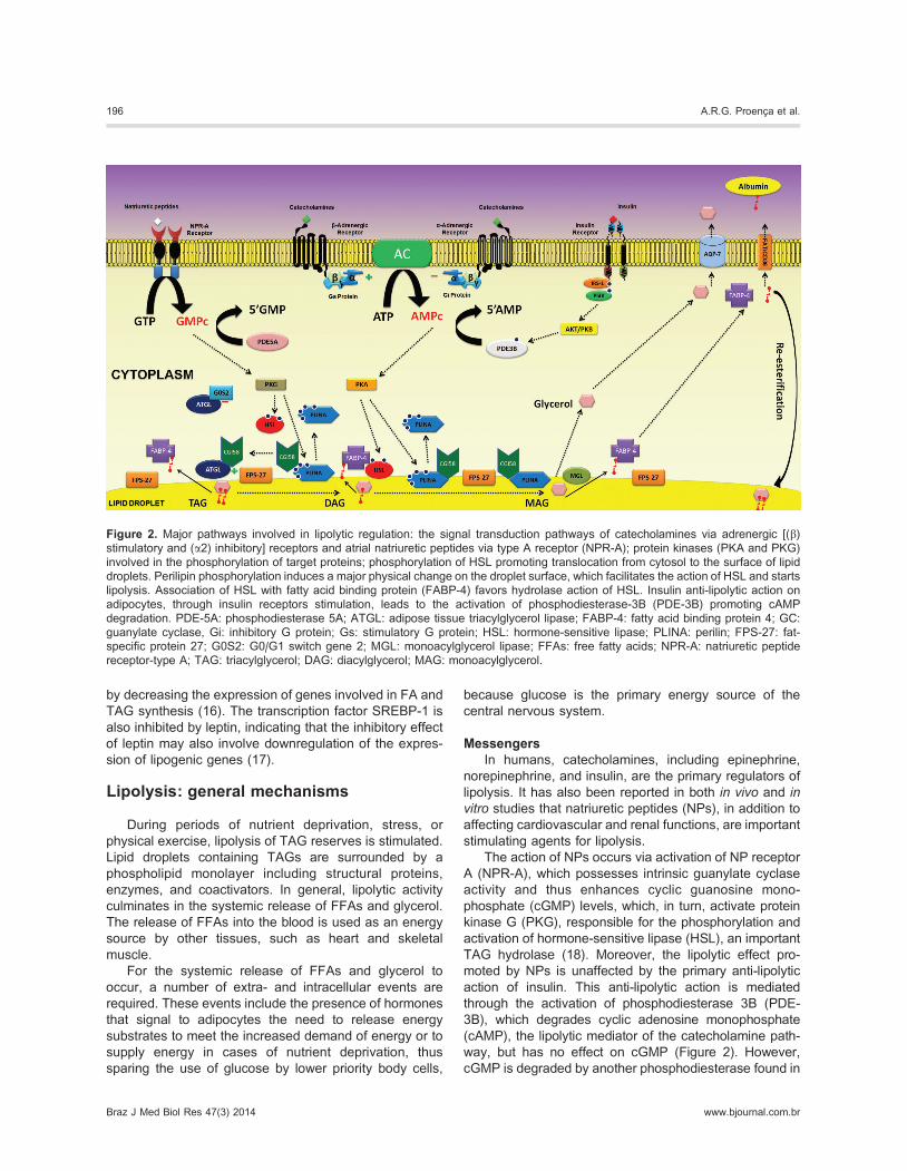
The action of NPs occurs via activation of NP receptor
A (NPR-A), which possesses intrinsic guanylate cyclase
activity and thus enhances cyclic guanosine mono-
phosphate (cGMP) levels, which, in turn, activate protein
kinase G (PKG), responsible for the phosphorylation and
activation of hormone-sensitive lipase (HSL), an important
TAG hydrolase (18). Moreover, the lipolytic effect pro-
moted by NPs is unaffected by the primary anti-lipolytic
action of insulin. This anti-lipolytic action is mediated
through the activation of phosphodiesterase 3B (PDE-
3B), which degrades cyclic adenosine monophosphate
(cAMP), the lipolytic mediator of the catecholamine path-
way, but has no effect on cGMP (Figure 2). However,
cGMP is degraded by another phosphodiesterase found in
Figure 2. Major pathways involved in lipolytic regulation: the signal transduction pathways of catecholamines via adrenergic [(b)stimulatory and (a2) inhibitory] receptors and atrial natriuretic peptides via type A receptor (NPR-A); protein kinases (PKA and PKG)
involved in the phosphorylation of target proteins; phosphorylation of HSL promoting translocation from cytosol to the surface of lipid
droplets. Perilipin phosphorylation induces a major physical change on the droplet surface, which facilitates the action of HSL and starts
lipolysis. Association of HSL with fatty acid binding protein (FABP-4) favors hydrolase action of HSL. Insulin anti-lipolytic action on
adipocytes, through insulin receptors stimulation, leads to the activation of phosphodiesterase-3B (PDE-3B) promoting cAMP
degradation. PDE-5A: phosphodiesterase 5A; ATGL: adipose tissue triacylglycerol lipase; FABP-4: fatty acid binding protein 4; GC:
guanylate cyclase, Gi: inhibitory G protein; Gs: stimulatory G protein; HSL: hormone-sensitive lipase; PLINA: perilin; FPS-27: fat-
specific protein 27; G0S2: G0/G1 switch gene 2; MGL: monoacylglycerol lipase; FFAs: free fatty acids; NPR-A: natriuretic peptide
receptor-type A; TAG: triacylglycerol; DAG: diacylglycerol; MAG: monoacylglycerol.
196 A.R.G. Proenca et al.
Braz J Med Biol Res 47(3) 2014 www.bjournal.com.br

the adipocyte, PDE-5A. NPs emerged as potent regulators
of lipolysis in humans, especially during exercise-stimu-
lated lipolysis (18). Lipolytic effects stimulated by growth
hormone, TNF-a, adrenocorticotropin, and glucocorticoids
have also been demonstrated (19-22).
Catecholamines remain the major lipolytic agents in
adipocytes. Particularly in humans, during periods of
fasting, catecholamines are the primary stimulators of
lipolysis. The regulatory effects of catecholamines occur
as a result of intracellular signaling triggered by activation
of several adrenergic receptors, namely, b-1, b-2, b-3,and a-2. In rodents, b-3-adrenergic receptor is the primary
lipolytic route, whereas in humans, b-1, b-2, and a-2-
adrenergic receptors play a regulatory role (23).
Adrenergic receptors have an extracellular and a trans-
membrane domain along with seven hydrophobic seg-
ments and an intracytoplasmic region that is coupled to
regulatory GTP/GDP-associated proteins or G proteins
(Figure 2). The ligand binding site of adrenergic receptors
is located within the transmembrane domain. The b-adrenergic receptors are Gs-protein coupled receptors,
which activate adenylate cyclase, a plasma membrane
enzyme anchored to the inner cytoplasmic leaflet that
catalyzes the formation of cAMP from ATP. In contrast, a-2-adrenergic receptors are Gi-protein coupled, inhibiting
adenylate cyclase activation and thus preventing the
formation of cAMP. Noradrenaline has a greater affinity
for the a-2-adrenergic receptor than for the b-adrenergicreceptors, suggesting a role for the a-adrenergic pathway
in the regulation of lipolysis in human subcutaneous AT
under conditions of exercise or stress (24).
The chemical messengers involved in the lipolytic
activity of AT, such as catecholamines, insulin, NPs, and
others cited earlier, are ultimately related to the synthesis
or degradation of second messengers, such as cAMP and
cGMP, which are involved in the activation of enzymes
responsible for the control of lipolysis.
Lipid dropletsWhite adipocytes characteristically exhibit a large
central fat droplet in the cytoplasm. In contrast, brown
adipocytes show multiple small cytoplasmic fat droplets
together with a rich amount of mitochondria, enabling these
adipocytes a high capacity to oxidize substrates, primarily
fatty acids. The lipid droplet is a vacuolar compartment
responsible for the storage of neutral lipids. Movement of
TAGs in and out of the lipid droplet is highly regulated (25).
Monoacylglycerol (MAG) and diacylglycerol (DAG) mole-
cules are intermediate compounds in the synthesis and
degradation of TAGs. This lipid arrangement within the lipid
droplet protects the adipocytes from harmful effects
associated with excess intracellular fat, known as lipotoxi-
city. Consequently, disordered processing of fat that can
potentially generate toxic fat metabolites, such as lipoper-
oxides, is avoided (25). Adipocyte hypertrophy and
hyperplasia prevent accumulation of fat in the cytoplasm
of other cell types in an organism unable to survive
abnormal fat deposition. Indeed, the accumulation of
abnormal fat may result in excess generation of ceramides,
lipoperoxides, and other reactive oxygen and nitrogen
species, such as nitric oxide and nitrolipids that trigger
cellular apoptosis (26-28). In light of the above-mentioned
studies, it may be assumed that lipid metabolism in
adipocytes comprises mechanisms that control lipid
mobilization via chemical messengers, with the lipid droplet
as a target of all intracellular signaling.
Enzymes, proteins, and coactivatorsIntracellular increases of cAMP and cGMP concentra-
tions culminate in activation of dependent protein kinase
A (PKA) and PKG, respectively. These enzymes transfer
high-energy phosphate groups from donor molecules,
such as ATP, to target proteins. The PKA enzyme
consists of four subunits: two regulatory and two catalytic.
In the presence of these activators, the regulatory subunit
undergoes conformational changes resulting in catalytic
activation.
Consequently, the b-adrenergic pathway, as described
previously, promotes PKA activation. Once activated, PKA
phosphorylates serine hydroxyl groups of HSL, resulting
in activation of HSL (Figure 2). Once activated, HSL
translocates from the cytosol to the lipid droplet, where it
binds to FABP-4, ALBP, or aP2, and begins to hydrolyze
TAGs, DAGs, and MAGs with relative hydrolytic rates of
1:10:0.5, respectively (29). Concurrently, PKA phosphor-
ylates the lipid droplet surface protein perilipin, promoting
displacement of perilipin to the cytosol. Such displacement
of perilipin is in the opposite direction to the translocation of
HSL, allowing space in the lipid droplet interface for contact
between the hydrolase and its substrate (30). Perilipin-1 is
a member of the PAT lipid droplet protein family, which also
includes adipophilin or adipocyte differentiation-related
protein (currently named perilipin-2), a tail-interacting
protein of 47 kDa (currently named perilipin-3), S3-12
(currently named perilipin-4), OXPAT/MLDP (currently
named perilipin-5), and fat-specific protein 27 (FSP27)
(31). These proteins cover the lipid droplet surface,
regulating and coordinating basal lipid storage and
mediating stimulated lipolysis (30,31). A study by Sztalryd
et al. (32) showed that the presence and phosphorylation of
perilipin-1 is essential for HSL translocation during lipolytic
activity, as adipocytes isolated from mice that did not
express perilipin-1 showed a loss of lipolytic activity
following b-adrenergic stimulation. Although perilipin-1
restricts HSL access to TAG reserves, thereby reducing
basal lipolysis, the phosphorylated form of perilipin-1 is
essential for hydrolytic HSL activity during stimulated
lipolysis.
In 2004, three independent groups reported the existence
of a second enzyme involved in the hydrolysis of triglycer-
ides, known as AT triglyceride lipase (ATGL), desnutrin,
adiponutrin, or calcium-independent phospholipase A2.
New concepts in white adipose tissue physiology 197
www.bjournal.com.br Braz J Med Biol Res 47(3) 2014
Consequently, the idea of HSL as the sole enzyme
responsible for TAG hydrolysis in mammals was abandoned
and replaced with the concept that HSL shares this ability
equally with another enzyme (33). The ATGL enzyme is
highly expressed in humans and rodents and has a high
hydrolyzing activity on TAGs (33). In a study conducted by
Haemmerle et al. (34) in 2006, ATGL knockout mice showed
more than a 75% decrease in FFA release, with consequent
accumulation of ectopic TAGs in muscle tissues, resulting in
severe myopathy in cardiac muscle and a general weakness
in energy balance, compromising animal survival.
The mechanisms that regulate activity of ATGL in
response to b-adrenergic stimulation remain unknown.
Although it is known that ATGL can be phosphorylated, it
is unclear whether this modification is vital for ATGL
activity (30). It is known, however, that ATGL activity is
greatly enhanced in the presence of the coactivator
comparative gene identification-58 (CGI-58), also known
as a/b-hydrolase domain-containing protein 5. Reports
suggest a mechanism involving perilipin-1 and CGI-58,
whereby the availability of CGI-58 is dependent on
perilipin-1 phosphorylation (Figure 2). In adipocytes with
no lipolytic stimulation, CGI-58 is located on the lipid
droplet surface next to perilipin-1, whereas, in the
stimulated state, perilipin-1 is phosphorylated via PKA or
PKG, resulting in displacement of perilipin-1 into the
cytoplasm and dissociation of CGI-58, leaving CGI-58
freely available for interaction with and activation of ATGL
(30). It has been reported that the presence of FSP27 on
the lipid droplet surface exerts an important role in lipolytic
activity of ATGL. Reduced expression of perilipin-1 and
FSP27 on the lipid droplet surface results in elevated basal
lipolysis (35). In contrast, when perilipin-1 and FSP27 work
in unison on the lipid droplet surface, adipocyte lipolytic
capacity is well maintained (36). Ultimately, FSP27 acts by
limiting the presence of ATGL at the lipid droplet interface,
whereas perilipin-1 is crucial in control of the b-adrenergic-mediated ATGL lipolytic response (36). To further increase
the complexity of lipolysis, another protein, identified as
G0/G1 switch gene 2, has been shown to interact with
ATGL, inhibiting its activity (37). Furthermore, it has been
shown that perilipin-5, expressed in both myocytes and
brown adipocytes, can negatively modulate ATGL activity
under basal conditions (38,39). Such reports indicate that
lypolytic action of ATGL is controlled by a variety of
mechanisms, emphasizing the essential role of ATGL on
lipid mobilization in adipocytes. Together, both ATGL and
HSL are responsible for more than 95% TAG hydrolysis in
mouse adipocytes.
The other hydrolase located in adipocytes is MAG
lipase (MGL). Unlike ATGL and HSL, MGL does not
hydrolyze TAG and DAG, but it performs a specific action
on MAG. Although its hydrolytic action is required for the
complete hydrolysis of TAGs in vitro, the fact that HSL is
also capable of hydrolyzing MAG indicates that the
presence of MGL in vivo is not vital (30). MGL activity
promotes dissociation of the last FA and glycerol of the
MAG molecule. There is no evidence that cellular
expression and activity of this enzyme are regulated by
hormones or the energy state of the cell.
Adipogenesis
Adipogenesis is commonly known as the transforma-
tion of undifferentiated preadipocytes in AT to adipocytes.
The balance between adipogenesis, triglyceride synth-
esis, and lipolysis is responsible for the quantity of AT in
an organism. Consequently, knowledge of the steps
involved in the regulation of adipogenesis is essential to
understand the formation of AT. In addition, determination
of the role of adipogenesis in metabolic conditions such as
diabetes, obesity, and lipodystrophies may be important in
the treatment of these diseases.
In obesity, the uncontrolled expansion of AT and
dysregulation of AT function cause the clinical symptoms
and comorbidities of this disease. Expansion of the AT
mass, seen in obesity, involves both hyperplasia and
hypertrophy of adipocytes. Thus, comprehension of the
molecular basis of adipogenesis that it is responsible for
hyperplasia and fat cell development in obesity would
provide important information about new biomarkers and
possible therapeutic targets for the development of anti-
obesity drugs.
Diabetes results in alterations in AT related to clinical
features of this disorder. Type 2 diabetes is related to
obesity and excess AT. In contrast, type 1 diabetes is
characterized by a loss of AT mass. AT thus plays an
important role in the maintenance of metabolic home-
ostasis. Therefore, as with obesity, research in adipogen-
esis may contribute to improvement in current treatments
and development of new therapies for diabetes and obesity.
Adipogenesis comprises three distinct phases: growth
arrest, clonal expansion, and terminal differentiation.
These three stages are governed by four key transcription
factors: the three CCAAT-binding proteins (C/EBPs) b, d,
and a and PPAR-c, expressed in a defined sequence and
thus coordinating the series of adipogenic stages.
Although both C/EBP-b and C/EBP-d are expressed
early in adipogenesis, they are not immediately active,
allowing them to bind to the C/EBP-regulatory element (in
the promoter region of C/EBP-a) close to the beginning of
clonal expansion (40). This delay in the ability of C/EBP-b
and C/EBP-d to bind to gene promoter regions in in vitrostudies differs from in vivo studies, whereby the onset
of C/EBP-b and C/EBP-d expression coincides with
their regulatory role on the C/EBP-a and PPAR-cpromoter regions as well as on specific adipocyte genes
(41). Expression of C/EBP-b is required for the clonal
expansion phase to occur (42). Increase in C/EBP-dexpression stimulates transcription of C/EBP-b, thus
inducing expression of both C/EBP-a and PPAR-c (41).
Both C/EBP-a and PPAR-c act on the promoter region of
198 A.R.G. Proenca et al.
Braz J Med Biol Res 47(3) 2014 www.bjournal.com.br
several specific adipocyte genes responsible for the
adipocyte phenotype (40,41).
In addition to these transcriptional factors, recent
research has identified several mechanisms involved in
the complex network that controls these adipogenic
processes. Siersbaek et al. (43) demonstrated the
important changes in chromatin structure in the adipogen-
esis process. During the first hours of adipogenesis, there
is a significant modulation of the chromatin landscape
that coincides with cooperative binding of multiple early
transcriptional factors (including glucocorticoid receptor,
retinoid X receptor, Stat5a, C/EBP-b, and C/EBP-d). Thisbinding enables chromatin remodeling and the binding of
other transcriptional factors such as PPAR-c.Other important transcription factors include ZNF-638
and p204 protein. ZNF-638 belongs to the zinc finger
family of proteins, is expressed in the early stages of
adipogenesis, and physically interacts with C/EBP-b and
C/EBP-d cooperating in the transcriptional stimulation of
PPAR-c production (44). C/EBPs recruit ZNF-638 to the
promoter region of PPAR-c, indicating that this protein
acts as a transcription cofactor (44).
Protein p204, which belongs to the interferon-inducible
murine p200 protein family, is translocated to the nucleus
in the early stages of adipogenesis, where it interacts with
C/EBP-d, essential for its binding to the promoter region of
PPAR-c (45).
Histone enzymatic modifications are also essential in
adipogenesis (46), because they induce chromatin con-
formational changes required for the binding of transcrip-
tional factors.
The histone lysine (K)-specific demethylase (KSD1)
participates in the regulation of adipogenesis by demethy-
lation of lysine 4 (K4) from histone H3 (H3K4) of the
promoter region of C/EBP-a, as well as other genes,
enabling changes in the chromatin that allow access of
transcriptional factors to the promoter regions (46).
Histone lysine-methyl-transferases are also involved
in the regulation of adipogenesis. These enzymes provide
gene silencing or activation by methylating histones into
lysine residues (47).
Other signaling pathways involved in the control and
development of adipogenesis is the MAP kinase (ERK1
and ERK2) pathway. Phosphorylation of ERK1 and ERK2
at specific sites is essential for the recruitment of
preadipocytes and formation of mature adipocytes (48).
Therefore, adipogenesis involves the expression of
key transcriptional factors (C/EBPs and PPAR-c) vital forcoordination of adipocyte differentiation, which is concur-
rently regulated by a large number of factors such as
enzymes, proteins, and hormones, resulting in a complex
but delicate regulatory system.
Endocrine role of AT
Initially considered an inert storage compartment for
triglycerides, several studies have demonstrated that
adipocytes are an abundant source of several proteins.
The secretory function is an important feature of AT.
Identification of leptin in 1994 (2) led to general recogni-
tion of AT as the owner of an important endocrine system
responsible for synthesis and secretion of proteins
(initially termed ‘‘adipocytokines’’ and currently known as
adipokines) with biological activity involving not only
adipocytes, but also other cells of the vascular stroma
(49). A wide variety of these proteins has been and is still
being identified, with the source attributed primarily to AT.
Although adipokines have been the focus of much
research in terms of their role as circulatory factors with
effects on metabolically active tissues, it should be noted
that adipocytes are responsive to various molecules
secreted by other cells and tissues, e.g., the recently
identified myokines (50). In the following section, the role
of these adipokines in the regulation of different organs
involved in cardiovascular, immune, reproductive, and
metabolic systems is reviewed.
LeptinLeptin, a 16-kDa protein hormone secreted primarily
by AT, participates in the processes of growth regulation,
metabolism, and behavior (especially feeding behavior).
Leptin acts on the hypothalamus, modulating body weight,
food intake, and lipid storage. Plasma levels of leptin
correlate positively with body adiposity, and leptin secre-
tion is many times higher in obese compared with lean
subjects. Other tissues also express leptin, such as the
placenta, mucosa of the gastric fundus, skeletal muscle,
and mammary gland epithelial cells (51).
Leptin exerts its effect on several peripheral tissues
by binding to its receptor, Ob-R, that belongs to Class 1
cytokine receptors as a member of the IL-6 family of
receptors. The long isoform of the leptin receptor, Ob-Rb
or Ob-RL, is found primarily in the brain, particularly in
hypothalamic areas involved in the control of food intake.
This receptor is also found in several peripheral tissues,
including AT, placenta, adrenal medulla, liver, pancreatic
beta cells, lung, intestinal cells, blood mononuclear cells,
articular chondrocytes, heart, and skeletal muscle. The
Ob-R gene also encodes an additional five short spliced
forms of the leptin receptor (Ob-Ra, Ob-Rc, Ob-Rd, Ob-
Re, and Ob-Rf) that are present in relatively low
concentrations in the hypothalamus, microvessels, cho-
roid plexus of the brain, as well as in all peripheral tissues
(52).
In the hypothalamus, leptin signals the status of body
energy reserves. When body energy reserves are
plentiful, circulating levels of leptin are high, resulting in
suppression of AMP-activated protein kinase (AMPK)
activity in the medial hypothalamus (particularly in the
arcuate nucleus), exerting anorexic effects that ultimately
lead to weight loss. The inhibition of AMPK promotes
activity of ACC in the arcuate and paraventricular nuclei of
New concepts in white adipose tissue physiology 199
www.bjournal.com.br Braz J Med Biol Res 47(3) 2014
the hypothalamus (53). As a consequence, there is an
increase in the level of malonyl-CoA, particularly in the
arcuate nucleus, and an increase in the level of palmitoyl-
CoA, particularly in the paraventricular nucleus, reducing
the release of orexigenic peptides neuropeptide Y and
agouti-related protein, resulting in reduced food intake (53).
However, the effect of leptin on metabolism is not
limited to the hypothalamus (51). Leptin acts directly on
skeletal muscle increasing fatty acid oxidation via AMPK
activation (54). Furthermore, it has been described in the
literature that high plasma levels of leptin in obesity are
related to insulin resistance. It has also been demon-
strated that leptin reduces insulin sensitivity in isolated
adipocytes and inhibits insulin secretion by pancreatic
beta cells. In addition to metabolic effects, leptin has been
recognized as playing an important role in modulating the
immune system (55).
A decrease in the signaling or function of leptin
receptors attenuates the inhibitory effect on food intake
and reduces energy expenditure and leptin deficiency,
ultimately resulting in severe obesity, hypogonadism,
hyperinsulinemia, hyperphagia, and immune deficiency
mediated by T-lymphocytes, which can be treated with
hormonal replacement [for a review of leptin hormonal
replacement, see Paz-Filho et al. (56)].
AdiponectinAdiponectin was discovered in the 1990s by four
independent groups, and it was originally named Acrp30,
AdipoQ, apM1, and GBP28. Until recently, it was believed
that adiponectin was secreted exclusively by AT; how-
ever, this has been challenged following demonstrations
that this adipokine is also produced and secreted by
murine and human cardiomyocytes and human and
mouse skeletal muscle (57).
Adiponectin plasma levels are inversely proportional to
bodymass index and visceral adiposity and can be found in
the circulation as a number of multimeric complexes: low
molecular weight trimers, medium molecular weight hex-
amers, and high molecular weight (HMW) multimers (12
to 18 mers). The HMW multimer appears to be the most
active form of adiponectin, as plasma concentration of
HMW multimers is related to insulin sensitivity, and failure
in the multimerization of adiponectin in humans is
associated with type 2 diabetes mellitus (58).
Two types of adiponectin receptor have been
described, AdipoR1 and AdipoR2. Depending on the type
of receptor bound by adiponectin, a specific intracellular
signaling pathway is activated: AMPK phosphorylation is
predominant for AdipoR1, whereas AdipoR2 is involved
in the activation of PPAR-a (59).
Adiponectin has several metabolic effects including
anti-inflammatory, insulin sensitizing, anti-atherogenic
(hypoadiponectemia is associated with a lipid profile
favoring atherosclerosis), and hepatoprotective, prevent-
ing the development of non-alcohol-induced steatosis in
ob/ob mice and LPL-induced liver damage in KK-Ay
obese mice. In addition, it has been reported that
adiponectin is involved in reduced risk of cardiovascular
disease, inhibition of tumorigenesis, and increased
production of IL-8 in human chondrocytes (57-59).
Consequently, it is important to note that adiponectin is
a promising therapeutic option for obesity-related dis-
eases.
IL-6IL-6 is involved in the pleiotropic effects implicated in
the regulation of both inflammation and lipid metabolism.
AT contributes approximately 35% of circulating IL-6,
indicating that the IL-6 released from AT may be
associated with subclinical inflammatory states that result
in insulin resistance. Indeed, in the liver IL-6 induces the
activation of pathways that impair insulin action. Such
effects of IL-6, however, are also apparently influenced by
chronic IL-6 persistence, since during a physical activity
the active muscles release IL-6 that then acts as an
activator of AMPK, stimulating glucose and fat burning in
the tissue. In AT, it has been shown that IL-6 acts as a
lipolytic agent (60,61). In addition, research has demon-
strated that a rise in IL-6 in the central nervous system is
capable of reducing body weight and visceral adiposity
without changing the amount of food ingested. Such body
weight regulatory mechanisms involve sympathetic activ-
ity in brown AT followed by a more intense expression of
mitochondrial uncoupling protein-1 (UCP-1), a likely
enhancement of UCP-1, and a probable increase in
thermogenesis (62). Finally, these data show that IL-6 has
several metabolic effects that must be considered
specific, depending on the site of action.
TNF-aThe proinflammatory cytokine TNF-a induces both
metabolic and immunological effects. Synthesized in both
adipocytes and AT-infiltrated macrophages, TNF-a reg-
ulates the function and development of white AT by
stimulating lipolysis and inhibiting lipogenesis and adipo-
genesis. Furthermore, TNF-a increases the expression of
leptin while reducing adiponectin secretion. At the
systemic level, TNF-a is known by its proinflammatory
characteristics, which include reduction of insulin sensi-
tivity in muscle, liver, and AT. Proposed TNF-a mechan-
isms for induction of insulin resistance include
enhancement of FFA release into the circulation from
lipolysis, decreased expression of glucose transporter
type 4, and the harm to the insulin signaling pathways due
to activation of serine protein kinases such as c-Jun
kinase (JNK) and IkB kinase that ultimately result in
phosphorylation of serine residues of insulin receptor
substrates IRS1 and IRS2 (63,64).
ResistinResistin is expressed in adipocytes in rodents,
200 A.R.G. Proenca et al.
Braz J Med Biol Res 47(3) 2014 www.bjournal.com.br
whereas in humans it is synthesized by macrophages.
Several tissues are responsive to resistin including AT,
liver, muscle, vascular endothelium, and leukocytes. The
main biological effects of resistin are associated with
blood glucose homeostatic disturbances and increases in
blood glucose levels in some animal models, partially
explained as a consequence of increased hepatic glucose
production. Furthermore, it has been reported that the
absence of resistin restores hepatic insulin sensitivity,
inhibiting the induction of hyperglycemia present in some
animal models of obesity. Research has also shown that
resistin reduces insulin-stimulated glucose uptake in
isolated adipocytes. The mechanisms underlying these
effects remain unclear, although data point to the
suppression of AMPK activity by resistin, primarily in the
liver, due to activation of the suppressor of cytokine
stimulation-3 (SOCS3) (63). It also appears that resistin
contributes to the pathogenesis of cardiovascular dis-
eases such as atherosclerosis. High resistin levels are
associated with elevated cardiovascular risk, unstable
angina, endothelial dysfunction, rise in atherogenic proin-
flammatory markers, vascular smooth muscle cell pro-
liferation, and unfavorable prognosis for coronary
vascular disease. In human vascular endothelial cells,
resistin augments the expression and secretion of
endothelin-1, monocyte chemotactic protein-1 (MCP-1),
and cell adhesion molecules such as ICAM-1 and VCAM-
1 and stimulates the migration and proliferation of these
cells. Finally, it has been suggested that resistin can bind
to certain endotoxin receptors, such as Toll-like receptor-4
(TLR4) (65).
The renin-angiotensin-aldosterone system (RAAS)White AT cells, particularly white AT adipocytes, are
capable of expressing all the RAAS components: angio-
tensinogen, renin, angiotensin-converting enzyme (ACE),
angiotensin II receptors (AT1 and AT2), as well as the
components of the nonclassical pathway including ACE2
and MAS receptors for angiotensin (1-7). Both metabolic
and developmental processes in AT are regulated by
RAAS. Both AT1 and AT2 receptors modulate AT mass
expansion through an increase in lipogenesis (by AT2)
and a reduction in lipolysis (by AT1). Therefore, both of
these receptors have synergistic and additive effects on
lipid storage in adipocytes. Associated with these, the
RAAS produces an anti-adipogenic effect on human
preadipocytes that assists in the expansion of the already
hypertrophic adipose mass, resulting in inflammation and
insulin resistance (66).
In 2012, it was reported that mature adipocytes are
able to produce aldosterone. Aldosterone synthase
mRNA (CYP11B2), as well as its resultant protein, were
found in 3T3-L1 adipocytes and in mice and human
mature adipocytes. Inhibition of CYP11B2 in 3T3-L1 cells
decreased expression of key transcriptional factors
related to adipogenesis, demonstrating a role for locally
produced aldosterone on adipocyte differentiation (67).
Recent research indicates that this system has a local
decisive influence on the development of obesity-related
hypertension (68).
VaspinVaspin, of visceral adipose tissue-derived serpin, is a
member of the serine protease inhibitor (serpin) family,
which is highly expressed by visceral adipose tissue in
obesity. It has been identified in an animal model of
visceral adiposity and diabetes mellitus. Due to its almost
exclusive expression in visceral AT, it was proposed that
vaspin contributed to a compensatory mechanism in the
pathogenesis of metabolic syndrome, as some vaspin
agonists were able to improve glucose tolerance and
insulin sensitivity (69). These effects were not confirmed,
however, because vaspin expression increased with the
development of insulin resistance in obesity (70).
Regarding the effects on the cardiovascular system, low
serum vaspin levels are assumed to be a predictor of
coronary artery disease, although this statement remains
controversial (71). As the physiological role of vaspin
remains incomplete, with some studies suggesting an
etiological participation and others proposing that vaspin
is only a biomarker for inflammation and cardiovascular
disease (72), further research is required to clarify the
importance of vaspin in AT biology.
VisfatinVisfatin was first recognized as a highly expressed
adipokine in visceral AT; however, it is now known that
expression of visfatin is far more ubiquitous, because it
has been detected in many fat depots and other cell
types. With a structure identical to two other molecules,
pre-B cell colony-enhancing factor and nicotinamide
phosphoribosyl-transferase (NAMPT), visfatin is capable
of producing some insulin effects (via insulin receptor
binding) in cell cultures and of diminishing blood glucose
in mice, stimulating glucose uptake in cell culture and fat
accumulation in preadipocytes (73-75).
It is important to note that visfatin has some catalytic
properties resulting in functions similar to that of cytokine-
enzymes. Visfatin (or NAMPT) regulates intracellular
activity of the NAD-consuming enzymes, stimulating the
production of inflammatory cytokines and affecting the cell
life span. Neutralizing the actions of visfatin brings
benefits in models of inflammation, promoting hypotheses
concerning the role of visfatin in metabolic and inflamma-
tory diseases and in the development of atherosclerosis
(74,75).
OmentinOmentin was so named because its main isoform is
predominantly expressed in omental and epicardic fat, but
not in subcutaneous depots. In vitro studies have shown
that omentin is involved in improvement of glucose uptake
New concepts in white adipose tissue physiology 201
www.bjournal.com.br Braz J Med Biol Res 47(3) 2014
and protein kinase B phosphorylation in human adipo-
cytes and that expression of omentin in AT is reduced
in obesity and insulin resistance (76). Rat arteries and
human endothelial cells treated with omentin led to
smooth muscle relaxation and to endothelial nitric oxide
synthase (eNOS) phosphorylation and reduction of
cyclooxygenase (COX)-2 expression and of TNF-a-induced activation of the nuclear factor kappa-light-
chain-enhancer of activated B cells (NFkB) and JNK-
signaling pathways in vascular endothelial cells, revealing
its anti-inflammatory properties (77,78).
ApelinApelin was first identified as an endogenous ligand of
the G-protein-coupled apelin receptor APJ that activates
intracellular pathways through the PI3K/AKT, ERK1/2,
and P70-S6K pathways. In AT, apelin is synthesized and
secreted by the adipocytes and vascular stromal cells.
The main effects of apelin are related to body fluid
homeostasis, such as control of thirst and diuresis,
cardiovascular effects, including vasodilation via nitric
oxide (NO) and opposing effects to the renin-angiotensin
system, which includes the inhibition of angiotensin II
signaling (79). Recently, it was demonstrated that lack of
apelin increases susceptibility for post-ischemia cardiac
lesions and that apelin analogs exhibit protective char-
acteristics by promoting local angiogenesis (80). In terms
of metabolism, apelin administration reduces body adi-
posity, improves glucose tolerance, and decreases
insulin, TAG, and leptin serum concentrations in animal
models of obesity. Apelin also appears to attenuate insulin
resistance by increasing adiponectinemia, energy con-
sumption, and the expression of mitochondrial UCPs in
brown AT (81).
Crosstalk between AT and other tissuesIn addition to the metabolic and endocrine functions of
white adipocytes, white adipocytes express receptors for
many molecules such as cytokines and hormones that
exert autocrine, paracrine, and endocrine action. These
molecules, which are released by several tissues and act
on adipocytes, have the ability to modulate 1) endocrine
function, regulating adipokine secretion; 2) cell number in
the fat pad, regulating cell turnover (adipogenesis and
apoptosis); and 3) metabolic regulation of lipogenesis,
lipolysis, and oxidation (10,82). Recent studies of meta-
bolic function have focused on the relationship between
the oxidative capacity control of white adipocytes and the
regulation of metabolic homeostasis and body adiposity.
This control is mediated by factors that regulate mito-
chondrial biogenesis and the expression of enzymes
involved in thermogenesis, such as UCP-1 (50).
Recently, it has been reported that muscle is able to
synthesize and secrete molecules known as myokines.
These molecules are cytokines or other peptides that may
act on different peripheral tissues such as liver, pancreas,
blood vessels, bone, and AT (83). It has also been shown
that the expression and secretion of these molecules is
increased during exercise and that the stimulus for this
increase is muscle contraction. The first myokine reported
to be secreted into the bloodstream in response to muscle
contractions was IL-6 (84), which is capable of inducing
lipolysis in AT (85). Another protein that also has an
important effect on AT is irisin, a fragment of a larger
protein, fibronectin type III domain-containing protein 5
(FNDC5), which is expressed in skeletal muscle. Irisin,
which is regulated by PPAR-c coactivator 1-alpha (PGC1-
a), is secreted from muscle into blood (in both mouse and
human) and activates thermogenic functions in AT,
increasing the expression of UCP-1 in mitochondria, as
well as the density of mitochondria in white adipocytes.
Irisin increases energy expenditure likely through stimula-
tion of UCP-1 and brown-fat-like development and has
been found to improve glucose tolerance in obese
animals (84). Thus, this crosstalk between AT and muscle
is a promising area of research in the study of new
therapeutic approaches for metabolic disorders such as
obesity and diabetes.
Final considerations
This review reports the impressive amount of research
regarding AT biology. Currently, there remains a strong
tendency to classify AT as an organ. As AT is diffusely
distributed throughout the body, and as the metabolic and
endocrine functions of AT vary depending on the
anatomical localization of the depot, a new approach
concerning the paracrine effects of AT is currently gaining
importance in recent research. Furthermore, as the
differences in AT are so specific to the tissue location,
many researchers are now considering the existence of
various adipose organs in the body. Another branch of
adipose research is related to brown AT. It is now widely
accepted that brown fat is not only functional but also
widely distributed in adult humans. As brown adipocytes
are diffusely and dispersedly distributed within the fat
pads, particularly in the subcutaneous depots, and the
amount of brown fat is decreased in obesity, attempts to
increase brown fat are important goals in future ther-
apeutic strategies to deal with obesity and the associated
complications. Therefore, understanding the functional
abilities of AT and the potential physiological and
pathophysiological roles of AT will bring new and
fundamental therapeutic tools to treat the obesity epi-
demic and related morbidities.
Acknowledgments
Research supported by FAPESP (#2009/54732-7),
CNPq, and CAPES.
202 A.R.G. Proenca et al.
Braz J Med Biol Res 47(3) 2014 www.bjournal.com.br

References
1. Fonseca-Alaniz MH, Takada J, Alonso-Vale MI, Lima FB.
[The adipose tissue as a regulatory center of the metabo-
lism]. Arq Bras Endocrinol Metabol 2006; 50: 216-229, doi:
10.1590/S0004-27302006000200008.
2. Zhang Y, Proenca R, Maffei M, Barone M, Leopold L,
Friedman JM. Positional cloning of the mouse obese gene
and its human homologue. Nature 1994; 372: 425-432, doi:
10.1038/372425a0.
3. Ahima RS. Adipose tissue as an endocrine organ. Obesity
2006; 14 (Suppl 5): 242S-249S, doi: 10.1038/oby.2006.317.
4. Diraison F, Dusserre E, Vidal H, Sothier M, Beylot M.
Increased hepatic lipogenesis but decreased expression of
lipogenic gene in adipose tissue in human obesity. Am J
Physiol Endocrinol Metab 2002; 282: E46-E51.
5. Swierczynski J, Goyke E, Wach L, Pankiewicz A, Kochan Z,
Adamonis W, et al. Comparative study of the lipogenic
potential of human and rat adipose tissue. Metabolism
2000; 49: 594-599, doi: 10.1016/S0026-0495(00)80033-5.
6. Girard J, Perdereau D, Foufelle F, Prip-Buus C, Ferre P.
Regulation of lipogenic enzyme gene expression by
nutrients and hormones. FASEB J 1994; 8: 36-42.
7. Park J, Rho HK, Kim KH, Choe SS, Lee YS, Kim JB.
Overexpression of glucose-6-phosphate dehydrogenase is
associated with lipid dysregulation and insulin resistance
in obesity. Mol Cell Biol 2005; 25: 5146-5157, doi: 10.1128/
MCB.25.12.5146-5157.2005.
8. Coleman RA, Lee DP. Enzymes of triacylglycerol synthesis
and their regulation. Prog Lipid Res 2004; 43: 134-176, doi:
10.1016/S0163-7827(03)00051-1.
9. Nye CK, Hanson RW, Kalhan SC. Glyceroneogenesis is the
dominant pathway for triglyceride glycerol synthesis in vivo
in the rat. J Biol Chem 2008; 283: 27565-27574, doi:
10.1074/jbc.M804393200.
10. Olswang Y, Cohen H, Papo O, Cassuto H, Croniger CM,
Hakimi P, et al. A mutation in the peroxisome proliferator-
activated receptor gamma-binding site in the gene for the
cytosolic form of phosphoenolpyruvate carboxykinase
reduces adipose tissue size and fat content in mice. Proc
Natl Acad Sci U S A 2002; 99: 625-630, doi: 10.1073/
pnas.022616299.
11. Agarwal AK, Garg A. Congenital generalized lipodystrophy:
significance of triglyceride biosynthetic pathways. Trends
Endocrinol Metab 2003; 14: 214-221, doi: 10.1016/S1043-
2760(03)00078-X.
12. Wong RH, Sul HS. Insulin signaling in fatty acid and fat
synthesis: a transcriptional perspective. Curr Opin
Pharmacol 2010; 10: 684-691, doi: 10.1016/j.coph.2010.08.
004.
13. Haugen F, Drevon CA. The interplay between nutrients and
the adipose tissue. Proc Nutr Soc 2007; 66: 171-182, doi:
10.1017/S0029665107005423.
14. Borland CA, Barber MC, Travers MT, Vernon RG. Inhibition
of adipose tissue lipogenesis by growth hormone: role of
polyamines. Biochem Soc Trans 1993; 21: 400S.
15. Rosenfeld RG, Hwa V. The growth hormone cascade and
its role in mammalian growth. Horm Res 2009; 71 (Suppl 2):
36-40, doi: 10.1159/000192434.
16. Oswal A, Yeo G. Leptin and the control of body weight: a
review of its diverse central targets, signaling mechanisms,
and role in the pathogenesis of obesity. Obesity 2010; 18:
221-229, doi: 10.1038/oby.2009.228.
17. Nogalska A, Sucajtys-Szulc E, Swierczynski J. Leptin
decreases lipogenic enzyme gene expression through
modification of SREBP-1c gene expression in white adipose
tissue of aging rats. Metabolism 2005; 54: 1041-1047, doi:
10.1016/j.metabol.2005.03.007.
18. Lafontan M, Moro C, Berlan M, Crampes F, Sengenes C,
Galitzky J. Control of lipolysis by natriuretic peptides and
cyclic GMP. Trends Endocrinol Metab 2008; 19: 130-137,
doi: 10.1016/j.tem.2007.11.006.
19. Gravholt CH, Schmitz O, Simonsen L, Bulow J, Christiansen
JS, Moller N. Effects of a physiological GH pulse on
interstitial glycerol in abdominal and femoral adipose tissue.
Am J Physiol 1999; 277: E848-E854.
20. Gasic S, Tian B, Green A. Tumor necrosis factor alpha
stimulates lipolysis in adipocytes by decreasing Gi protein
concentrations. J Biol Chem 1999; 274: 6770-6775, doi:
10.1074/jbc.274.10.6770.
21. Kiwaki K, Levine JA. Differential effects of adrenocortico-
tropic hormone on human and mouse adipose tissue. J
Comp Physiol B 2003; 173: 675-678, doi: 10.1007/s00360-
003-0377-1.
22. Campbell JE, Peckett AJ, D’souza AM, Hawke TJ, Riddell
MC. Adipogenic and lipolytic effects of chronic glucocorti-
coid exposure. Am J Physiol Cell Physiol 2011; 300: C198-
C209, doi: 10.1152/ajpcell.00045.2010.
23. Langin D. Adipose tissue lipolysis as a metabolic pathway to
define pharmacological strategies against obesity and the
metabolic syndrome. Pharmacol Res 2006; 53: 482-491,
doi: 10.1016/j.phrs.2006.03.009.
24. de Glisezinski I, Larrouy D, Bajzova M, Koppo K, Polak J,
Berlan M, et al. Adrenaline but not noradrenaline is a
determinant of exercise-induced lipid mobilization in human
subcutaneous adipose tissue. J Physiol 2009; 587: 3393-
3404, doi: 10.1113/jphysiol.2009.168906.
25. Greenberg AS, Coleman RA, Kraemer FB, McManaman JL,
Obin MS, Puri V, et al. The role of lipid droplets in metabolic
disease in rodents and humans. J Clin Invest 2011; 121:
2102-2110, doi: 10.1172/JCI46069.
26. Unger RH, Zhou YT. Lipotoxicity of beta-cells in obesity and
in other causes of fatty acid spillover. Diabetes 2001; 50
(Suppl 1): S118-S121, doi: 10.2337/diabetes.50.2007.S118.
27. Boland MP, O’Neill LA. Ceramide activates NFkappaB by
inducing the processing of p105. J Biol Chem 1998; 273:
15494-15500, doi: 10.1074/jbc.273.25.15494.
28. Lin KT, Xue JY, Nomen M, Spur B, Wong PY. Peroxynitrite-
induced apoptosis in HL-60 cells. J Biol Chem 1995; 270:
16487-16490, doi: 10.1074/jbc.270.28.16487.
29. Lampidonis AD, Rogdakis E, Voutsinas GE, Stravopodis
DJ. The resurgence of Hormone-Sensitive Lipase (HSL) in
mammalian lipolysis. Gene 2011; 477: 1-11, doi: 10.1016/
j.gene.2011.01.007.
30. Lass A, Zimmermann R, Oberer M, Zechner R. Lipolysis - a
highly regulated multi-enzyme complex mediates the
catabolism of cellular fat stores. Prog Lipid Res 2011; 50:
14-27, doi: 10.1016/j.plipres.2010.10.004.
31. Xu L, Zhou L, Li P. CIDE proteins and lipid metabolism.
Arterioscler Thromb Vasc Biol 2012; 32: 1094-1098, doi:
New concepts in white adipose tissue physiology 203
www.bjournal.com.br Braz J Med Biol Res 47(3) 2014
10.1161/ATVBAHA.111.241489.
32. Sztalryd C, Xu G, Dorward H, Tansey JT, Contreras JA,
Kimmel AR, et al. Perilipin A is essential for the translocation
of hormone-sensitive lipase during lipolytic activation. J Cell
Biol 2003; 161: 1093-1103, doi: 10.1083/jcb.200210169.
33. Zimmermann R, Strauss JG, Haemmerle G, Schoiswohl G,
Birner-Gruenberger R, Riederer M, et al. Fat mobilization in
adipose tissue is promoted by adipose triglyceride lipase.
Science 2004; 306: 1383-1386, doi: 10.1126/science.1100
747.
34. Haemmerle G, Lass A, Zimmermann R, Gorkiewicz G,
Meyer C, Rozman J, et al. Defective lipolysis and altered
energy metabolism in mice lacking adipose triglyceride
lipase. Science 2006; 312: 734-737, doi: 10.1126/science.
1123965.
35. Nishino N, Tamori Y, Tateya S, Kawaguchi T, Shibakusa T,
Mizunoya W, et al. FSP27 contributes to efficient energy
storage in murine white adipocytes by promoting the
formation of unilocular lipid droplets. J Clin Invest 2008;
118: 2808-2821.
36. Yang X, Heckmann BL, Zhang X, Smas CM, Liu J. Distinct
mechanisms regulate ATGL-mediated adipocyte lipolysis by
lipid droplet coat proteins. Mol Endocrinol 2013; 27: 116-
126, doi: 10.1210/me.2012-1178.
37. Yang X, Lu X, Lombes M, Rha GB, Chi YI, Guerin TM, et al.
The G(0)/G(1) switch gene 2 regulates adipose lipolysis
through association with adipose triglyceride lipase. Cell
Metab 2010; 11: 194-205, doi: 10.1016/j.cmet.2010.02.003.
38. Brasaemle DL. Perilipin 5: putting the brakes on lipolysis.
J Lipid Res 2013; 54: 876-877, doi: 10.1194/jlr.E036962.
39. Wang H, Bell M, Sreenivasan U, Hu H, Liu J, Dalen K, et al.
Unique regulation of adipose triglyceride lipase (ATGL) by
perilipin 5, a lipid droplet-associated protein. J Biol Chem
2011; 286: 15707-15715, doi: 10.1074/jbc.M110.207779.
40. Tang QQ, Lane MD. Activation and centromeric localization
of CCAAT/enhancer-binding proteins during the mitotic
clonal expansion of adipocyte differentiation. Genes Dev
1999; 13: 2231-2241, doi: 10.1101/gad.13.17.2231.
41. Salma N, Xiao H, Imbalzano AN. Temporal recruitment of
CCAAT/enhancer-binding proteins to early and late adipo-
genic promoters in vivo. J Mol Endocrinol 2006; 36: 139-
151, doi: 10.1677/jme.1.01918.
42. Tang QQ, Otto TC, Lane MD. CCAAT/enhancer-binding
protein beta is required for mitotic clonal expansion during
adipogenesis. Proc Natl Acad Sci U S A 2003; 100: 850-
855, doi: 10.1073/pnas.0337434100.
43. Siersbaek R, Nielsen R, John S, Sung MH, Baek S, Loft A,
et al. Extensive chromatin remodelling and establishment
of transcription factor ‘hotspots’ during early adipogenesis.
EMBO J 2011; 30: 1459-1472, doi: 10.1038/emboj.2011.65.
44. Meruvu S, Hugendubler L, Mueller E. Regulation of
adipocyte differentiation by the zinc finger protein ZNF638.
J Biol Chem 2011; 286: 26516-26523, doi: 10.1074/jbc.
M110.212506.
45. Xiao J, Sun B, Cai GP. Transient expression of interferon-
inducible p204 in the early stage is required for adipogen-
esis in 3T3-L1 cells. Endocrinology 2010; 151: 3141-3153,
doi: 10.1210/en.2009-1381.
46. Musri MM, Carmona MC, Hanzu FA, Kaliman P, Gomis R,
Parrizas M. Histone demethylase LSD1 regulates adipogen-
esis. J Biol Chem 2010; 285: 30034-30041, doi: 10.1074/
jbc.M110.151209.
47. Okamura M, Inagaki T, Tanaka T, Sakai J. Role of histone
methylation and demethylation in adipogenesis and obesity.
Organogenesis 2010; 6: 24-32, doi: 10.4161/org.6.1.11121.
48. Donzelli E, Lucchini C, Ballarini E, Scuteri A, Carini F,
Tredici G, et al. ERK1 and ERK2 are involved in recruitment
and maturation of human mesenchymal stem cells induced
to adipogenic differentiation. J Mol Cell Biol 2011; 3: 123-
131, doi: 10.1093/jmcb/mjq050.
49. Funahashi T, Nakamura T, Shimomura I, Maeda K,
Kuriyama H, Takahashi M, et al. Role of adipocytokines
on the pathogenesis of atherosclerosis in visceral obesity.
InternMed 1999; 38: 202-206, doi: 10.2169/internalmedicine.
38.202.
50. Pedersen BK, Febbraio MA. Muscles, exercise and obesity:
skeletal muscle as a secretory organ. Nat Rev Endocrinol
2012; 8: 457-465, doi: 10.1038/nrendo.2012.49.
51. Fried SK, Ricci MR, Russell CD, Laferrere B. Regulation of
leptin production in humans. J Nutr 2000; 130: 3127S-
3131S.
52. Tartaglia LA. The leptin receptor. J Biol Chem 1997; 272:
6093-6096.
53. Gao S, Kinzig KP, Aja S, Scott KA, Keung W, Kelly S, et al.
Leptin activates hypothalamic acetyl-CoA carboxylase to
inhibit food intake. Proc Natl Acad Sci U S A 2007; 104:
17358-17363, doi: 10.1073/pnas.0708385104.
54. Minokoshi Y, Kim YB, Peroni OD, Fryer LG, Muller C,
Carling D, et al. Leptin stimulates fatty-acid oxidation by
activating AMP-activated protein kinase. Nature 2002; 415:
339-343, doi: 10.1038/415339a.
55. Schaffler A, Scholmerich J, Salzberger B. Adipose tissue as
an immunological organ: Toll-like receptors, C1q/TNFs and
CTRPs. Trends Immunol 2007; 28: 393-399, doi: 10.1016/
j.it.2007.07.003.
56. Paz-Filho G, Wong ML, Licinio J. Ten years of leptin
replacement therapy. Obes Rev 2011; 12: e315-e323, doi:
10.1111/j.1467-789X.2010.00840.x.
57. Brochu-Gaudreau K, Rehfeldt C, Blouin R, Bordignon V,
Murphy BD, Palin MF. Adiponectin action from head to toe.
Endocrine 2010; 37: 11-32, doi: 10.1007/s12020-009-9278-
8.
58. Waki H, Yamauchi T, Kamon J, Ito Y, Uchida S, Kita S, et al.
Impaired multimerization of human adiponectin mutants
associated with diabetes. Molecular structure and multimer
formation of adiponectin. J Biol Chem 2003; 278: 40352-
40363, doi: 10.1074/jbc.M300365200.
59. Lee MH, Klein RL, El-Shewy HM, Luttrell DK, Luttrell LM.
The adiponectin receptors AdipoR1 and AdipoR2 activate
ERK1/2 through a Src/Ras-dependent pathway and stimu-
late cell growth. Biochemistry 2008; 47: 11682-11692, doi:
10.1021/bi801451f.
60. Kim JH, Bachmann RA, Chen J. Interleukin-6 and insulin
resistance. Vitam Horm 2009; 80: 613-633, doi: 10.1016/
S0083-6729(08)00621-3.
61. Hoene M, Weigert C. The role of interleukin-6 in insulin
resistance, body fat distribution and energy balance. Obes
Rev 2008; 9: 20-29.
62. Li G, Klein RL, Matheny M, King MA, Meyer EM, Scarpace
PJ. Induction of uncoupling protein 1 by central interleukin-6
gene delivery is dependent on sympathetic innervation of
brown adipose tissue and underlies one mechanism of body
204 A.R.G. Proenca et al.
Braz J Med Biol Res 47(3) 2014 www.bjournal.com.br
weight reduction in rats. Neuroscience 2002; 115: 879-889,
doi: 10.1016/S0306-4522(02)00447-5.
63. Galic S, Oakhill JS, Steinberg GR. Adipose tissue as an
endocrine organ. Mol Cell Endocrinol 2010; 316: 129-139,
doi: 10.1016/j.mce.2009.08.018.
64. Cai D, Yuan M, Frantz DF, Melendez PA, Hansen L, Lee J,
et al. Local and systemic insulin resistance resulting from
hepatic activation of IKK-beta and NF-kappaB. Nat Med
2005; 11: 183-190, doi: 10.1038/nm1166.
65. Schwartz DR, Lazar MA. Human resistin: found in transla-
tion from mouse to man. Trends Endocrinol Metab 2011; 22:
259-265.
66. Yvan-Charvet L, Quignard-Boulange A. Role of adipose
tissue renin-angiotensin system in metabolic and inflamma-
tory diseases associated with obesity. Kidney Int 2011; 79:
162-168, doi: 10.1038/ki.2010.391.
67. Briones AM, Nguyen Dinh CA, Callera GE, Yogi A, Burger
D, He Y, et al. Adipocytes produce aldosterone through
calcineurin-dependent signaling pathways: implications in
diabetes mellitus-associated obesity and vascular dysfunc-
tion. Hypertension 2012; 59: 1069-1078, doi: 10.1161/
HYPERTENSIONAHA.111.190223.
68. Yiannikouris F, Gupte M, Putnam K, Thatcher S, Charnigo
R, Rateri DL, et al. Adipocyte deficiency of angiotensinogen
prevents obesity-induced hypertension in male mice.
Hypertension 2012; 60: 1524-1530, doi: 10.1161/HYPERTE
NSIONAHA.112.192690.
69. Wada J. Vaspin: a novel serpin with insulin-sensitizing
effects. Expert Opin Investig Drugs 2008; 17: 327-333, doi:
10.1517/13543784.17.3.327.
70. Shaker OG, Sadik NA. Vaspin gene in rat adipose tissue:
relation to obesity-induced insulin resistance. Mol Cell
Biochem 2013; 373: 229-239, doi: 10.1007/s11010-012-
1494-5.
71. Kobat MA, Celik A, Balin M, Altas Y, Baydas A, Bulut M,
et al. The investigation of serum vaspin level in athero-
sclerotic coronary artery disease. J Clin Med Res 2012; 4:
110-113.
72. Wang Z, Nakayama T. Inflammation, a link between obesity
and cardiovascular disease.Mediators Inflamm 2010; 2010:
535918, doi: 10.1155/2010/535918.
73. Fukuhara A, Matsuda M, Nishizawa M, Segawa K, Tanaka
M, Kishimoto K, et al. Visfatin: a protein secreted by visceral
fat that mimics the effects of insulin. Science 2005; 307:
426-430, doi: 10.1126/science.1097243.
74. Romacho T, Sanchez-Ferrer CF, Peiro C. Visfatin/Nampt:
an adipokine with cardiovascular impact. Mediators Inflamm
2013; 2013: 946427, doi: 10.1155/2013/946427.
75. Moschen AR, Gerner RR, Tilg H. Pre-B cell colony
enhancing factor/NAMPT/visfatin in inflammation and obe-
sity-related disorders. Curr Pharm Des 2010; 16: 1913-
1920, doi: 10.2174/138161210791208947.
76. de Souza Batista CM, Yang RZ, Lee MJ, Glynn NM, Yu DZ,
Pray J, et al. Omentin plasma levels and gene expression
are decreased in obesity. Diabetes 2007; 56: 1655-1661,
doi: 10.2337/db06-1506.
77. Yamawaki H, Kuramoto J, Kameshima S, Usui T, Okada M,
Hara Y. Omentin, a novel adipocytokine inhibits TNF-
induced vascular inflammation in human endothelial cells.
Biochem Biophys Res Commun 2011; 408: 339-343, doi:
10.1016/j.bbrc.2011.04.039.
78. Yamawaki H, Tsubaki N, Mukohda M, Okada M, Hara Y.
Omentin, a novel adipokine, induces vasodilation in rat
isolated blood vessels. Biochem Biophys Res Commun
2010; 393: 668-672, doi: 10.1016/j.bbrc.2010.02.053.
79. Chun HJ, Ali ZA, Kojima Y, Kundu RK, Sheikh AY, Agrawal
R, et al. Apelin signaling antagonizes Ang II effects in
mouse models of atherosclerosis. J Clin Invest 2008; 118:
3343-3354.
80. Wang W, McKinnie SM, Patel VB, Haddad G, Wang Z,
Zhabyeyev P, et al. Loss of Apelin exacerbates myocardial
infarction adverse remodeling and ischemia-reperfusion
injury: therapeutic potential of synthetic Apelin analogues.
J Am Heart Assoc 2013; 2: e000249.
81. Higuchi K, Masaki T, Gotoh K, Chiba S, Katsuragi I, Tanaka
K, et al. Apelin, an APJ receptor ligand, regulates body
adiposity and favors the messenger ribonucleic acid
expression of uncoupling proteins in mice. Endocrinology
2007; 148: 2690-2697, doi: 10.1210/en.2006-1270.
82. Jensen MD, Ekberg K, Landau BR. Lipid metabolism during
fasting. Am J Physiol Endocrinol Metab 2001; 281: E789-
E793.
83. Bostrom P, Wu J, Jedrychowski MP, Korde A, Ye L, Lo JC,
et al. A PGC1-alpha-dependent myokine that drives brown-
fat-like development of white fat and thermogenesis. Nature
2012; 481: 463-468, doi: 10.1038/nature10777.
84. Pedersen BK, Febbraio MA. Muscle as an endocrine organ:
focus on muscle-derived interleukin-6. Physiol Rev 2008;
88: 1379-1406, doi: 10.1152/physrev.90100.2007.
85. Van Snick J. Interleukin-6: an overview. Annu Rev Immunol
1990; 8: 253-278, doi: 10.1146/annurev.iy.08.040190.
001345.
New concepts in white adipose tissue physiology 205
www.bjournal.com.br Braz J Med Biol Res 47(3) 2014