Embed Size (px)
Citation preview
REVIEW
New avenues for systematically inferring cell-cell communication: through single-celltranscriptomics data
Xin Shao1, Xiaoyan Lu1, Jie Liao1, Huajun Chen2,3, Xiaohui Fan1,4&
1 College of Pharmaceutical Sciences, Zhejiang University, Hangzhou 310058, China2 College of Computer Science and Technology, Zhejiang University, Hangzhou 310027, China3 The First Affiliated Hospital, School of Medicine, Zhejiang University, Hangzhou 310003, China4 The Save Sight Institute, Faculty of Medicine and Health, The University of Sydney, Sydney, NSW 2000, Australia& Correspondence: [email protected] (X. Fan)
Received February 4, 2020 Accepted April 12, 2020
ABSTRACT
For multicellular organisms, cell-cell communication isessential to numerous biological processes. Drawingupon the latest development of single-cell RNA-se-quencing (scRNA-seq), high-resolution transcriptomicdata have deepened our understanding of cellular phe-notype heterogeneity and composition of complex tis-sues, which enables systematic cell-cell communicationstudies at a single-cell level. We first summarize acommon workflow of cell-cell communication studyusing scRNA-seq data, which often includes datapreparation, construction of communication networks,and result validation. Two common strategies taken touncover cell-cell communications are reviewed, e.g.,physically vicinal structure-based and ligand-receptorinteraction-based one. To conclude, challenges andcurrent applications of cell-cell communication studiesat a single-cell resolution are discussed in details andfuture perspectives are proposed.
KEYWORDS cell-cell communication, single-cell RNAsequencing, physical contact-dependent communication,chemical signal-dependent communication, ligand-receptorinteraction, network biology
INTRODUCTION
Cell-cell communication, also known as cell-cell interaction,is an essential feature of multicellular organisms (Singer1992). The dynamic communicating network formed throughcommunication and cooperation between cells plays crucialroles in numerous biological processes (Petersen et al.,1999; Kirouac et al., 2009; Wang et al., 2013). Macrophagesin the bone marrow are known to directly interact with ery-throblasts in erythroblastic islands to facilitate their matura-tion (Ramos et al., 2013), while cancer-associatedfibroblasts (CAFs) collaborate with tumor-associated mac-rophages (TAMs) in the tumor microenvironment to promotetumor progression (Kumar et al., 2017). Increasing evidencehas demonstrated extensive cellular heterogeneity within agroup cells and the existence of previously unknown celltypes (Cheow et al., 2016). Therefore, investigation of cell-cell communication at a single-cell resolution within complextissues remains a challenge.
Fortunately, recent advances in single-cell RNAsequencing (scRNA-seq) have enabled the simultaneousclassification of thousands of cells in a single assay basedon transcriptome profiling (Macosko et al., 2015; Klein et al.,2015), which result in the characterization of several novel orrare cell-types that have been limitedly reported (Grun et al.,2015). For example, megakaryocyte-erythroid progenitorcells from the bone marrow (Nestorowa et al., 2016), Lgr5-positive stem cells from the intestine (Gao et al., 2018), andtype I spiral ganglion neurons from the mouse brain(Shrestha et al., 2018). These advances have shed light onthe understanding of cellular heterogeneity and provided themeans to investigate unknown cell-cell commutations
Xin Shao and Xiaoyan Lu contributed equally to this work.
© The Author(s) 2020
Protein Cell 2020, 11(12):866–880https://doi.org/10.1007/s13238-020-00727-5 Protein&Cell
Protein
&Cell
through single-cell transcriptomics data systematically.Recently some progresses have been made to identifyintercellular communication using scRNA-seq methodolo-gies. Here we first describe general workflow of scRNA-seqprocedures for cell-cell communication studies, includingdata preparation, construction of cell-cell communicatingnetworks, computational analysis and validation of results.Two common strategies, e.g., physically vicinal structure-based and ligand-receptor interaction-based strategiesaccording to physical contact-dependent and chemical sig-nal-dependent communications, are reviewed in details.Finally, we present the current applications and challengesby investigating cell-cell communication from single-cell levelas well as the future perspectives of this field.
GENERAL WORKFLOW
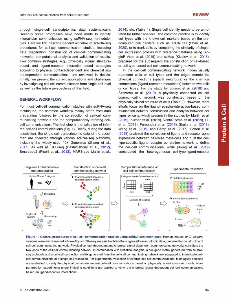
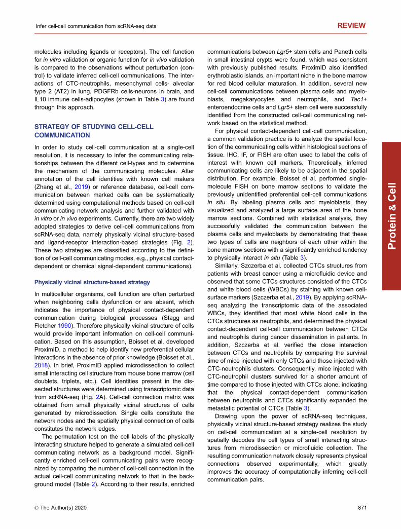
For most cell-cell communication studies with scRNA-seqtechniques, the common workflow mainly starts from datapreparation followed by the construction of cell-cell com-municating networks and the computationally inferring cell-cell communications. The last step is the validation of infer-red cell-cell communications (Fig. 1). Briefly, during the dataacquisition, the single-cell transcriptomic data of the speci-men are collected through various scRNA-seq platforms,including the widely-used 10x Genomics (Zheng et al.,2017), as well as CEL-seq (Hashimshony et al., 2012),Smart-seq2 (Picelli et al., 2013), MARS-seq (Jaitin et al.,
2014), etc. (Table 1). Single-cell identity needs to be anno-tated for further analysis. The common practice is to identifycell types with the known cell markers based on the pre-computed cell clusters such as scCATCH (Shao et al.,2020), or to mark cells by comparing the similarity of single-cell expression profiles with reference database using Sin-gleR Aran et al. (2019) and scMap (Kiselev et al., 2018),prepared for the subsequent the construction of cell-basedor cell-type-based cell-cell communicating network.
In the cell-cell communicating network, nodes usuallyrepresent cells or cell types and the edges denote thephysical connections (spatial neighbors) or the chemicalconnections (ligand-receptor interactions) between two cellsor cell types. For the study by Boisset et al. (2018) andSzczerba et al. (2019), a physically connected cell-cellcommunicating network was constructed based on thephysically vicinal structure of cells (Table 2). However, moreefforts focus on the ligand-receptor-interaction-based com-munication network construction and analysis between celltypes or cells, which present in the studies by Martin et al.(2019), Kumar et al. (2018), Vento-Tormo et al. (2018), Huet al. (2019), Fernandez et al. (2019), Skelly et al. (2018),Wang et al. (2019) and Camp et al. (2017). Cohen et al.(2018) analyzed the correlation of ligand and receptor geneexpression between pair-wise meta-cells and built the cell-type-specific ligand-receptor correlation network to definethe cell-cell communications, while (Xiong et al., 2019)constructed the heterogeneous cell-type-ligand-receptor
Single-cell transcriptomicdata preparation
Construction of cell-cellcommunicating network
Computational inference ofcell-cell communication Experimental validation
Dissecting
Single-cellcapture Sequencing
Human Mouse C.elegans
Chemical signal-dependentcommunicating network
Physical contact-dependentcommunicating network
Cell
Cell-gene matrix Cell-cell connectionmatrix
Methods of statistics
Cell
Gen
e
Cel
l
Perturbation experimentIn vitro
In vivo
Histological section
FISHIHCIF
Inhibitor
RNAi
Figure 1. General procedures of cell-cell communication studies using scRNA-seq techniques. Human, mouse, or C. elegans
samples were first dissected followed by scRNA-seq analysis to obtain the single-cell transcriptomic data, prepared for construction of
cell-cell communicating network. Physical contact-dependent and chemical signal-dependent communicating networks constitute the
two kinds of the cell-cell communicating network. In combination with statistical analysis, a cell-gene matrix generated from scRNA-
seq protocols and a cell-cell connection matrix generated from the cell-cell communicating network are integrated to investigate cell-
cell communications at a single-cell resolution. For experimental validation of inferred cell-cell communications, histological sections
are evaluated to verify the physical contact-dependent cell-cell communications based on physically vicinal structure of cells, while
perturbation experiments under inhibiting conditions are applied to verify the chemical signal-dependent cell-cell communications
based on ligand-receptor interactions.
© The Author(s) 2020 867
Protein
&Cell
Infer cell-cell communication from scRNA-seq data REVIEW
communicating network by filtering highly expressed ligandsand receptor for each cell types (Table 2). Statistical analysison the cell-cell communicating network identifies significantcell-cell communications between cell-types. In the work byBoisset et al. (2018), a 10,000 times permutation test on thevertices of physically connected cell-cell communicatingnetwork was performed using random sampling of cells,which leads to a distribution for each type of interaction in thenetwork. Significantly enriched and depleted interactions(P < 0.05) are found by the comparison with the experi-mental number of interactions. For ligand-receptor-interac-tion-based communication network, significantly enrichedligand-receptor pairs between cell types or cells are alsodefined by statistical methods such as the widely-used per-mutation test or Welch’s t-test, Wilcoxon rank-sum test andthe probability model as shown in Table 2. Then the fre-quency statistics of those significantly enriched pairs iscomputed to infer potential cell-cell communications with themost ligand-receptor pairs. What’s more, some scRNA-seq-based studies on cell-cell communication, e.g., Zhang et al.(2018), Zepp et al. (2017), Duan et al. (2018), Dong et al. (Liet al., 2017) and Rajbhandari et al. (2019), majorly rely onthe prior knowledge to define potential cell-cell communica-tion, wherein the mechanism underlying these
communications needs to be further elucidated at a single-cell resolution.
Validation of inferred cell-cell communicating pairs is thelast but most important step. Currently, there are mainly twoapproaches to validate the inferred pairs, namely histologicalsection analysis of the spatial location of communicatingcells and the interacting molecules (ligands and receptors)marked by fluorescence in situ hybridization (FISH),immunohistochemistry (IHC) or immunofluorescence (IF).Perturbation experiments (in vivo or in vitro) using inhibitorsor RNA interference (RNAi) are also taken to verify the keyligands and receptors that mediate the cell-cell communi-cation. Concordantly, most inferred communicating cellsexhibited observable spatial vicinity on the tissue sectionsuch as megakaryocytes-neutrophils in bone marrow, cir-culating tumor cells (CTC)-neutrophils, extravillous tro-phoblast (EVT)- decidual natural killer (dNK) in fetalplacenta, basophils-macrophages in lung, Oocytes- granu-losa cells (GCs), and fetal germ cells (FGCs)-gonadal nichecells in testis, etc. (Table 3).
For the significantly enriched ligand-receptor interactionsin cell-cell communication network, perturbation experimentsare currently conducted to verify the interaction betweencommunicating cells through inhibiting a key gene or proteinthat regulates the cell-cell communication (e.g., interacting
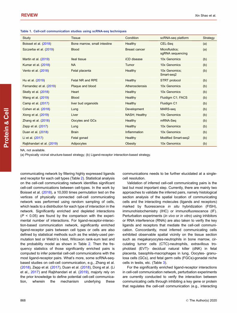
Table 1. Cell-cell communication studies using scRNA-seq techniques
Study Tissue Condition scRNA-seq platform Strategy
Boisset et al. (2018) Bone marrow, small intestine Healthy CEL-Seq (a)
Szczerba et al. (2019) Blood Breast cancer Microfluidics;sgRNA sequencing
(a)
Martin et al. (2019) Ileal tissue iCD disease 10x Genomics (b)
Kumar et al. (2018) NA Tumor 10x Genomics (b)
Vento et al. (2018) Fetal placenta Healthy 10x Genomics;Smart-seq2
(b)
Hu et al. (2019) Fetal NR and RPE Healthy STRT protocol (b)
Fernandez et al. (2019) Plaque and blood Atherosclerosis 10x Genomics (b)
Skelly et al. (2018) Heart Healthy 10x Genomics (b)
Wang et al. (2019) Blood Healthy Fluidigm C1; FACS (b)
Camp et al. (2017) liver bud organoids Healthy Fluidigm C1 (b)
Cohen et al. (2018) Lung Development MARS-seq (b)
Xiong et al. (2019) Liver NASH; Healthy 10x Genomics (b)
Zhang et al. (2018) Oocytes and GCs Healthy mRNA-Seq (b)
Zepp et al. (2017) Lung Healthy 10x Genomics (b)
Duan et al. (2018) Brain Inflammation 10x Genomics (b)
Li et al. (2017) Fetal gonad Healthy Modified Smart-seq2 (b)
Rajbhandari et al. (2019) Adipocytes Obesity 10x Genomics (b)
NA, not available.
(a) Physically vicinal structure-based strategy; (b) Ligand-receptor interaction-based strategy.
REVIEW Xin Shao et al.
868 © The Author(s) 2020
Protein
&Cell
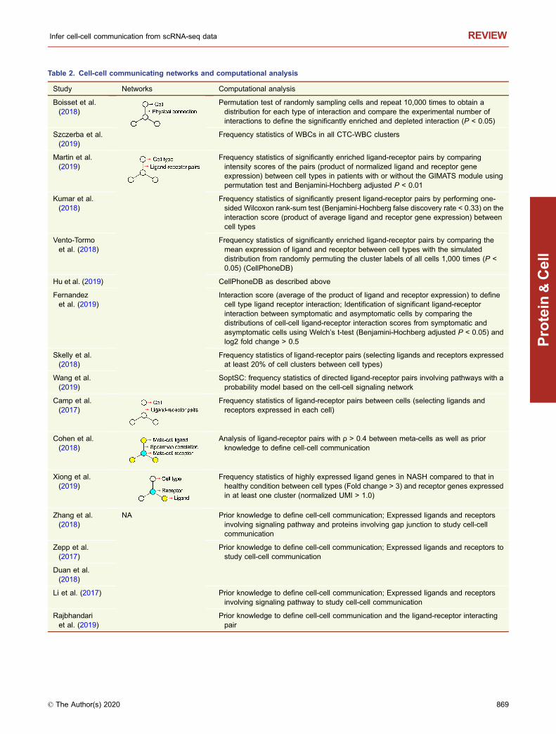
Table 2. Cell-cell communicating networks and computational analysis
Study Networks Computational analysis
Boisset et al.(2018)
Permutation test of randomly sampling cells and repeat 10,000 times to obtain adistribution for each type of interaction and compare the experimental number ofinteractions to define the significantly enriched and depleted interaction (P < 0.05)
Szczerba et al.(2019)
Frequency statistics of WBCs in all CTC-WBC clusters
Martin et al.(2019)
Frequency statistics of significantly enriched ligand-receptor pairs by comparingintensity scores of the pairs (product of normalized ligand and receptor geneexpression) between cell types in patients with or without the GIMATS module usingpermutation test and Benjamini-Hochberg adjusted P < 0.01
Kumar et al.(2018)
Frequency statistics of significantly present ligand-receptor pairs by performing one-sided Wilcoxon rank-sum test (Benjamini-Hochberg false discovery rate < 0.33) on theinteraction score (product of average ligand and receptor gene expression) betweencell types
Vento-Tormoet al. (2018)
Frequency statistics of significantly enriched ligand-receptor pairs by comparing themean expression of ligand and receptor between cell types with the simulateddistribution from randomly permuting the cluster labels of all cells 1,000 times (P <0.05) (CellPhoneDB)
Hu et al. (2019) CellPhoneDB as described above
Fernandezet al. (2019)
Interaction score (average of the product of ligand and receptor expression) to definecell type ligand receptor interaction; Identification of significant ligand-receptorinteraction between symptomatic and asymptomatic cells by comparing thedistributions of cell-cell ligand-receptor interaction scores from symptomatic andasymptomatic cells using Welch’s t-test (Benjamini-Hochberg adjusted P < 0.05) andlog2 fold change > 0.5
Skelly et al.(2018)
Frequency statistics of ligand-receptor pairs (selecting ligands and receptors expressedat least 20% of cell clusters between cell types)
Wang et al.(2019)
SoptSC: frequency statistics of directed ligand-receptor pairs involving pathways with aprobability model based on the cell-cell signaling network
Camp et al.(2017)
Frequency statistics of ligand-receptor pairs between cells (selecting ligands andreceptors expressed in each cell)
Cohen et al.(2018)
Analysis of ligand-receptor pairs with ρ > 0.4 between meta-cells as well as priorknowledge to define cell-cell communication
Xiong et al.(2019)
Frequency statistics of highly expressed ligand genes in NASH compared to that inhealthy condition between cell types (Fold change > 3) and receptor genes expressedin at least one cluster (normalized UMI > 1.0)
Zhang et al.(2018)
NA Prior knowledge to define cell-cell communication; Expressed ligands and receptorsinvolving signaling pathway and proteins involving gap junction to study cell-cellcommunication
Zepp et al.(2017)
Prior knowledge to define cell-cell communication; Expressed ligands and receptors tostudy cell-cell communication
Duan et al.(2018)
Li et al. (2017) Prior knowledge to define cell-cell communication; Expressed ligands and receptorsinvolving signaling pathway to study cell-cell communication
Rajbhandariet al. (2019)
Prior knowledge to define cell-cell communication and the ligand-receptor interactingpair
Infer cell-cell communication from scRNA-seq data REVIEW
© The Author(s) 2020 869
Protein
&Cell
Table 3. Inferred cell-cell communications and validation
Study Inferred cell-cell communication Validation of inferred cell-cell communication
Boisset et al.(2018)
Megakaryocytes-neutrophils, Lgr5+ stem cells-Paneth cells, Lgr5+ stem cells-Tac1+enteroendocrine cell, etc.
Marking the communicating cells by Single-molecule FISHstaining on bone marrow and small intestine sectionsindicated they are significant neighbors
Szczerbaet al.(2019)
CTC-neutrophils Marking the CTC and neutrophils by IF staining indicated theyare primarily neighbors; In vivo perturbation experimentsindicated mice injected with CTC-neutrophil clusters survivedfor a shorter amount of time compared to those injected withCTCs alone
Martin et al.(2019)
MNPs-T cells, etc. NA
Kumar et al.(2018)
Cancer cells-CAFs, Cancer cells-macrophages NA
Vento-Tormoet al.(2018)
EVT-dNK cells Marking the EVT and dNK cells by IHC staining on decidualserial sections indicated they are primarily neighbors.
Hu et al.(2019)
PCs-RPE cells NA
Fernandezet al.(2019)
T cells-macrophages NA
Skelly et al.(2018)
Macrophages-pericytes, Macrophages-fibroblasts NA
Wang et al.(2019)
HSPC-Monocytes; HSPC-granulocytes, etc. NA
Camp et al.(2017)
HE cells-macrophages, HE cells-endothelial cells In vitro perturbation experiments by knocking down the keyligand EDN1 of co-cultured endothelial cells indicated thedifferentiation of co-cultured HE cells was significantlyimpaired
Cohen et al.(2018)
Alveolus-Basophils, Basophils-macrophages Marking the communicating cells by IHC staining of lungsections indicated their spatial proximity to each other; In vitroperturbation experiments and in vivo IL-33 receptor knockoutmice experiments indicated basophils regulate alveolarmacrophage maturation and immunomodulation functions
Xiong et al.(2019)
HSCs-endothelial cells; HSCs-macrophages;HSCs-T cells, etc.
NA
Zhang et al.(2018)
Oocytes-GCs Marking the oocytes and GCs specific protein involving gapjunctions by IHC staining indicated they are primarilyneighbors
Zepp et al.(2017)
Mesenchymal cells-AT2 Spatial distance mapping using Leica indicated the adjacentlocation of Mesenchymal cells and AT2; In vitro perturbationexperiments using alveolar organoid indicated the ability ofmesenchymal lineages to promote alveolar organoid growth
Duan et al.(2018)
PDGFRb cells-neurons In vitro and in vivo perturbation experiments with RNAi indicatedPDGFRb cells communicate neurons by secreting chemokineCCL2 during early infection
Li et al.(2017)
FGCs-gonadal niche cells Marking the FGCs and gonadal niche cells specific proteininvolving BMP and Notch signaling by IF staining of testesindicated the communication between them
Rajbhandariet al.(2019)
IL10 immune cells-adipocytes In vivo perturbation experiments with adipocyte-specific IL10receptor-deficient mice indicated the communication of IL10immune cells-adipocytes in the modulation of the adiposeadrenergic response
REVIEW Xin Shao et al.
870 © The Author(s) 2020
Protein
&Cell
molecules including ligands or receptors). The cell functionfor in vitro validation or organic function for in vivo validationis compared to the observations without perturbation (con-trol) to validate inferred cell-cell communications. The inter-actions of CTC-neutrophils, mesenchymal cells- alveolartype 2 (AT2) in lung, PDGFRb cells-neurons in brain, andIL10 immune cells-adipocytes (shown in Table 3) are foundthrough this approach.
STRATEGY OF STUDYING CELL-CELLCOMMUNICATION
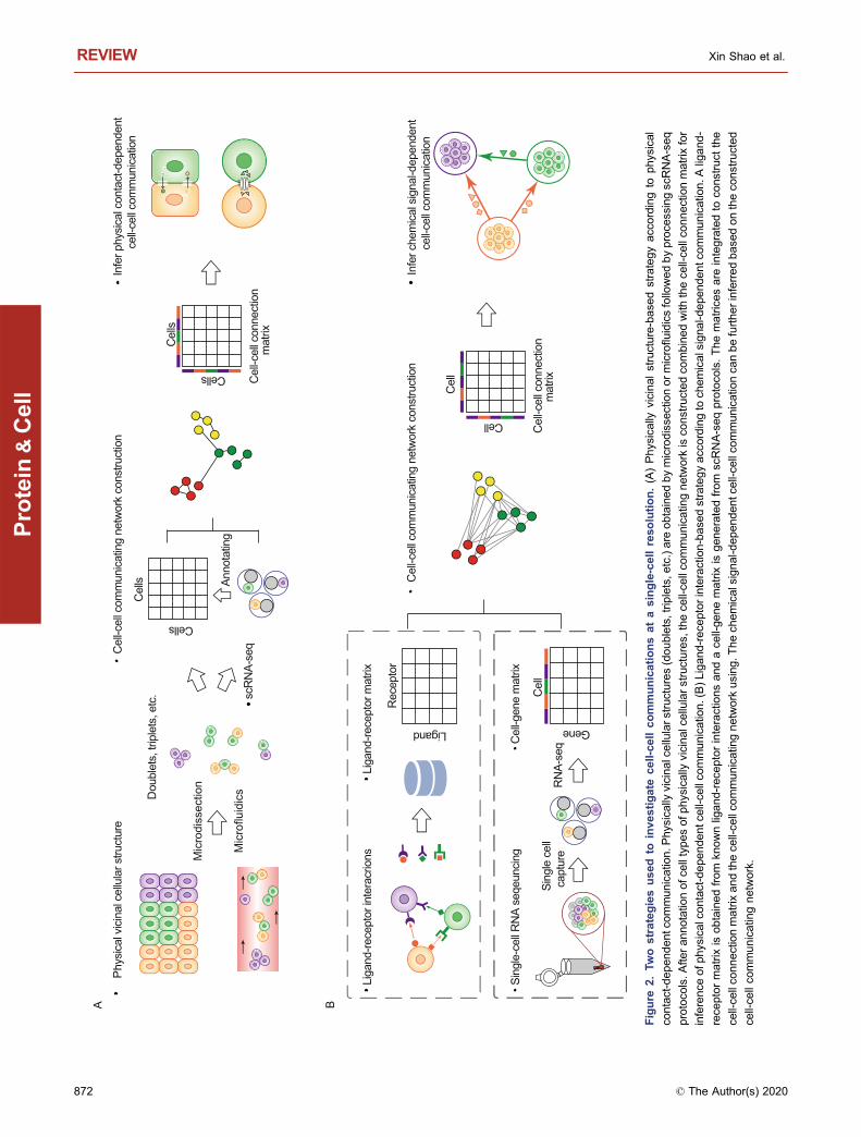
In order to study cell-cell communication at a single-cellresolution, it is necessary to infer the communicating rela-tionships between the different cell-types and to determinethe mechanism of the communicating molecules. Afterannotation of the cell identities with known cell makers(Zhang et al., 2019) or reference database, cell-cell com-munication between marked cells can be systematicallydetermined using computational methods based on cell-cellcommunicating network analysis and further validated within vitro or in vivo experiments. Currently, there are two widelyadopted strategies to derive cell-cell communications fromscRNA-seq data, namely physically vicinal structure-basedand ligand-receptor interaction-based strategies (Fig. 2).These two strategies are classified according to the defini-tion of cell-cell communicating modes, e.g., physical contact-dependent or chemical signal-dependent communications).
Physically vicinal structure-based strategy
In multicellular organisms, cell function are often perturbedwhen neighboring cells dysfunction or are absent, whichindicates the importance of physical contact-dependentcommunication during biological processes (Stagg andFletcher 1990). Therefore physically vicinal structure of cellswould provide important information on cell-cell communi-cation. Based on this assumption, Boisset et al. developedProximID, a method to help identify new preferential cellularinteractions in the absence of prior knowledge (Boisset et al.,2018). In brief, ProximID applied microdissection to collectsmall interacting cell structure from mouse bone marrow (celldoublets, triplets, etc.). Cell identities present in the dis-sected structures were determined using transcriptomic datafrom scRNA-seq (Fig. 2A). Cell-cell connection matrix wasobtained from small physically vicinal structures of cellsgenerated by microdissection. Single cells constitute thenetwork nodes and the spatially physical connection of cellsconstitutes the network edges.
The permutation test on the cell labels of the physicallyinteracting structure helped to generate a simulated cell-cellcommunicating network as a background model. Signifi-cantly enriched cell-cell communicating pairs were recog-nized by comparing the number of cell-cell connection in theactual cell-cell communicating network to that in the back-ground model (Table 2). According to their results, enriched
communications between Lgr5+ stem cells and Paneth cellsin small intestinal crypts were found, which was consistentwith previously published results. ProximID also identifiederythroblastic islands, an important niche in the bone marrowfor red blood cellular maturation. In addition, several newcell-cell communications between plasma cells and myelo-blasts, megakaryocytes and neutrophils, and Tac1+enteroendocrine cells and Lgr5+ stem cell were successfullyidentified from the constructed cell-cell communicating net-work based on the statistical method.
For physical contact-dependent cell-cell communication,a common validation practice is to analyze the spatial loca-tion of the communicating cells within histological sections oftissue. IHC, IF, or FISH are often used to label the cells ofinterest with known cell markers. Theoretically, inferredcommunicating cells are likely to be adjacent in the spatialdistribution. For example, Boisset et al. performed single-molecule FISH on bone marrow sections to validate thepreviously unidentified preferential cell-cell communicationsin situ. By labeling plasma cells and myeloblasts, theyvisualized and analyzed a large surface area of the bonemarrow sections. Combined with statistical analysis, theysuccessfully validated the communication between theplasma cells and myeloblasts by demonstrating that thesetwo types of cells are neighbors of each other within thebone marrow sections with a significantly enriched tendencyto physically interact in situ (Table 3).
Similarly, Szczerba et al. collected CTCs structures frompatients with breast cancer using a microfluidic device andobserved that some CTCs structures consisted of the CTCsand white blood cells (WBCs) by staining with known cell-surface markers (Szczerba et al., 2019). By applying scRNA-seq analyzing the transcriptomic data of the associatedWBCs, they identified that most white blood cells in theCTCs structures as neutrophils, and determined the physicalcontact-dependent cell-cell communication between CTCsand neutrophils during cancer dissemination in patients. Inaddition, Szczerba et al. verified the close interactionbetween CTCs and neutrophils by comparing the survivaltime of mice injected with only CTCs and those injected withCTC-neutrophils clusters. Consequently, mice injected withCTC-neutrophil clusters survived for a shorter amount oftime compared to those injected with CTCs alone, indicatingthat the physical contact-dependent communicationbetween neutrophils and CTCs significantly expanded themetastatic potential of CTCs (Table 3).
Drawing upon the power of scRNA-seq techniques,physically vicinal structure-based strategy realizes the studyon cell-cell communication at a single-cell resolution byspatially decodes the cell types of small interacting struc-tures from microdissection or microfluidic collection. Theresulting communication network closely represents physicalconnections observed experimentally, which greatlyimproves the accuracy of computationally inferring cell-cellcommunication pairs.
Infer cell-cell communication from scRNA-seq data REVIEW
© The Author(s) 2020 871
Protein
&Cell
A B
Rec
epto
r
Liga
nd-re
cept
or in
tera
crio
nsLi
gand
-rece
ptor
mat
rix
Ligand
Cel
l
Gene
Sing
le-c
ell R
NA
seqe
unci
ng
Sing
le c
ell
capt
ure
RN
A-se
qCel
l-gen
e m
atrix
Cel
l-cel
l con
nect
ion
mat
rix
Cel
l-cel
l com
mun
icat
ing
netw
ork
cons
truct
ion
Infe
r che
mic
al s
igna
l-dep
ende
ntce
ll-ce
ll com
mun
icat
ion
Cel
l
Cell
Mic
rodi
ssec
tion
Mic
roflu
idic
s
Phy
sica
l vic
inal
cel
lula
r stru
ctur
e
Dou
blet
s, tr
iple
ts, e
tc.
Cel
ls
scR
NA-
seq
Anno
tatin
g
Cel
l-cel
l con
nect
ion
mat
rix
Cells
Cel
ls
Cells
Infe
r phy
sica
l con
tact
-dep
ende
ntce
ll-ce
ll com
mun
icat
ion
Cel
l-cel
l com
mun
icat
ing
netw
ork
cons
truct
ion
Figure
2.Tw
ostrategiesused
toinvestigate
cell-ce
llcommunicationsatasingle-cellreso
lution.(A)Physically
vicinalstructure-base
dstrategyaccordingto
physical
contact-depen
dentc
ommunicatio
n.P
hysica
llyvicinalcellularstructures(doublets,trip
lets,e
tc.)are
obtainedbymicrodisse
ctionormicroflu
idicsfollowedbyproce
ssingsc
RNA-seq
protoco
ls.A
fterannotatio
nofc
elltypesofp
hys
icallyvicinal
cellularstructures,
thece
ll-ce
llco
mmunicatin
gnetwork
isco
nstructedco
mbinedwith
thece
ll-ce
llco
nnectionmatrix
for
inference
ofp
hys
ical
contact-depen
dentc
ell-ce
llco
mmunicatio
n.(B)Ligand
-rece
ptorinteraction-base
dstrategyacc
ordingto
chemicalsigna
l-depen
dentc
ommunicatio
n.A
ligand
-
rece
ptormatrix
isobtainedfrom
knownligand-rec
eptorinteractionsandace
ll-ge
nematrix
isgen
eratedfrom
scRNA-seqprotoco
ls.Thematrices
are
integratedto
cons
truct
the
cell-ce
llco
nnectionmatrixandthece
ll-ce
llco
mmunicatin
gnetwork
using
.Thech
emicalsignal-d
ependen
tcell-ce
llco
mmunica
tionca
nbefurtherinferredbase
dontheco
nstructed
cell-ce
llco
mmunicatin
gnetwork.
REVIEW Xin Shao et al.
872 © The Author(s) 2020
Protein
&Cell
However, small interacting structures were mainly col-lected by manual process based on microdissection devices,leading to the low capturing throughput in a single assay andpotential false negatives. Due to its limitation on definition ofcell-cell communication, distant cell-cell communicationswithin the tissue microenvironment may be missed by thisstrategy. In the statistical analysis on communication net-work, the question remained if the number of cell-cell con-nection pairs can correlate with the real communicationbetween these two cell types.
Ligand-receptor interaction-based strategy
For most single-cell studies, the physically spatial locationsof cells are lost during the frequently-used scRNA-seq pro-tocol such as 10x Genomics (Zheng et al., 2017). Commu-nication between cells are partly mediated through secretedsignaling molecules, such as cytokines and hormones (Si-card 1986; Gartner et al., 2017). Secreted signaling mole-cules play fundamental roles in chemical signal-dependentcell-cell communications for both physically vicinal cells anddistant cells (Braga 2002). With accumulating results aboutchemical signals for decades, thousands of ligand-receptorinteracting pairs have been defined and validated experi-mentally (Ramilowski et al., 2015), which enables the use ofscRNA-seq to construct cell-cell communicating networkbased on differentially expressed gene levels of ligands andreceptors to infer potential cell-cell communications such asCellPhoneDB (Efremova et al., 2020) and SoptSC (Wanget al., 2019). Using available ligand-receptor interactionsdatabase (Ramilowski et al., 2015), chemical signal-depen-dent cell-cell communication can be inferred from the con-structed cell-cell communicating network, where the edgesrepresent the interacting intensity integrated from the enri-ched ligand-receptor pairs between two cells and the nodesdenote single cells or cell-types (Fig. 2B).
Camp et al. (2017) sequenced three-dimensional liverbud organoids that were constituted with induced pluripotentstem cell-derived human hepatic endoderm (HE), macro-phages, and endothelial cells at a single-cell resolution toidentify the communications between these cell populationsduring liver bud development. By constructing and analyzingthe cell-cell communicating networks of receptor-ligandpairings, HE cells demonstrated more extensive crosstalkwith macrophages and endothelial cells compared to thatwith other HE cells (Tables 1–3).
To investigate the chemical signal-dependent cell-cellcommunications under the condition of non-alcoholicsteatohepatitis (NASH), Xiong et al. obtained a total of33,168 single-cell transcriptomes including 17,788 normalliver cells and 15,380 cells under NASH (Xiong et al., 2019).According to highly expressed ligands and receptors identi-fied in NASH, a liver cells’ ligand-receptor communicatingnetwork was constructed to investigate intercellular crosstalkwithin the liver microenvironment of NASH, which includedcholangiocytes, hepatic stellate cells (HSCs), hepatocytes,
as well as multiple immune cells. Consequently, they deter-mined that HSCs serve as a hub of intrahepatic signalingthrough the secretion of HSC-derived stellakines toendothelial cells, macrophages, and T cells during NASH byanalyzing the constructed communicating network(Tables 1–3)
Using the same strategy, Martin et al. sequenced 82,417lamina propria cells from 11 patients with ileal Crohn’s dis-ease (iCD) and characterized a GIMATS module (IgG PCs,inflammatory MNPs, and activated T and stromal cells) in asubset of iCD patients, namely IgG plasma B cells, inflam-matory mononuclear phagocytes (MNPs), and activated Tand stromal cells (Martin et al., 2019). For patients enrichedor lacking the GIMATS module, a ligand-receptor activitynetworks was constructed in which network edges referredto the normalized ligand and receptor expression from thesource to the target cell type. Computational analysis on theintensity scores of each ligand-receptor pair between eachpair-wise cell types determined several ligand-receptorinteractions related with receptors on T cells and ligandssecreted by MNPs were significantly enriched in GIMATSenriched iCD patients, including CCL19-CCR7, CCL2-CCR4, and IL6-IL6R interacting pairs. To explore cell-cellcommunication during early maternal–fetal interfaces inhumans, Vento et al. collected approximately 70,000 indi-vidual cells from first-trimester placentas and annotatedthem according to known marker genes, such as EVT, dNKcells and dendritic cells (DCs) (Vento-Tormo et al., 2018). Byconsidering the expression levels of ligands and receptorswithin each cell type, numerous significant ligand–receptorpairs involving immunomodulation, adhesion, and recruit-ment have been identified between EVT and dNK cells(Tables 1–3).
In addition, several novel chemical signal-dependent cell-cell communications underlying crucial biological processeshave been identified (Table 3) using this strategy. Thatinclude non-myocytic heart cells with normal cardiac function(Skelly et al., 2018), immune and non-immune cells duringlung development (Zepp et al., 2017; Cohen et al., 2018),malignant and non-malignant cells of tumor microenviron-ment (Kumar et al., 2018), and nervous and immune cellsagainst infection (Duan et al., 2018), etc. (Tables 1–3).
Perturbation experiments are usually conducted to vali-date the inferred cell-cell communication. It is expected thatthe function or population of one cell type will be influencedby inhibiting the key ligand of the other communicated celltype. For example, Camp et al. (2017) verified extensivecrosstalk between HE cells and endothelial cells by knockingdown EDN1, the key ligand of endothelial cells. Conse-quently, they found that the differentiation of co-cultured HEcells was significantly impaired. Beside the in vitro pertur-bation experiments with the co-culture on communicatingcells, Rajbhandari et al. applied in vivo perturbation experi-ments to verify the cell-cell communications between IL10-producing immune cells and adipocytes (Rajbhandari et al.,2019). Compared to the normal mice, mice with knocked out
Infer cell-cell communication from scRNA-seq data REVIEW
© The Author(s) 2020 873
Protein
&Cell
adipocyte-specific IL10 receptor were protected againstweight gain and observed with increased inguinal brownadipose tissue under high-fat diet, suggesting the import roleof IL10-producing immune cells and adipocytes’ communi-cation in regulating the thermogenesis and systemic energybalance involving diet-induced obesity (Table 3).
To date, intercellular crosstalk remain poorly understood,including the signals that initiate the communication and howthe communication is regulated and maintained. scRNA-seqtechniques provide new insights into the mechanismsinvolved in these known cell-cell communications at a single-cell level (Tables 1–3). As an example, Hu et al. analyzed2,421 individual cells isolated from human fetal neural reti-nas (NR) and retinal pigment epithelium (RPE) and analyzedthem by scRNA-seq (Hu et al., 2019). The results revealeddynamic expression patterns of the visual cycle and ligand-receptor interaction-related genes such as PTPRZ1, MDK,and PTN. Besides, it is known that bidirectional communi-cation of GCs is required for folliculogenesis. To betterunderstand the crosstalk between oocytes and GCs, Zhanget al. evaluated the transcriptomes of the cells using scRNA-seq and recapitulated the dynamic mechanism of transcrip-tional regulation between oocytes and GCs during folliculo-genesis (Zhang et al., 2018). In addition, it has been reportedthat the communication between epithelial progenitors andthe surrounding mesenchymal cells is able to modulate theability of the epithelial progenitors to proliferate and differ-entiate. Combined with scRNA-seq evaluation, Zepp et al.identified Pdgfra as the interacting molecule expressed bymesenchymal cells that mediates the growth and self-re-newal of epithelial cells (Zepp et al., 2017).
Ligand-receptor interaction-based strategy takes thegene expression level of known ligands and the corre-sponding receptors into account. Compared to physicallyvicinal structure-based strategy, this strategy is not only ableto infer the vicinal cell-cell communications, but also thedistant cell-cell communications through indirect ligand-re-ceptor interactions.
Whereas, it may be difficult to identify contact-dependentcommunication via transmembrane proteins or gap junctions(Evans 2015) rather than ligand-receptor interactions. Fur-thermore, the performance of this strategy heavily dependson reference databases of known ligand-receptor interac-tions. With notable variations on expression levels of theseligand and receptor genes, inference of cell-cell communi-cations based on ligand-receptor interaction may berestricted. It is still under debate how the edges in the net-work represent multiple bi-directional interactions betweencells.
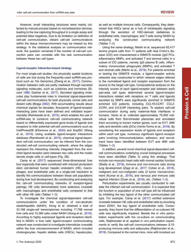
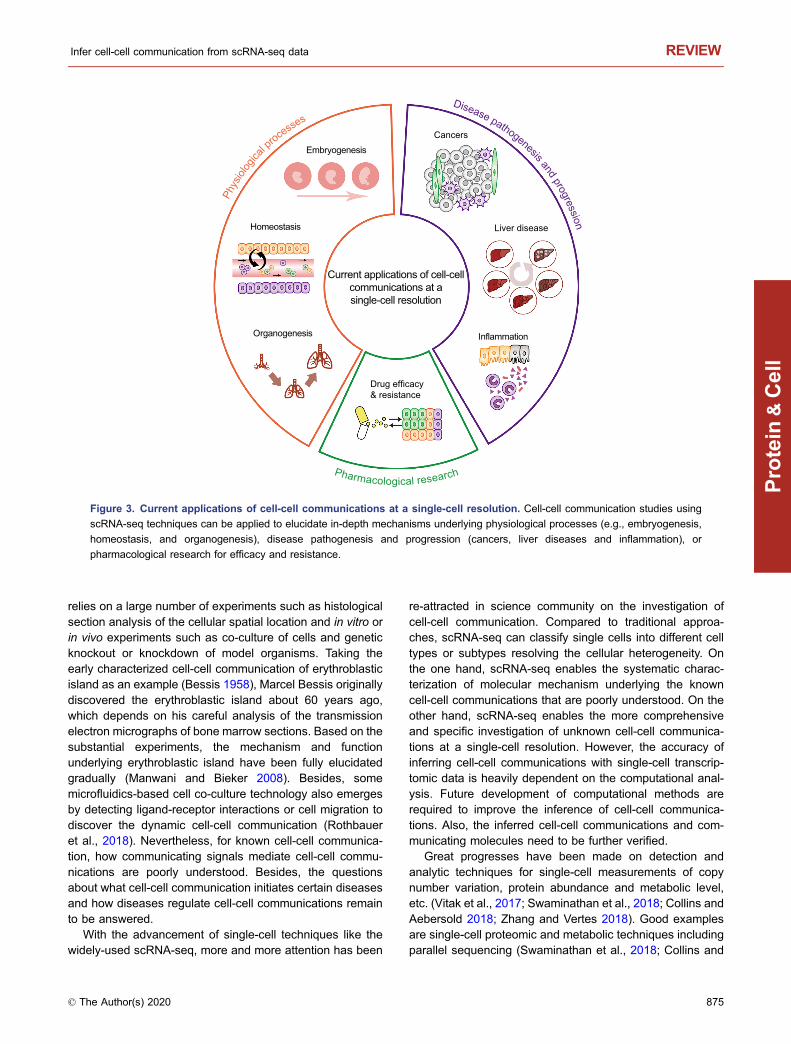
CURRENT APPLICATIONS
It is a common to study the biological processes includingphysiological processes, disease pathogenesis and pro-gression, and pharmacological research of pharmacologicaltreatment and drug resistance by focusing on the key genetic
events (copy number variations (Pan et al., 2020), mutations(Zheng et al., 2019, etc.), genes (Zhou 2020), RNAs (miR-NAs Xu et al. (2019), lncRNAs (Lin et al., 2019, etc.), pro-teins Shao et al. (2016), signaling pathways (Liao et al.,2018), or key cells (Mittal et al., 2019), etc. However, moreand more researchers in science community have realizedthe importance of cell-cell communication during biologicalprocesses. Increasing findings have indicated that cell-cellcommunication plays crucial roles in a vast of biologicalprocesses including growth, development, disease occur-rence and development, etc. in multicellular organisms.
Basically, scRNA-seq-based cell-cell communicationstudies can be applied to reveal the in-depth mechanisms asthey can elucidate the signals that initiate the communicationand how the communication is regulated and maintainedunderlying crucial biological processes (Shalek et al., 2014;Burns et al., 2015). Great efforts have been devoted torelated fields including physiological processes, such asembryogenesis (Li et al., 2017), homeostasis (Boisset et al.,2018), and organogenesis (Cohen et al., 2018; Scott andGuilliams 2018), as well as disease pathogenesis and pro-gression for cancers (Tirosh et al., 2016; Kumar et al., 2018),liver diseases (Xiong et al., 2019) and inflammation (Duanet al., 2018; Martin et al., 2019), and pharmacologicalresearch of pharmacological treatment and drug resistance(Martin et al., 2019) (Fig. 3). For example, Camp et al.revealed the key cell-cell communication that potentiallyregulates liver development and the key signaling molecules(Camp et al., 2017), while Zepp et al. sequenced lungmesenchymal cells at a single-cell resolution and identifiedepithelial-mesenchymal communications critical for lunghomeostasis and regeneration (Zepp et al., 2017). In addi-tion, Xiong et al. applied scRNA-seq to liver cells anddetermined the hepatic stellate cells as the core origin ofsecreted stellakines during the development of NASH (Xionget al., 2019). For pharmacological research, Martin et al.applied single-cell technologies to iCD lesions and con-cluded that the GIMATS module was driven by MNPs andobserved a high correlation exists between the GIMATSmodule and anti-TNF treatment resistance, suggesting thatthese may serve as novel biomarkers of treatment responseand may be exploited for tailored therapeutic opportunities(Martin et al., 2019). Obviously, a better understanding of thecell-cell communications during disease pathogenesis andprogression may provide insights into novel therapeuticstrategies and targets in pharmacological research (i.e., keycell types and key ligand-receptor interactions), despite thelimited number of studies available with respect to cell-cellcommunication at a single-cell resolution.
PERSPECTIVES
In multicellular organisms, cell-cell communication play cru-cial roles in numerous biological processes including growth,development, disease occurrence and development, etc.Traditionally, exploring cell-cell communications majorly
REVIEW Xin Shao et al.
874 © The Author(s) 2020
Protein
&Cell
relies on a large number of experiments such as histologicalsection analysis of the cellular spatial location and in vitro orin vivo experiments such as co-culture of cells and geneticknockout or knockdown of model organisms. Taking theearly characterized cell-cell communication of erythroblasticisland as an example (Bessis 1958), Marcel Bessis originallydiscovered the erythroblastic island about 60 years ago,which depends on his careful analysis of the transmissionelectron micrographs of bone marrow sections. Based on thesubstantial experiments, the mechanism and functionunderlying erythroblastic island have been fully elucidatedgradually (Manwani and Bieker 2008). Besides, somemicrofluidics-based cell co-culture technology also emergesby detecting ligand-receptor interactions or cell migration todiscover the dynamic cell-cell communication (Rothbaueret al., 2018). Nevertheless, for known cell-cell communica-tion, how communicating signals mediate cell-cell commu-nications are poorly understood. Besides, the questionsabout what cell-cell communication initiates certain diseasesand how diseases regulate cell-cell communications remainto be answered.
With the advancement of single-cell techniques like thewidely-used scRNA-seq, more and more attention has been
re-attracted in science community on the investigation ofcell-cell communication. Compared to traditional approa-ches, scRNA-seq can classify single cells into different celltypes or subtypes resolving the cellular heterogeneity. Onthe one hand, scRNA-seq enables the systematic charac-terization of molecular mechanism underlying the knowncell-cell communications that are poorly understood. On theother hand, scRNA-seq enables the more comprehensiveand specific investigation of unknown cell-cell communica-tions at a single-cell resolution. However, the accuracy ofinferring cell-cell communications with single-cell transcrip-tomic data is heavily dependent on the computational anal-ysis. Future development of computational methods arerequired to improve the inference of cell-cell communica-tions. Also, the inferred cell-cell communications and com-municating molecules need to be further verified.
Great progresses have been made on detection andanalytic techniques for single-cell measurements of copynumber variation, protein abundance and metabolic level,etc. (Vitak et al., 2017; Swaminathan et al., 2018; Collins andAebersold 2018; Zhang and Vertes 2018). Good examplesare single-cell proteomic and metabolic techniques includingparallel sequencing (Swaminathan et al., 2018; Collins and
Drug efficacy& resistance
Current applications of cell-cellcommunications at asingle-cell resolution
Cancers
Liver disease
Embryogenesis
Inflammation
Homeostasis
Organogenesis
Phys
iolog
ica
l processe
s
Pharmacological research
Disease pathogenesis and progression
Figure 3. Current applications of cell-cell communications at a single-cell resolution. Cell-cell communication studies using
scRNA-seq techniques can be applied to elucidate in-depth mechanisms underlying physiological processes (e.g., embryogenesis,
homeostasis, and organogenesis), disease pathogenesis and progression (cancers, liver diseases and inflammation), or
pharmacological research for efficacy and resistance.
Infer cell-cell communication from scRNA-seq data REVIEW
© The Author(s) 2020 875
Protein
&Cell
Aebersold 2018), single-cell mass cytometry (CyTOF)(Bandura et al., 2009), and single cell proteomics by massspectrometry (SCoPE-MS) (Budnik et al., 2018). Single-cellgenomics and transcriptomics reflect the cellular genealogyand can track cells as they evolve and change throughmutations (Marx 2019). Single-cell proteomics can be usedto classify cells into known cell types or to identify unknownor rare cell types/subtypes according to cell markers, for theinference of physical contact-dependent cell-cell communi-cations by annotating spatially vicinal identity of single cells.Given the inference of chemical signal-dependent cell-cellcommunications, single-cell proteomics can capture thedirect abundance of signaling proteins, while single-cellmetabonomics can examine the content of signalingmetabolites such as hormone (Pfaff and Baum 2018), neu-rotransmitter (Sugiyama et al., 2019).
However, high-throughput single-cell methods have notyet arrived in proteomics and metabonomics because of lotsof factors such as dyes falling off, low abundance in singlecell, infeasibility of amplification like DNA or RNA, challengesin sample and buffer preparation or high cost, etc. (Zhangand Vertes 2018; Marx 2019). Additionally, for some cell-cellcommunications via hormone, neurotransmitters, the chal-lenges might exist in the annotation of cells and
quantification of receptors when using single-cell metabo-nomics technique.
In consideration of the developed high-throughputscRNA-seq techniques (Cao et al., 2017), it has been acommon practice to use mRNA concentrations as proxies forthe concentrations and activities of the corresponding pro-teins, assuming that gene expression levels are the maindeterminant of protein abundances (Vogel and Marcotte2012). Compared to genome and proteome, transcriptomeanalysis provides knowledge of the molecular linkagesbetween genetic information and the proteome, leading to acomprehensive understanding of biological processesincluding the cell-cell communications (Song et al., 2019;Shao et al., 2019). Undoubtedly, there are some limitationsof scRNA-seq for the investigation of cell-cell communica-tions. First, scRNA-seq offers an indirect reflection of proteinlevels, not a direct measurement. Besides, for cell-cellcommunications via small signaling molecules such asdopamine and histamine, scRNA-seq can hardly infer thiskind of cell-cell communications. Even so, increasing studieshave focused on this technique to infer cell-cell communi-cations and the fact holds that scRNA-seq is proved to be anefficient approach to systematically infer and study cell-cell
Spatial reconstruction of single-cell transcriptomes
Dimension 1
Dim
ensi
on 2
X (Biological space)
Y (b
iolo
gica
l spa
ce)
A BNetwork topology of cell-cell
communicating network
CSpatial transcriptomics at a single-cell resolution
N NH2
NH
DSingle-cell multimodal
profiling
Genomics
Proteomics
Transcriptomics
Metabonomics
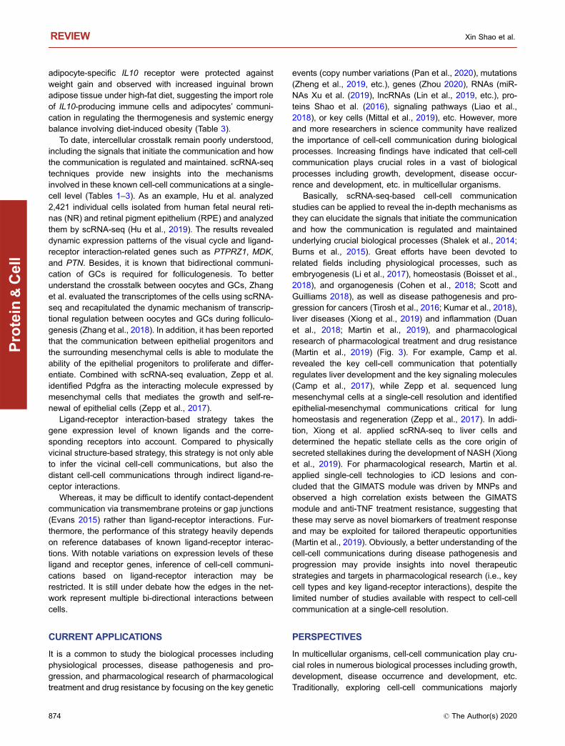
Figure 4. Challenges and opportunities of investigating cell-cell communication at a single-cell resolution. (A) Spatial
reconstruction of single-cell transcriptomes from single-cell transcriptomic data without spatial location will shed light on the
integration of physical contact-dependent and chemical signal-dependent cell-cell communications. (B) Incorporation of network
topology and features will help infer cell-cell communications. (C) Recent advances in spatial transcriptomics at a single-cell
resolution will facilitate the identification of single-cell intercellular communications in situ. (D) Establishing the comprehensive
molecular view of the cell by multimodal profiling in the future will definitely benefit the inference of cell-cell communicating modes.
REVIEW Xin Shao et al.
876 © The Author(s) 2020
Protein
&Cell
communications combined with computational analysis inrecent years.
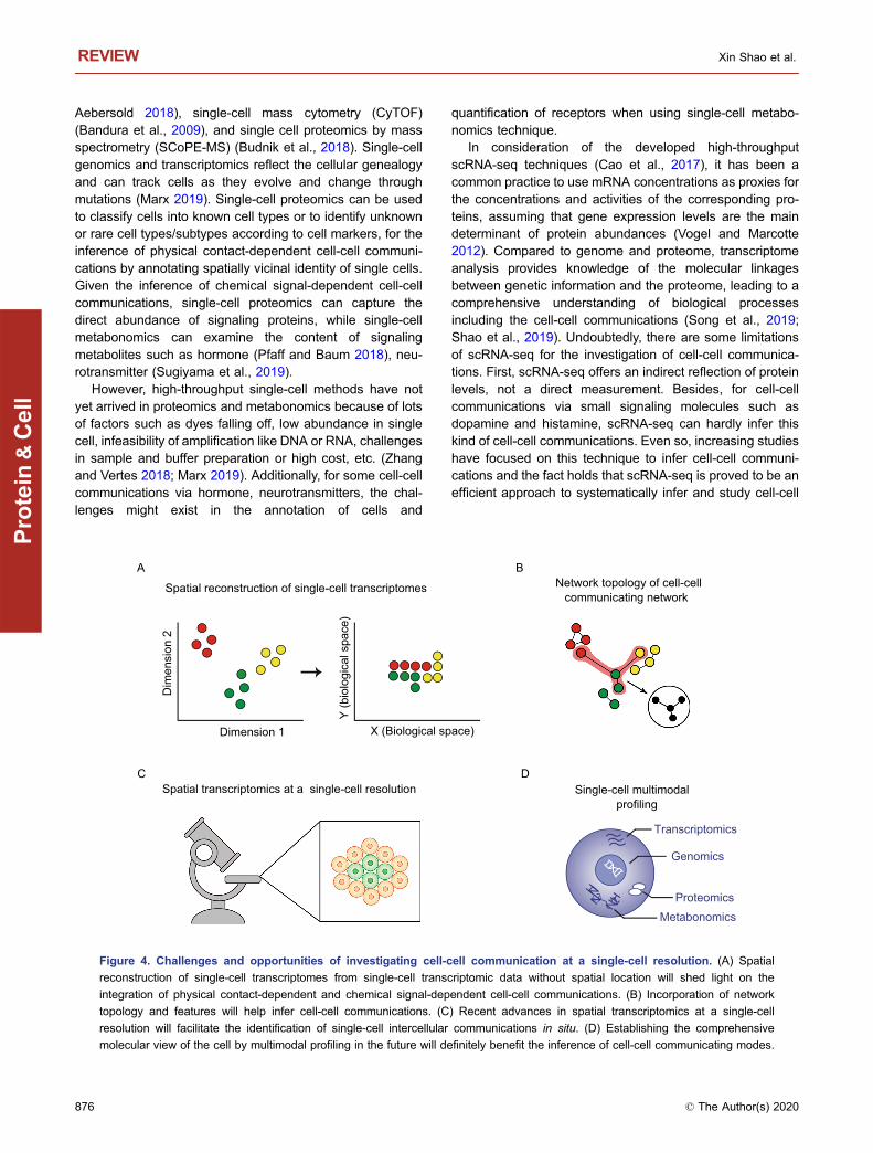
In general, cells communicate and interact with eachother intricately within the tissue microenvironment, whereinthe chemical signal communications may also occurbetween the physically vicinal cells (Stagg and Fletcher1990). It is possible for physical vicinal cells to communicatethrough chemical signals. If incorporated with the informationof physical cell-cell connection, it will be more reliable to inferthe cell-cell communications combined with ligand-receptorinteractions at a single-cell resolution. However, the fact isthat it is difficult for most studies currently to integrate twokinds of cell-cell communications within a single assay forthe widely-used scRNA-seq protocols lacking of the spatiallocation of cells. Therefore, the challenge becomes thespatial reconstruction of single-cell transcriptomes from sin-gle-cell transcriptomic data, which will shed light on theintegration of physical contact-dependent and chemicalsignal-dependent cell-cell communications (Fig. 4A). Fortu-nately, spatial reconstruction of single-cell transcriptomeshas attracted much attention recently and future improve-ments in this area may help address the limitations (Satijaet al., 2015; Halpern et al., 2017; Nitzan et al., 2019).
The concept of networks is important in many fields,including social sciences, physics, artificial intelligence,ecosystems, and systems biology. Analysis on networktopology and features may help scientists predict systembehavior (Albert et al., 2000; Barabasi and Oltvai 2004; Xueet al., 2020). Nevertheless, there are limited methods basedon network analysis for predicting both physical contact-dependent and chemical signal-dependent cell-cell com-munications from constructed cell-cell communicating net-work (Fig. 4B). More efforts should be directed to thedevelopment of network analysis methods for cell-cell com-municating networks.
With the great advances in spatial transcriptomics-relatedtechniques over the recent years (Wang et al. 2018; Enget al., 2019; Rodriques et al., 2019), it is likely that futuresingle-cell spatial position and single-cell transcriptomeanalysis will be able to simultaneously measure both thesingle cells and the sum of mRNA molecules with a highthroughput, especially for specimens originating from solidtissues (Fig. 4C). These future advancements will providehope for spatially reconstituted transcriptomes in situ atsingle-cell levels and will serve as new inspiration for theadvancement of both physical contact-dependent andchemical signal-dependent cell-cell communication studies.
More recently, techniques on multimodal single-cellmeasurements has drawn increasing attention (Fig. 4D),aiming at the simultaneous profiling on multiple types ofmolecule within a single cell (Stuart and Satija 2019; Zhuet al., 2020). For example, CITE-seq (Stoeckius et al., 2017)and REAP-seq (Peterson et al., 2017) have realized thesimultaneous measurements of whole transcriptome ofmRNA and proteins, while sci-CAR have enabled the mea-surements of whole transcriptome of mRNA and chromatin
accessibility simultaneously (Cao et al., 2018). The estab-lishment of a comprehensive molecular view of the cell bymultimodal profiling will definitely improve the classificationof cell identities and inference of multiple cell-cell commu-nicating modes via biomacromolecule and small signalingmolecules.
With the increasing researches on the investigation ofcell-cell communications in the future, we expect that morecell-cell communications related with physiological pro-cesses, disease pathogenesis and progression, and phar-macological research of pharmacological treatment and drugresistance will be discovered and verified, providing newinsights into the old biological and biomedical questionssuch as mechanism elucidation, identification of biomarkersand drug targets, and drug resistance, etc. As the relevanceof cell-cell communications involved in the initiation anddevelopment of physiological process and disease becomesbetter defined, novel or improved therapeutic strategies forpharmacological treatments targeting driving cell types andcommunicating molecules will become apparent.
ACKNOWLEDGEMENTS
This study was supported by the National Natural Science Foun-
dation of China (Grant Nos. 81774153 and 81973701), the Natural
Science Foundation of Zhejiang Province (LZ20H290002), and the
National Youth Top-notch Talent Support Program (W02070098).
COMPLIANCE WITH ETHICS GUIDELINES:
The authors declare that they have no conflict of interest. This
review does not contain any studies with human or animal subjects
performed by the any of the authors.
ABBREVIATIONS
AT2, alveolar type 2; CAFs, cancer-associated fibroblasts; CTCs,
circulating tumor cells; DCs, dendritic cells; dNK, decidual natural
killer; EVT, extravillous trophoblast; FGCs, fetal germ cells; FISH,
fluorescence in situ hybridization; GCs, granulosa cells; HE, hepatic
endoderm; iCD, ileal Crohn’s disease; IF, immunofluorescence; IHC,
immunohistochemistry; iPS, induced pluripotent stem; MNPs,
mononuclear phagocytes; NASH, non-alcoholic steatohepatitis;
NR, neural retina; PCs, photoreceptor cells; RNAi, RNA
interference; RPE, retinal pigment epithelium; scRNA-seq, single-
cell RNA-sequencing; TAMs, tumor-associated macrophages;
WBCs, white blood cells.
OPEN ACCESS
This article is licensed under a Creative Commons Attribution 4.0
International License, which permits use, sharing, adaptation,
distribution and reproduction in any medium or format, as long as
you give appropriate credit to the original author(s) and the source,
provide a link to the Creative Commons licence, and indicate if
changes were made. The images or other third party material in this
article are included in the article's Creative Commons licence, unless
Infer cell-cell communication from scRNA-seq data REVIEW
© The Author(s) 2020 877
Protein
&Cell
indicated otherwise in a credit line to the material. If material is not
included in the article's Creative Commons licence and your
intended use is not permitted by statutory regulation or exceeds
the permitted use, you will need to obtain permission directly from
the copyright holder. To view a copy of this licence, visit http://
creativecommons.org/licenses/by/4.0/.
REFERENCES
Albert R, Jeong H, Barabasi AL (2000) Error and attack tolerance of
complex networks. Nature 406:378–382
Aran D, Looney AP, Liu L, Wu E, Fong V, Hsu A, Chak S, Naikawadi
RP, Wolters PJ, Abate AR et al (2019) Reference-based analysis
of lung single-cell sequencing reveals a transitional profibrotic
macrophage. Nat Immunol 20:163–172
Bandura DR, Baranov VI, Ornatsky OI, Antonov A, Kinach R, Lou X,
Pavlov S, Vorobiev S, Dick JE, Tanner SD (2009) Mass
cytometry: technique for real time single cell multitarget
immunoassay based on inductively coupled plasma time-of-flight
mass spectrometry. Anal Chem 81:6813–6822
Barabasi AL, Oltvai ZN (2004) Network biology: understanding the
cell’s functional organization. Nat Rev Genet 5:101–113
Bessis M (1958) Erythroblastic island, functional unity of bone
marrow. Rev Hematol 13:8–11
Boisset JC, Vivie J, Grun D, Muraro MJ, Lyubimova A, van
Oudenaarden A (2018) Mapping the physical network of cellular
interactions. Nat Methods 15:547–553
Braga VM (2002) Cell-cell adhesion and signalling. Curr Opin Cell
Biol 14:546–556
Budnik B, Levy E, Harmange G, Slavov N (2018) SCoPE-MS: mass
spectrometry of single mammalian cells quantifies proteome
heterogeneity during cell differentiation. Genome Biol 19:161
Burns JC, Kelly MC, Hoa M, Morell RJ, Kelley MW (2015) Single-cell
RNA-Seq resolves cellular complexity in sensory organs from the
neonatal inner ear. Nat Commun 6:8557
Camp JG, Sekine K, Gerber T, Loeffler-Wirth H, Binder H, Gac M,
Kanton S, Kageyama J, Damm G, Seehofer D et al (2017)
Multilineage communication regulates human liver bud develop-
ment from pluripotency. Nature 546:533–538
Cao J, Packer JS, Ramani V, Cusanovich DA, Huynh C, Daza R, Qiu
X, Lee C, Furlan SN, Steemers FJ et al (2017) Comprehensive
single-cell transcriptional profiling of a multicellular organism.
Science 357:661–667
Cao J, Cusanovich DA, Ramani V, Aghamirzaie D, Pliner HA, Hill
AJ, Daza RM, McFaline-Figueroa JL, Packer JS, Christiansen L
et al (2018) Joint profiling of chromatin accessibility and gene
expression in thousands of single cells. Science 361:1380–1385
Cheow LF, Courtois ET, Tan Y, Viswanathan R, Xing Q, Tan RZ, Tan
DS, Robson P, Loh YH, Quake SR et al (2016) Single-cell
multimodal profiling reveals cellular epigenetic heterogeneity. Nat
Methods 13:833–836
Cohen M, Giladi A, Gorki AD, Solodkin DG, Zada M, Hladik A,
Miklosi A, Salame TM, Halpern KB, David E et al (2018) Lung
single-cell signaling interaction map reveals basophil role in
macrophage imprinting. Cell 175:1031–1044 e1018
Collins BC, Aebersold R (2018) Proteomics goes parallel. Nat
Biotechnol 36:1051–1053
Duan L, Zhang XD, Miao WY, Sun YJ, Xiong G, Wu Q, Li G, Yang P,
Yu H, Li H et al (2018) PDGFRbeta cells rapidly relay inflamma-
tory signal from the circulatory system to neurons via chemokine
CCL2. Neuron 100:183–200 e188
Efremova M, Vento-Tormo M, Teichmann SA, Vento-Tormo R (2020)
CellPhoneDB: inferring cell-cell communication from combined
expression of multi-subunit ligand-receptor complexes. Nat Pro-
toc 15(4):1484–1506
Eng CL, Lawson M, Zhu Q, Dries R, Koulena N, Takei Y, Yun J,
Cronin C, Karp C, Yuan GC et al (2019) Transcriptome-scale
super-resolved imaging in tissues by RNA seqFISH. Nature
568:235–239
Evans WH (2015) Cell communication across gap junctions: a
historical perspective and current developments. Biochem Soc
Trans 43:450–459
Fernandez DM, Rahman AH, Fernandez NF, Chudnovskiy A, Amir
ED, Amadori L, Khan NS, Wong CK, Shamailova R, Hill CA et al
(2019) Single-cell immune landscape of human atherosclerotic
plaques. Nat Med 25:1576–1588
Gao S, Yan L, Wang R, Li J, Yong J, Zhou X, Wei Y, Wu X, Wang X,
Fan X et al (2018) Tracing the temporal-spatial transcriptome
landscapes of the human fetal digestive tract using single-cell
RNA-sequencing. Nat Cell Biol 20:721–734
Gartner ZJ, Prescher JA, Lavis LD (2017) Unraveling cell-to-cell
signaling networks with chemical biology. Nat Chem Biol 13:564–
568
Grun D, Lyubimova A, Kester L, Wiebrands K, Basak O, Sasaki N,
Clevers H, van Oudenaarden A (2015) Single-cell messenger
RNA sequencing reveals rare intestinal cell types. Nature
525:251–255
Halpern KB, Shenhav R, Matcovitch-Natan O, Toth B, Lemze D,
Golan M, Massasa EE, Baydatch S, Landen S, Moor AE et al
(2017) Single-cell spatial reconstruction reveals global division of
labour in the mammalian liver. Nature 542:352–356
Hashimshony T, Wagner F, Sher N, Yanai I (2012) CEL-Seq: single-
cell RNA-Seq by multiplexed linear amplification. Cell Rep 2:666–
673
Hu Y, Wang X, Hu B, Mao Y, Chen Y, Yan L, Yong J, Dong J, Wei Y,
Wang W et al (2019) Dissecting the transcriptome landscape of
the human fetal neural retina and retinal pigment epithelium by
single-cell RNA-seq analysis. PLoS Biol 17:e3000365
Jaitin DA, Kenigsberg E, Keren-Shaul H, Elefant N, Paul F, Zaretsky
I, Mildner A, Cohen N, Jung S, Tanay A et al (2014) Massively
parallel single-cell RNA-seq for marker-free decomposition of
tissues into cell types. Science 343:776–779
Kirouac DC, Madlambayan GJ, Yu M, Sykes EA, Ito C, Zandstra PW
(2009) Cell-cell interaction networks regulate blood stem and
progenitor cell fate. Mol Syst Biol 5:293
Kiselev VY, Yiu A, Hemberg M (2018) scmap: projection of single-
cell RNA-seq data across data sets. Nat Methods 15:359–362
Klein AM, Mazutis L, Akartuna I, Tallapragada N, Veres A, Li V,
Peshkin L, Weitz DA, Kirschner MW (2015) Droplet barcoding for
single-cell transcriptomics applied to embryonic stem cells. Cell
161:1187–1201
REVIEW Xin Shao et al.
878 © The Author(s) 2020
Protein
&Cell
Kumar V, Donthireddy L, Marvel D, Condamine T, Wang F, Lavilla-
Alonso S, Hashimoto A, Vonteddu P, Behera R, Goins MA et al
(2017) Cancer-associated fibroblasts neutralize the anti-tumor
effect of CSF1 receptor blockade by inducing PMN-MDSC
infiltration of tumors. Cancer Cell 32:654–668 e655
Kumar MP, Du J, Lagoudas G, Jiao Y, Sawyer A, Drummond DC,
Lauffenburger DA, Raue A (2018) Analysis of single-cell RNA-
Seq identifies cell-cell communication associated with tumor
characteristics. Cell Rep 25:1458–1468e1454
Li L, Dong J, Yan L, Yong J, Liu X, Hu Y, Fan X, Wu X, Guo H, Wang
X et al (2017) Single-cell RNA-Seq analysis maps development
of human germline cells and gonadal niche interactions. Cell
Stem Cell 20:858–873 e854
Liao J, Hao C, Huang W, Shao X, Song Y, Liu L, Ai N, Fan X (2018)
Network pharmacology study reveals energy metabolism and
apoptosis pathways-mediated cardioprotective effects of Shenqi
Fuzheng. J Ethnopharmacol 227:155–165
Lin X, Spindler TJ, de Souza Fonseca MA, Corona RI, Seo JH,
Dezem FS, Li L, Lee JM, Long HW, Sellers TA et al (2019) Super-
enhancer-associated LncRNA UCA1 interacts directly with
AMOT to activate YAP target genes in epithelial ovarian cancer.
iScience 17:242–255
Macosko EZ, Basu A, Satija R, Nemesh J, Shekhar K, Goldman M,
Tirosh I, Bialas AR, Kamitaki N, Martersteck EM et al (2015)
Highly parallel genome-wide expression profiling of individual
cells using nanoliter droplets. Cell 161:1202–1214
Manwani D, Bieker JJ (2008) The erythroblastic island. Curr Top Dev
Biol 82:23–53
Martin JC, Chang C, Boschetti G, Ungaro R, Giri M, Grout JA,
Gettler K, Chuang LS, Nayar S, Greenstein AJ et al (2019)
Single-cell analysis of Crohn’s disease lesions identifies a
pathogenic cellular module associated with resistance to anti-
TNF therapy. Cell 178:1493–1508 e1420
Marx V (2019) A dream of single-cell proteomics. Nat Methods
16:809–812
Mittal K, Eremenko E, Berner O, Elyahu Y, Strominger I, Apelblat D,
Nemirovsky A, Spiegel I, Monsonego A (2019) CD4 T cells
induce a subset of MHCII-expressing microglia that attenuates
Alzheimer pathology. iScience 16:298–311
Nestorowa S, Hamey FK, Pijuan Sala B, Diamanti E, Shepherd M,
Laurenti E, Wilson NK, Kent DG, Gottgens B (2016) A single-cell
resolution map of mouse hematopoietic stem and progenitor cell
differentiation. Blood 128:e20–31
Nitzan M, Karaiskos N, Friedman N, Rajewsky N (2019) Gene
expression cartography. Nature 576:132–137
Pan G, Cavalli M, Carlsson B, Skrtic S, Kumar C, Wadelius C (2020)
rs953413 Regulates polyunsaturated fatty acid metabolism by
modulating ELOVL2 expression. iScience 23:100808
Petersen F, Bock L, Flad HD, Brandt E (1999) Platelet factor
4-induced neutrophil-endothelial cell interaction: involvement of
mechanisms and functional consequences different from those
elicited by interleukin-8. Blood 94:4020–4028
Peterson VM, Zhang KX, Kumar N, Wong J, Li L, Wilson DC, Moore
R, McClanahan TK, Sadekova S, Klappenbach JA (2017)
Multiplexed quantification of proteins and transcripts in single
cells. Nat Biotechnol 35:936–939
Pfaff DW, Baum MJ (2018) Hormone-dependent medial preoptic/
lumbar spinal cord/autonomic coordination supporting male
sexual behaviors. Mol Cell Endocrinol 467:21–30
Picelli S, Bjorklund AK, Faridani OR, Sagasser S, Winberg G,
Sandberg R (2013) Smart-seq2 for sensitive full-length transcrip-
tome profiling in single cells. Nat Methods 10:1096–1098
Rajbhandari P, Arneson D, Hart SK, Ahn IS, Diamante G, Santos LC,
Zaghari N, Feng AC, Thomas BJ, Vergnes L et al (2019) Single
cell analysis reveals immune cell-adipocyte crosstalk regulating
the transcription of thermogenic adipocytes. Elife 8:e49501
Ramilowski JA, Goldberg T, Harshbarger J, Kloppmann E, Lizio M,
Satagopam VP, Itoh M, Kawaji H, Carninci P, Rost B et al (2015)
A draft network of ligand-receptor-mediated multicellular sig-
nalling in human. Nat Commun 6:7866
Ramos P, Casu C, Gardenghi S, Breda L, Crielaard BJ, Guy E,
Marongiu MF, Gupta R, Levine RL, Abdel-Wahab O et al (2013)
Macrophages support pathological erythropoiesis in poly-
cythemia vera and beta-thalassemia. Nat Med 19:437–445
Rodriques SG, Stickels RR, Goeva A, Martin CA, Murray E,
Vanderburg CR, Welch J, Chen LM, Chen F, Macosko EZ
(2019) Slide-seq: a scalable technology for measuring genome-
wide expression at high spatial resolution. Science 363:1463–
1467
Rothbauer M, Zirath H, Ertl P (2018) Recent advances in microfluidic
technologies for cell-to-cell interaction studies. Lab Chip 18:249–
270
Satija R, Farrell JA, Gennert D, Schier AF, Regev A (2015) Spatial
reconstruction of single-cell gene expression data. Nat Biotech-
nol 33:495–502
Scott CL, Guilliams M (2018) Tissue unit-ed: lung cells team up to
drive alveolar macrophage development. Cell 175:898–900
Shalek AK, Satija R, Shuga J, Trombetta JJ, Gennert D, Lu D, Chen
P, Gertner RS, Gaublomme JT, Yosef N et al (2014) Single-cell
RNA-seq reveals dynamic paracrine control of cellular variation.
Nature 510:363–369
Shao X, Ai N, Xu D, Fan X (2016) Exploring the interaction between
Salvia miltiorrhiza and human serum albumin: Insights from herb-
drug interaction reports, computational analysis and experimental
studies. Spectrochim Acta A Mol Biomol Spectrosc 161:1–7
Shao X, Lv N, Liao J, Long J, Xue R, Ai N, Xu D, Fan X (2019) Copy
number variation is highly correlated with differential gene
expression: a pan-cancer study. BMC Med Genet 20:175
Shao X, Liao J, Lu X, Xue R, Ai N, Fan X (2020) scCATCH:
automatic annotation on cell types of clusters from single-cell
RNA sequencing data. iScience 23:100882
Shrestha BR, Chia C, Wu L, Kujawa SG, Liberman MC, Goodrich LV
(2018) Sensory neuron diversity in the inner ear is shaped by
activity. Cell 174:1229–1246 e1217
Sicard RE (1986) Hormones, neurosecretions, and growth factors as
signal molecules for intercellular communication. Dev Comp
Immunol 10:269–272
Singer SJ (1992) Intercellular communication and cell-cell adhesion.
Science 255:1671–1677
Skelly DA, Squiers GT, McLellan MA, Bolisetty MT, Robson P,
Rosenthal NA, Pinto AR (2018) Single-cell transcriptional profil-
ing reveals cellular diversity and intercommunication in the
mouse heart. Cell Rep 22:600–610
Infer cell-cell communication from scRNA-seq data REVIEW
© The Author(s) 2020 879
Protein
&Cell
Song Y, Xu X, Wang W, Tian T, Zhu Z, Yang C (2019) Single cell
transcriptomics: moving towards multi-omics. Analyst 144:3172–
3189
Stagg RB, Fletcher WH (1990) The hormone-induced regulation of
contact-dependent cell-cell communication by phosphorylation.
Endocr Rev 11:302–325
Stoeckius M, Hafemeister C, Stephenson W, Houck-Loomis B,
Chattopadhyay PK, Swerdlow H, Satija R, Smibert P (2017)
Simultaneous epitope and transcriptome measurement in single
cells. Nat Methods 14:865–868
Stuart T, Satija R (2019) Integrative single-cell analysis. Nat Rev
Genet 20:257–272
Sugiyama E, Guerrini MM, Honda K, Hattori Y, Abe M, Kallback P,
Andren PE, Tanaka KF, Setou M, Fagarasan S et al (2019)
Detection of a high-turnover serotonin circuit in the mouse brain
using mass spectrometry imaging. iScience 20:359–372
Swaminathan J, Boulgakov AA, Hernandez ET, Bardo AM, Bach-
man JL, Marotta J, Johnson AM, Anslyn EV, Marcotte EM (2018)
Highly parallel single-molecule identification of proteins in zepto-
mole-scale mixtures. Nat Biotechnol 36:1076–1082
Szczerba BM, Castro-Giner F, Vetter M, Krol I, Gkountela S, Landin
J, Scheidmann MC, Donato C, Scherrer R, Singer J et al (2019)
Neutrophils escort circulating tumour cells to enable cell cycle
progression. Nature 566:553–557
Tirosh I, Izar B, Prakadan SM, Wadsworth MH 2nd, Treacy D,
Trombetta JJ, Rotem A, Rodman C, Lian C, Murphy G et al
(2016) Dissecting the multicellular ecosystem of metastatic
melanoma by single-cell RNA-seq. Science 352:189–196
Vento-Tormo R, Efremova M, Botting RA, Turco MY, Vento-Tormo M,
Meyer KB, Park JE, Stephenson E, Polanski K, Goncalves A et al
(2018) Single-cell reconstruction of the early maternal-fetal
interface in humans. Nature 563:347–353
Vitak SA, Torkenczy KA, Rosenkrantz JL, Fields AJ, Christiansen L,
Wong MH, Carbone L, Steemers FJ, Adey A (2017) Sequencing
thousands of single-cell genomes with combinatorial indexing.
Nat Methods 14:302–308
Vogel C, Marcotte EM (2012) Insights into the regulation of protein
abundance from proteomic and transcriptomic analyses. Nat Rev
Genet 13:227–232
Wang X, Song W, Kawazoe N, Chen G (2013) The osteogenic
differentiation of mesenchymal stem cells by controlled cell-cell
interaction on micropatterned surfaces. J Biomed Mater Res A
101:3388–3395
Wang X, Allen WE, Wright MA, Sylwestrak EL, Samusik N, Vesuna
S, Evans K, Liu C, Ramakrishnan C, Liu J et al (2018) Three-
dimensional intact-tissue sequencing of single-cell transcriptional
states. Science 361:eaat5691
Wang S, Karikomi M, MacLean AL, Nie Q (2019) Cell lineage and
communication network inference via optimization for single-cell
transcriptomics. Nucleic Acids Res 47(11):e66
Xiong X, Kuang H, Ansari S, Liu T, Gong J, Wang S, Zhao XY, Ji Y, Li
C, Guo L et al (2019) Landscape of intercellular crosstalk in
healthy and NASH liver revealed by single-cell secretome gene
analysis. Mol Cell 75:644–660e645
Xu Y, Ji K, Wu M, Hao B, Yao KT, Xu Y (2019) A miRNA-HERC4
pathway promotes breast tumorigenesis by inactivating tumor
suppressor LATS1. Protein Cell 10:595–605
Xue R, Liao J, Shao X, Han K, Long J, Shao L, Ai N, Fan X (2020)
Prediction of adverse drug reactions by combining biomedical
tripartite network and graph representation model. Chem Res
Toxicol 33:202–210
Zepp JA, Zacharias WJ, Frank DB, Cavanaugh CA, Zhou S, Morley
MP, Morrisey EE (2017) Distinct mesenchymal lineages and
niches promote epithelial self-renewal and myofibrogenesis in the
lung. Cell 170:1134–1148 e1110
Zhang L, Vertes A (2018) Single-cell mass spectrometry approaches
to explore cellular heterogeneity. Angew Chem Int Ed Engl
57:4466–4477
Zhang Y, Yan Z, Qin Q, Nisenblat V, Chang HM, Yu Y, Wang T, Lu C,
Yang M, Yang S et al (2018) Transcriptome landscape of human
folliculogenesis reveals oocyte and granulosa cell interactions.
Mol Cell 72:1021–1034 e1024
Zhang X, Lan Y, Xu J, Quan F, Zhao E, Deng C, Luo T, Xu L, Liao G,
Yan M et al (2019) CellMarker: a manually curated resource of
cell markers in human and mouse. Nucleic Acids Res 47:D721–
D728
Zhang L, Vertes A (2018) Single-cell mass spectrometry approaches
to explore cellular heterogeneity. Angew Chem Int Ed Engl
57:4466–4477
Zheng GX, Terry JM, Belgrader P, Ryvkin P, Bent ZW, Wilson R,
Ziraldo SB, Wheeler TD, McDermott GP, Zhu J et al (2017)
Massively parallel digital transcriptional profiling of single cells.
Nat Commun 8:14049
Zheng G, Jiang C, Li Y, Yang D, Ma Y, Zhang B, Li X, Zhang P, Hu X,
Zhao X et al (2019) TMEM43-S358L mutation enhances NF-
kappaB-TGFbeta signal cascade in arrhythmogenic right ventric-
ular dysplasia/cardiomyopathy. Protein Cell 10:104–119
Zhou B, Liu C, Xu L, Yuan Y, Zhao J, Zhao W, Chen Y, Qiu J, Meng
M, Zheng Y et al (2020) N(6)-methyladenosine reader protein
Ythdc2 suppresses liver steatosis via regulation of mRNA
stability of lipogenic genes. Hepatology. https://doi.org/10.1002/
hep.31220
Zhu C, Preissl S, Ren B (2020) Single-cell multimodal omics: the
power of many. Nat Methods 17:11–14
REVIEW Xin Shao et al.
880 © The Author(s) 2020
Protein
&Cell